Article

In Vitro Antibacterial Activity of Selected Palestinian Medicinal Plants against Chlamydia trachomatis

1
Department of Life Sciences, College of Science and Technology, Al-Quds University, Jerusalem, Palestine
2
Faculty of Pharmacy, Al-Quds University, Jerusalem, Palestine
3
Department of Applied Biochemistry, Institute of Biotechnology, 4/3-2, Technical University of Berlin, Gustav-Meyer-Allee 25, 13355 Berlin, Germany
*
Authors to whom correspondence should be addressed.
These authors contributed equally to this work.
Microbiol. Res. 2021, 12(3), 656-662; https://doi.org/10.3390/microbiolres12030047
Submission received: 29 June 2021 / Revised: 30 July 2021 / Accepted: 5 August 2021 / Published: 8 August 2021

Abstract

:
Chlamydia spp. are intracellular pathogens of humans and animals that cause a wide range of diseases such as blinding trachoma and sexually transmitted infections. According to the World Health Organization (WHO), there are more than 127 million new infections each year worldwide. Chlamydial urogenital infections can cause cervicitis, urethritis, pelvic inflammatory disease and infertility. From within an intracellular niche, termed an inclusion, the Chlamydiae complete their life cycle shielded from host defenses. The host cell defense response used to eliminate the pathogen must subvert this protective shield and is thought to involve the gamma interferon-inducible family of immunity related GTPase proteins and nitric oxide. Typically, azithromycin and doxycycline are the first line drugs for the treatment of chlamydial infections. Although C. trachomatis is sensitive to these antibiotics in vitro, currently, there is increasing bacterial resistance to antibiotics including multidrug-resistant C. trachomatis, which have been described in many instances. Therefore, alternative drug candidates against Chlamydia should be assessed in vitro. In this study, we tested and quantified the activity of plant extracts against Chlamydia-infected HeLa cells with C. trachomatis inclusions. The in vitro results show that post-treatment with Artemisia inculta Delile extract significantly inhibits Chlamydia infection compared to DMSO-treated samples. In conclusion, plant extracts may contain active ingredients with antichlamydial activity potential and can be used as alternative drug candidates for treatment of Chlamydia infection which has significant socio-economic and medical impact.

1. Introduction

Chlamydia trachomatis is an obligate intracellular pathogenic bacterium associated with several important human diseases, such as trachoma, the leading cause of preventable blindness [1,2]. Although trachoma is preventable, it is currently the leading cause of eye disease in the world, causing blindness in approximately 1.9 million according to the WHO. In addition, more than 137 million people live in trachoma endemic areas or at risk of trachoma blindness [3].
In Palestine, there are no reports about chlamydia infection except a study in 2009 which was conducted among women attending gynecology and infertility centers in Gaza. The overall prevalence of C. trachomatis in the study population was 20.2% [4]
C. trachomatis is also the most common bacterium which causes sexually transmitted disease (STD) [1,2]. According to the WHO, there are an estimated 127 million new cases annually worldwide. Chlamydial urogenital infections can cause a range of diseases such as cervicitis, urethritis, pelvic inflammatory disease and infertility [1,2,5]. In infants, C. trachomatis is considered the most common cause of ophthalmia neonatorum and a significant cause of conjunctivitis and pneumonia in developing countries [6].
While antibiotic treatment against Chlamydia is available [7], most of the infected individuals are unlikely to seek treatment since infections in women (70 to 90%) and in men (50%) are asymptomatic. Recently, it has been reported that the standard antibiotic treatment against Chlamydia is less effective than expected, possibly due to persistent infections [8]. The unique life cycle of C. trachomatis leads to chronic infection and challenges in treatment using classical antibiotics. Further, many antibiotics target the reproductive reticulate body form of Chlamydia and this may lead to the need for prolonged treatment which accelerates the development of drug-resistant chlamydial strains. In addition, there are no vaccines or other effective prophylactic measures available to protect against Chlamydia infection. This indicates the need for new therapies against the disease. In addition, small molecules derived from medicinal chemistry and natural products are still major sources of innovative therapeutic agents for various conditions, including infectious diseases [9,10,11]. A large number of novel drugs have been isolated from natural plant sources, and many of these plants and their extracts were used in ethnobotanical medicine [12,13]. These natural products in the form of herbal remedies, could be explored based on information collected from local residents and specialists in ethnobotanical medicine in different parts of the world [14,15,16]. It has been reported that many compounds derived from plants regarded as effective drugs are currently in use, and the majority of these compounds were derived from ethnobotanical medicines [17,18,19,20]. However, inhibition of Chlamydia spp. infection using natural compounds and ethnobotanical approaches has not been thoroughly investigated.
The present study aimed to evaluate in vitro anti-Chlamydia activities of natural plant extracts from certain medicinal plants from various regions in Palestine and evaluated the potential of crude plant extracts to inhibit Chlamydia growth and replication.

2. Materials and Methods

2.1. Cultivation of HeLa Cells and Chlamydia trachomatis

Cell cultures of HeLa 229 (American Type Culture Collection CCL-2.1) epithelial cells were grown in cell growth medium (CGM) consisting of RPMI-1640 (Gibco-Invitrogen, Karlsruhe, Germany) supplemented with 10% FBS and 10 µg/mL gentamicin at 37 °C and 5% CO2 in a humidified tissue culture chamber. C. trachomatis Lymphogranuloma venereum (LGV) serovar L2 (7 × 10⁹ inclusion forming units/mL) were routinely propagated in HeLa cells using infection medium (IM) consisting of RPMI medium supplemented with 5% FBS only. The maintenance medium (MM) consisted of IM containing cycloheximide (1 µg/mL). The infectious progeny were purified, titrated and inclusion-forming units (IFU)/mL were determined [21,22].

2.2. Host Infection and Determination of Infectivity

For infection studies, HeLa cells were seeded onto coverslips in 12-well plates and inoculated with C. trachomatis diluted in IM at an MOI of 0.5, unless otherwise indicated. Cells inoculated with C. trachomatis were incubated for 2 h at 35 °C and 5% CO2. Cells infected with Chlamydia were washed before adding fresh IM and then incubated for specific time intervals in the humidified tissue culture chamber. In some experiments, crude plant extracts (10 μg/mL) (1–5) were added at the indicated times. The formation of infectious progeny of C. trachomatis was assessed by infectivity titration assays [23]. Briefly, infected cells were mechanically lysed using glass beads, and lysates were serially diluted in IM then inoculated onto HeLa 229 cells for 2 h. Cells were then washed and further incubated in MM for 24 h. The number of inclusions was expressed as inclusion-forming units per ml (IFU/mL).

2.3. Viability and Metabolic Activity of Host Cells

The effect of the drugs on the metabolic activity of host cells was determined using the WST-1 cell proliferation assay (Roche, Mannheim, Germany) according to the manufacturer’s instructions. Culture cells treated with the indicated plant extracts and DMSO were used as a control.

2.4. Selection and Collection of Medicinal Plants

Palestine is distinguished for its availability of medicinal plants because of the unique geographical location and biodiversity; these plants have been used for a long period of time to treat various illnesses [24,25,26].
A literature survey was first conducted to obtain ethnobotanical information on Palestinian flora used in folk medicine to treat bacterial, fungal and parasitic infections. Moreover, the selected plants with the desired activities were considered safe, since they are used by local people for medical purposes or as food. No history of toxicity or poisoning was documented for the selected plants. Four Palestinian medicinal plants were selected, and the same plant materials were previously tested by our group with promising antibacterial and antiparasitic activities [9]. The plant materials used in this study include Achillea fragrantissima (PS-Af01), Artemisia inculta Delile (PS-Ai10), Coridothymus capitatus (L.) Rchb.f. (PS-Cc19) and Malva sylvestris L. (PS-Ms50). Plant parts were collected and characterized by a botanist and the voucher numbers are kept at Al-Quds University Gardens (AQUG) and are available upon request. Plant parts were selected based on the recommended traditional reports [26,27,28,29]. The whole plant, flowers, fruits or seeds were collected and washed with distilled water, air dried in the shade for 20 days and powdered using an electric grinder.

2.5. Preparation of the Crude Extracts
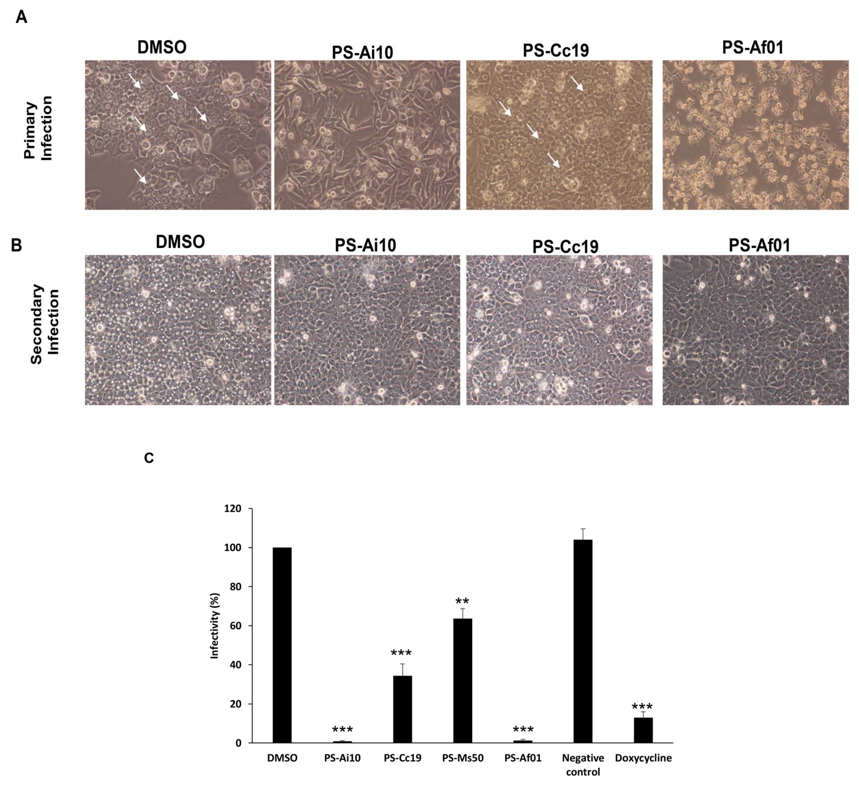
Aqueous and organic extractions were carried out for each plant. Each powdered whole plant sample (16 g) was soaked in 100 mL of absolute ethanol and dimethyl sulfoxide (DMSO) separately for 72 h at room temperature on a gentle shaker. The filtrate obtained was passed through Whatman No. 1 filter paper to obtain the crude extracts. Each extract yielded about 160 mg/mL (w/v), and all crude extracts were kept at room temperature until further processing. All the concentrations of the crude extracts are based on dry weight basis. The crude extracts were immediately tested for anti-Chlamydia activity.

3. Results

Activity of Plant Extracts against Chlamydia trachomatis

To determine the anti-Chlamydia trachomatis activity of the plant extracts, we initially infected HeLa 229 cells with C. trachomatis for 2 h. Subsequently, the infected cells were incubated in the presence of different plant extracts for an additional 46 h. Mock-treated cells infected with C. trachomatis served as a non-treated control (DMSO control). Following 48 h of incubation in the presence of different extracts, cells were analyzed by microscopy for inclusion development. Different plant extract treatments resulted in much smaller and substantially reduced numbers of C. trachomatis inclusions (Figure 1A). The extract from Artemisia inculta Delile (PS-Ai10) inhibited Chlamydia growth significantly compared with the control (DMSO treatment) with minimal toxic effect. Moreover, the extract from Achillea fragrantissima (PS-Af01) had a similar inhibitory effect on C. trachomatis. However, the extract showed an effect during primary infection and was excluded from further analysis (Figure 1A). The extract from Coridothymus capitatus (L.) Rchb.f (PS-Cc19) had a moderate inhibitory effect on Chlamydia growth in comparison with extract PS-Ai10. Afterwards, we tested the effect of different plant extracts on Chlamydia infectious progeny using an infectivity assay, described previously [21] (Figure 1B,C), with a significant inhibitory activity against infectious C. trachomatis EBs of more than 99% when cells were treated with PS-Ai10, while PS-Cc19 and PS-Ms50 did not show any inhibitory activity and the growth of chlamydial inclusions was prominent. Surprisingly, infected cells were sensitive to PS-Af01 treatment, which resulted in partial destruction and loss of infected host cells of the monolayer (Figure 1A). Therefore, the chlamydial infectivity measured in each sample had to be normalized to the surviving host cells, determined via an LDH release assay. These results suggested that plant extracts cause an inhibition of Chlamydia replication and not of the host cell, since the treatment was carried out after infection and is not from the binding to chlamydial ligands or host cell receptors. The yield of C. trachomatis infectious progeny was decreased considerably after treatment with different plant extracts (Figure 1C).

4. Discussion

Following the past few decades of intense research on host cell function analysis of infection, it has reached the point where the proof-of-concept for host cell-directed infection therapy has been demonstrated. Host cell-directed therapy could be an alternative or complementary therapy to conventional antibiotic and antiviral treatments, bearing the advantage of a substantially reduced risk of resistance development [30]. In the absence of a vaccine against Chlamydia, this anti-Chlamydia in vitro screening study has special importance as a way to find antichlamydial materials in safe medicinal plant extracts with future potential as effective drugs against this pathogen. This may pave the way to generating potentially new and promising anti-Chlamydia drugs.
Artemisia inculta Delile (PS-Ai10) extract significantly inhibited Chlamydia growth and reduced infectious C. trachomatis EBs by more than 99% with minimal toxic effect. This plant material contains several types of ingredients which have proven medicinal effects; camphor, thujone, chamazulene, 1,8-cineole and sabinol are among the active ingredients in this plant [31]. It was documented that Artemisia inculta Delile leaves are used by people in the Mediterranean basin for the treatment of various illnesses; it has anti-inflammatory, antiseptic, carminative, cholagogue, cold-treating, emmenagogue, hypoglycemic, gastric pain relief, rheumatic disease-treating, sedative, spasmolytic, stomachic and tonic properties [32]. However, this is the first report of antichlamydial activity for this plant in the region. Some studies revealed that a high percentage of people depend primarily on traditional and ethnobotanical medicine for the treatment of some infections, including sexually transmitted infections (STIs) [33]. A recent study from South Africa demonstrated that eight plant species are used for the treatment of STIs, including Chlamydia [34]. In Bangladesh, some plant species were used to treat syphilis and gonorrhea [35]. In Rwanda, it was reported that 25 different plant species were used to treat gonorrhea [36].
Further, it has been shown that an extract from desert truffles (Terfezia claveryi) has antibacterial activity against C. trachomatis [32]. Some plant species from Palestine have antileishmanial activity, but have never been tested previously on Chlamydia or other bacterial strains [9]. Further research is needed to establish the antimicrobial efficacies of these plant species against pathogens, especially sexually transmitted infections.
C. trachomatis has a unique life cycle which primarily leads to chronic infections and challenges in treatment using conventional antibiotics. For chlamydial infections in general, both azithromycin and doxycycline are recommended by the US Centers for Disease Control and Prevention and have a >95% microbiological cure rate [37]. Other antibiotics are used mainly to target the reproductive RBs, and this often leads to the development of drug resistance due to the prolonged treatment [38]. Therefore, the use of natural products from plants in traditional medicine provides valuable resources that may have potential against various infections, including C. trachomatis.
Our study is limited to the use of whole plant extracts which provide all plant ingredients. Therefore, fractionation and purification of active ingredients in these extracts should be carried out using chemical and biological assays. Moreover, plant materials used in this report were extracted from medicinal plants which were extensively used by local people in Palestine with no history of toxicity or poisoning. However, safety and toxicity profiles for these plants have to be established.
In conclusion, plants extracts may contain active ingredients with potential antichlamydial activity and can be used as alternative drug candidates for treatment of Chlamydia infection which has significant socio-economic and medical impact.

Author Contributions

O.H., A.A. and M.A.A.-Z. designed the study, O.H. and A.A. collected and extracted medicinal plants. M.A.A.-Z. designed and performed the experiments in the laboratory. O.H., A.A. and M.A.A.-Z. drafted and revised the manuscript. O.H. and A.A. contributed equally to this work. All authors have read and agreed to the published version of the manuscript.

Funding

This research was funded in part by the Palestinian Ministry of Higher Education, Palestine, provided to Omar Hamarsheh.

Institutional Review Board Statement

Not applicable.

Informed Consent Statement

Not applicable.

Acknowledgments

The authors would like to thank the Max Planck Institute, Berlin, Germany for providing support to Munir Al-Zeer to carry out the laboratory experiment at the institute and the Palestinian Ministry of Higher Education for funding the collection, extraction and analysis of the Palestinian plant materials used in this project.

Conflicts of Interest

The authors declare no conflict of interest.

References

  1. Hammerschlag, M.R. Chlamydia trachomatis and Chlamydia pneumoniae infections in children and adolescents. Pediatr. Rev. 2004, 25, 43–51. [Google Scholar] [CrossRef]
  2. Hammerschlag, M.R. Chlamydia pneumoniae and asthma in children: Diagnostic issues. Clin. Infect. Dis. 2004, 39, 1251–1252. [Google Scholar] [CrossRef]
  3. WHO. Global WHO alliance for the elimination of blinding trachoma by 2020. Wkly. Epidemiol. Rec. Relev. Épidémiol. Hebd. 2020, 87, 161–168. [Google Scholar]
  4. El Qouqa, I.A.; Shubair, M.E.; Al Jarousha, A.M.; Sharif, F.A. Prevalence of Chlamydia trachomatis among women attending gynecology and infertility clinics in Gaza, Palestine. Int. J. Infect. Dis. 2009, 13, 334–341. [Google Scholar] [CrossRef] [PubMed] [Green Version]
  5. Darville, T. Recognition and treatment of chlamydial infections from birth to adolescence. Adv. Exp. Med. Biol. 2013, 764, 109–122. [Google Scholar] [CrossRef]
  6. Zar, H.J. Neonatal chlamydial infections. Pediatri. Drugs 2005, 7, 103–110. [Google Scholar] [CrossRef]
  7. Sandoz, K.M.; Rockey, D.D. Antibiotic resistance in Chlamydiae. Future Microbiol. 2010, 5, 1427–1442. [Google Scholar] [CrossRef] [Green Version]
  8. Kissinger, P.J.; White, S.; Manhart, L.E.; Schwebke, J.; Taylor, S.N.; Mena, L.; Khosropour, C.M.; Wilcox, L.; Schmidt, N.; Martin, D.H. Azithromycin Treatment Failure for Chlamydia trachomatis Among Heterosexual Men with Nongonococcal Urethritis. Sex. Transm. Dis. 2016, 43, 599–602. [Google Scholar] [CrossRef] [PubMed] [Green Version]
  9. Hamarsheh, O.; Azmi, K.; Amro, A.; Schultheis, M.; Abdeen, Z.; Firdessa, R.; Sawalha, K.; Al-Rimawi, F.; Yaghmour, R.; Moll, H. Antileishmanial potential of crude plant extracts derived from medicinal plants in palestine. Ann. Clin. Cytol. Pathol. 2017, 3. [Google Scholar]
  10. Hamarsheh, O. Epidemiology of Scabies in Palestine. In Handbook of Healthcare in the Arab World; Laher, I., Ed.; Springer Nature: Cham, Switzerland, 2020. [Google Scholar] [CrossRef]
  11. Hamarsheh, O.; Amro, A. Epidemiology of Parasitic Infections in the West Bank and Gaza Strip, Palestine. Am. J. Trop. Med. Hyg. 2019, 102, 313–317. [Google Scholar] [CrossRef]
  12. Ahua, K.M.; Ioset, J.R.; Ioset, K.N.; Diallo, D.; Mauel, J.; Hostettmann, K. Antileishmanial activities associated with plants used in the Malian traditional medicine. J. Ethnopharmacol. 2007, 110, 99–104. [Google Scholar] [CrossRef] [PubMed]
  13. Braga, F.G.; Bouzada, M.L.; Fabri, R.L.; de Matos, O.M.; Moreira, F.O.; Scio, E.; Coimbra, E.S. Antileishmanial and antifungal activity of plants used in traditional medicine in Brazil. J. Ethnopharmacol. 2007, 111, 396–402. [Google Scholar] [CrossRef] [PubMed]
  14. Khan, N.; Abbasi, A.M.; Dastagir, G.; Nazir, A.; Shah, G.M.; Shah, M.M.; Shah, M.H. Ethnobotanical and antimicrobial study of some selected medicinal plants used in Khyber Pakhtunkhwa (KPK) as a potential source to cure infectious diseases. BMC Complement. Altern. Med. 2014, 14, 122. [Google Scholar] [CrossRef] [Green Version]
  15. Mathabe, M.C.; Nikolova, R.V.; Lall, N.; Nyazema, N.Z. Antibacterial activities of medicinal plants used for the treatment of diarrhoea in Limpopo Province, South Africa. J. Ethnopharmacol. 2006, 105, 286–293. [Google Scholar] [CrossRef]
  16. Tempone, A.G.; Sartorelli, P.; Teixeira, D.; Prado, F.O.; Calixto, I.A.; Lorenzi, H.; Melhem, M.S. Brazilian flora extracts as source of novel antileishmanial and antifungal compounds. Mem. Do Inst. Oswaldo Cruz 2008, 103, 443–449. [Google Scholar] [CrossRef] [PubMed] [Green Version]
  17. Baker, J.T.; Borris, R.P.; Carte, B.; Cordell, G.A.; Soejarto, D.D.; Cragg, G.M.; Gupta, M.P.; Iwu, M.M.; Madulid, D.R.; Tyler, V.E. Natural product drug discovery and development: New perspectives on international collaboration. J. Nat. Prod. 1995, 58, 1325–1357. [Google Scholar] [CrossRef]
  18. Fournet, A.; Munoz, V. Natural products as trypanocidal, antileishmanial and antimalarial drugs. Curr. Top. Med. Chem. 2002, 2, 1215–1237. [Google Scholar] [CrossRef]
  19. Izzo, A.A.; Ernst, E. Interactions between herbal medicines and prescribed drugs: An updated systematic review. Drugs 2009, 69, 1777–1798. [Google Scholar] [CrossRef]
  20. Ndjonka, D.; Rapado, L.N.; Silber, A.M.; Liebau, E.; Wrenger, C. Natural products as a source for treating neglected parasitic diseases. Int. J. Mol. Sci. 2013, 14, 3395–3439. [Google Scholar] [CrossRef] [PubMed] [Green Version]
  21. Al-Zeer, M.A.; Xavier, A.; Abu Lubad, M.; Sigulla, J.; Kessler, M.; Hurwitz, R.; Meyer, T.F. Chlamydia trachomatis Prevents Apoptosis Via Activation of PDPK1-MYC and Enhanced Mitochondrial Binding of Hexokinase II. EBioMedicine 2017, 23, 100–110. [Google Scholar] [CrossRef] [PubMed] [Green Version]
  22. Sayanjali, B.; Christensen, G.J.M.; Al-Zeer, M.A.; Mollenkopf, H.J.; Meyer, T.F.; Bruggemann, H. Propionibacterium acnes inhibits FOXM1 and induces cell cycle alterations in human primary prostate cells. Int. J. Med. Microbiol. 2016, 306, 517–528. [Google Scholar] [CrossRef] [Green Version]
  23. Abu-Lubad, M.; Meyer, T.F.; Al-Zeer, M.A. Chlamydia trachomatis inhibits inducible NO synthase in human mesenchymal stem cells by stimulating polyamine synthesis. J. Immunol. 2014, 193, 2941–2951. [Google Scholar] [CrossRef] [PubMed]
  24. Jaradat, N.A.; Al-Ramahi, R.; Zaid, A.N.; Ayesh, O.I.; Eid, A.M. Ethnopharmacological survey of herbal remedies used for treatment of various types of cancer and their methods of preparations in the West Bank-Palestine. BMC Complement. Altern. Med. 2016, 16, 93. [Google Scholar] [CrossRef] [Green Version]
  25. Jaradat, N.A.; Ayesh, O.I.; Anderson, C. Ethnopharmacological survey about medicinal plants utilized by herbalists and traditional practitioner healers for treatments of diarrhea in the West Bank/Palestine. J. Ethnopharmacol. 2016, 182, 57–66. [Google Scholar] [CrossRef]
  26. Jaradat, N.A. Medical plants utilized in Palestinian folk medicine for treatment of diabetes mellitus and cardiac diseases. J. Al-Aqsa Univ. 2005, 9, 1–28. [Google Scholar]
  27. Said, O.; Khalil, K.; Fulder, S.; Azaizeh, H. Ethnopharmacological survey of medicinal herbs in Israel, the Golan Heights and the West Bank region. J. Ethnopharmacol. 2002, 83, 251–265. [Google Scholar] [CrossRef]
  28. Duke, J.A.; Duke, P.-A.K.; Du Cellie, J.L. Duke’s Handbook of Medicinal Plants of the Bible; CRC Press, Taylor & Francis Group: Boca Raton, FL, USA, 2007. [Google Scholar]
  29. Ali-Shtayeh, M.S.; Jamous, R.M.; Al-Shafie, J.H.; Elgharabah, W.A.; Kherfan, F.A.; Qarariah, K.H.; Isra’S, K.; Soos, I.M.; Musleh, A.A.; Isa, B.A. Traditional knowledge of wild edible plants used in Palestine (Northern West Bank): A comparative study. J. Ethnobiol. Ethnomed. 2008, 4, 13. [Google Scholar] [CrossRef] [PubMed] [Green Version]
  30. Munguia, J.; Nizet, V. Pharmacological Targeting of the Host-Pathogen Interaction: Alternatives to Classical Antibiotics to Combat Drug-Resistant Superbugs. Trends Pharmacol. Sci. 2017, 38, 473–488. [Google Scholar] [CrossRef] [Green Version]
  31. Abad, M.J.; Bedoya, L.M.; Apaza, L.; Bermejo, P. The artemisia L. Genus: A review of bioactive essential oils. Molecules 2012, 17, 2542–2566. [Google Scholar] [CrossRef] [PubMed] [Green Version]
  32. Al-Marzooky, M.A. Truffles in eye disease. Proc. Int. Islam. Med 1981, 1, 353–357. [Google Scholar]
  33. Semenya, S.S.; Potgieter, M.J. Sexually transmitted infections and their diagnoses: Bapedi experience. Afr. Health Sci. 2013, 13, 1047–1053. [Google Scholar] [CrossRef] [PubMed] [Green Version]
  34. Semenya, S.S.; Potgieter, M.J.; Erasmus, L.J. Exotic and indigenous problem plants species used, by the Bapedi, to treat sexually transmitted infections in Limpopo Province, South Africa. Afr. Health Sci. 2013, 13, 320–326. [Google Scholar] [CrossRef] [PubMed] [Green Version]
  35. Hossan, S.; Hanif, A.; Agarwala, B.; Sarwar, S.; Karim, M.; Taufiq-Ur-Rahman, M.; Jahan, R.; Rahmatullah, M. Traditional Use of Medicinal Plants in Bangladesh to Treat Urinary Tract Infections and Sexually Transmitted Diseases. Ethnobot. 2010, 8, 61–74. [Google Scholar] [CrossRef] [Green Version]
  36. Van Puyvelde, L.; Geiser, I.; Rwangabo, P.C.; Sebikali, B. Rwandese herbal remedies used against gonorrhoea. J. Ethnopharmacol. 1983, 8, 279–286. [Google Scholar] [CrossRef]
  37. Bhengraj, A.R.; Dar, S.A.; Talwar, G.P.; Mittal, A. Potential of a novel polyherbal formulation BASANT for prevention of Chlamydia trachomatis infection. Int. J. Antimicrob. Agents 2008, 32, 84–88. [Google Scholar] [CrossRef]
  38. Vuorela, P.; Leinonen, M.; Saikku, P.; Tammela, P.; Rauha, J.-P.; Wennberg, T.; Vuorela, H. Natural products in the process of finding new drug candidates. Curr. Med. Chem. 2004, 11, 1375–1389. [Google Scholar] [CrossRef]
Figure 1. HeLa 229 cells were infected with C. trachomatis (MOI 0.5) and treated 2 hpi with different plant extracts (10 μg/mL), DMSO, doxycycline (10 μg/mL) (positive control) or left without treatment as a negative control. Cells were incubated in the presence of different natural compounds for an additional 46 h. (A) Bright field micrographs of HeLa cells infected with C. trachomatis after 48 h (primary infection). Natural compound treatment resulted in a small number of detectable small inclusions (arrows) in C. trachomatis-infected cells only in comparison with DMSO. Images taken using the same magnification. (B) Influence of different plant extracts on development of infectious progeny. Bright field micrographs of HeLa cells infected with cell lysates (A) after 24 h (secondary infection). The yield of C. trachomatis infectious progeny decreased considerably upon treatment of different plant extracts. (C) Infectivity percentage calculated as follows: IFU/mL estimated for each treated monolayer/IFU/mL of control cells ×100. Infectivity expressed as a percentage of control cells ± standard deviation (SD) from three independent experiments (n = 3). ** p < 0.01, *** p < 0.001.
Microbiolres 12 00047 g001
Publisher’s Note: MDPI stays neutral with regard to jurisdictional claims in published maps and institutional affiliations.

