Case Report

Tropheryma whipplei and Giardia intestinalis Co-Infection: Metagenomic Analysis During Infection and the Recovery Follow-Up

1
Defense Institute for Biomedical Sciences, 00184 Rome, Italy
2
Department of Infectious Diseases, Istituto Superiore di Sanità, 00161 Rome, Italy
3
Infectious Diseases Unit, ASST Papa Giovanni XXIII, 24127 Bergamo, Italy
4
Department of Clinical Sciences and Translational Medicine, University of Rome Tor Vergata, 00133 Rome, Italy
5
National Council of Research—Institute of Electronics, Information Engineering and Telecommunications, 20133 Milan, Italy
6
School of Medicine, University of Milano-Bicocca, 20126 Milan, Italy
*
Author to whom correspondence should be addressed.
Infect. Dis. Rep. 2025, 17(3), 62; https://doi.org/10.3390/idr17030062
Submission received: 28 March 2025 / Revised: 15 May 2025 / Accepted: 23 May 2025 / Published: 1 June 2025
(This article belongs to the Section Bacterial Diseases)

Abstract

:
Background: Whipple’s disease (WD) is a rare infection caused by Tropheryma whipplei. Diagnosis is challenging and requires a combination of several data sets, such as patient history, clinical and laboratory investigations, and endoscopy with histology analyses. While persistent diarrhea is a common symptom, WD can affect multiple organs. Case description: We present the case of a 66-year-old immunocompetent patient with WD and a history of Helicobacter pylori infection who developed chronic diarrhea. Colonoscopy and histopathological analysis revealed the presence of foamy macrophages with periodic acid-Schiff-positive particles. Subsequently, molecular methods confirmed the clinical WD diagnosis and metagenomic analyses further identified a co-infection with Giardia intestinalis. The patient fully recovered after 14 months of antibiotic therapy. During pharmacological treatment, clinical and laboratory follow-ups were conducted at 6 and 12 months, and microbiome profiles were also analyzed to identify the most abundant species in the samples. Conclusion: The metagenomic analyses showed the eradication of the two pathogens and a progressive restoration to a healthy/balanced status after antibiotic therapy.

1. Introduction

Whipple’s disease (WD) is caused by Tropheryma whipplei, a gram-positive bacterium with an environmental spreading. WD is a rare illness (3 cases per 1,000,000 population) predominantly affecting males, with a mean age at diagnosis of 55 years and with no evidence for human-to-human transmission. T. whipplei can be found in asymptomatic carriers and often causes self-limiting gastrointestinal symptoms. WD is a systemic disease, and relapsing or chronic infection can affect several organs, including intestinal mucosa, joints, and the central nervous system. Clinical manifestations are heterogeneous, and patients may present symptoms of diarrhea, weight loss, and arthralgias. Generally, the appropriate antibiotic therapy leads to a favorable prognosis, but if left untreated, the disease can be fatal. Genetic predisposition, individual risk factors, and immunological impairments likely contribute to disease manifestation, progression, relapses, and, in particular, cases evolving with neurological complications [1,2,3].
Several studies on familial cases showed a reduction of T-cell type 1 (Th1) and Th17 vs. an increase in Th2 immune responses. Moreover, an association with human major histocompatibility complex genes DRB1*13 and DQB1*06 has been demonstrated [4,5,6].
Due to the non-specific nature of its clinical presentation, WD is often underdiagnosed, making its identification a challenge. In patients with gastrointestinal symptoms, the diagnosis of WD is typically established by detecting foamy/amorphous macrophage positivity by periodic acid-Schiff (PAS) staining in intestinal biopsies. However, in patients with a clinical suspicion but lacking gastrointestinal symptoms, biopsies may be negative for PAS staining. In recent years, molecular diagnostic tools, including PCR and sequencing-based methods, have been developed to aid in WD diagnosis. These methods are particularly useful for laboratory diagnosis and WD confirmation as well as for challenging cases, including those with extra-intestinal involvement. Notably, PCR is the method of choice to detect T. whipplei DNA in cerebrospinal fluid when central nervous system involvement is suspected [7]. Since the T. whipplei infection may result from oral–oral or fecal–oral transmission, a co-infection with other enteric pathogens sharing the same type of transmission mode cannot be excluded. Bacterial and protozoan agents such as Helicobacter pylori and Giardia were found to be associated with WD, in particular in immunocompromised subjects [8,9,10,11].
In this study, we describe a case of a 66-year-old man affected by WD. We also performed gut microbiome analysis profiles by metagenomic next-generation sequencing (NGS) during the infection and the recovery follow-ups at 6 and 12 months. In particular, the NGS approach contributed to identifying clinically undetected T. whipplei and Giardia intestinalis co-infection. The pharmacological treatment with trimethoprim and sulfamethoxazole for 14 months was effective in the resolution of symptoms. The metagenomic analysis through NGS showed the eradication of the two pathogens and progressive restoration of their healthy/balanced status after antibiotic therapy.

2. Case Description

In July 2022, a 66-year-old male was admitted to the Hospital Papa Giovanni XXIII, Bergamo-Italy, with fever, loss of appetite and weight, nausea, upper abdominal pain, diarrhea, severe malabsorption syndrome, and marked microcytic anemia. He reported one year of recurrent febrile episodes, asthenia, gastrointestinal symptoms, and H. pylori infection treated with antibiotic therapy with bismuth subsalycilate, metronidazole, tetracycline, and proton pump inhibitors. The eradication of infection was assessed, and negative results were obtained for the H. pylori stool antigen performed two months after the end of treatment. The patient did not report any travel history. He lived in a rural area in South Italy as a countryman in the family field, and he reported exposure to sewer.
At admission, his blood tests showed microcytic anemia (Hb 6.6 g/dL, normal range 13 to 16 g/dL) with iron deficiency (9.0 mcg/dL, reference values 60–160 mcg/dL) and an increase of C-Reactive Protein (CRP 27 mg/dL, normal range < 0.5 mg/dL). Serum protein electrophoresis showed a slight increase in alphaglobulins (11%, normal range 2% to 5.8%) and hypoalbuminemia 2,7 g/dL (normal range > 3.5 g/dL). No abnormalities in gamma globulins were detected (Table 1). He was immunocompetent, and autoimmune factors (ANA, ENA, ANCA, anti-DNA) showed no positivity. The patient did n0t show any sign of immune system macroscopic abnormalities, and other immunological analyses were not performed. Microbiological blood cultures, including HIV, HBV, HCV, Brucella, Bartonella, Coxiella, and Leishmania serologies, were negative. Stool samples were collected for occult blood research, bacterial, Clostridium difficile, and parasitic studies without any findings.
The chest X-ray was normal. The abdominal CT scan showed diffuse concentric thickening of the walls of distal digiunal loops in the left iliac fossa, determining the lumen’s compression, mesenteral, retroperitoneal, and bilateral inguinal diffuse lymphadenopathies (20 × 15 mm), and significant hepatosplenomegaly. A PET scan, performed under the suspicion of lymphoproliferative disorder, showed irregular and intense uptake (SUV max 15.7) in the abdominal pelvic region, especially in the left quadrants. A diagnostic lumbar puncture was performed to investigate a potential CNS involvement. CSF blood count, glucose, and protein levels were normal. Esophagogastroduodenoscopy and colonoscopy were performed to obtain biopsy samples for laboratory tests and microbiological studies. Histological examination of duodenal biopsies revealed dilated villi and diffuse infiltration of foamy macrophages in the lamina propria. These macrophages turned positive upon periodic acid–Schiff (PAS) staining. This finding indicated a probable diagnosis of WD. Duodenal biopsies and CSF samples were sent to Istituto Superiore di Sanità, Department of Infectious Disease, Rome, Italy, for molecular investigations. According to the manufacturer’s protocol, DNA was extracted from samples by QIAamp DNA mini Kit (Qiagen, Hilden, Germany). DNA extracts were tested by Real-time PCR using whipp-frw2/whipp-rev primers specific for the hsp65 gene target of T. whipplei [12]. Duodenal biopsies were positive for WD, while the CSF was negative. Positive samples were genotyped by the amplification and sequencing of the 16S-23S rRNA intergenic spacer region and domain III of the 23S rRNA genes, as previously described [13,14]. WD was diagnosed on the basis of specific Real-Time PCRs and the resulting genotype 2A.
After diagnosis (T0), the patient received initial parenteral therapy of ceftriaxone 2 g once a day for 14 days in the hospital. Within the administration of the first dose, he developed symptoms consistent with a Jarisch–Herxheimer reaction, promptly resolved without any intervention, as also reported in the literature [15]. All the other ceftriaxone infusions were well tolerated and led to no significant complications. The highly intense arthralgias (wrists, elbows and knees, and fever) were resolved with paracetamol infusion. The patient recovered rapidly with complete resolution of symptoms and quickly regained weight. After patient discharge, the oral maintenance therapy was continued with co-trimoxazole (sulfamethoxazol 800 mg + trimethoprim 160 mg) twice a day for 14 months. The long course of oral trimethoprim-sulfamethoxazole therapy was well tolerated. The patient received concomitant folic acid and iron supplementation. The renal function was stable for the entire duration of treatment, as well as the hemoglobin levels (around 13 g/dL).
The follow-up blood examination showed normal results (Table 1), and duodenal biopsies after 6 (FU1) and 12 (FU2) months were PCR-negative for WD. The first gastroscopy (FU1) showed a significant macroscopic improvement of the gastric and duodenal mucosa, but histology still revealed PAS-positive foamy macrophages. The last endoscopy (FU2) and histological exam were completely normal. The abdominal CT scan was repeated after 9 months from antibiotic therapy, showing complete resolution of all alterations previously detected. The patient was fully asymptomatic for the duration of the treatment. His blood test never showed any sign of toxicity, hemoglobin (14 g/dL) and CRP levels and kidney function were stable without any symptoms recrudescence of WD.
To further investigate the characteristics of the biopsy samples, DNA from the three duodenal biopsies (T0, FU1, and FU2) were sent to the Defense Institute Biomedical Sciences, Rome, Italy, for shotgun metagenomics by using the Illumina platform. DNA integrity for T0, FU1, and FU2 samples was evaluated with Genomic DNA ScreenTape in TapeStation Systems (Agilent Technologies, Milano, Italy). DNA concentration was assessed using a Quantus fluorometer (Promega, Milano, Italy). For each sample, 1.5 ng of genomic DNA was used to prepare sequencing libraries following the Nextera XT DNA protocol according to the manufacturer’s instructions (Illumina, San Diego, CA, USA). The library fragment size distributions were evaluated on Agilent TapeStation Systems with D5000 Screen Tape (Agilent Technologies, Milano, Italy) according to the manufacturer’s instructions. Library concentration was obtained using a Quantus fluorometer. A final library concentration of 750 pM was loaded into the NextSeq 2000 P2 cartridge (Illumina, San Diego, CA, USA) (300 cycles) and run for approximately 30 h. A separate run was performed for each DNA sample library.
The raw sequencing output obtained through the Illumina NGS of the three clinical samples was around one billion paired-end reads for each sequenced sample. All the data were analyzed using the same pipeline in order to compare the results. The bioinformatics pipeline includes the following software to cover all the steps needed to derive the taxonomic composition of the samples under analysis: FastQC (v0.12.1) for reads quality checks [16]; Fastp (v.0.23.4) for trimming and adapter removal using the following parameters: q-score ≥ 20 and reads min length = 50 [17]; Bowtie2 (v2.5.3) with the default options to reduce the number of host genome reads in the dataset [18]; Kraken2 (v.2.1.3) with the following parameters: --threads 128 --use-names --paired --report --confidence 0.2 and the PlusPF_2024065 database for taxonomic classification [19]. The Standard plus Refeq protozoa and fungi contain Refeq archea, bacterial, viral, plasmid, human, UniVec_Core, protozoa, and fungi sequences. The reads classified as microbial organisms were extracted with Kraken Tools (v1.2) [20]. The species-specific reads were then aligned to the respective reference genomes using BWA-MEM (v0.7.17) and Samtools (v1.15.1) [21,22]. After the quality check and human reads depletion, the percentage of microbial reads that could be classified was 0.5%, 1.24%, and 0.6%, considering samples T0, FU1, and FU2, respectively. Table 2 reports the taxonomic classification results at the species level, considering species with at least 100 associated reads. The data related to the five most abundant species obtained by assigned reads from Kraken2 (confidence score of 0.2) were, for the sample T0 sample, T. whipplei, Cutibacterium acnes, G. intestinalis, Corynebacterium genitalium, and Rothia mucilaginosa; for the FU1 sample, C. acnes, Malassezia restricta, Moraxella osloensis, Staphylococcus epidermidis, and Prevotella intermedia; for the FU2 sample, C. acnes, Mycolicibacterium fluoranthenivorans, M. restricta, Schaalia odontolytica, and Solobacterium moorei (Table 2, underlined species). Overall, the microbial biodiversity (Table 2, Others) appeared to be progressively improved in the follow-up biopsies (T0: 15.64%, FU1: 21.64%, FU2: 25.20%).
Figure 1 shows the relative abundance of the best-represented microorganisms for each sample, from the taxonomic level domain up to the species level. In sample T0, the highest proportion of classified non-human reads was represented by T. whipplei (8497 classified reads), followed by C. acnes (3639 classified reads), and the flagellated protozoan G. intestinalis (727 classified reads). In terms of viruses, Roseolovirus humanbeta 7 was identified in the T0 sample (see highlighted areas in Figure 1a). In samples FU1 and FU2, the absence of T. whipplei and G. intestinalis and a high prevalence of C. acnes were established (see the highlighted areas in Figure 1b,c). At the species level, the only bacteria found in all the sequenced clinical samples are represented by C. acnes and M. osloensis. Overall, it is important to highlight the preponderant presence in sample T0 of Tropheryma whipplei (~39% of genome coverage) and the flagellated protozoan Giardia intestinalis (~3% of genome coverage for each chromosome), compared to the absence of T. whipplei and G. intestinalis replaced a high prevalence of C. acnes in the FU1 and FU2 samples. These findings were an expected output due to the pharmacological treatment.
Moreover, correlation analysis in the microbial composition changes at the three studied time points was performed by a non-parametric approach using the Spearman method. Correlations with p < 0.001 were considered as statistically significant. There was a positive correlation between T. whipplei and C. genitalium, both of which were present only at T0 and absent at FU1 and FU2. Other statistically significant positive correlations involved Dolosigranulum pigrum, Acidovorax temperans, Lactobacillus iners, Rhodococcus sp. B7740, Lactobacillus crispatus, and Staphylococcus pasteuri. All of these bacteria were present at FU1 but absent at T0 and FU2 (Table 2 and Figure 1).

3. Discussion

The human gut microbiome is a great example of symbiosis among various microorganisms that colonize the intestine and the host epithelial tissue. Changes in the microbiome ecosystem may run to dysbiosis, producing a decrease in microbiota biodiversity, immune response alteration, and favoring the susceptibility to infection [23,24].
Gastrointestinal symptoms, arthritis, and arthralgia are the main clinical manifestations, often treated with immunosuppressive drugs, leading to worsening or exacerbation of undiagnosed WD [25]. Most individuals acquire T. whipplei infection asymptomatically or show mild symptoms. However, infection susceptibility can be related to genetic factors, immunological predisposition or alteration in humoral and cellular immunity. The role of host factors is critical for the WD pathogenesis, as evidenced by a two- to threefold increase in the frequency of the HLA-B27 antigen among affected individuals [26,27].
In WD patients, the inflammatory response to the T. whipplei is impaired, characterized by an altered macrophage activation and dysfunction of Th1 response, favoring an activation of Th2 lymphocytes. Th2 lymphocyte polarization counteracts the Th1-mediated microbicidal activity by inducing greater expression of the anti-inflammatory cytokine IL-10. In particular, IL-10 plays a central role in immune tolerance, maintaining self-limited immune system response and tissue homeostasis. Moreover, in this scenario, the release and activation of IL-16, which is critical for monocytes and macrophage maturation/differentiation, is impaired, and the macrophages fail to undergo proper phagolysosome maturation, allowing T. whipplei to evade immune defenses [28,29].
Th2 drives the anti-inflammatory response, fostering the chronic infection with a consequent reduction of specific antibodies against T. whipplei. The immune dysregulation shapes a loop of persistent inflammation, facilitating the alteration of the intestinal barrier and spreading the pathogen through infected monocytes [29,30]. Moreover, T. whipplei targets immunoglobulin (Ig)A plasma cells, leading to reduced mucosal IgA levels [31,32]. This altered process may predispose WD patients to co-infections, such as Giardia [9].
Similarly to WD, Giardia infection shows a wide range of clinical manifestations, ranging from an asymptomatic status to a marked malabsorption syndrome, asthenia, abdominal pain, chronic diarrhea, and weight loss. Moreover, secretory IgA plays a critical role in immunity against Giardia, contributing to prolonged infection [10,33]. Studies on T. whipplei and Giardia co-infection have shown that the parasite was found either before WD diagnosis, before the antibiotic treatment or after therapeutic administration, similar to our case [8,9,34].
Since few co-infection cases have been reported and limited information is available, several hypotheses have been suggested. These include hypogammaglobulinemia status and the reduction of the macrophage CD11b expression, which is pivotal for phagocytosis [33,35,36,37]. Moreover, Giardia infection has been associated with a variety of gut bacteria, viruses, and parasites, including H. pylori. Ankarklev and colleagues, in a study conducted in Uganda, highlighted that Giardia and H. pylori co-infection was common, as it is three times more frequent than a single-species infection in asymptomatic children [38]. In particular, H. pylori infection impacts the biodiversity of the gut microbiome, generating dysbiosis and favoring the colonization of potential pathogens [39]. Our patient lived in a rural area and had a reported exposure to a sewer, and this might justify exposure to contaminated water and a potential co-transmission of these unusual pathogens [11]. In our case, the patient was previously treated for eradication of H. pylori infection, but he did not show specific symptoms for Giardia. However, the polymicrobial infection cannot be excluded, even before WD manifestation.
It is well known that H. pylori infection impacts the biodiversity of the gut microbiome, generating dysbiosis and favoring the colonization of potential pathogens [39]. In our patient, we can presume that the previous H. pylori infection and its eradication by antibiotic treatment may have created a gut-predisposing environment for different pathogens, particularly for T. whipplei and/or G. intestinalis. Giardia co-infection was a very interesting finding obtained retrospectively by NGS. We believe that this protozoa did not exacerbate symptoms in our patient. However, antibiotic therapy was effective in eradicating both pathogens. Co-trimoxazole is largely used in the antimicrobial therapy of diarrhea and is also effective against Giardia [40]. Our patient received ceftriaxone as first-line treatment for 2 weeks, followed by oral trimethoprim-sulfamethoxazole for 14 months as second-line therapy. The antibiotic treatment, generally with trimethoprim and sulfamethoxazole for 1–2 years, is crucial to avoid relapses [41]. This therapeutic approach led to the resolution of symptoms in our patient. The microbial composition analysis through NGS showed a significant shift after antibiotic therapy. T. whipplei and G. intestinalis were the most represented microorganisms in the microbiota at T0, but their complete eradication was observed at FU1 and FU2. The lack of T. whipplei and G. intestinalis at FU1 and FU2 suggested an effective therapeutic response consistent with the eradication of both species.
Moreover, the pharmacological therapy was also effective against C. genitalium, detected in T0 and absent in follow-up samples. From T0 to FU1 and FU2, a marked increase of C. acnes (T0: 22.18%, FU1: 70.50%, FU2: 66.84%) was observed, indicating a shift in the intestinal environment. C. acnes is a commensal bacterium that colonizes multiple tissues, including skin, oral cavity, respiratory tract, and gastrointestinal tract. Its presence in the gut may indicate a potential microbiome imbalance [42,43,44]. M. restricta, a commensal fungus of the skin, has recently been associated with intestinal dysbiosis and diseases such as Crohn’s disease. Sequential gut biopsies showed a progressive M. restricta increase (T0: 0.69%, FU1: 1.24%, FU2: 1.74%), probably reflecting an altered fungal balance, although the levels remain relatively low [45]. However, the clinical significance of these species requires further studies. Overall, the variety of microbial composition appears to improve in the follow-up biopsies (T0: 15.64%, FU1: 21.64%, FU2: 25.20%), indicating a positive signal and a progressive restoration of the healthy/balanced microbiome biodiversity [46].
Moreover, the variety of microbial species could be partially detected because they are possibly masked by the large amount of human DNA in the sequencing data. Our case presents some limitations. Sequencing technologies, such as NGS, are not widely available, and they are also expensive. In spite of the significant increase in attention and cost decrease, genome sequencing capability remains challenging. Investing limitations for facilities (reagents and instrument maintenance) is one of the main limited factors. Genome sequencing requires interdisciplinary knowledge in biology, chemistry and bioinformatics. The lack of access to genome sequencing facilities could be overcome by outsourcing, centralized sequencing facilities, international collaborations, specialized training courses, and increasing research funds. Other limitations of our study could be in the missing immunological profile of the patient that plays a role in disease pathogenesis and in the partial whole-genome sequencing of T. whipplei from biopsy samples to recognize antibiotic resistance genes or virulence factors or to identify in silico similarities and differences among T. whipplei strains.

4. Conclusions

The early recognition of WD disease since the onset of symptoms is crucial, and it can reduce morbidity and prevent more severe sequelae post-WD infection. Moreover, overlapping clinical symptoms with other diseases often represent a significant hurdle. For this purpose, the accurate diagnosis of WD, both with clinical and laboratory methods, is crucial and poses a diagnostic challenge. The reported case underscores the importance of a multidisciplinary approach in the care of patients affected by WD, integrating clinical evaluation, molecular diagnostics, and microbiome analysis to improve diagnostic accuracy and patient management. Our findings highlight the role of NGS as an innovative diagnostic tool for detecting co-infections that may go unnoticed with conventional methods. Indeed, in this study, NGS not only successfully confirmed T. whipplei infection but also identified G. intestinalis co-infection. Moreover, the metagenomics approach allowed monitoring of the patient’s microbiome and provided a dynamic view of microbiome variation, revealing microbial shifts related to disease progression and recovery. The eradication of pathogens, alongside the restoration of microbial diversity, suggests that microbiome-based monitoring could serve as valuable support for assessing therapeutic outcomes.
In conclusion, this case report emphasizes the critical role of NGS and whole metagenome sequencing in the management of infectious diseases through a personalized medicine approach. Further investigations are necessary to assess the long-term effects of microbiome alterations in patients with WD, refining effective therapeutic strategies. The future integration of NGS, particularly single-step detection via the shotgun metagenomics approach, into routine clinical workflows holds significant promise for enhancing diagnostic accuracy and supporting evidence-based interventions for complex infections such as WD.

Author Contributions

Conceptualization, A.C. (Alessandra Ciervo); microbiology and diagnostic laboratory analyses F.R., A.M.M. and S.C.; collecting clinical data and patient management, E.G. and L.B.; sequencing analyses and bioinformatic data curation, A.A., L.C., A.C. (Andrea Ciammaruconi), A.F. and S.F.; writing—original draft preparation, A.C. (Alessandra Ciervo), A.A. and E.G.; writing—review and editing, A.C. (Alessandra Ciervo), A.A., S.F. and F.L.; supervision, A.C. (Alessandra Ciervo) and F.L.; funding acquisition, F.L., S.F. and A.C. (Alessandra Ciervo). All authors have read and agreed to the published version of the manuscript.

Funding

The study was supported by the Italian General Secretariat of Defense and the National Armaments Directorate within the European Biodefence Laboratory Network 2 (EBLN2) Project, contract number B1519 ESM4 GP.

Institutional Review Board Statement

Personal data were processed in anonymized form in this manuscript. Ethical review and approval were waived for this case report due to its purely narrative nature.

Informed Consent Statement

Informed consent was obtained from the subject involved in the study.

Data Availability Statement

Data are contained within the article.

Acknowledgments

We thank Alessia Caratelli, Daniela Casale, and Sabrina Tocchio for technical and secretarial assistance.

Conflicts of Interest

The authors declare no conflicts of interest.

Abbreviations

The following abbreviations are used in this manuscript:
CNS Central Nervous System
CRP C-Reactive Protein
CT Computed Tomography
CSF Cerebrospinal Fluid
HLA Histocompatibility Leukocyte Antigen
IgImmunoglobulin
IFN-γ Interferon-γ
IL Interleukin
NGS Next-Generation Sequencing
PAS Periodic Acid-Schiff
PCR Polymerase Chain Reaction
PET Positron Emission Tomography
Th T-cell type
WD Whipple’s Diseases

References

  1. Fenollar, F.; Puéchal, X.; Raoult, D. Whipple’s disease. N. Engl. J. Med. 2007, 35, 55–66. [Google Scholar] [CrossRef]
  2. Rosain, J.; Casanova, J.L.; Bustamante, J. Human genetics of Whipple’s disease. Curr. Opin. Rheumatol. 2025, 37. [Google Scholar] [CrossRef]
  3. Schneider, T.; Moos, V.; Loddenkemper, C.; Marth, T.; Fenollar, F.; Raoult, D. Whipple’s disease: New aspects of pathogenesis and treatment. Lancet Infect. Dis. 2008, 8, 179–190. [Google Scholar] [CrossRef]
  4. Marth, T.; Kleen, N.; Stallmach, A.; Ring, S.; Aziz, S.; Schmidt, C.; Strober, W.; Zeitz, M.; Schneider, T. Dysregulated peripheral and mucosal Th1/Th2 response in Whipple’s disease. Gastroenterology. 2002, 123, 1468–1477. [Google Scholar] [CrossRef]
  5. Moos, V.; Kunkel, D.; Marth, T.; Feurle, G.E.; LaScola, B.; Ignatius, R.; Zeitz, M.; Schneider, T. Reduced peripheral and mucosal Tropheryma whipplei-specific Th1 response in patients with Whipple’s disease. J. Immunol. 2006, 177, 2015–2022. [Google Scholar] [CrossRef]
  6. Martinetti, M.; Biagi, F.; Badulli, C.; Feurle, G.E.; Müller, C.; Moos, V.; Schneider, T.; Marth, T.; Marchese, A.; Trotta, L.; et al. The HLA alleles DRB1*13 and DQB1*06 are associated to Whipple’s disease. Gastroenterology 2009, 136, 2289–2294. [Google Scholar] [CrossRef]
  7. Dolmans, R.A.; Boel, C.H.; Lacle, M.M.; Kusters, J.G. Clinical Manifestations, treatment, and diagnosis of Tropheryma whipplei infections. Clin. Microbiol. Rev. 2017, 30, 529–555. [Google Scholar] [CrossRef]
  8. Bassotti, G.; Pelli, M.A.; Ribacchi, R.; Miglietti, M.; Cavalletti, M.L.; Rossodivita, M.E.; Giovenali, P.; Morelli, A. Giardia lamblia infestation reveals underlying Whipple’s disease in a patient with longstanding constipation. Am. J. Gastroenterol. 1991, 86, 371–374. [Google Scholar]
  9. Fenollar, F.; Lepidi, H.; Gérolami, R.; Drancourt, M.; Raoult, D. Whipple disease associated with giardiasis. J. Infect. Dis. 2003, 188, 828–834. [Google Scholar] [CrossRef]
  10. Gil Ruiz, J.A.; Gil Simón, P.; Aparicio Duque, R.; Mayor Jerez, J.L. Association between Whipple’s disease and Giardia lamblia infection. Rev. Esp. Enferm. Dig. 2005, 97, 521–526. [Google Scholar] [CrossRef]
  11. Moro, L.; Pomari, E.; Leonardi, M.; La Marca, G.; Pajola, B.; Mazzi, C.; Piubelli, C.; Beltrame, A. Tropheryma whipplei, Helicobacter pylori, and intestinal protozoal co-infections in Italian and immigrant populations: A cross-sectional study. Microorganisms 2022, 10, 769. [Google Scholar] [CrossRef]
  12. Dreier, J.; Szabados, F.; von Herbay, A.; Kroger, T.; Kleesiek, K. Tropheryma whipplei infection of an acellular porcine heart valve bioprosthesis in a patient who did not have intestinal Whipple’s disease. J. Clin. Microbiol. 2004, 42, 4487–4493. [Google Scholar] [CrossRef]
  13. Hinrikson, H.P.; Dutly, F.; Nair, S.; Altwegg, M. Detection of three different types of “Tropheryma whippelii” directly from clinical specimens by sequencing, single-strand conformation polymorphism (SSCP) analysis and type-specific PCR of their 16S-23S ribosomal intergenic spacer region. Int. J. Syst. Bacteriol. 1999, 49, 1701–1706. [Google Scholar] [CrossRef]
  14. Hinrikson, H.P.; Dutly, F.; Altwegg, M. Evaluation of a specific nested PCR targeting domain III of the 23S rRNA gene of “Tropheryma whippelii” and proposal of a classification system for its molecular variants. J. Clin. Microbiol. 2000, 38, 595–599. [Google Scholar] [CrossRef]
  15. Clarke, M.C.C.; Price, R.N. Delayed diagnosis of Whipple’s disease complicated by Jarisch-Herxheimer reaction to ceftriaxone treatment: A case report and literature review. Trop. Med. Infect. Dis. 2022, 7, 40. [Google Scholar] [CrossRef]
  16. Andrews, S. FastQC: A Quality Control Tool for High Throughput Sequence Data [Online]. 2010. Available online: http://www.bioinformatics.babraham.ac.uk/projects/fastqc/ (accessed on 20 January 2025).
  17. Chen, S.; Zhou, Y.; Chen, Y.; Gu, J. fastp: An ultra-fast all-in-one FASTQ preprocessor. Bioinformatics 2018, 34, i884–i890. [Google Scholar] [CrossRef]
  18. Langmead, B.; Salzberg, S.L. Fast gapped-read alignment with Bowtie 2. Nat. Methods 2012, 9, 357–359. [Google Scholar] [CrossRef]
  19. Wood, D.E.; Lu, J.; Langmead, B. Improved metagenomic analysis with Kraken 2. Genome Biol. 2019, 20, 257. [Google Scholar] [CrossRef]
  20. Lu, J.; Rincon, N.; Wood, D.E.; Breitwieser, F.P.; Pockrandt, C.; Langmead, B.; Salzberg, S.L.; Steinegger, M. Metagenome analysis using the Kraken software suite. Nat. Protoc. 2022, 17, 2815–2839. [Google Scholar] [CrossRef]
  21. Li, H. Aligning sequence reads, clone sequences and assembly contigs with BWA-MEM. arXiv 2013, arXiv:13033997. [Google Scholar]
  22. Danecek, P.; Bonfield, J.K.; Liddle, J.; Marshall, J.; Ohan, V.; Pollard, M.O.; Whitwham, A.; Keane, T.; McCarthy, S.A.; Davies, R.M.; et al. Twelve years of SAMtools and BCFtools. Giga Sci. 2021, 10, giab008. [Google Scholar] [CrossRef]
  23. Sultan, S.; El-Mowafy, M.; Elgaml, A.; Ahmed, T.A.E.; Hassan, H.; Mottawea, W. Metabolic Influences of gut microbiota dysbiosis on inflammatory bowel disease. Front. Physiol. 2021, 27, 715506. [Google Scholar] [CrossRef]
  24. Maciel-Fiuza, M.F.; Muller, G.C.; Campos, D.M.S.; do Socorro Silva Costa, P.; Peruzzo, J.; Bonamigo, R.R.; Veit, T.; Vianna, F.S.L. Role of gut microbiota in infectious and inflammatory diseases. Front. Microbiol. 2023, 27, 1098386. [Google Scholar] [CrossRef]
  25. Marth, T. Tropheryma whipplei, immunosuppression and Whipple’s Disease: From a low-pathogenic, environmental infectious organism to a rare, multifaceted inflammatory complex. Dig. Dis. 2015, 33, 190–199. [Google Scholar] [CrossRef]
  26. Moos, V.; Schneider, T. Changing paradigms in Whipple’s disease and infection with Tropheryma whipplei. Eur. J. Clin. Microbiol. Infect. Dis. 2011, 30, 1151–1158. [Google Scholar] [CrossRef]
  27. Cappellini, A.; Minerba, P.; Maimaris, S.; Biagi, F. Whipple’s disease: A rare disease that can be spotted by many doctors. Eur. J. Intern. Med. 2024, 121, 25–29. [Google Scholar] [CrossRef]
  28. Iyer, S.S.; Cheng, G. Role of interleukin 10 transcriptional regulation in inflammation and autoimmune disease. Crit. Rev. Immunol. 2012, 32, 23–63. [Google Scholar] [CrossRef]
  29. Ghigo, E.; Barry, A.O.; Pretat, L.; Al Moussawi, K.; Desnues, B.; Capo, C.; Kornfeld, H.; Mege, J.L. IL-16 promotes T. whipplei replication by inhibiting phagosome conversion and modulating macrophage activation. PLoS ONE 2010, 5, e13561. [Google Scholar] [CrossRef]
  30. Moos, V.; Schmidt, C.; Geelhaar, A.; Kunkel, D.; Allers, K.; Schinnerling, K.; Loddenkemper, C.; Fenollar, F.; Moter, A.; Raoult, D.; et al. Impaired immune functions of monocytes and macrophages in Whipple’s disease. Gastroenterology 2010, 138, 210–220. [Google Scholar] [CrossRef]
  31. Eck, M.; Kreipe, H.; Harmsen, D.; Müller-Hermelink, H.K. Invasion and destruction of mucosal plasma cells by Tropheryma whippelii. Hum. Pathol. 1997, 28, 1424–1428. [Google Scholar] [CrossRef]
  32. Huangfu, L.; Li, R.; Huang, Y.; Wang, S. The IL-17 family in diseases: From bench to bedside. Signal Transduct. Target Ther. 2023, 8, 402. [Google Scholar] [CrossRef] [PubMed]
  33. Faubert, G. Immune Response to Giardia duodenalis. Clin. Microbiol. Rev. 2000, 13, 35–54. [Google Scholar] [CrossRef]
  34. Gisbertz, I.A.; Bergmans, D.C.; van Marion-Kievit, J.A.; Haak, H.R. Concurrent Whipple’s disease and Giardia lamblia infection in a patient presenting with weight loss. Eur. J. Intern. Med. 2001, 12, 525–528. [Google Scholar] [CrossRef] [PubMed]
  35. Ruttenberg, D.; Ress, S.R.; Price, S.K.; Girdwood, A.H.; Marks, I.N. Common variable hypogammaglobulinemia. A case report. J. Clin. Gastroenterol. 1990, 12, 336–340. [Google Scholar] [CrossRef]
  36. Dutly, F.; Altwegg, M. Whipple’s disease and “Tropheryma whippelii”. Clin. Microbiol. Rev. 2001, 14, 561–583. [Google Scholar] [CrossRef]
  37. Fekete, E.; Allain, T.; Siddiq, A.; Sosnowski, O.; Buret, A.G. Giardia spp. and the gut microbiota: Dangerous liaisons. Front. Microbiol. 2021, 11, 618106. [Google Scholar] [CrossRef] [PubMed]
  38. Ankarklev, J.; Hestvik, E.; Lebbad, M.; Lindh, J.; Kaddu-Mulindwa, D.H.; Andersson, J.O.; Tylleskär, T.; Tumwine, J.K.; Svärd, S.G. Common coinfections of Giardia intestinalis and Helicobacter pylori in non-symptomatic Ugandan children. PLoS Negl. Trop. Dis. 2012, 6, e1780. [Google Scholar] [CrossRef]
  39. Chen, C.C.; Liou, J.M.; Lee, Y.C.; Hong, T.C.; El-Omar, E.M.; Wu, M.S. The interplay between Helicobacter pylori and gastrointestinal microbiota. Gut Microbes 2021, 13, 1909459. [Google Scholar] [CrossRef]
  40. Church, J.A.; Fitzgerald, F.; Walker, A.S.; Gibb, D.M.; Prendergast, A.J. The expanding role of co-trimoxazole in developing countries. Lancet Infect. Dis. 2015, 15, 327–339. [Google Scholar] [CrossRef]
  41. Boumaza, A.; Ben Azzouz, E.; Arrindell, J.; Lepidi, H.; Mezouar, S.; Desnues, B. Whipple’s disease and Tropheryma whipplei infections: From bench to bedside. Lancet Infect. Dis. 2022, 22, e280–e291. [Google Scholar] [CrossRef]
  42. Delgado, S.; Suárez, A.; Mayo, B. Identification, typing and characterization of Propionibacterium strains from healthy mucosa of the human stomach. Int. J. Food Microbiol. 2011, 149, 65–72. [Google Scholar] [CrossRef] [PubMed]
  43. Kumpitsch, C.; Koskinen, K.; Schöpf, V.; Moissl-Eichinger, C. The microbiome of the upper respiratory tract in health and disease. BMC Biol. 2019, 17, 87. [Google Scholar] [CrossRef] [PubMed]
  44. Mayslich, C.; Grange, P.A.; Dupin, N. Cutibacterium acnes as an opportunistic pathogen: An update of its virulence-associated factors. Microorganisms 2021, 9, 303. [Google Scholar] [CrossRef] [PubMed]
  45. Limon, J.J.; Skalski, J.H.; Underhill, D.M. Commensal fungi in health and disease. Cell Host Microbe. 2017, 22, 156–165. [Google Scholar] [CrossRef] [PubMed]
  46. Lozupone, C.A.; Stombaugh, J.I.; Gordon, J.I.; Jansson, J.K.; Knight, R. Diversity, stability, and resilience of the human gut microbiota. Nature 2012, 489, 220–230. [Google Scholar] [CrossRef]
Figure 1. Sankey diagrams of the microorganisms found in the T0 (a), FU1 (b), and FU2 (c) samples. The top 10 species are represented for each sample. The most abundant species are shown in highlighted areas.
Idr 17 00062 g001aIdr 17 00062 g001b
Table 1. Laboratory examinations at T0 (WD diagnosis), FU1 (follow-up 1), and FU2 (follow-up 2). Abnormal values are underlined.
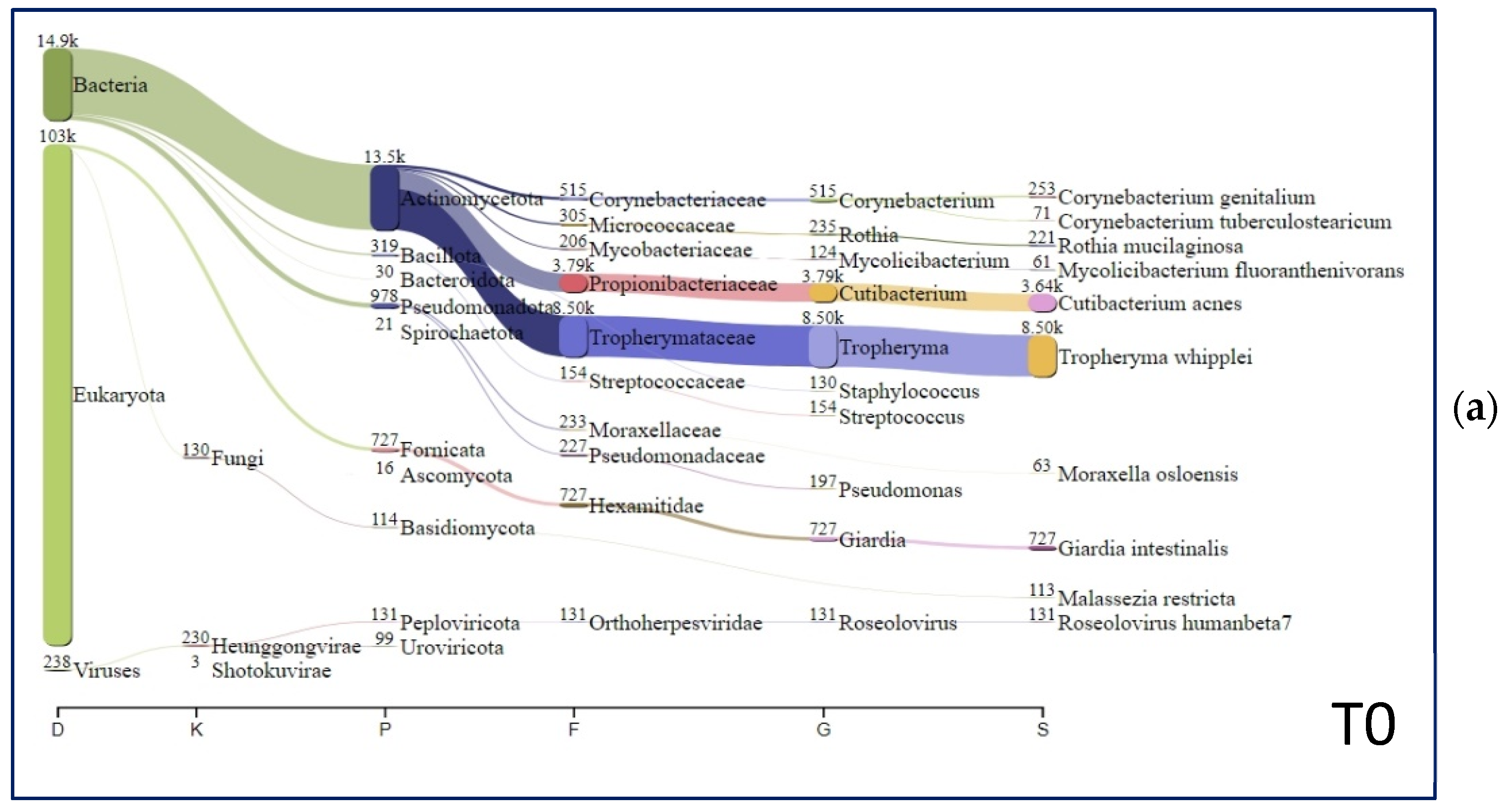
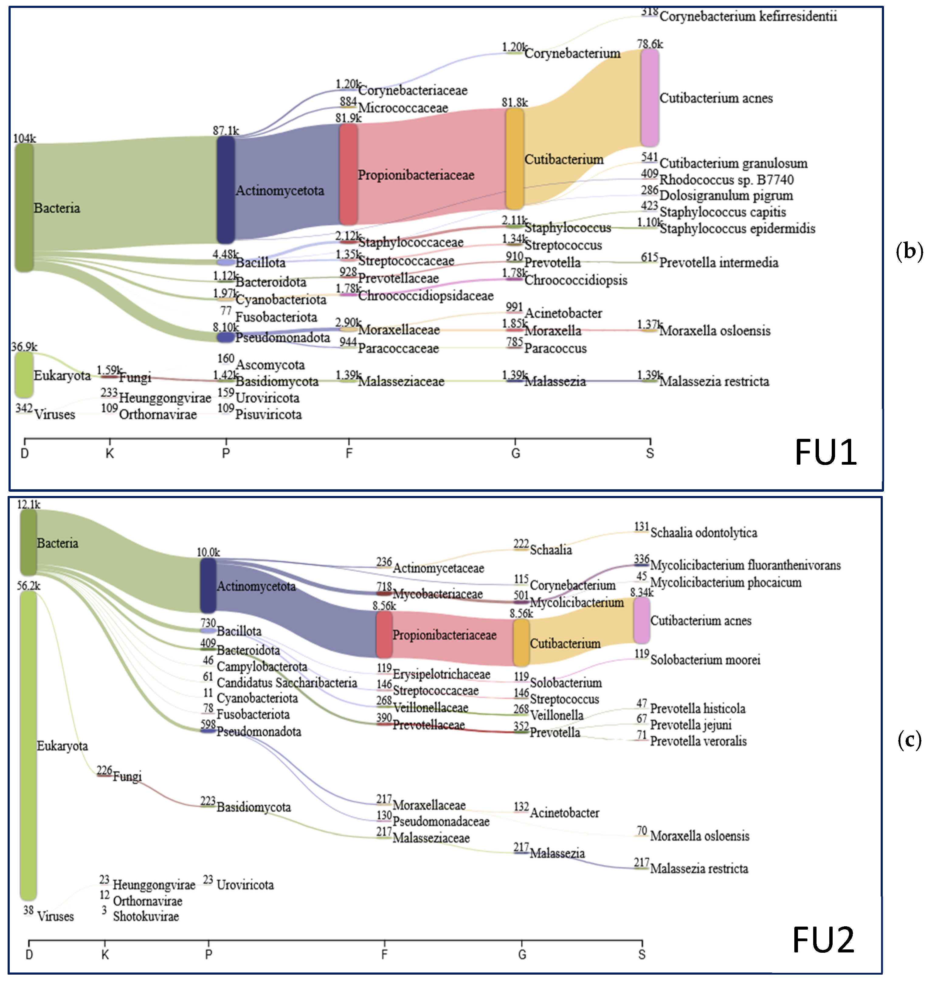
Laboratory Examinations
(Reference Range)
T0FU1FU2
Blood cell count, ×1012/L (4.3–5.9)2.584.54.9
Hemoglobin, g/dL (13–16)6.613.613.9
Platelet, ×109/L (150–450)171201183
White blood cells, ×109/L (4.0–10)5.06.36.9
Erythrocyte sedimentation rate, mm/h (0–20)281312
INR, (0.91–1.08)0.920.960.93
Iron, mcg/dL (60–160)9.07085
Sodium, mmol/L (130–150)135136135
Potassium, nmol/L (3.5–5.0)3.63.73.9
C-Reactive protein, mg/L (<5)2762
Ferritin, mcg/L (20–270)98101107
Transferrin saturation, % (15–50)3.21820
Alpha-globulins, g/dL (0.11–0.35)0.560.220.28
Gamma-globulins, g/dL (0.58–1.75)0.840.910.85
IgM, g/dL (0.5–2.0)0.60.81.1
IgG, g/dL (5.3–16.5)9.08.99.2
IgA, g/dL (0.8–5.3)3.62.53.1
Serum creatinine, mg/dL (0.67–1.2)0.980.860.91
Albumin, g/dL (>3.5)2.74.64.7
Bilirubin, mg/dL (0.3–1.2)0.30.60.8
Glutamic oxaloacetic Transaminase, IU/L (<34)332120
Glutamic pyruvic transaminase, IU/L (<49)201111
Alkaline phosphatase, IU/L (46–116)1009785
Lactate dehydrogenase, IU/L (<250)160135130
Table 2. The most abundant species identified in T0 (WD diagnosis), FU1 (follow-up 1), and FU2 (follow-up 2) samples. The five most abundant species for the three studied time points are underlined.
SpeciesT0 (%)FU1 (%)FU2 (%)
Tropheryma whipplei51.790.000.00
Cutibacterium acnes22.1870.5066.84
Giardia intestinalis4.430.000.03
Corynebacterium genitalium1.540.000.00
Rothia mucilaginosa1.350.010.06
Roseolovirus humanbeta70.800.070.00
Malassezia restricta0.691.241.74
Moraxella osloensis0.381.230.56
Mycolicibacterium fluoranthenivorans0.370.122.69
Staphylococcus epidermidis0.320.990.10
Staphylococcus capitis0.090.380.05
Cutibacterium granulosum0.080.490.00
Corynebacterium kroppenstedtii0.080.130.19
Corynebacterium kefirresidentii0.050.290.09
Streptococcus thermophilus0.050.100.03
Schaalia odontolytica0.040.001.05
Paracoccus yeei0.040.190.01
Kocuria rhizophila0.040.090.03
Acinetobacter johnsonii0.020.100.03
Finegoldia magna0.010.090,00
Streptococcus sanguinis0.010.120.02
Dolosigranulum pigrum0.000.260.00
Streptococcus mitis0.000.150.02
Prevotella intermedia0.000.550.02
Acidovorax temperans0.000.180.00
Lactobacillus iners0.000.170.00
Haemophilus parainfluenzae0.000.160.26
Rhodococcus sp. B77400.000.370.00
Solobacterium moorei0.000.000.95
Corynebacterium accolens0.000.170.02
Lactobacillus crispatus0.000.130.00
Staphylococcus pasteuri0.000.110.00
Others15.6421.6425.20
Disclaimer/Publisher’s Note: The statements, opinions and data contained in all publications are solely those of the individual author(s) and contributor(s) and not of MDPI and/or the editor(s). MDPI and/or the editor(s) disclaim responsibility for any injury to people or property resulting from any ideas, methods, instructions or products referred to in the content.

