1. Introduction
Esophageal, gastric and rectal cancers are a worldwide issue; their high incidence is a consequence of unhealthy behaviors (smoking, alcohol consumption, high red/processed meat intake) or pre-neoplastic conditions (Barrett’s esophagus, atrophic gastritis, Helicobacter pylori infection, dysplastic lesions, central obesity, high risk genetic conditions such as mutation of the CDH1 gene, Lynch syndrome, juvenile polyposis syndrome, familial adenomatous polyposis, Peutz-Jeghers syndrome). Given the lack of symptoms till advanced stages, an early diagnosis and a resulting curative treatment are still uncommon. Nevertheless, thanks to the technical improvement of the endoscopes and the campaigns of screening and follow-up directed at high-risk patients, the rate of early diagnoses is increasing.
In superficial GI (gastrointestinal) cancers the choice between a minimally invasive treatment (endoscopic mucosal resection—EMR—endoscopic mucosal dissection—ESD—transanal endoscopic microsurgery—TEM) and resective surgery is mainly based on the assessment of the depth of neoplastic infiltration alongside the GI wall and the consequent risk of lymph node metastases. Moreover, the therapeutic strategy must take into consideration other factors such as the site and the size of the lesion, the anesthesiological and procedural risks, and the balance between oncological benefits and costs.
Historically, after the bioptic confirmation of an esophageal, gastric or rectal carcinoma, a distant and locoregional staging by various combinations of TC scan, PET scan, MRI and EUS has been the cornerstone through which the treatment was planned. Nevertheless, for superficial GI tumors, due to conflicting results, the utility of EUS staging is under debate. For esophageal cancers EUS is still the gold standard for local staging of not-metastatic tumors, with an overall accuracy of 90%. However, according to some papers, its accuracy can drop up to 65% in early tumors, when there is the need for distinguishing T1a (mucosal) from T1b (submucosal) tumors [
1,
2,
3,
4,
5], even when high-frequency intracanalar miniprobes are used [
6]. On the other side, a meta-analysis found better results, with a sensitivity and a specificity of 84% and 91% for T1a, and of 83% and 89% for T1b, respectively [
7]. In staging gastric cancers a meta-analysis showed that the overall EUS performance is satisfying, although it seems suboptimal in diagnosing superficial tumors (T1a versus T1b), with a sensitivity and a specificity of 87% and 75% respectively [
8]. Finally, for staging rectal cancers, EUS should still be strongly considered for T staging, although for early stages its performance is slightly conflicting; in predicting a T1a tumor Zhao [
3] found an accuracy of 84.6% while Puli reported a sensitivity and a specificity of 97.3% and 96.3% [
9]; according to a recent meta-analysis EUS was able to differentiate a slight (<1 mm) submucosal from a deep (>1 mm) submucosal colorectal infiltration with a sensitivity of 88% and a specificity of 87% [
10].
On the other side, the recent technical improvement of endoscopic instruments allows for an increasingly better visualization of early lesions: high-definition endoscopes, magnification imaging, dye-based or virtual chromoendoscopy, and the use of acetic acid, possibly coupled with artificial intelligence, can improve the detection, the characterization and the depth assessment of the epithelial lesions. This endoscopic staging has recently gained trust as an accurate method to distinguish intramucosal tumors from cancers with submucosal invasion [
11,
12,
13,
14,
15], although the high interobserver variability can be a limit for its application and reproducibility [
16]. Anyway, for superficial-appearing lesions, the general current tendency is to plan the treatment according to their peculiar endoscopic pattern. When the lesion is likely to be intramucosal, an endoscopic resection is done with no other prior staging assessment; the pathologic evaluation of the resected specimen will prove if a radical resection has been achieved or if further staging procedures or treatments are needed. In the early stages an endoscopic resection, mainly ESD, can offer the same oncological radicality as surgery with less hospitalization, fewer adverse events and fewer costs. On the other hand, if the tumor firstly shows endoscopic signs of deep submucosal infiltration, the risk of lymph node metastasis or local relapse after endoscopic resection is significant and an additional distant and local staging must precede the therapeutic decisions [
17,
18,
19,
20].
2. Materials and Methods
This is a retrospective study on the patients that were evaluated in our center for an early-appearing neoplastic lesion of the esophagus, stomach or rectum, with a bioptic diagnosis of high-grade dysplasia (HGD), squamous cell carcinoma (SCC) or adenocarcinoma (ADK).
We included the patients that underwent: (1) a high-definition white light endoscopic assessment of the lesion, coupled with acetic acid-enhancement and dye-based or virtual chromoendoscopy; as magnification imaging endoscopes were introduced during the period of recruitment and only a few patients were assessed with them, this kind of evaluation was not considered in this study; (2) an EUS local T and N staging of the lesion with a radial echoendoscope; our policy is to use radial echoendoscopes to stage GI tumors in order to achieve a more complete and fast 360° evaluation of the GI wall and the surrounding structures; or (3) the pathological diagnosis of the lesion, after resection of any type.
We excluded the patients: (1) under 18 years old and (2) that underwent previous endoscopic attempts of resection of the target lesion.
2.1. Endoscopic Evaluation
The endoscopic assessment of the lesions was performed using Olympus H190 (Tokyo, Japan) series endoscopes; it was based on: the Paris classification [
21], their size, the presence of ulceration, and the histological diagnosis on the pretreatment biopsy; the lesions were finally labeled as fit or unfit for an attempt of en bloc endoscopic resection with a curative aim.
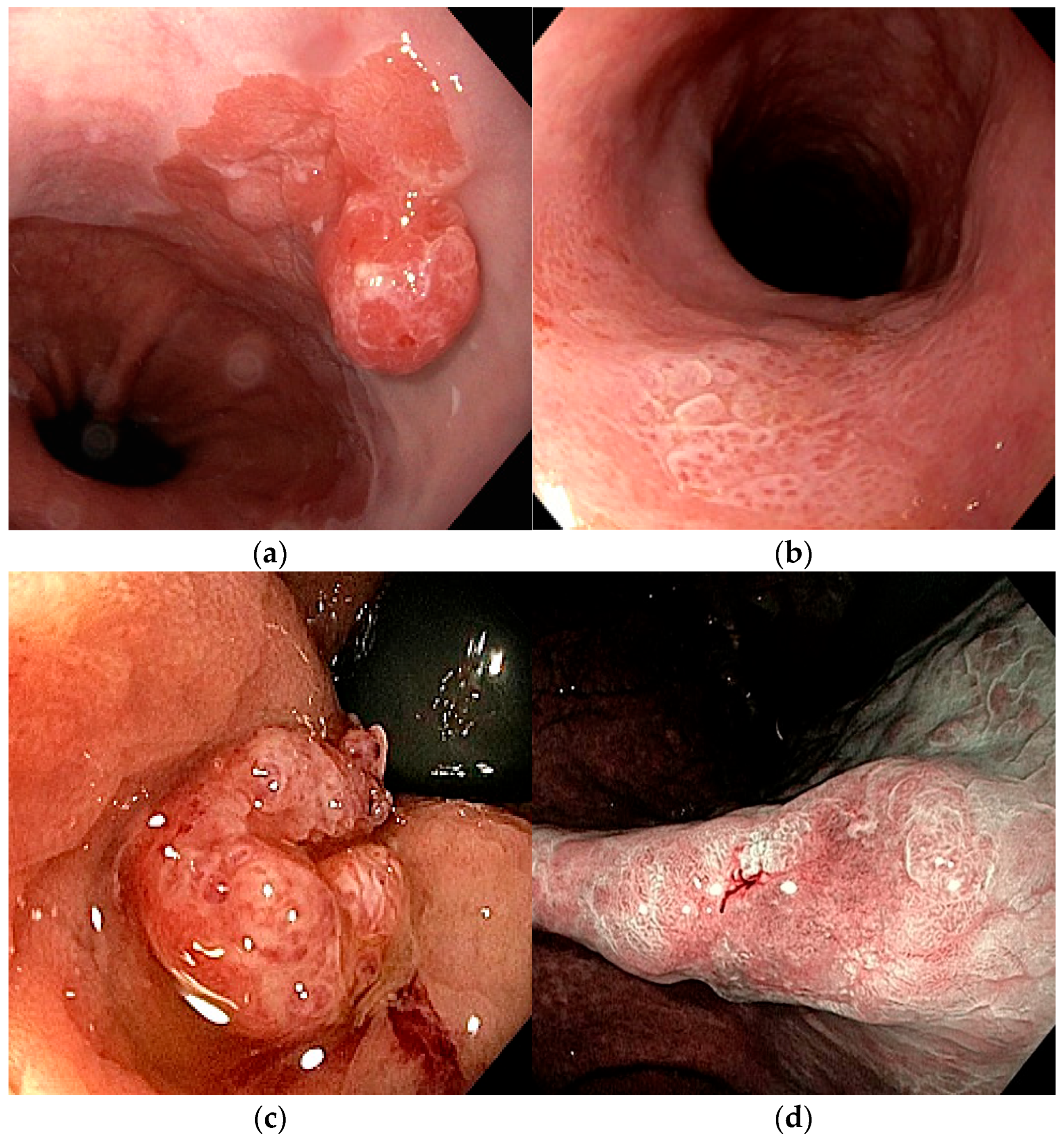
For esophageal squamous lesions (HGD or SCC), we classified as fit for endoscopic resection, preferably with ESD, the Paris 0-Is and 0-II non-circumferential lesions ≤ 20 mm, which have a high probability of only intramucosal infiltration [
22] (
Figure 1a). The Japan Esophageal Society magnifying endoscopy classification [
23], based on the superficial microvascular pattern, was not applied.
For esophageal Barrett-associated lesions (HGD or ADK), according to the ESGE (European Society of Gastrointestinal Endoscopy), we considered as fit for EMR the Paris 0-IIa and 0-IIb lesions ≤ 20 mm (and larger or multifocal dysplastic lesions), while ESD was preferred for the Paris 0-Is and 0-IIc lesions, for malignant lesions > 20 mm, and for lesions located in fibrotic areas [
24] (
Figure 1b).
For gastric lesions (HGD or ADK), we set the ESD indication as dysplastic lesions of any size (with en bloc EMR being an alternative only for Paris 0-IIa lesions ≤ 10 mm), and asParis 0-Is and 0-II intramucosal not ulcerated differentiated-type ADK of any size (and for those ≤30 mm when ulcerated) (
Figure 1c). Moreover, for apparently superficial submucosal (SM1) differentiated lesions ≤ 30 mm or poorly differentiated intramucosal lesions ≤ 20 mm, both without ulceration, ESD was considered on an individualized basis. On the contrary, among endoscopic features suggestive of deep submucosal invasion (SM2 or SM3), that served as exclusion criteria, we included deep ulceration, markedly elevated margins, nodularity, and the fusion/convergence/clubbing of mucosal folds [
25].
For rectal lesions (HGD, SCC or ADK) ESD was recommended for lesions > 20 mm, or if a bulky/protruding component or a demarcated depressed area with irregular surface architecture was present, according to the LST classification [
26] (
Figure 1d). According to the NICE classification [
27], based on the use of NBI to assess the color, the vessels and the surface pattern of the lesions, we excluded Type 3 lesions, due to their high probability of deep submucosal infiltration. The JNET magnification-based classification was not applied.
2.2. EUS Staging
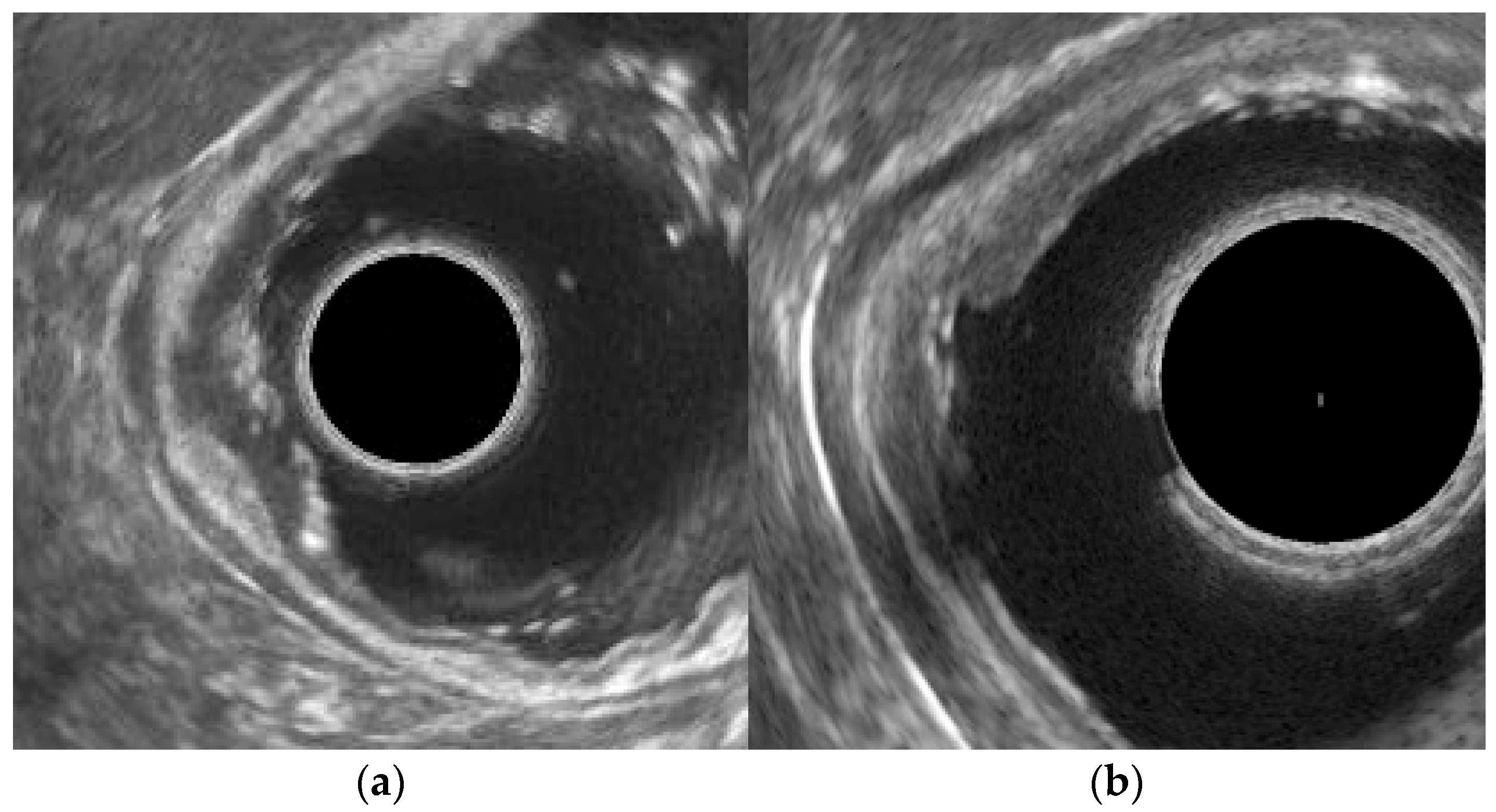
The EUS staging was done using Pentax EG3670URK radial echoendoscopes (Tokyo, Japan); the lesions were classified according to the TNM classification (8th edition) and, based on the local uT (ultrasound-assessed Tumor extent) and uN (ultrasound-assessed Nodal involvement) evaluation, they were labeled as fit or unfit for an attempt at endoscopic resection. The lesions were classified as uT1a in case of thickening of the mucosa, with normal thickness and stratification of the other deeper wall layers (
Figure 2a). Given the great difficulty, or rather the impossibility, of discrimination between an early (SM1) and a deep (SM2 or SM3) submucosal involvement with the probes available in our center, the lesions were considered as uT1b instead when the submucosa was irregularly thickened or thin, or if it looked somehow fusioned with the mucosa, with normal underlying muscularis propria and adventitia/serosa (
Figure 2b). Regarding the nodal staging, the lesions were considered as uN0 in case of absence of regional enlarged lymph nodes and uN1 if at least one local hypoechoic roundish lymph node > 1cm was visible. Lesions staged as T1a N0 were classified as fit for endoscopic resection, while lesions uT1b or uN1 were considered unfit for endoscopic treatment.
The above-mentioned endoscopic and EUS assessments were blindly effected under conscious (using Midazolam and Fentanyl), or deep (using Propofol) sedation by two experienced endoscopists. The therapeutic strategy, based on the endoscopic pattern, the EUS staging, and the clinical conditions of the patient, was planned by a multidisciplinary group composed by the endoscopist, the surgeon and the oncologist. The lesions, when a deep submucosal infiltration seemed unlikely, were preferably resected with ESD with potentially curative purpose. A more aggressive approach was planned instead if a radical endoscopic resection seemed unfeasible, or if it was attempted but the result was not curative after the histological assessment.
2.3. Statistical Analysis
Categorical variables were expressed as absolute numbers and percentages, while continuous variables were reported as mean ± standard deviation or median with interquartile range, as appropriate. The final pathological assessment after resection was considered the reference standard. Lesions were classified as intramucosal when final histology showed HGD or T1a cancer (with or without nodal assessment), as submucosal in case of T1b invasion, and T2 if muscularis propria was involved.
The performance of endoscopic assessment and EUS in identifying lesions suitable for endoscopic resection was evaluated. Comparisons between endoscopic assessment and EUS were performed using the McNemar test for paired data.
EUS T-staging errors were further analyzed by distinguishing overstaging from understaging. Factors associated with incorrect EUS T staging were explored using univariate logistic regression analysis. Variables with a p value < 0.10 at univariate analysis were entered into a multivariate logistic regression model to identify independent predictors of EUS misclassification. Results of logistic regression analyses were reported as odds ratios (ORs) with 95% confidence intervals (CIs). A two-sided p value < 0.05 was considered statistically significant. Statistical analyses were performed using the MedCalc 14.0 statistical software.
2.4. Aim of the Study
The aim of the study was to assess the performance of endoscopy and EUS in staging superficial GI neoplastic lesions and their capacity to identify which are fit for endoscopic resection. The endoscopic and EUS results regarding the indication for endoscopic treatment were compared to the final pathological staging, after any type of resection, done by experienced dedicated pathologists.
3. Results
Overall 57 patients were included in the study (37 male, 20 female, age 69.3 ± 12.3 years). Bioptic histology of the lesions before resection yielded HGD in 16 cases, SSC in four, and ADK in 37. Lesions were located in the esophagus (19 patients), stomach (29 patients), and rectum (nine patients) with a median size of 20 mm [interquartile range: 15–26 mm].
3.1. Endoscopic Evaluation
After the endoscopic assessment 42 (73.7%) lesions were considered as fit for endoscopic resection. Six patients underwent EMR, yielding HGD (one patient), T1a (four patients), and T1b N0 (one patient, who underwent surgery after not-radical endoscopic resection). Twenty-eight patients underwent ESD: six yielded HGD (one of whom underwent surgery for intraprocedural perforation), 10 T1a (two of them underwent surgery: one due to not-radical endoscopic resection, one for iatrogenic perforation), and 12 T1b (after not-radical resection one of them underwent TEM and five of them underwent surgery). Three patients underwent upfront TEM, yielding HGD (one patient), T1a (one patient), and T1b (one patient). Five patients underwent upfront surgery due to the patient’s preference: they yielded HGD (one patient) and T1a N0 (four patients).
Meanwhile, 15 lesions were considered as unfit for endoscopic resection. Thirteen patients underwent surgery, yielding T1a N0 (one patient), T1b N0 (10 patients), and T1b N1 (two patients). One patient underwent an attempt of ESD due to her poor general conditions but for an intraprocedural perforation she underwent surgery, yielding T1b N0. One patient underwent TEM, yielding T1b.
The presence of a sessile component was shown in 21 (36.6%) cases while a depressed or excavated (Paris 0-IIc or 0-III) component was shown in 17 (29.8%) cases.
Overall, after the endoscopic evaluation, among the 42 lesions considered fit for endoscopic resection nine were HGD, 19 were T1a cancers, and 14 were T1b cancers; among the 15 lesions considered unfit for endoscopic resection one was a T1a cancer and 14 were T1b cancers (two of whom were N1). The sensibility, specificity, PPV, NPV, and accuracy of the endoscopic assessment for detecting lesions limited within the mucosa (HGD or T1a tumors) were 96.6%, 50%, 66.7%, 93.3%, and 73.7% respectively.
3.2. EUS Staging
Regarding the T assessment, EUS found 29 (50.9%) lesions fit for endoscopic resection (uT1a): eight were HGD, eight were T1a cancers, and 13 were T1b cancers at final histology. At the same time, EUS staged 28 lesions as unfit for endoscopic resection (uT1b): one was HGD, 12 were T1a cancers, and 15 were T1b cancers (two of whom were N1) at pathological assessment after resection. EUS overstaged 19 lesions and understaged seven lesions. The sensibility, specificity, PPV, NPV, and accuracy of EUS for detecting lesions limited within the mucosa (HGD or T1a tumors) were 55.2%, 53.6%, 55.2%, 53.6%, and 54.4% respectively.
Regarding the N staging, 55 out of 57 patients were uN0. Twenty-seven of them underwent surgery (17 upfront, 10 after endoscopic failure) and lymphadenectomy was done in 24, revealing lymph node metastases in two patients. Among the 31 patients that were treated with EMR, ESD, TEM, or surgery (upfront or rescue) without lymphadenectomy, the confirmation of the N0 status is lacking, but they are currently showing no nodal metastases at follow-up. EUS staged two out of 57 patients as uN1; they did not undergo EUS-guided needle biopsy before therapy: one underwent ESD yieldingT1a, the other underwent an upfront surgical resection of a T1b N0 esophageal squamous cancer. Thus, considering only the limited group of the 25 patients that underwent lymphadenectomy, the sensibility, specificity, PPV, NPV, and accuracy of EUS in establishing the absence of regional lymph node metastases (N0 tumors) were 95.7%, 0%, 91.7%, 0%, and 88.0% respectively. In
Table 1 are shown all the results.
The endoscopic assessment was more accurate (73.7%) than EUS (54.4%) in identifying lesions limited to the mucosa (
p = 0.03). The performance of EUS was associated neither with the age and gender of the patient, nor with the sessile or depressed component, or the location or the size of the lesion. In multivariate analysis the presence of an initial bioptic diagnosis of ADK and an endoscopic pattern fit for endoscopic resection were independent factors related to a wrong EUS T staging of the lesion (
Table 2). As a confirmation of the low concordance between these two techniques, 15 patients were considered fit for endoscopic resection according to the endoscopic assessment but unfit after the EUS staging; five of them underwent TEM or surgery; 13 (86.7%) of them yielded HGD or T1a cancers after resection. We did not see any difference between the diagnostic performance of endoscopy or EUS according to the use of conscious or deep sedation.
3.3. Type and Outcomes of Treatment
Among the 57 patients in the study the following treatments were carried out: EMR in six patients (four in the esophagus, two in the stomach), ESD in 29 patients (10 in the esophagus, 15 in the stomach, four in the rectum), upfront TEM in four patients, and upfront surgical resection in 18 patients (five in the esophagus, 12 in the stomach, one in the rectum).
The upfront minimally invasive treatments (EMR, ESD or TEM) were technically successful and histologically curative in 28 out of 39 (71.8%) patients. Meanwhile, a further rescue treatment was needed in 11 (28.2%) patients: one TEM after a not-curative rectal ESD, one surgical resection due a to not-curative gastric EMR, seven surgical resections after a not-curative ESD (two in the esophagus, five in the stomach), and two surgical resections for gastric perforation during ESD, a potential complication of this procedure.
The final pathologic diagnosis after resection was HGD (nine patients), T1a Nx cancers (14 patients), T1a N0 cancers (six patients), T1b Nx cancers (nine patients), T1b N0 cancers (17 patients), and T1b N1 cancers (two patients). It is notable that among the 16 lesions that had an initial bioptic diagnosis of HGD, seven (43.8%) yielded a cancer at final histology after resection: three T1a (two in the esophagus, one in the stomach) and four T1b (two in the esophagus, two in the rectum).
4. Discussion
GI cancers show a high incidence worldwide and the prognosis of the patients depends on the timing of the diagnosis and treatment. Recently, the campaigns of screening and follow-up for high-risk patients and the technical improvement of the endoscopes have increased the rate of early diagnosis of such tumors. Moreover, in the last two decades, in the early stages, mini-invasive techniques in the form of endoscopic treatment, mainly ESD, have gradually demonstrated to achieve the same oncological benefit as surgical resection with fewer costs and fewer adverse events. Thus, a precise staging of the tumor is mandatory in order to select the patients fit for an endoscopic treatment; for this purpose EUS and, more recently, the high-definition endoscopic assessment of the superficial pattern have been used.
In this study the endoscopic assessment proved superior to EUS in distinguishing intramucosal lesions from tumors with submucosal invasion, in accordance with other published data [
28,
29]. Although both techniques showed only fair overall accuracy, endoscopy achieved a markedly higher sensitivity and NPV for intramucosal disease; endoscopic judgment was not significantly influenced by patient-related variables, supporting a lesion-centered decision-making process. This finding is clinically relevant, as the primary objective of staging in superficial-appearing lesions is not to precisely subclassify T1 disease, but rather to safely exclude deep submucosal invasion and to identify patients suitable for curative endoscopic resection.
By contrast, EUS showed a limited discriminative ability in early T staging, with an accuracy barely exceeding 50%. Notably, EUS tended to overstage intramucosal lesions, classifying a substantial proportion of HGD and T1a cancers as uT1b. Such systematic overstaging undermines the utility of EUS in this setting and may negatively influence the therapeutic decision-making by diverting patients from potentially curative endoscopic treatment toward more invasive approaches. EUS overstaging was independently associated with ADK histology and with lesions considered suitable for endoscopic resection at endoscopic assessment. The tendency of EUS to overstage early lesions has important clinical implications. In our series, several lesions deemed unsuitable for endoscopic resection based on EUS findings were ultimately confirmed as intramucosal at histology. This suggests that, in superficial-appearing lesions, EUS may act as a driver of overtreatment (i.e., surgery) rather than as a safeguard against undertreatment. Importantly, EUS inaccuracy was more pronounced in lesions that appeared endoscopically suitable for endoscopic resection, highlighting a potential conflict between these two staging modalities when used in parallel. These findings support the concept that confident endoscopic assessment should not be overridden by EUS-based T staging in early GI neoplasia. Rather than functioning as an equal or confirmatory test, EUS appears poorly suited for refining invasion depth within the T1 category.
Despite its limitations in T staging, EUS retained a high PPV for the absence of nodal disease in patients who underwent lymphadenectomy. This observation suggests that the main contribution of EUS in superficial GI tumors may lie in nodal rather than mural assessment. In this context, EUS could be considered as a complementary tool aimed at excluding suspicious lymph nodes before endoscopic resection, rather than as a decisive instrument for determining invasion depth.
Overall, our data support a staging paradigm in which endoscopic morphology represents the primary determinant for treatment selection in superficial GI lesions, while EUS plays a complementary, supportive role focused mainly on nodal evaluation.
Regarding the outcomes of minimally invasive treatments without lymphadenectomy, EMR, ESD and TEM were curative in 71.8% of patients, while 23.1% and 5.1% required further rescue treatments after a not-curative resection or iatrogenic perforation, respectively. This is in line with other papers [
30] and confirms the central role of endoscopic resections in the setting of superficial GI tumors, where a great proportion of patients will benefit from a curative and safe sparing-surgery therapy, and only a minority will need further surgical treatment.
We want to stress the importance of a multidisciplinary management of such patients; a team of experienced and dedicated endoscopists, surgeons, pathologists and oncologists should take care of the initial evaluation, share the therapeutic decision and carefully monitor the outcomes after treatment during the follow-up.
This study has some limits. First, we included only patients that have been studied without magnification imaging endoscopes and without high-frequency intracanalar EUS probes, which probably are the best performing instruments for these staging procedures [
31,
32]. This could have caused us to underestimate the real performance of these techniques; this choice had the purpose of reproducing the everyday setting of the majority of hospitals, where only standard instruments are available. Second, in the study we also included patients with a pre-resective bioptic diagnosis of HGD, which could not be considered a proper malignant lesion and was thus suitable for staging evaluation. Nevertheless, almost half of such lesions appeared to be T1a or even T1b cancers after resection, showing that the initial bioptic diagnosis of HGD could not completely represent the real histology of the whole lesion, especially in the esophagus. So, we argue that all the early-appearing neoplastic lesions that are evaluated for an endoscopic resection could benefit from a sort of staging assessment regardless of the initial not-malignant bioptic diagnosis.
5. Conclusions
An early diagnosis and a precise staging of early GI tumors are mandatory in order to assess the possibility of an endoscopic resection. In this series the endoscopic performance in differentiating superficial GI lesions fit for an endoscopic treatment from deeper T1 lesion was better than that of EUS, that tended to overstage such lesions; regarding the evaluation of tumor depth, endoscopy selected patients correctly while EUS tended to deselect them incorrectly. Nevertheless, EUS had a high PPV for the N0 status, thus showing an interesting role, in combination with the endoscopic assessment, in the decision-making for early GI tumors.
Larger multicenter comparative studies are advisable to confirm these data, also including the use of the best devices currently available such as magnification imaging endoscopes and high-frequency intracanalar EUS probes, which have not been evaluated in this study.
Author Contributions
Conceptualization, T.T.; methodology, T.T. and A.L.; software, A.L.; validation, P.F. and V.G.M.; formal analysis, A.L.; investigation, T.T., F.T., A.M., A.G., M.T. and M.M.; data curation, A.L.; writing, review and editing, T.T. and A.L.; supervision, P.F. and V.G.M. All authors have read and agreed to the published version of the manuscript.
Funding
This research received no external funding.
Institutional Review Board Statement
The study was conducted in accordance with the principles of the Declaration of Helsinki. Due to the retrospective and observational design of the study, which involved the analysis of anonymized clinical data collected during routine clinical practice and did not require any additional diagnostic or therapeutic procedures, formal approval from the Institutional Review Board was waived according to institutional policies and applicable national regulations. In particular, the study complies with the framework for observational clinical research defined by the Italian Medicines Agency (AIFA) Determination of 20 March 2008, “Guidelines for the classification and conduct of observational studies on medicines”, published in the Italian Official Journal (Gazzetta Ufficiale n. 76, 31 March 2008). Available at:
https://www.gazzettaufficiale.it/eli/id/2008/03/31/08A02109/sg (accessed on 1 April 2022). Finally, the study protocol was reviewed by the local ethics committee, which confirmed that formal approval was not required due to the retrospective nature of the study.
Informed Consent Statement
Informed consent was obtained from all subjects involved in the study.
Data Availability Statement
The data presented in this study are available upon request from the corresponding author due to privacy and regulatory issues.
Conflicts of Interest
Andrea Lisotti and Pietro Fusaroli received consultancy fees from Boston Scientific Corporation and Olympus Company. The remaining authors declare that the research was conducted in the absence of any commercial or financial relationships that could be construed as a potential conflict of interest.
Abbreviations
The following abbreviations are used in this manuscript:
| EUS | Endoscopic Ultrasound |
| EMR | Endoscopic Mucosal Resection |
| ESD | Endoscopic Submucosal Dissection |
| TEM | Transanal Endoscopic Microsurgery |
| HGD | High-Grade Dysplasia |
| GI | Gastrointestinal |
| CT | Computed Tomography |
| PET | Positron Emission Tomography |
| MRI | Magnetic Resonance Imaging |
| NBI | Narrow Band Imaging |
| PPV | Positive Predictive Value |
| NPV | Negative Predictive Value |
| SCC | Squamous Cell Cancer |
| ADK | Adenocarcinoma |
References
- Young, P.E.; Gentry, A.B.; Acosta, R.D.; Greenwald, B.D.; Riddle, M. Endoscopic ultrasound does not accurately stage early adenocarcinoma or high-grade dysplasia of the esophagus. Clin. Gastroenterol. Hepatol. 2010, 8, 1037–1041. [Google Scholar] [CrossRef] [Scilit]
- Pouw, R.E.; Heldoorn, N.; Alvarez Herrero, L.; ten Kate, F.J.; Visser, M.; Busch, O.R.; van Berge Henegouwen, M.I.; Krishnadath, K.K.; Weusten, B.L.; Fockens, P.; et al. Do we still need EUS in the workup of patients with early esophageal neoplasia? A retrospective analysis of 131 cases. Gastrointest. Endosc. 2011, 73, 662–668. [Google Scholar] [CrossRef] [Scilit]
- Kahlon, S.; Aamar, A.; Butt, Z.; Urayama, S. Role of endoscopic ultrasound for pre-intervention evaluation in early esophageal cancer. World J. Gastrointest. Endosc. 2023, 15, 447–457. [Google Scholar] [CrossRef] [Scilit] [PubMed]
- Zhang, J.; Chen, M.; Gao, Y.; Liu, J.; Li, Z.; Wang, D. Endoscopic ultrasound with submucosal saline injection improves the accuracy of T1a and T1b staging in superficial esophageal squamous cell carcinoma. Front. Med. 2025, 12, 1509628. [Google Scholar] [CrossRef] [Scilit]
- Maes, S.; Haidry, R.; Bisschops, R. Can the depth of invasion of early esophageal cancer be predicted based on endoscopic evidence? Minerva Chir. 2018, 73, 385–393. [Google Scholar] [CrossRef] [Scilit]
- Rodríguez-Carrasco, M.; Esposito, G.; Libânio, D.; Pimentel-Nunes, P.; Dinis-Ribeiro, M. Image-enhanced endoscopy for gastric preneoplastic conditions and neoplastic lesions: A systematic review and meta-analysis. Endoscopy 2020, 52, 1048–1065. [Google Scholar] [CrossRef] [Scilit] [PubMed]
- Backes, Y.; Moss, A.; Reitsma, J.B.; Siersema, P.D.; Moons, L.M. Narrow band imaging, magnifying chromoendoscopy, and gross morphological features for the optical diagnosis of T1 colorectal cancer and deep submucosal invasion: A systematic review and meta-analysis. Am. J. Gastroenterol. 2017, 112, 54–64. [Google Scholar] [CrossRef] [Scilit] [PubMed]
- Kawaguti, F.S.; Franco, M.C.; Martins, B.C.; Segateli, V.; Marques, C.F.S.; Nahas, C.S.R.; Pinto, R.A.; Safatle-Ribeiro, A.V.; Ribeiro-Junior, U.; Nahas, S.C.; et al. Role of Magnification Chromoendoscopy in the Management of Colorectal Neoplastic Lesions Suspicious for Submucosal Invasion. Dis. Colon Rectum 2019, 62, 422–428. [Google Scholar] [CrossRef] [Scilit] [PubMed]
- Nemoto, D.; Guo, Z.; Peng, B.; Zhang, R.; Nakajima, Y.; Hayashi, Y.; Yamashina, T.; Aizawa, M.; Utano, K.; Lefor, A.K.; et al. Computer-aided diagnosis of serrated colorectal lesions using non-magnified white-light endoscopic images. Int. J. Color. Dis. 2022, 37, 1875–1884. [Google Scholar] [CrossRef] [Scilit] [PubMed]
- ASGE STANDARDS OF PRACTICE COMMITTEE; Qumseya, B.; Sultan, S.; Bain, P.; Jamil, L.; Jacobson, B.; Anandasabapathy, S.; Agrawal, D.; Buxbaum, J.L.; Fishman, D.S.; et al. ASGE guideline on screening and surveillance of Barrett’s esophagus. Gastrointest. Endosc. 2019, 90, 335–359.e2. [Google Scholar] [CrossRef] [Scilit] [PubMed]
- Ishihara, R.; Mizusawa, J.; Kushima, R.; Matsuura, N.; Yano, T.; Kataoka, T.; Fukuda, H.; Hanaoka, N.; Yoshio, T.; Abe, S.; et al. Assessment of the Diagnostic Performance of Endoscopic Ultrasonography After Conventional Endoscopy for the Evaluation of Esophageal Squamous Cell Carcinoma Invasion Depth. JAMA Netw. Open 2021, 4, e2125317. [Google Scholar] [CrossRef] [Scilit] [PubMed]
- Dinis-Ribeiro, M.; Libânio, D.; Uchima, H.; Spaander, M.C.W.; Bornschein, J.; Matysiak-Budnik, T.; Tziatzios, G.; Santos-Antunes, J.; Areia, M.; Chapelle, N.; et al. Management of epithelial precancerous conditions and early neoplasia of the stomach (MAPS III): European Society of Gastrointestinal Endoscopy (ESGE), European Helicobacter and Microbiota Study Group (EHMSG) and European Society of Pathology (ESP) Guideline update 2025. Endoscopy 2025, 57, 504–554. [Google Scholar] [CrossRef] [Scilit]
- Weusten, B.L.A.M.; Bisschops, R.; Dinis-Ribeiro, M.; Di Pietro, M.; Pech, O.; Spaander, M.C.W.; Baldaque-Silva, F.; Barret, M.; Coron, E.; Fernández-Esparrach, G.; et al. Diagnosis and management of Barrett esophagus: European Society of Gastrointestinal Endoscopy (ESGE) Guideline. Endoscopy 2023, 55, 1124–1146. [Google Scholar] [CrossRef] [Scilit]
- Mizumoto, T.; Hiyama, T.; Oka, S.; Yorita, N.; Kuroki, K.; Kurihara, M.; Yoshifuku, Y.; Sanomura, Y.; Urabe, Y.; Arihiro, K.; et al. Diagnosis of superficial esophageal squamous cell carcinoma invasion depth before endoscopic submucosal dissection. Dis. Esophagus 2018, 31, dox142. [Google Scholar] [CrossRef] [Scilit] [PubMed]
- Ishihara, R.; Muto, M. Current status of endoscopic detection, characterization and staging of superficial esophageal squamous cell carcinoma. Jpn. J. Clin. Oncol. 2022, 52, 799–805. [Google Scholar] [CrossRef] [Scilit] [PubMed]
- Tamura, E.; Kudo, S.E.; Iwasaki, S.; Semba, S.; Shibuya, T.; Kato, S.; Kuroki, T.; Sato, Y.; Sakurai, T.; Ogawa, Y.; et al. White Light, Magnifying Endoscopy, Endocytoscopy, and Artificial Intelligence in Diagnosis of Early Colorectal Cancer: A Comparative Study. DEN Open 2025, 6, e70240. [Google Scholar] [CrossRef] [Scilit] [PubMed]
- Choi, J.; Chung, H.; Lee, A.; Kim, J.L.; Cho, S.J.; Kim, S.G. Role of Endoscopic Ultrasound in Selecting Superficial Esophageal Cancers for Endoscopic Resection. Ann. Thorac. Surg. 2021, 111, 1689–1695. [Google Scholar] [CrossRef] [Scilit] [PubMed]
- Pech, O.; Günter, E.; Dusemund, F.; Ell, C. Value of high-frequency miniprobes and conventional radial endoscopic ultrasound in the staging of early Barrett’s carcinoma. Endoscopy 2010, 42, 98–103. [Google Scholar] [CrossRef] [Scilit] [PubMed]
- Luo, L.N.; He, L.J.; Gao, X.Y.; Huang, X.X.; Shan, H.B.; Luo, G.Y.; Li, Y.; Lin, S.Y.; Wang, G.B.; Zhang, R.; et al. Endoscopic Ultrasound for Preoperative Esophageal Squamous Cell Carcinoma: A Meta-Analysis. PLoS ONE 2016, 11, e0158373. [Google Scholar] [CrossRef] [Scilit] [PubMed]
- Mocellin, S.; Pasquali, S. Diagnostic accuracy of endoscopic ultrasonography (EUS) for the preoperative locoregional staging of primary gastric cancer. Cochrane Database Syst. Rev. 2015, 2015, CD009944. [Google Scholar] [CrossRef] [Scilit] [PubMed]
- Zhao, Y.; Ren, M.; Jia, A.; Zhang, J.; Wang, S.; Zhao, Q.; Cai, G.; He, S. The factors influencing the accuracy of pre-operative endoscopic ultrasonography assessment in endoscopic treatments for gastrointestinal tumors. Cancer Med. 2023, 12, 4321–4331. [Google Scholar] [CrossRef] [Scilit]
- Puli, S.R.; Bechtold, M.L.; Reddy, J.B.; Choudhary, A.; Antillon, M.R. Can endoscopic ultrasound predict early rectal cancers that can be resected endoscopically? A meta-analysis and systematic review. Dig. Dis. Sci. 2010, 55, 1221–1229. [Google Scholar] [CrossRef] [Scilit] [PubMed]
- Chen, R.; Huang, Y.; Liu, F. Estimation of Invasion Depth of Early Colorectal Cancer Using Endoscopic Ultrasonography and Magnifying Chromoendoscopy: A Meta-Analysis. Digestion 2025, 106, 245–264. [Google Scholar] [CrossRef] [Scilit]
- Younis, F.; Rösch, T.; Beyna, T.; Ebigbo, A.; Faiss, S.; May, A.; Pech, O.; Dautel, P.; Anders, M.; Clauditz, T.; et al. Expert assessment of infiltration depth and recommendation of endoscopic resection technique in early Barrett cancer. United Eur. Gastroenterol. J. 2024, 12, 848–858. [Google Scholar] [CrossRef] [Scilit] [PubMed]
- Endoscopic Classification Review Group. Update on the Paris classification of superficial neoplastic lesions in the digestive tract. Endoscopy 2005, 37, 570–578. [Google Scholar] [CrossRef] [Scilit] [PubMed]
- Kodama, M.; Kakegawa, T. Treatment of superficial cancer of the esophagus: A summary of responses to a questionnaire on superficial cancer of the esophagus in Japan. Surgery 1998, 123, 432–439. [Google Scholar] [CrossRef] [PubMed]
- Ishihara, R.; Arima, M.; Iizuka, T.; Oyama, T.; Katada, C.; Kato, M.; Goda, K.; Goto, O.; Tanaka, K.; Yano, T.; et al. Endoscopic submucosal dissection/endoscopic mucosal resection guidelines for esophageal cancer. Dig. Endosc. 2020, 32, 452–493. [Google Scholar] [CrossRef] [Scilit] [PubMed]
- Pimentel-Nunes, P.; Libânio, D.; Bastiaansen, B.A.J.; Bhandari, P.; Bisschops, R.; Bourke, M.J.; Esposito, G.; Lemmers, A.; Maselli, R.; Messmann, H.; et al. Endoscopic submucosal dissection for superficial gastrointestinal lesions: European Society of Gastrointestinal Endoscopy (ESGE) Guideline—Update 2022. Endoscopy 2022, 54, 591–622. [Google Scholar] [CrossRef] [Scilit] [PubMed]
- Choi, J.; Kim, S.G.; Im, J.P.; Kim, J.S.; Jung, H.C.; Song, I.S. Endoscopic prediction of tumor invasion depth in early gastric cancer. Gastrointest. Endosc. 2011, 73, 917–927. [Google Scholar] [CrossRef] [Scilit] [PubMed]
- Bisschops, R.; East, J.E.; Hassan, C.; Hazewinkel, Y.; Kamiński, M.F.; Neumann, H.; Pellisé, M.; Antonelli, G.; Bustamante Balen, M.; Coron, E.; et al. Advanced imaging for detection and differentiation of colorectal neoplasia: ESGE Guideline—Update 2019. Endoscopy 2019, 51, 1155–1179. [Google Scholar] [CrossRef] [Scilit] [PubMed]
- Hayashi, N.; Tanaka, S.; Hewett, D.G.; Kaltenbach, T.R.; Sano, Y.; Ponchon, T.; Saunders, B.P.; DRex, D.K.; Soetikno, R.M. Endoscopic prediction of deep submucosal invasive carcinoma: Validation of the narrow-band imaging international colorectal endoscopic (NICE) classification. Gastrointest. Endosc. 2013, 78, 625–632. [Google Scholar] [CrossRef] [Scilit] [PubMed]
- Misra, S.; Badwal, S.; Rao, S.; Khare, S.; Anikhindi, A.; Arora, A.; Dhawan, S. Clinico-pathological findings in a series of gastrointestinal endoscopic submucosal dissection specimens: A retrospective 5-year study. Ann. Diagn. Pathol. 2026, 80, 152575. [Google Scholar] [CrossRef] [Scilit] [PubMed]
| Disclaimer/Publisher’s Note: The statements, opinions and data contained in all publications are solely those of the individual author(s) and contributor(s) and not of MDPI and/or the editor(s). MDPI and/or the editor(s) disclaim responsibility for any injury to people or property resulting from any ideas, methods, instructions or products referred to in the content. |




 ,
,