Review

Liquid Biopsy in Pancreatic Ductal Adenocarcinoma: Clinical Utility, Trials, and Future Directions

1
Department of Pathology and Laboratory Medicine, Montefiore Medical Center, Bronx, NY 10467, USA
2
Department of Pathology and Laboratory Medicine, Lenox Hill Hospital, New York, NY 10075, USA
3
Department of Pathology and Laboratory Medicine, Molecular Genetic Pathology Division, Mount Sinai Hospital, New York, NY 10029, USA
4
Department of Medical Biochemistry, Faculty of medicine, Zagazig University, Zagazig 44519, Al-Sharqia Governorate, Egypt
5
Department of Pathology, Faculty of Medicine, Menoufia University, Shebin El-Kom 32511, Menoufia Governorate, Egypt
*
Author to whom correspondence should be addressed.
Gastroenterol. Insights 2025, 16(4), 39; https://doi.org/10.3390/gastroent16040039
Submission received: 29 July 2025 / Revised: 15 October 2025 / Accepted: 17 October 2025 / Published: 19 October 2025
(This article belongs to the Collection Advances in Gastrointestinal Cancer)

Abstract

Pancreatic ductal adenocarcinoma (PDAC) is a lethal malignancy marked by late diagnosis, rapid progression, and poor prognosis, with a 5-year survival rate of 2–9%. Traditional tissue biopsy faces limitations in accessibility and real-time monitoring. Liquid biopsy—a minimally invasive technique analyzing tumor-derived materials such as circulating tumor DNA (ctDNA), circulating tumor cells (CTCs), exosomes, tumor-educated platelets (TEPs), and cell-free RNAs (cfRNAs)—offers dynamic insights into PDAC biology. This review advances beyond the prior literature by offering a unified synthesis that bridges molecular mechanisms, biomarker dynamics, and clinical translation within the context of PDAC. It also summarizes key clinical trials evaluating liquid biopsy in PDAC, underscoring its growing impact on precision oncology.

1. Introduction

Pancreatic ductal adenocarcinoma (PDAC) is an aggressive malignancy with one of the poorest prognoses among solid tumors, largely due to late diagnosis, early metastasis, and resistance to existing therapies. Despite advances in imaging, molecular profiling, and systemic treatment, the overall 5-year survival rate remains below 2–9% [1,2,3,4,5]. Traditional diagnostic approaches—such as imaging and tissue biopsy—are invasive, technically challenging, and limited in their ability to capture tumor heterogeneity or provide dynamic molecular information [6,7,8].
Liquid biopsy has emerged as a minimally invasive strategy capable of detecting tumor-derived analytes such as circulating tumor DNA (ctDNA), circulating tumor cells (CTCs), exosomes, and tumor-educated platelets (TEPs) in body fluids. These biomarkers offer real-time insights into tumor evolution, genetic alterations, and therapeutic response, positioning liquid biopsy as a promising complement to conventional diagnostics in PDAC.
Despite the extensive and rapidly evolving literature on liquid biopsy in PDAC, most prior reviews have examined either the technological foundations of biomarker detection or their clinical relevance in isolation. In contrast, this review provides a comprehensive and integrative synthesis that connects molecular mechanisms, biomarker kinetics, and translational implementation within a single conceptual framework. We particularly emphasize emerging frontiers such as multi-omic profiling, longitudinal disease monitoring, and artificial intelligence-driven data integration, which remain underrepresented in existing analyses. Moreover, this review critically evaluates the current clinical trial landscape and regulatory environment to delineate the barriers to clinical translation.

2. Methods

A focused literature review was conducted using PubMed, Scopus, and Web of Science for English-language studies published from 2015 to 2025. Search terms included “liquid biopsy”, “pancreatic ductal adenocarcinoma”, “ctDNA”, “CTCs”, “exosomes”. “cfRNA”, and “TEPs.” Eligible studies involved human subjects and evaluated diagnostic, prognostic, or therapeutic applications of liquid biopsy in PDAC. Non-English, non-human, or in vitro-only reports were excluded. Reference lists of included papers and recent reviews were manually screened to identify additional relevant studies. Data extracted included study design, analyte type, detection platform, and key clinical outcomes. Clinical trials were identified through searches of ClinicalTrials.gov and cross-referencing cited registries, with verification of recruitment status and completion dates as of October 2025.

3. Pathophysiology of Pancreatic Ductal Adenocarcinoma (PDAC)

PDAC arises from the exocrine portion of the pancreas and accounts for more than 90% of all pancreatic malignancies. It typically originates from precursor lesions such as pancreatic intraepithelial neoplasia (PanIN), intraductal papillary mucinous neoplasm (IPMN), or mucinous cystic neoplasm (MCN). These precursor lesions gradually accumulate genetic and epigenetic alterations, ultimately progressing to invasive carcinoma. [9].

3.1. Genetic Landscape of PDAC

The genetic evolution of PDAC is driven by four major mutations. KRAS, mutated in over 90–95% of cases, represents the earliest and most frequent alteration, primarily at codon 12 (e.g., p.G12D, p.G12V), leading to constitutive activation of MAPK and PI3K pathways that promote proliferation and survival [5,10,11]. In liquid biopsy studies, KRAS mutations—particularly G12D and Q61 variants—correlate with significantly reduced overall survival [10]. CDKN2A, inactivated in 35–40% of PDACs through mutation, deletion, or promoter methylation, encodes a key cell-cycle regulator; its concurrent loss with KRAS mutations in ctDNA further predicts poor prognosis [9,10,11,12,13,14]. TP53, a master regulator of apoptosis and genomic stability, is mutated in about 70–75% of cases [5,13,14]. SMAD4 (DPC4), a downstream effector of TGF-β signaling, is altered in ~30% and associated with enhanced tumor progression and metastasis [5,14].
Somatic mutations in PDAC drive signaling alterations that manifest as the classic “hallmarks of cancer,” including sustained proliferation, evasion of apoptosis, angiogenesis, and metastasis [15]. Core mutations are often accompanied by alterations in epigenetic and chromatin remodeling genes such as ARID1A, MLL3, and ARID2, which may inform targeted therapies [16]. Beyond these canonical drivers, next-generation sequencing (NGS) has identified mutations in DNA damage repair genes—BRCA1, BRCA2, ATM, and PALB2—in 5–10% of PDACs, conferring sensitivity to platinum agents and PARP inhibitors [16,17]. Additional alterations include RNF43, GNAS, and BRAF mutations [18]. Per NCCN Guidelines (version 2.2025), comprehensive molecular profiling of tumor tissue—or cell-free DNA when tissue is unavailable—is recommended to detect actionable alterations in BRAF, BRCA1/2, KRAS, and PALB2, as well as gene fusions in ALK, NRG1, NTRK, ROS1, FGFR2, and RET. HER2 overexpression is recommended to be evaluated with immunohistochemistry (IHC) and/or fluorescence in situ hybridization (FISH) [19]. Figure 1 depicts histologic and genetic evolution of PDAC.

3.2. Tumor Microenvironment

The tumor microenvironment (TME) in PDAC is characterized by a dense desmoplastic stroma and marked immunosuppression, both of which contribute to tumor aggressiveness and therapeutic resistance. Activated pancreatic stellate cells and cancer-associated fibroblasts (CAFs) generate extracellular matrix components, including collagen and hyaluronan, creating a physical barrier to drug penetration and promoting tumor progression [20]. Immune evasion results from an abundance of regulatory T cells, myeloid-derived suppressor cells, and M2-polarized macrophages that inhibit cytotoxic T-cell activity through cytokine release and immune checkpoint signaling [21,22,23]. CAFs are functionally diverse—comprising myofibroblastic, inflammatory, and antigen-presenting subtypes—that differentially modulate immune responses and tumor behavior [24,25]. In addition, tumor hypoxia activates hypoxia-inducible factor (HIF) pathways, driving metabolic reprogramming, and resistance to apoptosis [26].

3.3. Mechanisms of Tumor Dissemination

PDAC demonstrates early and aggressive spread through local invasion, lymphatic and perineural infiltration, and distant metastasis, largely driven by epithelial-to-mesenchymal transition (EMT) [27]. Tumor cells degrade the extracellular matrix via matrix metalloproteinases and enter vascular and lymphatic channels [21]. During this process, CTCs, ctDNA, and exosomes are shed into circulation, forming the basis for liquid biopsy detection [28]. CTCs reflect metastatic potential, ctDNA mirrors tumor-specific mutations and burden, and exosomes carry oncogenic cargo that promotes immune evasion and niche formation [29,30]. These mechanisms collectively underpin the promise of liquid biopsy for early detection, prognosis, and disease monitoring in PDAC.

4. Overview of Liquid Biopsy in Oncology

Liquid biopsy is a non-invasive technique that enables real-time cancer monitoring by analyzing circulating biomarkers in blood and other body fluids. It offers major advantages over tissue biopsy, including lower risk, repeatability, and the ability to capture tumor heterogeneity and temporal evolution [31]. In PDAC—where conventional biopsy is often invasive and limited by anatomical constraints—liquid biopsy provides a practical alternative for diagnosis, prognosis, and treatment monitoring [29].

4.1. Biological Basis of Liquid Biopsy

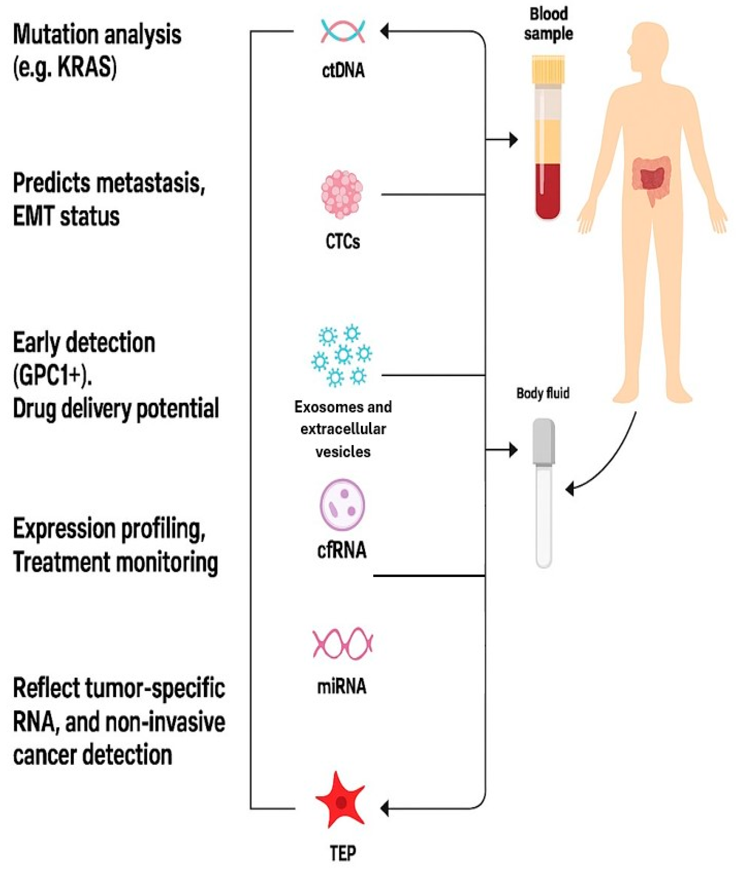
Liquid biopsy is a minimally invasive approach that detects tumor-derived components in body fluids, mainly blood. As tumors grow and undergo turnover, they release biomarkers such as ctDNA, CTCs, extracellular vesicles (EVs), and TEPs that reflect tumor heterogeneity and molecular alterations [31]. ctDNA, derived from apoptotic or viable tumor cells, mirrors the genetic and epigenetic landscape of primary and metastatic lesions, enabling early detection, response assessment, and resistance monitoring [32,33]. CTCs represent viable disseminating cells with prognostic and therapeutic relevance [34]. EVs, particularly exosomes, carry DNA, RNA, and proteins that mediate intercellular communication, immune evasion, and drug resistance [30]. TEPs sequester tumor-derived RNA, providing a robust source for RNA-based cancer diagnostics [35]. (Figure 2 depicts the types of molecular and cellular component).

4.2. Comparison with Tissue Biopsy

Tissue biopsy remains the diagnostic gold standard, providing essential histopathologic and molecular data. However, it is invasive, carries procedural risks, and may not capture tumor heterogeneity. Liquid biopsy offers a minimally invasive alternative, analyzing ctDNA, CTCs, and EVs in body fluids to assess tumor characteristics in real time [33]. Unlike tissue biopsy, it enables longitudinal monitoring of tumor evolution, treatment response, and resistance mutations without repeated invasive procedures [36]. Yet, tissue biopsy remains indispensable for initial diagnosis and subtyping, as liquid biopsy sensitivity is limited in early-stage disease due to low ctDNA shedding [37]. In PDAC—where tissue access is often challenging—liquid biopsy serves as a valuable adjunct, providing complementary molecular insights for diagnosis, prognosis, and therapeutic stratification [29,38].

5. Molecular and Cellular Components of Liquid Biopsy in PDAC

Each analyte in liquid biopsy has unique advantages, limitations, and applications. The following subsections explore the utility of ctDNA, CTCs, exosomes, cfRNA, and TEPs in pancreatic cancer.

5.1. Circulating Tumor DNA (ctDNA)

ctDNA refers to fragments of DNA shed from tumor cells into the bloodstream, often due to apoptosis, necrosis, or active secretion. In PDAC, a malignancy characterized by poor prognosis and late-stage diagnosis, ctDNA has emerged as a minimally invasive biomarker with significant diagnostic, prognostic, and monitoring potential [39]. Per the 2018 joint review of American Society of Clinical Oncology and College of American Pathologists the optimal specimen for ctDNA analysis in patient with cancer is plasma collected in EDTA tubes (processed within 6 h of collection) or leukocyte stabilization tubes.

5.1.1. Detection Techniques

Due to the low abundance of ctDNA in plasma, especially in early-stage PDAC, sensitive and specific detection methods are crucial. Among the most widely used techniques is digital droplet PCR (ddPCR), which offers high sensitivity for detecting known hotspot mutations, particularly KRAS mutations found in over 90% of PDAC cases [29]. ddPCR enables the quantification of mutant alleles even at very low frequencies (as low as 0.01%) in a background of wild-type DNA.
Another common method is BEAMing (Beads, Emulsion, Amplification, and Magnetics), which combines PCR and flow cytometry to detect mutant DNA molecules with high precision. BEAMing has been used to assess KRAS mutations in plasma with excellent concordance with tissue samples [40].
NGS platforms have broadened ctDNA analysis by enabling the simultaneous detection of multiple mutations, gene amplifications, and structural rearrangements. Hybrid-capture-based NGS approaches like CAPP-Seq (Cancer Personalized Profiling by deep Sequencing) are particularly useful in PDAC due to their ability to detect low-frequency variants and track tumor evolution [41].

5.1.2. Clinical Applications

Early Detection and Diagnosis: While ctDNA alone may have limited sensitivity for detecting early-stage PDAC, combining it with protein biomarkers (e.g., CA19-9) improves diagnostic accuracy. Firpo et al. reported that a multianalyte serum biomarker panel integrating ctDNA and protein markers achieved a sensitivity of 92.8% and specificity of 100% for detecting PDAC, underscoring the value of multimodal liquid biopsy strategies for identifying resectable disease at an earlier stage [42].
Elevated ctDNA levels are associated with increased tumor burden and poorer outcomes in PDAC. In a cohort of 66 resected patients, post-operative ctDNA positivity correlated with significantly shorter disease-free and overall survival and independently predicted recurrence (HR ≈ 2.68) [43]. For treatment monitoring, ctDNA allows for real-time assessment of treatment response. Decreases in ctDNA levels during chemotherapy have been associated with clinical benefit, while increases may precede radiographic progression [44]. This dynamic monitoring can guide therapeutic decisions earlier than imaging modalities. Moreover, it helps in detection of resistance mechanisms since ctDNA analysis can uncover acquired resistance mutations, such as secondary KRAS mutations or alterations in genes involved in DNA repair pathways. This is particularly important for patients on targeted therapies or clinical trials, as it may inform the selection of next-line treatments [45].

5.2. Circulating Tumor Cells (CTCs)

CTCs are malignant cells that detach from the primary tumor or metastatic sites and enter the bloodstream. In PDAC, CTCs are a promising biomarker for non-invasive tumor profiling, prognosis, and monitoring treatment response. Although PDAC is a hypovascular and fibrotic tumor, making CTC detection challenging, advances in isolation technologies have improved their detection and analysis [46].

5.2.1. Isolation Techniques

CTC isolation is technically demanding due to their rarity—often just 1–10 CTCs per 10 mL of blood—and the need to distinguish them from billions of hematopoietic cells. Current isolation methods fall into two broad categories: label-dependent and label-independent.
Label-Dependent (Immunoaffinity-Based) Techniques:
The FDA-approved CellSearch® system remains the most widely used platform. It relies on immunomagnetic separation using antibodies against epithelial cell adhesion molecules (EpCAM) and cytokeratins, along with negative selection against CD45 (a leukocyte marker) [47]. While it has shown utility in PDAC, its reliance on epithelial markers limits detection of CTCs that have undergone EMT, a common feature in PDAC [48].
Label-Independent (Physical Property-Based) Techniques:
Techniques based on size, deformability, and electrical properties have been developed to overcome limitations of epithelial marker loss. Microfluidic devices such as the “CTC-iChip” combine size filtration with inertial focusing and immunomagnetic depletion of leukocytes [49]. Another approach, the Parsortix™ system, captures CTCs based on size and compressibility, allowing for viable cell recovery [50].
The development of microfluidic platforms has significantly enhanced the capture efficiency of CTCs in PDAC. One such example is the “NanoVelcro” chip, which uses nanosubstrates and immunoaffinity capture to isolate CTCs with high sensitivity [51]. These advanced platforms allow downstream molecular analysis, including single-cell sequencing.

5.2.2. Clinical Relevance in PDAC

Multiple studies have demonstrated that the presence of CTCs correlates with worse outcomes in PDAC. Yasui et al. (2025) [52] demonstrated that sequential changes in CTC counts during pre-operative chemotherapy strongly correlated with treatment response. Patients exhibiting a reduction in CTC levels achieved better clinical outcomes, while persistent or rising CTCs predicted disease progression. These findings highlight the potential of CTC kinetics as a non-invasive biomarker for assessing therapeutic efficacy and disease trajectory in pancreatic cancer [52]. These dynamics may be more sensitive than imaging for assessing response.
Isolated CTCs can be subjected to genomic and transcriptomic analyses to identify mutations (e.g., KRAS, TP53) and resistance mechanisms. This is particularly valuable in PDAC, where repeat biopsies are often infeasible. Single-cell sequencing of CTCs has revealed heterogeneity and evolution under treatment pressure [53]. Moreover, it could allow early detection and recognize minimal residual disease (MRD).
While sensitivity for early-stage PDAC remains low, combining CTCs with other biomarkers like CA19-9 or ctDNA may enhance early detection. Additionally, post-surgical detection of CTCs could indicate minimal residual disease and impending relapses.

5.3. Exosomes and Extracellular Vesicles

Exosomes are small EVs (30–150 nm in diameter) secreted by various cell types, including cancer cells. These vesicles play essential roles in intercellular communication by transferring proteins, lipids, and nucleic acids (including DNA, mRNA, and microRNAs). In PDAC, exosomes have emerged as valuable tools for diagnosis, prognosis, and therapeutic monitoring due to their stability in bodily fluids and ability to reflect the molecular characteristics of the tumor [54].

5.3.1. Isolation Techniques

Detecting and isolating exosomes from plasma or serum involves several strategies, with ongoing efforts to improve sensitivity and specificity. Common techniques include ultracentrifugation and Size-Exclusion Chromatography (SEC). Differential ultracentrifugation remains the gold standard. It involves sequential centrifugation steps to remove cells, debris, and larger vesicles, ultimately isolating exosomes at ~100,000 g. However, this method is time-consuming and may co-isolate non-exosomal particles [55].
SEC separates vehicles based on size using porous beads, providing high purity with minimal damage to exosomal contents [56].
Immunoaffinity Capture: This method uses antibodies against exosomal surface markers like CD63, CD81, or tumor-specific proteins such as glypican-1 (GPC1). Immunocapture enhances specificity but may miss subpopulations lacking the targeted marker [57].
Nanoparticle Tracking Analysis (NTA) and Tunable Resistive Pulse Sensing (TRPS): These tools allow sizing and quantification of exosomes. NTA tracks Brownian motion of particles to estimate size and concentration [58].
Microfluidic Devices: Emerging microfluidic platforms provide rapid, label-free exosome isolation and analysis with high sensitivity. These “lab-on-a-chip” technologies are especially useful for clinical applications due to minimal sample volume requirements [59].

5.3.2. Diagnostic Potential

Early Detection and Diagnosis: Exosomes from PDAC patients contain tumor-specific cargo that distinguishes them from healthy individuals. Exosomal glypican-1 (GPC1) has shown strong potential as a diagnostic marker for PDAC, achieving 98% sensitivity and 86% specificity in distinguishing pancreatic cancer from chronic pancreatitis, with better performance than CA19-9 [57]. These exosomes not only differentiated PDAC from benign pancreatic disease but also detected early-stage tumors with high sensitivity and specificity.
Prognostic Significance: Exosomal content correlates with tumor burden and aggressiveness. Elevated levels of exosomal microRNAs (miRNAs) such as miR-21 and miR-17-5p have been linked to poor survival in PDAC patients [60]. Measuring these biomarkers could aid in patient stratification.
Monitoring Treatment Response: Exosomal RNA and protein profiles change dynamically with treatment. Exosomal KRAS mutations in plasma have been used to monitor chemotherapy resistance and minimal residual disease [39]. Unlike ctDNA, exosomal RNA is more stable and may provide a richer source of dynamic information.
Therapeutic Potential: Exosomes may also be exploited therapeutically. They can serve as delivery vehicles for drugs or therapeutic RNA molecules due to their biocompatibility and ability to cross biological barriers [61]. In a preclinical PDAC model, exosomes loaded with siRNA targeting mutant KRAS suppressed tumor growth significantly.

5.4. Tumor-Educated Platelets (TEPs)

TEPs are blood platelets that undergo molecular and functional reprogramming in response to tumor-derived signals. In PDAC, TEPs have gained attention as a novel liquid biopsy component due to their ability to sequester tumor-related RNA, proteins, and biomolecules. This interaction transforms platelets into dynamic biosensors capable of reflecting the presence, type, and molecular profile of tumors [35].

5.4.1. Detection and Isolation Techniques

Platelet Isolation
TEPs are typically isolated from peripheral blood using differential centrifugation techniques. Blood is first processed to obtain platelet-rich plasma, followed by low-speed centrifugation and washing to remove leukocyte contamination. Careful handling is crucial to prevent platelet activation, which can alter RNA profiles [62].
RNA Sequencing of TEPs
The core approach to detect tumor signals within TEPs is transcriptomic profiling. RNA is extracted from purified platelets and subjected to high-throughput RNA sequencing (RNA-seq). Mantini et al. has demonstrated that platelets in PDAC undergo systematic transcriptomic, miRNA, and proteomic reprogramming compared to benign pancreatic disease. These molecular changes underscore the potential for TEP-based biomarker development, though further validation is needed to assess classification performance in early disease settings [35]. Specific RNA signatures from TEPs, including overexpression of KRAS and MET transcripts, have been detected in PDAC patients [35,63].
Bioinformatics and Machine Learning
Advanced bioinformatics tools and machine learning algorithms are employed to classify cancer types and molecular subtypes based on TEP RNA profiles. These computational models increase diagnostic accuracy and may even predict mutations, such as KRAS or TP53, found in the tumor itself [63].
Multiplexed Platforms
Emerging multiplexed technologies combine TEP RNA analysis with other biomarkers (e.g., circulating tumor DNA or exosomal RNA) for enhanced sensitivity and specificity. Platforms such as NanoString and digital PCR are being explored for their ability to validate TEP-derived gene panels in clinical settings.

5.4.2. Diagnostic Potential

  • -Early Detection and Diagnosis
TEPs have shown promise in early detection of PDAC. Given the asymptomatic nature of early PDAC and the limitations of current markers like CA19-9, TEPs could represent a non-invasive and sensitive alternative.
  • -Prognostic Value
Changes in TEP RNA profiles have been associated with tumor burden and disease progression. Platelet-derived transcripts related to angiogenesis, inflammation, and tumor proliferation correlate with poor prognosis and decreased overall survival in PDAC patients [63,64].
  • -Monitoring Therapeutic Response
TEP profiles are dynamic and can reflect treatment-induced tumor changes. For example, RNA signatures may shift in response to chemotherapy or targeted therapy, enabling real-time monitoring of response and detection of resistance mechanisms [63]. This real-time surveillance could guide therapeutic adjustments without the need for repeated imaging or invasive tissue biopsies.
  • -Potential for Personalized Medicine
TEPs offer a molecular window into tumor biology, potentially aiding in the selection of personalized therapies. For instance, detecting TEP-associated mutant KRAS RNA could influence eligibility for KRAS-targeting agents or clinical trials [63].

5.5. Cell-Free RNAs (cfRNAs) and miRNAs

Non-coding RNAs—especially miRNAs—are stable in circulation and often dysregulated in PDAC.
miR-21, miR-155, and miR-196a are consistently upregulated in PDAC.
cfRNAs can be used as companion markers for ctDNA to enhance diagnostic yield. Table 1 summarizes detection techniques and clinical relevance of liquid biopsy components in PDAC.)

6. Techniques and Analytical Platforms in Liquid Biopsy

6.1. Sample Types and Collection

The most common biological fluids used in liquid biopsy are peripheral blood, plasma, serum, and, in some cases, urine, saliva, or cerebrospinal fluid [33]. For ctDNA and cfRNA analysis, plasma is preferred due to lower contamination with genomic DNA from lysed leukocytes. Blood is typically collected in EDTA or specialized tubes like Streck Cell-Free DNA BCT to prevent nucleic acid degradation and leukocyte lysis. Standardized processing, such as centrifugation within 2–4 h, is crucial to preserve sample integrity [69].

6.2. Biomarker-Specific Isolation Techniques

ctDNA is isolated using silica-based spin columns, magnetic beads, or automated systems (e.g., QIAamp Circulating Nucleic Acid Kit). These methods are designed to recover short DNA fragments (~160–200 bp) that originate from apoptotic tumor cells [38].
CTCs are isolated based on size, density, or cell-surface antigens like EpCAM. Platforms such as CellSearch (FDA-approved) use immunomagnetic enrichment, while microfluidic devices (e.g., CTC-iChip) enable label-free capture based on size and deformability [65].
Exosomes are separated using ultracentrifugation, Size-Exclusion Chromatography, or commercial kits based on polymer precipitation or immunoaffinity (e.g., ExoQuick, ExoEasy). Microfluidic devices integrated with capture antibodies (e.g., CD63, CD81) are increasingly used for high-throughput exosome enrichment [66]
TEPs are isolated via differential centrifugation to obtain platelet-rich plasma. Careful handling prevents activation that could alter RNA content. Subsequent RNA extraction and analysis reflect tumor-induced alterations in the platelet transcriptome [35].
CfRNAs and miRNAs are extracted from plasma or serum using phenol-chloroform-based or column-based kits. Exosomal RNA and protein co-isolation techniques are also used for profiling miRNAs encapsulated in vesicles, enhancing stability and specificity [67].

6.3. Molecular Detection Techniques

The detection of nucleic acids relies on various platforms:
Quantitative PCR (qPCR) and ddPCR provide high sensitivity for known mutations or transcripts (e.g., KRAS in PDAC).
NGS allows broad genomic or transcriptomic profiling, enabling identification of somatic mutations, copy number variations, fusions, and expression changes [33].
Methylation-specific PCR and bisulfite sequencing detect epigenetic alterations in ctDNA, important for cancer-specific signatures.

6.4. Bioinformatics and Data Interpretation

Raw data from sequencing or PCR are processed using robust bioinformatics pipelines. Variant calling tools (e.g., GATK, MuTect2) identify somatic mutations from ctDNA. In transcriptomics, alignment tools (e.g., STAR, HISAT2) and differential expression analysis (e.g., DESeq2) are employed for TEPs and cfRNAs. Machine learning models are also used to distinguish cancer-specific signatures from background noise [70].
Databases such as COSMIC and TCGA help contextualize genomic alterations. Integration of multi-omics data (DNA, RNA, epigenetics) enhances diagnostic and prognostic power. Cloud-based platforms and AI-driven analytics are becoming central in managing large datasets generated by liquid biopsy studies.

6.5. Biological Limitations

Despite the growing promise of liquid biopsy, several biological limitations constrain its utility in PDAC. One of the most critical challenges is the inherently low abundance of circulating biomarkers due to the dense stromal architecture and relatively low vascularity of PDAC tumors. This desmoplastic reaction limits the shedding of ctDNA, CTCs, and exosomes into the bloodstream, particularly in early-stage disease, resulting in low sensitivity for early detection [29,42].
Moreover, the short half-life of ctDNA (~2 h) and the rapid clearance of CTCs by the immune system or hepatic filtration reduce the window for their detection [38]. Tumor heterogeneity adds another layer of complexity; spatial and temporal genetic variability may lead to sampling bias, where detected alterations in liquid biopsies may not fully represent the primary or metastatic tumor molecular landscape [69]. This can impact the accuracy of therapeutic decisions based on liquid biopsy results.
Another limitation arises from background noise created by non-tumor-derived cfDNA released during apoptosis or necrosis of normal cells, particularly in inflammatory or benign pancreatic conditions such as pancreatitis [33]. Additionally, EMT can reduce the expression of epithelial markers used to capture CTCs, leading to underestimation of tumor burden [31,65].

7. Clinical Trials and Ongoing Research

Recent advances in liquid biopsy have significantly impacted the management of PDAC, offering a minimally invasive approach for diagnosis, prognosis, treatment monitoring, and early detection of recurrence. ctDNA, CTCs, and exosomes are extensively studied as biomarkers. Numerous clinical trials are ongoing to validate their clinical utility in different PDAC settings, including resectable, borderline, and metastatic stages [45]. These trials aim to integrate liquid biopsy into standard care by enabling real-time tumor profiling and response assessment [71]. The table below summarizes key ongoing and completed clinical trials exploring liquid biopsy in PDAC. Table 2 summarizes clinical trials in liquid biopsy for PDAC.

8. Future Directions in Liquid Biopsy for Pancreatic Cancer

Recent advances in liquid biopsy have significantly impacted the diagnostic and therapeutic landscape of PDAC, a malignancy notorious for late presentation and limited therapeutic options. Although current applications center around ctDNA, exosomes, and CTCs, future advances aim to go beyond single-analyte tests and into the realm of multianalyte, multi-omics, and artificial intelligence (AI)-driven predictive modeling.

8.1. Multianalyte and Multi-Omics Liquid Biopsy Platforms

One of the major future directions involves combining multiple analytes (ctDNA, CTCs, exosomes, miRNAs, cfRNA, and TEPs) to enhance sensitivity and specificity. This multianalyte approach seeks to overcome the limitations of single biomarkers, particularly in early-stage PDAC where ctDNA yield may be low due to limited tumor shedding [71].
Multi-omics approaches integrate genomic, epigenomic, transcriptomic, proteomic, and metabolomic data from liquid biopsy components. For example, methylation profiling of cfDNA has shown promise in detecting early-stage PDAC with higher accuracy than mutation-based panels alone [72]. Combining DNA methylation signatures with KRAS mutation detection and exosomal RNA content may yield more robust classifiers.
Moreover, TEPs, which are altered in RNA profiles by tumor-secreted factors, have emerged as novel liquid biopsy tools capable of providing information about the tumor transcriptome [35]. Their use, combined with ctDNA and exosomal profiling, is under exploration for early diagnosis and treatment response.
An example of this direction is the CancerSEEK test, which combines mutations, protein biomarkers, and machines learning to detect multiple cancer types, including PDAC, with high specificity [42]. While not yet clinically adopted for PDAC, such multianalyte strategy will likely become the cornerstone for early detection.

8.2. Artificial Intelligence and Predictive Modeling

The molecular complexity and heterogeneity of PDAC necessitate AI-driven integration of liquid biopsy data. Machine learning algorithms are increasingly applied to large-scale, multidimensional datasets to identify novel biomarker signatures and predict treatment response or disease progression.
AI models can synthesize inputs from multiple platform mutational profiles, fragmentomics (cfDNA fragment sizes), methylation landscapes, and exosomal miRNA panels—into comprehensive predictive tools. For example, convolutional neural networks (CNNs) and support vector machines (SVMs) have demonstrated efficacy in classifying PDAC from healthy controls based on exosomal protein and RNA patterns [68].
Fragmentomics, the analysis of cfDNA fragment length and nucleosome footprinting, is also showing promise. Combined with deep learning, these features may differentiate tumor-derived cfDNA from normal cfDNA with high precision [73].
Furthermore, AI enables longitudinal modeling, allowing the prediction of relapse, treatment resistance, and survival. One study developed a Bayesian predictive model incorporating serial ctDNA levels to anticipate recurrence months before radiological evidence [74].
Projects such as the PANCAID study (NCT06283576) are actively exploring AI-integrated platforms using multimodal data—including ctDNA, imaging, and clinical metadata—for early detection and risk stratification.

8.3. Minimal Residual Disease (MRD) and Personalized Surveillance

The future of post-surgical management in PDAC will likely include MRD detection using ultra-sensitive ctDNA methods, such as personalized digital PCR or whole-genome sequencing-based assays. These technologies aim to detect minute traces of ctDNA after curative-intent surgery, predicting recurrence before clinical symptoms or imaging changes emerge [41].
By incorporating AI, MRD assays can dynamically adjust surveillance strategies, recommending imaging, adjuvant therapy adjustments, or enrollment in early-intervention trials based on predicted relapse risk.

8.4. Integration into Precision Oncology and Adaptive Trials

Liquid biopsy will increasingly support adaptive clinical trial designs, allowing for real-time therapeutic stratification based on evolving molecular profiles. Platforms like the AMPLIFY-7P trial (NCT05726864) use ctDNA dynamics to guide immunotherapy administration, marking a shift toward response-adaptive designs.
In future models, AI-enhanced liquid biopsy will allow for not just static genetic snapshots, but dynamic monitoring of tumor evolution, clonal shifts, and emergence of resistance mutations—guiding rational therapy modifications.

8.5. Knowledge Gaps and Limitations

Despite substantial progress, critical knowledge gaps continue to impede the full clinical integration of liquid biopsy in PDAC. Current technologies remain insufficiently sensitive for early detection, reflecting the biological heterogeneity of PDAC and its characteristically low levels of tumor-derived analytes. Standardization across pre-analytical workflows, analytical platforms, and reporting criteria is urgently needed to ensure reproducibility and cross-study comparability. Furthermore, the biological interpretation of molecular alterations—particularly in distinguishing tumor-specific variants from clonal hematopoiesis—remains incompletely resolved.

8.6. Future Directions

Prospective, large-scale trials are required to validate predictive and prognostic biomarkers and to define clinically actionable thresholds. Integration of multi-omic profiling, artificial intelligence–driven analytics, and longitudinal monitoring represents a promising frontier for real-time disease stratification. Finally, the path toward clinical adoption demands parallel progress in regulatory clarity, cost-effectiveness analysis, and ethical oversight to ensure equitable and evidence-based implementation.

9. Conclusions

PDAC remains one of the most lethal human malignancies due to its late presentation, aggressive biology, and limited therapeutic options. Liquid biopsy has emerged as a transformative tool with wide-ranging clinical applications in this disease—from early detection and prognostication to therapy selection, resistance monitoring, and surveillance of minimal residual disease.
While significant challenges remain—particularly in standardization, sensitivity in early-stage disease, and integration into routine clinical practice—the future of liquid biopsy in PDAC is promising. The continued evolution of technology, multianalyte integration, and AI-based interpretation may ultimately position liquid biopsy as a cornerstone in the personalized management of PDAC.

Author Contributions

Conceptualization: A.B. (Ahmed Bendari); methodology, A.B. (Ahmed Bendari); software: A.B. (Ahmed Bendari), A.B. (Alaa Bendari); investigation: A.B. (Ahmed Bendari), O.V., A.B. (Alaa Bendari), M.S., J.L.G.M., resources: A.B. (Ahmed Bendari), A.B. (Alaa Bendari); formal analysis: A.B. (Ahmed Bendari), A.B. (Alaa Bendari); data curation: A.B. (Ahmed Bendari), O.V., A.B. (Alaa Bendari); writing—original draft preparation: A.B. (Ahmed Bendari), O.V., B.B.; writing—review and editing: K.K. and S.A., supervision A.B. (Ahmed Bendari). All authors have read and agreed to the published version of the manuscript.

Funding

This research received no external funding.

Institutional Review Board Statement

Not applicable.

Informed Consent Statement

Not applicable.

Data Availability Statement

All data is included in the current manuscript.

Conflicts of Interest

The authors declare no conflict of interest.

References

  1. Garrido-Laguna, I.; Hidalgo, M. Pancreatic cancer: From state-of-the-art treatments to promising novel therapies. Nat. Rev. Clin. Oncol. 2015, 12, 319–334. [Google Scholar] [CrossRef] [PubMed]
  2. Siegel Rebecca, L.; Miller, K.D.; Jemal, A. Cancer statistics, 2018. CA A Cancer J. Clin. 2018, 68, 7–30. [Google Scholar]
  3. McGuigan, A.; Kelly, P.; Turkington, R.C.; Jones, C.; Coleman, H.G.; McCain, R.S. Pancreatic cancer: A review of clinical diagnosis, epidemiology, treatment and outcomes. World J. Gastroenterol. 2018, 24, 4846–4861. [Google Scholar] [CrossRef] [PubMed]
  4. Sarantis, P.; Koustas, E.; Papadimitropoulou, A.; Papavassiliou, A.G.; Karamouzis, M.V. Pancreatic ductal adenocarcinoma: Treatment hurdles, tumor microenvironment and immunotherapy. World J. Gastrointest. Oncol. 2020, 12, 173–181. [Google Scholar] [CrossRef] [PubMed] [PubMed Central]
  5. Knox, J.J.; Jang, G.H.; Grant, R.C.; Zhang, A.; Ma, L.; Elimova, E.; Jang, R.; Moore, M.; Biagi, J.; Tehfe, M.; et al. Whole genome and transcriptome profiling in advanced pancreatic cancer patients on the COMPASS trial. Nat. Commun. 2025, 16, 5919. [Google Scholar] [CrossRef]
  6. Bilreiro, C.; Andrade, L.; Santiago, I.; Marques, R.M.; Matos, C. Imaging of pancreatic ductal adenocarcinoma—An update for all stages of patient management. Eur. J. Radiol. Open 2024, 12, 100553. [Google Scholar] [CrossRef]
  7. Dhar, J.; Samanta, J.; Nabi, Z.; Aggarwal, M.; Conti Bellocchi, M.C.; Facciorusso, A.; Frulloni, L.; Crinò, S.F. Endoscopic Ultrasound-Guided Pancreatic Tissue Sampling: Lesion Assessment, Needles, and Techniques. Medicina 2024, 60, 2021. [Google Scholar] [CrossRef] [PubMed] [PubMed Central]
  8. Grossberg, A.J.; Chu, L.C.; Deig, C.R.; Fishman, E.K.; Hwang, W.L.; Maitra, A.; Marks, D.L.; Mehta, A.; Nabavizadeh, N.; Simeone, D.M.; et al. Multidisciplinary standards of care and recent progress in pancreatic ductal adenocarcinoma. CA A Cancer J. Clin. 2020, 70, 375–403. [Google Scholar] [CrossRef] [PubMed] [PubMed Central]
  9. Ren, B.; Liu, X.; Suriawinata, A.A. Pancreatic ductal adenocarcinoma and its precursor lesions: Histopathology, cytopathology, and molecular pathology. Am. J. Pathol. 2019, 189, 9–21. [Google Scholar] [CrossRef]
  10. Yousef, M.M.; Hurd, M.W.; Yousef, A.M.; A Snyder, R.; Knafl, M.; Fanaeian, M.M.; Chacko, R.; Peterson, J.; Smaglo, B.G.; Wolff, R.A.; et al. Frequency and oncologic outcomes of KRAS mutations in circulating tumor DNA of patients with pancreatic ductal adenocarcinoma. J. Clin. Oncol. 2024, 42, 636. [Google Scholar] [CrossRef]
  11. Waters, A.M.; Der, C.J. KRAS: The Critical Driver and Therapeutic Target for Pancreatic Cancer. Cold Spring Harb. Perspect. Med. 2018, 8, a031435. [Google Scholar] [CrossRef] [PubMed] [PubMed Central]
  12. Iwatate, Y.; Hoshino, I.; Ishige, F.; Itami, M.; Chiba, S.; Arimitsu, H.; Yanagibashi, H.; Nagase, H.; Yokota, H.; Takayama, W. Prognostic significance of p16 protein in pancreatic ductal adenocarcinoma. Mol. Clin. Oncol. 2020, 13, 83–91. [Google Scholar] [CrossRef] [PubMed] [PubMed Central]
  13. Mello, S.S.; Flowers, B.M.; Mazur, P.K.; Lee, J.J.; Müller, F.; Denny, S.K.; Ferreira, S.; Hanson, K.; Kim, S.K.; Greenleaf, W.J.; et al. Multifaceted role for p53 in pancreatic cancer suppression. Proc. Natl. Acad. Sci. USA 2023, 120, e2211937120. [Google Scholar] [CrossRef]
  14. Stefanoudakis, D.; Frountzas, M.; Schizas, D.; Michalopoulos, N.V.; Drakaki, A.; Toutouzas, K.G. Significance of TP53, CDKN2A, SMAD4 and KRAS in Pancreatic Cancer. Curr. Issues Mol. Biol. 2024, 46, 2827–2844. [Google Scholar] [CrossRef]
  15. Hanahan, D. Hallmarks of cancer: New dimensions. Cancer Discov. 2022, 12, 31–46. [Google Scholar] [CrossRef]
  16. Pishvaian, M.J.; Bender, R.J.; Halverson, D.; Rahib, L.; Hendifar, A.E.; Mikhail, S.; Chung, V.; Picozzi, V.J.; Sohal, D.; Blais, E.M.; et al. Molecular Profiling of Patients with Pancreatic Cancer: Initial Results from the Know Your Tumor Initiative. Clin. Cancer Res. 2018, 24, 5018–5027. [Google Scholar] [CrossRef] [PubMed]
  17. Bailey, P.; Chang, D.K.; Nones, K.; Johns, A.L.; Patch, A.-M.; Gingras, M.-C.; Miller, D.K.; Christ, A.N.; Bruxner, T.J.C.; Quinn, M.C.; et al. Genomic analyses identify molecular subtypes of pancreatic cancer. Nature 2016, 531, 47–52. [Google Scholar] [CrossRef]
  18. Furukawa, T.; Kuboki, Y.; Tanji, E.; Yoshida, S.; Hatori, T.; Yamamoto, M.; Shibata, N.; Shimizu, K.; Kamatani, N.; Shiratori, K. Whole-exome sequencing uncovers frequent GNAS mutations in intraductal papillary mucinous neoplasms of the pancreas. Sci. Rep. 2011, 531, 161. [Google Scholar] [CrossRef]
  19. National Comprehensive Cancer Network. NCCN Clinical Practice Guidelines in Oncology: Pancreatic Adenocarcinoma; Version 2.2025; National Comprehensive Cancer Network: Plymouth Meeting, PA, USA, 2025; Available online: https://www.nccn.org/professionals/physician_gls/pdf/pancreatic.pdf (accessed on 8 October 2025).
  20. Neesse, A.; Algül, H.; Tuveson, D.A.; Gress, T.M. Stromal biology and therapy in pancreatic cancer. Gut 2015, 64, 1379–1389. [Google Scholar] [CrossRef] [PubMed]
  21. Sherman, M.H.; Beatty, G.L. Tumor microenvironment in pancreatic cancer pathogenesis and therapeutic resistance. Annu. Rev. Pathol. Mech. Dis. 2023, 18, 123–148. [Google Scholar] [CrossRef] [PubMed]
  22. Beatty, G.L.; Winograd, R.; Evans, R.A.; Long, K.B.; Luque, S.L.; Lee, J.W.; Clendenin, C.; Gladney, W.L.; Knoblock, D.M.; Guirnalda, P.D.; et al. Exclusion of T cells from pancreatic carcinomas in mice is regulated by Ly6Clow F4/80+ extratumoral macrophages. Cancer Immunol. Res. 2015, 3, 233–239. [Google Scholar] [CrossRef]
  23. Ju, Y.; Xu, D.; Liao, M.; Sun, Y.; Bao, W.-D.; Yao, F.; Ma, L. Barriers and opportunities in pancreatic cancer immunotherapy. NPJ Precis. Oncol. 2024, 8, 199. [Google Scholar] [CrossRef] [PubMed]
  24. Elyada, E.; Bolisetty, M.; Laise, P.; Flynn, W.F.; Courtois, E.T.; Burkhart, R.A.; Teinor, J.A.; Belleau, P.; Biffi, G.; Lucito, M.S.; et al. Cross-species single-cell analysis reveals a conserved myeloid population in pancreatic ductal adenocarcinoma. Cancer Discov. 2019, 9, 1102–1123. [Google Scholar] [CrossRef]
  25. Öhlund, D.; Handly-Santana, A.; Biffi, G.; Elyada, E.; Almeida, A.S.; Ponz-Sarvise, M.; Corbo, V.; Oni, T.E.; Hearn, S.A.; Lee, E.J.; et al. Distinct populations of inflammatory fibroblasts and myofibroblasts in pancreatic cancer. Cancer Cell. 2017, 31, 19–34. [Google Scholar] [CrossRef] [PubMed]
  26. Mello, A.M.; Ngodup, T.; Lee, Y.; Donahue, K.L.; Li, J.; Rao, A.; Carpenter, E.S.; Crawford, H.C.; di Magliano, M.P.; Lee, K.E. Hypoxia promotes an inflammatory phenotype of fibroblasts in pancreatic cancer. Oncogenesis 2022, 11, 56. [Google Scholar] [CrossRef]
  27. Yang, J.; Liu, Y.; Liu, S. The role of epithelial-mesenchymal transition and autophagy in pancreatic ductal adenocarcinoma invasion. Cell Death Dis. 2023, 14, 506. [Google Scholar] [CrossRef]
  28. Zheng, X.; Carstens, J.; Kim, J.; Scheible, M.; Kaye, J.; Sugimoto, H.; Wu, C.-C.; LeBleu, V.S.; Kalluri, R. Epithelial-to-mesenchymal transition is dispensable for metastasis but induces chemoresistance in pancreatic cancer. Nature 2015, 527, 525–530. [Google Scholar] [CrossRef]
  29. Kinugasa, H.; Nouso, K.; Miyahara, K.; Morimoto, Y.; Dohi, C.; Tsutsumi, K.; Kato, H.; Matsubara, T.; Okada, H.; Yamamoto, K. Detection of K-ras gene mutation by liquid biopsy in patients with pancreatic cancer. Cancer 2015, 121, 2271–2280. [Google Scholar] [CrossRef] [PubMed]
  30. Kalluri, R.; LeBleu, V.S. The biology, function, and biomedical applications of exosomes. Science 2020, 367, eaau6977. [Google Scholar] [CrossRef] [PubMed] [PubMed Central]
  31. Pantel, K.; Alix-Panabières, C. Liquid biopsy in 2016: Circulating tumour cells and cell-free DNA in gastrointestinal cancer. Nat. Rev. Gastroenterol. Hepatol. 2017, 14, 73–74. [Google Scholar] [CrossRef] [PubMed]
  32. Bartolomucci, A.; Nobrega, M.; Ferrier, T.; Dickinson, K.; Kaorey, N.; Nadeau, A.; Castillo, A.; Burnier, J.V. Circulating tumor DNA to monitor treatment response in solid tumors and advance precision oncology. NPJ Precis. Oncol. 2025, 9, 84. [Google Scholar] [CrossRef]
  33. Wan, J.C.M.; Massie, C.; Garcia-Corbacho, J.; Mouliere, F.; Brenton, J.D.; Caldas, C.; Pacey, S.; Baird, R.; Rosenfeld, N. Liquid biopsies come of age: Towards implementation of circulating tumour DNA. Nat. Rev. Cancer 2017, 17, 223–238. [Google Scholar] [CrossRef] [PubMed]
  34. Lee, H.S.; Jung, E.H.; Shin, H.; Park, C.S.; Park, S.B.; Jung, D.E.; Leem, G.; Kim, S.J.; Jo, J.H.; Chung, M.J.; et al. Phenotypic characteristics of circulating tumor cells and predictive impact for efficacy of chemotherapy in patients with pancreatic cancer: A prospective study. Front. Oncol. 2023, 13, 1206565. [Google Scholar] [CrossRef] [PubMed]
  35. Mantini, G.; Meijer, L.L.; Glogovitis, I.; In ‘t Veld, S.G.J.G.; Paleckyte, R.; Capula, M.; Le Large, T.Y.S.; Morelli, L.; Pham, T.V.; Piersma, S.R.; et al. Omics Analysis of Educated Platelets in Cancer and Benign Disease of the Pancreas. Cancers 2020, 13, 66. [Google Scholar] [CrossRef] [PubMed] [PubMed Central]
  36. Heitzer, E.; Ulz, P.; Geigl, J.B. Circulating tumor DNA as a liquid biopsy for cancer. Clin. Chem. 2015, 61, 112–123. [Google Scholar] [CrossRef] [PubMed]
  37. Bettegowda, C.; Sausen, M.; Leary, R.J.; Kinde, I.; Wang, Y.; Agrawal, N.; Bartlett, B.R.; Wang, H.; Luber, B.; Alani, R.M.; et al. Detection of Circulating Tumor DNA in Early- and Late-Stage Human Malignancies. Sci. Transl. Med. 2014, 6, 224. [Google Scholar] [CrossRef]
  38. Siravegna, G.; Marsoni, S.; Siena, S.; Bardelli, A. Integrating liquid biopsies into the management of cancer. Nat. Rev. Clin. Oncol. 2017, 14, 531–548. [Google Scholar] [CrossRef] [PubMed]
  39. Allenson, K.; Castillo, J.; San Lucas, F.A.; Scelo, G.; Kim, D.U.; Bernard, V.; Davis, G.; Kumar, T.; Katz, M.; Overman, M.J.; et al. High prevalence of mutant KRAS in circulating exosome-derived DNA from early-stage pancreatic cancer patients. Ann. Oncol. 2017, 28, 741–747. [Google Scholar] [CrossRef] [PubMed] [PubMed Central]
  40. Ma, L.; Guo, H.; Zhao, Y.; Liu, Z.; Wang, C.; Bu, J.; Sun, T.; Wei, J. Liquid biopsy in cancer: Current status, challenges and future prospects. Signal Transduct. Target. Ther. 2024, 9, 336. [Google Scholar] [CrossRef] [PubMed]
  41. Parikh, A.R.; Leshchiner, I.; Elagina, L.; Goyal, L.; Levovitz, C.; Siravegna, G.; Livitz, D.; Rhrissorrakrai, K.; Martin, E.E.; Van Seventer, E.E.; et al. Liquid versus tissue biopsy for detecting acquired resistance and tumor heterogeneity in gastrointestinal cancers. Nat. Med. 2019, 25, 1415–1421. [Google Scholar] [CrossRef]
  42. Firpo, M.A.; Boucher, K.M.; Bleicher, J.; Khanderao, G.D.; Rosati, A.; Poruk, K.E.; Kamal, S.; Marzullo, L.; De Marco, M.; Falco, A.; et al. Multianalyte Serum Biomarker Panel for Early Detection of Pancreatic Adenocarcinoma. JCO Clin. Cancer Inform. 2023, 7, e2200160. [Google Scholar] [CrossRef] [PubMed] [PubMed Central]
  43. Hata, T.; Mizuma, M.; Motoi, F.; Ohtsuka, H.; Nakagawa, K.; Morikawa, T.; Unno, M. Prognostic impact of postoperative circulating tumor DNA as a molecular minimal residual disease marker in patients with pancreatic cancer undergoing surgical resection. J. Hepato-Biliary-Pancreat. Sci. 2023, 30, 815–824. [Google Scholar] [CrossRef] [PubMed]
  44. Tjensvoll, K.; Lapin, M.; Buhl, T.; Oltedal, S.; Steen-Ottosen Berry, K.; Gilje, B.; Søreide, J.A.; Javle, M.; Nordgård, O.; Smaaland, R. Clinical relevance of circulating KRAS mutated DNA in plasma from patients with advanced pancreatic cancer. Mol. Oncol. 2016, 10, 635–643. [Google Scholar] [CrossRef] [PubMed] [PubMed Central]
  45. Zill, O.A.; Greene, C.; Sebisanovic, D.; Siew, L.M.; Leng, J.; Vu, M.; Hendifar, A.E.; Wang, Z.; Atreya, C.E.; Kelley, R.K.; et al. Cell-Free DNA Next-Generation Sequencing in Pancreatobiliary Carcinomas. Cancer Discov. 2015, 5, 1040–1048. [Google Scholar] [CrossRef] [PubMed] [PubMed Central]
  46. Poruk, K.E.; Blackford, A.L.; Weiss, M.J.; Cameron, J.L.; He, J.; Goggins, M.; Rasheed, Z.A.; Wolfgang, C.L.; Wood, L.D. Circulating Tumor Cells Expressing Markers of Tumor-Initiating Cells Predict Poor Survival and Cancer Recurrence in Patients with Pancreatic Ductal Adenocarcinoma. Clin. Cancer Res. 2017, 23, 2681–2690. [Google Scholar] [CrossRef] [PubMed] [PubMed Central]
  47. Stoecklein, N.H.; Fluegen, G.; Guglielmi, R.; Neves, R.P.; Hackert, T.; Birgin, E.; Cieslik, S.A.; Sudarsanam, M.; Driemel, C.; van Dalum, G.; et al. Ultra-sensitive CTC-based liquid biopsy for pancreatic cancer enabled by large blood volume analysis. Mol. Cancer 2023, 22, 181. [Google Scholar] [CrossRef]
  48. Zhao, X.H.; Wang, Z.R.; Chen, C.L.; Di, L.; Bi, Z.F.; Li, Z.H.; Liu, Y.M. Molecular detection of epithelial-mesenchymal transition markers in circulating tumor cells from pancreatic cancer patients: Potential role in clinical practice. World J. Gastroenterol. 2019, 25, 138–150. [Google Scholar] [CrossRef] [PubMed] [PubMed Central]
  49. Mishra, A.; Huang, S.B.; Dubash, T.; Burr, R.; Edd, J.F.; Wittner, B.S.; Cunneely, Q.E.; Putaturo, V.R.; Deshpande, A.; Antmen, E.; et al. Tumor cell-based liquid biopsy using high-throughput microfluidic enrichment of entire leukapheresis product. Nat. Commun. 2025, 16, 32. [Google Scholar] [CrossRef]
  50. Franses, J.W.; Philipp, J.; Missios, P.; Bhan, I.; Liu, A.; Yashaswini, C.; Tai, E.; Zhu, H.; Ligorio, M.; Nicholson, B.; et al. Pancreatic circulating tumor cell profiling identifies LIN28B as a metastasis driver and drug target. Nat. Commun. 2020, 11, 3303. [Google Scholar] [CrossRef]
  51. Macaraniag, C.; Khan, I.; Barabanova, A.; Valle, V.; Zhou, J.; Giulianotti, P.C.; Borgeat, A.; Votta-Velis, G.; Papautsky, I. Benchmarking microfluidic and immunomagnetic platforms for isolating circulating tumor cells in pancreatic cancer. Lab A Chip 2025, 25, 5292–5301. [Google Scholar] [CrossRef]
  52. Yasui, K.; Saito, T.; Ueda, S.; Shinohara, K.; Fukami, Y.; Sano, T.; Nakanishi, H. Sequential Changes in Circulating Tumor Cells in the Peripheral Blood of Pancreatic Cancer Patients with Preoperative Chemotherapy Using a New Immunocytology-Based, Light Microscopic CTC Detection Platform. Diagnostics 2025, 15, 752. [Google Scholar] [CrossRef] [PubMed] [PubMed Central]
  53. Dimitrov-Markov, S.; Perales-Patón, J.; Bockorny, B.; Dopazo, A.; Muñoz, M.; Baños, N.; Bonilla, V.; Menendez, C.; Duran, Y.; Huang, L.; et al. Discovery of New Targets to Control Metastasis in Pancreatic Cancer by Single-cell Transcriptomics Analysis of Circulating Tumor Cells. Mol. Cancer Ther. 2020, 19, 1751–1760. [Google Scholar] [CrossRef] [PubMed]
  54. Kalluri, R. The biology and function of extracellular vesicles in immune response and immunity. Immunity 2024, 57, 1752–1768. [Google Scholar] [CrossRef] [PubMed] [PubMed Central]
  55. Dilsiz, N. A comprehensive review on recent advances in exosome isolation and characterization: Toward clinical applications. Transl. Oncol. 2024, 50, 102121. [Google Scholar] [CrossRef] [PubMed] [PubMed Central]
  56. Lobb, R.J.; Becker, M.; Wen, S.W.; Wong, C.S.; Wiegmans, A.P.; Leimgruber, A.; Möller, A. Optimized exosome isolation protocol for cell culture supernatant and human plasma. J. Extracell. Vesicles 2015, 4, 27031. [Google Scholar] [CrossRef] [PubMed] [PubMed Central]
  57. Moutinho-Ribeiro, P.; Adem, B.; Batista, I.; Silva, M.; Silva, S.; Ruivo, C.F.; Morais, R.; Peixoto, A.; Coelho, R.; Costa-Moreira, P.; et al. Exosomal glypican-1 discriminates pancreatic ductal adenocarcinoma from chronic pancreatitis. Dig. Liver Dis. 2022, 54, 871–877. [Google Scholar] [CrossRef] [PubMed]
  58. Kowkabany, G.; Bao, Y. Nanoparticle Tracking Analysis: An Effective Tool to Characterize Extracellular Vesicles. Molecules 2024, 29, 4672. [Google Scholar] [CrossRef] [PubMed] [PubMed Central]
  59. Liga, A.; Vliegenthart, A.D.; Oosthuyzen, W.; Dear, J.W.; Kersaudy-Kerhoas, M. Exosome isolation: A microfluidic road-map. Lab A Chip 2015, 15, 2388–2394. [Google Scholar] [CrossRef] [PubMed]
  60. Abue, M.; Yokoyama, M.; Shibuya, R.; Tamai, K.; Yamaguchi, K.; Sato, I.; Tanaka, N.; Hamada, S.; Shimosegawa, T.; Sugamura, K.; et al. Circulating miR-483-3p and miR-21 is highly expressed in plasma of pancreatic cancer. Int. J. Oncol. 2015, 46, 539–547. [Google Scholar] [CrossRef] [PubMed] [PubMed Central]
  61. Kamerkar, S.; LeBleu, V.S.; Sugimoto, H.; Yang, S.; Ruivo, C.F.; Melo, S.A.; Lee, J.J.; Kalluri, R. Exosomes facilitate therapeutic targeting of oncogenic KRAS in pancreatic cancer. Nature 2017, 546, 498–503. [Google Scholar] [CrossRef] [PubMed] [PubMed Central]
  62. Best, M.G.; Sol, N.; Kooi, I.; Tannous, J.; Westerman, B.A.; Rustenburg, F.; Schellen, P.; Verschueren, H.; Post, E.; Koster, J.; et al. RNA-Seq of Tumor-Educated Platelets Enables Blood-Based Pan-Cancer, Multiclass, and Molecular Pathway Cancer Diagnostics. Cancer Cell 2015, 28, 666–676. [Google Scholar] [CrossRef] [PubMed] [PubMed Central]
  63. Sol, N.; Veld, S.G.I.; Vancura, A.; Tjerkstra, M.; Leurs, C.; Rustenburg, F.; Schellen, P.; Verschueren, H.; Post, E.; Zwaan, K.; et al. Tumor-educated platelet RNA for the detection and (pseudo) progression monitoring of glioblastoma. Cell Rep. Med. 2020, 1, 100101. [Google Scholar] [CrossRef]
  64. Krishnan, A.; Thomas, S. Toward platelet transcriptomics in cancer diagnosis, prognosis and therapy. Br. J. Cancer 2022, 126, 316–322. [Google Scholar] [CrossRef] [PubMed]
  65. Ju, S.; Chen, C.; Zhang, J.; Xu, L.; Zhang, X.; Li, Z.; Chen, Y.; Zhou, J.; Ji, F.; Wang, L. Detection of circulating tumor cells: Opportunities and challenges. Biomark. Res. 2022, 10, 58. [Google Scholar] [CrossRef]
  66. Théry, C.; Witwer, K.W.; Aikawa, E.; Alcaraz, M.J.; Anderson, J.D.; Andriantsitohaina, R.; Antoniou, A.; Arab, T.; Archer, F.; Atkin-Smith, G.K.; et al. Minimal information for studies of extracellular vesicles 2018 (MISEV2018): A position statement of the International Society for Extracellular Vesicles and update of the MISEV2014 guidelines. J. Extracell. Vesicles 2018, 7, 1535750. [Google Scholar] [CrossRef] [PubMed]
  67. Metcalf, G.A.D. MicroRNAs: Circulating biomarkers for the early detection of imperceptible cancers via biosensor and machine-learning advances. Oncogene 2024, 43, 2135–2142. [Google Scholar] [CrossRef]
  68. Huang, B.; Huang, H.; Zhang, S.; Zhang, D.; Shi, Q.; Liu, J.; Guo, J. Artificial intelligence in pancreatic cancer. Theranostics 2022, 12, 6931–6954. [Google Scholar] [CrossRef] [PubMed] [PubMed Central]
  69. Heitzer, E.; Haque, I.S.; Roberts, C.E.S.; Speicher, M.R. Current and future perspectives of liquid biopsies in genomics-driven oncology. Nat. Rev. Genet. 2019, 20, 71–88. [Google Scholar] [CrossRef] [PubMed]
  70. Aravanis, A.M.; Lee, M.; Klausner, R.D. Next-Generation Sequencing of Circulating Tumor DNA for Early Cancer Detection. Cell 2017, 168, 571–574. [Google Scholar] [CrossRef] [PubMed]
  71. Jensen, T.J.; Goodman, A.M.; Kato, S.; Ellison, C.K.; Daniels, G.A.; Kim, L.; Nakashe, P.; McCarthy, E.; Mazloom, A.R.; McLennan, G.; et al. Genome-Wide Sequencing of Cell-Free DNA Identifies Copy-Number Alterations That Can Be Used for Monitoring Response to Immunotherapy in Cancer Patients. Mol. Cancer Ther. 2019, 18, 448–458. [Google Scholar] [CrossRef] [PubMed]
  72. Guler, G.D.; Ning, Y.; Ku, C.J.; Phillips, T.; McCarthy, E.; Ellison, C.K.; Bergamaschi, A.; Collin, F.; Lloyd, P.; Scott, A.; et al. Detection of early stage pancreatic cancer using 5-hydroxymethylcytosine signatures in circulating cell free DNA. Nat. Commun. 2020, 11, 5270. [Google Scholar] [CrossRef] [PubMed] [PubMed Central]
  73. Cristiano, S.; Leal, A.; Phallen, J.; Fiksel, J.; Adleff, V.; Bruhm, D.C.; Jensen, S.Ø.; Medina, J.E.; Hruban, C.; White, J.R.; et al. Genome-wide cell-free DNA fragmentation in patients with cancer. Nature 2019, 570, 385–389. [Google Scholar] [CrossRef] [PubMed] [PubMed Central]
  74. Lapin, M.; Edland, K.H.; Tjensvoll, K.; Oltedal, S.; Austdal, M.; Garresori, H.; Rozenholc, Y.; Gilje, B.; Nordgård, O. Comprehensive ctDNA Measurements Improve Prediction of Clinical Outcomes and Enable Dynamic Tracking of Disease Progression in Advanced Pancreatic Cancer. Clin. Cancer Res. 2023, 29, 1267–1278. [Google Scholar] [CrossRef] [PubMed] [PubMed Central]
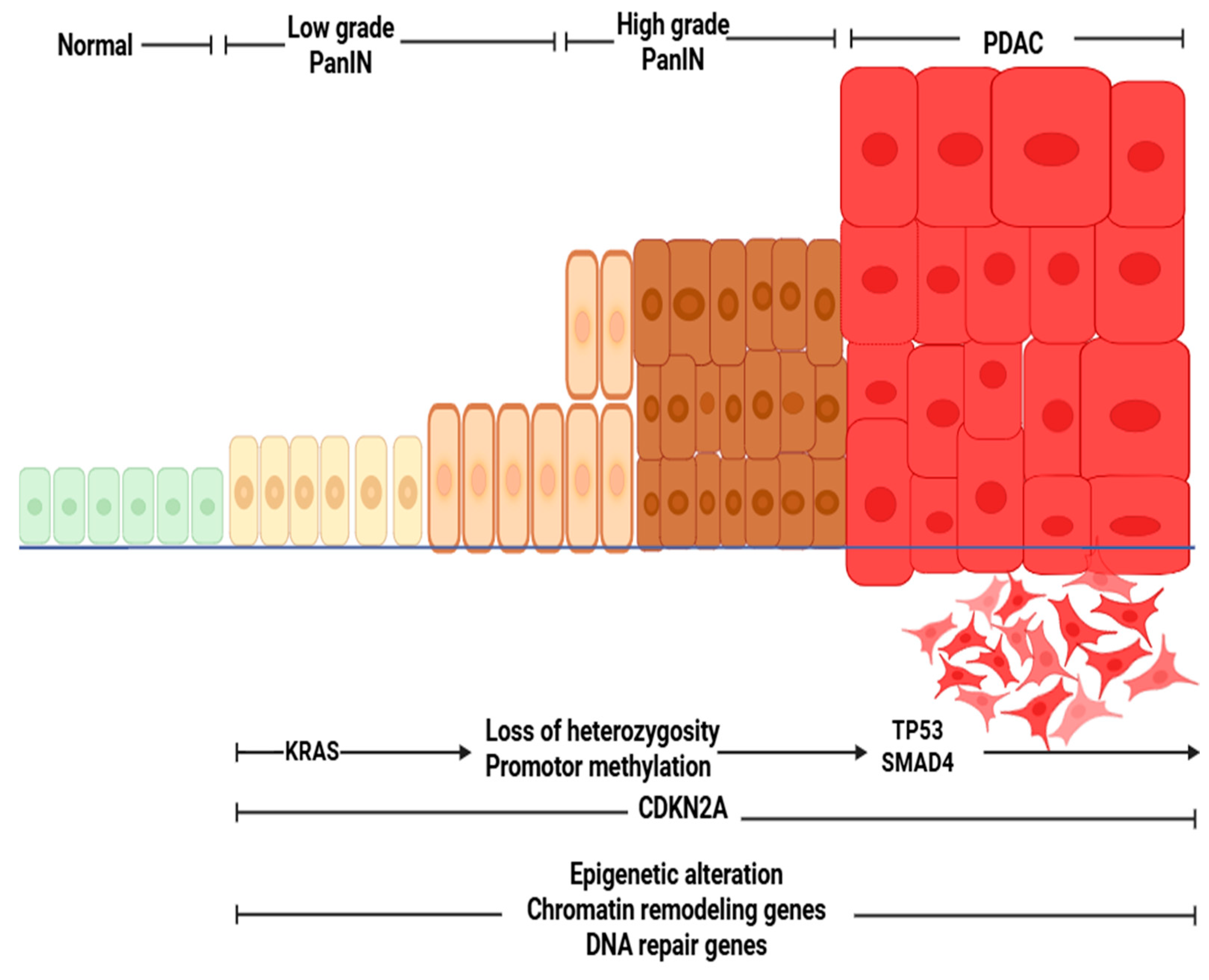
Figure 1. Histologic and genetic evolution of PDAC (image generated on Biorender app).
Gastroent 16 00039 g001
Figure 2. Liquid biopsy in PDAC: This figure depicts the types of molecular and cellular components. CtDNA: circulating tumor DNA, CTCs: Circulating tumor cells, miRNA: microRNA, cfRNA: cell-free RNA, and TEP: tumor-educated platelets. (Image was generated on biorender app).
Gastroent 16 00039 g002
Table 1. Summary of liquid biopsy analytes, clinical applications, analytical platforms, and pre-analytical considerations in PDAC.
AnalyteMain Clinical ApplicationsTypical Sensitivity/Specificity RangeSample TypeAnalytical Platform(s)Critical Pre-Analytic Considerations
TEPs [35]Early detection, treatment response, molecular profilingSensitivity: 70–95%/Specificity: 85–95%Whole blood (platelet-rich plasma)RNA-seq, qRT-PCR, NanoString, bioinformatics classifiersProcess within 2 h; avoid platelet activation; centrifuge gently
cfDNA/ctDNA mutations [38]Early detection, prognosis, treatment monitoring, MRD detectionSensitivity: ~72%
Specificity: >90%
Plasma (preferred) or serumddPCR, BEAMing, NGS (CAPP-Seq, Guardant360)Use EDTA or Streck tubes; plasma separation within 2–4 h; avoid hemolysis
CTCs [65]Prognosis, treatment monitoring, MRD detectionSensitivity: 40–70%/Specificity: >95%Whole bloodCellSearch®, microfluidic chips (CTC-iChip, Parsortix), immunomagnetic enrichmentCollect in CellSave or EDTA tubes; process within 24 h; gentle handling to preserve viability
Exosomes/EVs [66]Early detection, prognosis, therapy resistance monitoringSensitivity: 70–90%/Specificity: 80–95%Plasma, serum, urineUltracentrifugation, SEC, microfluidic chips, NTAStore at −80 °C; avoid freeze–thaw cycles; standardize isolation method
cfRNA/miRNA [67]Diagnosis, prognosis, treatment responseSensitivity: 60–90%/Specificity: 80–95%Plasma, serum, salivaqRT-PCR, NGS, microarraysUse RNA-stabilizing tubes; rapid plasma separation; store at −80 °C
Fragmentomics (cfDNA size) [68]Early detection, tumor origin classificationSensitivity: 70–85%/Specificity: 85–95%PlasmaWGS/WES, cfDNA fragmentation analysis, AI-based modelsSpecialized software; prompt processing to preserve fragment integrity
Protein biomarkers (CA19-9, CEA)Diagnosis, disease monitoring, risk stratificationSensitivity: 60–80%/Specificity: 70–90%Serum, plasmaELISA, mass spectrometry, multiplex immunoassaysAvoid hemolysis; standardize sample dilution and temperature
Table 2. Summarizes clinical trials in liquid biopsy for PDAC (Status Verified on ClinicalTrials.gov, accessed 8 October 2025).
Trial Name/IdentifierPhase & DesignObjectivePatient PopulationStatus & Completion
AMPLIFY--201(NCT04853017)Phase IEvaluate safety & efficacy of KRAS-targeted vaccine ELI--002--2P using ctDNA clearancePost-surgery PDAC with KRAS mutationCompleted (January 2023)
AMPLIFY--7P(NCT05726864)Phase I/IIAssess ELI--002--2P in delaying recurrence via ctDNA monitoringPost-surgery RAS-mutated PDACRecruiting (April 2023–November 2026)
CASPER(NCT05634931)Observational
cohort
Prognostic assessment of ctDNA in surgical resectability & treatment responseResectable PDACRecruiting (December 2022–May 2026)
DYNAMIC--Pancreas (NCT03899636)Phase IIPost-op ctDNA informing adjuvant chemo decisionsResectable PDACCompleted (November 2023)
PROJECTION (NCT04246203)ObservationalPrognostic role of pre-op ctDNA on disease-free survival (DFS)Resectable PDACCompleted (March 2025)
ctDNA Diagnostic (pre-op) (NCT03524677)ObservationalDiagnostic role of ctDNA (KRAS, CDKN2A, SMAD4, TP53) pre-opNon-metastatic PDACCompleted (January 2020)
ctDNA for recurrence surveillance (NCT02934984)ObservationalctDNA for recurrence surveillanceResectable PDACCompleted (January 2021)
Exosomal RNA Biomarker (NCT04636788)Prospective cohortExosomal small RNA diagnostic biomarkerPDAC & pancreatic lesionsCompleted (November 2022)
PRIMUS--002(NCT04176952)Phase II non-randomizedPrognostic ctDNA in neoadjuvant treatment regimensResectable/borderline PDACCompleted August 2021
ctDNA-based MRD(NCT05479708) ObservationalctDNA as a biomarker for early detection of MRD and predicting relapse in resected PDACafter PDAC resection Recruiting (August 2024–November 2025)
PLATON(NCT04484636)Observational multicohortTargetable mutations via tissue + liquid biopsyGI cancer including PDAC (n ≈ 400)Completed (December 2024)
Multi-biomarker Screening (NCT03334708)ObservationalMulti-biomarker (ctDNA, exosome, CTC) screening & treatment responseGI cancers including PDAC (n ≈ 700)Recruiting (October 2017–October 2025)
PANCAID (NCT06283576)Multiple-cohort diagnosticAI-driven liquid biopsy for early detectionEarly PDAC, high-risk populationsRecruiting (May 2024–December 2027)
CIRCPAC (NCT05788744)Observational case–control studyTumor DNA and Circular DNA Analysis
in localized PDAC to Optimize the Pre- and Post-operative Treatment
Localized PDAC (stage 1–3)Recruiting (January 2023–January 2030)
LIPAC (NCT05400681)ObservationalPLF+ and KRAS ctDNA for curative surgery and to study the prognostic impact in PPAC patients PDAC pancreatic resection
specimen age > 18 years
Completed (December 2024)
The Role of MicroRNA in the Diagnosis, Prognosis and Response to Treatment in Pancreatic Cancer
(NCT04406831)
ObservationalMicroRNA in the diagnosis, prognosis and response to treatment in PDACNew unresectable
PDAC
Recruiting (April 2015-April 2027)
DNA Promoter Hypermethylation as a Blood Based Maker for Pancreatic Cancer
(NCT02079363)
ObservationalCell-free DNA Promoter Hypermethylation in Plasma From patients PDAC.
Diagnostic, prognostic
Patients with chronic
pancreatitis and detecting patients with
particularly high risk of
developing pancreatic cancer.
Completed (January 2018)
Disclaimer/Publisher’s Note: The statements, opinions and data contained in all publications are solely those of the individual author(s) and contributor(s) and not of MDPI and/or the editor(s). MDPI and/or the editor(s) disclaim responsibility for any injury to people or property resulting from any ideas, methods, instructions or products referred to in the content.