Case Report

An Extreme Case of Liver Adenomatosis: Are They All the Same?

by
Carolina Chálim Rebelo
1,*,
Diogo Bernardo Moura
1,
Margarida Flor de Lima
1,
Eduardo Dutra
2,
Rui Amaral
3,
José Renato Pereira
1 and
Maria Antónia Duarte
1
1
Gastroenterology Department, Hospital Divino Espírito Santo de Ponta Delgada, 9500-307 Ponta Delgada, Portugal
2
Pathology Department, Curry Cabral Hospital, Central Lisbon University Hospital Center, Nova Medical School, Nova University, 1050-099 Lisbon, Portugal
3
Radiology Deparment, Hospital Divino Espírito Santo de Ponta Delgada, 9500-307 Ponta Delgada, Portugal
*
Author to whom correspondence should be addressed.
Gastroenterol. Insights 2022, 13(1), 68-76; https://doi.org/10.3390/gastroent13010008
Submission received: 8 January 2022 / Revised: 14 February 2022 / Accepted: 16 February 2022 / Published: 18 February 2022
(This article belongs to the Section Liver)

Abstract

:
Hepatocellular adenoma (HCA) is a rare and benign liver tumor that affects predominantly young and middle-aged women, especially between 30–40 years old. Liver adenomatosis (LA) is defined as the presence of 10 or more HCA. There are authors that report eight different subtypes of HCA, that correlates with clinical and histopathological features, being the inflammatory subtype the most common. We present a case of a 32-year-old Caucasian woman with a history of self-limited episodes of right abdominal pain and an abdominal ultrasound with multiple hypoechogenic liver nodules, suspected of metastasis. She was taking combined oral contraceptive for 6 years. Magnetic Resonance Imaging (MRI) disclosed around 40 nodules, suggestive of HCA. Liver biopsy confirmed HCA, inflammatory subtype. Oral contraceptive was stopped and control MRI 6 months later disclosed reduction of nodules’ dimensions. Management of patients with LA should be based on the size of the largest tumor, as clinical presentation and risk of bleeding or malignancy do not differ between patients with single or multiple HCAs. However, even with biopsy, there is a risk of missampling, raising concern about the real risk of bleeding and malignant potential in patients with different subtypes coexisting in the same liver.

1. Introduction

Hepatocellular adenoma (HCA) is a benign liver tumor that affects predominantly young and middle-aged women, especially between 30–40 years old [1,2,3,4,5]. Although there was an increase in HCAs frequency after the wide prescription of oral contraceptive pills in the 1960s, HCA is still a rare tumor, being 10 times less common than focal nodular hyperplasia [1,5]. The prevalence of HCAs ranges between 0.001 and 0.004% [3,6]. There is a clear female predominance, with a female to male ratio of 10:1 [3,7]. Long term users of oral contraceptive pills have a 30 to 40-fold increase in the incidence of HCA [1,2]. HCAs incidence is also amplified in anabolic steroids users [8] and patients with elevated levels of endogenously produced androgens or sex hormone imbalance [9,10]. Nonetheless, the role of estrogen in the pathogenesis of HCA remains unclear [11].
Most commonly HCA are solitary lesions of the right liver lobe and can also be exophytic [1,12]. Liver adenomatosis (LA) was first described by Monges et al. [13] and Monaco et al. [14] in the early 1960s. The definition assumed since 1985, described by Flejou et al. [15], refers to the presence of 10 or more HCA.
The majority of HCAs are discovered incidentally when patients are being evaluated for other conditions; however, they can be symptomatic when the lesion reaches several centimeters, causing epigastric or right upper quadrant pain, and severe pain may even precede bleeding [12].
Molecular classifications allowed a better understanding of the pathogenesis of HCA and have been recently updated [3,16]. The Clinical Practice Guidelines published by the European Association for the Study of the Liver (EASL), in 2016, reported four subtypes of HCAs, according to genomic analysis [1]. Since then, new classifications are evolving [17,18,19,20] and now there are authors that report eight different subtypes of HCA, that correlates with clinical and histopathological features, namely: (1) hepatocyte nuclear factor 1A mutated HCA (H-HCA) accounting for 30 to 40% of HCAs and usually massively steatotic [1,3]; (2) β-catenin activated HCAs with exon 3 mutations, associated with a higher risk of malignant transformation; (3) β-catenin activated HCAs with exon 7 or 8 mutations, associated with a lower risk of malignant transformation; (4) inflammatory HCA (I-HCA), which occurs in 40–55% of cases and is characterized by mutations in either IL6ST, STAT3, JAK1, GNAS, FRK, or IL6, somatic alterations, and is associated with metabolic syndrome and alcohol intake; (5) sonic hedgehog activated HCA (shHCA) with aberrant overexpression of GLI1. The last three subtypes are mixed inflammatory HCA with activated β-catenin, with (6) a mutation in exon 3 or (7) in exon 7/8 of the CTNNB1 gene and (8) the tumors that remained unclassified (U-HCA) [1,3,17,18,19,20]. In LA the most common lesions encountered are the I-HCA [3].
As seen, according to the HCA subtype, they may have the potential for complications with risk of bleeding and malignant transformation [1,21,22]. The risk of rupture and, hence, hemorrhage is highest in lesions >5 cm [23]. The cutoff of 5 cm is also clinically relevant for its higher risk of hepatocellular carcinoma (HCC) development [1]. Male sex has a larger risk of malignant transformation [24], which may traduce the greater percentage of β-catenin activated HCA subtype in this group of patients [25].
Nowadays, the different tumor subtypes of HCA can be frequently identified by imaging characteristics, allowing for non-invasive stratification of their risk of bleeding and malignant transformation. Though multiple imaging techniques guide clinical hypothesis, only biopsy or surgical resection allow confirmation of the HCA subtype [12].

2. Case Report

We present a case of a 32-year-old Caucasian woman referred to hepatology consultation with a history of self-limited episodes of right abdominal pain for several months and an abdominal ultrasound showing hepatomegaly and multiple hypoechogenic liver nodules, suspected of metastasis. There were no history of weight loss, anorexia, or other accompanying symptoms. Four years before she had an abdominal ultrasound without any liver nodule. She was taking combined oral contraceptive for 6 years and had a personal history of gallstones, that led to cholecystectomy in 2015.
Liver biochemical work-up showed a cholestatic pattern (AST 27 U/L, ALT 35 U/L, alkaline phosphatase 131 U/L, ɣ-glutamyl transferase 108 U/L, normal bilirrubin). Remaining analytical workup was normal, with negative tumoral markers (alpha-fetoprotein (AFP), CA125, CA15-3, CA19-9, CEA and B2-microglobulin).
MRI showed hepatomegaly of 19.5 cm in longitudinal diameter, without morphological alterations and with regular borders; the sign intensity was heterogeneous, given the presence of around 40 nodular lesions, dispersed along the parenchyma. The largest lesions were in segment VIII (37 mm), segment II (24 mm) and segment VII (24 mm). They were well-delimited lesions, hyperintense on T2-weighted sequences, practically imperceptible on T1-weighted sequences, one of them with partial loss of signal in the chemical opposition of fat, translating the presence of intracellular fat, with restriction in diffusion but without hyposignal in the ADC map—suggesting benign lesions (Figure 1). There were no other alterations worth of notice.
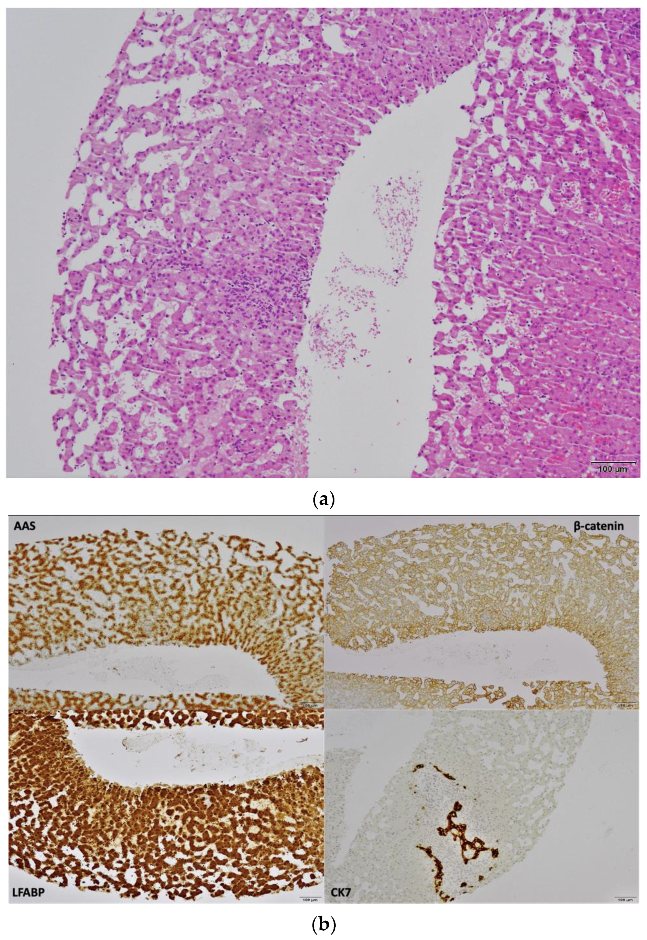
To establish the definitive diagnosis an ultrasound-guided liver biopsy was performed. Histologic assessment showed two portal spaces, hepatic parenchyma with sinusoidal dilation and congestion, discrete to moderate inflammatory infiltrate, with lymphocyte predominance (Figure 2a—Hematoxylin and Eosin staining). Arteries had thickened walls, without accompanying bile ducts and with ductular proliferation. There were no atypical hepatocytes. Immunohistochemistry disclosed positive serum amyloid A, positive membrane β-catenin staining and negative glutamine synthetase (Figure 2b—immunostaining (GS and CD34 not available). With that information, we were able to make the final diagnosis of hepatic adenomatosis, inflammatory subtype.
The case was discussed in a hepato-biliary multidisciplinary group and was decided to discontinue oral contraceptives and maintain surveillance at 6 months.
The MRI 6 months later showed a global decrease of the largest nodules, ranging from a 3 to 9 mm reduction (Figure 3). She has been followed for two years now and the lesions are stable, with roughly the same dimensions.

3. Discussion

This case highlights the challenges when facing multiple hypoechoic nodules in a young woman. The first imaging method performed is usually an ultrasound [3]. On ultrasound HCA appear as hypo, iso, or hyperechoic nodule, depending on the fat content and presence of intralesional hemorrhage [26], as so, it has limited utility in diagnosing HCA [27]. On the other side, on computed tomography (CT), HCA appear as a round lesion, with late enhancing peripheral capsule [27]. The main differential diagnosis for HCA is focal nodular hyperplasia (FNH), a benign lesion that rarely requires surgical resection [28]. Small HCA lesions on CT tend to homogenously enhance on arterial phase, while larger lesions may enhance heterogeneously due to areas of necrosis [27]. At CT, simple HCA are commonly isodense with the surrounding liver on unenhanced CT, become visible in the arterial phase with a homogeneous blush of contrast enhancement, and then fade to isodensity in the portal or delayed phase [26].
Magnetic Resonance Imaging (RMI) is the most sensitive imaging method to diagnose HCA [1] and it is also more sensitive in detecting subtle hemorrhage on T1-weighted imaging [27]. Nonetheless, in some cases, it can be difficult to distinguish HCA from FNH. Use of contrast agents facilitate this distinction and even aid in subtype differentiation. Inflammatory HCA can be specifically identified using MRI: they are characterized by their telangiectatic features, a strong hyperintense signal on T2-weighted images (as strong as the signal of the spleen), which may be either diffuse or as a rim-like band in the periphery of the lesion and defines the atoll sign. On T1-weighted sequences, lesion signal intensity is variably iso- to hyperintense [1]. H-HCA on MRI appears as a diffuse and homogeneous signal dropout on out-of-phase T1 imaging [29]. The other subtypes, especially β-catenin activated, do not have specific signs on MRI and cannot be differentiated from HCC [1,29].
When there are no specific imaging characteristics, biopsy is mandatory to exclude dysplasia and establish the subtype. Pathology study allows classification of the subtype based on the use of an immunohistochemical panel, that commonly includes Liver Fatty Acid-Binding Protein (LFABP), serum amyloid A (SAA), C-reactive protein (CRP), beta-catenin (BC), and glutamine synthetase (GS) in tandem with attention to the histological features of the tumor and non-tumor liver [30].
I-HCA has varying degrees of inflammatory infiltrate, ductular proliferation and telangiectasia; is also most likely to arise in a liver showing adjacent changes of steatosis and steatohepatitis [31]. Immunohistochemical analysis using both SAA and CRP can be used for the diagnosis of I-HCA, two acute phase reactants overexpressed in the adenoma hepatocyte cytoplasm; however, some authors state that CRP is more reliable than SAA with more consistent and stronger staining patterns [28,30,32].
As LA is a rare entity and has a heterogeneous clinical course, its management imposes a multidisciplinary approach. According to the EASL clinical practice guidelines, in a general picture, the management must consider the gender and the size of the biggest lesion, independently of how many lesions there is [1]. However, even with biopsy, the real risk of bleeding and malignant potential in a liver with >40 nodules is unknown.
In patients under oral contraceptive pills with single or multiple tumors, the first therapeutic approach passes by its withdrawal and imaging surveillance [1,16]. Cessation of exogenous hormone replacement therapies frequently causes a near-total to complete regression in the size of the tumor [16]. Asymptomatic patients with tumors smaller than 5 cm generally have a benign and uncomplicated clinical course, so conservative management with surveillance has been suggested [28,30]. In the absence of worrisome features, follow-up with CT or MRI every 6 months for the first 2 years and annually thereafter is an acceptable approach [16,33]. However, the duration of follow-up is not fully established, and some authors advocate that follow-up must be carried out until menopause in women [33]. In men, given the higher incidence of malignant transformation of HCA, resection or curative treatment always is recommended (see below) [1]. No follow-up recommendation can be found regarding non curative treatment of HCA in men. Historically women have been advised against pregnancy; however, although pregnancy may be associated with an increased risk of HCA complications, the presence of small asymptomatic HCA is not an absolute contraindication for pregnancy [16].
Surgical resection is recommended for patients at risk of developing complications, namely lesions >5 cm, HCA increasing in size during follow-up and β-catenin subtype (especially exon 3). It is also recommended on male gender, rising levels of AFP, imaging suspicious for malignancy or inability to rule out HCC, or when dysplasia is present on biopsy [16,34].
The number of lesions has not been associated with an additional risk of complications, so it is acceptable to follow these same criteria for surgical resection, even when there are multiple lesions [1,33,34], as is the case of this patient. Timing of resection must be considered, according to current guidelines, in patients with HCA > 5 cm and whose tumor does not regress, or even progresses, during the 6-month interval following interruption of oral contraceptives [33]. However, some authors advocate a longer surveillance period before considering surgery, of up to 12 months, as many patients have longer time to regression to <5 cm, especially with larger tumors [33,35].
Surgical resection must be adapted according to the number and distribution of lesions. When HCAs are localized, they can be treated with hepatectomy (minor or major hepatectomies), while resection of the largest adenoma may be the choice towards a widespread HCA’s distribution [1,3]. Efficacy and safety of laparoscopic liver resection is comparable to laparotomy, with no differences in postoperative morbidity. However, laparoscopic approach may be limited by lesion size and location [16,33].
Transarterial embolization (TAE) is a first option in the management of hemodynamically stable patients with bleeding HCA and can also be performed electively as an alternative to surgical intervention [16,36]. Surgery may be avoided in up to 45% of patients when using TAE [37]. Other alternatives, such as thermal ablation (microwave and radiofrequency), may be considered in specialized centers, for patients that are not surgical candidates due to clinical or technical reasons [16,38,39]. Thermal ablation for small tumors (<5 cm) seems to have a similar complication rate to surgery and embolization [38], but further head-to-head studies are needed to access safety and efficacy.
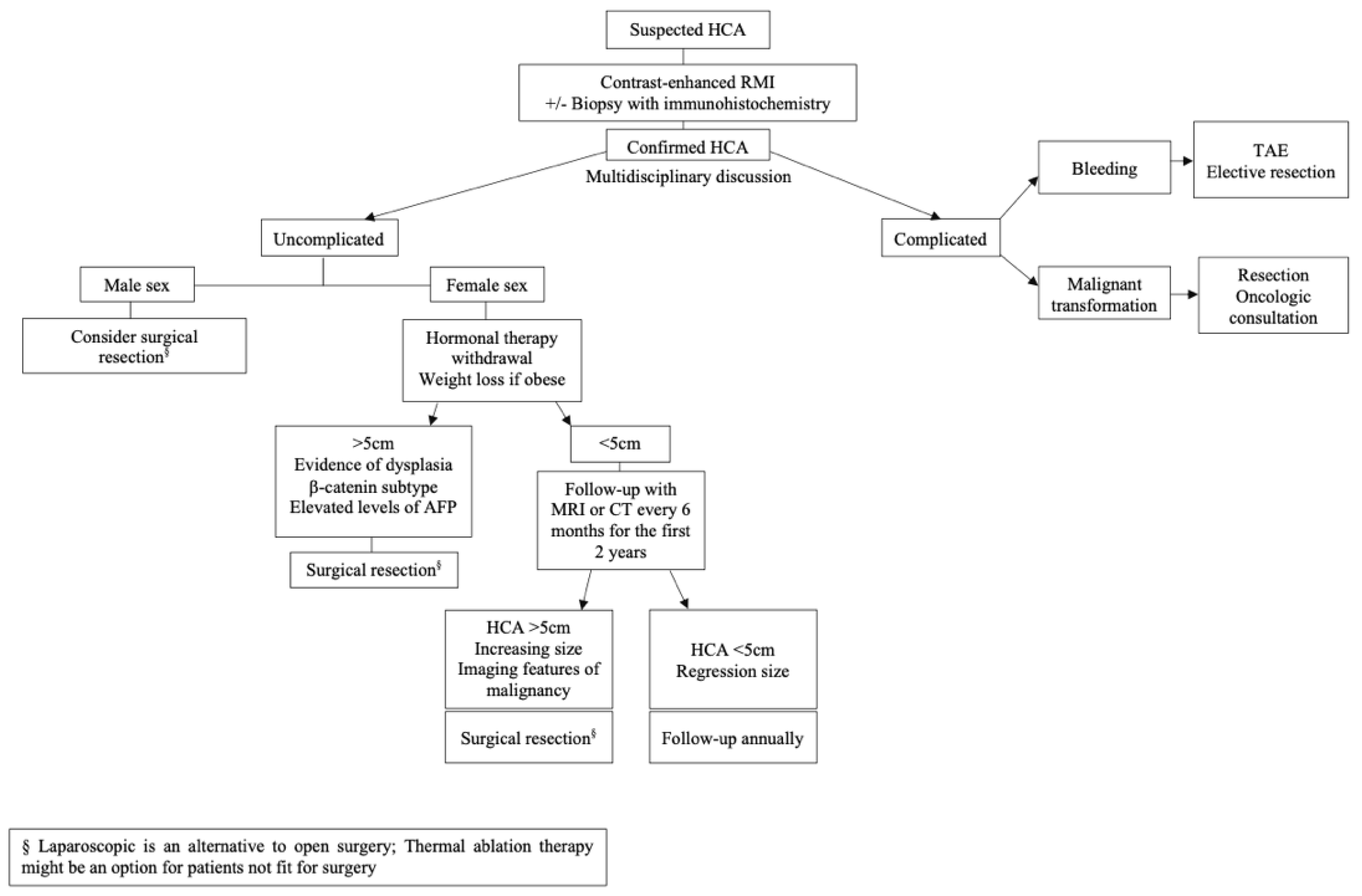
Liver transplantation for LA is restricted to very selected situations [4,16], with the main indication being the presence of multiple lesions with suspicious or proven malignant transformation, not amenable to surgical resection [16]. Another indication is the presence of a portosystemic venous shunt [16]. An algorithm comprising diagnosis and treatment is proposed (Figure 4).
In this case the diagnosis was performed two years ago, and the patient showed an important reduction in HCA size 6 months after stopping oral contraceptive pills. The lesions are now stable, with roughly the same dimensions after one year of follow-up and she remains asymptomatic.
As described by Barbier et al., on the longest follow-up study of LA’s patients (26 years), 13% different subtypes coexisted in the same liver, although most HCA subtypes were homogenous [3]. The controversial aspect is the management approach in a patient with >40 lesions, as in this case, since biopsy has a limited diagnosis accuracy of the subtype, given the inability to sample all the lesions.

4. Conclusions

LA is a rare disease, that affects predominantly women between 30–40 years old. The inflammatory subtype is the most common subtype. Approach to HCA is based on gender and the size of the biggest lesion, independently of how many lesions there is. However, even with biopsy, the real risk of bleeding and malignant potential in a liver with >40 nodules is unknown, as there are cases of patients with different subtypes coexisting in the same liver.

Author Contributions

Conceptualization, C.C.R. and M.A.D.; methodology, C.C.R., D.B.M. and M.F.d.L.; validation, J.R.P. and M.A.D.; resources, E.D. and R.A.; writing—original draft preparation, C.C.R.; writing—review and editing, D.B.M., M.F.d.L., J.R.P. and M.A.D.; visualization, C.C.R. and J.R.P.; supervision, J.R.P. and M.A.D.; All authors have read and agreed to the published version of the manuscript.

Funding

This research received no external funding.

Institutional Review Board Statement

Ethical review and approval were waived for this study as the paper describes an individual clinical case with patient’s approval.

Informed Consent Statement

Written informed consent has been obtained from the patient to publish this paper.

Data Availability Statement

The data presented in this study are available on request from the corresponding author. The data are not publicly available due to privacy reasons.

Conflicts of Interest

The authors declare no conflict of interest.

References

  1. European Association for the Study of the Liver. EASL Clinical Practice Guidelines on the management of benign liver tumours. J. Hepatol. 2015, 65, 386–398. [Google Scholar]
  2. Rooks, J.B.; Ory, H.W.; Ishak, K.G.; Strauss, L.T.; Greenspan, J.R.; Hill, A.P.; Tyler, C.W., Jr. Epidemiology of hepatocellular adenoma. The role of oral contraceptive use. JAMA 1979, 242, 644–648. [Google Scholar] [CrossRef] [PubMed]
  3. Barbier, L.; Nault, J.C.; Dujardin, F.; Scotto, B.; Besson, M.; de Muret, A.; Bourlier, P.; Zucman-Rossi, J.; Salamé, E.; Bacq, Y. Natural history of liver adenomatosis: A long-term observational study. J. Hepatol. 2019, 71, 1184–1192. [Google Scholar] [CrossRef] [PubMed]
  4. Sundar Alagusundaramoorthy, S.; Vilchez, V.; Zanni, A.; Sourianarayanane, A.; Maynard, E.; Shah, M.; Daily, M.F.; Pena, L.R.; Gedaly, R. Role of Transplantation in the Treatment of Benign Solid Tumors of the Liver: A Review of the United Network of Organ Sharing Data Set. JAMA Surg. 2015, 150, 337–342. [Google Scholar] [CrossRef] [PubMed] [Green Version]
  5. Karhunen, P.J. Benign hepatic tumours and tumour like conditions in men. J. Clin. Pathol. 1986, 39, 183–188. Available online: https://pubmed.ncbi.nlm.nih.gov/3950039 (accessed on 8 January 2022). [CrossRef] [PubMed] [Green Version]
  6. Bonder, A.; Afdhal, N. Evaluation of liver lesions. Clin. Liver Dis. 2012, 16, 271–283. [Google Scholar] [CrossRef] [PubMed]
  7. Giannitrapani, L.; Soresi, M.; La Spada, E.; Cervello, M.; D’Alessandro, N.; Montalto, G. Sex hormones and risk of liver tumor. Ann. N. Y. Acad. Sci. 2006, 1089, 228–236. [Google Scholar] [CrossRef] [PubMed]
  8. Dokmak, S.; Belghiti, J. Will weight loss become a future treatment of hepatocellular adenoma in obese patients? Liver Int. Off. J. Int. Assoc. Study Liver 2015, 35, 2228–2232. [Google Scholar] [CrossRef]
  9. Grangé, J.D.; Guéchot, J.; Legendre, C.; Giboudeau, J.; Darnis, F.; Poupon, R. Liver adenoma and focal nodular hyperplasia in a man with high endogenous sex steroids. Gastroenterology 1987, 93, 1409–1413. [Google Scholar] [CrossRef]
  10. Triantafyllopoulou, M.; Whitington, P.F.; Melin-Aldana, H.; Benya, E.C.; Brickman, W. Hepatic adenoma in an adolescent with elevated androgen levels. J. Pediatr. Gastroenterol. Nutr. 2007, 44, 640–642. [Google Scholar] [CrossRef]
  11. Renzulli, M.; Clemente, A.; Tovoli, F.; Cappabianca, S.; Bolondi, L.; Golfieri, R. Hepatocellular adenoma: An unsolved diagnostic enigma. World J. Gastroenterol. 2019, 25, 2442–2449. [Google Scholar] [CrossRef] [PubMed]
  12. Baker, F.A.; Zeina, A.R.; Mouch, S.A.; Mari, A. Benign Hepatic Tumors: From Incidental Imaging Finding to Clinical Management. Korean J. Fam. Med. 2021, 42, 2–28. [Google Scholar] [CrossRef] [PubMed]
  13. Monges, H.; Payan, H.; Legre, M.; Vignoli, R. Considerations on a case of benign multinodular adenoma of the liver. Arch. Mal. Appar. Dig. Mal. Nutr. 1963, 52, 725–737. [Google Scholar] [PubMed]
  14. Monaco, A.P.; Hallgrimsson, J.; Mcdermott, W.V.J. Multiple adenoma (hamartoma) of the liver treated by subtotal (90 percent) resection: Morphological and functional studies of regeneration. Ann. Surg. 1964, 159, 513–519. [Google Scholar] [CrossRef]
  15. Flejou, J.F.; Barge, J.; Menu, Y.; Degott, C.; Bismuth, H.; Potet, F.; Benhamou, J.P. Liver adenomatosis. An entity distinct from liver adenoma? Gastroenterology 1985, 89, 1132–1138. [Google Scholar] [CrossRef]
  16. Tsilimigras, D.I.; Rahnemai-Azar, A.A.; Ntanasis-Stathopoulos, I.; Gavriatopoulou, M.; Moris, D.; Spartalis, E.; Cloyd, J.M.; Weber, S.M.; Pawlik, T.M. Current Approaches in the Management of Hepatic Adenomas. J. Gastrointest. Surg. 2019, 23, 199–209. [Google Scholar] [CrossRef]
  17. Bioulac-Sage, P.; Laumonier, H.; Couchy, G.; Le Bail, B.; Sa Cunha, A.; Rullier, A.; Laurent, C.; Blanc, J.F.; Cubel, G.; Trillaud, H.; et al. Hepatocellular adenoma management and phenotypic classification: The Bordeaux experience. Hepatology 2009, 50, 481–489. [Google Scholar] [CrossRef]
  18. Bioulac-Sage, P.; Rebouissou, S.; Thomas, C.; Blanc, J.-F.; Saric, J.; Sa Cunha, A.; Rullier, A.; Cubel, G.; Couchy, G.; Imbeaud, S.; et al. Hepatocellular adenoma subtype classification using molecular markers and immunohistochemistry. Hepatology 2007, 46, 740–748. [Google Scholar] [CrossRef]
  19. Rebouissou, S.; Bioulac-Sage, P.; Zucman-Rossi, J. Molecular pathogenesis of focal nodular hyperplasia and hepatocellular adenoma. J. Hepatol. 2008, 48, 163–170. [Google Scholar] [CrossRef] [Green Version]
  20. Nault, J.C.; Couchy, G.; Balabaud, C.; Morcrette, G.; Caruso, S.; Blanc, J.F.; Bacq, Y.; Calderaro, J.; Paradis, V.; Ramos, J.; et al. Molecular Classification of Hepatocellular Adenoma Associates with Risk Factors, Bleeding, and Malignant Transformation. Gastroenterology 2017, 152, 880–894.e6. [Google Scholar] [CrossRef] [Green Version]
  21. Cho, S.W.; Marsh, J.W.; Steel, J.; Holloway, S.E.; Heckman, J.T.; Ochoa, E.R.; Geller, D.A.; Gamblin, T.C. Surgical management of hepatocellular adenoma: Take it or leave it? Ann. Surg. Oncol. 2008, 15, 2795–2803. [Google Scholar] [CrossRef] [PubMed]
  22. Ribeiro Junior, M.A.F.; Chaib, E.; Saad, W.A.; D’Albuquerque, L.A.C.; Cecconello, I. Surgical management of spontaneous ruptured hepatocellular adenoma. Clinics 2009, 64, 775–779. [Google Scholar] [CrossRef] [PubMed] [Green Version]
  23. van Aalten, S.M.; de Man, R.A.; IJzermans, J.N.M.; Terkivatan, T. Systematic review of haemorrhage and rupture of hepatocellular adenomas. Br. J. Surg. 2012, 99, 911–916. [Google Scholar] [CrossRef] [PubMed]
  24. Farges, O.; Ferreira, N.; Dokmak, S.; Belghiti, J.; Bedossa, P.; Paradis, V. Changing trends in malignant transformation of hepatocellular adenoma. Gut 2011, 60, 85–89. [Google Scholar] [CrossRef]
  25. Zucman-Rossi, J.; Jeannot, E.; Nhieu, J.T.; Van Scoazec, J.-Y.; Guettier, C.; Rebouissou, S.; Bacq, Y.; Leteurtre, E.; Paradis, V.; Michalak, S.; et al. Genotype-phenotype correlation in hepatocellular adenoma: New classification and relationship with HCC. Hepatology 2006, 43, 515–524. [Google Scholar] [CrossRef] [PubMed] [Green Version]
  26. Hussain, S.M.; van den Bos, I.C.; Dwarkasing, R.S.; Kuiper, J.W.; den Hollander, J. Hepatocellular adenoma: Findings at state-of-the-art magnetic resonance imaging, ultrasound, computed tomography and pathologic analysis. Eur. Radiol. 2006, 16, 1873–1886. [Google Scholar] [CrossRef]
  27. Thapar, M.; Grapp, O.; Fisher, C. Management of Hepatic Adenomatosis. Curr. Gastroenterol. Rep. 2015, 17, 12. [Google Scholar] [CrossRef]
  28. Krause, K.; Tanabe, K.K. A Shifting Paradigm in Diagnosis and Management of Hepatic Adenoma. Ann. Surg. Oncol. 2020, 27, 3330–3338. [Google Scholar] [CrossRef]
  29. Nault, J.C.; Paradis, V.; Cherqui, D.; Vilgrain, V.; Zucman-Rossi, J. Molecular classification of hepatocellular adenoma in clinical practice. J. Hepatol. 2017, 67, 1074–1083. [Google Scholar] [CrossRef] [Green Version]
  30. Pilati, C.; Letouzé, E.; Nault, J.-C.; Imbeaud, S.; Boulai, A.; Calderaro, J.; Poussin, K.; Franconi, A.; Couchy, G.; Morcrette, G.; et al. Genomic profiling of hepatocellular adenomas reveals recurrent FRK-activating mutations and the mechanisms of malignant transformation. Cancer Cell 2014, 25, 428–441. [Google Scholar] [CrossRef] [Green Version]
  31. Torbenson, M. Hepatic Adenomas: Classification, Controversies, and Consensus. Surg. Pathol. Clin. 2018, 11, 351–366. [Google Scholar] [CrossRef] [PubMed]
  32. Miller, G.C.; Campbell, C.M.; Manoharan, B.; Bryant, R.; Cavallucci, D.; O’Rourke, N.; Clouston, A.D. Subclassification of hepatocellular adenomas: Practical considerations in the implementation of the Bordeaux criteria. Pathology 2018, 50, 593–599. [Google Scholar] [CrossRef] [PubMed]
  33. Thomeer, M.G.; Broker, M.; Verheij, J.; Doukas, M.; Terkivatan, T.; Bijdevaate, D.; de Man, R.A.; Moelker, A.; IJzermans, J.N. Hepatocellular adenoma: When and how to treat? Update of current evidence. Ther. Adv. Gastroenterol. 2016, 9, 898–912. [Google Scholar] [CrossRef]
  34. Liau, S.-S.; Qureshi, M.S.; Praseedom, R.; Huguet, E. Molecular pathogenesis of hepatic adenomas and its implications for surgical management. J. Gastrointest. Surg. Off. J. Soc.Surg. Aliment. Tract. 2013, 17, 1869–1882. [Google Scholar] [CrossRef] [Green Version]
  35. Klompenhouwer, A.J.; de Man, R.A.; Dioguardi Burgio, M.; Vilgrain, V.; Zucman-Rossi, J.; Ijzermans, J.N.M. New insights in the management of Hepatocellular Adenoma. Liver Int. 2020, 40, 1529–1537. Available online: https://pubmed.ncbi.nlm.nih.gov/32464711 (accessed on 8 January 2022). [CrossRef] [PubMed]
  36. Agrawal, S.; Agarwal, S.; Arnason, T.; Saini, S.; Belghiti, J. Management of Hepatocellular Adenoma: Recent Advances. Clin. Gastroenterol. Hepatol. 2015, 13, 1221–1230. [Google Scholar] [CrossRef] [PubMed]
  37. van Rosmalen, B.V.; Coelen, R.J.S.; Bieze, M.; van Delden, O.M.; Verheij, J.; Dejong, C.H.C.; van Gulik, T.M. Systematic review of transarterial embolization for hepatocellular adenomas. Br. J. Surg. 2017, 104, 823–835. [Google Scholar] [CrossRef] [PubMed]
  38. Mironov, O.; Jaberi, A.; Beecroft, R.; Kachura, J.R. Retrospective Single-Arm Cohort Study of Patients with Hepatocellular Adenomas Treated with Percutaneous Thermal Ablation. Cardiovasc. Interv. Radiol. 2018, 41, 935–941. [Google Scholar] [CrossRef]
  39. Smolock, A.R.; Cristescu, M.M.; Potretzke, T.A.; Ziemlewicz, T.J.; Lubner, M.G.; Hinshaw, J.L.; Brace, C.L.; Lee, F.T., Jr. Microwave Ablation for the Treatment of Hepatic Adenomas. J. Vasc. Interv. Radiol. 2016, 27, 244–249. [Google Scholar] [CrossRef]
Figure 1. Images at diagnosis. (a,b) Coronal T2-weighted image, showing multiple nodular lesions with bilobar distribution, hyperintense on T2-WI. (c,d) On arterial axial T1-weighted GRE 3D FS image, the lesions show high signal intensity compared with surrounding liver parenchyma. (e) axial T1-weighted 3D GRE FS—the lesions are hypointense on 20-min delayed hepatobiliary phase image, with hepatospecific contrast (Gd-EOB-DTPA), because of the lack of normal functioning hepatocytes.
Gastroent 13 00008 g001
Figure 2. (a) Hematoxylin and Eosin, 100×. Liver biopsy showing an hepatocellular lesion lacking portal structures while retaining the trabecular architecture of the hepatocytes. The trabeculae are not thickened. There is inflammation and prominent sinusoidal dilatation. (b) Tumour cells expressing SAA (serum Amyloid A) and LFABP (liver fatty acid binding protein). β-catenin expression is restricted to the cell membrane. In the setting of an hepatic adenoma, this immunohistochemical profile is consistent with the inflammatory subtype. CK7 stain demonstrates the presence of bile ductules, typical of this subtype.
Gastroent 13 00008 g002
Figure 3. Re-evaluation, comparative with Figure 1, dimensional improvement of nodular lesions; (a,b) Coronal section, lesion is hyperintense on T2-weighted image; (c,d) Axial GRE 3D FS T1-weighted sequence post-Gadoxetic acid intravenous administration at arterial phase, with hyperintense lesions.
Gastroent 13 00008 g003
Figure 4. Algorithm with proposed diagnosis and treatment.
Gastroent 13 00008 g004
Publisher’s Note: MDPI stays neutral with regard to jurisdictional claims in published maps and institutional affiliations.