

Acute Anterior Choroidal Artery Territory Infarction: A Case Series Report


Abstract
1. Introduction
2. Materials and Methods
3. Results
3.1. CASE 1
3.2. CASE 2
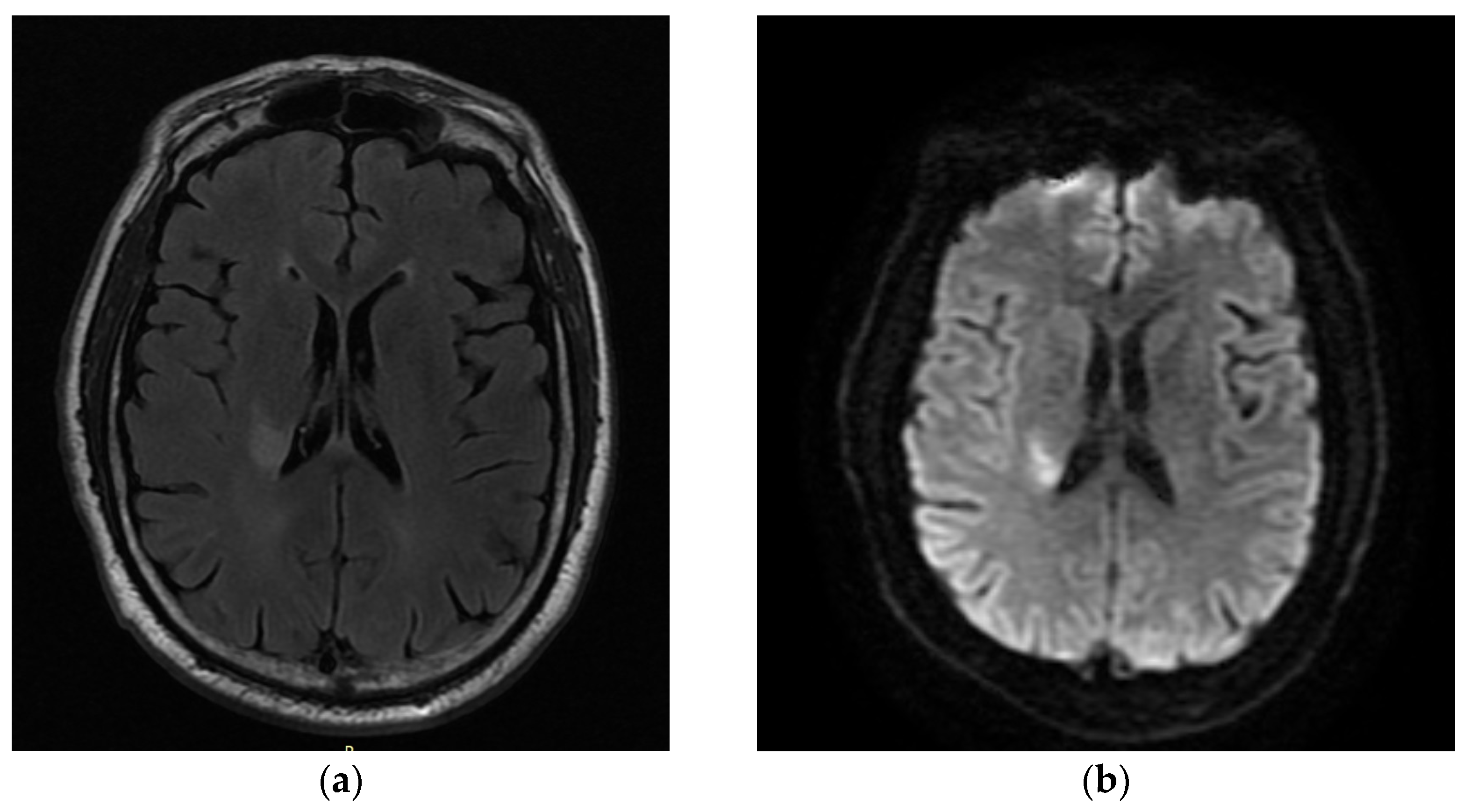
3.3. CASE 3
3.4. CASE 4
3.5. CASE 5
4. Discussion
5. Conclusions
Author Contributions
Funding
Institutional Review Board Statement
Informed Consent Statement
Data Availability Statement
Conflicts of Interest
References
- Alqahtani, S.A.; Luby, M.; Nadareishvili, Z.; Benson, R.T.; Hsia, A.W.; Leigh, R.; Lynch, J.K. Perfusion Deficits and Association with Clinical Outcome in Patients with Anterior Choroidal Artery Stroke. J. Stroke Cerebrovasc. Dis. 2017, 26, 1755–1759. [Google Scholar] [CrossRef] [PubMed]
- Leys, D.; Mounier-Vehier, F.; Lavenu, I.; Rondepierre, P.; Pruvo, J.P. Anterior choroidal artery territory infarcts. Study of presumed mechanisms. Stroke 1994, 25, 837–842. [Google Scholar] [CrossRef] [PubMed]
- Cheng, Z.; Duan, H.; Meng, F.; Du, H.; Zhang, W.; Li, H.; Geng, X.; Tong, Y. Acute Anterior Choroidal Artery Territory Infarction: A Retrospective Study. Clin. Neurol. Neurosurg. 2020, 195, 105826. [Google Scholar] [CrossRef] [PubMed]
- Sohn, H.; Kang, D.W.; Kwon, S.U.; Kim, J.S. Anterior choroidal artery territory infarction: Lesions confined to versus beyond the internal capsule. Cerebrovasc. Dis. 2013, 35, 228–234. [Google Scholar] [CrossRef] [PubMed]
- Nelles, M.; Gieseke, J.; Flacke, S.; Lachenmayer, L.; Schild, H.H.; Urbach, H. Diffusion tensor pyramidal tractography in patients with anterior choroidal artery infarcts. AJNR Am. J. Neuroradiol. 2008, 29, 488–493. [Google Scholar] [CrossRef] [PubMed]
- Pezzella, F.R.; Vadalà, R. Anterior choroidal artery territory infarction. Front. Neurol. Neurosci. 2012, 30, 123–127. [Google Scholar] [CrossRef] [PubMed]
- Ois, A.; Cuadrado-Godia, E.; Solano, A.; Perich-Alsina, X.; Roquer, J. Acute ischemic stroke in anterior choroidal artery territory. J. Neurol. Sci. 2009, 281, 80–84. [Google Scholar] [CrossRef] [PubMed]
- Derflinger, S.; Fiebach, J.B.; Böttger, S.; Haberl, R.L.; Audebert, H.J. The Progressive Course of Neurological Symptoms in Anterior Choroidal Artery Infarcts. Int. J. Stroke 2015, 10, 134–137. [Google Scholar] [CrossRef] [PubMed]
- Chausson, N.; Joux, J.; Saint-Vil, M.; Edimonana, M.; Jeannin, S.; Aveillan, M.; Cabre, P.; Olindo, S.; Smadja, D. Infarction in the anterior choroidal artery territory: Clinical progression and prognosis factors. J. Stroke Cerebrovasc. Dis. 2014, 23, 2012–2017. [Google Scholar] [CrossRef] [PubMed]
- Turan, T.N.; Makki, A.A.; Tsappidi, S.; Cotsonis, G.; Lynn, M.J.; Cloft, H.J.; Chimowitz, M.I. Risk factors associated with severity and location of intracranial arterial stenosis. Stroke 2010, 41, 1636–1640. [Google Scholar] [CrossRef] [PubMed]
- Palomeras, E.; Fossas, P.; Cano, A.T.; Sanz, P.; Floriach, M. Anterior choroidal artery infarction: A clinical, etiologic and prognostic study. Acta Neurol. Scand. 2008, 118, 42–47. [Google Scholar] [CrossRef] [PubMed]


| Clinical Presentation | Case 1 | Case 2 | Case 3 | Case 4 | Case 5 |
|---|---|---|---|---|---|
| Dysarthria | + | - | + | + | + |
| VII central paresis | + | + | - | - | - |
| Hemiparesis | + | + | + | + | + |
| Hemisensory loss | - | - | - | - | - |
| Hemianopsia | - | - | - | - | - |
| Ataxia | + | - | - | - | + |
| Medical history | Case 1 | Case 2 | Case 3 | Case 4 | Case 5 |
|---|---|---|---|---|---|
| Hypertension | - | + | + | + | + |
| Diabetes | - | - | - | + | + |
| Dyslipidemia | - | - | + | + | - |
| Smoking | - | + | + | - | + |
| Case 1 | Case 2 | Case 3 | Case 4 | Case 5 | |
|---|---|---|---|---|---|
| Sex | F | M | M | M | M |
| Age | 53 | 52 | 58 | 77 | 54 |
| NIHSS | 5–7 | 4 | 3–6 | 3–7 | 2–10 |
| Thrombolysis | - | + | - | - | - |
| Follow-up mRS | 3 | 3 | 3 | 4 | 2 |
| TOAST | ESUS | ESUS | LAA | LAA | ESUS |
Disclaimer/Publisher’s Note: The statements, opinions and data contained in all publications are solely those of the individual author(s) and contributor(s) and not of MDPI and/or the editor(s). MDPI and/or the editor(s) disclaim responsibility for any injury to people or property resulting from any ideas, methods, instructions or products referred to in the content. |
© 2024 by the authors. Licensee MDPI, Basel, Switzerland. This article is an open access article distributed under the terms and conditions of the Creative Commons Attribution (CC BY) license (https://creativecommons.org/licenses/by/4.0/).
Share and Cite
Tsika, A.; Stamati, P.; Tsouris, Z.; Provatas, A.; Papa, A.; Tsimoulis, D.; Ralli, S.; Siokas, V.; Dardiotis, E. Acute Anterior Choroidal Artery Territory Infarction: A Case Series Report. Neurol. Int. 2024, 16, 289-298. https://doi.org/10.3390/neurolint16020020
Tsika A, Stamati P, Tsouris Z, Provatas A, Papa A, Tsimoulis D, Ralli S, Siokas V, Dardiotis E. Acute Anterior Choroidal Artery Territory Infarction: A Case Series Report. Neurology International. 2024; 16(2):289-298. https://doi.org/10.3390/neurolint16020020
Chicago/Turabian StyleTsika, Antonia, Polyxeni Stamati, Zisis Tsouris, Antonios Provatas, Alexandra Papa, Dimitrios Tsimoulis, Stylliani Ralli, Vasileios Siokas, and Efthimios Dardiotis. 2024. "Acute Anterior Choroidal Artery Territory Infarction: A Case Series Report" Neurology International 16, no. 2: 289-298. https://doi.org/10.3390/neurolint16020020
APA StyleTsika, A., Stamati, P., Tsouris, Z., Provatas, A., Papa, A., Tsimoulis, D., Ralli, S., Siokas, V., & Dardiotis, E. (2024). Acute Anterior Choroidal Artery Territory Infarction: A Case Series Report. Neurology International, 16(2), 289-298. https://doi.org/10.3390/neurolint16020020