Systematic Review

MicroRNA and DNA Methylation Adaptation Mechanism to Endurance Training in Cardiovascular Disease: A Systematic Review

by
Jil Delhez
1,
Jeanne Ougier
1,
Francisco Xavier de Araujo
2,
Raphael Martins de Abreu
1,3 and
Camilo Corbellini
1,3,*
1
Department of Health, LUNEX University of Applied Sciences, 50, Avenue du Parc des Sports, 4671 Differdange, Luxembourg
2
Department of Gymnastics and Health, Universidade Federal de Pelotas (Federal University of Pelotas), Pelotas 96010-610, Brazil
3
Luxembourg Health & Sport Sciences Research Institute A.s.b.l., 50, Avenue du Parc des Sports, 4671 Differdange, Luxembourg
*
Author to whom correspondence should be addressed.
Cardiogenetics 2025, 15(4), 28; https://doi.org/10.3390/cardiogenetics15040028
Submission received: 8 August 2025 / Revised: 27 September 2025 / Accepted: 28 September 2025 / Published: 11 October 2025
(This article belongs to the Section Cardiovascular Genetics in Clinical Practice)

Abstract

Background: Regular endurance training induces physiological changes in cardiac structure and function. The precise epigenetic mechanisms by which cardiovascular adaptations are mediated are still unclear. This review seeks to clarify the role of epigenetic regulation in exercise-induced cardiovascular adaptation. Methods: This systematic review was conducted in accordance with the PRISMA guidelines up to 30 April 2025, using the databases PubMed, VHL, and LILACS Plus. Studies were included if they focused on microRNA expression and DNA methylation in individuals with cardiovascular disease who underwent endurance training. Results: Six articles, including 384 participants with heart failure, coronary artery disease, and hypertension, were included in the final analysis. Changes in DNA methylation and microRNA expression of specific genes involved in cardiovascular structural and functional adaptation were observed. Significant improvements were found in body composition, VO2peak, systolic and diastolic blood pressure, and left ventricular function and structure. Conclusions: Endurance training has a positive impact on epigenetic mechanisms related to cardiovascular structural and functional adaptation. A clear causal link between epigenetic modifications and clinical outcomes remains to be established.

Graphical Abstract

1. Introduction

Cardiovascular diseases (CVDs) remain the leading cause of death globally, responsible for one in four deaths in the United States since 1975 [1]. In 2015, CVDs caused an estimated 17.7 million deaths, presenting a significant economic burden and ranking as the most costly condition, followed by diabetes and Alzheimer’s disease [1]. By the age of 45, half of the general population is at risk of developing CVD [1]. Given this increasing burden, there is a rising need to investigate non-pharmacological strategies such as exercise to reduce cardiovascular risk and enhance long-term health outcomes [2].
Regular endurance exercise is a well-established protective factor against CVDs, improving cardiorespiratory fitness, enhancing cardiac function, and lowering mortality risk [3,4]. One of the strongest predictors of cardiovascular health, peak oxygen consumption (VO2max), improves through aerobic training and is associated with a reduced risk of cardiovascular mortality [5]. Physiological adaptations to aerobic exercise include increased stroke volume, myocardial contractility, and left ventricular hypertrophy, all of which enhance cardiovascular function [6]. Both acute and chronic exercise induce significant adaptations in the cardiovascular system via changes at cellular and molecular levels [7]. In addition to these structural and functional changes, growing evidence suggests that exercise-induced modifications in gene expression play a vital role in cardiovascular adaptation [8,9].
Recent research has highlighted the role of epigenetic mechanisms in mediating the cardiovascular benefits of exercise [8]. Epigenetics refers to heritable changes in gene expression that occur without altering the DNA sequence itself [10]. Among the key mechanisms, microRNA (miRNA) expression regulation and DNA methylation have emerged as major modulators of gene expression in response to aerobic exercise [11]. MiRNAs are small non-coding RNA molecules that regulate gene expression post-transcriptionally by binding to specific messenger RNAs (mRNAs), leading to mRNA degradation or inhibition of its translation into protein [12]. MiRNAs influence pathways involved in cardiac remodelling and vascular function [12].
Meanwhile, DNA methylation, which involves the addition of methyl groups to DNA, plays a crucial role in silencing or activating gene expression [10]. In the cardiovascular system, changes in DNA methylation, including hypomethylation and hypermethylation of genes, contribute to cardiac protection [10]. Both mechanisms have been linked to various cardiovascular outcomes, including cardiac remodelling, which is a vital process in maintaining heart health in response to exercise [10,12,13].
Despite substantial evidence linking endurance exercise to improved cardiovascular outcomes, the exact molecular pathways through which miRNA regulation and DNA methylation facilitate these adaptations remain only partly understood. Although the impact of exercise on skeletal muscle gene expression is well established, fewer studies have investigated its effects on cardiac gene expression [14]. Animal models have consistently demonstrated predictable physiological responses to endurance training, suggesting that exercise-induced epigenetic regulation could play a fundamental role in human cardiac remodelling [7,8]. However, a systematic synthesis of the available evidence on epigenetic animal models have consistently shown predictable physiological responses to endurance training, implying that exercise-induced epigenetic regulation might play a key role in human cardiac remodelling [7,8]. However, a systematic review of the existing evidence on epigenetic mechanisms in adults with CVD is absent [15]. Prior studies using animal models and healthy individuals limit clinical relevance and generalisation to people with CVD, highlighting the need to focus on clinical populations [5,6,7,9]. Mechanisms in adults with CVD is lacking [15]. The previous use of animal models and healthy individuals limits clinical applicability and generalisation to individuals with CVD, creating a strong rationale for focusing on clinical populations [5,6,7,9].
This systematic review aims to explore how endurance training affects gene regulation, particularly miRNA expression and DNA methylation, in individuals with cardiovascular diseases. It considers how these epigenetic modifications influence cardiac structure and function, emphasising the molecular basis of exercise-induced cardiovascular adaptation and potentially guiding future therapies for preventing and managing CVD.

2. Materials and Methods

2.1. Search Strategy

This systematic review was conducted in accordance with the PRISMA guidelines [16]. The search was performed up to 30 April 2025 across the databases PubMed, Virtual Health Library (VHL), and LILACS Plus. The search string was developed using the main components of the research question with the Boolean operators ‘AND’ and ‘OR’, combining these key elements. PubMed was chosen as the primary database because of its advanced search capabilities, which enable more precise query formulation. The search string was then adapted for the VHL and LILACS Plus databases. The PubMed search string was ((“aerobic exercise” [Title/Abstract] OR “endurance training” [Title/Abstract] OR “endurance exercise” [Title/Abstract] OR “aerobic training” [Title/Abstract]) AND (“microRNA” [Title/Abstract] OR “miRNA” [Title/Abstract] OR “DNA methylation” [Title/Abstract] OR “epigenetic regulation” [Title/Abstract] OR “epigenetic modulation” [Title/Abstract]) AND “cardiac” [Title/Abstract] OR “cardiovascular modulation” [Title/Abstract]). The search string for VHL and LILACS Plus was (Aerobic exercise) OR (aerobic training) AND (epigenetic) OR (gene regulation) OR (MicroRNA) OR (DNA methylation) AND (Cardiac) OR (Cardiovascular).
This systematic review was registered in the PROSPERO (International Online Prospective Register of Health-related Reviews) database under the registration number CRD420251009415. Since then, minor amendments have been made: the keywords were changed and the review stages were updated to reflect progress.

2.2. Eligibility Criteria

The eligibility criteria were established based on the ‘Participant-Intervention-Comparison-Outcome (PICO)’ framework. Studies were included if they investigated adult patients with any form of cardiovascular disease and any type of aerobic exercise. The primary outcomes of interest were miRNAs and DNA methylation, as well as their effects on cardiac structural and functional adaptation. Only peer-reviewed articles, randomised controlled trials, experimental, and observational studies in English, German, and French were considered, as the researchers are fluent in these languages. To maintain high research quality and minimise selection bias, secondary sources such as systematic reviews, meta-analyses, opinion papers, case reports, studies that fail to report quantitative data, grey literature, non-cardiovascular-related diseases, and populations under 18 years were excluded.

2.3. Selection Process and Data Extraction

The selection process employed a systematic, step-by-step filtering method. To aid with screening, the online tool ‘Rayyan’ [17] was utilised, which allows independent and systematic assessment of titles and abstracts for inclusion. The screening was carried out blindly by two researchers. Initially, all duplicates were identified and eliminated. Titles and abstracts were screened based on eligibility criteria. Articles were compared between researchers, and any discrepancies were discussed until consensus was reached. This process was repeated after full-text screening to finalise the selection of articles for inclusion in the systematic review. Each stage of the selection process was documented and summarised in a PRISMA flow diagram [16]. After inclusion, the researchers divided tasks and independently extracted relevant data and results into tables and text. Table 1 summarises the study and population characteristics (first author, year, study design, disease focus, sample size, sex distribution, and age). Table 2 details the intervention characteristics (type, duration, and frequency). Table 3 summarises the outcome characteristics, including molecular outcomes and clinical parameters, along with their respective results.
The outcomes of the six included studies were organised into thematic headings due to their heterogeneity. This allowed different concepts and domains to be presented and compared in the discussion. Effect measures are reported, as in the case of the authors, in the included studies; only one study reported effect sizes with a 95% confidence interval, and none of the studies reported risk ratios or mean differences as outcomes of interest.

2.4. Quality Assessment

Quality assessment of the included studies was conducted using the Joanna Briggs Institute critical appraisal tool [18,19]. This tool offers a framework to evaluate study quality, methodological rigour, assessment reliability, and outcomes to identify potential risks of bias in each study. The variation in study designs necessitated the use of the ‘Revised JBI critical appraisal tool for the assessment of risk of bias for quasi-experimental studies’ [19] and the ‘Revised JBI critical appraisal tool for the assessment of risk of bias for randomised controlled trials’ [18].

3. Results

3.1. Selection Process

A total of 3212 articles was identified through electronic database searches in PubMed, VHL, and LILACS Plus. After removing 17 duplicates, 3295 articles remained for title and abstract screening. We excluded 3255 articles for not meeting the eligibility criteria. The full texts of the remaining 40 articles were then screened. Twenty-seven were excluded due to incorrect population, four because of inappropriate interventions, and three based on unsuitable study design. Consequently, six articles met all the inclusion criteria and were included in this review. This selection process was illustrated in Figure 1 below.

3.2. Study Characteristics

Among the six included studies, five were quasi-experimental with pre–post measurements [20,21,22,23,24], while one was a double-blind randomised clinical trial [25]. The total sample included 384 participants, with individual study sizes ranging from 6 to 226 participants. One study had a single group [21], one had four equal-sized groups [25], two had two equal-sized groups [22,24], and two had groups of unequal sizes [20,23]. Two studies [22,23] involved patients with coronary artery diseases (CAD), two involved patients with heart failure (HF) [21,24], and two involved patients with hypertension [20,25]. Regarding molecular aspects, two studies investigated changes in DNA methylation [20,21], and four assessed miRNA expression. The study and population characteristics are reported in Table 1.
Table 1. Study and population characteristics.
First Author,
(Year)
Study DesignPopulation DiseaseSample Size (Groups)Sex Distribution M/FAge (Years) Mean ± SD
Yamada et al. [24]
(2020)
Non RCT pre- and post-interventionHFIG: n = 3 (HF)3 M60.0 ± 12.2
CG: n = 3 (healthy controls)3 M58.7 ± 0.6
Ferrari et al. [20]
(2019)
Non RCT pre- and post-interventionHypertensionIG: n = 44 (hypertensive)33 M/11 F49.5 (no SD)
RG: n = 24 (normotensive)17 M/7 FNot specified
Mayr et al. [22]
(2021)
Non RCT pre- and post- interventionCADIG: n = 10 (male)10 M53.2 ± 4.1
IG: n = 10 (female)10 F62.7 ± 7.6
Taurino et al. [23]
(2010)
Mixed: cross-sectional and pre- and post-interventionCADn = 12 (CAD CABG)12 M66 ± 11
CG: n =12 (healthy)12 M59 ± 7
IG: n = 10 (CAD CABG performing CRP)10 M69 ± 9
Hsu et al. [21]
(2023)
Non RCT pre- and post-interventionHFIG: n = 12 (HF)11 M/1 F56.5 ± 3.9
Masoumi-Ardakani et al. [25]
(2022)
Double blind RCTHypertensionIG: n = 13 (placebo)13 M49 ± 0.7
IG: n = 13 (MitoQ)13 M49 ± 0.7
IG: n = 13 (ET)13 M48 ± 0.9
IG: n = 13 (MitoQ + ET)13 M47 ± 1.1
Abbreviations: Standard deviation (SD), randomised clinical trial (RCT), heart failure (HF), coronary artery disease (CAD), intervention group (IG), control group (CG), reference group (RG), male (M), female (F), coronary artery bypass graft (CABG), endurance training (ET), cardiac rehabilitation programme (CRP).

3.3. Intervention Characteristics

Three studies utilised a bicycle ergometer, either during a single session [22] (Article S3), as part of a cardiac rehabilitation programme [24] (Article S1), or in high-intensity interval training (HIIT) [21] (Article S5). One study allowed participants to choose between jogging and a stationary bike within an aerobic exercise training programme [20] (Article S2), while another implemented a non-specified cardiac rehabilitation programme [23] (Article S4). The randomised clinical trial [25] (Article S6) included four intervention arms: placebo intervention, endurance training (ET) on a bicycle ergometer, MitoQ supplementation, and a combination of ET and MitoQ supplementation. One study conducted a single exercise session [22], whereas the others ranged from 18 to 48 sessions over periods of 2 to 18 weeks. The study assessors supervised all sessions. The intervention characteristics are detailed in Table 2.
Table 2. Intervention characteristics.
First Author, (Year)Type of InterventionDuration and Frequency of Intervention
Yamada et al. [24] (2020)Cardiac rehabilitation programme
(bicycle ergometer)
20 min, 2 x/day, 5 x/week
2 weeks
Pre-determined anaerobic threshold intensity
Ferrari et al. [20] (2019)Aerobic exercise-training programme
(stationary bike or jogging)
40 min (minimum 30 min of HR at anaerobic threshold)
4 x/week
12 continuous weeks
Mayr et al. [22] (2021)Maximal ergospirometryIncreasing intensity by 10–20 watts every minute to reach maximal fatigue after 12.44 ± 3.23 min
1 session
Taurino et al. [23] (2010)CABG surgery

Cardiac rehabilitation programme (4–6 weeks post-CABG procedure)
/

60 min exercise (15 min warm-up, 30 min cardiovascular training, 15 min warming down)
2 x/week for 10 weeks
Hsu et al. [21] (2023)HIIT programme using a bicycle ergometer3-min intervals of 80% VO2peak and 3-min intervals of 40% VO2peak for 30 min
36 sessions, 2–3 x/week
Masoumi-Ardakani et al. [25] (2022)Placebo capsules

MitoQ capsules

Endurance training on cycle ergometer

Simultaneous MitoQ (capsules) + ET (cycle ergometer)
6 weeks

20 mg/day for 6 weeks

40–60% VO2peak, 3 x/week for 6 weeks

MitoQ: 20 mg/day for 6 weeks
ET: 40–60% VO2peak, 3 x/week for 6 weeks
First session for 15 min, in subsequent sessions, an average of 2 min was added to the training time until the duration reached ~45 min
Abbreviations: Heart rate (HR), elective coronary artery bypass graft (CABG), high intensity interval training (HIIT), exercise training (ET).

3.4. Outcome Results

3.4.1. Molecular Outcomes

DNA Methylation
Ferrari et al. [20] reported notable changes in DNA methylation of two repetitive elements and six specific genes following 12 weeks of aerobic exercise training in both hypertensive and normotensive participants. The DNA methylation levels in the repetitive elements ALU and long interspersed nuclear element 1 (LINE-1) increased, while the methylation of endothelin-1 (EDN1) and nitric oxide synthase 2 (NOS2) significantly decreased after the aerobic exercise-training programme. Methylation changes in four specific genes—nitric oxide synthase (NOS3), intercellular adhesion molecule 1 (ICAM1), toll-like receptor 2 (TLR2), and tumour necrosis factor alpha (TNF)—were not significant.
Hsu et al. [21] conducted a proteome profiling of human cardiac fibroblasts (HCFs) treated with serum from HF patients before and after a HIIT programme. They observed a significant upregulation of DNMT1, with levels nearly four times higher than pre-HIIT, indicating increased global DNA methylation activity. Whole-genome DNA methylation analysis revealed altered methylation patterns in 6977 genes, with 3830 being hypermethylated. Among these, 1192 genes showed a notable increase in methylation after HIIT, with the acyl-CoA dehydrogenase very long chain (ACADVL) gene exhibiting the most significant hypermethylation. ACADVL encodes very long-chain acyl-CoA dehydrogenase (VLCAD), a mitochondrial enzyme essential for fatty acid oxidation. To assess the functional relevance of this epigenetic regulation, the authors performed in vitro knockdown of ACADVL in HCFs. This resulted in maladaptive cellular responses, including reduced mitochondrial fluorescence and disruption of actin filaments. Further proteomic and Western blot analyses revealed a significant decrease in VLCAD and β-actin, alongside increased expression of apoptotic markers, cytochrome c (Cyto C), and caspase-3 (CASP3).
miRNA Expression
Yamada et al. [24] found that 61 miRNAs were significantly differentially expressed between HF patients and controls at the baseline. After the cardiac rehabilitation programme, three miRNAs (hsa-miR-125b-1-3p, hsa-miR-200c-3p, and hsa-miR-3181) were significantly higher, and two (hsa-miR-1290 and hsa-miR-196b-3p) were significantly lower in HF patients compared to before the programme. Hsa-miR-125b-1-3p, which was downregulated, and hsa-miR-1290, which was upregulated before the cardiac rehabilitation programme in HF patients, both returned to normal afterwards. When comparing HF patients before cardiac rehabilitation to controls and HF patients after the programme, hsa-miR-24-3p and hsa-miR-3661 showed non-significant downregulation, while hsa-miR-30c-1-3p, hsa-miR-196b-3p, hsa-miR-3945, and hsa-miR-7151-3p showed non-significant upregulation [24].
Mayr et al. [22] identified notable gender-based differences in the expression of 16 out of 187 targeted miRNAs in CAD patients. After a single bout of exercise and applying a Benjamini–Hochberg correction to limit false positives, 33 miRNAs were significantly differentially expressed compared to the baseline in the overall population. These 33 miRNAs were organised into clusters based on their functions. They exhibited pro- and anti-angiogenic, inflammatory, hypoxic, and atherogenic properties, along with roles in lipid metabolism, cardiovascular diseases, and exercise. Nine miRNAs demonstrated gender-specific differences in post-exercise expression, including let-7e-5p, miR-1, miR-19b-1-5p, miR-103a-3p, miR-148b-3p, miR-181b-5p, miR-188-5p, miR-423-5p, and miR-874-3p [22].
Taurino et al. [23] reported 365 genes that were differentially expressed in CAD patients compared to healthy controls. After the cardiac rehabilitation programme, 645 genes were found to be differentially regulated before and after the intervention. Hsa-miR-140-3p and hsa-miR-182 were upregulated in CAD patients, which was associated with reduced expression of their predicted target genes. Post-intervention, hsa-miR-92a and hsa-miR-92b were also upregulated [23].
Pathway analysis revealed significant modulation of mitochondrial dysfunction and oxidative phosphorylation [23]. In patients with CAD, genes such as NDUFB3 and COX7C were upregulated. The expression of NDUFB3, UQCRQ, COX7C, and ATP5I was significantly higher in CAD patients than in control subjects.
Post-rehabilitation expression of genes, including NDUFA1, CASP3, COX7C, ATP5I, and ATP5L, was downregulated [23].
Masoumi-Ardakani et al. [25] reported significant reductions in serum miR-21 after the intervention across all groups (MitoQ, ET, and MitoQ + ET), and decreases in serum miR-222 within the ET and MitoQ + ET groups [25].

3.4.2. Clinical Parameters

Body Composition
Yamada et al. [24] observed non-significant reductions in body weight and BMI following a cardiac rehabilitation programme. In contrast, Masoumi-Ardakani et al. [25] found a significant decrease in body weight, BMI, and body fat percentage in the ET group and the combined intervention group. At the baseline, all the demographic and clinical characteristics were similar across all groups, including age, history of hypertension, physical activity, systolic blood pressure (SBP), diastolic blood pressure (DBP), BMI, and body weight [25].
Biochemical Markers
Masoumi-Ardakani et al. [25] reported significant increases in total serum antioxidant capacity (TAC) and a significant reduction in malondialdehyde (MDA) and IL-6 levels across all intervention groups, with the combined therapy (MitoQ + ET) demonstrating the most significant changes in the biochemical markers.
Cardiovascular Function
Yamada et al. [24] observed a significant reduction in SBP and a smaller reduction in DBP and heart rate (HR) after a cardiac rehabilitation programme.
Ferrari et al. [20] showed increases in VO2peak and decreases in resting SBP and DBP post-training-programme. Using an adjusted multivariate model, some of the methylation markers and repetitive elements showed a significant association with VO2peak, SBP, and DBP. VO2peak positively correlated with ALU, EDN1, NOS2, and TNF. Resting SBP was inversely associated with LINE-1, EDN1, and NOS2, while resting DBP was inversely associated with EDN1 and NOS2 [20].
Hsu et al. [21] found that HIIT resulted in significant improvements in VO2peak, which was positively correlated with the oxygen uptake efficiency slope and negatively with the left ventricular end-systolic volume (LVESV). Both the LVESV and left ventricular end-diastolic volume (LVEDV) were notably reduced after HIIT, as verified by chest X-ray and Cardiovascular Magnetic Resonance (CMR) imaging. Peak cardiac output (COex) and left ventricular ejection fraction (LVEF) rose significantly, while B-type natriuretic peptide (BNP) levels fell [21]. Masoumi-Ardakani et al. [25] observed no significant changes in ejection fraction (EF) or left ventricular (LV) filling (E/A ratio). However, SBP decreased markedly across all intervention groups, while DBP only did so in the combined group (MitoQ + ET) compared to the baseline. The resting and peak HR and VO2 remained within expected limits for moderate endurance training across all groups [25].
Cardiovascular Structure
Hsu et al. [21] observed a trend of decreased heart size on chest X-rays following HIIT. Extracellular volume (ECV), an indicator of myocardial fibrosis, was significantly reduced in the middle, apex, and total LV segments, but not at the LV base [21].
Masoumi-Ardakani et al. [25] reported significant reductions in the LV mass and LV end-systolic diameter in the combined intervention group. In contrast, the LV mass index, left ventricular end-diastolic diameter (LVEDD), and relative wall thickness (RWT) showed no notable changes. Table 3 summarises the outcome characteristics of each included study.
Table 3. Outcome characteristics.
AuthorMolecular OutcomesResultsClinical ParametersResults
Yamada et al. (2020) [24]miRNA expression Body structure
hsa-miR-125b-1-3p↓↓ (pre vs. post CR in HF)Body weight↓ (CR)
hsa-miR-200c-3p↓↓ (pre vs. post CR in HF)BMI↓ (CR)
hsa-miR-3181↓↓ (pre vs. post CR in HF)Cardiovascular function
hsa-miR-1290↑↑ (pre vs. post CR in HF)SBP↓↓ (CR)
hsa-miR-196b-3p↑↑ (pre vs. post CR in HF), ↑ (pre vs. post CR)DBP↓ (CR)
hsa-miR-24-3p↓ (pre vs. post CR)HR↓ (CR)
hsa-miR-3661↓ (pre vs. post CR)
hsa-miR-30c-1-3p↑ (pre vs. post CR)
hsa-miR-3945↑ (pre vs. post CR)
hsa-miR-7151-3p↑ (pre vs. post CR)
Ferrari et al. (2019) [20]DNA methylation Cardiovascular function
ALU↑↑ (ETP)VO2peak↑ (ETP)
LINE-1↑↑ (ETP)SBP↓(ETP)
EDN1↓↓ (ETP)DBP↓ (ETP)
NOS2↓↓ (ETP)
NOS3↑ (ETP)
ICAM1↑ (ETP)
TLR2↑ (ETP)
TNF↑ (ETP)
Mayr et al. (2021) [22]miRNA expression
miR-338-3p↑↑ (ME)
miR-223-3p↑↑ (ME)
miR-197-3p↑↑ (ME)
miR-199a-3p↑↑ (ME)
miR-99b-5p↑↑ (ME)
let-7f-5p↑↑ (ME)
miR-146a-5p↑↑ (ME)
miR-342-3p↑↑ (ME)
miR-23b-3p↑↑ (ME)
miR-150-5p↑↑ (ME)
miR-23a-3p↑↑ (ME)
miR-24-3p↑↑ (ME)
miR-30b-5p↑↑ (ME)
miR-26a-5p↑↑ (ME)
miR-192-5p↓↓ (ME)
miR-22-3p↓↓ (ME)
let-7i-5p↓↓ (ME)
miR-186-6p↓↓ (ME)
miR-423-5p↓↓ (ME)
miR-25-3p↓↓ (ME)
miR-92a-3p↓↓ (ME)
miR-185-5p↓↓ (ME)
miR-17-5p↓↓ (ME)
miR-16-5P↓↓ (ME)
miR-425-5P↓↓ (ME)
miR-320a↓↓ (ME)
miR-130a-3p↓↓ (ME)
miR-140-3p↓↓ (ME)
miR-363-3p↓↓ (ME)
let-7b-5p↓↓ (ME)
miR-16-2-3p↓↓ (ME)
miR-451a↓↓ (ME)
miR-101-3p↓↓ (ME)
Taurino et al. (2010) [23]miRNA expression
hsa-miR-140-3p↑ (CAD vs. controls
hsa-miR182↑ (CAD vs. controls)
hsa-miR-92a↑ (EP)
hsa-miR-92b↑ (EP)
Gene expression profiling
NDUFB3↑↑ (CAD vs. controls)
COX7C↑↑ (CAD vs. controls), ↓ (EP)
UQCRQ↑↑ (CAD vs. controls), ↓ (EP)
ATP5I↑↑ (CAD vs. controls), ↓ (EP)
NDUFA1↓ (EP)
CASP3,↓ (EP)
ATP5L↓ (EP)
Hsu et al. (2023) [21]DNA methylation Cardiovascular function
DNMT1↑↑ (pre vs. post HIIT)VO2peak↑↑
ACADVL↑↑↑LVESV↓↓
After knockdown of ACADVL:LVEDV↓↓
↓↓VLCADCOex↑↑
↓↓Β-actinLVEF↑↑
↑ Cyto CBNP
↑ CASP3Cardiovascular structure
Heart size↓↓
ECV↓↓ (middle, apex, total LV segments), = (LV base)
Masoumi-Ardakani et al. (2022) [25]miRNA expression Body structure
miR-21↓↓ (MitoQ, ET, MitoQ + ET)Body weight↓↓ (ET, MitoQ + ET)
miR-222↓↓ (MitoQ, MitoQ + ET)BMI↓↓ (ET, MitoQ + ET)
Body fat percentage↓↓ (ET, MitoQ + ET)
Biochemical makers
TAC↑↑ (MitoQ, ET, MitoQ + ET)
MDA↓↓ (MitoQ, ET, MitoQ + ET)
IL-6↓↓ (MitoQ, ET, MitoQ + ET)
Cardiovascular function
EF=
LV filling=
SBP↓↓ (ET, MitoQ + ET)
DBP↓ (MitoQ + ET)
HR=
VO2=
Cardiovascular structure
LV mass↓↓ (MitoQ + ET)
LVESD↓↓ (MitoQ + ET)
LV mass index=
LVEDD=
RWT=
↑ Non-significant increase (p > 0.05), ↑↑ significant increase (p < 0.05), ↓ non-significant decrease (p > 0.05), ↓↓ significant decrease (p < 0.05), = no change. Abbreviations: heart failure (HF), cardiac rehabilitation (CR), body mass index (BMI), diastolic blood pressure (DBP), systolic blood pressure (SBP), heart rate (HR), exercise training programme (ETP), maximal ergospirometry (ME), coronary artery disease (CAD), exercise programme (EP), high intensity interval training (HIIT), left-ventricular end-systolic volume (LVESV), left ventricular end-diastolic volume (LVEDV); peak cardiac output (COex), left ventricular ejection fraction (LVEF), b-type natriuretic peptide (BNP), extracellular volume (ECV), left ventricle (LV), endurance training (ET), total oxidant capacity (TAC), malondialdehyde (MDA), interukin-6 (IL-6), ejection fraction (EF), left ventricular end-diastolic diameter (LVEDD), relative wall thickness (RWT).

3.5. Article Appraisal

Quality assessment of the included studies was conducted using the domain-based JBI critical appraisal tool for quasi-experimental studies and for randomised controlled trials [18,19]. This tool evaluates whether a specific type of bias is present, absent, unclear, or not applicable, with each response requiring a yes, no, unclear, or N/A answer, respectively.
The majority of quasi-experimental studies demonstrated internal validity through clear temporal precedence (cause/effect, causal relationship) and rigorous, reliable and consistent assessment and outcome measurements. Two studies included proper control groups [23,24], while the remaining either used a reference group with different baseline characteristics [20], a placebo group [25], or lacked a control group [21,22]. Multiple studies [21,22,23] presented comparable participants, and two of them [21,22] ensured participants received similar care other than the intervention, while in the remaining studies [20,24,25], participants received concurrent medical treatment.
DNA methylation and miRNA outcomes were regularly measured at multiple time points, while other clinical parameters (e.g., cardiorespiratory fitness, cardiac fibrosis, patient characteristics) were typically assessed before and after the intervention. All outcomes were evaluated using consistent and reliable methods. Follow-up was complete in four articles, while another [21] did not finish follow-up due to insufficient or refused sample collection after the intervention.
The randomised clinical trial (RCT) [25] demonstrated proper randomisation during group allocation, with participants who were similar at baseline. The allocation concealment procedure was not described. The participants and assessors were blinded during the pre-intervention assessment phase for baseline measurements but not during the intervention phase. It was unclear whether participants received any concurrent medical treatment other than the intervention. All outcomes were measured using standardised methods, including blinding during the assessment of outcome measures. No dropouts were reported, and follow-up was complete for all outcomes.
All quasi-experimental studies and the RCT demonstrated statistical conclusion validity by employing appropriate statistical analysis methods and suitable study designs. A critical appraisal summary of each included article is provided in Table 4 and Table 5 below. The full versions of each study’s critical appraisal can be found in the appendices.
Table 4. Critical appraisal (revised JBI critical appraisal tool for the assessment of risk of bias for quasi-experimental studies [19]).
StudyYamada et al. [24]Ferrari et al. [20]Mayr et al. [22]Taurino et al. [23]Hsu et al. [21]
Q1Cardiogenetics 15 00028 i001Cardiogenetics 15 00028 i002Cardiogenetics 15 00028 i003Cardiogenetics 15 00028 i004Cardiogenetics 15 00028 i005
Q2
Cardiogenetics 15 00028 i006
4
Cardiogenetics 15 00028 i007
9
Cardiogenetics 15 00028 i008

Cardiogenetics 15 00028 i009
11
Cardiogenetics 15 00028 i010

Cardiogenetics 15 00028 i011
Q31
Cardiogenetics 15 00028 i012
5
Cardiogenetics 15 00028 i013

Cardiogenetics 15 00028 i014

Cardiogenetics 15 00028 i015
12
Cardiogenetics 15 00028 i016

Cardiogenetics 15 00028 i017
Q42
Cardiogenetics 15 00028 i018
6
Cardiogenetics 15 00028 i019

Cardiogenetics 15 00028 i020
13
Cardiogenetics 15 00028 i021
14
Cardiogenetics 15 00028 i022

Cardiogenetics 15 00028 i023
Q5
Cardiogenetics 15 00028 i024
3
Cardiogenetics 15 00028 i025
7
Cardiogenetics 15 00028 i026

Cardiogenetics 15 00028 i027
10
Cardiogenetics 15 00028 i028

Cardiogenetics 15 00028 i029

Cardiogenetics 15 00028 i030

Cardiogenetics 15 00028 i031
Q6Cardiogenetics 15 00028 i032Cardiogenetics 15 00028 i033Cardiogenetics 15 00028 i034Cardiogenetics 15 00028 i035Cardiogenetics 15 00028 i036
Q7Cardiogenetics 15 00028 i037Cardiogenetics 15 00028 i038Cardiogenetics 15 00028 i039Cardiogenetics 15 00028 i040Cardiogenetics 15 00028 i041
Q8
Cardiogenetics 15 00028 i042

Cardiogenetics 15 00028 i043
8
Cardiogenetics 15 00028 i044

Cardiogenetics 15 00028 i045
15
Cardiogenetics 15 00028 i046
16
Cardiogenetics 15 00028 i047

Cardiogenetics 15 00028 i048
Q9Cardiogenetics 15 00028 i049Cardiogenetics 15 00028 i050Cardiogenetics 15 00028 i051Cardiogenetics 15 00028 i052Cardiogenetics 15 00028 i053
+ yes, − no, ? maybe, / not assessable 1 Different treatments and NT-pro BNP levels, 2 possible difference in treatment, 3 control group, 4 reference group, 5 different size (no inter-group comparison), 6 medication, 7 participants characteristics only at baseline, 8 participant characteristics, 9 intervention for all groups, 10 patient characteristics and ergospirometry, 11 pre- and post-intervention comparison (within-subject control), 12 difference in pathology (CAD vs. healthy controls), 13 healthy controls, 14 CAD receiving no intervention, 15 aerobic capacity, 16 cardiac fibrosis, cell migration speed, cell proliferation, immunofluorescence staining, proteomic analysis, DNA methylation profiling, protein analysis. Q1: Is it clear in the study what is the “cause” and what is the “effect” (i.e., there is no confusion about which variable comes first)? Q2: Was there a control group? Q3: Were participants included in any comparisons similar? Q4: Were the participants included in any comparisons receiving similar treatment/care, other than the exposure or intervention of interest? Q5: Were there multiple measurements of the outcome, both pre and post the intervention/exposure? Q6; Were the outcomes of participants included in any comparisons measured in the same way? Q7: Were outcomes measured in a reliable way? Q8: Was follow-up complete and if not, were differences between groups in terms of their follow-up adequately described and analysed? Q9: Was appropriate statistical analysis used?
Table 5. Critical appraisal (revised JBI critical appraisal tool for the assessment of risk of bias for randomised controlled trials [18]).
StudyQ1Q2Q3Q4Q5Q6Q7
Masoumi-Ardakani et al. [25]Cardiogenetics 15 00028 i054Cardiogenetics 15 00028 i055Cardiogenetics 15 00028 i056Cardiogenetics 15 00028 i057Cardiogenetics 15 00028 i058Cardiogenetics 15 00028 i059Cardiogenetics 15 00028 i060
Q8Q9Q10Q11Q12Q13
Cardiogenetics 15 00028 i061Cardiogenetics 15 00028 i062Cardiogenetics 15 00028 i063Cardiogenetics 15 00028 i064Cardiogenetics 15 00028 i065Cardiogenetics 15 00028 i066
+ yes, − no, ? maybe, Q1: Was true randomisation used for assignment of participants to treatment groups? Q2: Was allocation to treatment groups concealed? Q3: Were treatment groups similar at the baseline? Q4: Were participants blind to treatment assignment? Q5: Were those delivering the treatment blind to treatment assignment? Q6: Were treatment groups treated identically other than the intervention of interest? Q7: Were outcome assessors blind to treatment assignment? Q8: Were outcomes measured in the same way for treatment groups? Q9: Were outcomes measured in a reliable way? Q10: Was follow up complete and if not, were differences between groups in terms of their follow up adequately described and analysed? Q11: Were participants analysed in the groups to which they were randomised? Q12: Was appropriate statistical analysis used? Q13: Was the trial design appropriate and any deviations from the standard RCT design (individual randomisation, parallel groups) accounted for in the conduct and analysis of the trial?

4. Discussion

This systematic review aimed to investigate how endurance training influences gene regulation in individuals with CVD by examining miRNA expression and DNA methylation, as well as their subsequent effects on cardiovascular adaptation. Across the six included studies, five quasi-experimental and one RCT, consistent patterns emerged showing that different forms of endurance training cause molecular changes in DNA methylation and miRNA expression in populations with hypertension, CAD, and HF. These changes are linked to improvements in mitochondrial function, vascular health, reduction in fibrosis, and clinical parameters such as VO2peak, LV function, and blood pressure (BP). Our systematic review provides a comprehensive synthesis of how endurance exercise affects miRNA and DNA methylation, along with their key regulatory pathways relevant to cardiovascular adaptation. Our findings highlight additional benefits from combining endurance training with pharmacological treatment, particularly MitoQ supplementation, suggesting that a combined lifestyle and pharmacological approach may enhance cardiac recovery in individuals with CVD.

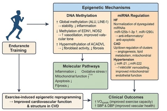
4.1. DNA Methylation

Two included studies [20,21] demonstrated that endurance training can induce changes in DNA methylation in individuals with HF and hypertension, respectively. Ferrari et al. [20] observed increased methylation in the repetitive elements ALU and LINE-1, which are markers of global DNA methylation, and decreased methylation of EDN1 and NOS2, genes related to inflammation and vascular tone. These changes were accompanied by improved VO2peak and lower SBP and DBP [20]. Reduced methylation of EDN1 and NOS2 suggests a potential increase in the expression of these genes, possibly enhancing vasodilation and reducing vascular resistance, which improves cardiovascular performance. Increased methylation in ALU and LINE-1 suggests greater epigenetic stability and reduced systemic inflammation, supported by a study from Newell–Price et al. [26], although their influence on VO2peak and BP still requires clarification. These results highlight that exercise-induced changes in DNA methylation may be associated with cardiovascular improvements by modulating specific gene expression involved in vascular function and inflammation.
Hsu et al. [21] demonstrated the importance of exercise-induced DNA methylation in HF patients. The identification of ACADVL as the most hypermethylated gene after HIIT is notable, given its role in mitochondrial fatty acid oxidation and energy regulation [27]. Usually, hypermethylation suppresses gene activity, which is associated with maladaptive cellular behaviour, but it appears to support the fibrotic process in this context. The in vitro knockdown experiment showed that inhibiting ACADVL impaired mitochondrial function and increased apoptotic markers (CASP3 and Cyto C) in cardiac fibroblasts, which are associated with decreased fibroblast activity and a possible reduction in myocardial fibrosis.
The hypermethylation of ACADVL in this case seemed to suppress excessive fibroblast activity, thus reducing myocardial fibrosis, which suggests a protective effect in the context of HF and facilitates structural remodelling. This dual evidence corroborates the findings of Ghazal et al. [28], who identified ACADVL as an epigenetic target involved in regulating fibrosis. Knottnerus et al. [27] emphasise the importance of regulated VLCAD expression levels, which determine the rate of ACADVL activity for a controlled remodelling process in HF.

4.2. MiRNA Expression

Exercise-induced changes in specific miRNAs were observed in four studies. They were identified as key regulators of inflammation, oxidative stress, mitochondrial function, and apoptosis in response to endurance training.
Mayr et al. [22] observed changes in miRNA expression following a single maximal ergospirometry exercise session in CAD patients. The 33 differentially regulated miRNAs influenced inflammation, angiogenesis, atherogenesis, hypoxia, lipid metabolism, and cardiovascular diseases. The literature explains that specific gene roles, such as the downregulation of miR-92a-3p, are associated with reduced endothelial dysfunction [29], while the upregulation of miR-223-3p, miR-146a-5p, miR-150-5p, and miR-23a-3p induces anti-inflammatory effects [30]. Let-7e-5P and Let-7f-5p are two miRNAs from the let-7 family involved in regulating inflammation, cellular processes, and protective functions [31]. These findings suggest that even a single exercise session can elicit acute molecular responses, demonstrating the sensitivity of miRNA to endurance exercise.
Yamada et al. [24] found that two dysregulated miRNAs in HF patients normalised after cardiac rehabilitation. Hsa-miR-125b-1-3p expression increased, which is linked to cellular stress, apoptosis, and inflammatory control [32], while hsa-miR-1290, known for its implication in cancer and inflammatory pathways [33], showed decreased expression. These findings support the idea that exercise-induced miRNA regulation has anti-inflammatory and anti-apoptotic effects [34]. The authors also observed the upregulation of hsa-miR-200c-3p and hsa-miR-3181, and the downregulation of hsa-miR-196b-3p. While hsa-miR-200c-3p was found to be associated with prolonged life expectancy [35], the current knowledge of the specific roles in cardiovascular adaptations of these miRNAs remains unclear. Their changes were associated with reduced SBP, but a direct link between changes in miRNA expression and clinical outcomes remains unknown.
Taurino et al. [23] demonstrated that their endurance intervention programme altered miRNA and gene expression profiles in CAD patients. Specific miRNAs hsa-miR-140-3p and hsa-miR-182 were upregulated in CAD, while the expression of their target genes related to vascular function was reduced [23]. After the rehabilitation programme, the upregulation of hsa-miR-92a and hsa-miR-92b was correlated with a decreased expression of mitochondrial and apoptotic genes, including COX7C, NDUFA1, ATP5I, and CASP3 [23]. Pathway analysis revealed oxidative phosphorylation and mitochondrial dysfunction as key modifiable processes, supported by Klimczak–Tomaniak et al. [36] and Ghazal et al. [28], who described oxidative stress and mitochondrial impairment as key features of cardiovascular pathology. These findings suggest that endurance exercise may be associated with changes in mitochondrial and apoptotic activity. This is supported by Pernaute et al. [37] and Schüttler et al. [38], who linked miRNA regulation of apoptosis with mitochondrial pathway activity and their contribution to reversing disease-related molecular changes, which underlines their role in the pathogenesis and progression of CAD.
Masoumi-Ardakani et al. [25] demonstrated that ET, MitoQ supplementation, and especially their combined effect, led to a significant downregulation of miR-21 and miR-222 in hypertensive individuals. The changes are most prominent in the ET and combined groups, suggesting that exercise may be the primary modulator of these miRNAs. miR-21 promotes vascular remodelling, oxidative stress, and inflammation pathways involving reactive oxygen species (ROS) and C-reactive protein (CRP), while miR-222 is linked to endothelial and mitochondrial dysfunction [25]. Their reduction suggests improvements in vascular health, mitochondrial activity, and inflammatory status. These findings are supported by Klimczak–Tomaniak et al. [36], who explain that oxidative stress is a key contributor to CVD, and by Ghazal et al. [28] and Schüttler et al. [38], who found that epigenetic regulation influences mitochondrial control, affecting endothelial function and vascular health. The concurrent findings that miR-21 was associated with a decrease in LV mass and LVESD in the combined groups highlight a potential link between miRNA modulation and structural cardiac improvements [25].

4.3. Clinical Parameters

Two studies reported outcomes on body composition [24,25]. While a decrease in BMI, body weight, and body fat percentage can be expected after following an endurance training programme, only Masoumi-Ardakani et al. [25] reported a significant decrease in those outcomes in hypertensive individuals, with the most pronounced effects in the ET group and combined intervention group, likely due to the longer intervention period of 6 weeks. This supports the association between structured endurance programmes and improving metabolic and body composition measurements, particularly when combined with MitoQ supplementation.
BP characteristics were reported in several studies. SBP decreased significantly in Yamada et al. [24], Ferrari et al. [20], and Masoumi-Ardakani et al. [25], while DBP decreased in Ferrari et al. [20] and in the combined intervention group of Masoumi-Ardakani et al. [25]. This suggests that adding antioxidant supplements might enhance the effect of endurance training on hypertension.
Studies by Ferrari et al. [20], Hsu et al. [21], and Masoumi-Ardakani et al. [25] reported outcomes on cardiac function and structure. Hsu et al. [21] and Ferrari et al. [20] observed significant improvements in VO2peak following HIIT and aerobic exercise training, respectively. Hsu et al. [21] found additional improvements in COex, LVEF, and reductions in end-systolic and end-diastolic volumes, which indicate improved contractility and reduced dilatation in HF patients. Additionally, Hsu et al. [21] reported a decrease in ECV and heart size, suggesting a reduction in myocardial fibrosis. Masoumi-Ardakani et al. [25] observed a decrease in LV mass and LVESD in the combined group which suggests significant structural remodelling in hypertensive individuals. These findings suggest a potential influence of endurance exercise on reverse cardiac remodelling in HF and hypertensive individuals.
Hsu et al. [21] and Masoumi-Ardakani et al. [25] reported on the systemic benefits of biochemical markers induced by exercise. Masoumi-Ardakani et al. [25] found a reduction in MDA and IL-6 and an increase in TAC, with the most pronounced effect in the combined intervention group. This suggests an overall improvement in oxidative and inflammatory status, aligning with the observed changes in cardiac structure and function. The study by Ghazal et al. [28] supports these findings and identifies IL-6 as a key regulator of fibroblast activity in cardiac fibrosis through exercise-induced epigenetic regulation. Hsu et al. [21] also observed an impact on cardiac stress via a reduction in BNP levels after HIIT, which reinforces the systemic benefits of endurance exercise in biochemical markers.

4.4. Quality and Limitations

All six included studies were limited by a relatively small sample size, with intervention groups ranging from 3 to 64 participants [20,21,22,23,24,25]. This reduces the statistical power of the findings and limits their generalisability, potentially resulting in several non-significant outcomes. None of the included articles provided long-term follow-up assessments, which prevented the interpretation of the longevity of the exercise-induced epigenetic and clinical effects. Heterogeneity among study populations, including three different pathologies, limits the comparison of results across the studies. Another source of heterogeneity is the variation in exercise protocols implemented across studies, including differences in exercise modality, intensity, or frequency, which further complicates the interpretation of outcomes and likely influences molecular and clinical outcomes. Several studies reported the use of concurrent pharmacological treatment, which could act as a confounding factor and reduce the ability to attribute effects exclusively to exercise training. [16,19,20]. Heterogeneity in CVD populations and exercise interventions limits direct comparisons and reduces the strength of conclusions. The impact of exercise training on epigenetic modifications may vary depending on disease severity, baseline medical treatment, and underlying pathophysiology. Consequently, generalisation of findings across all forms of CVD is limited.
Overall, heterogeneity in study designs, populations, interventions, and outcomes complicates the interpretation of results and limits the ability to draw conclusions or establish causal relationships. In the RCT [25], the combination of ET and MitoQ supplementation makes it difficult to isolate the effects of exercise alone on the outcomes. Variation in laboratory techniques used to extract and measure DNA methylation and miRNA expression may affect the consistency of molecular outcome assessments. Only two studies included a proper control group [23,24], which limits causal inference and reduces internal validity. While one study employed a randomised clinical trial design [25], the other five were quasi-experimental designs, which carry a higher risk of bias. The prevalence of quasi-experimental methods, coupled with the absence of randomisation and blinding, increases the likelihood of selection and performance bias. Flaws such as small sample sizes, inadequate or absent control groups, inconsistent methodologies, or concurrent medical treatments introduce bias, weaken the evidence, and hinder causal inference. Several studies also failed to establish a clear link between observed epigenetic changes and cardiovascular improvements, constraining the interpretation and clinical relevance of the findings.

4.5. Future Perspectives

Further research is needed to clarify the causal relationship between exercise-induced epigenetic changes and cardiovascular adaptations. While associations between miRNA expression, DNA methylation changes, and positive cardiovascular outcomes have been observed, several causal inferences are based on supporting evidence from additional literature rather than direct outcomes within the included studies. Additional relevant cardiovascular parameters, such as heart rate reserve or stroke volume, are seldom reported despite their importance in physiological adaptation. The current literature remains largely centred on animal models. Larger clinical studies, including those involving human populations, are needed to translate and validate these findings and to better understand their clinical significance for improving cardiovascular outcomes in individuals with CVD.

5. Conclusions

This systematic review emphasises the beneficial effect of endurance training on epigenetic markers, such as miRNA expression and DNA methylation, as well as on cardiovascular structure and function in individuals with various CVDs. Results show both up- and downregulation of specific miRNAs and different patterns of DNA methylation, which are associated with pathways involved in inflammation, fibrosis, mitochondrial function, and oxidative stress. These molecular changes may be associated with improvements in BP, VO2peak, LV function, and myocardial fibrosis. However, most studies were unable to establish a clear causal link between epigenetic modifications and clinical outcomes. The small sample sizes and lack of long-term follow-up highlight the need for future studies with larger samples and stronger methodological designs that directly investigate the therapeutic potential of exercise-induced epigenetic changes.

Supplementary Materials

The following supporting information can be downloaded at: https://www.mdpi.com/article/10.3390/cardiogenetics15040028/s1, Articles S1–S6: JBI critical appraisal.

Author Contributions

Conceptualisation, J.D. and J.O.; methodology, J.D., J.O. and R.M.d.A.; investigation, J.D. and J.O.; resources, J.D. and J.O.; writing—original draft preparation, J.D., J.O. and C.C.; writing—review and editing, J.D., J.O., R.M.d.A., F.X.d.A. and C.C.; visualisation, J.D. and J.O.; supervision, C.C.; project administration, J.D., J.O., F.X.d.A. and C.C. All authors have read and agreed to the published version of the manuscript.

Funding

This research received no external funding.

Institutional Review Board Statement

Not applicable.

Data Availability Statement

No new data were created or analysed in this study. Data sharing is not applicable to this article. Extracted data, search strategies, and other materials are included in full within this thesis and Supplementary Materials.

Conflicts of Interest

The authors declare no conflicts of interest.

References

  1. Olvera Lopez, E.; Ballard, B.D.; Jan, A. Cardiovascular Disease. In StatPearls; StatPearls Publishing: Treasure Island, FL, USA, 2025. Available online: http://www.ncbi.nlm.nih.gov/books/NBK535419/ (accessed on 16 June 2025).
  2. Visseren, F.L.J.; Mach, F.; Smulders, Y.M.; Carballo, D.; Koskinas, K.C.; Bäck, M.; Benetos, A.; Biffi, A.; Boavida, J.-M.; Capodanno, D.; et al. 2021 ESC Guidelines on cardiovascular disease prevention in clinical practice: Developed by the Task Force for cardiovascular disease prevention in clinical practice with representatives of the European Society of Cardiology and 12 medical societies With the special contribution of the European Association of Preventive Cardiology (EAPC). Eur. Heart J. 2021, 42, 3227–3337. [Google Scholar] [CrossRef]
  3. Morici, G.; Gruttad’Auria, C.I.; Baiamonte, P.; Mazzuca, E.; Castrogiovanni, A.; Bonsignore, M.R. Endurance training: Is it bad for you? Breathe Sheff. Engl. 2016, 12, 140–147. [Google Scholar] [CrossRef]
  4. Isath, A.; Koziol, K.J.; Martinez, M.W.; Garber, C.E.; Martinez, M.N.; Emery, M.S.; Baggish, A.L.; Naidu, S.S.; Lavie, C.J.; Arena, R.; et al. Exercise and cardiovascular health: A state-of-the-art review. Prog. Cardiovasc. Dis. 2023, 79, 44–52. [Google Scholar] [CrossRef]
  5. Huang, G.; Gibson, C.A.; Tran, Z.V.; Osness, W.H. Controlled Endurance Exercise Training and VO2max Changes in Older Adults: A Meta-Analysis. Prev. Cardiol. 2005, 8, 217–225. [Google Scholar] [CrossRef]
  6. Wilmore, J.H.; Knuttgen, H.G. Aerobic Exercise and Endurance: Improving Fitness for Health Benefits. Phys. Sportsmed. 2003, 31, 45–51. [Google Scholar] [CrossRef] [PubMed]
  7. Seo, D.Y.; Kwak, H.-B.; Kim, A.H.; Park, S.H.; Heo, J.W.; Kim, H.K.; Ko, J.R.; Lee, S.J.; Bang, H.S.; Sim, J.W.; et al. Cardiac adaptation to exercise training in health and disease. Pflugers Arch. 2020, 472, 155–168. [Google Scholar] [CrossRef]
  8. Soci, U.P.R.; Melo, S.F.S.; Gomes, J.L.P.; Silveira, A.C.; Nóbrega, C.; de Oliveira, E.M. Exercise Training and Epigenetic Regulation: Multilevel Modification and Regulation of Gene Expression. Adv. Exp. Med. Biol. 2017, 1000, 281–322. [Google Scholar] [CrossRef] [PubMed]
  9. Radak, Z.; Pan, L.; Zhou, L.; Mozaffaritabar, S.; Gu, Y.; Pinho, R.A.; Zheng, X.; Ba, X.; Boldogh, I. Epigenetic and “redoxogenetic” adaptation to physical exercise. Free Radic. Biol. Med. 2024, 210, 65–74. [Google Scholar] [CrossRef]
  10. Wu, G.; Zhang, X.; Gao, F. The epigenetic landscape of exercise in cardiac health and disease. J. Sport Health Sci. 2021, 10, 648–659. [Google Scholar] [CrossRef] [PubMed]
  11. Plaza-Diaz, J.; Izquierdo, D.; Torres-Martos, Á.; Baig, A.T.; Aguilera, C.M.; Ruiz-Ojeda, F.J. Impact of Physical Activity and Exercise on the Epigenome in Skeletal Muscle and Effects on Systemic Metabolism. Biomedicines 2022, 10, 126. [Google Scholar] [CrossRef]
  12. Fernandes, T.; Baraúna, V.G.; Negrão, C.E.; Phillips, M.I.; Oliveira, E.M. Aerobic exercise training promotes physiological cardiac remodeling involving a set of microRNAs. Am. J. Physiol. Heart Circ. Physiol. 2015, 309, H543–H552. [Google Scholar] [CrossRef]
  13. Fulghum, K.; Hill, B.G. Metabolic Mechanisms of Exercise-Induced Cardiac Remodeling. Front. Cardiovasc. Med. 2018, 5, 127. [Google Scholar] [CrossRef]
  14. Gibb, A.A.; Hill, B.G. Metabolic Coordination of Physiological and Pathological Cardiac Remodeling. Circ. Res. 2018, 123, 107–128. [Google Scholar] [CrossRef]
  15. Sun, Y.; Peng, Z.; Liang, H. Role of physical activity in cardiovascular disease prevention: Impact of epigenetic modifications. Front. Cardiovasc. Med. 2025, 12, 1511222. [Google Scholar] [CrossRef]
  16. Page, M.J.; McKenzie, J.E.; Bossuyt, P.M.; Boutron, I.; Hoffmann, T.C.; Mulrow, C.D.; Shamseer, L.; Tetzlaff, J.M.; Akl, E.A.; Brennan, S.E.; et al. The PRISMA 2020 statement: An updated guideline for reporting systematic reviews. BMJ 2021, 372, n71. [Google Scholar] [CrossRef]
  17. Ouzzani, M.; Hammady, H.; Fedorowicz, Z.; Elmagarmid, A. Rayyan-a web and mobile app for systematic reviews. Syst. Rev. 2016, 5, 210. [Google Scholar] [CrossRef]
  18. Barker, T.H.; Stone, J.C.; Sears, K.; Klugar, M.; Tufanaru, C.; Leonardi-Bee, J.; Aromataris, E.; Munn, Z. The revised JBI critical appraisal tool for the assessment of risk of bias for randomized controlled trials. JBI Evid. Synth. 2023, 21, 494–506. [Google Scholar] [CrossRef] [PubMed]
  19. Barker, T.H.; Habibi, N.; Aromataris, E.; Stone, J.C.; Leonardi-Bee, J.; Sears, K.; Hasanoff, S.; Klugar, M.; Tufanaru, C.; Moola, S.; et al. The revised JBI critical appraisal tool for the assessment of risk of bias for quasi-experimental studies. JBI Evid. Synth. 2024, 22, 378–388. [Google Scholar] [CrossRef] [PubMed]
  20. Ferrari, L.; Vicenzi, M.; Tarantini, L.; Barretta, F.; Sironi, S.; Baccarelli, A.A.; Guazzi, M.; Bollati, V. Effects of Physical Exercise on Endothelial Function and DNA Methylation. Int. J. Environ. Res. Public Health 2019, 16, 2530. [Google Scholar] [CrossRef] [PubMed]
  21. Hsu, C.-C.; Wang, J.-S.; Shyu, Y.-C.; Fu, T.-C.; Juan, Y.-H.; Yuan, S.-S.; Wang, C.-H.; Yeh, C.-H.; Liao, P.-C.; Wu, H.-Y.; et al. Hypermethylation of ACADVL is involved in the high-intensity interval training-associated reduction of cardiac fibrosis in heart failure patients. J. Transl. Med. 2023, 21, 187. [Google Scholar] [CrossRef]
  22. Mayr, B.; Müller, E.E.; Schäfer, C.; Droese, S.; Schönfelder, M.; Niebauer, J. Exercise-induced changes in miRNA expression in coronary artery disease. Clin. Chem. Lab. Med. 2021, 59, 1719–1727. [Google Scholar] [CrossRef]
  23. Taurino, C.; Miller, W.H.; McBride, M.W.; McClure, J.D.; Khanin, R.; Moreno, M.U.; Dymott, J.A.; Delles, C.; Dominiczak, A.F. Gene expression profiling in whole blood of patients with coronary artery disease. Clin. Sci. Lond. Engl. 1979 2010, 119, 335–343. [Google Scholar] [CrossRef]
  24. Yamada, R.; Okumura, S.; Kono, Y.; Miyazaki, A.; Niwa, Y.; Ito, T.; Ueda, S.; Ishiguro, T.; Yoshinaga, M.; Fujiwara, W.; et al. Effect of cardiac rehabilitation on circulating microRNA expressknown for its implication inion in heart failure: A preliminary study. Fujita Med. J. 2021, 7, 76–82. [Google Scholar] [CrossRef] [PubMed]
  25. Masoumi-Ardakani, Y.; Najafipour, H.; Nasri, H.R.; Aminizadeh, S.; Jafari, S.; Safi, Z. Moderate Endurance Training and MitoQ Improve Cardiovascular Function, Oxidative Stress, and Inflammation in Hypertensive Individuals: The Role of miR-21 and miR-222: A Randomized, Double-Blind, Clinical Trial. Cell J. 2022, 24, 577–585. [Google Scholar] [CrossRef] [PubMed]
  26. Newell-Price, J.; Clark, A.J.L.; King, P. DNA Methylation and Silencing of Gene Expression. Trends Endocrinol. Metab. 2000, 11, 142–148. [Google Scholar] [CrossRef] [PubMed]
  27. Knottnerus, S.J.G.; Bleeker, J.C.; Ferdinandusse, S.; Houtkooper, R.H.; Langeveld, M.; Nederveen, A.J.; Strijkers, G.J.; Visser, G.; Wanders, R.J.A.; Wijburg, F.A.; et al. Subclinical effects of long-chain fatty acid β-oxidation deficiency on the adult heart: A case-control magnetic resonance study. J. Inherit. Metab. Dis. 2020, 43, 969–980. [Google Scholar] [CrossRef]
  28. Ghazal, R.; Wang, M.; Liu, D.; Tschumperlin, D.J.; Pereira, N.L. Cardiac Fibrosis in the Multi-Omics Era: Implications for Heart Failure. Circ. Res. 2025, 136, 773–802. [Google Scholar] [CrossRef]
  29. Shang, F.; Wang, S.-C.; Hsu, C.-Y.; Miao, Y.; Martin, M.; Yin, Y.; Wu, C.-C.; Wang, Y.-T.; Wu, G.; Chien, S.; et al. MicroRNA-92a Mediates Endothelial Dysfunction in CKD. J. Am. Soc. Nephrol. JASN 2017, 28, 3251–3261. [Google Scholar] [CrossRef]
  30. Das, D.; Kumar, A.; Sharma, M. A systematic review of work-related musculoskeletal disorders among handicraft workers. Int. J. Occup. Saf. Ergon. JOSE 2020, 26, 55–70. [Google Scholar] [CrossRef]
  31. Bernstein, D.L.; Jiang, X.; Rom, S. let-7 microRNAs: Their Role in Cerebral and Cardiovascular Diseases, Inflammation, Cancer, and Their Regulation. Biomedicines 2021, 9, 606. [Google Scholar] [CrossRef]
  32. Wang, Y.; Tan, J.; Wang, L.; Pei, G.; Cheng, H.; Zhang, Q.; Wang, S.; He, C.; Fu, C.; Wei, Q. MiR-125 Family in Cardiovascular and Cerebrovascular Diseases. Front. Cell Dev. Biol. 2021, 9, 799049. [Google Scholar] [CrossRef] [PubMed]
  33. Guz, M.; Jeleniewicz, W.; Cybulski, M. An Insight into miR-1290: An Oncogenic miRNA with Diagnostic Potential. Int. J. Mol. Sci. 2022, 23, 1234. [Google Scholar] [CrossRef]
  34. Xu, H.; Cui, Y.; Liu, X.; Zheng, X.; Liu, J.; Hu, X.; Gao, F.; Hu, X.; Li, M.; Wei, X.; et al. miR-1290 promotes IL-8-mediated vascular endothelial cell adhesion by targeting GSK-3β. Mol. Biol. Rep. 2022, 49, 1871–1882. [Google Scholar] [CrossRef]
  35. miRBase Entry: hsa-mir-200c. Available online: https://mirbase.org/hairpin/MI0000650?acc=MI0000650 (accessed on 20 June 2025).
  36. Klimczak-Tomaniak, D.; Haponiuk-Skwarlińska, J.; Kuch, M.; Pączek, L. Crosstalk between microRNA and Oxidative Stress in Heart Failure: A Systematic Review. Int. J. Mol. Sci. 2022, 23, 15013. [Google Scholar] [CrossRef] [PubMed]
  37. Pernaute, B.; Spruce, T.; Smith, K.M.; Sánchez-Nieto, J.M.; Manzanares, M.; Cobb, B.; Rodríguez, T.A. MicroRNAs control the apoptotic threshold in primed pluripotent stem cells through regulation of BIM. Genes Dev. 2014, 28, 1873–1878. [Google Scholar] [CrossRef] [PubMed]
  38. Schüttler, D.; Clauss, S.; Weckbach, L.T.; Brunner, S. Molecular Mechanisms of Cardiac Remodeling and Regeneration in Physical Exercise. Cells 2019, 8, 1128. [Google Scholar] [CrossRef] [PubMed]
Figure 1. PRISMA flow diagram.
Cardiogenetics 15 00028 g001
Disclaimer/Publisher’s Note: The statements, opinions and data contained in all publications are solely those of the individual author(s) and contributor(s) and not of MDPI and/or the editor(s). MDPI and/or the editor(s) disclaim responsibility for any injury to people or property resulting from any ideas, methods, instructions or products referred to in the content.

