Highlights
What are the main findings?
- We found that bidirectional changes (an increase in the nuclear fraction and a decrease in the mitochondrial fraction) in patients with type 2 diabetes mellitus are associated with adverse cardiac remodeling, renal damage and inflammation.
- We found a superior discriminatory ability in cell-free nuclear DNA compared with N-terminal brain natriuretic peptide for heart failure in patients with type 2 diabetes mellitus.
- They may improve the conventional predictive models for the development of any phenotype of chronic heart failure in diabetics.
- They suggest a new methodology for the identification of patients and the application of early treatment responses through serial measuring of circulating levels of cell-free-DNA.
Abstract
Cell-free nuclear (cf-nDNA) and mitochondrial (cf-mDNA) DNA are released from damaged cells in type 2 diabetes mellitus (T2DM) patients, contributing to adverse cardiac remodeling, vascular dysfunction, and inflammation. The purpose of this study was to correlate the presence and type of cf-DNAs with HF in T2DM patients. A total of 612 T2DM patients were prescreened by using a local database, and 240 patients (120 non-HF and 120 HF individuals) were ultimately selected. The collection of medical information, including both echocardiography and Doppler imagery, as well as the assessment of biochemistry parameters and the circulating biomarkers, were performed at baseline. The N-terminal brain natriuretic pro-peptide (NT-proBNP) and cf-nDNA/cf-mtDNA levels were measured via an ELISA kit and real-time quantitative PCR tests, respectively. We found that HF patients possessed significantly higher levels of cf-nDNA (9.9 ± 2.5 μmol/L vs. 5.4 ± 2.7 μmol/L; p = 0.04) and lower cf-mtDNA (15.7 ± 3.3 μmol/L vs. 30.4 ± 4.8 μmol/L; p = 0.001) than those without HF. The multivariate log regression showed that the discriminative potency of cf-nDNA >7.6 μmol/L (OR = 1.07; 95% CI = 1.03–1.12; p = 0.01) was higher that the NT-proBNP (odds ratio [OR] = 1.10; 95% confidence interval [CI] = 1.04–1.19; p = 0.001) for HF. In conclusion, we independently established that elevated levels of cf-nDNA, originating from NT-proBNP, were associated with HF in T2DM patients.
1. Introduction
Type 2 diabetes mellitus (T2DM) is one of most significant risk factors of heart failure (HF) in the general population [1]. The Framingham Heart Study revealed that T2DM independently increases the risk of HF up to twofold and fivefold in men and women, respectively, when compared with age-matched controls [2]. With respect to this, patients with known HF demonstrate a two- to threefold increase in risk in regard to developing T2DM [3]. The absolute risk for developing HF in younger patients with T2DM was sufficiently higher than that observed in older participants with diabetes (14% vs. 7%) included in the pooled population of the Framingham Heart Study, the Prevention of Renal and Vascular End-stage Disease Study, and the Multi-Ethnic Study of Atherosclerosis [4].
By possessing a complex overlap in the underlying pathophysiological mechanisms—oxidative stress, the metabolic memory phenomenon, glucose and lipid toxicity, hyperinsulinemia, insulin resistance, mitochondrial dysfunction, accelerating atherosclerosis, altered endothelial tone regulation, and myocardial ischemia/necrosis—T2DM and HD exacerbate both cardiac and vascular remodeling. Furthermore, each also significantly worsens the prognosis of the other [5,6]. Despite well-developed approaches to antidiabetic therapies, a persistence of hyperglycemia in T2DM patients continues to be a factor, which links to progression in the metabolic abnormalities with adverse cardiac remodeling, thus resulting in the high prevalence of HF in T2DM patients [7,8]. Glycemic variability, along with additional cardiovascular risk (CV) factors beyond hyperglycemia, may contribute to an increased HF risk and mortality in T2DM through several molecular mechanisms, including oxidative stress-mediated neutrophil extracellular traps (NET) [9,10,11].
The immune milieu concerning the occurrence and natural evolution of HF in T2DM patients include mitochondrial-mediated necrosis, autophagy-dependent cell death, apoptosis/ferroptosis, and immunogenic cell death, as well as cardiac and microvascular inflammation, which partially result from NET [12]. The most important factors contributing to cardiac myocyte death, apoptosis and necrosis, ischemia/reperfusion injury, extracellular matrix remodeling, and endothelial dysfunction—which together play a crucial role in the development of HF in T2DM—are released products, such as inflammatory cytokines, chemokines, oxidative stress components, and cell-free DNAs [11,12]. Although the roles of immunogenic reactions in regards to adverse cardiac remodeling with respect to HF with concomitant T2DM is being actively investigated, its methods are not yet fully understood.
Moreover, cell-free DNAs (cf-DNAs) are extracellular circulating fragments of DNA, derived from numerous cells under physiological and pathological conditions [13]. The current nomenclature of cf-DNAs includes both the nuclear (n-cfDNA) and mitochondrial (mt-cfDNA) variants, which exert their own function and possess different molecular structures [14]. In addition, NET is a common process that corresponds to the occurrence of cf-DNA in circulation, mainly by playing a crucial role in the host defense and innate immune reaction via its regulation of complement activities [15]. Moreover, NET is involved in the pathogenesis of transplant rejection, autoimmunity, cancer metastasis, acute myocardial infarction, and sepsis [16,17]. Along with these, cf-DNA can originate not only from activated mononuclear cells—which are involved in NET—but also from circulating pathogens, gut microbiota, cancer cells, and other human cells originating damaged organs [14]. However, there is a large quantity of evidence indicating that NET is induced by hyperglycemia, oxidized lipids, and inflammatory cytokines. This is in addition to the fact that it contributes to the pathogenesis of diabetes and its complications, such as microvascular inflammation, accelerating atherosclerosis, and thrombosis [18,19]. Indeed, among T2DM patients NET products such as extracellular fragments of DNA were found to be significantly elevated. Further, their concentrations were correlated with the presence of diabetes nephropathy and CV disease [20]. It is important to note that there are two subpopulations of circulating cf-DNAs—nuclear (cf-nDNA) and mitochondrial (cf-mtDNA)—which differ in their correspondence to NET [14]. Cf-nDNA characterizes apoptosis and necrosis in close connection with NET activity, whereas cell-free mitochondrial DNA is an indicator of the non-selective permeability of cell membranes, which is due to oxidative stress and mitochondrial dysfunction [20,21,22]. There is a large amount of evidence relating to the fact that the progression of target organ damage in numerous diseases, including T2DM, is closely related to the circulating levels and clearance of cf-DNA [14].
In contrast to T2DM, the role of NET with respect to HF pathogenesis appears to be controversial. Indeed, in acute ischemia-induced HF, damage-related molecular patterns may intervene in pattern recognition receptors in order to cause NETs and worse myocardial perfusion. However, it is still uncertain whether products of NET, such as cf-DNAs, directly contribute to the adverse cardiac remodeling in T2DM patients [23]. Although there are limited data regarding the discriminative potency of cf-DNAs among patients with HF, cf-DNAs serve not only as a cause of T2DM-related complications, but also as a marker for the risk of HF [24,25]. As such, the purpose of this study was to correlate the presence and type of cf-DNAs with HF in T2DM patients.
2. Materials and Methods
2.1. Study Patients
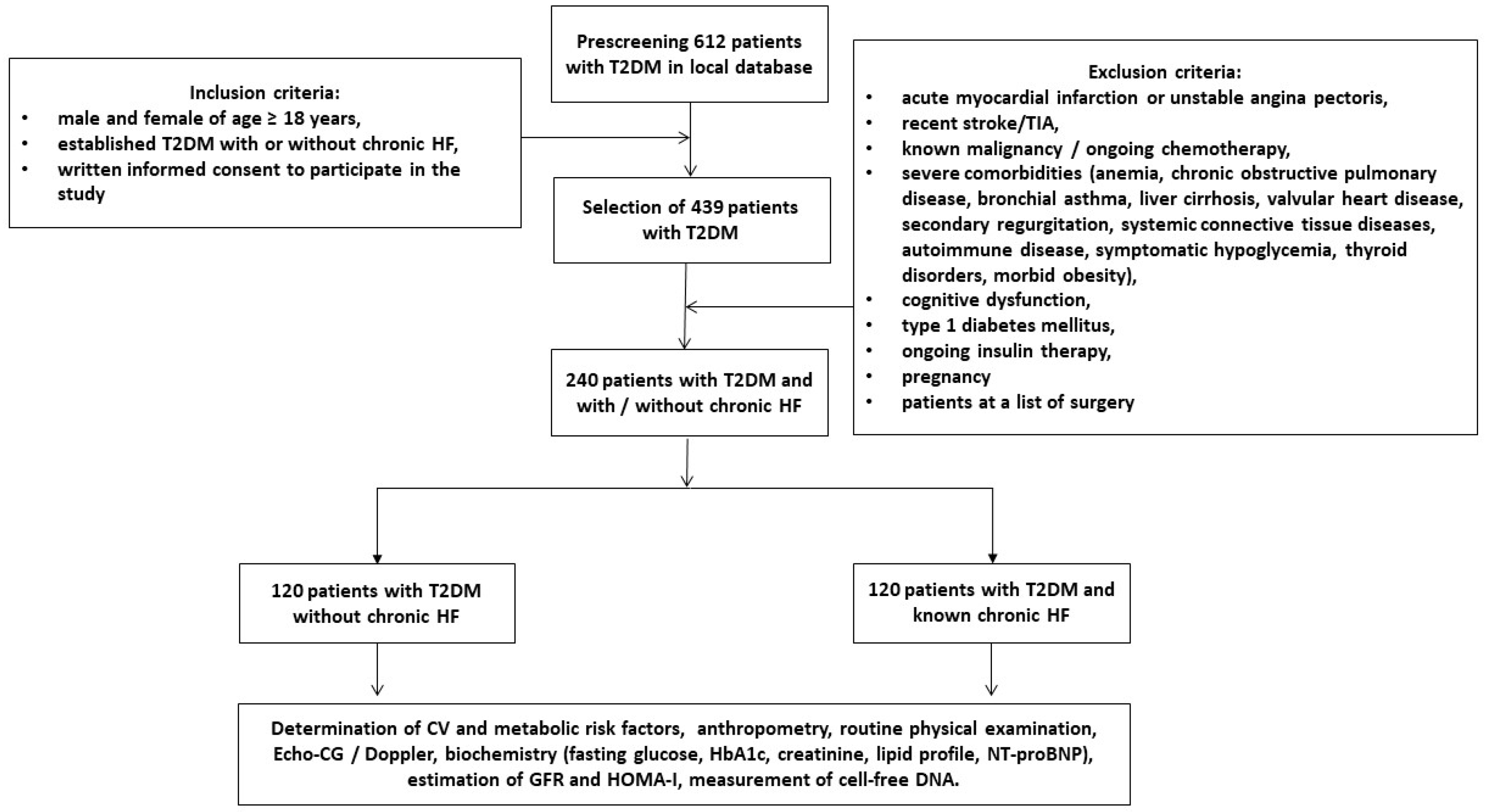
A total of 612 patients with T2DM were prescreened using a local database from the Vita Center (Zaporozhye, Ukraine). By using specific inclusion criteria (i.e., male/female with an age of ≥18 years; with established T2DM; with or without any phenotypes of chronic HF; as well as informed consent to participate in the study), we enrolled 439 patients with T2DM, with and without chronic HF (Figure 1). The exclusion criteria were: acute coronary syndrome/myocardial infarction or unstable angina pectoris, recent stroke/transient ischemic attack, known malignancy, severe co-morbidities (anemia, chronic lung and liver diseases, known inherited and acquired heart defects, symptomatic severe hypoglycemia, morbid obesity, systemic connective tissue diseases, autoimmune disease, cognitive dysfunction, and thyroid disorders), pregnancy, and type 1 diabetes mellitus or current insulin therapy. Finally, we selected 240 patients with T2DM and divided them into two cohorts, depending on whether or not chronic HF was present.
Figure 1.
Study design and procedure flow chart. Abbreviations—HF: heart failure; HbA1c: glycated hemoglobin; HOMA-IR: homeostatic assessment model of insulin resistance; NT-proBNP: N-terminal brain natriuretic pro-peptide; T2DM: type 2 diabetes mellitus; and TIA: transient ischemic attack.
2.2. Medical Information Collection
The basic clinical data (age, gender, height, weight, waist circumference, hip-to-waist ratio (WHR), body mass index (BMI), and body surface area (BSA)), comorbidities (hypertension, diabetes history, dyslipidemia, etc.), and smoking status were all collected. Microalbuminuria was defined as albumin urine excretion in the range of 30–299 mg/g of creatinine [26]. Furthermore, the T2DM and HF status were established according to conventional clinical recommendations [27,28]. The European Society of Cardiology’s (ESC) clinical guidelines were used in order to determine hypertension [29], dyslipidemia [30], and coronary artery disease [31]. Chronic kidney disease in the T2DM patients was detected in accordance with the Kidney Disease Improving Global Outcomes (KDIGO) Consensus Report [32].
2.3. Examination of Hemodynamics
The B-mode echocardiography and Doppler examinations of the patients were performed by a blinded ultrasonographer using the Vivid T8 (“GE Medical Systems”, Freiburg, Germany) diagnostic system in compliance with current guidelines [33,34]. The left ventricular end-diastolic (LVEDV) and end-systolic (LVESV) volumes, as well as the left atrial volume (LAV), were measured in the apical 4-chamber view. Moreover, the LAV index (LAVI) was calculated as a ratio of LAV to BSA. The left ventricular (LV) ejection fraction (LVEF) was estimated by the Simpson method. In addition, the diastolic parameters were also calculated, included early diastolic blood filling (E), as well as the longitudinal strain ratio (è) and their combined ratio (E/è). Furthermore, the estimated E/è ratio was expressed as the ratio equation of the E wave velocity to an averaged medial and lateral e’ velocity [34]. Instances of left ventricular hypertrophy (LVH) were detected according to conventional recommendations [34], which used the LV myocardial mass index (LVMMI) ≥125 g/m2 or ≥110 g/m2, as a marker of LVH, in both males and females, respectively.
2.4. Blood Sampling and Biomarker Measures
The patients’ fasting blood samples were collected from an antecubital vein (3–5 mL) and maintained at 4 °C. After centrifugation (3000 r/min, 30 min), the polled serum aliquots were immediately stored at ≤−70 °C until required for analysis. The serum concentrations of NT-proBNP and the high-sensitivity C-reactive protein (hs-CRP) were determined via commercially available enzyme-linked immunosorbent assay (ELISA) kits (Elabscience, Houston, TX, USA), according to the manufacturer’s instructions. All ELISA data were analyzed according to the standard curve; additionally, each sample was measured in duplicate, and the mean value was finally analyzed. Both the intra- and inter-assay coefficient of variability for each biomarker were <10%.
Conventional biochemistry parameters were routinely measured at the local biochemical laboratory of the Vita Center (Zaporozhye, Ukraine) using a Roche P800 analyzer (Basel, Switzerland). In addition, we used the CKD-EPI formula in order to estimate the glomerular filtration rate (GFR) [35]. Moreover, the insulin resistance was evaluated by using the homeostatic assessment model of insulin resistance (HOMA-IR) [36].
2.5. Cell-Free DNA Extraction
In this study, we isolated the cell-free DNA from the 4 mL plasma samples by applying the Biosystems MagMAX Cell-Free DNA Kit (Thermo Fisher Scientific, Wien, Austria), according to the manufacturer’s instructions. Plasma samples were received from the EDTA whole blood samples through two steps of centrifugation at 4 °C for each sample. The first step included centrifugation at 2000× g for 10 min. The plasma samples were collected and transferred into silicon tubes for a second centrifugation (20,000× g at 4 °C, for 5 min) in order to thoroughly remove the cell debris. Then, the supernatant was pooled and eluted in a 2 mL Tris-EDTA-buffer. Next, it was quantified with a Nanodrop (ND-1000 Spectrophotometer v 3.7.1, Waltham, MA, USA) device via spectrophotometric analysis at 260/280 nm.
2.6. Measurement of Cell-Free DNAs in Plasma Samples
Real-time quantitative PCR (qPCR) assays targeting the human GAPDH (glyceraldehyde 3-phosphate dehydrogenase) gene (gene ID 2597) for the purposes of determining cf-nDNA and the mitochondrial ATPase 8 gene (ID 4509) for cf-mtDNA were used for measurement of the concentration of cell-free DNA. The length of the amplicons, which were selected for the evaluation of cf-nDNA, was 97 and 229 bp, respectively. Regarding cf-mtDNA, 78 and 218 bp lengths were used. In order to estimate the fragmentation of cf-nDNA and cf-mtDNA, we used a ratio between the longer and the shorter amplicons. The qPCR reactions were carried out using SYBR Green Technology (Thermo Fisher Scientific, Waltham, MA, USA). The sequences of the primers for the nDNA were the following: forward primer—CCCCACACACATGCACTTACC and reverse primer—ATCAAACTCAAAGGGCAGGA; the forward primer for mtDNA—AATATTAAACACAAACTACC, and the reverse primer for mtDNA—TGGGTGGTGATTAGTCGGTTG. In addition, we used 20 μL of the reaction volume in total, which comprised: 0.1 mL of TaqMan1Universal PCR Master Mix (Applied Biosystems, Branchburg, NJ, USA); 0.5 mL of ultra-clear water; 0.25 μL of each of the primers (Sigma-Aldrich, St. Louis, MO, USA); 1 μL of a FAM-labeled MT-ATP 8-probe; 1 μL of a MVIC-labeled GAPDH probe; and 2 μL of Tris-EDTA buffer containing cell-free DNA isolated from plasma. The concentrations of primers and probes in the reaction volume were 0.6 μmol/L and 0.4 μmol/L, respectively. The negative control was 2 μL of Tris–EDTA buffer. The measures were provided with the 7500 HT Real-time PCR System (Applied Biosystems, Branchburg, NJ, USA) using the high-resolution melt software v. 2.0 (Applied Biosystems, Branchburg, NJ, USA), according to conventional methods [37].
2.7. Statistical Analysis
Statistical analysis was executed by using SPSS 11.0 for Windows and the v. 9 Graph Pad Prism (Graph Pad Software, San Diego, CA, USA). In addition, the continuous variables were expressed as the means (M) ± SD for the parametric data, median (Me), and interquartile range [IQR], according to whether or not they were normally distributed. Furthermore, the Kolmogorov–Smirnov test was used to check for normal distribution. Moreover, the distribution of dichotomous values was assessed with the Chi-square test. We also performed a t-test and a one-way analysis of variance (ANOVA), in conjunction with the Tukey test, for the purposes of obtaining the comparisons of two or three variables between cohorts, respectively. In addition, Spearman’s correlation coefficient (r) was used for determining the correlations between the levels of cf-nDNA/cf-mtDNA and other parameters. The predictors of HF were determined by univariate and multivariate logistic regression analysis. We reported an odds ratio (OR) and 95% confidence interval (95% CI) for each variable included in the regression analysis. The predictive value of cf-nDNA for HF was reclassified using the integrated discrimination indices (IDI) and net reclassification improvement (NRI). The differences were considered significant with a level of statistical significance of p < 0.05.
3. Results
3.1. General Patient Characteristics
The entire patient population was composed of 240 patients (136 male, i.e., 56.7%, and 104 female, i.e., 43.3%) with an age average of 52 years (Table 1). The mean values with respect to the body mass index (BMI), waist circumference, and waist-to-hip ratio (WHR) were 25.2 ± 2.9 kg/m2, 96.4 ± 3.7 cm, and 0.86 ± 0.06 units, respectively. The male patients possessed a higher waist circumference (98.6 ± 3.0 cm) and WHR (0.87 ± 0.04 units) than the female patients (95.1 ± 3.5 cm and 0.84 ± 0.05 units), respectively, with a p = 0.02 and p = 0.01. The cardiovascular risk profile included: dyslipidemia (81.7%); hypertension (63.3%); stable coronary artery disease (33.8%); smoking (40.8%); abdominal obesity (45.4%); left hypertrophy (LV) (80.8%); chronic kidney disease, grades 1–3 (25.8%); and microalbuminuria (18.8%). Among the entire patient population, we noted the following medical traits: 7.1% possessed a background with paroxysmal or persistent atrial fibrillation (AF), 15.8% possessed HF with a preserved ejection fraction (HFpEF), 15.4% possessed HF with a mildly reduced ejection fraction (HFmrEF), and 18.8% possessed HF with a reduced ejection fraction (HFrEF). Moreover, 33.8% of the patients demonstrated a I/II HF NYHA class, and 16.65% indicated a III HF NYHA class. All of the patients were hemodynamically stable and possessed an average LVEF that was equal to 55% (46–65%). In addition, the patients possessed a left ventricular (LV) myocardial mass index (LVMMI) of 126 ± 9 g/m2 and a left atrial volume index (LAVI) of 39 mL/m2 (33–47 mL/m2). Along with above, the mean fasting glucose concentration, HbA1c, as well as the circulating level of NT-proBNP and hs-CRP, were 6.09 mmol/L, 6.4%, 1043 pmol/mL, and 4.06 mg/L, respectively.

Table 1.
The baseline general characteristics of eligible T2DM patients.
There were no significant differences between the cohorts of patients in terms of age, gender, BMI, waist circumference, WHR (including waist circumference and WHR by gender). Nor were there any significant differences with respect to: the presence of dyslipidemia, hypertension, smoking, abdominal obesity, LV hypertrophy, microalbuminuria, systolic and diastolic blood pressure, HOMA-IR, HbA1c, and high-density lipoprotein cholesterol (HDL-C). On the contrary, patients from the HF cohort frequently possessed AF (p = 0.01), CKD grades 1–3 (p = 0.02), and a stable CAD (p = 0.04). A total of 67.5% and 32.5% of these patients demonstrated the I/II HF NYHA class and III HF NYHA class, respectively. However, instances of HFpEF, HfmrEF, and HFrEF were detected in 32.0%, 30.8%, and 37.5% of patients, respectively. These patients possessed significantly lower LVEF and eGFR, as well as higher cardiac volumes, LVMMI, LAVI, E/è, levels of creatinine, low-density lipoprotein cholesterol (LDL-C), triglycerides, hs-CRP, and NT-proBNP than those without HF. The patients from both cohorts received conventional therapies, which were adjusted according to their concomitant disease presence. In fact, the patients from the HF cohort frequently received ACE inhibitors, angiotensin receptor neprilysin inhibitors, beta-blockers, SGLT2 inhibitors, the i/f blocker ivabradine, calcium channel blockers, mineralocorticoid receptor antagonists, loop diuretics, and antiplatelet/anticoagulants. However, it must be noted that there were significant differences in terms of prescription frequency between these cohorts regarding angiotensin-II receptor blockers, statins, and metformin.
3.2. The Levels of Circulating Cell-Free DNAs
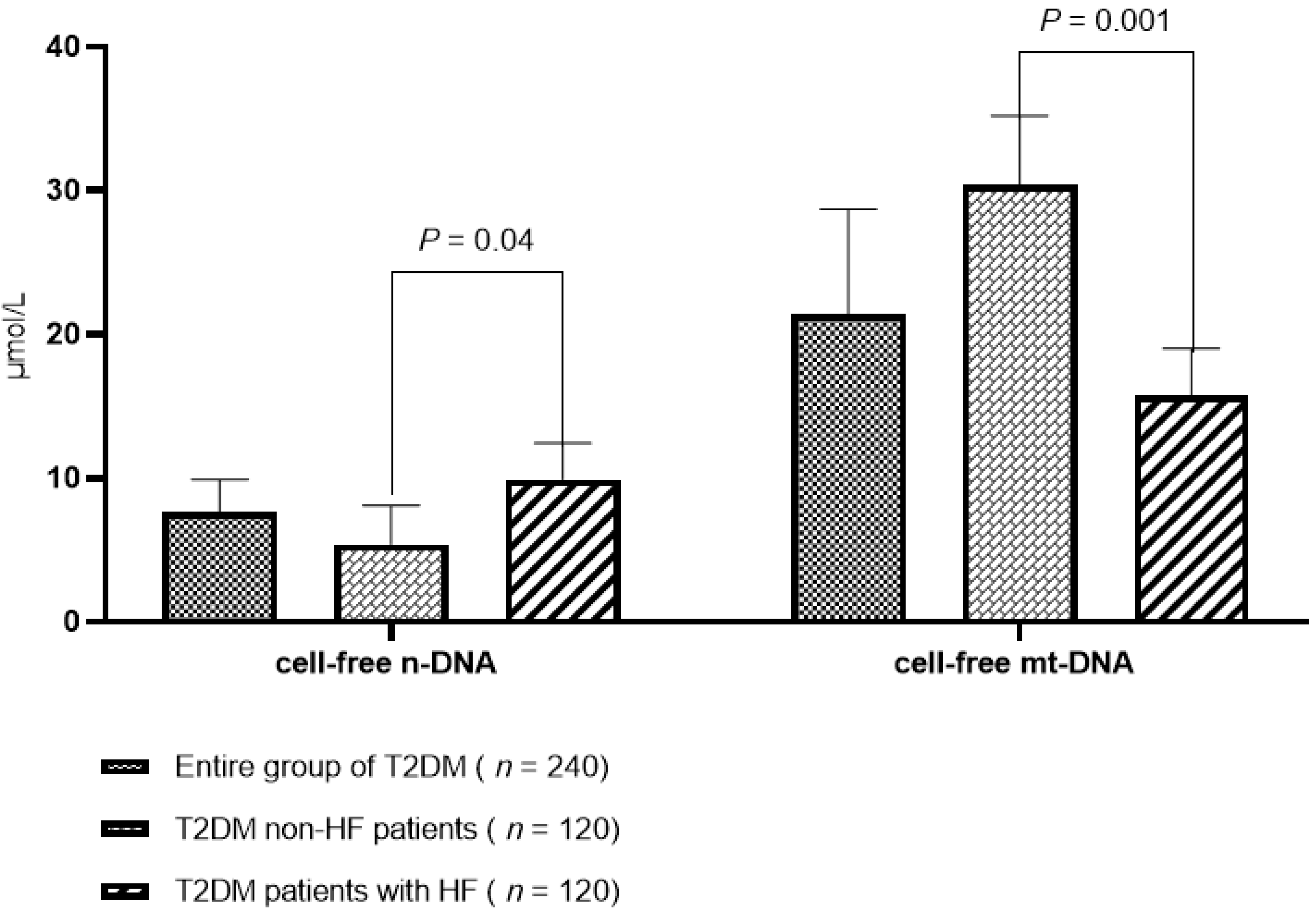
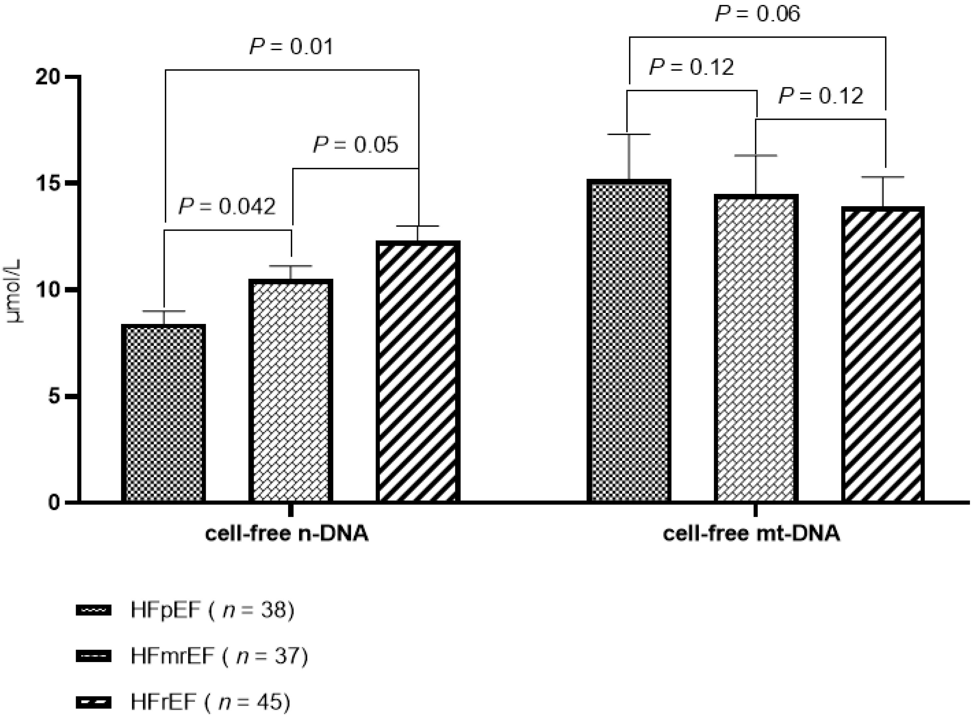
The concentrations of cell-free nuclear and mitochondrial DNA in terms of the circulating blood of patients from the entire population were 7.6 ± 2.3 μmol/L and 21.4 ± 7.3 μmol/L, respectively (Figure 2). Among the patients with T2DM, with and without HF, the levels of cell-free nuclear and mitochondrial DNA were significantly different. Moreover, the T2DM patients with HF possessed significantly higher levels of cf-nDNA (9.9 ± 2.5 μmol/L versus 5.4 ± 2.7 μmol/L; p = 0.04) and lower cf-mtDNA (15.7 ± 3.3 μmol/L versus 30.4 ± 4.8 μmol/L; p = 0.001) than those without HF. The patients with HF exhibited an overwhelming similarity in their levels of cf-mtDNA; whereas there was significant difference regarding the levels of cf-nDNA among the patients with HFpEF versus those who possessed HfmrEF/HfrEF (Figure 3).
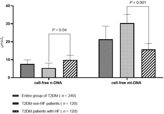
Figure 2.
Plasma cell-free nuclear and mitochondrial DNA concentration by cohort. A box and whisker plot shows the cell-free DNA concentrations in μmol/L at the median and 25–75% interquartile range. A comparison of the variables was provided with a t-test. The p-value illustrates the difference between T2DM patient cohorts, with and without HF. Abbreviations—T2DM: type 2 diabetes mellitus; HF: heart failure; n-DNA: nuclear DNA; and mt-DNA: mitochondrial DNA.
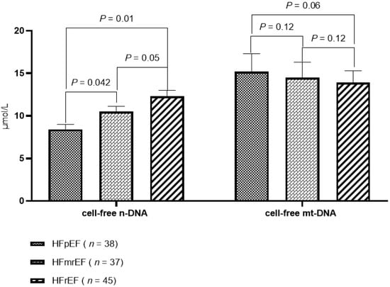
Figure 3.
Plasma cell-free nuclear and mitochondrial DNA concentration by HF phenotypes. A box and whisker plot showing the cell-free DNA concentration in μmol/L as the median and 25–75% interquartile range. Comparisons of the variables were provided with an ANOVA Tukey test. The p-values show differences among the T2DM patient cohorts who were with any of the phenotypes of HF. Abbreviations—T2DM: type 2 diabetes mellitus; HF: heart failure; n-DNA: nuclear DNA; and mt-DNA: mitochondrial DNA.
3.3. Spearman’s Correlation between the Circulating Levels of Cell-Free DNA and Other Parameters
In the entire group of patients, we found positive correlations between the levels of cf-nDNA and NT-proBNP (r = 0.34; p = 0.001); LAVI (r = 0.30; p = 0.001); hs-CRP (r = 0.30; p = 0.001); E/è (r = 0.30; p = 0.02); microalbuminuria (r = 0.29; p = 0.01); NYHA class (r = 0.26; p = 0.02); and TG (r = 0.24; p = 0.04), as well as a negative correlation with LVEF (r = −0.33; p = 0.001). There was no association determined with respect to the levels of cf-nDNA with: fasting glucose, the HOMA index, BMI, HbA1c, LDL-C, and TG. The levels of cf-mtDNA were associated positively with LVEF (r = 0.31; p = 0.01) and negatively associated with NT-proBNP (r = −0.36; p = 0.001), hs-CRP (r = −0.34; p = 0.001), NYHA class (r = −0.30; p = 0.02), and LAVI (r = −0.30; p = 0.001). However, it must be noted that there were no significant correlations between the levels of cf-nDNA and cf-mtDNA with respect to concomitant medications.
In T2DM patients, the levels of cf-nDNA and cf-mtDNA correlated with hs-CRP (r = 0.32; p = 0.001 and r = 0.28; p = 0.001, respectively), microalbuminuria (r = 0.32; p = 0.001 and r = 0.31; p = 0.01, respectively), and eGFR (r = 0.26; p = 0.01 and r = 0.24; p = 0.01, respectively), but not with BMI. However, a positive correlation between cf-mtDNA and HOMA was found (r = 0.28; p = 0.02) in this group. In regard to the T2DM patients with HF, the levels of cf-nDNA and cf-mtDNA correlated with: NT-proBNP (r = 0.36; p = 0.001 and r = −0.30; p = 0.001, respectively); LAVI (r = 0.30; p = 0.001 and r = −0.26; p = 0.01, respectively); hs-CRP (r = 0.30; p = 0.001 and r = −0.24; p = 0.02, respectively); the NYHA class (r = 0.26; p = 0.02 and r = 0.26; p = 0.02, respectively); and LVEF (r = −0.33; p = 0.001 and r = 0.29; p = 0.01, respectively).
3.4. The Factors Associated with HF in T2DM Patients: The Univariate and Multivariate Logistic Regression
We identified several factors that corresponded to HF in the T2DM patients by using the univariate logistic regression model (Table 2). For this analysis, we used the median of the cell-free n-DNA (7.6 μmol/L) and the median of the cf-mtDNA (21.4 μmol/L). We established that: NT-proBNP (OR = 1.09; 95% CI = 1.05–1.16; p = 0.001); cf-nDNA > 7.6 μmol/L (OR = 1.05; 95% CI = 1.02–1.08; p = 0.02); cf-mtDNA < 21.4 μmol/L (OR = 1.03; 95% CI = 1.01–1.06; p = 0.04); and LAVI (OR = 1.05; 95% CI = 1.02–1.09; p = 0.02) were predicted with respect to HF in the T2DM patients. The multivariate logistic regression yielded NT-proBNP (OR = 1.10; 95% CI = 1.04–1.19; p = 0.001) and cf-nDNA > 7.6 μmol/L (OR = 1.07; 95% CI = 1.03–1.12; p = 0.01), which remained independent predictors for HF.

Table 2.
Factors influencing HF occurrence in the study population. The results of the univariate and multivariate logistic regression analysis were adjusted to LVEF and AF.
3.5. Comparison of the Models
In order to compare the two models, we used an estimation of the area under the curve (AUC), which showed a superiority with respect to the discriminative ability of cf-nDNA when compared to NT-proBNP (p = 0.001) (Table 3). Nevertheless, the presence of cf-nDNA enhanced the risk differentiation by independently increasing the prognostic impact on HF from the presence of NT-proBNP. However, we noticed that the discriminative value of the combined biomarker model (NT-proBNP + cf-nDNA) was not better than that of cf-nDNA alone (AUC = 0.83; 95% CI = 0.74–0.92 vs. AUC = 0.80; 95% CI = 0.75–0.88; p = 0.80). Having said this, it was still better than the reference model constructed using the NT-proBNP levels detected in the peripheral blood.

Table 3.
The comparisons of NT-proBNP and cell-free n-DNA discriminative potencies for HF.
4. Discussion
The results of our investigation showed that the bidirectional changes in the circulating levels of cf-DNA (i.e., an increase in cf-nDNA and a decrease in cf-mtDNA) in T2DM patients were associated with hemodynamic performance. These were characterized by adverse cardiac remodeling (LVEF, LAVI, and E/è), as well as corresponded to the indicators of systemic inflammation (hs-CRP), cardiac stretching (NT-proBNP), the clinical status of the patients (heart failure, NYHA functional class), and kidney damage (microalbuminuria, estimated GFR). However, cf-nDNAs, but not cf-mtDNAs, were independently associated with HF in the T2DM patient population. Furthermore, we established that cf-nDNA appears to show better discriminative potency than NT-proBNP in the multivariate logistic regression, which was adjusted to LVEF and AF.
Previous studies have tested a hypothesis in which the circulating cf-DNAs may be promising biomarkers with respect to the microvascular complications of T2DM, such as retinopathy and nephropathy [38,39,40], as well as skeletal muscle damage/myopathy after exercise [41] and obesity [42]. However, the association of the cf-DNA profile with macrovascular complications and cardiac remodeling, specifically due to a T2DM advance, was incompletely understood [43]. It is, therefore, suggested that cf-DNA should serve as an integrated part of HF patient management, as well as a surveillance tool for T2DM patients with variable levels of the classic biomarkers of HF, such as NT-proBNP [21,44]. Indeed, NT-proBNP is a well-established circulating biomarker in HF, but its discriminative values for mortality and morbidity differ significantly in patients with obesity vs. non-obesity, T2DM versus non-T2DM, HfrEF versus HfpEF, and chronic kidney disease (CKD) versus non-CKD [45,46]. Moreover, the circulating levels of natriuretic peptides, including NT-proBNP in HF patients with T2DM, were frequently lower than those in non-T2DM petients, regardless of HF phenotypes [45,47,48,49]. All of these are open perspectives with respect to the discovery of novel biomarker-guided approaches in the management of T2DM patients at risk of HF.
We hypothesized that the epigenetically modified circulating DNA fragments, which are detected as cf-nDNA and cf-mDNA and associated with NET, can reflect adverse cardiac remodeling due to the direct damage of cardiac myocytes and the inflammatory condition, which is due to mitochondrial dysfunction. Consequently, in the circulation, both fragments of cf-DNA occur, and their persistence was remarkably supported by metabolic disturbances that were closely related to T2DM. Although the first part of this hypothesis is successfully confirmed, surprisingly, we did not find a significant correlation between the levels of cf-nDNA/cf-mDNA, nor with respect to HOMA, BMI, HbA1c, and the fasting glucose levels within the entire population, nor in the T2DM non-HF patients. However, the circulating levels of cf-nDNA mildly correlated with concentrations of TG in T2DM non-HF patients. Meanwhile, there are limiting data regarding the correlations of any types of cf-DNAs with these metabolic parameters in T2DM. However, the many factors contributing to insulin resistance—such as age, obesity, BMI, HbA1c, and fasting glucose—are potential triggers for impaired mitochondrial structure and function, such that they may interfere with NET [50,51,52]. Having said this, we did not observe any interaction between the circulating levels of cf-mtDNA and cf-nDNA with respect to sex or age. However, there is an obvious similarity between these issues in other studies [53].
Conversely, T2DM patients with HF demonstrated higher frequencies of AF, chronic kidney disease (CKD), and coronary artery disease (CAD) than those who exhibited T2DM without HF, whereas there was no significant difference in smoking between these groups. When taking into consideration the fact that previous studies revealed that these concomitant conditions were associated with elevated levels of cf-DNA [54,55], it is reasonable to suggest that a profile of comorbidities including AF, CKD, and CAD may intervene in HF through underlying mechanisms, which thus engage NET.
Along with the aforementioned, in each cohort of the patients enrolled in this study, we established clear positive correlations between cf-nDNA and the inflammatory cytokine hs-CRP, NT-proBNP, and LAVI, as well as a negative association with the global LVEF. However, in our study, positive associations of cf-mtDNA and HOMA were found in the T2DM patients without HF. Previous studies have shown that cf-nDNA positively correlates with hs-CRP, but the association of the levels in regard to the molecule in peripheral blood that describe insulin resistance, such as HOMA, were found to be controversial [53,56].
Perhaps, with respect to the underlying mechanisms by which cf-nDNA/cf-mDNA were released into circulation in HF individuals, there may be a significant difference in regards to those who had no HF. Indeed, all patients with T2DM included in the study were treated with metformin, which is reported to decrease the levels of inflammatory cytokines in diabetics. However, this effect was regarded, in the end, not to be powerful in HF patients with concomitant T2DM. As such, we suggested that hs-CRP may directly induce NET in HF patients, regardless of antidiabetic management, such that the basal levels of NET become higher than those found in T2DM individuals without HF [57,58]. With respect to this, Carestia A. et al. (2016) [58] reported that despite well-controlled levels of fasting glucose and/or HbA1c among T2DM patients—as well as a tendency to a restoration of normal values after six months of metformin treatment—the basal formation of NETs and the failure of neutrophils to form these DNA structures after TNFα stimulation, along with a presence of nucleosomes and DNA complexes in the plasma of the patients, continued to be retained at a higher level than those found in healthy individuals [58]. Thus, the increased levels of cf-nDNA in circulation within hemodynamically stable, well-treated T2DM patients with HF can be caused by pro-inflammatory activation, which is beyond glycemic control. However, we agree that hyperglycemia can worsen the already elevated baseline NET in T2DM patients, which can increase its detrimental effects on cardiac myocytes and vasculature [59]. Indeed, the variability of glycemia is considered a silent detrimental factor with respect to NET formation when compared with true hyperglycemia [60]. In addition, it stimulates oxidative stress and induces mitochondrial dysfunction, which negatively influences cell membrane permeability and phagocyte activity. Perhaps this was a primary reason for the changes in cf-mtDNA levels within the patients enrolled in our study. In fact, it is reasonable to speculate regarding the causes of bidirectional changes in cf-nDNA and cf-mtDNA in the patients through the context of providing conventional treatment. However, cf-nDNA, but not cf-mtDNA, added predictive information to NT-proBNP. As such, the underlying molecular mechanism by which NET intervenes in cardiac remodeling was treated in accordance with the recommended guidelines. However, this requires further elucidation. In other studies, low levels of cf-mtDNA were detected as a biomarker in atherosclerosis and coronary artery disease patients with T2DM [61,62]. Perhaps cf-mtDNA may play an adaptive role in the regulation of phagocyte activity with respect to antigen-presenting cells, whereby the low levels of cf-mtDNA may indicate tissue damage due to oxidative stress [63].
Although the lowered cf-mtDNA was important in regards to the HF associations, the effect is not clearly observed in the discrimination between the various phenotypes of HF. This is due to a borderline statistical significance (p = 0.06) between HFpEF and HFrEF. Conversely, cf-nDNA is likely to be important in the discriminatory capability (p = 0.01) between HFpEF and HFrEF. Perhaps the significance of this difference may also be disputed in terms of non-diabetics in order to place the role of the altered proportion of cf-DNA in the proper context for all HF patients, as cf-DNA could be an important diagnostic modality, regardless of hyperglycemia. In regard to this connection, the altered interrelationship between cf-nDNA and cf-mtDNA appears to be a promising indicator for the continuous monitoring of HF evolution, although this hypothesis require further investigation. Last, but not least, the obtained findings should be validated in a study with a larger sample size. Furthermore, future studies should also endeavor to identify the economic burden for the novel biomarker model when it is implemented in clinical practice. All these speculative and problematic issues are likely to be addressed in further investigations. However, the impact of metformin and the SGLT2 inhibitor, with respect to the circulating pool of NET biomarkers, appears to be promising in the context of future studies.
5. Study Limitations
This study possesses several limitations. The first relates to the origin of DNA molecules. Although certain molecular features, such as the pattern of methylation, the proportion of circular and single-stranded forms, and the distribution of cell-free DNA fragments, yield important information in regard to their tissue sources, such features also reflect the severity with respect to the target organ damage. Regarding this, we assessed a circulating pool of cf-nDNA and cf-mtDNA without categorizing their correspondence to cardiac myocytes. This may be reasonable for HF patients with different etiologies and variable signatures of concomitant disease, due to an impact of NET on the progression of cardiac remodeling and where the manifestation of HF can be complex. Along with the aforementioned, in this study, we used the median of circulating levels of cf-nDNA and cf-mtDNA in order to perform univariate and multivariate logistic analysis. We did not compare the sensitivity and specificity of different cut-offs for these molecules, even though the other cut-off points may be a more optimistic tool for the purposes of reclassification analysis. The second limitation may stem from the design of the study, in which the patients were enrolled proportionally according to the presence of HF, but were not randomized. Moreover, a respectively small sample size can also be considered as one of the study’s limitations. Notably, the cf-DNA, in response to ROS, underwent subsequent fragmentation to smaller DNA fragments. We have chosen the primer pairs for cf-nDNA and cf-mtDNA such that we could better identify the detectable molecules, which were stable over a long period. As such, we neglected a portion of the small fragments of cf-DNA. Therefore, we did not examine the methylation status to bind our findings with the target genes. In the future, we will extend our investigation by conducting a randomization of the larger population of HF patients, and we will not restrict this process to those who possess T2DM. We believed that these limitations would not imply a serious impact on the interpretation of the results of our study, but we still wish to emphasize the ways in which we will investigate our hypothesis in the future.
6. Conclusions
In this study, we established that the elevated circulating levels of cf-nDNA, but not cf-mtDNA, were independently associated with HF in T2DM patients due to the detection of their levels of NT-proBNP. Moreover, the concentrations of both molecules engendered mild-to-moderate associations with the global left ventricular function, hs-CRP, NT-proBNP, and with the clinical status of the patients and the parameters of kidney damage. These findings thus provide new perspectives in regard to the reclassification of T2DM patients who are at a higher risk of HF, regardless of their phenotypes and clinical presentation. However, a large clinical study is required in order to further investigate whether this hypothesis is valid. As such, this paper currently serves as a proof-of-concept for the central hypothesis.
Author Contributions
Conceptualization, A.A.B. and A.E.B.; methodology, M.L. and M.P.K.; software, A.A.B. and O.V.P.; validation, T.A.B., A.E.B., M.L. and Z.O.; formal analysis, A.A.B., T.A.B., L.S., M.P.K., O.V.P. and Z.O.; investigation, A.A.B. and T.A.B.; resources, T.A.B., A.E.B. and Z.O.; data curation, A.E.B. and T.A.B.; writing—original draft preparation, A.A.B., M.L., Z.O., L.S., A.E.B. and A.E.B.; writing—review and editing, A.A.B., A.E.B., M.L., M.P.K., O.V.P., L.S. and Z.O.; visualization, A.A.B. and T.A.B.; supervision, A.E.B.; project administration, T.A.B. and A.A.B. All authors have read and agreed to the published version of the manuscript.
Funding
This research received no external funding.
Institutional Review Board Statement
This study was conducted according to the guidelines of the Declaration of Helsinki and was approved by the Institutional Ethics Committee of Zaporozhye, Medical Academy of Post-Graduate Education (protocol number: 8; date of approval: 10 October 2020).
Informed Consent Statement
Informed consent was obtained from all subjects involved in the study.
Data Availability Statement
Not applicable.
Acknowledgments
We would like to thank all patients who gave their consent to participate in the study and all administrative staff and doctors of the Vita-Centre LTD private hospital for their study assistance.
Conflicts of Interest
The authors declare no conflict of interest.
References
- Yap, J.; Tay, W.T.; Teng, T.K.; Anand, I.; Richards, A.M.; Ling, L.H.; MacDonald, M.R.; Chandramouli, C.; Tromp, J.; Siswanto, B.B.; et al. ASIAN-HF (Asian Sudden Cardiac Death in Heart Failure) Registry Investigators. Association of Diabetes Mellitus on Cardiac Remodeling, Quality of Life, and Clinical Outcomes in Heart Failure With Reduced and Preserved Ejection Fraction. J. Am. Heart Assoc. 2019, 8, e013114. [Google Scholar] [CrossRef] [PubMed]
- Kannel, W.B.; McGee, D.L. Diabetes and cardiovascular disease. The Framingham study. JAMA 1979, 241, 2035–2038. [Google Scholar] [CrossRef] [PubMed]
- Kenny, H.C.; Abel, E.D. Heart Failure in Type 2 Diabetes Mellitus. Circ. Res. 2019, 124, 121–141. [Google Scholar] [CrossRef]
- Tromp, J.; Paniagua, S.M.A.; Lau, E.S.; Allen, N.B.; Blaha, M.J.; Gansevoort, R.T.; Hillege, H.L.; Lee, D.E.; Levy, D.; Vasan, R.S.; et al. Age dependent associations of risk factors with heart failure: Pooled population based cohort study. BMJ 2021, 372, n461. [Google Scholar] [CrossRef]
- Randhawa, V.K.; Dhanvantari, S.; Connelly, K.A. How Diabetes and Heart Failure Modulate Each Other and Condition Management. Can. J. Cardiol. 2021, 37, 595–608. [Google Scholar] [CrossRef]
- Park, J.J. Epidemiology, Pathophysiology, Diagnosis and Treatment of Heart Failure in Diabetes. Diabetes Metab. J. 2021, 45, 146–157. [Google Scholar] [CrossRef]
- Pop-Busui, R.; Januzzi, J.L.; Bruemmer, D.; Butalia, S.; Green, J.B.; Horton, W.B.; Knight, C.; Levi, M.; Rasouli, N.; Richardson, C.R. Heart Failure: An Underappreciated Complication of Diabetes. A Consensus Report of the American Diabetes Association. Diabetes Care 2022, 45, 1670–1690. [Google Scholar] [CrossRef]
- Berezin, A. Metabolic memory phenomenon in diabetes mellitus: Achieving and perspectives. Diabetes Metab. Syndr. 2016, 10 (Suppl. S1), S176–S183. [Google Scholar] [CrossRef]
- Caprnda, M.; Mesarosova, D.; Ortega, P.F.; Krahulec, B.; Egom, E.; Rodrigo, L.; Kruzliak, P.; Mozos, I.; Gaspar, L. Glycemic Variability and Vascular Complications in Patients with Type 2 Diabetes Mellitus. Folia Med. 2017, 59, 270–278. [Google Scholar] [CrossRef]
- Cardoso, C.R.L.; Leite, N.C.; Moram, C.B.M.; Salles, G.F. Long-term visit-to-visit glycemic variability as predictor of micro- and macrovascular complications in patients with type 2 diabetes: The Rio de Janeiro Type 2 Diabetes Cohort Study. Cardiovasc. Diabetol. 2018, 17, 33. [Google Scholar] [CrossRef]
- Berezin, A. Neutrophil extracellular traps: The core player in vascular complications of diabetes mellitus. Diabetes Metab. Syndr. 2019, 13, 3017–3023. [Google Scholar] [CrossRef] [PubMed]
- Del Re, D.P.; Amgalan, D.; Linkermann, A.; Liu, Q.; Kitsis, R.N. Fundamental Mechanisms of Regulated Cell Death and Implications for Heart Disease. Physiol. Rev. 2019, 99, 1765–1817. [Google Scholar] [CrossRef] [PubMed]
- De Miranda, F.S.; Barauna, V.G.; Dos Santos, L.; Costa, G.; Vassallo, P.F.; Campos, L.C.G. Properties and Application of Cell-Free DNA as a Clinical Biomarker. Int. J. Mol. Sci. 2021, 22, 9110. [Google Scholar] [CrossRef] [PubMed]
- Bronkhorst, A.J.; Ungerer, V.; Diehl, F.; Anker, P.; Dor, Y.; Fleischhacker, M.; Gahan, P.B.; Hui, L.; Holdenrieder, S.; Thierry, A.R. Towards systematic nomenclature for cell-free DNA. Hum. Genet. 2021, 140, 565–578. [Google Scholar] [CrossRef] [PubMed]
- De Bont, C.M.; Boelens, W.C.; Pruijn, G.J.M. NETosis, complement, and coagulation: A triangular relationship. Cell Mol. Immunol. 2019, 16, 19–27. [Google Scholar] [CrossRef] [PubMed]
- De Vlaminck, I.; Valantine, H.A.; Snyder, T.M.; Strehl, C.; Cohen, G.; Luikart, H.; Neff, N.F.; Okamoto, J.; Bernstein, D.; Weisshaar, D.; et al. Circulating cell-free DNA enables noninvasive diagnosis of heart transplant rejection. Sci. Transl. Med. 2014, 6, 241ra77. [Google Scholar] [CrossRef]
- Dawson, S.-J.; Tsui, D.W.; Murtaza, M.; Biggs, H.; Rueda, O.M.; Chin, S.-F.; Dunning, M.J.; Gale, D.; Forshew, T.; Mahler-Araujo, B.; et al. Analysis of circulating tumor DNA to monitor metastatic breast cancer. N. Engl. J. Med. 2013, 368, 1199–1209. [Google Scholar] [CrossRef]
- Antonatos, D.; Patsilinakos, S.; Spanodimos, S.; Korkonikitas, P.; Tsigas, D. Cell-free DNA levels as a prognostic marker in acute myocardial infarction. Ann. N. Y. Acad. Sci. 2006, 1075, 278–281. [Google Scholar] [CrossRef]
- Cheng, Z.; Abrams, S.T.; Toh, J.; Wang, S.S.; Wang, Z.; Yu, Q.; Yu, W.; Toh, C.H.; Wang, G. The Critical Roles and Mechanisms of Immune Cell Death in Sepsis. Front. Immunol. 2020, 11, 1918. [Google Scholar] [CrossRef]
- Menegazzo, L.; Ciciliot, S.; Poncina, N.; Mazzucato, M.; Persano, M.; Bonora, B.; Albiero, M.; Vigili de Kreutzenberg, S.; Avogaro, A.; Fadini, G.P. NETosis is induced by high glucose and associated with type 2 diabetes. Acta Diabetol. 2015, 52, 497–503. [Google Scholar] [CrossRef]
- Dou, H.; Kotini, A.; Liu, W.; Fidler, T.; Endo-Umeda, K.; Sun, X.; Olszewska, M.; Xiao, T.; Abramowicz, S.; Yalcinkaya, M.; et al. Oxidized Phospholipids Promote NETosis and Arterial Thrombosis in LNK(SH2B3) Deficiency. Circulation 2021, 144, 1940–1954. [Google Scholar] [CrossRef]
- Njeim, R.; Azar, W.S.; Fares, A.H.; Azar, S.T.; Kfoury Kassouf, H.; Eid, A.A. NETosis contributes to the pathogenesis of diabetes and its complications. J. Mol. Endocrinol. 2020, 65, R65–R76. [Google Scholar] [CrossRef] [PubMed]
- Ling, S.; Xu, J.W. NETosis as a Pathogenic Factor for Heart Failure. Oxid. Med. Cell. Longev. 2021, 2021, 6687096. [Google Scholar] [CrossRef] [PubMed]
- Yokokawa, T.; Misaka, T.; Kimishima, Y.; Shimizu, T.; Kaneshiro, T.; Takeishi, Y. Clinical Significance of Circulating Cardiomyocyte-Specific Cell-Free DNA in Patients With Heart Failure: A Proof-of-Concept Study. Can. J. Cardiol. 2020, 36, 931–935. [Google Scholar] [CrossRef]
- Ren, J.; Jiang, L.; Liu, X.; Liao, Y.; Zhao, X.; Tang, F.; Yu, H.; Shao, Y.; Wang, J.; Wen, L.; et al. Heart-specific DNA methylation analysis in plasma for the investigation of myocardial damage. J. Transl. Med. 2022, 20, 36. [Google Scholar] [CrossRef]
- Toto, R.D. Microalbuminuria: Definition, detection, and clinical significance. J. Clin. Hypertens. 2004, 6 (Suppl. S3), 2–7. [Google Scholar] [CrossRef]
- American Diabetes Association. 2. Classification and Diagnosis of Diabetes: Standards of Medical Care in Diabetes—2021. Diabetes Care 2021, 44 (Suppl. S1), S15–S33. [Google Scholar] [CrossRef] [PubMed]
- McDonagh, T.A.; Metra, M.; Adamo, M.; Gardner, R.S.; Baumbach, A.; Bohm, M.; Burri, H.; Butler, J.; Čelutkienė, J.; Chioncel, O.; et al. 2021 ESC Guidelines for the diagnosis and treatment of acute and chronic heart failure. Eur. Heart J. 2021, 42, 3599–3726. [Google Scholar] [CrossRef]
- Williams, B.; Mancia, G.; Spiering, W.; Agabiti Rosei, E.; Azizi, M.; Burnier, M.; Burnier, M.; Clement, D.L.; Coca, A.; de Simone, G.; et al. 2018 ESC/ESH Guidelines for the management of arterial hypertension. Eur. Heart J. 2018, 39, 3021–3104. [Google Scholar] [CrossRef]
- Mach, F.; Baigent, C.; Catapano, A.L.; Koskinas, K.C.; Casula, M.; Badimon, L.; Chapman, M.J.; De Backer, G.G.; Delgado, V.; Ference, B.A.; et al. 2019 ESC/EAS Guidelines for the management of dyslipidaemias: Lipid modification to reduce cardiovascular risk. Eur. Heart J. 2020, 41, 111–188. [Google Scholar] [CrossRef]
- Knuuti, J.; Wijns, W.; Saraste, A.; Capodanno, D.; Barbato, E.; Funck-Brentano, C.; Prescott, E.; Storey, R.F.; Deaton, C.; Cuisset, T.; et al. 2019 ESC Guidelines for the diagnosis and management of chronic coronary syndromes. Eur. Heart J. 2020, 41, 407–477. [Google Scholar] [CrossRef] [PubMed]
- De Boer, I.H.; Khunti, K.; Sadusky, T.; Tuttle, K.R.; Neumiller, J.J.; Rhee, C.M.; Rosas, S.E.; Rossing, P.; Bakris, G. Diabetes Management in Chronic Kidney Disease: A Consensus Report by the American Diabetes Association (ADA) and Kidney Disease: Improving Global Outcomes (KDIGO). Diabetes Care 2022, 45, 3075–3090. [Google Scholar] [CrossRef] [PubMed]
- Yamamoto, J.; Wakami, K.; Muto, K.; Kikuchi, S.; Goto, T.; Fukuta, H.; Seo, Y.; Ohte, N. Verification of Echocardiographic Assessment of Left Ventricular Diastolic Dysfunction in Patients With Preserved Left Ventricular Ejection Fraction Using the American Society of Echocardiography and European Association of Cardiovascular Imaging 2016 Recommendations. Circ. Rep. 2019, 1, 525–530. [Google Scholar] [CrossRef] [PubMed]
- Nagueh, S.F.; Smiseth, O.A.; Appleton, C.P.; Byrd, B.F., 3rd; Dokainish, H.; Edvardsen, T.; Flachskampf, F.A.; Gillebert, T.C.; Klein, A.L.; Lancellotti, P.; et al. Recommendations for the Evaluation of Left Ventricular Diastolic Function by Echocardiography: An Update from the American Society of Echocardiography and the European Association of Cardiovascular Imaging. J. Am. Soc. Echocardiogr. 2016, 29, 277–314. [Google Scholar] [CrossRef] [PubMed]
- Levey, A.S.; Stevens, L.A.; Schmid, C.H.; Zhang, Y.L.; Castro, A.F., 3rd; Feldman, H.I.; Kusek, J.W.; Eggers, P.; Van Lente, F.; Greene, T.; et al. CKD-EPI (Chronic Kidney Disease Epidemiology Collaboration). A new equation to estimate glomerular filtration rate. Ann. Intern. Med. 2009, 150, 604–612. [Google Scholar] [CrossRef] [PubMed]
- Matthews, D.R.; Hosker, J.P.; Rudenski, A.S.; Naylor, B.A.; Treacher, D.F.; Turner, R.C. Homeostasis model assessment: Insulin resistance and beta-cell function from fasting plasma glucose and insulin concentrations in man. Diabetologia 1985, 28, 412–419. [Google Scholar] [CrossRef]
- Hasenleithner, S.O.; Speicher, M.R. A clinician’s handbook for using ctDNA throughout the patient journey. Mol. Cancer 2022, 21, 81. [Google Scholar] [CrossRef] [PubMed]
- Han, L.; Chen, C.; Lu, X.; Song, Y.; Zhang, Z.; Zeng, C.; Chiu, R.; Li, L.; Xu, M.; He, C.; et al. Alterations of 5-hydroxymethylcytosines in circulating cell-free DNA reflect retinopathy in type 2 diabetes. Genomics 2021, 113 Pt 1, 79–87. [Google Scholar] [CrossRef]
- Yuzefovych, L.V.; Pastukh, V.M.; Ruchko, M.V.; Simmons, J.D.; Richards, W.O.; Rachek, L.I. Plasma mitochondrial DNA is elevated in obese type 2 diabetes mellitus patients and correlates positively with insulin resistance. PLoS ONE 2019, 14, e0222278. [Google Scholar] [CrossRef]
- Bryk, A.H.; Prior, S.M.; Plens, K.; Konieczynska, M.; Hohendorff, J.; Malecki, M.T.; Butenas, S.; Undas, A. Predictors of neutrophil extracellular traps markers in type 2 diabetes mellitus: Associations with a prothrombotic state and hypofibrinolysis. Cardiovasc. Diabetol. 2019, 18, 49. [Google Scholar] [CrossRef]
- Belcher, D.J.; Sousa, C.A.; Carzoli, J.P.; Johnson, T.K.; Helms, E.R.; Visavadiya, N.P.; Zoeller, R.F.; Whitehurst, M.; Zourdos, M.C. Time course of recovery is similar for the back squat, bench press, and deadlift in well-trained males. Appl. Physiol. Nutr. Metab. 2019, 44, 1033–1042. [Google Scholar] [CrossRef] [PubMed]
- Camuzi Zovico, P.V.; Gasparini Neto, V.H.; Venâncio, F.A.; Soares Miguel, G.P.; Graça Pedrosa, R.; Kenji Haraguchi, F.; Barauna, V.G. Cell-free DNA as an obesity biomarker. Physiol Res. 2020, 69, 515–520. [Google Scholar] [CrossRef]
- Martinod, K.; Witsch, T.; Erpenbeck, L.; Savchenko, A.; Hayashi, H.; Cherpokova, D.; Gallant, M.; Mauler, M.; Cifuni, S.M.; Wagner, D.D. Peptidylarginine deiminase 4 promotes age-related organ fibrosis. J. Exp. Med. 2017, 214, 439–458. [Google Scholar] [CrossRef] [PubMed]
- Devaux, Y. Cardiomyocyte-Specific Cell-Free DNA as a Heart Failure Biomarker? Can. J. Cardiol. 2020, 36, 807–808. [Google Scholar] [CrossRef] [PubMed]
- Buckley, L.F.; Canada, J.M.; Del Buono, M.G.; Carbone, S.; Trankle, C.R.; Billingsley, H.; Kadariya, D.; Arena, R.; Van Tassell, B.W.; Abbate, A. Low NT-proBNP levels in overweight and obese patients do not rule out a diagnosis of heart failure with preserved ejection fraction. ESC Heart Fail. 2018, 5, 372–378. [Google Scholar] [CrossRef]
- Nah, E.H.; Kim, S.Y.; Cho, S.; Kim, S.; Cho, H.I. Plasma NT-proBNP levels associated with cardiac structural abnormalities in asymptomatic health examinees with preserved ejection fraction: A retrospective cross-sectional study. BMJ Open 2019, 9, e026030. [Google Scholar] [CrossRef]
- Oremus, M.; McKelvie, R.; Don-Wauchope, A.; Santaguida, P.L.; Ali, U.; Balion, C.; Hill, S.; Booth, R.; Brown, J.A.; Bustamam, A.; et al. A systematic review of BNP and NT-proBNP in the management of heart failure: Overview and methods. Heart Fail. Rev. 2014, 19, 413–419. [Google Scholar] [CrossRef]
- Jhund, P.S.; Anand, I.S.; Komajda, M.; Claggett, B.L.; McKelvie, R.S.; Zile, M.R.; Carson, P.E.; McMurray, J.J. Changes in N-terminal pro-B-type natriuretic peptide levels and outcomes in heart failure with preserved ejection fraction: An analysis of the I-Preserve study. Eur. J. Heart Fail. 2015, 17, 809–817. [Google Scholar] [CrossRef]
- Rørth, R.; Jhund, P.S.; Yilmaz, M.B.; Kristensen, S.L.; Welsh, P.; Desai, A.S.; Køber, L.; Prescott, M.F.; Rouleau, J.L.; Solomon, S.D.; et al. Comparison of BNP and NT-proBNP in Patients With Heart Failure and Reduced Ejection Fraction. Circ. Heart Fail. 2020, 13, e006541. [Google Scholar] [CrossRef]
- Kwak, S.H.; Park, K.S. Role of mitochondrial DNA variation in the pathogenesis of diabetes mellitus. Front. Biosci. 2016, 21, 1151–1167. [Google Scholar] [CrossRef]
- Walczak, K.; Stawski, R.; Perdas, E.; Brzezinska, O.; Kosielski, P.; Galczynski, S.; Budlewski, T.; Padula, G.; Nowak, D. Circulating cell free DNA response to exhaustive exercise in average trained men with type I diabetes mellitus. Sci. Rep. 2021, 11, 4639. [Google Scholar] [CrossRef] [PubMed]
- Rautenberg, E.K.; Hamzaoui, Y.; Coletta, D.K. Mini-review: Mitochondrial DNA methylation in type 2 diabetes and obesity. Front. Endocrinol. 2022, 13, 968268. [Google Scholar] [CrossRef]
- Wang, W.; Luo, J.; Willems van Dijk, K.; Hägg, S.; Grassmann, F.; T Hart, L.M.; van Heemst, D.; Noordam, R. Assessment of the bi-directional relationship between blood mitochondrial DNA copy number and type 2 diabetes mellitus: A multivariable-adjusted regression and Mendelian randomisation study. Diabetologia 2022, 65, 1676–1686. [Google Scholar] [CrossRef]
- Li, X.; Hu, R.; Luo, T.; Peng, C.; Gong, L.; Hu, J.; Yang, S.; Li, Q. Serum cell-free DNA and progression of diabetic kidney disease: A prospective study. BMJ Open Diabetes Res. Care 2020, 8, e001078. [Google Scholar] [CrossRef] [PubMed]
- Ishida, M.; Ueda, K.; Sakai, C.; Ishida, T.; Morita, K.; Kobayashi, Y.; Okazaki, Y.; Baba, A.; Horikoshi, Y.; Yoshizumi, M.; et al. Cigarette smoke induces mitochondrial DNA damage and activates cGAS-STING pathway -Application to a biomarker for atherosclerosis. Clin. Sci. 2023, 137, CS20220525. [Google Scholar] [CrossRef]
- Park, J.H.; Kim, J.E.; Gu, J.Y.; Yoo, H.J.; Park, S.H.; Kim, Y.I.; Nam-Goong, I.S.; Kim, E.S.; Kim, H.K. Evaluation of Circulating Markers of Neutrophil Extracellular Trap (NET) Formation as Risk Factors for Diabetic Retinopathy in a Case-Control Association Study. Exp. Clin. Endocrinol Diabetes 2016, 124, 557–561. [Google Scholar] [CrossRef]
- Vulesevic, B.; Lavoie, S.S.; Neagoe, P.E.; Dumas, E.; Räkel, A.; White, M.; Sirois, M.G. CRP Induces NETosis in Heart Failure Patients with or without Diabetes. Immunohorizons 2019, 3, 378–388. [Google Scholar] [CrossRef]
- Carestia, A.; Frechtel, G.; Cerrone, G.; Linari, M.A.; Gonzalez, C.D.; Casais, P.; Schattner, M. NETosis before and after Hyperglycemic Control in Type 2 Diabetes Mellitus Patients. PLoS ONE 2016, 11, e0168647. [Google Scholar] [CrossRef]
- Thiam, H.R.; Wong, S.L.; Wagner, D.D.; Waterman, C.M. Cellular Mechanisms of NETosis. Annu. Rev. Cell Dev. Biol. 2020, 36, 191–218. [Google Scholar] [CrossRef]
- Miyoshi, A.; Yamada, M.; Shida, H.; Nakazawa, D.; Kusunoki, Y.; Nakamura, A.; Miyoshi, H.; Tomaru, U.; Atsumi, T.; Ishizu, A. Circulating Neutrophil Extracellular Trap Levels in Well-Controlled Type 2 Diabetes and Pathway Involved in Their Formation Induced by High-Dose Glucose. Pathobiology 2016, 83, 243–251. [Google Scholar] [CrossRef]
- Liu, J.; Zou, Y.; Tang, Y.; Xi, M.; Xie, L.; Zhang, Q.; Gong, J. Circulating cell-free mitochondrial deoxyribonucleic acid is increased in coronary heart disease patients with diabetes mellitus. J. Diabetes Investig. 2016, 7, 109–114. [Google Scholar] [CrossRef] [PubMed]
- Liu, L.P.; Cheng, K.; Ning, M.A.; Li, H.H.; Wang, H.C.; Li, F.; Chen, S.Y.; Qu, F.L.; Guo, W.Y. Association between peripheral blood cells mitochondrial DNA content and severity of coronary heart disease. Atherosclerosis 2017, 261, 105–110. [Google Scholar] [CrossRef] [PubMed]
- Berezin, A.E. Circulating Cell-Free Mitochondrial DNA as Biomarker of Cardiovascular risk: New Challenges of Old Findings. Angiology 2015, 3, 161. [Google Scholar] [CrossRef]
Disclaimer/Publisher’s Note: The statements, opinions and data contained in all publications are solely those of the individual author(s) and contributor(s) and not of MDPI and/or the editor(s). MDPI and/or the editor(s) disclaim responsibility for any injury to people or property resulting from any ideas, methods, instructions or products referred to in the content. |
© 2023 by the authors. Licensee MDPI, Basel, Switzerland. This article is an open access article distributed under the terms and conditions of the Creative Commons Attribution (CC BY) license (https://creativecommons.org/licenses/by/4.0/).

