Article

Epigallocatechin Gallate and Glutathione Attenuate Aflatoxin B1-Induced Acute Liver Injury in Ducklings via Mitochondria-Mediated Apoptosis and the Nrf2 Signalling Pathway

1
Department of Animal Nutrition and Feed Science, College of Animal Science and Technology, Huazhong Agricultural University, Wuhan 430070, China
2
Institute of Animal Husbandry and Veterinary Sciences, Wuhan Academy of Agricultural Sciences, Wuhan 430208, China
3
Department of Animal Feed and Production, Faculty of Veterinary and Animal Sciences, Muhammad Nawaz Shareef University of Agriculture, Multan 66000, Pakistan
*
Author to whom correspondence should be addressed.
Toxins 2022, 14(12), 876; https://doi.org/10.3390/toxins14120876
Submission received: 3 November 2022 / Revised: 4 December 2022 / Accepted: 13 December 2022 / Published: 15 December 2022
(This article belongs to the Special Issue Novel Strategies for Biodegradation and Detoxification of Mycotoxins)

Abstract

:
Aflatoxin B1 (AFB1) exists widely in feed and food with severe hazards, posing a serious threat to human and animal health. Epigallocatechin gallate (EGCG) and glutathione (GSH) have been reported as having anti-oxidative and other functions. The present study aimed to investigate the detoxification effect of EGCG and GSH alone or in combination on AFB1 exposure in ducklings. Fifty one-day-old male ducklings were randomly assigned into five experimental groups (n = 10): 1. Control (CTR); 2. 0.3 mg/kg BW AFB1 (AFB1); 3. 0.3 mg/kg BW AFB1 + 100 mg/kg BW EGCG (AFB1 + EGCG); 4. 0.3 mg/kg BW AFB1 + 30 mg/kg BW GSH (AFB1 + GSH); 5. 0.3 mg/kg BW AFB1 + 100 mg/kg BW EGCG + 30 mg/kg BW GSH (AFB1 + EGCG + GSH). The experiment lasted for seven days. Compared with the CTR group, AFB1 reduced growth performance, total serum protein and albumin content, increased serum enzyme activity (alanine aminotransferase, aspartate aminotransferase, alkaline phosphatase, and γ-glutamyl transpeptidase), and caused pathological damage to the ducklings’ livers. AFB1 exposure increased malondialdehyde content and decreased superoxide dismutase, total antioxidant capacity, catalase, glutathione peroxidase activities, and glutathione content in the liver. EGCG and GSH alone or in combination mitigated these adverse effects. Meanwhile, EGCG and GSH attenuate apoptosis of hepatocytes, and regulated AFB1-induced changes in the abundance of genes contained in the Keap1/Nrf2 signalling and apoptotic pathways. Collectively, these results suggest that EGCG and GSH alleviate the hepatocyte injury induced by AFB1 by inhibiting oxidative stress and attenuating excessive mitochondria-mediated apoptosis.
Key Contribution: EGCG and GSH alleviate AFB1-induced liver injury in ducklings by increasing antioxidant capacity and antagonising apoptosis.

1. Introduction

Mycotoxins are harmful naturally occurring secondary metabolites produced by fungi [1]. Aflatoxins (AFs) are poisonous mycotoxins produced principally by Aspergillus flavus and Aspergillus parasiticus, of which Aflatoxin B1 (AFB1) is the most hepatotoxic [2]. Corn, wheat, and other grains have a high detection rate of AFB1 [3], which seriously affects the health of poultry and humans throughout the food chain [4]. AFB1 has been reported to cause diarrhoea, poor feather quality, weight loss, multifocal hepatic necrosis, bile duct hyperplasia, skeletal deformation, and altered muscle alignment in poultry [5,6]. Liver cancer, immunosuppression, and stunted children have all been linked with foods contaminated with AFB1 [7]. Over 70% of the world’s ducks are raised in China [8]. As ducklings are more sensitive to AFB1 than turkeys, lower doses of AFB1 can cause them bodily damage [9]. AFB1 exhibits its toxic action by being metabolised to exo-8, 9-epoxide (AFBO) in the liver [10]. The Keap1 (Kelch-like ECH-associated protein 1)/Nrf2 (nuclear factor erythroid 2-related factor 2) pathway enables cells to adapt to oxidative stress caused by external stimuli, and some plant extracts may activate this pathway [11]. Apoptosis plays an essential role in maintaining stability in the internal environment [12]. AFB1 can dysregulate the Keap1/Nrf2 pathway and cause excessive apoptosis in hepatocytes [13]. Hence, it will be an effective measure to improve the antioxidant capacity and inhibit excessive apoptosis caused by liver injury.
Epigallocatechin gallate (EGCG) is the main active ingredient in green tea. Other catechins include epicatechin-3-gallate, epigallocatechin and epicatechin, but EGCG is the most abundant [14]. It not only promotes animal growth performance and egg quality, ameliorates body fatty acid metabolism, and regulates intestinal health [15,16], but also contributes to cardioprotection, renoprotection, hepatoprotection, and neuroprotection in humans [17]. More importantly, EGCG is a scavenger of reactive oxygen/nitrogen and has potent antioxidant capacities. Moreover, it reduces the damage to cells caused by oxidative stress by capturing oxygen free radicals, restoring their redox status and mitochondrial function [18,19]. Previous studies have shown that EGCG can attenuate bleomycin-induced pulmonary fibrosis through the Keap1/Nrf2 signalling pathway [20]. Four hundred mg/kg of EGCG in the diet significantly alleviated heat stress in quail [21]. The preventive effect of EGCG against AFB1-induced liver injury and the mechanisms involved are not clarified. Therefore, it is necessary to proceed with this work.
Glutathione (GSH) is a tripeptide comprising cysteine, glutamic acid, and glycine. It exists in two forms in animals, oxidised (GSSG) and reduced (GSH), both of which play a significant role in bodily redox status [22]. The leading absorption site of exogenous GSH is the small intestine. Oral GSH in animals and humans can increase the content of GSH in the body [23], improve antioxidant capacity (i.e., protect cells from oxidative damage), protect intestinal mucosa, and enhance the transport and absorption of nutrients [22]. Studies have shown that GSH can increase the resistance of carp to nitric oxide stress and lipopolysaccharide (LPS) stimulation [24]. Adding GSH to the diet alleviated the oxidative damage caused by ochratoxin A and significantly inhibited cell apoptosis in rats [25]. Therefore, GSH may exhibit a positively beneficial effect in mitigating the hazards of AFB1.
The present study used EGCG and GSH to alleviate the damage caused by AFB1 in ducklings. In particular, we investigated whether EGCG and GSH could alleviate liver damage through the Keap1/Nrf2 signalling pathway and the inhibition of apoptosis and whether there was a mutual effect between them. The present experiment hoped to prompt the individual and combined use of EGCG and GSH to ameliorate toxin damage in animals.

2. Results

2.1. EGCG and GSH Inhibit AFB1-Induced Changes in Growth Performance and Liver Index of Ducklings

The effects of AFB1 and EGCG or GSH on ducklings’ growth are shown in Figure 1A,B. In this experiment, ducks were treated with gavage, and each group of ten ducks was individually numbered but housed in a combined pen, so only the mean values of feed intake were calculated. As can be seen from the Figure 1A,B, there was no significant difference in the initial body weight, but after acute attacks, the AFB1 group had significantly reduced body weight (p < 0.01) compared with the CTR group, and the feed intake decreased by 13.1%. However, the EGCG and GSH alone and in combination significantly increased the ducklings’ body weights compared with the AFB1 group, while the feed intake increased by 7.7%, 2.5%, and 9.8%, respectively, showing that the combination of EGCG and GSH was more effective. As Figure 1C shows, AFB1 significantly increased the relative weight of the ducklings’ livers (p < 0.01), while EGCG and GSH significantly decreased as compared with the AFB1 group. The results indicate that EGCG and GSH alleviated the damage caused by AFB1, but a combination was more effective.

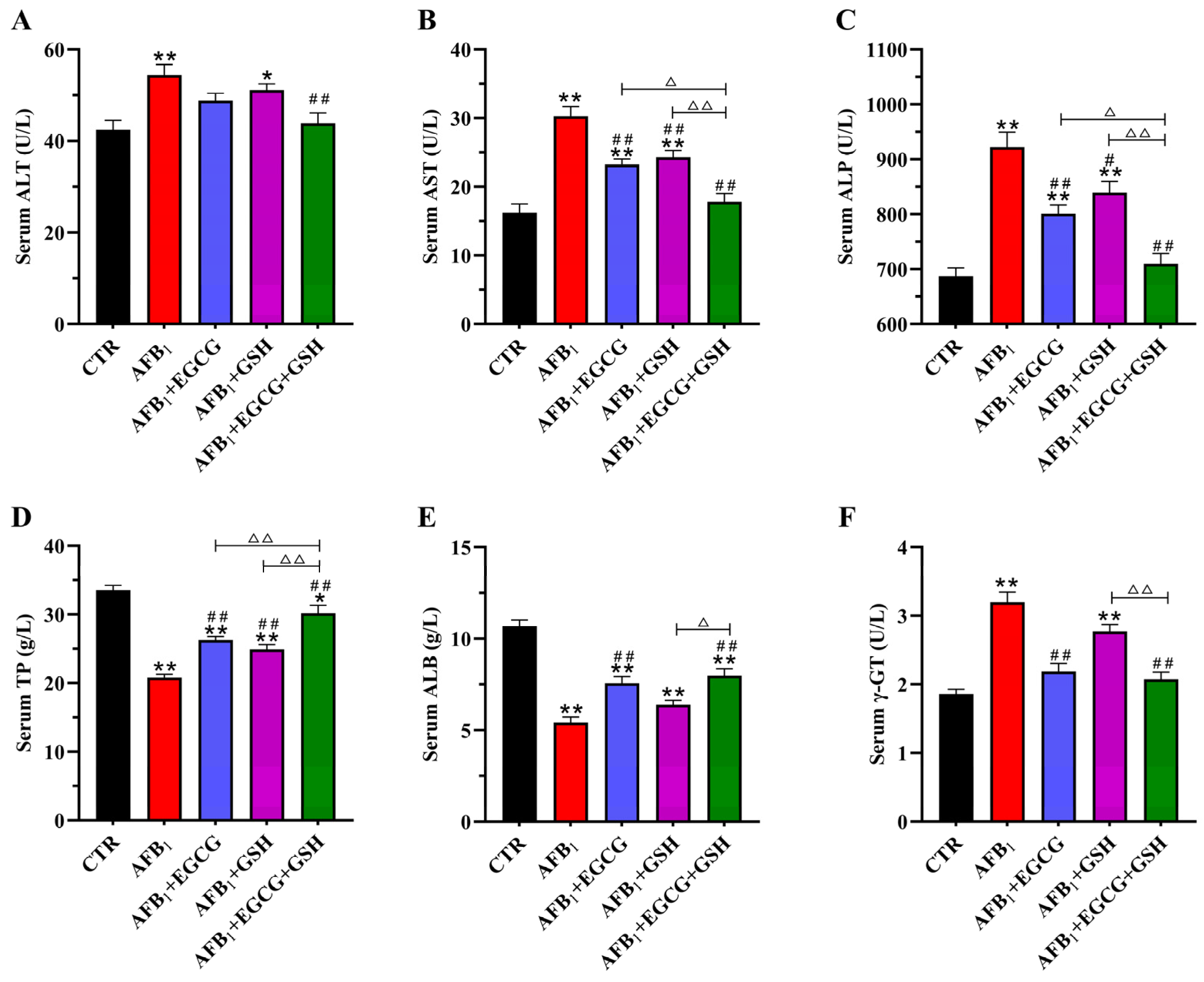
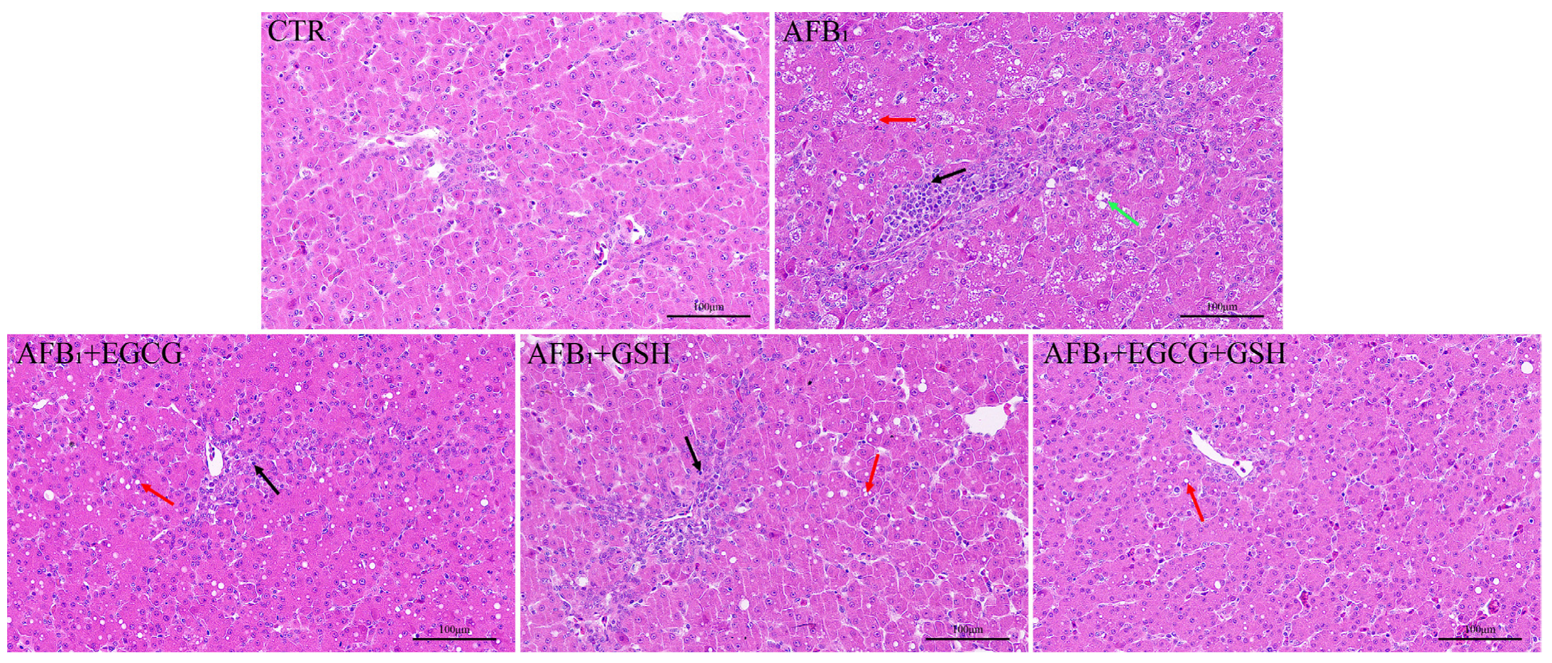
2.2. EGCG and GSH Protect against AFB1-Induced Liver Damage in Ducklings

The effects of EGCG and GSH alone or in combination on the serum biochemistry of AFB1-exposed ducklings are shown in Figure 2. Serum biochemistry was affected adversely by AFB1 as the enzyme activities of alanine aminotransferase (ALT), aspartate aminotransferase (AST), alkaline phosphatase (ALP), and γ-glutamyl transpeptidase (γ-GT) were elevated (p < 0.01, Figure 2A–C,F). The use of EGCG or GSH mitigated these adverse effects. Compared with the AFB1 group, the enzyme activities of ALT, AST, ALP, and γ-GT decreased by 19.4%, 41.2%, 23.0%, and 35.2%, respectively, in the combined detoxification group. The AFB1-treated group reduced total serum protein (TP, 37.9%) and albumin (ALB, 49.2%) levels extremely significantly (p < 0.01; Figure 2D,E). Both EGCG or GSH increased the levels of TP and ALB compared with the AFB1 group, with the combined group increasing by 45.0% and 47.1%, respectively. This was still lower than the control group. The results indicate that EGCG and GSH alleviated the negative effects caused by AFB1 on the serum biochemistry of ducklings, but a combination was more effective.

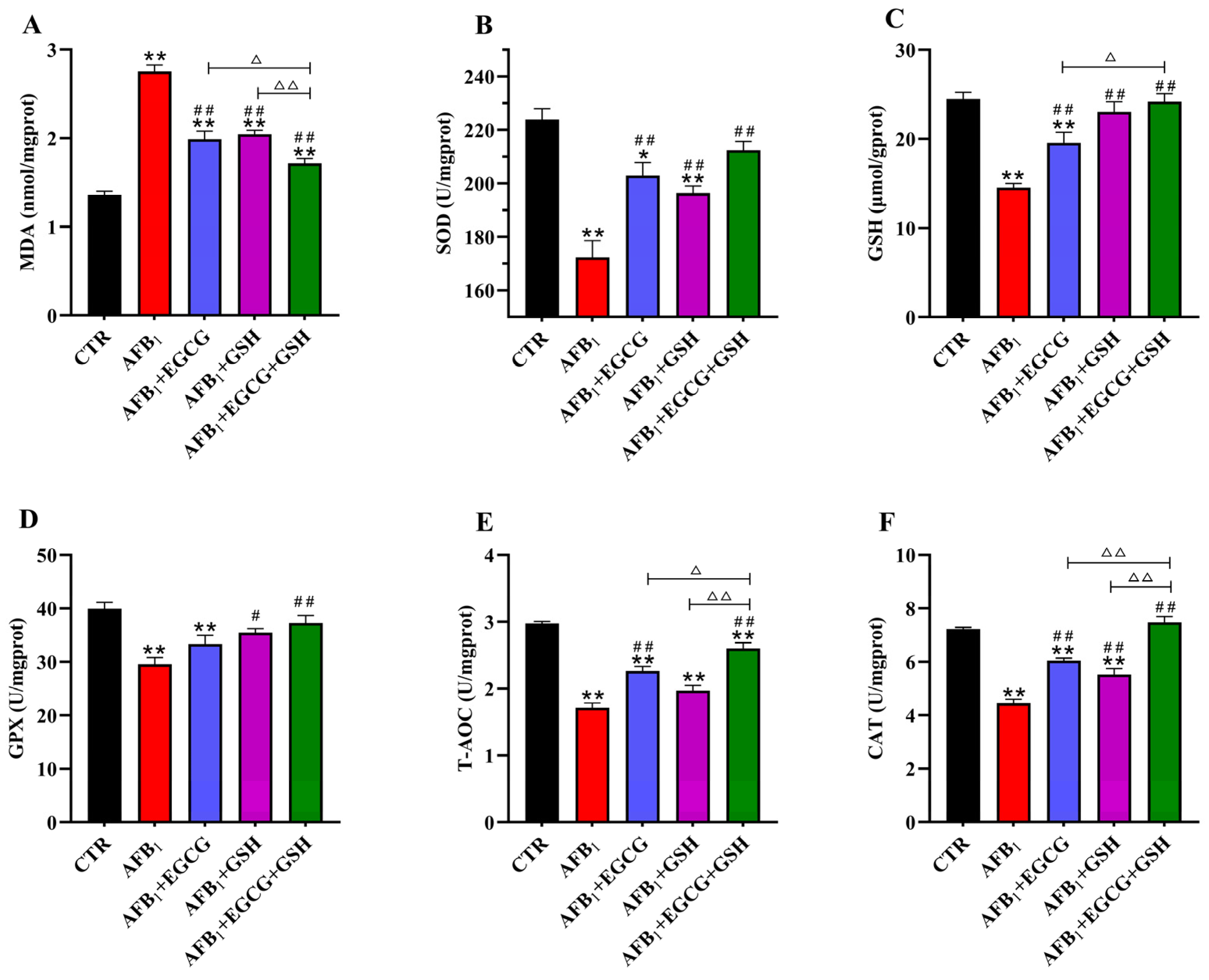
2.3. EGCG and GSH Mitigate AFB1-Induced Oxidative Stress in the Livers of Ducklings

To evaluate the damage caused by AFB1 and the protective effect of EGCG and GSH, we examined the antioxidant capacity of the ducklings’ livers. Compared with the CTR group, the AFB1 group highly significantly elevated malondialdehyde (MDA) content (p < 0.01), while EGCG, GSH, and a combination of both decreased MDA content by 27.8%, 25.7%, and 37.7%, respectively (Figure 3A). Meanwhile, the levels of antioxidant enzymes and GSH were also negatively affected, with the enzymatic activities of superoxide dismutase (SOD), glutathione peroxide (GPX), total antioxidant capacity (T-AOC), and catalase (CAT) decreasing by 23.0%, 26.0%, 34.2%, and 38.4% (Figure 3B,D–F), respectively, compared with the CTR group, while the levels of GSH decreased by 40.6% (Figure 3C). Thus, EGCG and GSH effectively prevented their alteration, especially in MDA, T-AOC, and CAT, where there was a significant joint effect (p < 0.05). The results indicate that EGCG or GSH alleviated the oxidative damage caused by AFB1, but a combination was more effective.

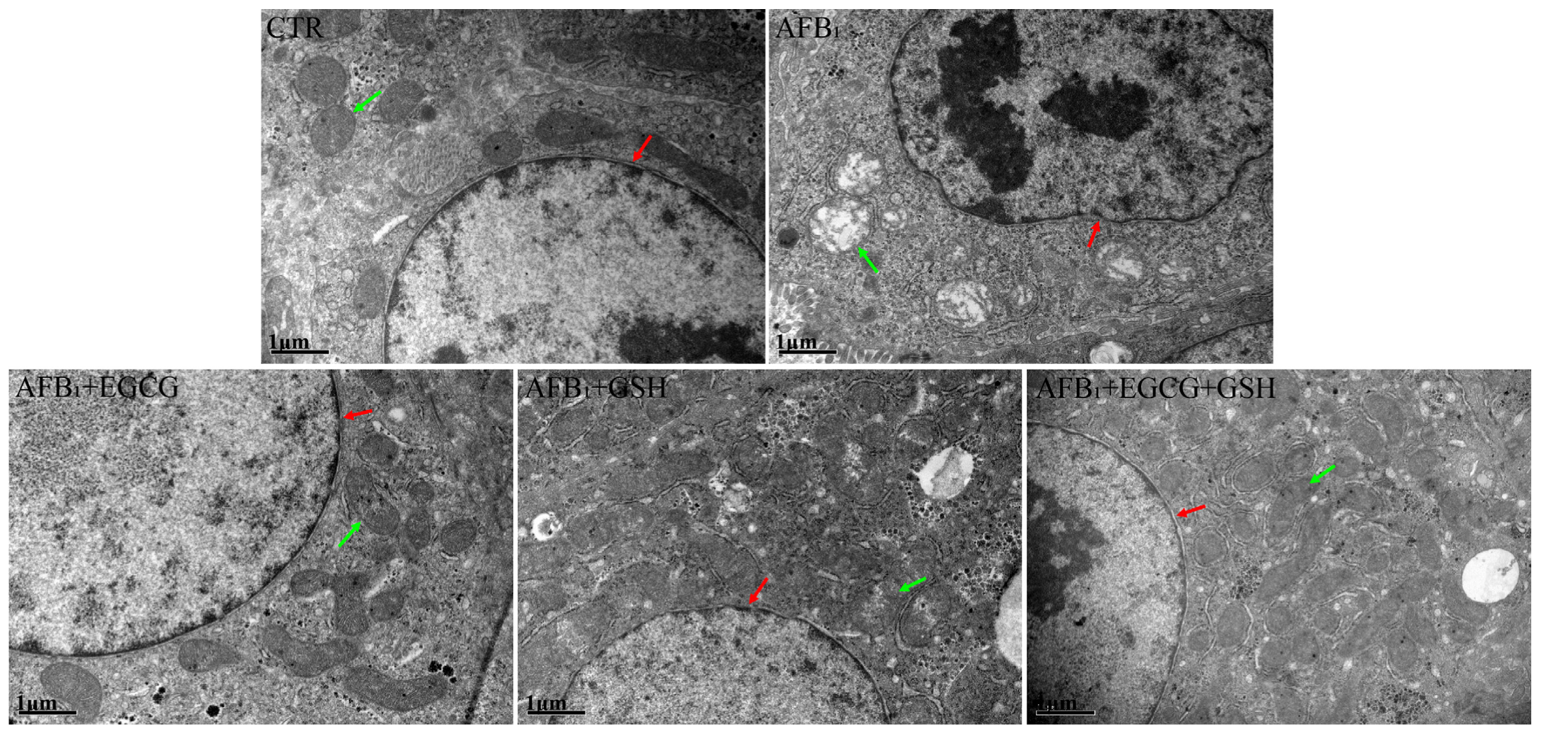
2.4. EGCG and GSH Prevent AFB1-Induced Alterations in the Microstructure and Ultrastructure of Duckling Livers

The liver is the target organ of AFB1 action, so we observed its microscopic and ultrastructural structure. As Figure 4 shows, the liver tissue structure of the CTR group was normal, with an intact hepatocyte structure and no fatty degeneration, necrosis, or inflammatory cell infiltration. However, we observed that large areas of hepatocytes were ill-defined, some cells were swollen and necrotic, and disappeared nuclei or pyknosis was present in the AFB1 group. Compared with the AFB1 group, inflammatory cell infiltration and hepatocyte necrosis were reduced in the detoxification group alone or in combination, especially in the combined detoxification group. However, lipid droplets were still present in some hepatocytes.
To examine the internal structure of hepatocytes more closely, we performed transmission electron microscopy scans (Figure 5). In the CTR group, the nuclei and mitochondrial structures were normal, while in the AFB1 group, the nuclei underwent significant wrinkling, and the mitochondrial structures were heavily abnormal, with swelling and disrupted mitochondrial ridges. Although the mitochondrial structure was also lesioned to varying degrees in the EGCG or GSH groups, it was largely improved relative to the AFB1 group. The best results were seen in the combined group.

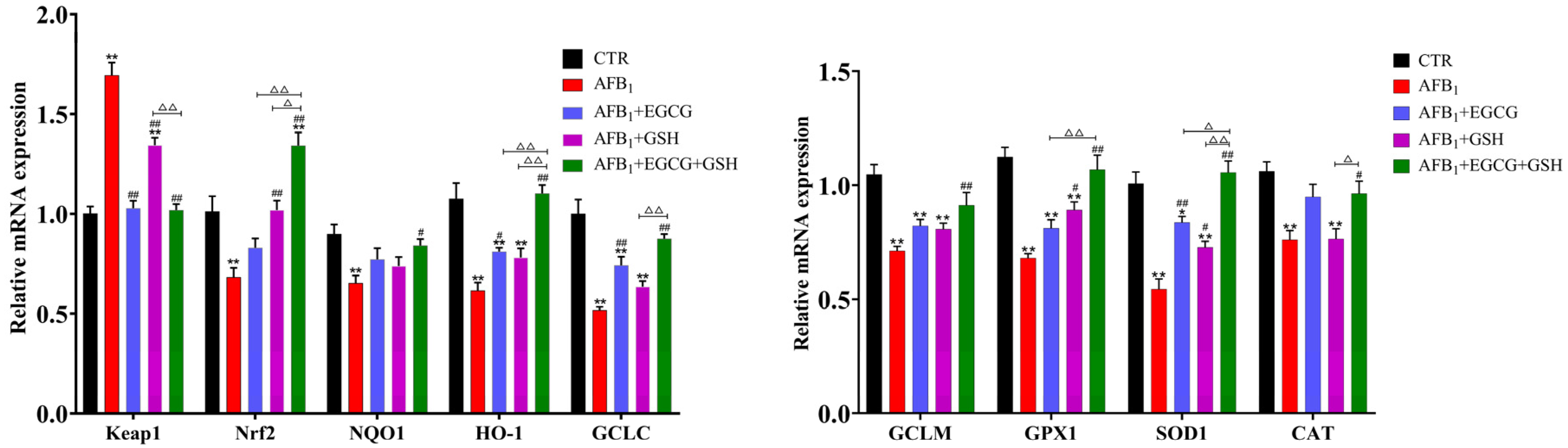
2.5. EGCG and GSH Alleviate the Interference of AFB1 on the Keap1-Nrf2 Antioxidant Signalling Pathway

As Figure 6 shows, the abundance of related genes in the Nrf2 signalling pathway was significantly downregulated in the AFB1 group compared with the CTR group (p < 0.01). However, the gene expression of Keap1, an Nrf2 repressor, was significantly elevated (p < 0.01). These changes were back-regulated to varying degrees in the EGCG and GSH groups and in the combined group. The combined group Nrf2, HO-1 and SOD1 gene expression reached significant levels (p < 0.05) compared to the group used alone. The results indicate that EGCG and GSH can alleviate the oxidative damage caused by AFB1 and that they interact to some degree.

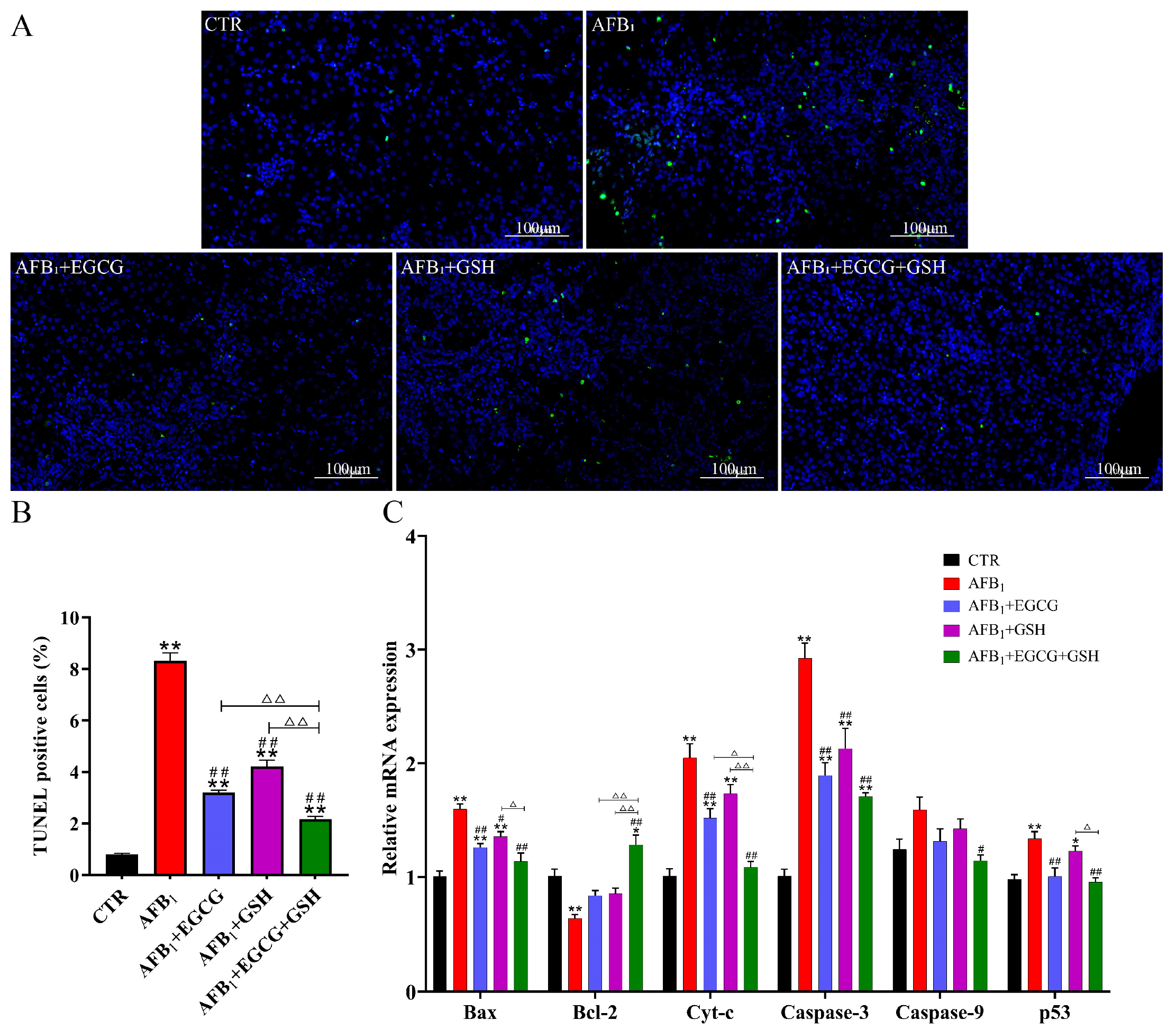
2.6. Protective Effects of EGCG and GSH on AFB1-Induced Apoptosis of Duckling Hepatocytes

In order to evaluate the protective effect of EGCG and GSH alone or in combination against AFB1-induced apoptosis, hepatocyte apoptosis and the expression of genes related to apoptosis mediated by mitochondria were examined by terminal deoxynucleotidyl transferase dUTP nick end-labelling (TUNEL) staining and RT-qPCR, respectively. Green fluorescence was significantly enhanced (as evidenced in the increased number of apoptotic cells) in all AFB1-exposed groups (Figure 7A); the apoptosis rate (TUNEL positive rate) was elevated to 8.31% in the AFB1 group, while the apoptosis rate decreased by 61.5%, 49.2%, and 74.0% in the EGCG, GSH, and combined groups, respectively, compared with the AFB1 group (Figure 7B). Compared with the CTR group, the gene abundance of the pro-apoptotic gene Bax, as well as Cyt-c, Caspase-3, and p53 were significantly up-regulated (p < 0.01), and the anti-apoptotic gene Bcl-2 was significantly down-regulated (p < 0.01, Figure 7C) in the AFB1 group; EGCG and GSH alone or in combination had a positive effect. It was concluded that EGCG and GSH attenuated AFB1-induced apoptosis in hepatocytes.

3. Discussion

It is well-known that AFB1 is commonly found in feed and causes severe damage to commercial animals, especially ducks [26,27]. Growth retardation and hepatic lesion are among the most important symptoms of AFB1 poisoning. In the present study, we discovered that AFB1 reduced the ducklings’ feed intake and body weight, as well as caused liver damage. Our findings are consistent with previous research showing that AFB1 causes a decrease in food intake, metabolic capacity, body weight, and significantly higher liver coefficients [28,29,30]. The results of the present study indicated that EGCG and GSH significantly increased body weight and decreased liver indices. Serum ALT, AST, and ALP are the most sensitive indicators for evaluating liver damage, and AFB1 in the diet can increase the levels of these enzymes [31]. In one study, when ducklings were fed a diet of 0.1 mg/kg AFB1, the levels of AST, ALT, and the ratio of AST/ALT increased [32]. Because AFB1 inhibits protein biosynthesis, serum TB and ALB can be used to evaluate its impact [33]. Our results confirmed this: AFB1 elevated the levels of ALT, AST, ALP, and γ-GT while decreasing the content of TB and ALB compared with the CTR group. The addition of EGCG and GSH slowed down the change.
Oxidative stress can promote the formation of reactive oxygen species (ROS) in animal target organs [34]. Numerous studies have shown that excessive ROS can damage macromolecules such as proteins and nucleic acids, thereby producing a large amount of MDA. Therefore, MDA, as the end product of lipid peroxidation, is an important indicator for detecting oxidative damage [35,36]. However, excessive ROS in the body can be scavenged by antioxidant enzymes, including SOD, GPX, CAT, and GSH. In the present study, exposure to AFB1 increased the amount of MDA, while the content of GSH and the enzymatic activities of T-AOC, SOD, CAT, and GPX decreased. Apparently, AFB1 induced oxidative stress in the ducklings’ livers. The addition of EGCG and GSH also alleviated oxidative stress. Previous studies have shown that EGCG can attenuate carbon tetrachloride-induced oxidative stress in mouse livers and protect against H2O2-induced cellular oxidative damage [37,38]. Exogenous GSH has been found to have similar antioxidant effects in acute kidney injury in rats [39]. At the same time, we observed that AFB1 induced pathological changes in the liver. These results indicated that AFB1 caused oxidative damage to the liver, but EGCG and GSH could protect it by enhancing its antioxidant status. The antioxidant properties of EGCG and GSH themselves, as well as the fact that GSH can act as a substrate for GPX and GST, may explain the common effect exhibited by them.
Oxidative stress caused by AFB1 can regulate the expression of a series of genes involved in the antioxidant system through the Keap1-Nrf2 signalling pathway. Nrf2 is the main regulator of cells that respond to environmental stress, inducing the expression of detoxification and antioxidant enzymes. The activity of Nrf2 is dependent on the regulation of the Keap1 adaptor protein, which is a negative regulator of the former [40,41]. Under non-stress conditions, Nrf2 binds to Keap1 in the cytoplasm to promote Nrf2 ubiquitination and proteasomal degradation; when stimulated, Nrf2 dissociates from Keap1 into the nucleus and combines with nuclear receptors to regulate the expression of downstream target genes (NQO1, HO-1, GCLC, GCLM, and so on) [42], thereby performing antioxidant or detoxification functions. It has been demonstrated that EGCG can strengthen cellular defences against chemical carcinogens as well as ultraviolet (UV) and oxidative stress through the Keap1-Nrf2 signalling pathway [43]. In one experiment, the EGCG treatment group normalised the expression of Keap1-Nrf2 and its downstream regulatory proteins in fluoride-treated rat kidneys [44]. We noted a significant upregulation of Keap1 mRNA expression in the AFB1-treated group compared with the CTR group, indicating an enhanced negative regulation of Nrf2 by Keap1, while Nrf2 and its related genes (NQO1, HO-1, GCLC, GCLM, SOD1, GPX1, and CAT) were downregulated. Treatment with EGCG significantly reversed these effects (a finding that is consistent with previous studies). However, the effect of GSH on this pathway has not been investigated, so we speculated that it might regulate the expression of genes by balancing ROS production. This deserves more investigation. We concluded that EGCG and GSH contribute to the antioxidant capacity of the body through the Keap1-Nrf2 signalling pathway and that they are most effective in combination.
Apoptosis (i.e., programmed cell death) plays an essential role in controlling cell numbers and maintaining the homeostasis of multicellular organisms. Abnormal regulation of apoptosis has been associated with the development of a variety of diseases [45]. AFB1 has been reported to induce apoptosis in hepatic, pulmonary, and bone marrow cells [46,47]. The present study found hepatocytes undergoing significant apoptosis in the AFB1 group. It is widely known that mitochondria perform a central role in apoptosis initiated by many kinds of stimuli and that key events in apoptosis are associated with mitochondria [48]. Livers have been observed with severe mitochondrial lesions. In such cases, membrane permeability is altered, and Cyt-c enters the cytoplasm from the mitochondria, binding to the apoptosis protease activator Apaf-1 and caspase-9 and activating caspase-9, which in turn induces the activation of caspase-3 and subsequently triggers apoptosis mediated by the mitochondria [49]. However, mitochondria-mediated apoptosis is regulated by the pro-apoptotic protein Bax and the anti-apoptotic protein Bcl-2 [50]. The present study showed that AFB1 reduced the mRNA expression of Bcl-2 and elevated the mRNA expression of Bax, while the expression of associated apoptotic genes (Cyt-c, caspase-3, caspase-9, and so on) was significantly elevated. However, EGCG and GSH inhibited the excessive apoptosis of hepatocytes caused by AFB1 by regulating the expression of these genes. Studies have shown that EGCG protects against apoptosis in human umbilical vein endothelial cells by regulating the mitochondria-dependent apoptotic signalling pathway [51], while exogenous GSH defends IPEC-J2 cells from oxidative stress-induced apoptosis [52]. Apoptosis can be activated by oxidative stress [36]. Therefore, we speculate that EGCG and GSH alleviate apoptosis caused by AFB1 in hepatocytes either directly or by inhibiting oxidative stress. However, the mechanism of interaction between EGCG and GSH needs to be further researched.

4. Conclusions

In the present study, AFB1 caused serious damage to the ducklings. The results suggest that EGCG and GSH can alleviate acute liver injury by improving hepatic antioxidant capacity through the Keap1-Nrf2 signalling pathway and inhibiting the excessive apoptosis of hepatocytes mediated by mitochondria. This explains the protective mechanism of EGCG and GSH alone or in combination against AFB1-induced liver injury. The present study also provides a theoretical basis for their application, and we suggest that EGCG and GSH could be used as promising duck feed additives to counteract AFB1 damage.

5. Materials and Methods

5.1. Animals and Experimental Design

Age is an important factor affecting the bird’s resistance to AFB1, and male ducklings are more sensitive (male ducklings produce more AFBO than females), so we chose younger males to complete the experiment [53]. One-day-old male Cherry Valley ducks were purchased from Wuhan Yongsheng Duck Industry Co., Ltd. (Wuhan, China). The ducklings were kept in a controlled environment at a temperature of 30 ± 2 °C and 60 ± 5% humidity. The experiment (No. HZAUDU-2022-0002) was approved by the Animal Ethics Committee of Huazhong Agricultural University (Wuhan, China).
After three days of acclimatisation, 50 male ducklings were randomly divided into five groups (n = 10): 1. Control group (CTR); 2. Treated with AFB1 (>99%, Pribolab, Qingdao, China) 0.3 mg/kg BW (AFB1); 3. Treated with AFB1 0.3 mg/kg BW + EGCG (98%, Shanghai Yuanye Biotech Co., Ltd., Shanghai, China) 100 mg/kg BW (AFB1 + EGCG); 4. Treated with AFB1 0.3 mg/kg BW + GSH (Reduced, 98%, Aladdin, Shanghai, China) 30 mg/kg BW (AFB1 + GSH); 5. Treated with AFB1 0.3 mg/kg BW + EGCG 100 mg/kg BW + GSH 30 mg/kg BW (AFB1 + EGCG + GSH). Each group of ten ducklings was kept in a pen, marked and weighed individually. All ducklings were gavaged with the same concentration and 1 mL Volume/200 g BW. The acute liver injury experiment cycle lasted for 7 days. On Days 1–6, they were weighed daily and gavaged with distilled water, distilled water, EGCG, GSH, and EGCG + GSH, respectively. On Day 4, Groups 2 to 5 were treated with AFB1 0.5 h after the first gavage, and the control group was given the corresponding solvent gavage (4% dimethyl sulfoxide). Slaughter sampling took place on Day 7. The composition and nutrient levels of the basal diet are shown in Appendix A, Table A1. The acute toxic dose of AFB1 was determined based on previous reports [54,55] and preliminary experiments. Gavage doses of EGCG and GSH refer to preliminary experiment. The health status of the ducklings was strictly observed, and the body weight and feed intake were recorded during the experiment.

5.2. Sample Collection

After the ducklings fasted for 12 h, blood samples were collected using wing venipuncture into a tube. The blood samples were centrifuged at 3500 rpm for 10 min to obtain serum, which was divided and stored at −80 °C for biochemical analysis. The ducklings were immediately sacrificed and dissected to remove the liver, rinsed in cold saline, and weighed. A portion of liver tissue was cut and placed in paraformaldehyde fixative and 2.5% glutaraldehyde, respectively, for hematoxylin and eosin (H&E) staining or TUNEL detection and ultrastructural observation. The remaining part of each liver was stored at −80 °C in a refrigerator to detect antioxidant indexes, gene expression, and so on. The relative weight of the livers was calculated using the following formula:
Relative weight = liver weight (g)/body weight (g) × 100%

5.3. Determination of Serum Biochemical Indicators

The serum enzyme activities of ALT, AST, ALP, and γ-GT, as well as the content of ALB and TP, reflect the function of the liver. These indicators were measured using an automatic biochemical analyser according to the manufacturer’s set procedure (Mindray, Shenzhen, China). The serum samples were placed in a cryogenic sample tray. ALT, AST, ALP, and γ-GT were expressed as U/L, while ALB and TP were expressed as g/L. All of these kits were purchased from the same manufacturer (Mindray, Shenzhen, China).

5.4. Detection of Antioxidant Capacity of the Liver

Liver tissue homogenates were prepared according to the requirements of the corresponding kits, and the protein concentration of the homogenate supernatant was determined by the BCA protein assay kit (Beyotime Biotechnology, Shanghai, China). SOD, GPX, MDA, T-AOC, CAT, and GSH kits were procured and operated according to the manufacturer’s instructions (Nanjing Jiancheng Biotech, Nanjing, China). Absorbance was measured by a microplate reader (Multiskan MK3, Thermo Fisher Scientific, Waltham MA, USA) or visible light spectrophotometer (722E, Shanghai Spectrum Instruments Co., Ltd., Shanghai, China), and the enzyme activity or substance content was calculated and analysed.

5.5. Histopathological Analysis

Fresh liver tissue samples were placed in 4% paraformaldehyde and fixed for more than 24 h. The tissues were dehydrated with different concentrations of alcohol and embedded in wax. The wax blocks were placed in a microtome (Leica RM2016, Wetzlar, Germany) and cut into sections of 4 μm thickness. Staining with hematoxylin and eosin was performed for histopathological observation.

5.6. Ultrastructural Pathology Observation

Liver samples were cut to around 1 mm3 in size and placed in 2.5% glutaraldehyde for 24 h. After 24 h, the samples were washed three times with 0.1 M PBS and fixed with 1% osmium acid for 2 h. The samples were rewashed with 0.1 M PBS, then dehydrated with gradient acetone and embedded in resin. The embedded samples were cut into 60 nm sections using an ultramicrotome (Leica UC5, Wetzlar, Germany), stained with uranyl acetate and lead citrate solution, and observed under a transmission electron microscope (Hitachi H-7650, Tokyo, Japan) for scanning and photographing [56].

5.7. Detection of Apoptosis by TUNEL Staining

Following the TUNEL kit manufacturer’s instructions (Roche, Basel, Switzerland), the embedded liver sections were dewaxed, rehydrated, and then incubated with proteinase K at 37 °C for 30 min and washed three times with PBS. Fifty 50 μL of TUNEL reaction solution were added dropwise to the tissue, incubated at 37 °C for 2 h in the dark, washed again with PBS three times, and then incubated with 4,6-diamidino-2-phenylindole (DAPI) staining solution for 10 min while keeping it out of the light. After blocking, the images were observed and collected using an inverted fluorescence microscope (Olympus IX51, Tokyo, Japan). Fluorescence signals were analysed with Image-Pro Plus 6.0 (Media Cybernetics, Silver Spring, MD, USA), and apoptosis rates were calculated.

5.8. Quantitative Real-Time PCR

Total RNA was extracted from the ducklings’ livers using Trizol reagent (TaKaRa, Dalian, China), and the quality (A260/A280) and concentration were evaluated using a NanoDrop 2000 spectrophotometer (Thermo Fisher Scientific, Waltham, MA, USA). Genomic DNA was removed, and RNA was reverse transcribed into cDNA using PrimeScriptTM RT reagent Kit with gDNA Eraser (TaKaRa, Dalian, China) according to the steps in the instructions, and expressed genes were evaluated by Real-Time PCR using TB Green® Premix Ex TaqTM II (TaKaRa, Dalian, China). The primer sequences are shown in Appendix A Table A2. All primers were designed by Sangon Biotech (Shanghai, China) and synthesised by Tsingke Biotechnology Co., Ltd. (Beijing, China). The relative mRNA abundance was analysed following the 2−∆∆Ct formula and normalised with the housekeeping gene GAPDH [57].

5.9. Statistical Analysis

The results were analysed using one-way ANOVA with SPSS Version 26 (SPSS Incorporated, Armonk, NY, USA) and Tukey’s multiple comparisons as the post-hoc test. Outcomes were expressed as mean ± standard error (SEM). GraphPad Prism Version 9.0 (GraphPad Prism, San Diego, CA, USA) was used to visualise the data. In all statistical analyses, p < 0.05 was considered significant and p < 0.01 was considered highly significant.

Author Contributions

D.Q., conceptualisation, writing—review and editing, project administration and funding acquisition; Y.W., methodology, formal analysis, data curation and writing—original draft preparation; J.W., L.W. and P.Y., formal analysis, data curation and visualisation; Z.L., S.A.R. and M.H., writing—review and editing. All authors have read and agreed to the published version of the manuscript.

Funding

This project was supported by the National Key Research and Development Program of China (2016YFD0501207).

Institutional Review Board Statement

This experiment was approved by the Animal Ethics Committee of Huazhong Agricultural University (No. HZAUDU-2022-0002).

Informed Consent Statement

Not applicable.

Data Availability Statement

The data presented in this study are available on request from the corresponding author.

Conflicts of Interest

The authors declare no conflict of interest.

Appendix A

Table A1. Composition and nutrient level of basal diet.
IngredientsContent (%)Nutrition Component 2Content (%)
Corn52.98Crude protein20.16
Soybean meal (44% CP)30.36ME (MJ/kg)12.34
Fish meal2.50Calcium0.91
Wheat bran3.00Available phosphorus0.42
Rice bran5.25Methionine0.45
Soybean oil1.91Lysine1.12
Premix 14.00AFB1 (μg/kg)2.1
Total100.00
1 Premix provided per kilogram of diet: 10,000 IU of vitamin A, 2500 IU of vitamin D3, 35 IU of vitamin E, 2.5 mg of vitamin K3, 2.5 mg of vitamin B1, 9 mg of vitamin B2, 0.02 mg of vitamin B12, 15 mg of calcium pantothenate, 60 mg of niacin, 1.5 mg of folic acid, and 0.2 mg of biotin; 12 mg of Cu, 80 mg of Fe, 60 mg of Zn, 92 mg of Mn, 0.3 mg of Se, and 0.3 mg of I. 2 All nutrient levels were calculated.
Table A2. Primer sequences used in qRT-PCR.
GenesPrimer Sequences (5′ to 3′)Product Lengths (bp)Accession No.
Keap1F: TCAAGACCTCACCCTCCATAAACCC
R: AGTAGCCCAAGGACTGCCGATAG
110KU048807.1
Nrf2F: TGGCTGAGGTAAGCACAATCACAAG
R: GGCTCTCAACAGTCTCCAGGAAATC
138NM_001310777.1
NQO1F: TGTCACCATCTCCGACCTCTATGC
R: TCCTTCCACGCTTCTCCCATCTC
126XM_027466610.2
HO-1F: ATGAATGCCCTTGAGATGGACCTTG
R: GTGACCGTTCTCCTGGCTCTTTG
132XM_005015345.5
GCLCF: TTCAGGTGACATTCCAGGCTTGC
R: AGAACGGAGATGCAGCACTCAATG
108XM_027455103.2
GCLMF: TGTTGTGTGATGCCACCTGATCTC
R: GTGCTTTGACGTTCTGGATGCTTTC
142XM_027462629.2
SOD1F: TCATCTCTCTGACTGGACCACACTG
R: GTTAGCGTGCTCTCGTTGTCTCC
103KU048808.1
GPX1F: CCACCAGGAGAATGCCACCAAC
R: CTTCCCGTTCACCTCGCACTTC
115XM_027467953.2
CATF: ATGGACCAATGTGCGTGACTGAC
R: CATGCGGCTCTCCTTCACAACAG
104XM_027458335.2
Cyt-cF: CCAGTGCCATACGGTTGAGAAGG
R: TCTGTGTAGGAGAAGCCCTCAGC
105XM_027447873.2
BaxF: TCGTCGCCCTCTTCTACTTCGC
R: CAGGAGACGATGGTGCGGAAAAG
88KY788660.1
Bcl-2F: CATGTGCGTGGAGAGCGTCAAC
R: ACTGATCCAGCCTCCGTTGTCC
124XM_027451679.2
Caspase-3F: TGAGGCAGACAGTGGACCAGATG
R: TCCTTCAGCACCCTACACAGAGAC
156XM_021279218.3
Caspase-9F: TGGATTGCGATTCACCCGAAGATG
R: ATTACCCGAGGGAGCCTGGAAAG
83XM_038166520.1
p53F: AGGAGGAGAACTTCCGCAAGAGG
R: GCAGGCAGAAGATCTCGTTGTCG
129XM_038171818.1
GAPDHF: GTCTCCTGCGACTTCAACGG
R: CCTTGGATGCCATGTGGACC
160XM_038180584.1

References

  1. Reverberi, M.; Ricelli, A.; Zjalic, S.; Fabbri, A.A.; Fanelli, C. Natural functions of mycotoxins and control of their biosynthesis in fungi. Appl. Microbiol. Biotechnol. 2010, 87, 899–911. [Google Scholar] [CrossRef] [PubMed]
  2. da Silva, J.B.; Dilkin, P.; Fonseca, H.; Correa, B. Production of aflatoxins by Aspergillus flavus and of fumonisins by Fusarium species isolated from Brazilian sorghum. Braz. J. Microbiol. 2004, 35, 182–186. [Google Scholar] [CrossRef] [Green Version]
  3. Girolami, F.; Barbarossa, A.; Badino, P.; Ghadiri, S.; Cavallini, D.; Zaghini, A.; Nebbia, C. Effects of turmeric powder on aflatoxin M1 and aflatoxicol excretion in milk from dairy cows exposed to aflatoxin B1 at the EU maximum tolerable levels. Toxins 2022, 14, 430. [Google Scholar] [CrossRef] [PubMed]
  4. Sana, S.; Anjum, A.A.; Yaqub, T.; Nasir, M.; Ali, M.A.; Abbas, M. Molecular approaches for characterisation of aflatoxin producing Aspergillus flavus isolates from poultry feed. Pak. Vet. J. 2019, 39, 169–174. [Google Scholar] [CrossRef] [PubMed]
  5. Chen, K.; Fang, J.; Peng, X.; Cui, H.; Chen, J.; Wang, F.; Chen, Z.; Zuo, Z.; Deng, J.; Lai, W.; et al. Effect of selenium supplementation on aflatoxin B-1-induced histopathological lesions and apoptosis in bursa of Fabricius in broilers. Food Chem. Toxicol. 2014, 74, 91–97. [Google Scholar] [CrossRef]
  6. Morrison, D.M.; Ledoux, D.R.; Chester, L.F.; Samuels, C.A.J.V.S. A limited survey of aflatoxins in poultry feed and feed ingredients in Guyana. Vet. Sci. 2017, 4, 60. [Google Scholar] [CrossRef] [Green Version]
  7. Guo, Y.; Zhao, L.; Ma, Q.; Ji, C. Novel strategies for degradation of aflatoxins in food and feed: A review. Food Res. Int. 2021, 140, 109878. [Google Scholar] [CrossRef]
  8. Deng, M.; Zhu, F.; Yang, Y.; Yang, F.; Hao, J.; Chen, S.; Hou, Z. Genome-wide association study reveals novel loci associated with body size and carcass yields in Pekin ducks. BMC Genom. 2019, 20, 1. [Google Scholar] [CrossRef]
  9. Diaz, G.J.; Murcia, H.W. An unusually high production of hepatic aflatoxin B-1-dihydrodiol, the possible explanation for the high susceptibility of ducks to aflatoxin B-1. Sci. Rep. 2019, 9, 8010. [Google Scholar] [CrossRef] [Green Version]
  10. Rushing, B.R.; Selim, M.I. Aflatoxin B1: A review on metabolism, toxicity, occurrence in food, occupational exposure, and detoxification methods. Food Chem. Toxicol. 2019, 124, 81–100. [Google Scholar] [CrossRef]
  11. Stepkowski, T.M.; Kruszewski, M.K. Molecular cross-talk between the NRF2/KEAP1 signaling pathway, autophagy, and apoptosis. Free Radic. Biol. Med. 2011, 50, 1186–1195. [Google Scholar] [CrossRef] [PubMed]
  12. Chen, Y.; Feng, X.; Hu, X.; Sha, J.; Li, B.; Zhang, H.; Fan, H. Dexmedetomidine ameliorates acute stress-induced kidney injury by attenuating oxidative stress and apoptosis through inhibition of the ROS/JNK signaling pathway. Oxid. Med. Cell. Longev. 2018, 2018, 4035310. [Google Scholar] [CrossRef] [PubMed]
  13. Liu, Y.; Wang, W. Aflatoxin B1 impairs mitochondrial functions, activates ROS generation, induces apoptosis and involves Nrf2 signal pathway in primary broiler hepatocytes. Anim. Sci. J. 2016, 87, 1490–1500. [Google Scholar] [CrossRef]
  14. Rains, T.M.; Agarwal, S.; Maki, K.C. Antiobesity effects of green tea catechins: A mechanistic review. J. Nutr. Biochem. 2011, 22, 1–7. [Google Scholar] [CrossRef] [PubMed]
  15. Abd El-Hack, M.E.; Elnesr, S.S.; Alagawany, M.; Gado, A.; Noreldin, A.E.; Gabr, A.A. Impact of green tea (Camellia sinensis) and epigallocatechin gallate on poultry. World’s Poult. Sci. J. 2020, 76, 49–63. [Google Scholar] [CrossRef]
  16. Marin, L.; Miguelez, E.M.; Villar, C.J.; Lombo, F. Bioavailability of dietary polyphenols and gut microbiota metabolism: Antimicrobial properties. BioMed Res. Int. 2015, 2015, 905215. [Google Scholar] [CrossRef] [Green Version]
  17. Chiu, H.; Venkatakrishnan, K.; Wang, C. The role of nutraceuticals as a complementary therapy against various neurodegenerative diseases: A mini-review. J. Tradit. Complement. Med. 2020, 10, 434–439. [Google Scholar] [CrossRef]
  18. Aydogan, I.; Karsli, M.A.; Basalan, M.; Yildirim, E.; Cinar, M.; Sen, G.; Sumer, T. Effects of supplemental epigallocatechin gallate in the diet of broilers exposed to fluoride intoxication. Biol. Trace Elem. Res. 2018, 186, 258–266. [Google Scholar] [CrossRef]
  19. Kim, H.-S.; Quon, M.J.; Kim, J.-a. New insights into the mechanisms of polyphenols beyond antioxidant properties; lessons from the green tea polyphenol, epigallocatechin 3-gallate. Redox Biol. 2014, 2, 187–195. [Google Scholar] [CrossRef] [Green Version]
  20. Sriram, N.; Kalayarasan, S.; Sudhandiran, G. Epigallocatechin-3-gallate augments antioxidant activities and inhibits inflammation during bleomycin-induced experimental pulmonary fibrosis through Nrf2-Keap1 signaling. Pulm. Pharmacol. Ther. 2009, 22, 221–236. [Google Scholar] [CrossRef]
  21. Orhan, C.; Tuzcu, M.; Gencoglu, H.; Sahin, N.; Hayirli, A.; Sahin, K. Epigallocatechin-3-gallate exerts protective effects against heat stress through modulating stress-responsive transcription factors in poultry. Br. Poult. Sci. 2013, 54, 447–453. [Google Scholar] [CrossRef] [PubMed]
  22. Lui, S.M.; Eady, S.J. Glutathione: Its implications for animal health, meat quality, and health benefits of consumers. Aust. J. Agric. Res. 2005, 56, 775–780. [Google Scholar] [CrossRef]
  23. Lomaestro, B.M.; Malone, M. Glutathione in health and disease: Pharmacotherapeutic issues. Ann. Pharmacother. 1995, 29, 1263–1273. [Google Scholar] [CrossRef]
  24. Saeij, J.P.J.; van Muiswinkel, W.B.; van de Meent, M.; Amaral, C.; Wiegertjes, G.F. Different capacities of carp leukocytes to encounter nitric oxide-mediated stress: A role for the intracellular reduced glutathione pool. Dev. Comp. Immunol. 2003, 27, 555–568. [Google Scholar] [CrossRef]
  25. Atef, H.A.; El-Shafei, H.M.; Mansour, M.K.; Al Kalamawy, N.M.; Hassan, N.; Lashin, A.J.I.J.C.M.A.S. Prevalence of ochratoxigenic fungi and ochratoxin A residues in animal feeds and modulation of their toxic effects by glutathione. Int. J. Curr. Microbiol. Appl. Sci. 2018, 7, 2559–2582. [Google Scholar] [CrossRef]
  26. Fan, Y.; Liu, L.; Zhao, L.; Wang, X.; Wang, D.; Huang, C.; Zhang, J.; Ji, C.; Ma, Q. Influence of Bacillus subtilis ANSB060 on growth, digestive enzyme and aflatoxin residue in Yellow River carp fed diets contaminated with aflatoxin B-1. Food Chem. Toxicol. 2018, 113, 108–114. [Google Scholar] [CrossRef] [PubMed]
  27. Limaye, A.; Yu, R.; Chou, C.; Liu, J.; Cheng, K. Protective and detoxifying effects conferred by dietary selenium and curcumin against AFB1-mediated toxicity in livestock: A Review. Toxins 2018, 10, 25. [Google Scholar] [CrossRef] [PubMed] [Green Version]
  28. Mehta, R.; Campbell, J.S.; Laver, G.W.; Stapley, R.; Mueller, R. Acute hepatic response to aflatoxin B1 in rats fed a methyl-deficient, amino acid-defined diet. Cancer Lett. 1993, 69, 93–106. [Google Scholar] [CrossRef]
  29. Ruggeberg, K.-G.; O’Sullivan, P.; Kovacs, T.J.; Dawson, K.; Capponi, V.J.; Chan, P.P.; Golobish, T.D.; Gruda, M.C. Hemoadsorption improves survival of rats exposed to an acutely lethal dose of aflatoxin B-1. Sci. Rep. 2020, 10, 799. [Google Scholar] [CrossRef] [PubMed] [Green Version]
  30. Yan, J.; Chen, L.; Zhang, L.; Zhang, Z.; Zhao, Y.; Wang, Y.; Ou, J. New insights into the persistent effects of acute exposure to AFB(1) on rat liver. Front Microbiol. 2022, 13, 911757. [Google Scholar] [CrossRef]
  31. Saei, M.M.; Sadeghi, A.A.; Ahmadvand, H. The effect of Myrtus communis oil extract on growth performance, serum biochemistry and humoral immune responses in broiler chicks fed diet containing aflatoxin B1. Arch. Tierz. 2013, 56, 842–850. [Google Scholar] [CrossRef] [Green Version]
  32. Mendez-Albores, A.; Del Rio-Garcia, J.C.; Moreno-Martinez, E. Decontamination of aflatoxin duckling feed with aqueous citric acid treatment. Anim. Feed Sci. Technol. 2007, 135, 249–262. [Google Scholar] [CrossRef]
  33. Mojgani, N.; Razmgah, N.; Torshizi, M.A.K.; Sanjabi, M.R. Effects of three Bacillus specious on hatchability, growth performance and serum biochemistry in Japanese quails fed diet contaminated with Aflatoxin B1. Acta Sci. Anim. Sci. 2020, 42, e50184. [Google Scholar] [CrossRef]
  34. Yener, Z.; Celik, I.; Ilhan, F.; Bal, R. Effects of Urtica dioica L. seed on lipid peroxidation, antioxidants and liver pathology in aflatoxin-induced tissue injury in rats. Food Chem. Toxicol. 2009, 47, 418–424. [Google Scholar] [CrossRef] [PubMed]
  35. Bai, K.; Huang, Q.; Zhang, J.; He, J.; Zhang, L.; Wang, T. Supplemental effects of probiotic Bacillus subtilis fmbJ on growth performance, antioxidant capacity, and meat quality of broiler chickens. Poult. Sci. 2017, 96, 74–82. [Google Scholar] [CrossRef] [PubMed]
  36. Li, X.; Lv, Z.; Chen, J.; Nepovimova, E.; Long, M.; Wu, W.; Kuca, K. Bacillus amyloliquefaciens B10 can alleviate liver apoptosis and oxidative stress induced by aflatoxin B1. Food Chem. Toxicol. 2021, 151, 112124. [Google Scholar] [CrossRef] [PubMed]
  37. Cia, D.; Vergnaud-Gauduchon, J.; Jacquemot, N.; Doly, M. Epigallocatechin gallate (EGCG) prevents H2O2-induced oxidative stress in primary rat retinal pigment epithelial cells. Curr. Eye Res. 2014, 39, 944–952. [Google Scholar] [CrossRef]
  38. Tipoe, G.L.; Leung, T.M.; Liong, E.C.; Lau, T.Y.H.; Fung, M.L.; Nanji, A.A. Epigallocatechin-3-gallate (EGCG) reduces liver inflammation, oxidative stress and fibrosis in carbon tetrachloride (CCl4)-induced liver injury in mice. Toxicology 2010, 273, 45–52. [Google Scholar] [CrossRef]
  39. Babaeenezhad, E.; Dezfoulian, O.; Hadipour Moradi, F.; Rahimi Monfared, S.; Fattahi, M.D.; Nasri, M.; Amini, A.; Ahmadvand, H.J.D.; Toxicology, C. Exogenous glutathione protects against gentamicin-induced acute kidney injury by inhibiting NF-κB pathway, oxidative stress, and apoptosis and regulating PCNA. Drug Chem. Toxicol. 2022. [Google Scholar] [CrossRef]
  40. Kovesi, B.; Pelyhe, C.; Zandoki, E.; Mezes, M.; Balogh, K. Changes of lipid peroxidation and glutathione redox system, and expression of glutathione peroxidase regulatory genes as effect of short-term aflatoxin B-1 exposure in common carp. Toxicon 2018, 144, 103–108. [Google Scholar] [CrossRef]
  41. Suzuki, T.; Yamamoto, M. Molecular basis of the Keap1-Nrf2 system. Free Radic. Biol. Med. 2015, 88, 93–100. [Google Scholar] [CrossRef] [PubMed] [Green Version]
  42. Baird, L.; Yamamoto, M. The molecular mechanisms regulating the KEAP1-NRF2 pathway. Mol. Cell. Biol. 2020, 40, e00099-20. [Google Scholar] [CrossRef] [PubMed]
  43. Na, H.K.; Surh, Y.J. Modulation of Nrf2-mediated antioxidant and detoxifying enzyme induction by the green tea polyphenol EGCG. Food Chem. Toxicol. 2008, 46, 1271–1278. [Google Scholar] [CrossRef]
  44. Thangapandiyan, S.; Miltonprabu, S. Epigallocatechin gallate supplementation protects against renal injury induced by fluoride intoxication in rats: Role of Nrf2/HO-1 signaling. Toxicol. Rep. 2014, 1, 12–30. [Google Scholar] [CrossRef] [PubMed] [Green Version]
  45. Patel, B.K. Targeting apoptosis pathways for cancer therapy. In Cancer Drug Discovery and Development: The Oncogenomics Handbook; LaRochelle, W.J., Shimkets, R.A., Eds.; Humana Press: Totowa, NJ, USA, 2005; pp. 229–244. ISBN 978-1-59259-893-9. [Google Scholar]
  46. Meki, A.R.M.; Abdel-Ghaffar, S.K.; El-Gibaly, I.J.N.L. Aflatoxin B1 induces apoptosis in rat liver: Protective effect of melatonin. Neuroendocrinol. Lett. 2001, 22, 417–426. [Google Scholar] [PubMed]
  47. Raj, H.G.; Kohli, E.; Rohil, V.; Dwarakanath, B.S.; Parmar, V.S.; Malik, S.; Adhikari, J.S.; Tyagi, Y.K.; Goel, S.; Gupta, K.; et al. Acetoxy-4-methylcoumarins confer differential protection from aflatoxin B-1-induced micronuclei and apoptosis in lung and bone marrow cells. Mutat. Res., Genet. Toxicol. Environ. Mutagen. 2001, 494, 31–40. [Google Scholar] [CrossRef] [PubMed]
  48. Bensassi, F.; Gallerne, C.; El Dein, O.S.; Lemaire, C.; Hajlaoui, M.R.; Bacha, H. Involvement of mitochondria-mediated apoptosis in deoxynivalenol cytotoxicity. Food Chem. Toxicol. 2012, 50, 1680–1689. [Google Scholar] [CrossRef]
  49. Twiddy, D.; Brown, D.G.; Adrain, C.; Jukes, R.; Martin, S.J.; Cohen, G.M.; MacFarlane, M.; Cain, K. Pro-apoptotic proteins released from the mitochondria regulate the protein composition and caspase-processing activity of the native Apaf-1/caspase-9 apoptosome complex. J. Biol. Chem. 2004, 279, 19665–19682. [Google Scholar] [CrossRef] [Green Version]
  50. Brunelle, J.K.; Letai, A. Control of mitochondrial apoptosis by the Bcl-2 family. J. Cell Sci. 2009, 122, 437–441. [Google Scholar] [CrossRef] [Green Version]
  51. Liu, S.; Sun, Z.; Chu, P.; Li, H.; Ahsan, A.; Zhou, Z.; Zhang, Z.; Sun, B.; Wu, J.; Xi, Y.; et al. EGCG protects against homocysteine-induced human umbilical vein endothelial cells apoptosis by modulating mitochondrial-dependent apoptotic signaling and PI3K/Akt/eNOS signaling pathways. Apoptosis 2017, 22, 672–680. [Google Scholar] [CrossRef]
  52. Chen, Q.; Yu, M.; Tian, Z.; Cui, Y.; Deng, D.; Rong, T.; Liu, Z.; Song, M.; Li, Z.; Ma, X.; et al. Exogenous glutathione protects IPEC-J2 cells against oxidative stress through a mitochondrial mechanism. Molecules 2022, 27, 2416. [Google Scholar] [CrossRef]
  53. Dohnal, V.; Wu, Q.; Kuca, K. Metabolism of aflatoxins: Key enzymes and interindividual as well as interspecies differences. Arch. Toxicol. 2014, 88, 1635–1644. [Google Scholar] [CrossRef] [PubMed]
  54. Monson, M.S.; Coulombe, R.A.; Reed, K.M.J.A. Aflatoxicosis: Lessons from toxicity and responses to aflatoxin B1 in poultry. Agriculture 2015, 5, 742–777. [Google Scholar] [CrossRef] [Green Version]
  55. Yunus, A.W.; Razzazi-Fazeli, E.; Bohm, J. Aflatoxin B-1 in affecting broiler’s performance, immunity, and gastrointestinal tract: A review of history and contemporary issues. Toxins 2011, 3, 566–590. [Google Scholar] [CrossRef] [Green Version]
  56. Cao, J.; Song, Y.; Wu, H.; Qin, L.; Hu, L.; Hao, R. Ultrastructural studies on the natural leaf senescence of cinnamomum camphora. Scanning 2013, 35, 336–343. [Google Scholar] [CrossRef] [PubMed]
  57. Zhong, G.; Wan, F.; Lan, J.; Jiang, X.; Wu, S.; Pan, J.; Tang, Z.; Hu, L. Arsenic exposure induces intestinal barrier damage and consequent activation of gut-liver axis leading to inflammation and pyroptosis of liver in ducks. Sci. Total Environ. 2021, 788, 147780. [Google Scholar] [CrossRef] [PubMed]
Figure 1. Effect of Epigallocatechin gallate (EGCG) and Glutathione (GSH) on Aflatoxin B1 (AFB1)-induced changes in growth performance and liver index of ducklings. (A) Average total feed intake per duckling during the experiment. (B) Body weight of each duckling at the beginning and end of the experiment. (C) Relative weight of the liver. Results are expressed as means ± SEM (n = 10). * p < 0.05, ** p < 0.01 vs. control (CTR) group; # p < 0.05, ## p < 0.01 vs. AFB1 group; ∆ p < 0.05, ∆∆ p < 0.01 significant difference between AFB1 + EGCG + GSH and AFB1 + EGCG or AFB1 + GSH groups.
Toxins 14 00876 g001
Figure 2. Effect of EGCG and GSH on AFB1-induced changes in serum biochemical parameters. (A) ALT, alanine aminotransferase; (B) AST, aspartate aminotransferase; (C) ALP, alkaline phosphatase; (D) TP, total protein; (E) ALB, albumin; (F) γ-GT, γ-glutamyl transpeptidase. Results are expressed as means ± SEM (n = 10). * p < 0.05, ** p < 0.01 vs. CTR group; # p < 0.05, ## p < 0.01 vs. AFB1 group; ∆ p < 0.05, ∆∆ p < 0.01 significant difference between AFB1 + EGCG + GSH and AFB1 + EGCG or AFB1 + GSH groups.
Toxins 14 00876 g002
Figure 3. Effect of EGCG and GSH on AFB1-induced oxidative stress in the livers of ducklings. (A) MDA, malondialdehyde; (B) SOD, superoxide dismutase; (C) GSH, glutathione; (D) GPX, glutathione peroxidase; (E) T-AOC, total antioxidant capacity; (F) CAT, catalase. Results are expressed as means ± SEM (n = 10). * p < 0.05, ** p < 0.01 vs. CTR group; # p < 0.05, ## p < 0.01 vs. AFB1 group; ∆ p < 0.05, ∆∆ p < 0.01 significant difference between AFB1 + EGCG + GSH and AFB1 + EGCG or AFB1 + GSH groups.
Toxins 14 00876 g003
Figure 4. Effect of EGCG and GSH on the microscopic pathological structure of the livers of ducklings exposed to AFB1. Magnification 200×, scale bars = 100 µm. Green arrows: cell swelling and necrosis, nuclear pyknosis; black arrows: inflammatory cell infiltration in the hepatic parenchyma; red arrows: a small number of lipid droplets can be seen in the cytoplasm.
Toxins 14 00876 g004
Figure 5. Effect of EGCG and GSH on the ultramicro-pathological structure of the livers of ducklings exposed to AFB1. Magnification 20,000×, scale bars = 1 µm. Red arrows represent the nucleus of the cell, and green arrows represent mitochondria.
Toxins 14 00876 g005
Figure 6. Effect of EGCG and GSH on the abundance of genes involved in the Keap1-Nrf2 signalling pathway in the livers of ducklings exposed to AFB1. All results are expressed as means ± SEM (n = 6). * p < 0.05, ** p < 0.01 vs. CTR group; # p < 0.05, ## p < 0.01 vs. AFB1 group; ∆ p < 0.05, ∆∆ p < 0.01 significant difference between AFB1 + EGCG + GSH and AFB1 + EGCG or AFB1 + GSH groups.
Toxins 14 00876 g006
Figure 7. Effects of EGCG and GSH on AFB1-induced apoptosis of ducklings’ hepatocytes. (A) Terminal deoxynucleotidyl transferase dUTP nick end-labelling (TUNEL) staining to detect apoptosis. Magnification 200 ×, scale bars = 100 µm. Fluorescently labelled green indicates apoptotic cells, and blue indicates the nucleus. (B) TUNEL positive cells. (C) Expression of genes associated with mitochondria-mediated apoptosis, Cyt-c, Bax, Bcl-2, Caspase-3, Caspase-9, p53. All results are expressed as means ± SEM (n = 6). * p < 0.05, ** p < 0.01 vs. CTR group; # p < 0.05, ## p < 0.01 vs. AFB1 group; ∆ p < 0.05, ∆∆ p < 0.01 significant difference between AFB1 + EGCG + GSH and AFB1 + EGCG or AFB1 + GSH groups.
Toxins 14 00876 g007
Publisher’s Note: MDPI stays neutral with regard to jurisdictional claims in published maps and institutional affiliations.

