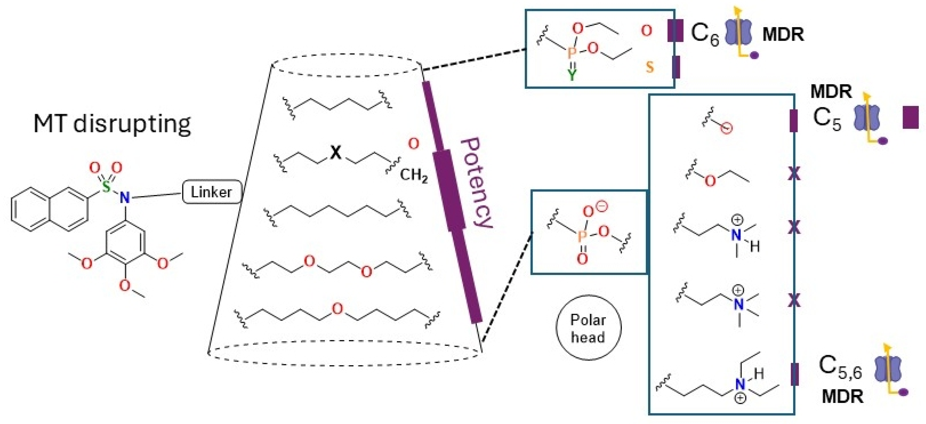
2. Results and Discussion
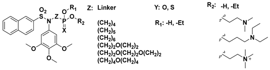
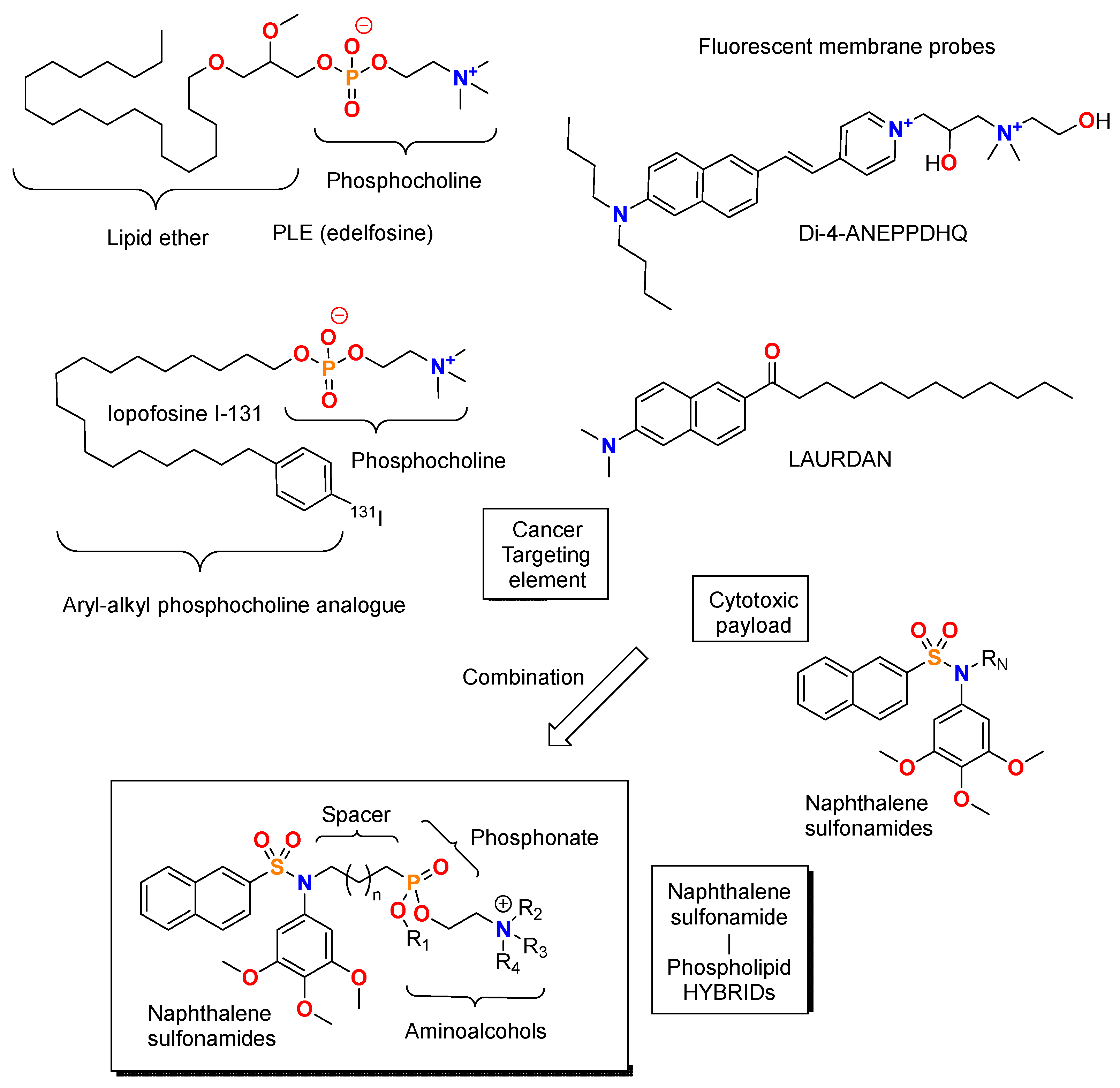
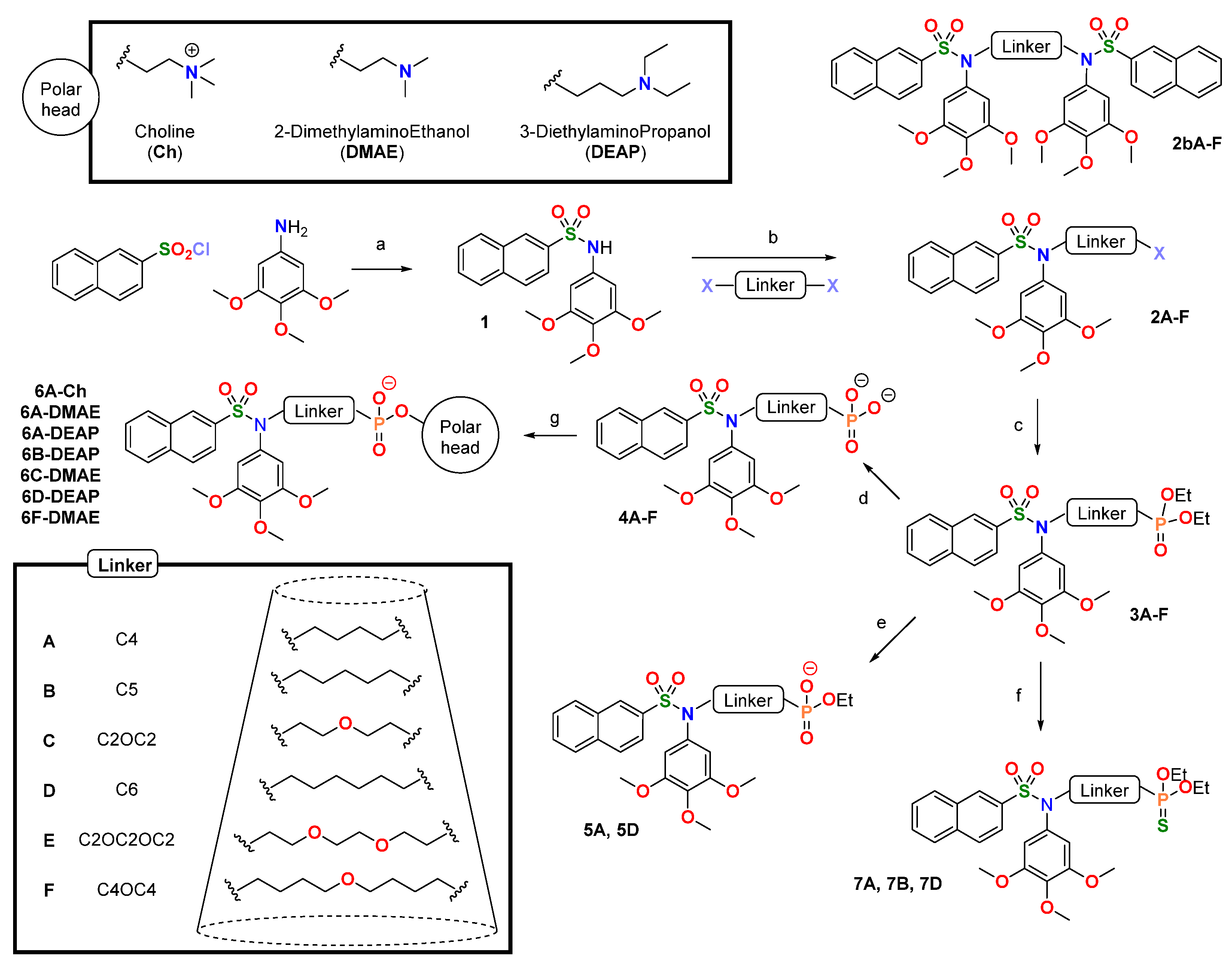
Seven naphthalene sulfonamide—phospholipid hybrids with different spacer lengths and polarity (hydrocarbons or ethers) and amino alcohol phosphonates (2-dimethylaminoethanol, 3-diethylaminopropanol, or choline) were prepared according to synthetic
Scheme 1. The synthetic route involved assembly of the sulfonamide (a), followed by nitrogen alkylation (b) and introduction of a terminal phosphonic acid (c,d), which finally yielded the phosphonates by esterification with amino alcohols (g). Partial hydrolysis of the intermediate diethyl phosphonates (e) and conversion to the thiophosphonates (f) on selected derivatives to study their effect on the activity were performed.
Naphthalene sulfonamide
1 was obtained in good yield by reacting trimethoxyaniline with naphthalene-2-sulfonyl chloride as described [
32]. Alkylation of the sulfonamide nitrogen with dihalogenated spacers, in situ converted into the iodides by the Finkelstein reaction with KI, was performed under basic catalysis with cesium carbonate to activate the sulfonamide nitrogen to achieve alkylated sulfonamides (
2A-
E) as mixtures of the possible terminal halogens (iodine and bromine or chlorine coming from either the KI or the dihalogenated reagent). Despite using a large excess (3–5 moles per mole of sulfonamide) of the dihalogenated reagent, products of the double substitution (
2bA-
F) were obtained in proportions exceeding the stoichiometric expectation. The proportion varies with the linker, thus suggesting structure–dependent reactivity. The sulfonamide nitrogen was substituted with alkyls or alkyl ethers of different lengths (A-F: from four to nine atoms) to explore the effect of these variations on the biological activity.
The diethyl phosphonates were prepared in moderate yields by means of a Michaelis–Arbuzov rearrangement, heating the halogen-capped alkyl sulfonamides in neat triethylphosphite. Subsequent hydrolysis with trimethylsilyl bromide (TMSBr), generated in situ with TMSCl and NaBr, gave the phosphonic acids in varied yields, mainly dependent on the isolation difficulties. Partial hydrolysis was carried out with NaI in acetone (
7A) [
33] or hydrazine hydrate (
7D) [
34] with good to excellent yields. Reaction of the diethylphosphonates with Lawesson’s reagent yielded the diethylthiophosphonates in moderate (
7A and
7B) to good (
7D) yields. The naphthalene sulfonamide—APL hybrids were synthesized by esterification of the phosphonic acids with the aminoalcohols using a carbodiimide (EDC) to activate the acid. The isolated yields were low due to the difficulty in purifying the desired phosphonates from the intermediate phosphonyl isoureas, which do not fully react to the desired phosphonates due to steric hindrance and the low nucleophilicity of the alcohols. The purification is further complicated by the formation of salts of 4-DMAP. Therefore, several chromatographic separations and solid-liquid extractions with different solvents are required to achieve the pure products for the biological assays (
Supplementary Material: Supplementary Figures S16–S38).
- b.
Biology
The antiproliferative effect of the phosphorus-containing naphthalene sulfonamides was evaluated against the human cancer cell lines HeLa (cervix epithelioid carcinoma), a sensitive cell-line towards anti-tubulin sulfonamides [
21,
26,
28,
29,
35], and HT-29 (colon adenocarcinoma), that shows much less sensitivity to colchicine-site agents such as combretastatin A-4 [
36] but is sensitive to alkylphospholipids such as edelfosine [
37], after 72 h of treatment at a concentration of 10 µM using the MTT method. Compounds that inhibited proliferation by more than 40% compared with the untreated control in three independent experiments were assayed at different concentrations, and their IC
50 values were calculated (
Table 1). The diethyl phosphonates reach potencies in the low micromolar to the submicromolar range for both cell lines (HeLa: 1.24–0.26 µM, HT-29: 1.54–0.34 µM), well under the value for miltefosine (7.26 µM and 2.61 µM for HeLa and HT-29, respectively), and in the same order as the antimitotic sulfonamide ABT-751 (0.20 µM and 0.25 µM for HeLa and HT-29, respectively) used as a reference. The phosphonic acids with a butyl or a 5-oxanonyl spacer have IC
50 values comparable to miltefosine in HeLa cells, whereas the remaining acids do not show antiproliferative potency under the chosen cutoff. Against HT-29, only the first one is active. The thiophosphonates are less potent than their phosphonate pairs but still active in the low micromolar range for HeLa cells, while only the five-carbon spacer analogue is active against HT-29. The potencies against HeLa are slightly but systematically higher than against HT-29, and these IC
50 increases in this cell line probably push them above the selected cutoff of 10 µM. The partially hydrolysed products are also inactive, even if, for the butyl derivative, both the diethylphosphonate and the phosphonic acid are active. None of the aminoalcohol ethers (also having a partially hydrolysed phosphonate) is active.
Those compounds that showed IC
50 values below 10 µM against HeLa or HT-29 cells were assayed against the human glioblastoma cancer cell line U87 MG, as glioblastomas are very chemoresistant cell lines that have recently been shown to be specially sensitive to microtubule-targeting agents [
38,
39] and in particular anti-tubulin sulfonamides, and the MCF7 (human breast adenocarcinoma) that is especially sensitive to some types of sulfonamides [
28]. As before, an initial screen of cell proliferation inhibition at 10 μM was followed by the establishment of the IC
50 values. Antiproliferative assays on HEK-293 (human embryonic kidney), a non-tumorigenic cancer cell line, as a surrogate for normal cells, were performed. ABT-751, a representative of colchicine-site sulfonamide in the clinic, and miltefosine, a PLE, were used as positive controls (
Table 1,
Supplementary Material: Supplementary Table S1, ST1 and Supplementary Figure S1, SF1).
Overall, all the cell lines show similar sensitivities towards the compounds, with modest differences in their respective IC50 values. MCF7 shows sensitivities similar to HeLa for phosphonates and slightly higher IC50 values for phosphonic acids and thiophosphonates. U87 behaves similarly, with higher increases in IC50 values for phosphonic acids and thiophosphonates. HEK-293 cells show similar sensitivities as HeLa cells, with low selectivity indexes, except for phosphonic acid 4F, which shows a selectivity index comparable to miltefosine. On the other hand, sulfonamide ABT-751 shows similar potencies against all the cell lines, including HEK-293, and therefore has poor selectivity indexes.
These results suggest that the naphthalene sulfonamides tolerate substitutions on the sulfonamide nitrogen, incorporating spacers capped with phosphorus groups of different polarity. Intriguingly, the partial hydrolysis of diethyl phosphonates abrogates potency, whereas complete hydrolysis to the phosphonates does not. The zwitterionic species 6 do not show antiproliferative potency below 10 μM against any of the cell lines. Amongst the active series, the highest potency is observed for the 5-carbon spacer, with the four- and six-carbon ones following, in what seems an odd-even effect. Introducing an oxygen atom in the 5-carbon spacer substantially reduces potency against all cell lines. Longer (8- or 9-atom) oxygenated spacers are well tolerated, with lower IC50 values than the four- and six-carbon spacers. The four-carbon spacer is less effective for the phosphonates, suggesting it is not long enough. The potency modulation by the spacers is maintained over all the active capping groups, suggesting that they reflect binding to a common pocket, which somehow accommodates end groups of different polarity, except for the zwitterionic species.
- ii.
MDR pumps are an important determinant of the activity of the modified naphthalene sulfonamides
The multidrug-resistant (MDR) phenotype is one of the main mechanisms by which cancer cells exert resistance against chemotherapy, both innate and induced [
40]. This mechanism is also one of the main ways cancer cells develop resistance in the clinic against microtubule-targeting drugs [
41], thus reducing their efficacy. The MDR phenotype is due to membrane pumps that transfer drugs from inside to outside the cells, thus lowering their intracellular concentrations, reducing their binding to their targets, and their anticancer activity [
40,
42,
43]. We have undertaken a pharmacological approach to study the effect of MDR pumps on the activity of the compounds, comparing the IC
50 values of the treatments with the compounds in cotreatments with verapamil (VP) at concentrations that are not toxic to the cell line (i.e., HT-29) with those obtained in its absence (
Table 1). Verapamil is a P-glycoprotein 1 and multidrug resistance protein 1 (MRP1) [
44,
45,
46] inhibitor; therefore, if the compound is an MDR substrate, its IC
50 is significantly reduced upon cotreatment with VP. We quantified the effect of verapamil cotreatment as the quotient of the IC
50 value in the absence of verapamil over the IC
50 value in the presence of verapamil (Relative sensitivity, RS) and used a three-fold decrease as the cutoff, indicating that the compound is a substrate of the efflux pumps. RS = (IC
50 non-cotreatment/IC
50 cotreated with verapamil).
The cotreatment with verapamil results in changes in the potency of many compounds (
Scheme 2), turning some inactive compounds (with IC
50 values > 10 µM) into active ones with IC
50 values < 10 µM and improving the potency of some already active ones, but not all, thus indicating a structure-dependent effect. This is quite different from what is observed for our previously described naphthalene sulfonamides with
N-substituents of a different nature, which are not substrates of the MDR efflux pumps. Amongst the diethyl phosphonates, the cotreatment increases the potency, but only significantly for the analogue with a six-carbon spacer
3D, which then equals the potency of the five-carbon analogue
3B. This suggests that the apparent odd-even effect suggested by the antiproliferative data alone is indeed a reflection of two different effects: the four-carbon spacer is too short, and the six-carbon spacer results in enhanced efflux, therefore reducing the potency with respect to the five-carbon spacer. The similar potency of the five- and six-carbon spacers suggests that the optimal length is in this range.
The co-treatment with verapamil has a dramatic effect on the potencies of the phosphonic acids, turning all of them, except the one with the oxygenated, nine-atom spacer 4F, active (with IC50 values < 10 µM). In this case, the effect on 4B, with the five-carbon spacer, stands out, becoming again the most potent of the series with a submicromolar IC50 value after a more than one-fold potency improvement. In this case, the improvement of the six-carbon spacer just puts it at the limit, being much less potent than the five-carbon analogue. Again, the oxygen atom in 4C results in a lower potency, which does not match that of 4B, thus suggesting the detrimental effect of the oxygen atom is due to the binding to the target. For the thiophosphonates, there is no substantial potency increase upon treatment with VP, thus suggesting that their lower potency compared with the phosphonates is due to the interaction with the target and not to the uptake. The same applies to the ethyl hydrogenophosphonates, which do not improve upon verapamil co-treatment. Finally, a significant recovery is seen for some of the amino alcohol esters with a diethylaminopropanol cap (6B-DEAP and 6D-DEAP), with the five (again most potent) and six-carbon spacers upon verapamil co-treatment. This suggests that the zwitterions can bind to the target, presumably at the interfacial space of the tubulin dimer, but they are rendered inactive by efflux proteins. These results highlight the difficulties in obtaining small-molecule drug conjugates, requiring not only compliance with the pharmacophoric requirements of the target(s) but also avoiding escape from the efflux pumps.
With the aim of characterizing the mechanism of action of the most active compounds, the phosphonates
3B and
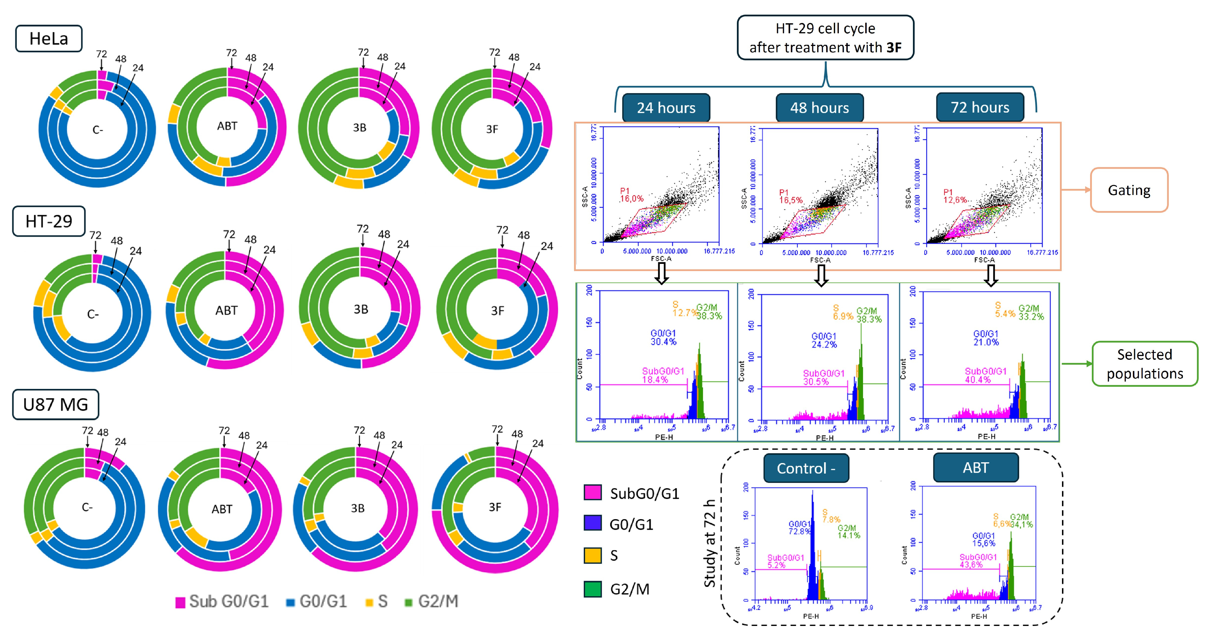
3F, we studied their effect on the distribution of cells in the different populations along the cell cycle. We assessed the effect by flow cytometric analysis of the DNA content of HeLa, HT-29, and U-87 MG cells at 24 h, 48 h, and 72 h post-treatment. ABT-751 was a positive control, representative of antimitotic sulfonamides binding to the colchicine site of tubulin (
Figure 2,
Supplementary Material: Supplementary Tables S2–S7, ST2–ST7).
The assay is based on the fluorescence intensity enhancement that results from the Propidium Iodide’s (PI) intercalation between the DNA bases, that allows quantification of the DNA within each cell and its assignment to populations based on their DNA contents: cells with a 2N DNA content are assigned to the G
0/G
1 phases, those with 4N to the G
2/M phases, cells with DNA amounts in between 2N and 4N are assigned to the S phase, and DNA contents less than 2N to the subG
0/G
1 population, usually considered as apoptotic cells [
47].
3B was tested at 1 µM for the three cell lines,
3F at 1 µM for HeLa and HT-29, and 1.5 µM for U87-MG, and ABT-751 at 0.5 µM for HeLa and HT-29 and 1 µM for U87-MG. Negative controls were treated with the same volume of DMSO used for the treatments.
The cell populations of HeLa cells treated with solvent (negative controls) remain similar over time: G0/G1 (77–82%), G2/M (12–15%), S (3.0–3,5%), and subG0/G1 (3–6%). Treatment with ABT-751 arrests HeLa cells at G2/M at 24 h, with a reduction at 48 h and close to control at 72 h (45–38–19%, respectively). These changes are accompanied by an initial increase (26%) of the subG0/G1 population that falls to half (14%) at 48 h, then sharply increases at 72 h, when it accounts for 50% of the cells. 3B and 3F show similar profiles, with a larger 24 h G2/M arrest than ABT-751 (61 and 56%, respectively), which are slightly reduced at 48 h (46 and 43%, respectively) and 72 h (44 and 39%, respectively). These are accompanied by progressive increases in subG0/G1 populations (17–28–34% for 3B and 13–23–31% for 3F). The positive antimitotic control and the compounds show quite similar trends, with 3B and 3F arresting more at G2/M and ABT-751 inducing more apoptosis.
Untreated HT-29 cells distribute similarly to HeLa cells, with slightly larger G2/M (15–25%) and smaller G0/G1 populations (62–74%). ABT-751 modestly arrests cells in G2/M at 24 h, then brings it back to control levels at 48 h and 72 h (38–26–18%, respectively). Cell counts in subG0/G1 rise at 24 h and 48 h up to 40%, with a final boost to 55% at 72 h. 3B initially arrests cells at G2/M (52%), sustains it at 48 h (49%), and falls to 29% at 72 h. 3F shows a slower increase in G2/M at 24 h (39%) than 3B, which continues to increase at 48 h to 48% and ends up with an abrupt fall (32%) at 72 h. This apparent earlier start of 3B is also observed for the SubG0/G1 populations, which increase at 24 h (27% for 3B and 12% for 3F) to proportionately progress with time (31% at 48 h and 49% at 72 h for 3B vs. 20% at 48 h and 39% at 72 h for 3F). The overall trends for the ABT-751 control and the compounds are similar, with subtle kinetic differences.
The untreated U-87 MG cells show higher basal G2/M levels (30–34%) than HeLa or HT-29, and lower G0/G1 (56–57%). ABT-751 treatment in this cell line does not arrest cells at G2/M (control-like values of 34% and 29% at 24 h and 48 h, respectively), with a fall to 14% at 72 h, while the subG0/G1 populations steeply increase over time (17–47–63%). Similar effects are observed for the treatments with 3B and 3F, with equal to control G2/M levels (no arrest) at 24 h, maintained for 3B and slightly increased for 3F, to fall in both of them in a similar way to the ABT-751 treatment. These changes are accompanied by a persistent increase in subG0/G1 at 24 h and 48 h, larger than the ABT-751 treatment, that finishes with a large increase at 72 h. These differences can well be explained by small kinetic differences in the onset of the cell effects.
The two compounds behave similarly, and differences across the three cell lines are few, mostly arising from kinetic differences in the evolution of the events. These results are consistent with the uniformity of anti-proliferative potencies of the compounds across the cell lines and suggest a common mechanism of action.
- iv.
3B and 3F induce cell death 72 h post-treatment
Aiming to better characterize the mechanism of action we studied the cell death induced in the HeLa, HT-29, and U87-MG cells after 72 h of treatment with
3B and
3F at the same concentrations used in the cell cycle experiments (i.e.,
3B was tested at 1 µM for the three cell lines,
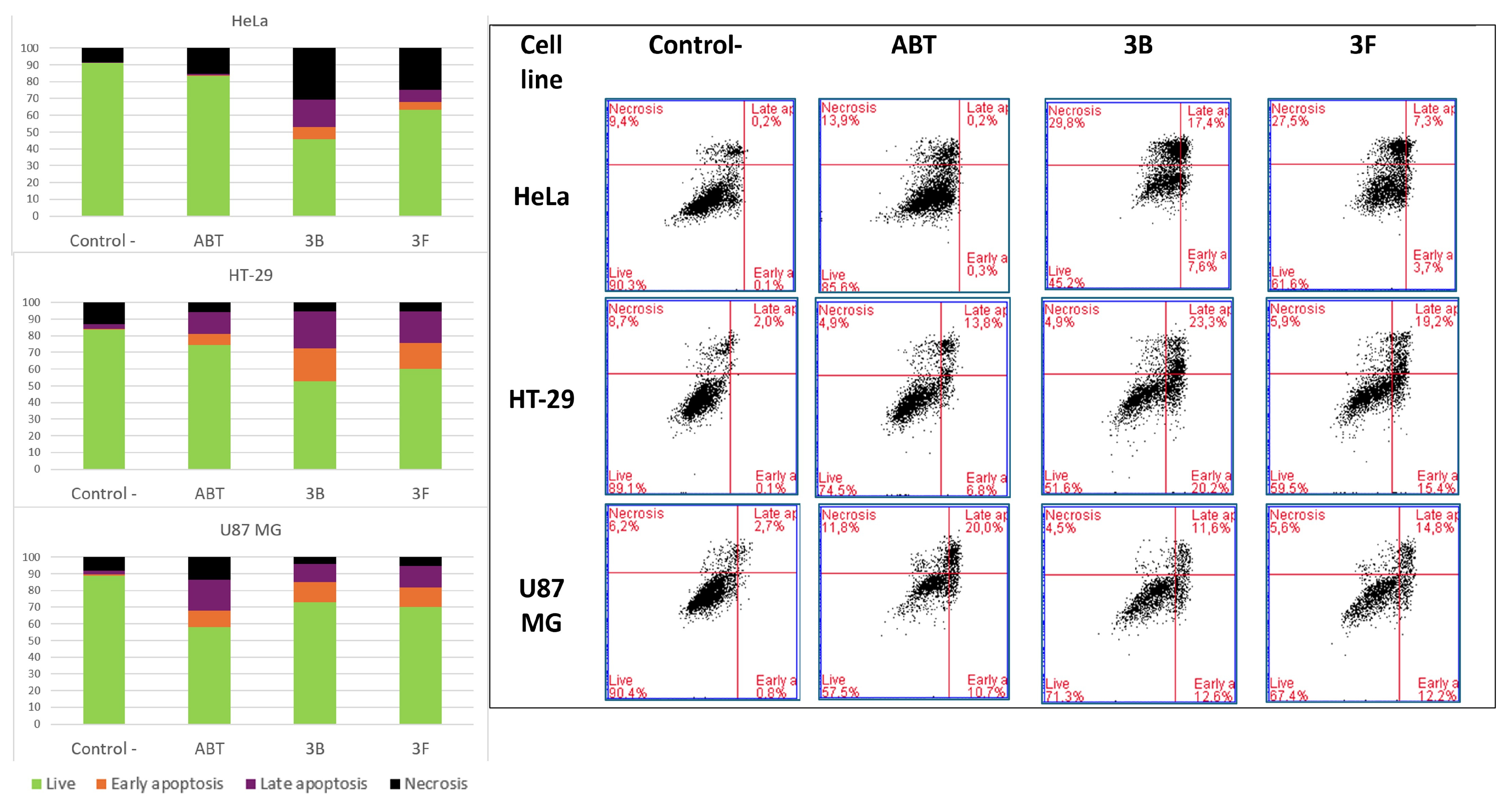
3F at 1 µM for HeLa and HT-29, and 1.5 µM for U87-MG). ABT-751 at 0.5 µM for HeLa and HT-29 and 1 µM for U87-MG were positive controls. Cells were stained with propidium iodide (PI) and fluorescein isothiocyanate-labeled Annexin V (AnV) (
Figure 3,
Supplementary Material: Supplementary Tables S8 and S9, ST8 and ST9) and subjected to bidimensional flow cytometry. Phosphatidylserine is translocated to the outer leaflet of the cell membrane in apoptotic cells, which are therefore labelled by fluorescent Annexin V (AnV
+). PI is only taken up by cells in late apoptosis or necrotic stages with damaged cellular membranes (PI
+). Accordingly, cells were classified by dual-channel flow cytometry into four populations depending on the labeling with the two stains as healthy (PI
−, AnV
−), early apoptotic (PI
−, AnV
+), late apoptotic (PI
+, AnV
+), or necrotic (PI
+, AnV
−) [
48].
In HeLa cells, treatments with 3B and 3F induce cell death substantially above the levels of the untreated control and of those treated with ABT-751. ABT increases apoptosis through the necrotic population, while 3B increases necrotic and late apoptotic cells to a lesser degree, and even fewer early apoptotic cells. 3F is similar to 3B but with a lesser increase in every apoptotic group. 3F increases cell death compared to ABT-751, and 3B does so to a larger extent.
For HT-29 cells, increased apoptosis, early and late, is observed for ABT-751, and more in both 3B and 3F, which are similar to each other. 3B and 3F show a large increase in early and late apoptosis; in both cases, more for 3B.
In U-87 MG cells, the effects of ABT-751 are larger than those of 3B and 3F. The ABT-751 increase is due, in increasing order, to late apoptosis, early apoptosis, and necrosis. For 3B and 3F, there is almost no increase in necrotic cells, with smaller increases in late apoptosis than for ABT-751 and larger early apoptosis. Contrary to the other two cell lines, 3F is here the more potent inducer of the naphthalene-sulfonamides.
As observed for the cell cycle, the compounds and the control behave qualitatively similarly, with the main differences arising between the cell lines. This suggests similar underlying mechanisms for the three, and the differences might arise from kinetic differences.
- v.
3B and 3F disrupt the microtubule network of tumor cell lines
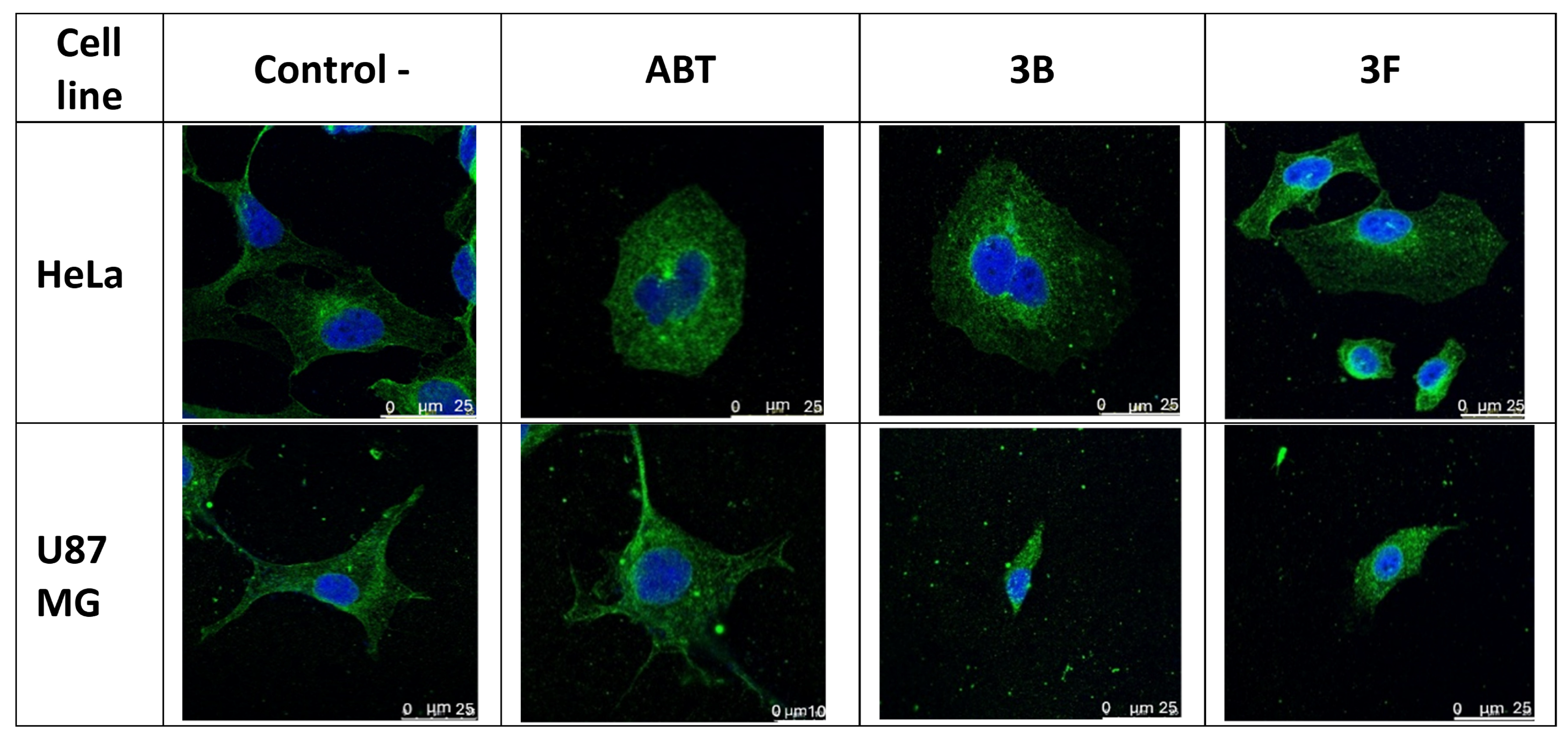
Trying to determine whether the compounds act on the microtubule network, we carried out immunostaining experiments with anti-alpha-tubulin antibodies and DAPI for staining the nuclei (
Figure 4). HeLa and U-87 MG cells were treated for 24 h with ABT-751 (at 0.5 µM for HeLa and at 1 µM for U-87 MG),
3B (at 1 µM), and
3F (at 1 µM for HeLa and 1.5 µM for U-87 MG) and stained.
The two naphthalene sulfonamides result in apparent changes in cell shape, with cells losing their typical shapes and becoming round, a characteristic phenotype of cytoskeleton disassembly that occurs early during apoptosis. Microtubule network integrity is lost upon treatment in both cell lines, with cells having smaller cytoplasms and many with multilobed nuclei. The effects of ABT-751, a typical microtubule inhibitor, are very similar to those of 3B and 3F, thus suggesting a common mechanism of action involving microtubule disorganization inside the cells, reinforcing their proposed mechanism of action as antimitotic agents.
- vi.
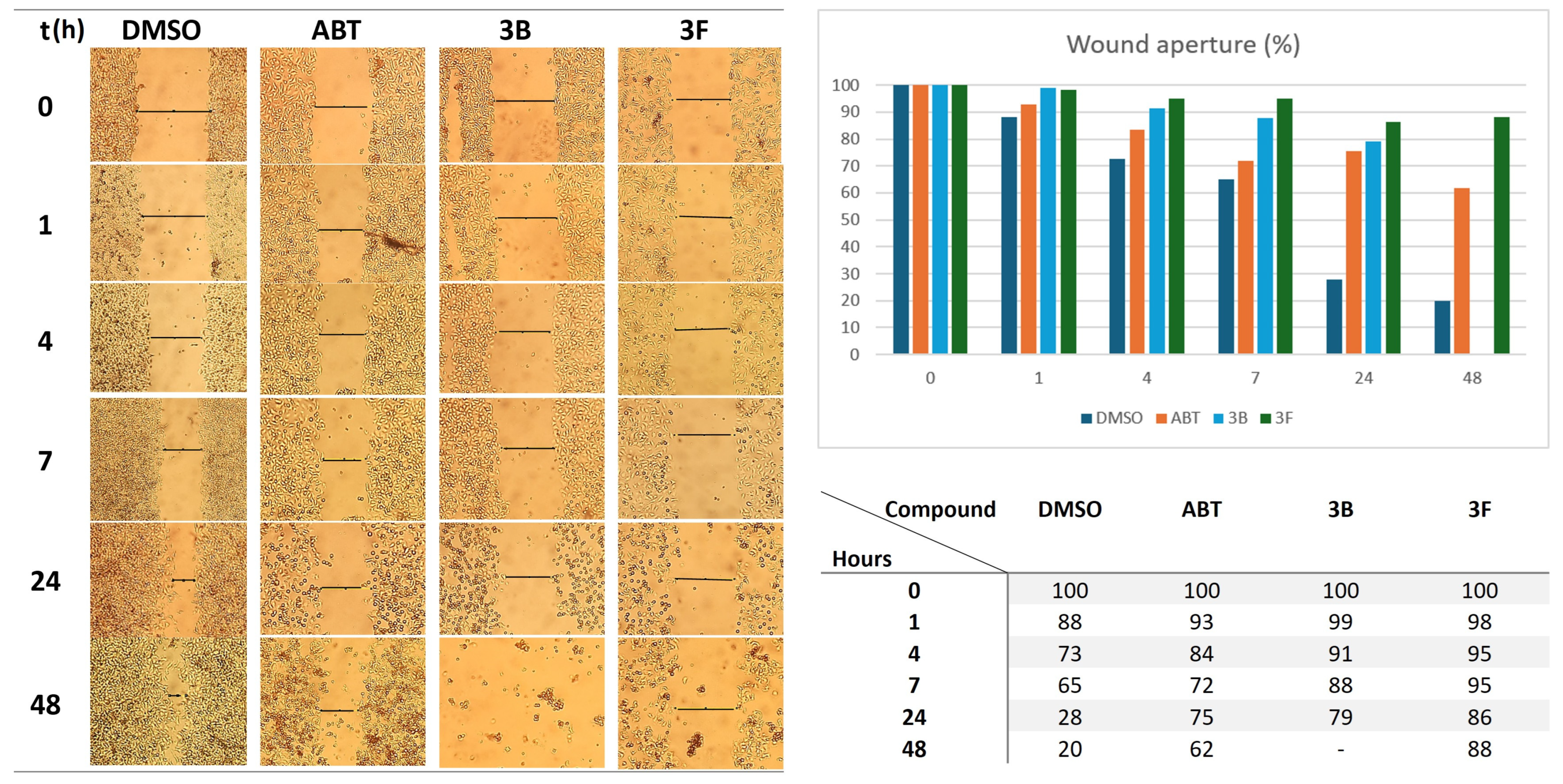
3B and 3F impair the migration of HeLa cells
Metastasis is the main cause of cancer-related deaths, with migration being critical for cancer cell invasion and metastasis. In the in vitro wound-healing assay, cells are grown in monolayers, scratched, and images are taken at regular intervals to measure the gap closure, a surrogate of cell migration. Microtubules play an essential role in cell migration through their involvement in signaling, cell mechanics, intracellular trafficking, and interactions with actin. Tubulin inhibitors are, therefore, potential migration and metastasis inhibitors. The alkylphospholipids, involved in modifying lipid membrane fluidity, are also considered to have anti-migratory effects. Therefore, we set out to study the antimigratory effect of compounds 3B and 3D by scratch healing assays on monolayers of HeLa cells.
In this assay, HeLa cells are allowed to grow up to confluence, and then a scratch is made, thus generating a cell-free gap. Then, the compounds are added at the desired concentration, and pictures are taken at different intervals (
Figure 5). Quantification of the closure of the gap with time gives an estimation of the migration rate of the cells.
ABT-751 and compounds 3B and 3F slow down the closure of the wound compared to the untreated control, which at 48 h almost completely healed. This suggests that the three sulfonamides are able to impair cell migration at the tested concentration. 3B and 3F show larger migration inhibitory effects than ABT-751, although 3B causes a large cell detachment at the final (48 h) time point. It is noteworthy that 3F, at this concentration lower than its IC50 value against HeLa cells (0.64 µM), maintains the gap mostly open at this time.
- c.
In silico studies
N-trimethoxyphenyl naphthalene sulfonamides are moderately flexible compounds with conformations arising from rotations about the sulfonamide (S-N) bond, the 2-naphthyl—sulfur and phenyl-nitrogen single bonds, and the aromatic methoxy groups. We will refer to this common substructure of compounds
1–7 as the naphthalene sulfonamide, whose conformational preferences determine the possibility of binding to the different subpocket combinations of the colchicine site of tubulin, which require different relative dispositions of the rings due to the geometrical arrangements in the site. Binding to the AB zones requires a cisoid (as a matter of fact, one of the many possible cisoid dispositions) naphthalene sulfonamide to put the two aryl rings in a close, non-coplanar arrangement. The
N-substituted naphthalene sulfonamides show a clear conformational preference for these cisoid dispositions of the aromatic rings. The spacers on the sulfonamide nitrogen greatly increase the conformational possibilities of the compounds, more so as they become longer. The all-carbon spacers show preferentially anti-dispositions, whereas the oxygen-containing spacers show an increased tendency towards gauche dispositions along the chain, thus enhancing the conformational variability (
Supplementary Material: Supplementary Figure S2, SF2).
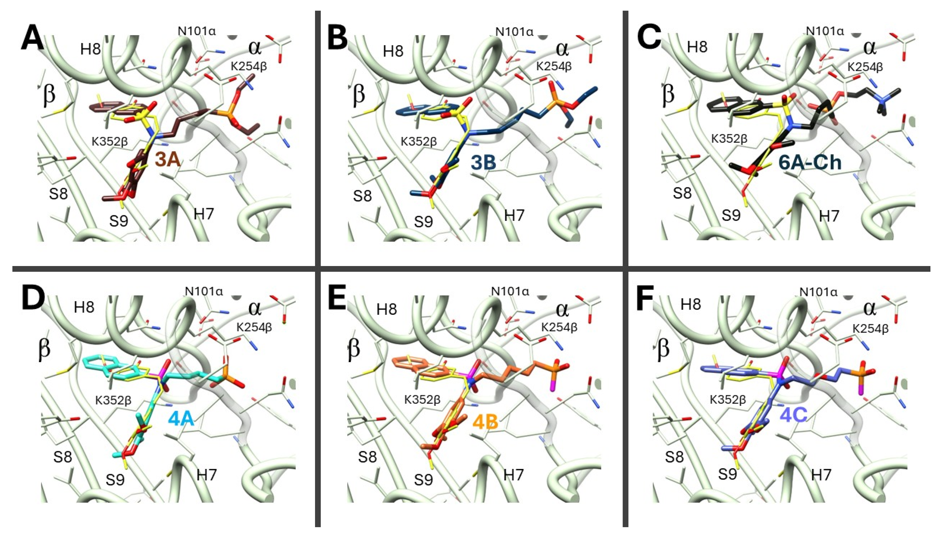
The chemical structure of the compounds resembles that of colchicine-site binders, and therefore the putative binding of the synthesized compounds at the colchicine site of tubulin (
Figure 6) was studied by docking experiments. Ligand flexibility was explicitly accounted for by the docking programs. The flexibility of the target was accounted for by docking the ligands in many binding site arrangements, the so-called ensemble docking approach [
49], found in the available X-ray crystal structures of tubulin in complex with colchicine site ligands that occupy the different binding sub-pockets (sub-sites A, B, and C) in the PDB [
30,
31,
50]. These ligands can vary greatly in size and nature, even among structurally related ligands, and they induce changes in the corresponding subpockets that sample diverse dispositions [
21,
30,
31]. These configurations are increased by separately considering binding sites with water molecules deemed important for binding, along with “dry” pockets [
49], giving 147 models coming from X-ray crystal structures [
50]. Additionally, six models representative of the main clusters of a molecular dynamics simulation of tubulin in complex with indole isocombretastatins [
51] were included for a final sum of 153 models [
49].
AutoDock 4.2 [
52] and PLANTS [
53] were the two docking programs with different scoring functions used for the docking experiments, looking for complementary exploration and scoring for each ligand binding pose. The best-scoring poses from each software that were similar to each other were selected as the binding mode(s). To compare the binding poses, each pose was assigned as binding to the different sub-pockets by a semiautomatic process that involves several steps: 1) if the shortest distance of any atom of the pose to the geometrical centre of the subpocket, as previously defined in ensemble pharmacophore docking experiments [
49], was bellow a threshold, then the pose was assigned as binding to that sub-pocket; 2) the RMSDs between the poses and undecorated scaffolds placing the centroids of phenyl rings at the above mentioned centers of the A, B, or C sub-sites or the X-ray coordinates of reference ligands such as of combretastatin A-4 (PDB ID 5LYJ, A-B sites), MI-181 (PDB ID 4YJ2, A-C sites), or ABT-751 (PDB ID 3HKC, A-B-C sites) [
27,
30,
31,
50]. Every pose of all the ligands is allocated in this way to the binding site sub-pockets. The energy scores of the poses of each ligand were scaled to a relative scale between 0 (worse scored) and 1 (best scored), and their Z-scores were calculated. These values allowed us to compare the scores of the two programs. The pair of similar poses with the best possible scores for the two software was taken as the binding mode. Application of this protocol to ligands with known X-ray structures retrieved, in most cases, the correct binding modes [
27].
The consensus poses for all the ligands placed the
N-trimethoxy-2-naphthalene sulfonamide portion of the molecules within the BA sub-pockets of the colchicine site (
Figure 6), with the 3,4,5-trimethoxyphenyl ring in the A zone and the naphthalene in the B zone, contacting tubulin as previously described for naphthalene sulfonamides [
32]. The spacer projects towards the interfacial space of the tubulin dimer, as designed, and the hydrogen-bond-accepting atoms of the polar capping groups are placed within hydrogen-bond distances of residues of α and β tubulin in the interdimer interface. The phosphonates put the oxygen atom doubly bonded to the phosphorus within a hydrogen-bond distance of the side chains’ NH of K254β or N101α, except for the shorter four-carbon spacer, which does not reach this far and hydrogen bonds instead to the backbone NH of N249β of the T7 loop. The phosphonic acids place the negatively charged oxygens to make hydrogen bonds with the NHs of the side chains of K254β, K352β, or N101α. The charged head groups of the choline derivative
6A-Ch and the diethylamoniumpropyl groups of
6B-DEAP and
6D-DEAP are placed close to the negatively charged phosphates of the GTP at the non-exchangeable site of the α subunit, and the negatively charged carboxylate of E71α; the DEAP derivatives hydrogen-bond to it with their protonated ammonium.
The docking of ligands into multiple protein structures selects the most favorable poses for each ligand but also picks some protein structures preferentially (
Supplementary Material: Supplementary Table S10, ST10), reflecting the required adaptations of the target to the structural particularities of the ligands. The PDB IDs most frequently retrieved for the naphthalene sulfonamides with polar caps differ from those previously shown to be selected when shorter and less polar substituents are placed at the sulfonamide nitrogen atom [
32]. The procedure selects instead proteins with T7 loop dispositions opening the gorges for the spacer, mostly associated with X-ray ligands with larger bridges between the A and B binding moieties, and mainly directing the spacer towards the interfacial space in the direction towards N101α, while maintaining A and B zones bound by moieties of similar shapes as those of
N-3,4,5-trimethoxyphenyl naphthalene sulfonamides (e.g., 5XAF and 5XAG for AD4, with bulky β-lactams as bridges, or 6GJ4 for PLANTS, with a pyridine bridge). As previously seen, the two software programs select different proteins, a selectivity attributed to the sought-after differences in scoring, justifying the application of the ensemble docking strategy combined with consensus scoring. The docking results are in good agreement with the design hypothesis that the active ligands bind with the naphthalene in zone A and the 3,4,5-trimethoxyphenyl in zone B, with the sulfonamide bridge opening towards the interfacial space and directing the cap groups towards the polar surface of the α and β subunits at the interfacial space.
The SwissADME web tool [
54] (
Supplementary Material: Supplementary Table S11, ST11) was used to predict physicochemical properties, pharmacokinetic parameters, and drug-likeness predictions for the compounds. All the compounds show fractions of Csp
3 atoms well above the target of 0.25, TPSA above 100 in all cases, a good predictor of low nonspecific toxicity [
55], with most above 130 (although the calculated geometries suggest that part of the polar surfaces might not be totally exposed), and moderate to poor solubilities. None of the compounds is predicted to be CNS penetrant; all have low gastrointestinal (GI) absorption, but
4C has high. All the compounds are classified as substrates of P-glycoprotein, although our experimental results of co-treatment with verapamil only show clear P-gp efflux for
3D,
4B,
6B-DEAP, and possibly for
4C. Overall, these results suggest that the molecules are good candidates for antitumor treatment. Although P-gp is considered an efflux system mainly for hydrophobic neutral or cationic substrates, the efflux of anionic substrates, such as the anionic phosphonates coming from phosphonic acid deprotonation, has also been observed. As already mentioned, the charged oxygens are partially hidden from the molecular surface by intramolecular interactions with the polar aromatic CHs.
The sites of potential metabolic transformations by CYP1A2, CYP2C9, CYP2C19, CYP2D6, CYP3A4, and UGT were predicted by SOMP [
56] and FAME3 and GLORYx (within NERDD) [
57] (
Supplementary Material: Supplementary Table S12, ST12). For the phosphonic acids, the main metabolic sites of transformation were oxidative hydroxylation of the phosphonate carbon, followed by hydroxylation of the methoxy groups and subsequent
O-demethylation. For the diethyl phosphonates, the most likely metabolic transformations are phosphonate hydrolysis and hydroxylation of the phosphonate carbon, both mediated by aliphatic hydroxylation. The four-carbon atom spacer significantly protects 3A from these metabolic transformations, and the presence of oxygen atoms on the phosphonate moderately reduces the probability of these reactions. The partially hydrolyzed ethyl and amino alcohol hydrogenophosphonates mainly suffer hydroxylation of the phosphonate carbon or hydrolysis of the phosphonate ester bonds.
3. Methods
Purchased reagents were used as received. We dried and stored solvents over molecular sieves. Precoated silica gel polyester plates (0.25 mm thickness) with a UV fluorescence indicator 254 (Polychrom SI F254, Merck KGaA, Darmstadt, Germany) were used for thin-layer chromatography (TLC). Preparative TLC and flash silica gel column chromatography (Kieselgel 40, 0.040–0.063 mm; Merck KGaA, Darmstadt, Germany) were eluted with the indicated mobile phase mixtures. NMR spectra were run on a Bruker (Billerica, MA, USA) SY spectrometer operating at 400 MHz for 1H and 100 MHz for 13C with the samples dissolved in CDCl3, CD3OD, or DMSO-d6, as indicated. DEPT-135 and DEPT-90 experiments were used to distinguish C, CH, CH2, and CH3 carbon signals. Chemical shifts (δ) are in ppm relative to the residual solvent signals. High-resolution mass spectra (HRMS) were performed with a hybrid QSTAR XL quadrupole/time-of-flight (QTOF) mass spectrometer (Agilent Technologies Inc., Santa Clara, CA, US).
To a stirred solution of the aniline (1.3–2 eq) in 50 mL of CH2Cl2 and pyridine (400 µL), the corresponding sulfonyl chloride (1 eq) was slowly added. The mixture was stirred at room temperature for 24 h. After that, the reaction was washed with 2N HCl, 5% NaHCO3, and brine until neutral pH. The organic layers were dried over anhydrous Na2SO4, filtered, and evaporated under reduced pressure. Crystallization or flash column chromatography afforded the sulfonamides.
- b.
General procedure b for the alkylation of the sulfonamide bridge
KI was added to a stirred solution of the halogen derivative (4 eq) at room temperature in CH3CN (25 mL), and after 5 min, a solution of the sulfonamide (1 eq) in CH3CN (25 mL) and Cs2CO3 (2.5 eq) was added dropwise to the first one. The mixture was stirred under an Ar atmosphere at room temperature for 48 to 72 h or refluxed for 24 h. After that, the reaction was evaporated under reduced pressure, dissolved in EtOAc, and washed with brine until neutral pH. The organic layers were dried over anhydrous Na2SO4, filtered, and evaporated under reduced pressure. Flash column chromatography and/or crystallization afforded the products.
- c.
General procedure c for the introduction of diethyl phosphonates
A stirred solution of the alkyl halide (1 eq) in 1.5 mL of triethyl phosphite was refluxed at 150 °C for 24 h. After that, the mixture was precipitated with 50 mL of hexane 3 times, and flash column chromatography, crystallization, or precipitation afforded the diethyl phosphonates.
- d.
General procedure d for the formation of phosphonic acids
To a stirred solution of the corresponding diethyl phosphonate (1 eq) in 3 mL of DMF, NaBr (4 eq) and TMS-Cl (4 eq) were added. The solution was stirred at 75 °C for 18 h in a sealed tube. After that, 3 mL of 2N HCl was added, and the solution was stirred for 5 min at room temperature until the formation of a precipitate. The precipitate was redissolved in EtOAc, and the aqueous layer was extracted two more times with EtOAc. The organic layers were washed 5 times with brine, dried over anhydrous Na2SO4, filtered, and evaporated under reduced pressure. Precipitation, flash column chromatography, and/or crystallization afforded the products.
- e.
General procedure e for the formation of diethyl tiophosphonates
The corresponding diethyl phosphonate (1 eq) was dissolved in 20 mL of toluene and, under stirring, Lawesson’s reagent (1.1 eq) was added to the solution. The reaction was stirred at 120 °C under an Ar atmosphere for 24 h. After that, evaporation of the reaction under reduced pressure and flash column chromatography or crystallization afforded the diethyl tiophosphonates.
- f.
General procedure f the formation of phosphonic acid esters
To a stirred solution of the corresponding phosphonic acid (1 eq) in 25 mL of CH2Cl2, DMAP (1.6 eq) and EDC (2.3 eq) were added, and the solution was stirred at room temperature for 5 min. After that, the alcohol (1.1 eq) was added, and the solution was refluxed for 24 h under an Ar atmosphere. The reaction was then poured onto ice and carefully washed with brine until neutral pH. The organic layers were dried over anhydrous Na2SO4, filtered, and evaporated under reduced pressure. Flash column chromatography and/or precipitation afforded the phosphonates.
- 2.
Synthesis and characterization of the compounds
Following the general procedure, 3,4,5-trimethoxyaniline (1.72 g, 9.40 mmol) and naphthalene-2-sulfonyl chloride (1.979 g, 8.73 mmol) gave a white solid that was purified by crystallization to afford 2.483 g (6.65 mmol, 76%) of 1 as white crystals.
N-(3,4,5-trimethoxyphenyl)naphthalene-2-sulfonamide (1): 1H-NMR (CDCl3): 8.34 (1 H, d, J = 2.0 Hz); 7.93–7.87 (3 H, m); 7.73 (1 H, dd, J = 8.8 Hz, J = 0.6 Hz); 7.64 (1 H, bt, J = 7.0 Hz); 7.59 (1 H, bt, J = 7.0 Hz); 6.35 (NH, s); 6.28 (2 H, s); 3.75 (3 H, s); 3.68 (6 H, s) ppm. 13C-NMR (CDCl3): 153.4 (2 C); 135.7 (2 C); 134.9 (C); 132.3 (C); 132.0 (C); 129.4 (CH); 129.2 (CH); 129.0 (2 CH); 127.9 (CH); 127.7 (CH); 122.9 (CH); 99.9 (2 CH); 60.9 (CH3); 56.0 (2 CH3) ppm. M.p. (Hexane/CH2Cl2): 161–162 °C. HRMS: Calculated for C19H19NO5SNa+ 396.0876, found 396.0875 (M + Na+).
- b.
Synthesis of N-(4-bromobutyl)-N-(3,4,5-trimethoxyphenyl)naphthalene-2-sulfonamide (2A-Br):
Following the general procedure b, compound 1 (0.892 g, 2.39 mmol) was alkylated with 1,4-dibromobutane (1141 µL, 9.56 mmol), and the reaction was stirred at room temperature for 72 h. The crude was purified by flash column chromatography (hexane/EtOAc 7:3) giving 581 mg (1.11 mmol, 47%) of a mixture of bromine (2A-Br, 374 mg, 0.74 mmol, 31%) and iodine (2A-I, 207 mg, 0.37 mmol, 16%) derivatives as a white solid and 94 mg (0.12 mmol, 10%) of 2bA as a white solid. Compounds 2A-Br and 2A-I co-crystallized in hexane/CH2Cl2, giving 280 mg of white crystals with a m.p. of 123–125 °C.
N-(4-bromobutyl)-N-(3,4,5-trimethoxyphenyl)naphthalene-2-sulfonamide (2A-Br): 1H-NMR (CDCl3): 8.18 (1 H, s); 7.92–7.86 (3 H, m); 7.63–7.55 (3 H, m); 6.18 (2 H, s); 3.83 (6 H, s); 3.54 (3 H, s); 3.58–3.54 (2 H, m); 3.42 (2 H, t, J = 6.6 Hz); 1.99–1.90 (2 H, m); 1.62–1.55 (2 H, m) ppm. 13C-NMR (CDCl3): 153.1 (2 C); 138.0 (C); 134.9 (C); 134.8 (C); 134.3 (C); 132.0 (C); 129.1 (2 CH); 128.9 (2 CH); 127.9 (CH); 127.7 (CH); 123.1 (CH); 106.4 (2 CH); 60.9 (CH3); 56.1 (2 CH3); 50.0 (CH2); 33.2 (CH2); 29.4 (CH2); 26.6 (CH2) ppm. HRMS: Calculated for C23H26BrNO5SNa+ 530.0607, found 530.0599 (M + Na+).
N-(4-iodobutyl)-N-(3,4,5-trimethoxyphenyl)naphthalene-2-sulfonamide (2A-I): 1H-NMR (CDCl3): 8.18 (1 H, s); 7.92–7.86 (3 H, m); 7.63–7.55 (3 H, m); 6.18 (2 H, s); 3.83 (6 H, s); 3.54 (3 H, s); 3.58–3.54 (2 H, m); 3.18 (2 H, t, J = 6.8 Hz); 1.99–1.90 (2 H, m); 1.62–1.55 (2 H, m) ppm. 13C-NMR (CDCl3): 153.1 (2 C); 138.0 (C); 134.9 (C); 134.8 (C); 134.3 (C); 132.0 (C); 129.1 (2 CH); 128.9 (2 CH); 127.9 (CH); 127.7 (CH); 123.1 (CH); 106.4 (2 CH); 60.9 (CH3); 56.1 (2 CH3); 49.8 (CH2); 30.0 (CH2); 28.8 (CH2); 6.2 (CH2) ppm. HRMS: Calculated for C23H26INO5SNa+ 578.0469, found 578.0458 (M + Na+).
N,N’-(butane-1,4-diyl)bis(N-(3,4,5-trimethoxyphenyl)naphthalene-2-sulfonamide) (2bA): 1H-NMR (CDCl3): 8.20 (2 H, s); 7.94–7.90 (6 H, m); 7.67–7.60 (6 H, m); 6.17 (4 H, s); 3.84 (6 H, s); 3.58 (12 H, s); 3.65–3.49 (4 H, m); 1.65–1.53 (4 H, m) ppm. 13C-NMR (CDCl3): 153.1 (4 C); 137.9 (2 C); 134.8 (4 C); 134.5 (2 C); 132.0 (2 C); 129.2 (4 CH); 128.9 (2 CH); 128.8 (2 CH); 127.8 (2 CH); 127.6 (2 CH); 123.2 (2 CH); 106.4 (4 CH); 60.8 (2 CH3); 56.0 (4 CH3); 50.1 (2 CH2); 25.0 (2 CH2) ppm. M.p. (MeOH): 215–216 °C.
- c.
Synthesis of N-(5-bromopentyl)-N-(3,4,5-trimethoxyphenyl)naphthalene-2-sulfonamide (2B-Br):
Following general procedure b, compound 1 (1.982 g, 5.30 mmol) was alkylated using 1,5-dibromopentane (2200 µL, 16.29 mmol), and the reaction was stirred at room temperature for 72 h. The crude was purified by flash column chromatography (hexane/EtOAc 7:3) giving 1545 mg (2.90 mmol, 50%) of a mixture of bromine (2B-Br, 1214 mg, 2.32 mmol, 44%) and iodine (2B-I, 331 mg, 0.58 mmol, 11%) derivatives as a white solid and 251 mg (0.30 mmol, 12%) of 2bB as a white solid. Compounds 2B-Br and 2B-I co-crystallized in hexane/EtOAc, giving 421 mg of white crystals with a m.p. of 115–117 °C.
N-(5-bromopentyl)-N-(3,4,5-trimethoxyphenyl)naphthalene-2-sulfonamide (2B-Br): 1H-NMR (CDCl3): 8.20 (1 H, bs); 7.93–7.89 (3 H, m); 7.65–7.58 (3 H, m); 6.19 (2 H, s); 3.83 (3 H, s); 3.60 (6 H, s); 3.53 (2 H, t, J = 7.0 Hz); 3.37 (2 H, t, J = 6.6 Hz); 1.88–1.77 (2 H, m); 1.56–1.43 (4 H, m) ppm. 13C-NMR (CDCl3): 153.1 (2 C); 137.9 (C); 135.0 (C); 134.7 (C); 134.5 (C); 131.9 (C); 129.1 (2 CH); 128.8 (2 CH); 127.8 (CH); 127.6 (CH); 123.2 (CH); 106.4 (2 CH); 60.8 (CH3); 56.0 (2 CH3); 50.8 (CH2); 33.7 (CH2); 32.1 (CH2); 27.4 (CH2); 24.9 (CH2) ppm.
N-(5-iodopentyl)-N-(3,4,5-trimethoxyphenyl)naphthalene-2-sulfonamide (2B-I): 1H-NMR (CDCl3): 8.20 (1 H, bs); 7.93–7.89 (3 H, m); 7.65–7.58 (3 H, m); 6.19 (2 H, s); 3.83 (3 H, s); 3.60 (6 H, s); 3.53 (2 H, t, J = 7.0 Hz); 3.15 (2 H, t, J = 6.8 Hz); 1.88–1.77 (2 H, m); 1.56–1.47 (4 H, m) ppm. 8.15 (1 H, s); 7.89–7.83 (3 H, m); 7.61–7.53 (3 H, m); 6.16 (2 H, s); 3.79 (3 H, s); 3.55 (6 H, s); 3.53 (2 H, t, J = 7.0 Hz); 3.15 (2 H, t, J = 6.8 Hz); 1.80–1.74 (2 H, m); 1.56–1.43 (4 H, m) ppm. 13C-NMR (CDCl3): 153.1 (2 C); 137.9 (C); 135.0 (C); 134.7 (C); 134.5 (C); 131.9 (C); 129.1 (2 CH); 128.8 (2 CH); 127.8 (CH); 127.6 (CH); 123.2 (CH); 106.4 (2 CH); 60.8 (CH3); 56.0 (2 CH3); 50.8 (CH2); 32.8 (CH2); 27.3 (CH2); 27.2 (CH2); 6.5 (CH2) ppm.
N,N’-(pentane-1,5-diyl)bis(N-(3,4,5-trimethoxyphenyl)naphthalene-2-sulfonamide) (2bB): 1H-NMR (CDCl3): 8.19 (2 H, s); 7.94–7.90 (6 H, m); 7.67–7.60 (6 H, m); 6.18 (4 H, s); 3.84 (6 H, s); 3.66 (12 H, s); 3.51 (4 H, t, J = 6.4 Hz); 1.55–1.48 (6 H, m).
- d.
Synthesis of N-(2-(2-chloroethoxy)ethyl)-N-(3,4,5-trimethoxyphenyl)naphthalene-2-sulfonamide (2C):
Following general procedure b, compound 1 (1.026 g, 2.75 mmol) was alkylated with 1-chloro-2-(2-chloroethoxy)ethane (970 µL, 8.25 mmol), and the reaction was refluxed for 24 h. The crude product was purified by flash column chromatography (hexane/EtOAc 7:3), giving 840 mg (1.75 mmol, 64%) of 2C as a white solid and 116 mg (0.14 mmol, 5%) of 2bC, which were crystallized to afford 100 mg (0.12 mmol, 4%) of white crystals.
N-(2-(2-chloroethoxy)ethyl)-N-(3,4,5-trimethoxyphenyl)naphthalene-2-sulfonamide (2C): 1H-NMR (CDCl3): 8.24 (1 H, d, J = 1.8 Hz); 7.94–7.87 (3 H, m); 7.69 (1 H, dd, J = 8.8 Hz, J = 1.8 Hz); 7.63 (1 H, bt, J = 7.0 Hz); 7.59 (1 H, bt, J = 7.0 Hz); 6.25 (2 H, s); 3.82 (3 H, s); 3.77 (2 H, t, J = 6.0 Hz); 3.64 (2 H, t, J = 5.7 Hz); 3.59 (6 H, s); 3.61 (2 H, m); 3.51 (2 H, t, J = 6.0 Hz) ppm. 13C-NMR (CDCl3): 153.1 (2 C); 138.0 (C); 135.4 (C); 134.8 (C); 134.8 (C); 132.0 (C); 129.2 (CH); 129.1 (CH); 128.9 (CH); 128.8 (CH); 127.8 (CH); 127.6 (CH); 123.3 (CH); 106.7 (2 CH); 71.0 (CH2); 68.8 (CH2); 60.9 (CH3); 56.0 (2 CH3); 50.8 (CH2); 42,8 (CH2) ppm. HRMS: Calculated for C23H26ClNO6SNa+ 502.1062, found 502.1053 (M + Na+).
N,N’-(oxybis(ethane-2,1-diyl))bis(N-(3,4,5-trimethoxyphenyl)naphthalene-2-sulfonamide) (2bC): 1H-NMR (CDCl3): 8.21 (2 H, s); 7.95–7.87 (6 H, m); 7.66 (2 H, dd, J = 8.8 Hz, J = 2.0 Hz); 7.64 (2 H, m); 7.59 (2 H, dt, J = 7.0 Hz, J = 1.5 Hz); 6.18 (4 H, s); 3.82 (6 H, s); 3.66 (4 H, t, J = 6.3 Hz); 3.56 (12 H, s); 3.50 (4 H, t, J = 6.3 Hz) ppm. 13C-NMR (CDCl3): 153.1 (4 C); 138.0 (2 C); 135.2 (2 C); 134.8 (4 C); 132.0 (2 C); 129.2 (4 CH); 128.9 (2 CH); 128.8 (2 CH); 127.8 (2 CH); 127.6 (2 CH); 123.2 (2 CH); 106.5 (4 CH); 68.5 (2 CH2); 60.9 (2 CH3); 56.0 (4 CH3); 50.7 (2 CH2) ppm. M.p. (CH2Cl2/MeOH): 169–170 °C.
- e.
Synthesis of N-(6-bromohexyl)-N-(3,4,5-trimethoxyphenyl)naphthalene-2-sulfonamide (2D-Br):
Following general procedure b, compound 1 (1.089 g, 2.92 mmol) was alkylated with 1,6-dibromohexane (1340 µL, 8.75 mmol), and the reaction was stirred at room temperature for 72 h. The crude was purified by flash column chromatography (hexane/EtOAc 7:3) giving 541 mg (0.98 mmol, 33%) of a mixture of bromine (2D-Br, 304 mg, 0.57 mmol, 20%) and iodine (2D-I, 237 mg, 0.41 mmol, 14%) derivatives as a white solid, and 546 mg (0.66 mmol, 46%) of 2bD that were crystallized to afford 201 mg (0.24 mmol, 17%) of white crystals. Compounds 2D-Br and 2D-I co-crystallized in hexane/EtOAc, giving 113 mg (0.20 mmol, 7%) of white crystals with a m.p. of 125–126 °C.
N-(6-bromohexyl)-N-(3,4,5-trimethoxyphenyl)naphthalene-2-sulfonamide (2D-Br): 1H-NMR (CDCl3): 8.19 (1 H, s); 7.92–7.87 (3 H, m); 7.64 (1 H, dd, J = 8.6 Hz, J = 1.7 Hz); 7.62–7.55 (2 H, m); 6.19 (2 H, s); 3.83 (3 H, s); 3.59 (6 H, s); 3.52 (2 H, t, J = 6.8 Hz); 3.35 (2 H, t, J = 6.8 Hz); 1.81–1.36 (8 H, m) ppm. 13C-NMR (CDCl3): 153.1 (2 C); 137.8 (C); 135.1 (C); 134.7 (C); 134.6 (C); 132.0 (C); 129.1 (2 CH); 128.8 (2 CH); 127.9 (CH); 127.6 (CH); 123.2 (CH); 106.4 (2 CH); 60.9 (CH3); 56.1 (2 CH3); 50.9 (CH2); 33.7 (CH2); 32.5 (CH2); 28.1 (CH2); 27.6 (CH2); 25.5 (CH2) ppm. HRMS: Calculated for C25H30BrNO5SNa+ 558.0920, found 558.0919 (M + Na+).
N-(6-iodohexyl)-N-(3,4,5-trimethoxyphenyl)naphthalene-2-sulfonamide (2D-I): 1H-NMR (CDCl3): 8.19 (1 H, s); 7.92–7.87 (3 H, m); 7.64 (1 H, dd, J = 8.6 Hz, J = 1.7 Hz); 7.62–7.55 (2 H, m); 6.19 (2 H, s); 3.83 (3 H, s); 3.59 (6 H, s); 3.52 (2 H, t, J = 6.8 Hz); 3.13 (2 H, t, J = 7.0 Hz); 1.81–1.36 (8 H, m) ppm.13C-NMR (CDCl3): 153.1 (2 C); 137.8 (C); 135.1 (C); 134.7 (C); 134.6 (C); 132.0 (C); 129.1 (2 CH); 128.8 (2 CH); 127.9 (CH); 127.6 (CH); 123.2 (CH); 106.4 (2 CH); 60.9 (CH3); 56.1 (2 CH3); 50.9 (CH2); 33.2 (CH2); 29.9 (CH2); 28.1 (CH2); 25.3 (CH2); 6.98 (CH2) ppm.
N,N’-(hexane-1,6-diyl)bis(N-(3,4,5-trimethoxyphenyl)naphthalene-2-sulfonamide) (2bD): 1H-NMR (CDCl3): 8.19 (2 H, s); 7.95–7.87 (6 H, m); 7.67–7.56 (6 H, m); 6.18 (4 H, s); 3.83 (6 H, s); 3.58 (12 H, s); 3.50 (4 H, t, J = 6.9 Hz); 1.50–1.30 (8 H, m) ppm. 13C-NMR (CDCl3): 153.1 (4 C); 137.9 (2 C); 135.1 (2 C); 134.7 (2 C); 134.6 (2 C); 132.0 (2 C); 129.2 (2 CH); 129.1 (2 CH); 128.8 (4 CH); 127.8 (2 CH); 127.6 (2 CH); 123.2 (2 CH); 106.4 (4 CH); 60.9 (2 CH3); 56.0 (4 CH3); 50.9 (2 CH2); 28.2 (2 CH2); 25.9 (2 CH2) ppm. M.p. (MeOH): 148–149 °C.
- f.
Synthesis of N-(2-(2-(2-chloroethoxy)ethoxy)ethyl)-N-(3,4,5-trimethoxyphenyl)naphthalene-2-sulfonamide (2E):
Following general procedure b, compound 1 (1.51 g, 4.04 mmol) was alkylated with 1-chloro-2-(2-chloroethoxy)ethane (2536 µL, 16.17 mmol), and the reaction was refluxed for 24 h. The crude product was purified by flash column chromatography (hexane/EtOAc 7:3), giving 1216 mg (2.32 mmol, 57%) of 2E as a brown oil and 451 mg (0.52 mmol, 26%) of 2bE, which were crystallized to afford 169 mg (0.20 mmol, 10%) of white crystals.
N-(2-(2-(2-chloroethoxy)ethoxy)ethyl)-N-(3,4,5-trimethoxyphenyl)naphthalene-2-sulfonamide (2E): 1H-NMR (CDCl3): 8.23 (1 H, s); 7.94–7.88 (3 H, m); 7.67 (1 H, dd, J = 8.6 Hz, J = 1.9 Hz); 7.65 (1 H, m); 7.60 (1 H, t, J = 7.0 Hz); 6.23 (2 H, s); 3.83 (3 H, s); 3.76 (2 H, t, J = 6.4 Hz); 3.70–3.65 (2 H, m); 3.60 (6 H, s); 3.60–3.55 (8 H, m) ppm. 13C-NMR (CDCl3): 153.1 (2 C); 138.0 (C); 135.3 (C); 134.9 (C); 134.8 (C); 132.0 (C); 129.2 (CH); 129.1 (CH); 128.9 (CH); 128.8 (CH); 127.8 (CH); 127.6 (CH); 123.2 (CH); 106.7 (2 CH); 72.5 (CH2); 70.4 (CH2); 70.3 (CH2); 68.8 (CH2); 61.7 (CH2); 60.9 (CH3); 56.0 (2 CH3); 50.7 (CH2) ppm. HRMS: Calculated for C25H29NO7SNa+ 546.1324, found 546.1326 (M + Na+).
N,N’-((ethane-1,2-diylbis(oxy))bis(ethane-2,1-diyl))bis(N-(3,4,5-trimethoxyphenyl)naphthalene-2-sulfonamide) (2bE): 1H-NMR (CDCl3): 8.22 (2 H, d, J = 1.8 Hz); 7.93–7.85 (6 H, m); 7.68 (2 H, dd, J = 8.6 Hz, J = 1.9 Hz); 7.64 (2 H, td, J = 7.0 Hz, J = 1.4 Hz); 7.60 (2 H, td, J = 7.0 Hz, J = 1.4 Hz); 6.22 (4 H, s); 3.82 (6 H, s); 3.74 (4 H, t, J = 6.4 Hz); 3.56 (12 H, s); 3.56–3.52 (4 H, m); 3.50 (4 H, bs) ppm. 13C-NMR (CDCl3): 153.1 (4 C); 138.0 (2 C); 135.4 (2 C); 134.9 (2 C); 134.8 (2 C); 132.0 (2 C); 129.2 (2 CH); 129.1 (2 CH); 128.9 (2 CH); 128.8 (2 CH); 127.8 (2 CH); 127.6 (2 CH); 123.2 (2 CH); 106.6 (4 CH); 70.2 (2 CH2); 68.8 (2 CH2); 60.8 (2 CH3); 56.0 (4 CH3); 50.7 (2 CH2) ppm. M.p. (Hexane/EtOAc): 181–183 °C.
- g.
Synthesis of N-(4-(4-chlorobutoxy)butyl)-N-(3,4,5-trimethoxyphenyl)naphthalene-2-sulfonamide (2F):
Following general procedure b, compound 1 (1.003 g, 2.69 mmol) was alkylated with 1-chloro-4-(4-chlorobutoxy)butane (1163 µL, 8.07 mmol) under reflux for 24 h. The crude product was purified by flash column chromatography (hexane/EtOAc 6:4), giving 974 mg (1.82 mmol, 68%) of 2F as a white solid and 30 mg (0.03 mmol, 2%) of 2bF that crystallized as 28 mg (0.03 mmol, 1%) of white crystals.
N-(4-(4-chlorobutoxy)butyl)-N-(3,4,5-trimethoxyphenyl)naphthalene-2-sulfonamide (2F): 1H-NMR (CDCl3): 8.21 (1 H, bs); 7.96–7.89 (3 H, m); 7.69–7.62 (2 H, m); 7.61 (1 H, bt, J = 7.0 Hz); 6.20 (2 H, s); 3.84 (3 H, s); 3.61 (6 H, s); 3.56 (2 H, t, J = 6.6 Hz); 3.54 (2 H, t, J = 6.6 Hz); 3.43–3.38 (4 H, m); 1.90–1.80 (2 H, m); 1.72–1.50 (6 H, m) ppm. 13C-NMR (CDCl3): 153.1 (2 C); 137.9 (C); 135.1 (C); 134.7 (C); 134.5 (C); 132.0 (C); 129.1 (2 CH); 128.8 (2 CH); 127.8 (CH); 127.6 (CH); 123.2 (CH); 106.5 (2 CH); 70.1 (CH2); 69.9 (CH2); 60.9 (CH3); 56.0 (2 CH3); 50.8 (CH2); 44.9 (CH2); 29.5 (CH2); 27.0 (CH2); 26.5 (CH2); 25.2 (CH2) ppm. M.p. (Hexane/CH2Cl2): 76–77 °C. HRMS: Calculated for C27H34ClNO6SNa+ 558.1688, found 558.1697 (M + Na+).
N,N’-(oxybis(butane-4,1-diyl))bis(N-(3,4,5-trimethoxyphenyl)naphthalene-2-sulfonamide) (2bF): 1H-NMR (CDCl3): 8.19 (2 H, d, J = 1.8 Hz); 7.94–7.86 (6 H, m); 7.64 (2 H, dd, J = 8.6 Hz, J = 1.8 Hz); 7.62 (2 H, td, J = 7.0 Hz, J = 1.4 Hz); 7.61 (2 H, bt, J = 7.0 Hz, J = 1.4 Hz); 6.19 (4 H, s); 3.82 (6 H, s); 3.56 (12 H, s); 3.54 (4 H, t, J = 6.8 Hz); 3.37 (4 H, t, J = 6.3 Hz); 1.65–1.56 (4 H, m); 1.55–1.46 (4 H, m) ppm. 13C-NMR (CDCl3): 153.1 (4 C); 137.9 (2 C); 135.1 (2 C); 134.7 (2 C); 134.5 (2 C); 132.0 (2 C); 129.1 (4 CH); 128.8 (4 CH); 127.8 (2 CH); 127.6 (2 CH); 123.2 (2 CH); 106.5 (4 CH); 70.0 (2 CH2); 60.9 (2 CH3); 56.0 (4 CH3); 50.8 (2 CH2); 26.5 (2 CH2); 25.1 (2 CH2) ppm. M.p. (Hexane/CH2Cl2): 142–143 °C.
- h.
Synthesis of diethyl (4-(N-(3,4,5-trimethoxyphenyl)naphthalene-2-sulfonamido)butyl)phosphonate (3A):
Following general procedure c, a mixture of the compounds 2ABr (662 mg, 1.30 mmol) and 2AI (367 mg, 0.66 mmol) was reacted with triethyl phosphite, and the crude product was purified by flash column chromatography (hexane/acetone 4:6) to afford 418 mg (0.74 mmol, 38%) of 3A as a light brown oil.
1H-NMR (CDCl3): 8.14 (1 H, d, J = 2.0 Hz); 7.91–7.81 (3 H, m); 7.62–7.52 (3 H, m); 6.14 (2 H, s); 4.08–3.95 (4 H, m); 3.78 (3 H, s); 3.55 (6 H, s); 3.50 (2 H, t, J = 6.8 Hz); 1.73–1.48 (6 H, m); 1.25 (6 H, t, J = 7.0 Hz) ppm. 13C-NMR (CDCl3): 153.1 (2 C); 137.9 (C); 134.9 (C); 134.7 (C); 134.4 (C); 132.0 (C); 129.1 (2 CH); 128.8 (2 CH); 127.8 (CH); 127.6 (CH); 123.1 (CH); 106.3 (2 CH); 61.5 (2 CH2, d, J = 6.5 Hz); 60.8 (CH3); 56.0 (2 CH3); 50.3 (CH2); 28.9 (CH2, d, J = 15.7 Hz); 25.0 (CH2, d, J = 141.1 Hz); 19.4 (CH2, d, J = 5.1 Hz); 16.4 (2 CH3, d, J = 6.0 Hz) ppm. HRMS: Calculated for C27H37NO8PS+ 566.1972, found 566.1967 (M + H+).
- i.
Synthesis of diethyl (5-(N-(3,4,5-trimethoxyphenyl)naphthalene-2-sulfonamido)pentyl)phosphonate (3B):
Following general procedure c, a mixture of compound 2BBr (944 mg, 1.81 mmol) and 2BI (257 mg, 0.45 mmol) was reacted with triethyl phosphite to afford 1087 mg (1.88 mmol, 83%) of a very clean crude product that was purified by crystallization to afford 95 mg (0.16 mmol, 7%) of 3B as pale pink crystals.
1H-NMR (CDCl3): 8.16 (1 H, d, J = 1.8 Hz); 7.90–7.86 (3 H, m); 7.64–7.58 (2 H, m); 7.56 (1 H, td, J = 6.9 Hz, J = 1.3 Hz); 6.15 (2 H, s); 4.11–3.97 (4 H, m); 3.81 (3 H, s); 3.57 (6 H, s); 3.49 (2 H, t, J = 6.4 Hz); 1.70–1.41 (8 H, m); 1.27 (6 H, t, J = 7.0 Hz). 13C-NMR (CDCl3): 153.1 (2 C); 137.9 (C); 135.0 (C); 134.7 (C); 134.5 (C); 132.0 (C); 129.1 (2 CH); 128.8 (2 CH); 127.8 (CH); 127.6 (CH); 123.2 (CH); 106.4 (2 CH); 61.4 (CH2, d, J = 6.5 Hz); 60.9 (CH3); 56.0 (2 CH3); 50.8 (CH2); 27.9 (CH2, d, J = 1.2 Hz); 27.4 (CH2, d, J = 17.0 Hz); 25.5 (CH2, d, J = 140.9 Hz); 22.0 (CH2, d, J = 5.1 Hz); 16.4 (2 CH3, d, J = 5.8 Hz) ppm. M.p. (Hexane): 82–83 °C. HRMS: Calculated for C28H39NO8PS+ 580.2129, found 580.2120 (M + H+).
- j.
Synthesis of diethyl (2-(2-(N-(3,4,5-trimethoxyphenyl)naphthalene-2-sulfonamido)ethoxy)ethyl)phosphonate (3C):
Following general procedure c, compound 2C (752 mg, 1.57 mmol) reacted with triethyl phosphite, and the crude product was purified by flash column chromatography (EtOAc/MeOH 95:5) to afford 604 mg (1.04 mmol, 66%) of 3C as a brown oil.
1H-NMR (CDCl3): 8.20 (1 H, d, J = 2.2 Hz); 7.92–7.85 (3 H, m); 7.65 (1 H, dd, J = 1.9, 8.6 Hz); 7.63–7.54 (2 H, m); 6.19 (2 H, s); 4.09–4.00 (4 H, m); 3.80 (3 H, s); 3.72 (2 H, t, J = 6.2 Hz); 3.59–3.64 (2 H, m); 3.56 (6 H, s); 3.51 (2 H, t, J = 6.2 Hz); 1.96 (2 H, dt, J = 7.1, 18.4 Hz); 1.27 (6 H, t, J = 7.1 Hz) ppm. 13C-NMR (CDCl3): 153.0 (2 C); 137.9 (C); 135.3 (C); 134.6 (2 C); 131.8 (C); 129.0 (CH); 128.8 (2 CH); 128.7 (CH); 127.7 (CH); 127.5 (CH); 123.0 (CH); 106.5 (2 CH); 68.1 (CH2); 64.7 (CH2); 61.5 (2 CH2, d, J = 2.4 Hz); 60.6 (CH3); 55.8 (2 CH3); 50.7 (CH2); 26.7 (CH2, d, J = 139.6 Hz); 16.3 (2 CH3, d, J = 6.3 Hz) ppm. HRMS: Calculated for C27H37NO9PS+ 582.1921, found 582.1913 (M + H+).
- k.
Synthesis of diethyl (6-(N-(3,4,5-trimethoxyphenyl)naphthalene-2-sulfonamido)hexyl)phosphonate (3D):
Following general procedure c, a mixture of the compounds 2DBr (533 mg, 0.99 mmol) and 2DI (416 mg, 0.71 mmol) was reacted with triethyl phosphite, and the crude product was purified by flash column chromatography (hexane/acetone 4:6) to afford 595 mg (1.00 mmol, 59%) of 3D as a light brown oil.
1H-NMR (CDCl3): 8.19 (1 H, s); 7.94–7.88 (3 H, m); 7.66–7.61 (2 H, m); 7.59 (1 H, dd, J = 7.0 Hz); 6.18 (2 H, s); 4.14–4.00 (4 H, m); 3.83 (3 H, s); 3.59 (6 H, s); 3.51 (2 H, t, J = 7.0 Hz); 1.76–1.33 (10 H, m); 1.30 (6 H, bt, J = 7.0 Hz) ppm. 13C-NMR (CDCl3): 153.2 (2 C); 138.0 (C); 135.2 (C); 134.8 (C); 134.7 (C); 132.1 (C); 129.2 (2xCH); 128.9 (2 CH); 127.9 (CH); 127.7 (CH); 123.3 (CH); 106.5 (2 CH); 61.5 (2 CH2, d, J = 6.6 Hz); 61.0 (CH3); 56.1 (2 CH3); 51.1 (CH2); 30.1 (CH2, d, J = 17.0 Hz); 28.2 (CH2); 26.0 (CH2); 25.7 (CH2, d, J = 140.7 Hz); 22.5 (CH2, d, J = 5.2 Hz); 16.5 (2 CH3, d, J = 5.9 Hz) ppm. HRMS: Calculated for C29H41NO8PS+ 594.2285, found 594.2275 (M + H+).
- l.
Synthesis of diethyl (2-(2-(2-(N-(3,4,5-trimethoxyphenyl)naphthalene-2-sulfonamido)ethoxy)ethoxy)ethyl)phosphonate (3E):
Following general procedure c, compound 2E (1023 mg, 1.95 mmol) reacted with triethyl phosphite, and the crude product was purified by flash column chromatography (EtOAc/MeOH 95:5) to afford 394 mg (0.63 mmol, 32%) of 3E as a brown oil.
1H-NMR (CDCl3): 8.23 (1 H, d, J = 1.6 Hz); 7.93–7.89 (3 H, m); 7.67 (1 H, dd, J = 8.7, J = 1.8 Hz); 7.64 (1 H, td, J = 7.0, J = 1.7 Hz); 7.60 (1 H, dd, J = 7.0, J = 1.6 Hz); 6.22 (2 H, s); 4.13–4.02 (4 H, m); 3.82 (3 H, s); 3.75 (2 H, t, J = 6.5 Hz); 3.71–3.62 (2 H, m); 3.59 (6 H, s); 3.59–3.48 (6 H, s); 2.08 (2 H, dt, J = 7.6; 18.8 Hz); 1.30 (6 H, t, J = 7.1 Hz) ppm. 13C-NMR (CDCl3): 153.1 (4 C); 138.0 (C); 135.3 (C); 134.9 (C); 134.7 (C); 132.0 (C); 129.2 (CH); 129.1 (CH); 128.9 (CH); 128.8 (CH); 127.8 (CH); 127.6 (CH); 123.2 (CH); 106.6 (2 CH); 70.2 (CH2); 70.0 (CH2); 68.8 (CH2); 65.1 (CH2); 61.6 (2 CH2, d, J = 6.2 Hz); 60.8 (CH3); 56.0 (2 CH3); 50.7 (CH2); 26.9 (CH2, d, J = 139.3 Hz); 16.4 (2 CH3, d, J = 6.1 Hz) ppm. HRMS: Calculated for C29H40NO10PSNa+ 648.2003, found 648.1989 (M + Na+).
- m.
Synthesis of diethyl (4-(4-(N-(3,4,5-trimethoxyphenyl)naphthalene-2-sulfonamido)butoxy)butyl)phosphonate (3F):
Following general procedure c, compound 2F (1403 mg, 2.62 mmol) reacted with triethyl phosphite, and the crude product was purified by precipitation with hexane to afford 801 mg (1.26 mmol, 48%) of 3F as a brown oil.
1H-NMR (CDCl3): 8.12 (1 H, d, J = 1.9 Hz); 7.87–7.80 (3 H, m); 7.59–7.49 (3 H, m); 6.11 (2 H, s); 4.09–3.92 (4 H, m); 3.75 (3 H, s); 3.51 (6 H, s); 3.48 (2 H, t, J = 6.8 Hz); 3.30 (4 H, m); 1.72–1.38 (10 H, m); 1.25 (6 H, t, J = 7.0 Hz) ppm. 13C-NMR (CDCl3): 153.1 (2 C); 137.9 (C); 135.1 (C); 134.7 (C); 134.5 (C); 132.0 (C); 129.1 (2 CH); 128.8 (CH); 128.7 (CH); 127.8 (CH); 127.6 (CH); 123.2 (CH); 106.4 (2 CH); 70.1 (2 CH2); 61.4 (CH2, d, J = 3.5 Hz); 60.9 (CH3); 56.0 (2 CH3); 50.8 (CH2); 30.5 (CH2, d, J = 16.4 Hz); 26.5 (CH2); 25.5 (CH2, d, J = 141.1 Hz); 25.1 (CH2); 19.3 (CH2, d, J = 5.0 Hz); 16.4 (CH3, d, J = 6.0 Hz) ppm. HRMS: Calculated for C31H44NO9PSNa+ 660.2367, found 660.2355 (M + Na+).
- n.
Synthesis of (4-(N-(3,4,5-trimethoxyphenyl)naphthalene-2-sulfonamido)butyl)phosphonic acid (4A):
Following the general procedure d, compound 3A (376 mg, 0.66 mmol) was hydrolyzed, and the crude product was purified by crystallization to afford 185 mg (0.36 mmol, 55%) of 4A as white crystals.
1H-NMR (CD3OD): 8.19 (1 H, d, J = 1.8 Hz); 8.04 (1 H, d, J = 8.6 Hz); 8.00–7.95 (2 H, m); 7.67 (1 H, t, J = 6.9 Hz); 7.64 (1 H, d, J = 8.6 Hz); 7.62 (1 H, t, J = 6.9 Hz); 6.26 (2 H, s); 3.73 (3 H, s); 3.64 (2 H, t, J = 6.6 Hz); 3.54 (6 H, s); 1.80–1.65 (4 H, m); 1.59–1.51 (2 H, m). 13C-NMR (CD3OD): 153.1 (2 C); 137.8 (C); 134.9 (C); 134.7 (C); 134.6 (C); 132.1 (C); 128.9 (2 CH); 128.8 (CH); 128.7 (CH); 127.6 (CH); 127.4 (CH); 122.8 (CH); 106.5 (2 CH); 59.8 (CH3); 55.1 (2 CH3); 50.1 (CH2); 28.6 (CH2, d, J = 16.2 Hz); 26.1 (CH2, d, J = 138.3 Hz); 19.6 (CH2, d, J = 4.6 Hz). M.p. (Hexane/MeOH): 175–177 °C. HRMS: Calculated for C23H27NO8PS− 508.1200, found 508.1199 (M–H+).
- o.
Synthesis of (5-(N-(3,4,5-trimethoxyphenyl)naphthalene-2-sulfonamido)pentyl)phosphonic acid (4B):
Following the general procedure d, compound 3B (1121 mg, 1.93 mmol) was hydrolyzed, and the crude product was purified by precipitation with EtOAc, giving 565 mg (1.08 mmol, 56%) of 4B, which was crystallized to afford 208 mg (0.40 mmol, 21%) of white crystals.
1H-NMR (CDCl3): 8.17 (1 H, d, J = 1.8 Hz); 7.93–7.86 (3 H, m); 7.65–7.60 (2 H, m); 7.58 (1 H, bt, J = 6.9 Hz); 6.16 (2 H, s); 3.81 (3 H, s); 3.57 (6 H, s); 3.54–3.48 (2 H, m); 1.80–1.41 (8 H, m). 13C-NMR (CDCl3): 153.1 (2 C); 137.9 (C); 135.0 (C); 134.7 (C); 134.5 (C); 132.0 (C); 129.1 (2 CH); 128.8 (2 CH); 127.8 (CH); 127.6 (CH); 123.2 (CH); 106.4 (2 CH); 60.9 (CH3); 56.0 (2 CH3); 50.9 (CH2); 27.8 (CH2); 27.2 (CH2, d, J = 16.7 Hz); 25.3 (CH2, d, J = 145.6 Hz); 21.7 (CH2, d, J = 5.7 Hz) ppm. M.p. (EtOAc): 178–179 °C. HRMS: Calculated for C24H29NO8PS 522.1357, found 522.1358 (M–H+).
- p.
Synthesis of (2-(2-(N-(3,4,5-trimethoxyphenyl)naphthalene-2-sulfonamido)ethoxy)ethyl)phosphonic acid (4C):
Following general procedure d, compound 3C (561 mg, 0.96 mmol) was hydrolyzed, giving 506 mg (0.96 mmol, 99%) of a very clean crude product that was purified by crystallization to afford 122 mg (0.23 mmol, 24%) of 4C as white crystals.
1H-NMR (CDCl3): 8.22 (1 H, d, J = 1.8 Hz); 7.95–7.86 (3 H, m); 7.68–7.56 (3 H, m); 7.68 (1 H, d, J = 8.6 Hz); 7.63 (1 H, t, J = 7.0 Hz); 7.58 (1 H, t, J = 7.3 Hz); 6.23 (2 H, s); 3.81 (3 H, s); 3.80–3.68 (4 H, m); 3.57 (6 H, s); 3.58–3.48 (2 H, m); 2.21–2.05 (2 H, m). 13C-NMR (CDCl3): 153.2 (2 C); 138.0 (C); 135.1 (C); 134.8 (C); 134.5 (C); 132.0 (C); 129.2 (2 CH); 128.9 (2 CH); 127.8 (CH); 127.6 (CH); 123.1 (CH); 106.7 (2 CH); 68.1 (2 CH2); 65.0 (CH2); 60.8 (CH3); 56.0 (2 CH3); 50.9 (CH2); 27.3 (CH2, d, J = 138.5 Hz). M.p. (Hexane/CHCl3): 152–154 °C. HRMS: Calculated for C23H27NO9PS− 524.1150, found 524.1151 (M–H+).
- q.
Synthesis of (6-(N-(3,4,5-trimethoxyphenyl)naphthalene-2-sulfonamido)hexyl)phosphonic acid (4D):
Following the general procedure d, compound 3D (489 mg, 0.82 mmol) was hydrolyzed, and the crude product was purified by precipitation with diethyl ether, giving 266 mg (0.49 mmol, 60%) of 4D, which was crystallized to afford 51 mg (0.09 mmol, 11%) of pale brown crystals.
1H-NMR (CDCl3): 8.19 (1 H, d, J = 1.8 Hz); 7.94–7.87 (3 H, m); 7.67–7.50 (3 H, m); 6.18 (2 H, s); 3.83 (3 H, s); 3.59 (6 H, s); 3.55–3.47 (2 H, m); 1.90–1.40 (4 H, m); 1.50–1.26 (6 H, m).13C-NMR (CDCl3): 153.1 (2 C); 137.9 (C); 135.1 (C); 134.7 (C); 134.6 (C); 132.0 (C); 129.1 (2 CH); 128.8 (2 CH); 127.8 (CH); 127.6 (CH); 123.2 (CH); 106.4 (2 CH); 60.9 (CH3); 56.0 (2 CH3); 51.0 (CH2); 33.8 (CH2); 32.4 (CH2); 28.1 (2 CH2); 27.0 (CH2, d, J = 130.0 Hz); 25.9 (CH2) ppm. M.p. (Hexane/CH2Cl2): 172–175 °C. HRMS: Calculated for C25H31NO8PS− 536.1513, found 536.1510 (M-H+).
- r.
Synthesis of (4-(4-(N-(3,4,5-trimethoxyphenyl)naphthalene-2-sulfonamido)butoxy)butyl)phosphonic acid (4F):
Following general procedure d, compound 3F (801 mg, 1.26 mmol) was hydrolyzed, giving 578 mg (0.99 mmol, 79%) of a very clean crude that was purified by crystallization to afford 48 mg (0.08 mmol, 6%) of 4F as white crystals.
1H-NMR (CDCl3): 8.19 (1 H, d, J = 1.8 Hz); 7.93–7.98 (3 H, m); 7.65–7.59 (3 H, m); 6.18 (2 H, s); 3.82 (3 H, s); 3.58 (6 H, s); 3.54 (2 H, t, J = 6.8 Hz); 3.38 (4 H, m); 1.79–1.49 (10 H, m). 13C-NMR (CD3OD): 153.0 (2 C); 137.8 (C); 135.0 (C); 134.7 (C); 134.5 (C); 132.0 (C); 129.1 (2 CH); 128.8 (2 CH); 127.8 (CH); 127.6 (CH); 123.1 (CH); 106.4 (2 CH); 70.2 (CH2); 70.1 (CH2); 60.8 (CH3); 56.0 (2 CH3); 50.8 (CH2); 30.2 (CH2, d, J = 17.2 Hz); 26.3 (CH2); 25.0 (CH2, d, J = 145.9 Hz); 19.0 (CH2, d, J = 5.0 Hz). M.p. (Hexane/EtOAc): 93–95 °C. HRMS: Calculated for C27H35NO9PS− 580.1776, found 580.1778 (M–H+).
- s.
Synthesis of ethyl hydrogen (4-(N-(3,4,5-trimethoxyphenyl)naphthalene-2-sulfonamido)butyl)phosphonate (5A):
To a stirred solution of compound 3A (520 mg, 0.92 mmol) in 5 mL of acetone, NaI (140 mg, 0.92 mmol) was added, and the solution was stirred at 100 °C in a sealed tube for 18 h. The crude product was purified by flash column chromatography (EtOAc/MeOH 8:2), giving 496 mg (0.92 mmol, 99%) of 5A as a white solid.
1H-NMR (CDCl3): 8.19 (1 H, d, J = 1.8 Hz); 8.05 (1 H, d, J = 8.6 Hz); 8.02–7.97 (2 H, m); 7.70–7.61 (3 H, m); 6.25 (2 H, s); 3.91–3.80 (2 H, m); 3.73 (3 H, s); 3.62 (2 H, t, J = 6.9 Hz); 3.55 (6 H, s); 1.70–1.58 (2 H, m); 1.56–1.46 (4 H, m); 1.21 (6 H, t, J = 7.0 Hz) ppm. 13C-NMR (CD3OD): 153.1 (2 C); 137.8 (C); 135.0 (C); 134.9 (C); 134.8 (C); 132.2 (C); 129.0 (CH); 128.9 (CH); 128.8 (2 CH); 127.7 (CH); 127.5 (CH); 123.0 (CH); 106.5 (2 CH); 59.8 (CH3); 59.3 (CH2, d, J = 5.7 Hz); 55.2 (2 CH3); 50.4 (CH2); 29.4 (CH2, d, J = 15.8 Hz); 26.3 (CH2, d, J = 135.4 Hz); 20.5 (CH2, d, J = 4.6 Hz); 15.9 (CH3, d, J = 6.6 Hz) ppm. HRMS: Calculated for C25H33NO8PS+ 538.1659, found 538.1652 (M + H+).
- t.
Synthesis of ethyl hydrogen (6-(N-(3,4,5-trimethoxyphenyl)naphthalene-2-sulfonamido)hexyl)phosphonate (5D):
A solution of compound 3D (244 mg, 0.41 mmol) in hydrazine hydrate (8 eq) was refluxed at 130 °C for 24 h. The crude product was purified by flash column chromatography (EtOAc/MeOH 1:1), giving 140 mg (0.25 mmol, 61%) of 5D as a white solid.
1H-NMR (CD3OD): 8.18 (1 H, d, J = 1.8 Hz); 8.00 (1 H, d, J = 8.8 Hz); 7.94 (1 H, d, J = 8.8 Hz); 7.92 (1 H, d, J = 8.8 Hz); 7.68–7.55 (3 H, m); 6.25 (2 H, s); 4.00–3.91 (2 H, m); 3.70 (3 H, s); 3.59 (2 H, t, J = 6.3 Hz); 3.52 (6 H, s); 1.67–1.50 (4 H, m); 1.43–1.27 (6 H, m); 1.25 (3 H, t, J = 7.0 Hz) ppm. 13C-NMR (CD3OD): 153.1 (2 C); 137.8 (C); 135.0 (C); 134.9 (C); 134.7 (C); 132.0 (C); 128.9 (3 CH); 128.8 (CH); 127.7 (CH); 127.5 (CH); 122.9 (CH); 106.5 (2 CH); 60.2 (CH2, d, J = 5.9 Hz); 59.8 (CH3); 55.2 (2 CH3); 50.7 (CH2); 29.9 (CH2, d, J = 16.2 Hz); 27.8 (CH2); 25.9 (CH2, d, J = 137.6 Hz); 25.7 (CH2); 22.7 (CH2, d, J = 4.9 Hz); 15.7 (CH3, d, J = 5.9 Hz) ppm. HRMS: Calculated for C27H35NO8PS− 564.1826, found 564.1815 (M–H+).
- u.
Synthesis of O,O-diethyl (4-(N-(3,4,5-trimethoxyphenyl)naphthalene-2-sulfonamido)butyl)phosphonothioate (7A):
Following the general procedure e, compound 3A (104 mg, 0.18 mmol) reacted with Lawesson’s reagent, giving a crude product that was purified by flash column chromatography (hexane/EtOAc 8:2), giving 43 mg (0.07 mmol, 39%) of 7A, which was crystallized to afford 6 mg (0.01 mmol, 6%) of white crystals.
1H-NMR (CDCl3): 8.20 (1 H, d, J = 1.8 Hz); 7.95–7.89 (3 H, m); 7.67–7.62 (2 H, m); 7.60 (1 H, td, J = 6.9 Hz, J = 1.3 Hz); 6.19 (2 H, s); 4.17–3.98 (4 H, m); 3.84 (3 H, s); 3.60 (6 H, s); 3.54 (2 H, t, J = 7.0 Hz); 1.98–1.88 (2 H, m); 1.74–1.68 (2 H, m); 1.60–1.52 (2 H, m); 1.27 (6 H, t, J = 7.0 Hz) ppm. 13C-NMR (CDCl3): 153.1 (2 C); 137.9 (C); 134.9 (C); 134.8 (C); 134.4 (C); 132.0 (C); 129.2 (CH); 129.1 (CH); 128.8 (2 CH); 127.8 (CH); 127.6 (CH); 123.2 (CH); 106.4 (2 CH); 60.9 (CH3); 62.3 (2 CH2, d, J = 7.0 Hz); 56.0 (2 CH3); 50.5 (CH2); 33.9 (CH2, d, J = 112.2 Hz); 28.8 (CH2, d, J = 17.2 Hz); 19.9 (CH2, d, J = 3.2 Hz); 16.2 (2 CH3, d, J = 6.9 Hz) ppm. M.p. (Hexane/CH2Cl2): 79–80 °C. HRMS: Calculated for C27H37NO7PS2+ 582.1744, found 582.1739 (M + H+). White solid.
- v.
Synthesis of O,O-diethyl (5-(N-(3,4,5-trimethoxyphenyl)naphthalene-2-sulfonamido)pentyl)phosphonothioate (7B):
Following the general procedure e, compound 3B (113 mg, 0.19 mmol) reacted with Lawesson’s reagent, giving a crude product that was purified by flash column chromatography (hexane/EtOAc 8:2) to afford 55 mg (0.09 mmol, 47%) of 7B as a white solid.
1H-NMR (CDCl3): 8.19 (1 H, d, J = 1.8 Hz); 7.94–7.87 (3 H, m); 7.66–7.61 (2 H, m); 7.59 (1 H, t, J = 7.0 Hz); 6.18 (2 H, s); 4.18–3.98 (4 H, m); 3.83 (3 H, s); 3.59 (6 H, s); 3.52 (2 H, t, J = 6.4 Hz); 1.96–1.85 (2 H, m); 1.68–1.56 (2 H, m); 1.53–1.41 (4 H, m); 1.28 (6 H, t, J = 7.0 Hz) ppm. 13C-NMR (CDCl3): 153.0 (2 C); 137.8 (C); 135.0 (C); 134.6 (C); 134.5 (C); 131.9 (C); 129.0 (2 CH); 128.7 (2 CH); 127.7 (CH); 127.5 (CH); 123.1 (CH); 106.3 (2 CH); 62.2 (2 CH2, d, J = 7.0 Hz); 60.8 (CH3); 55.9 (2 CH3); 50.8 (CH2); 34.3 (CH2, d, J = 111.8 Hz); 27.8 (CH2); 27.0 (CH2, d, J = 18.4 Hz); 22.4 (CH2, d, J = 3.7 Hz); 16.1 (2 CH3, d, J = 6.9 Hz) ppm. HRMS: Calculated for C28H39NO7PS2+ 596.1900, C28H39NO7PS2Na+ 618.1720, found 596.1893 (M + H+), 618.1710 (M + Na+).
- w.
Synthesis of O,O-diethyl (6-(N-(3,4,5-trimethoxyphenyl)naphthalene-2-sulfonamido)hexyl)phosphonothioate (7D):
Following general procedure e, compound 3D (123 mg, 0.21 mmol) reacted with Lawesson’s reagent, and the crude product was purified by flash column chromatography (hexane/EtOAc 8:2), giving 90 mg (0.15 mmol, 71%) of 7D as a white solid.
1H-NMR (CDCl3): 8.19 (1 H, d, J = 1.8 Hz); 7.94–7.88 (3 H, m); 7.66–7.61 (2 H, m); 7.59 (1 H, t, J = 7.0 Hz); 6.18 (2 H, s); 4.19–3.99 (4 H, m); 3.83 (3 H, s); 3.59 (6 H, s); 3.51 (2 H, t, J = 6.9 Hz); 1.95–1.85 (2 H, m); 1.66–1.53 (2 H, m); 1.50–1.40 (2 H, m); 1.41–1.32 (4 H, m); 1.28 (6 H, t, J = 7.0 Hz) ppm. 13C-NMR (CDCl3): 153.0 (2 C); 137.8 (C); 135.0 (C); 134.7 (C); 134.5 (C); 131.9 (C); 129.1 (CH); 129.0 (CH); 128.7 (2 CH); 127.8 (CH); 127.5 (CH); 123.1 (CH); 106.4 (2 CH); 62.2 (2 CH2, d, J = 7.1 Hz); 60.8 (CH3); 56.0 (2 CH3); 50.9 (CH2); 34.3 (CH2, d, J = 111.6 Hz); 29.6 (CH2, d, J = 18.1 Hz); 28.0 (CH2); 25.9 (CH2, d, J = 1.2 Hz); 22.6 (CH2, d, J = 3.9 Hz); 16.1 (2 CH3, d, J = 6.9 Hz) ppm. HRMS: Calculated for C29H41NO7PS2+ 610.2057, C29H40NO7PS2Na+ 632.1868, found 610.2050 (M + H+), 632.1876 (M + Na+).
- x.
Synthesis of 2-(dimethylamino)ethyl hydrogen (4-(N-(3,4,5-trimethoxyphenyl)naphthalene-2-sulfonamido)butyl)phosphonate (6A-DMAE):
Following the general procedure f, compound 4A (110 mg, 0.22 mmol) reacted with 2-(dimethylamino)ethan-1-ol (24 µL, 0.24 mmol). The crude product was purified by flash column chromatography (EtOAc/MeOH 1:9) to afford 58 mg (0.10 mmol, 45%) of 6A-DMAE as a colorless oil.
1H-NMR (CDCl3): 8.14 (1 H, d, J = 1.7 Hz); 7.89–7.84 (3 H, m); 7.63–7.51 (3 H, m); 6.13 (2 H, s); 4.92 (1 H, s); 4.02–3.90 (2 H, m); 3.77 (3 H, s); 3.55–3.51 (2 H, m); 3.51 (6 H, s); 2.69 (2 H, t, J = 5.3 Hz); 2.39 (6 H, s); 1.71–1.43 (6 H, m). 13C-NMR (CDCl3): 153.0 (2 C); 137.8 (C); 134.9 (C); 134.7 (C); 134.5 (C); 132.0 (C); 129.2 (CH); 129.1 (CH); 128.8 (2 CH); 127.8 (CH); 127.5 (CH); 123.1 (CH); 106.5 (2 CH); 60.8 (CH3); 60.5 (CH2, d, J = 5.2 Hz); 59.4 (CH2, d, J = 6.4 Hz); 56.0 (2 CH3); 50.8 (CH2); 44.8 (2 CH3); 29.5 (CH2, d, J = 16.9 Hz); 26.2 (CH2, d, J = 136.0 Hz); 20.2 (CH2, d, J = 2.6 Hz). HRMS: Calculated for C27H36N2O8PS− 579.1935, found 579.1936 (M–H+).
- y.
Synthesis of 2-(dimethylamino)ethyl hydrogen (2-(2-(N-(3,4,5-trimethoxyphenyl)naphthalene-2-sulfonamido)ethoxy)ethyl)phosphonate (6C-DMAE):
Following the general procedure f, compound 4C (141 mg, 0.27 mmol) reacted with 2-(dimethylamino)ethan-1-ol (30 µL, 0.30 mmol). The crude product was purified by flash column chromatography (EtOAc/MeOH 2:8) and precipitation with EtOAc to afford 58 mg (0.10 mmol, 36%) of 6C-DMAE as a colorless oil.
1H-NMR (CDCl3): 8.21 (1 H, d, J = 1.8 Hz); 7.97–7.89 (3 H, m); 7.69 (1 H, d, J = 8.8 Hz); 7.66 (1 H, t, J = 7.5 Hz); 7.60 (1 H, t, J = 7.5 Hz); 6.19 (2 H, s); 4.34–4.26 (2 H, m); 3.81 (3 H, s); 3.74 (2 H, d, J = 6.0 Hz); 3.72–3.61 (2 H, m); 3.57 (6 H, s); 3.50 (2 H, d, J = 6.0 Hz); 3.24–3.17 (2 H, m); 2.85 (6 H, s); 2.04–1.92 (2 H, m) ppm. 13C-NMR (CDCl3): 153.1 (2 C); 138.0 (C); 135.2 (C); 134.7 (C); 134.6 (C); 132.0 (C); 129.2 (2 CH); 128.9 (2 CH); 127.8 (CH); 127.6 (CH); 123.1 (CH); 106.6 (2 CH); 67.8 (CH2); 66.9 (CH2); 60.8 (CH3); 59.2 (CH2, d, J = 4.0 Hz); 59.0 (CH2, d, J = 4.3 Hz); 56.0 (2 CH3); 50.9 (CH2); 44.2 (2 CH3); 28.0 (CH2, d, J = 133.2 Hz) ppm. HRMS: Calculated for C27H36N2O9PS− 595.1885, found 595.1888 (M–H+).
- z.
Synthesis of 2-(dimethylamino)ethyl hydrogen (4-(4-(N-(3,4,5-trimethoxyphenyl)naphthalene-2-sulfonamido)butoxy)butyl)phosphonate (6F-DMAE):
Following the general procedure f, compound 4F (113 mg, 0.19 mmol) reacted with 2-(dimethylamino)ethan-1-ol (22 µL, 0.21 mmol). The crude product was purified by flash column chromatography (EtOAc/MeOH 2:8) and precipitation with EtOAc to afford 40 mg (0.06 mmol, 32%) of 6F-DMAE as a white solid.
1H-NMR (CDCl3): 8.15 (1 H, d, J = 1.8 Hz); 7.92–7.85 (3 H, m); 7.64–7.59 (2 H, m); 7.56 (1 H, td, J = 7.0 Hz, J = 1.3 Hz); 6.16 (2 H, s); 3.91–3.84 (2 H, m); 3.80 (3 H, s); 3.55 (6 H, s); 3.53 (2 H, t, J = 7.0 Hz); 3.37–3.30 (4 H, m); 2.51 (2 H, t, J = 5.3 Hz); 2.36 (6 H, s); 1.64–1.44 (10 H, m) ppm. 13C-NMR (CDCl3): 153.0 (2 C); 137.9 (C); 135.0 (C); 134.7 (C); 134.5 (C); 132.0 (C); 129.1 (2 CH); 128.8 (2 CH); 127.8 (CH); 127.6 (CH); 123.2 (CH); 106.4 (2 CH); 70.6 (CH2); 70.1 (CH2); 61.0 (CH2, d, J = 5.3 Hz); 60.9 (CH3); 59.8 (CH2, d, J = 6.2 Hz); 56.0 (2 CH3); 50.9 (CH2); 45.1 (2 CH3); 31.3 (CH2, d, J = 16.5 Hz); 26.7 (CH2, d, J = 134.3 Hz); 26.6 (CH2); 25.1 (CH2); 20.7 (CH2, d, J = 4.1 Hz) ppm. HRMS: Calculated for C31H46N2O9PS+ 653.2656, found 653.2646 (M + H+).
- aa.
Synthesis of 3-(diethylamino)propyl hydrogen (4-(N-(3,4,5-trimethoxyphenyl)naphthalene-2-sulfonamido)butyl)phosphonate (6A-DEAP):
Following the general procedure f, compound 4A (195 mg, 0.38 mmol) reacted with 3-(diethylamino)propan-1-ol (62 µL, 0.42 mmol). The crude product was purified by flash column chromatography (EtOAc/MeOH 2:8) to afford 67 mg (0.11 mmol, 29%) of 6A-DEAP as a colorless oil.
1H-NMR (CDCl3): 8.12 (1 H, d, J = 1.8 Hz); 7.89–7.82 (3 H, m); 7.60 (1 H, dd, J = 8.8 Hz, J = 1.8 Hz); 7.58 (1 H, m); 7.53 (1 H, td, J = 7.0 Hz, J = 1.3 Hz); 6.12 (2 H, s); 3.91 (2 H, bq, J = 6.7 Hz); 3.76 (3 H, s); 3.51 (6 H, s); 3.43–3.54 (2 H, m); 2.96–2.78 (6 H, m); 1.88 (2 H, quint, J = 6.8 Hz); 1.66–1.48 (6 H, m); 1.15 (6 H, t, J = 7.2 Hz). 13C-NMR (CDCl3): 153.0 (2 C); 137.8 (C); 135.0 (C); 134.7 (C); 134.6 (C); 131.9 (C); 129.1 (CH); 129.0 (CH); 128.8 (2 CH); 127.8 (CH); 127.5 (CH); 123.1 (CH); 106.4 (2 CH); 61.1 (CH2, d, J = 5.2 Hz); 60.8 (CH3); 56.0 (2 CH3); 50.9 (CH2); 49.1 (CH2); 45.7 (2 CH2); 29.6 (CH2, d, J = 15.9 Hz); 26.7 (CH2, d, J = 135.7 Hz); 26.3 (CH2, d, J = 4.3 Hz); 21.0 (CH2, d, J = 4.7 Hz); 9.3 (2 CH3) ppm. HRMS: Calculated for C30H44N2O8PS+ 623.2551, found 623.2539 (M + H+).
- bb.
Synthesis of 3-(diethylamino)propyl hydrogen (5-(N-(3,4,5-trimethoxyphenyl)naphthalene-2-sulfonamido)pentyl)phosphonate (6B-DEAP):
Following the general procedure f, compound 4B (170 mg, 0.32 mmol) reacted with 3-(diethylamino)propan-1-ol (55 µL, 0.37 mmol). The crude product was purified by flash column chromatography (EtOAc/MeOH 5:95) to afford 46 mg (0.07 mmol, 22%) of 6B-DEAP as a colorless oil.
1H-NMR (CDCl3): 8.14 (1 H, d, J = 1.8 Hz); 7.90–7.84 (3 H, m); 7.59 (1 H, dd, J = 8.6 Hz, J = 1.9 Hz); 7.59 (1 H, m); 7.54 (1 H, td, J = 7.0 Hz, J = 1.2 Hz); 6.13 (2 H, s); 3.97–3.90 (2 H, m); 3.79 (3 H, s); 3.54 (6 H, s); 3.48 (2 H, t, J = 6.7 Hz); 3.04–2.88 (6 H, m); 1.93 (2 H, quint, J = 6.5 Hz); 1.59–1.33 (8 H, m); 1.21 (6 H, t, J = 7.2 Hz). 13C-NMR (CDCl3): 153.0 (2 C); 137.8 (C); 135.1 (C); 134.7 (C); 134.6 (C); 132.0 (C); 129.1 (CH); 129.0 (CH); 128.8 (CH); 128.7 (CH); 127.8 (CH); 127.5 (CH); 123.2 (CH); 106.4 (2 CH); 60.8 (CH2, d, J = 5.5 Hz); 60.8 (CH3); 56.0 (2 CH3); 51.1 (CH2) 49.0 (CH2); 45.5 (2x CH2); 28.1 (CH2); 27.9 (CH2, d, J = 16.6 Hz); 25.7 (CH2, d, J = 4.8 Hz); 27.3 (CH2, d, J = 136.1 Hz); 23.6 (CH2, d, J = 4.5 Hz); 8.7 (2 CH3) ppm. HRMS: Calculated for C31H46N2O8PS+ 637.2707, found 637.2695 (M + H+).
- cc.
Synthesis of 3-(diethylamino)propyl hydrogen (6-(N-(3,4,5-trimethoxyphenyl)naphthalene-2-sulfonamido)hexyl)phosphonate (6D-DEAP):
Following general procedure f, compound 4D (149 mg, 0.28 mmol) reacted with 3-(diethylamino)propan-1-ol (45 µL, 0.31 mmol). The crude product was purified by flash column chromatography (EtOAc/MeOH 5:95) to afford 38 mg (0.06 mmol, 21%) of 6D-DEAP as a colorless oil.
1H-NMR (CDCl3): 8.15 (1 H, d, J = 1.8 Hz); 7.92–7.84 (3 H, m); 7.63–7.57 (2 H, m); 7.56 (1 H, bt, J = 7.0 Hz); 6.14 (2 H, s); 3.98–3.88 (2 H, m); 3.79 (3 H, s); 3.55 (6 H, s); 3.47 (2 H, t, J = 7.0 Hz); 2.98 (2 H, t, J = 7.0 Hz); 2.92 (4 H, q, J = 7.2 Hz); 1.92 (2 H, quint, J = 6.6 Hz); 1.57–1.45 (4 H, m); 1.45–1.36 (2 H, m); 1.36–1.25 (4 H, m); 1.20 (6 H, t, J = 7.2 Hz). 13C-NMR (CDCl3): 153.0 (2 C); 137.8 (C); 135.1 (C); 134.7 (C); 134.6 (C); 132.0 (C); 129.1 (2 CH); 128.8 (2 CH); 127.8 (CH); 127.6 (CH); 123.2 (CH); 106.4 (2 CH); 61.0 (CH2, d, J = 5.4 Hz); 60.8 (CH3); 56.0 (2 CH3); 51.1 (CH2); 49.1 (CH2); 45.6 (2 CH2); 30.6 (CH2, d, J = 16.8 Hz); 28.2 (CH2); 27.2 (CH2, d, J = 135.5 Hz); 26.1 (CH2); 25.9 (CH2, d, J = 2.4 Hz); 23.8 (CH2, d, J = 4.1 Hz); 8.8 (2 CH3) ppm. HRMS: Calculated for C32H48N2O8PS+ 651.2864, found 651.2853 (M + H+).
- dd.
Synthesis of 2-((hydroxy(4-(N-(3,4,5-trimethoxyphenyl)naphthalene-2-sulfonamido)butyl)phosphoryl)oxy)-N,N,N-trimethylethan-1-aminium (6A-Ch):
Following general procedure f, compound 4A (61 mg, 0.12 mmol) reacted with 2-hydroxy-N,N,N-trimethylethan-1-aminium hydrochloride (19 mg, 0.13 mmol). The crude product was purified by precipitation with hexane and preparative thin-layer chromatography (MeOH 100%) to afford 7 mg (0.01 mmol, 8%) of 6A-Ch as a colorless oil.
1H-NMR (CDCl3): 8.18 (1 H, d, J = 1.6 Hz); 8.05 (1 H, d, J = 8.7 Hz); 8.02–7.98 (2 H, m); 7.71–7.62 (3 H, m); 6.24 (2 H, s); 4.22 (2 H, m); 3.81 (3 H, s); 3.64 (2 H, t, J = 6.6 Hz); 3.62–3.59 (2 H, m); 3.55 (6 H, s); 3.22 (9 H, s); 1.70–1.50 (6 H, m) ppm. HRMS: Calculated for C28H40N2O8PS+ 595.2238, found 595.2230 (M + H+).
- b.
Biology
HeLa (human cervical carcinoma), MCF7 (human breast carcinoma), HT-29 (human colorectal adenocarcinoma), U87 (human glioblastoma), and HEK-293 (human embryonic kidney cells) cell lines were purchased from ATTC (Manassas, VA, USA). They were cultured in GlutaMAX™ Dulbecco’s Modified Eagle’s Medium (DMEM) (Gibco, Thermo Fisher Scientific, Grand Island, NY, USA) containing 10% (v/v) heat-inactivated fetal bovine serum (HIFBS) (Lonza-Cambrex, Karlskoga, Sweden) and 1% streptomycin-penicillin (Sigma-Aldrich, St. Louis, MO, USA) and incubated at 37 °C in 95% humid air and 5% CO2.
- ii.
Cell Proliferation Assay
The proliferation in all cell lines, when treated with the corresponding compounds, was determined using MTT (3-(4,5-dimethylthiazol-2-yl)-2,5-diphenyl-2H-tetrazolium bromide) (Sigma-Aldrich, St. Louis, MO, USA). MTT in Phosphate Buffered Saline (PBS) (5 mg/mL) was added to cells (10 µL/well in 96-well plates; total volume of 120 µL/well). After 4 h of incubation, the medium was aspirated, and formazan violet crystals were dissolved in dimethyl sulfoxide (DMSO) (Sigma- Aldrich, St. Louis, MO, USA) (100 µL/well). Absorbance was measured at 570 nm in a microplate reader (ASYS® UVM-340, connected to DigiRead software).
To determine cell viability, cells in exponential growth phase were seeded (100 µL/well in 96-well plates) with the appropriate cell line concentration (1.5·104 cells/mL for HeLa, 4·104 cells/mL for MCF7 and HT-29, 2·104 cells/mL for HEK-293, and 5·104 cells/mL for U87) in GlutaMAX™ DMEM medium and incubated at 37 °C and 5% CO2 atmosphere. After 24 h of incubation to allow cells to attach to the plates, all compounds were added to a final 1 or 10 µM concentration, and the effect on proliferation was evaluated 72 h post-treatment. Compounds showing antiproliferative effects at the evaluated concentration were selected for IC50 calculation (50% inhibitory concentration with respect to the untreated controls) from 10−5 to 10−9 M or 10−4 to 10−8 M concentrations. Non-linear curve fitting of the experimental data was carried out for each compound, and IC50 values were determined using the software GraphPad Prism 8. Compounds were dissolved in DMSO, and the final solvent concentrations never exceeded 0.5% (v/v). The control wells included treated cells with 0.5% (v/v) DMSO and the positive control. A final concentration of 10 µM verapamil was included as a control for the HT-29 cell line to study the sensitivity to MDR pumps. Measurements were performed in triplicate, and each experiment was repeated three times.
HeLa cells were seeded in 12-well plates at 3·10
5 cells/well in GlutaMAX™ DMEM medium containing 2% (
v/
v) HIFBS and 1% streptomycin-penicillin and incubated at 37 °C in humidified 95% air and 5% CO
2. After 24 h, using a 200 µL pipette tip, the well was scratched to create a scar in the monolayer and marked with a pen on the outside to track progression over the next 48 h. Each well was carefully washed twice with 0.5 mL of PBS to remove the detached cells, and fresh medium was added with the final concentration of the treatment or DMSO as a negative control. Pictures were taken with a Samsung Galaxy A51 phone from a Motic
® inverted light microscope at 0, 1, 4, 7, 24, and 48 h, with a 4x objective. The images were then processed with ImageJ software downloaded from
https://imagej.net/ij/ (accessed on 9 January 2026). The healing of the scar was calculated with the following formula:
- iv.
Flow cytometry assay: Cell cycle distribution
Cells were seeded in 6-well plates at the appropriate cell line concentration (5·104 cells/mL for HeLa, 7.5·104 cells/mL for HT-29, and 8.75·105 cells/mL for U87) in GlutaMAX™ DMEM medium containing 10% (v/v) HIFBS and 1% streptomycin-penicillin and incubated at 37 °C in humidified 95% air and 5% CO2. After 24 h, the culture medium was removed, and fresh medium was added with the final concentration of the treatments or DMSO as a negative control. All the samples were performed in triplicate. After 24, 48, and 72 h, cells were detached from the plates with trypsin TrypLETM Express (Gibco, Thermo Fisher Scientific, Grand Island, NY, USA), the rest recovered with PBS, and centrifuged at 1300 rpm, 24 °C for 10 min. Then, the supernatant was discarded, and the cells within the pellet were fixed with 500 µL of a 7:3 mixture of EtOH/MilliQ H2O, then incubated at 4 °C for less than 5 days. 24 h before the experiment, the fixed cells were centrifuged at 1300 rpm, 4 °C for 10 min, resuspended in PBS, and centrifuged again at 1300 rpm, 4 °C for 10 min. The supernatant was discarded, and the pellet was resuspended in 400 µL DNA marking mixture and incubated in the dark at 24 °C.
For each plate, a DNA marking mixture was made of 8 mL of PBS, 8 µL TritonTM X-100 (Sigma-Aldrich, St. Louis, MO, USA), 80 µL RNase A (10 mg/mL) (Roche Diagnostics GmbH, Mannheim, Germany), and 320 µL propidium iodide (1 mg/mL) (Sigma-Aldrich, St. Louis, MO, USA).
A BD Accuri™ C6 Plus Flow Cytometer (BD Biosciences, Franklin Lakes, NJ, USA) was used to analyze the samples, applying a scatter gate (FSC-A vs. SSC-A) gating strategy for 3000 events. Data analysis was performed in BD Accuri™ C6 Software (version 1.0.264.21).
Cells were seeded in 12-well plates at the appropriate cell line concentration (5·104 cells/mL for HeLa, 7.5·104 cells/mL for HT-29, and 8.75·105 cells/mL for U87) in GlutaMAX™ DMEM medium containing 10% (v/v) HIFBS and 1% streptomycin-penicillin and incubated at 37 °C in humid 95% air and 5% CO2. After 24 h, the culture medium was removed, and fresh medium was added with the final concentration of the treatments or DMSO as a negative control. All samples were performed in triplicate. After 72 h, cells were detached with TrypLETM Express and centrifuged at 1500 rpm for 7 min at 24 °C. The supernatant was discarded, and the cells within the pellet were incubated for 15 min in the marking mixture using an Annexin V-FITC/PI apoptosis detection kit (Immunostep, Salamanca, Spain), following the manufacturer’s guidelines.
A BD Accuri™ C6 Plus Flow Cytometer (BD Biosciences, Franklin Lakes, NJ, USA) was used to analyze the samples. The data analysis was performed in BD Accuri™ C6 Software (version 1.0.264.21).
0.01% Poly-L-Lysine pre-coated square glass coverslips (22 mm2) were placed in 6-well plates, one coverslip per well. After that, cells were seeded in each well at the appropriate cell line concentration (5·104 cells/mL for HeLa and 8.75·105 cells/mL for U87) in GlutaMAX™ DMEM medium containing 10% (v/v) HIFBS and 1% streptomycin-penicillin and incubated at 37 °C in humidified 95% air and 5% CO2. After 24 h, the culture medium was carefully removed, and fresh medium was added with the final concentration of the treatments or DMSO as a negative control. Cells were incubated for 24 h, after which the medium was removed. Coverslips were washed with PBS, fixed with a 4% formaldehyde (Sigma-Aldrich, St. Louis, MO, USA) in PBS solution and incubated for 10 min, permeabilized with a 0.25% Tryton ™ X-100 in PBS solution for 10 min, blocked with a 10% BSA (Sigma-Aldrich, St. Louis, MO, USA) in PBS solution for 30 min, and finally washed with PBS. After that, a 1:200 anti-α-tubulin mouse monoclonal antibody (Sigma-Aldrich, St. Louis, MO, USA) in PBS solution was added to each coverslip, and they were incubated for 1.5 h. The coverslips were washed again with PBS, and a 1:400 Alexa Fluor 488 goat anti-mouse IgG (Molecular Probes, Invitrogen, Eugene, OR, USA) in PBS solution was added as the secondary antibody and incubated in the dark for 1 h. Coverslips were washed with PBS, and a drop of ProLong™ Gold Antifade Mountant containing DAPI was added for cell nuclei staining, and the samples were incubated for 24 h at 4 °C in the dark before the analysis. The samples were analyzed via confocal microscopy performed on a Leica SP5 microscope DMI-6000V model coupled to a Leica LAS AF software computer.
- c.
In silico studies
The virtual structures of the compounds were built within Spartan’08, and conformers were generated using the conformational search engine at the molecular mechanics (MMFF) level. The conformations were ordered by energy, and the most stable conformers were selected and superimposed.
The virtual compounds were built in Marvin in 2D and transformed to 3D structures [
58]. The 3D models were saved as mol2 or pdbqt using AutoDockTools [
59]. The virtual compounds were docked into multiple structures representing different configurations of the colchicine site of tubulin (ensemble docking) as described [
49]. We downloaded 139 structures, reduced them to a single tubulin dimer, cleaned them to allow docking, and removed the ligands from the binding site. Structures with water bridging the ligands and the protein were used in duplicate, one with the water and the other without it, giving up to 147 structures. We added six models from a previous molecular dynamics simulation, for a total of 153 binding sites. All the refined models were superimposed with Chimera to best superimpose the colchicine sites 6Å from any colchicine site ligand. Docking experiments were run in parallel with AutoDock 4.2 [
52] and PLANTS [
53]. AutoDock used the Lamarckian genetic algorithm (LGA) 100–300 times for a maximum of 2.5 10
6 energy evaluations, 150 individuals, and a maximum of 27,000 generations. PLANTS was run 10 instances per ligand with default settings. In-house written scripts and KNIME pipelines [
60] were used to semiautomatically process the outputs from the docking programs, allocating the poses to the subzones A, B, C, and D. The programs’ energy scores for each ligand were converted to relative scales from 0 to 1, and Z-scores were calculated. This allowed us to compare the energies of the two software packages. We visually selected the two common poses, one from each program, having the best Z-scores, and took them as the consensus binding mode. LigRMSD [
61] calculated RMSDs between the poses and undecorated (with no substituents) model scaffolds and with colchicine binding-site ligands occupying different subzones. Analysis of the poses was done with Chimera [
62], OpenEye [
63], and JADOPPT [
64].
- iii.
Calculation of physicochemical properties, pharmacokinetic parameters, and metabolic transformations
The structures of the molecules in the most likely protonation states were drawn with ChemDraw and converted to SMILES. The SMILES codes were submitted to the Swiss ADME web tool [
54] to obtain their physicochemical properties and pharmacokinetic parameters. Significant properties for the pharmacokinetic behavior of the compounds were compared between compounds: topological polar surface area (TPSA), fraction of sp
3 carbons, logP, water solubility, GI absorption, BBB penetration, and predicted sensitivity to glycoprotein efflux pumps (Pgp substrate). The predicted sites of metabolism were estimated with the SOMP and NERDD (FAME3 and GLORYx) web tools [
56,
57].