Proteomic Stability and Ex Vivo Compatibility of a Processed Phospholipoproteic Secretome-Derived Formulation

 ,
, 
Abstract
1. Introduction
2. Materials and Methods
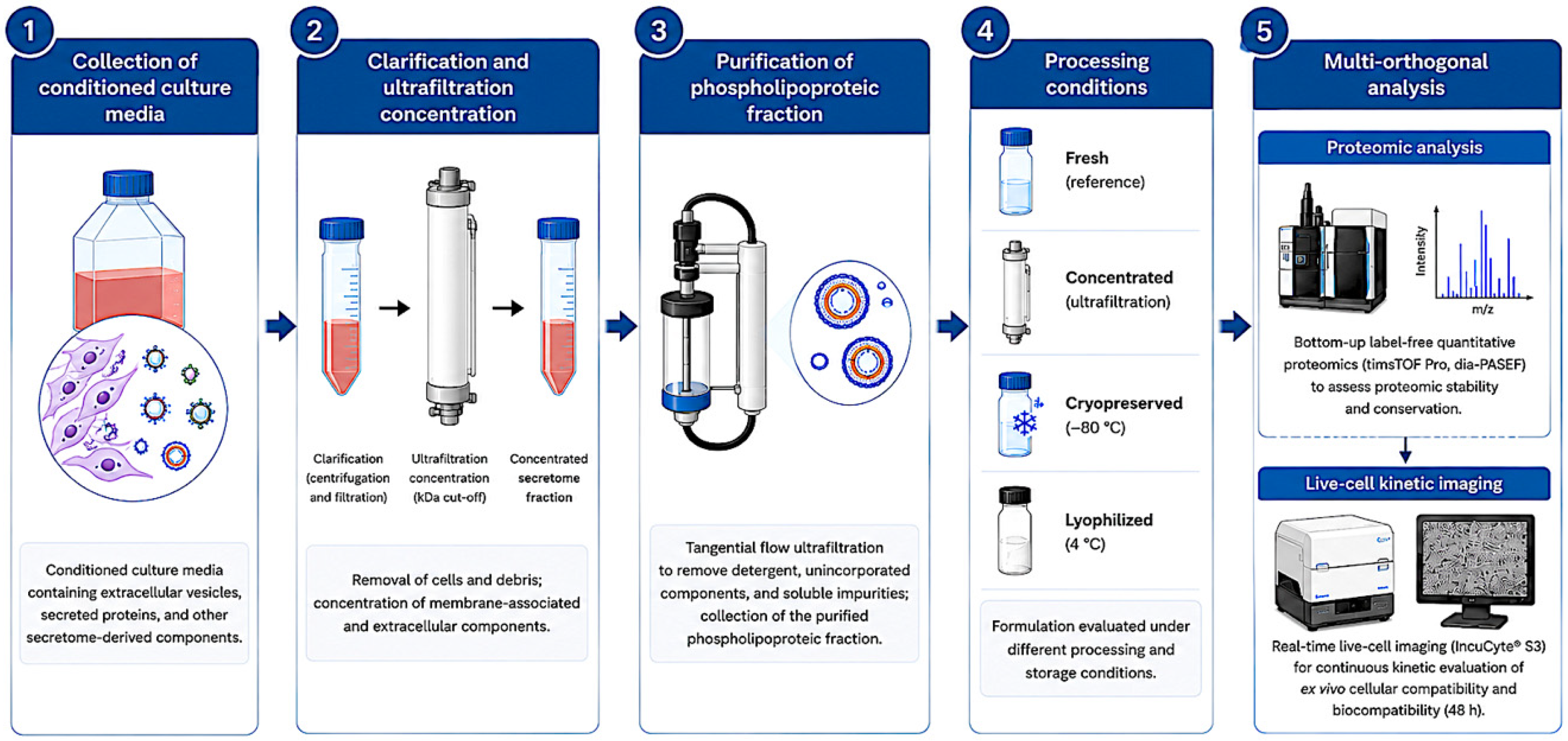
2.1. Preparation of the Processed Phospholipoproteic Secretome-Derived Formulation
2.2. Processing Conditions
2.3. Proteomic Characterization
2.4. Short-Term Real-Time Live-Cell Kinetic Profiling
2.5. Data Integration and Quality Control
2.6. Statistical Analysis
2.7. Quality Control and Acceptance Criteria
3. Results
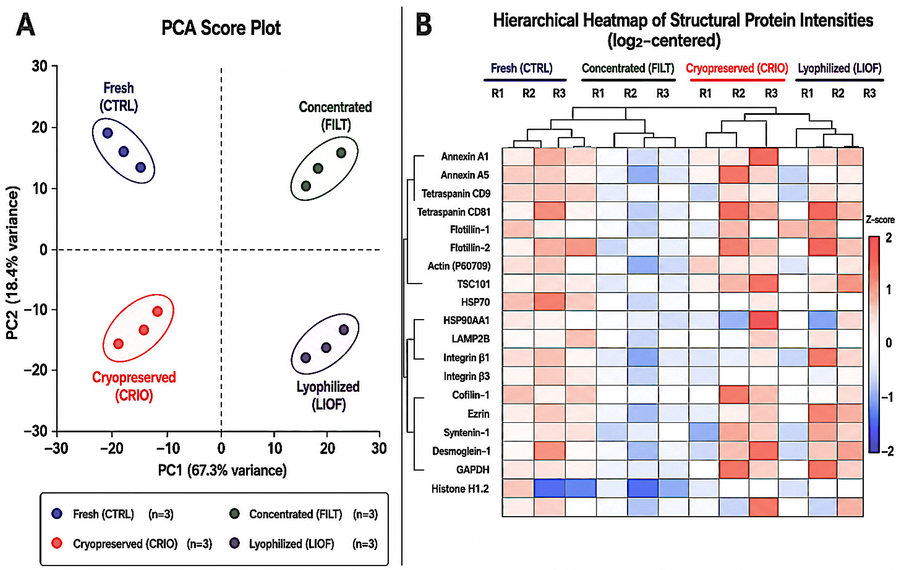
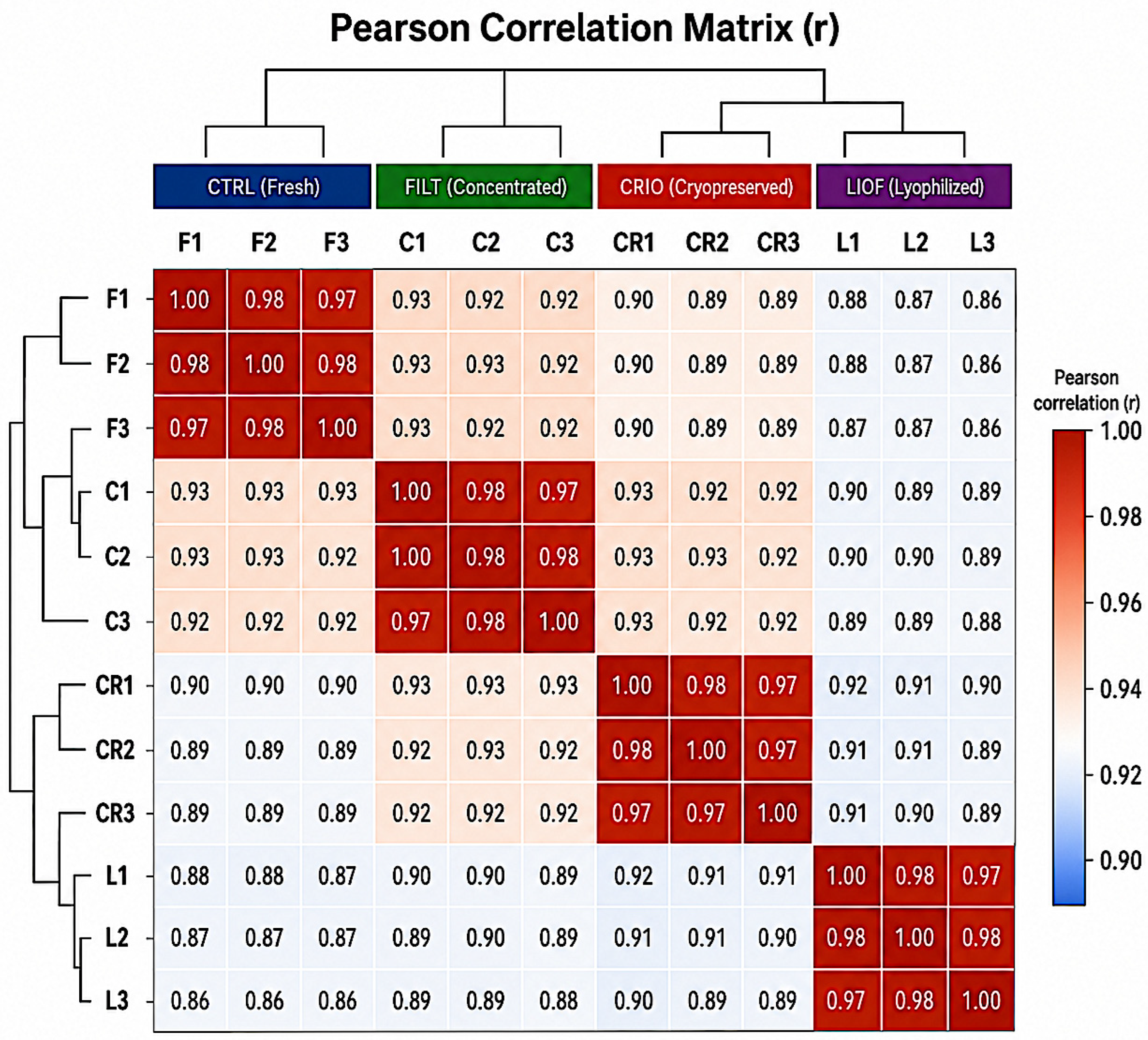
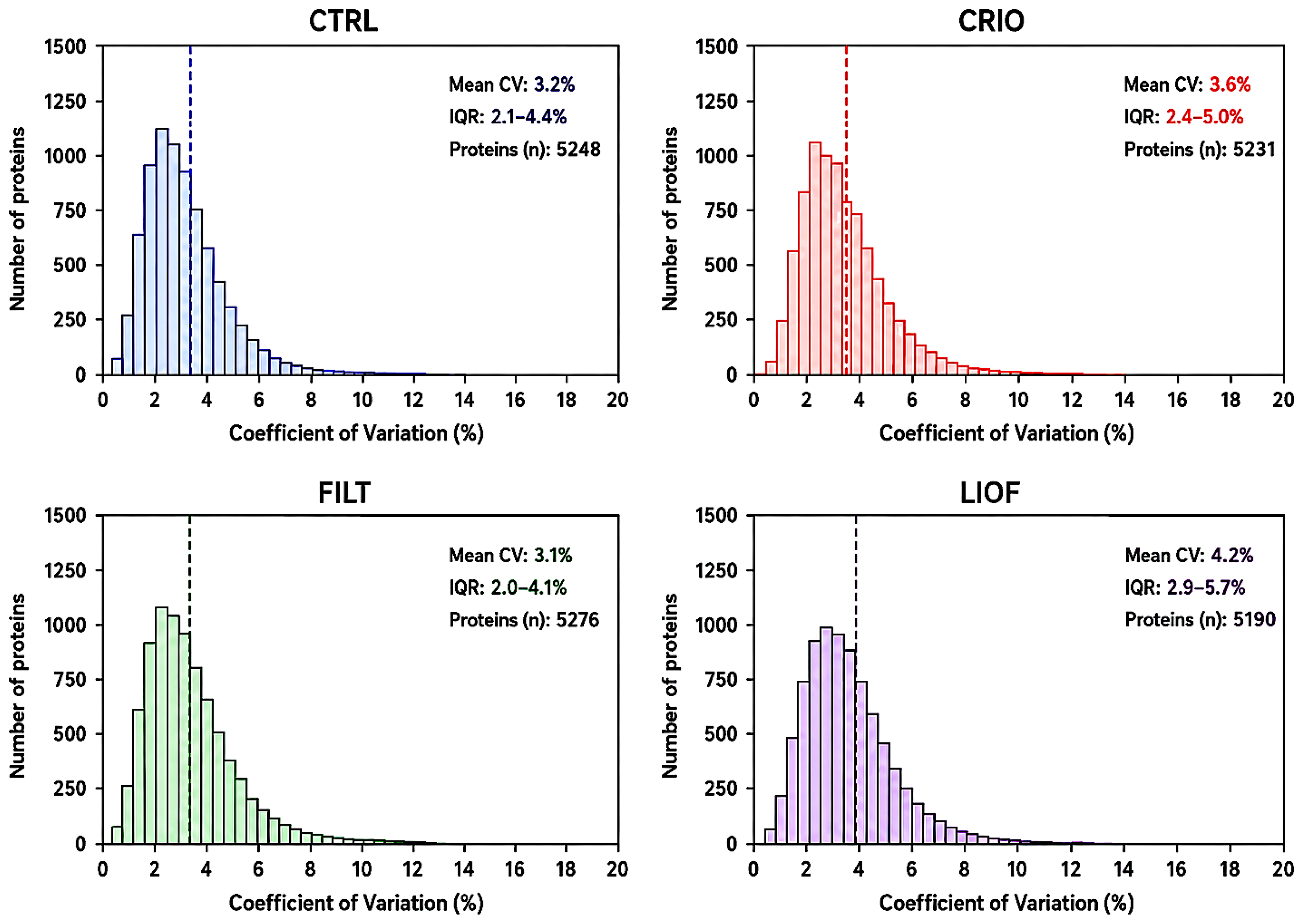
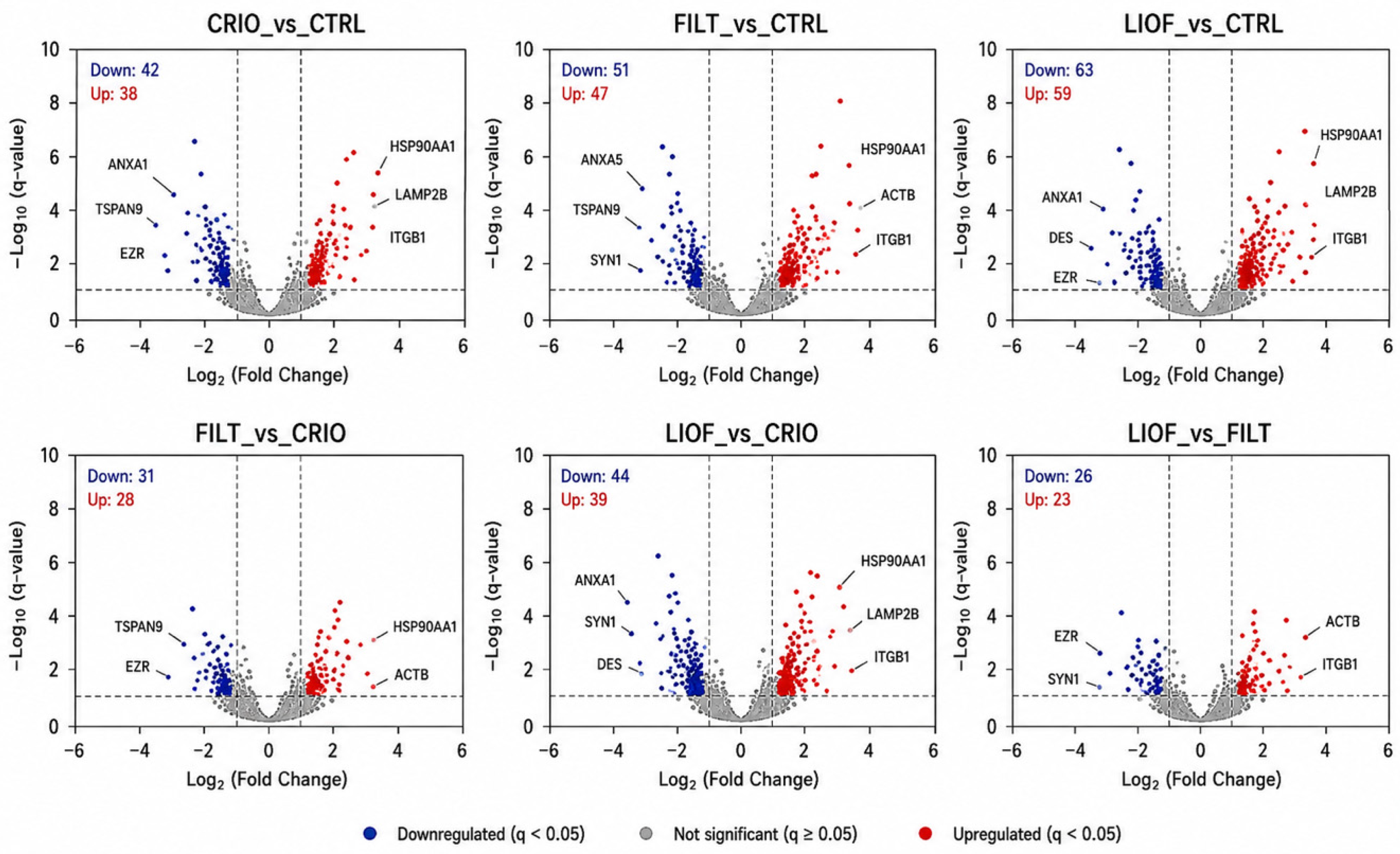
3.1. Proteomic Fingerprint and Structural Protein Conservation
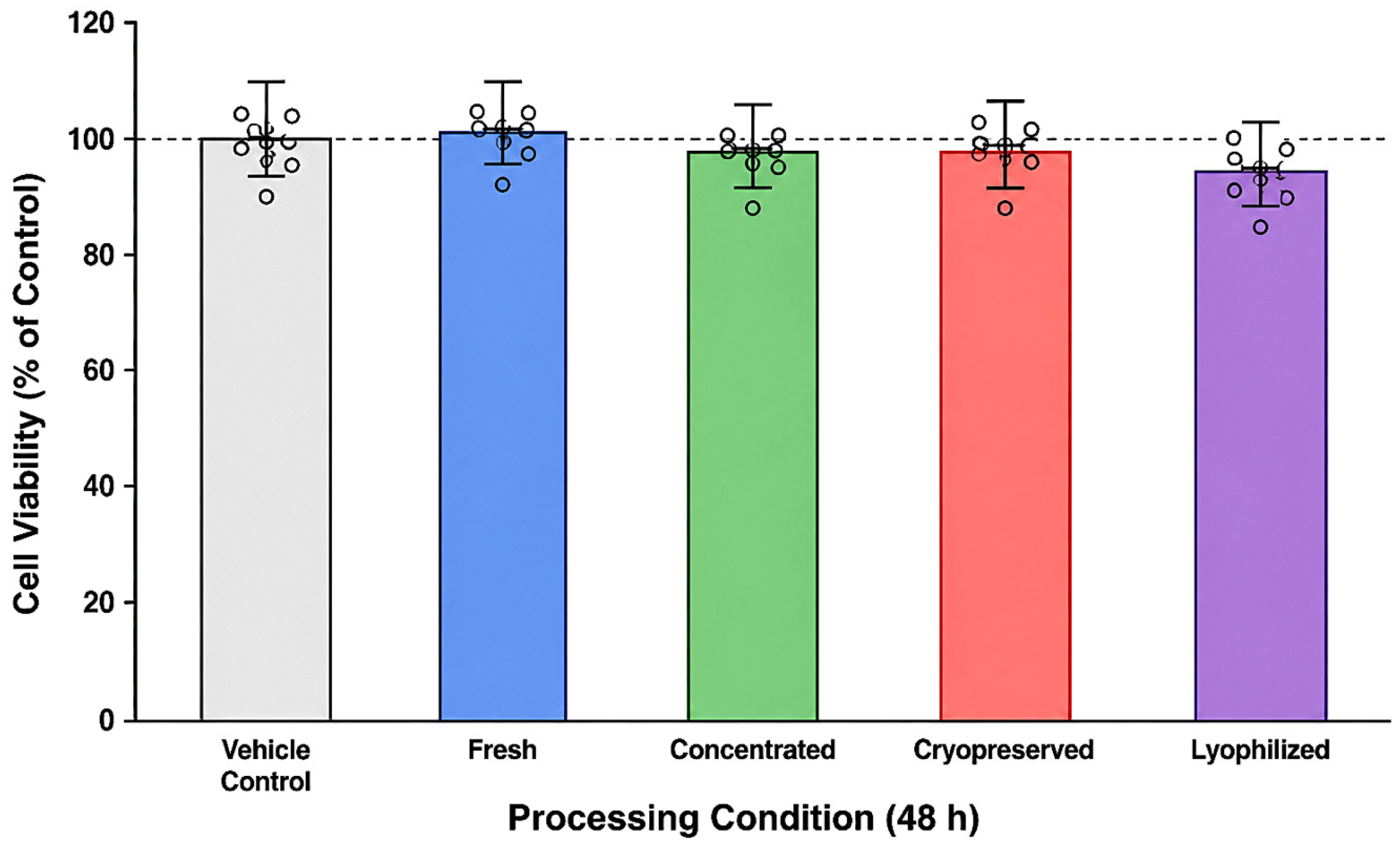
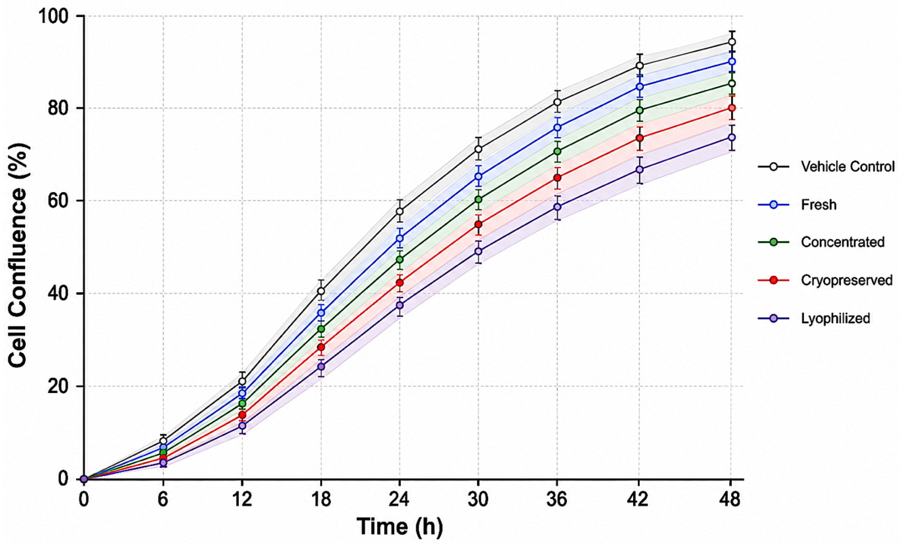
3.2. Short-Term Real-Time Kinetic Profiling and Ex Vivo Compatibility
3.3. Functional Annotation of Processing-Associated Proteomic Changes
4. Discussion
5. Conclusions
Supplementary Materials
Author Contributions
Funding
Institutional Review Board Statement
Informed Consent Statement
Data Availability Statement
Acknowledgments
Conflicts of Interest
Abbreviations
| Abbreviation | Full Term |
| CTRL | Fresh control formulation |
| CRIO | Cryopreserved formulation |
| FILT | Concentrated formulation |
| LIOF | Lyophilized formulation |
| LFQ | Label-Free Quantification |
| DEP | Differentially Expressed Protein |
| CV | Coefficient of Variation |
| PCA | Principal Component Analysis |
| FDR | False Discovery Rate |
| dia-PASEF | Data-Independent Acquisition Parallel Accumulation–Serial Fragmentation |
| timsTOF Pro | Trapped Ion Mobility Spectrometry Time-of-Flight Pro mass spectrometer |
| SD | Standard Deviation |
| Lo | Liquid-Ordered phase |
| Ld | Liquid-Disordered phase |
| IncuCyte® S3 | Real-time live-cell kinetic imaging system |
| UniProt | Universal Protein Resource database |
| ANOVA | Analysis of Variance |
| Z-score | Standardized score normalization metric |
| PC1 | Principal Component 1 |
| PC2 | Principal Component 2 |
References
- Levental, I.; Levental, K.R.; Heberle, F.A. Lipid rafts: Controversies resolved, mysteries remain. Trends Cell Biol. 2020, 30, 341–353. [Google Scholar] [CrossRef] [PubMed]
- Gutiérrez-Sandoval, R.; Gutiérrez-Castro, F.; Muñoz-Godoy, N.; Rivadeneira, I.; Lagos, A.; Iturra, J.; Krakowiak, F.; Peña-Vargas, C.; Vidal, M.; Toledo, A.; et al. Structural determinants and repair of membrane microdomains in dendritic cell-mediated antitumor immunity: An integrative mechanistic synthesis. Int. J. Mol. Sci. 2026, 27, 2305. [Google Scholar] [CrossRef] [PubMed]
- Zhang, S.; Zhu, N.; Gu, J.; Li, H.F.; Qiu, Y.; Liao, D.F.; Qin, L. Crosstalk between lipid rafts and aging: New frontiers for delaying aging. Aging Dis. 2022, 13, 1042. [Google Scholar] [CrossRef] [PubMed]
- Nicolaou, A.; Kendall, A.C. Bioactive lipids in the skin barrier mediate its functionality in health and disease. Pharmacol. Ther. 2024, 260, 108681. [Google Scholar] [CrossRef] [PubMed]
- Gutiérrez-Sandoval, R.; Gutiérrez-Castro, F.; Muñoz-Godoy, N.; Rivadeneira, I.; Sobarzo, A.; Iturra, J.; Krakowiak, F.; Alarcón, L.; Dorado, W.; Lagos, A.; et al. Phospholipid-Rich DC-Vesicles with Preserved Immune Fingerprints: A Stable and Scalable Platform for Precision Immunotherapy. Biomedicines 2025, 13, 1299. [Google Scholar] [CrossRef] [PubMed]
- Wculek, S.K.; Cueto, F.J.; Mujal, A.M.; Melero, I.; Krummel, M.F.; Sancho, D. Dendritic cells in cancer immunology and immunotherapy. Nat. Rev. Immunol. 2020, 20, 7–24. [Google Scholar] [PubMed]
- Marjanovic, J.; Jurczuk, V.; Tose, L.V.; Cintron Diaz, Y.; Fernandez Lima, F.; Abdo Abujamra, B.; Danker, S.; Jabori, S.; Singh, D.; Burgess, J.L.; et al. Scaffolds with spatiotemporally controlled growth factor delivery and cyclodextrin-enabled antagonism of growth factor receptor sequestration promote cutaneous wound healing. npj Regen. Med. 2025, 10, 42. [Google Scholar] [CrossRef] [PubMed]
- Vicente-da-Silva, J.V.; da Silva Lopes Pereira, J.O.; do Carmo, F.A.; de Carvalho Patricio, B.F. Skin and wound healing: Conventional dosage versus nanobased emulsions forms. ACS Omega 2025, 10, 12837–12855. [Google Scholar] [CrossRef] [PubMed]
- Isik, O.A.; Cizmecioglu, O. Rafting on the plasma membrane: Lipid rafts in signaling and disease. In Cell Biology and Translational Medicine, Volume 20; Springer: Cham, Switzerland, 2023; Volume 1436, pp. 87–108. [Google Scholar]
- Kulkarni, R.; Wiemer, E.A.C.; Chang, W. Role of lipid rafts in pathogen-host interaction—A mini review. Front. Immunol. 2022, 12, 815020. [Google Scholar] [CrossRef] [PubMed]
- Choudhary, V.; Choudhary, M.; Bollag, W.B. Exploring skin wound healing models and the impact of natural lipids on the healing process. Int. J. Mol. Sci. 2024, 25, 3790. [Google Scholar] [CrossRef] [PubMed]
- Deben, C.; Cardenas De La Hoz, E.; Rodrigues Fortes, F.; Le Compte, M.; Seghers, S.; Vanlanduit, S.; Vercammen, H.; Van Den Bogert, B.; Dusetti, N.; Lin, A.; et al. Development and validation of the Normalized Organoid Growth Rate (NOGR) metric in brightfield imaging-based assays. Commun. Biol. 2024, 7, 1612. [Google Scholar] [CrossRef] [PubMed]
- Philip, M.; Schietinger, A. CD8+ T cell differentiation and dysfunction in cancer. Nat. Rev. Immunol. 2022, 22, 209–223. [Google Scholar] [PubMed]
- Garcia, E.; Ismail, S. Spatiotemporal Regulation of Signaling: Focus on T Cell Activation and the Immunological Synapse. Int. J. Mol. Sci. 2020, 21, 3283. [Google Scholar] [CrossRef] [PubMed]
- Bagaev, A.; Kotlov, N.; Nomie, K.; Svekolkin, V.; Gafurov, A.; Isaeva, O.; Osokin, N.; Kozlov, I.; Frenkel, F.; Gancharova, O.; et al. Conserved pan-cancer microenvironment subtypes predict response to immunotherapy. Cancer Cell 2021, 39, 845–865.e7. [Google Scholar] [CrossRef] [PubMed]
- Vu, S.H.; Vetrivel, P.; Kim, J.; Lee, M.S. Cancer resistance to immunotherapy: Molecular mechanisms and tackling strategies. Int. J. Mol. Sci. 2022, 23, 10906. [Google Scholar] [CrossRef] [PubMed]
- Liu, Y.J.; Wang, C. A Review of the Regulatory Mechanisms of Extracellular Vesicles-Mediated Intercellular Communication. Cell Commun. Signal. 2023, 21, 77. [Google Scholar] [CrossRef] [PubMed]
- Steinberger, S.; Karuthedom George, S.; Lauková, L.; Weiss, R.; Tripisciano, C.; Marchetti-Deschmann, M.; Weber, V.; Allmaier, G.; Weiss, V.U. Targeting the structural integrity of extracellular platforms via nano electrospray gas-phase electrophoretic mobility molecular analysis (nES GEMMA). Membranes 2022, 12, 872. [Google Scholar] [CrossRef] [PubMed]
- Wurster, S.; Kumaresan, P.R.; Albert, N.D.; Hauser, P.J.; Lewis, R.E.; Kontoyiannis, D.P. Live monitoring and analysis of fungal growth, viability, and mycelial morphology using the IncuCyte NeuroTrack processing module. mBio 2019, 10, e00673-19. [Google Scholar] [CrossRef] [PubMed]
- Holme, B.; Bjørnerud, B.; Pedersen, N.M.; Rodriguez de la Ballina, L.; Wesche, J.; Haugsten, E.M. Automated tracking of cell migration in phase contrast images with CellTraxx. Sci. Rep. 2023, 13, 22982. [Google Scholar] [CrossRef] [PubMed]
- Yu, F.; Haynes, S.E.; Nesvizhskii, A.I. IonQuant Enables Accurate and Sensitive Label-Free Quantification with FDR-Controlled Match-Between-Runs. Mol. Cell. Proteom. 2021, 20, 100077. [Google Scholar] [CrossRef]
- Gidda, A.K.; Karuthedom George, S.; Lauková, L.; Weiss, R.; Tripisciano, C.; Marchetti-Deschmann, M.; Weber, V.; Allmaier, G.; Weiss, V.U. Real-Time IncuCyte® Assay for the Dynamic Assessment of Live and Dead Cells in 2D Cultures. Bio Protoc. 2025, 15, e5209. [Google Scholar] [CrossRef] [PubMed]
- Cheng, C.; Thrash, J.C. Sparse-growth-curve: A computational pipeline for parsing cellular growth curves with low temporal resolution. Microbiol. Resour. Announc. 2021, 10, e00296-21. [Google Scholar] [CrossRef] [PubMed]
- Vitko, D.; Chou, W.F.; Nouri Golmaei, S.; Lee, J.Y.; Belthangady, C.; Blume, J.; Chan, J.K.; Flores-Campuzano, G.; Hu, Y.; Liu, M.; et al. timsTOF HT Improves Protein Identification and Quantitative Reproducibility for Deep Unbiased Plasma Protein Biomarker Discovery. J. Proteome Res. 2024, 23, 929–938. [Google Scholar] [CrossRef] [PubMed]
- Lai, J.J.; Chau, Z.L.; Chen, S.Y.; Hill, J.J.; Korpany, K.V.; Liang, N.W.; Lin, L.H.; Liu, J.K. Exosome processing and characterization approaches for research and technology development. Adv. Sci. 2022, 9, e2103222. [Google Scholar] [CrossRef]
- Distler, U.; Yoo, H.B.; Kardell, O.; Hein, D.; Sielaff, M.; Scherer, M.; Jozefowicz, A.M.; Leps, C.; Gomez-Zepeda, D.; von Toerne, C.; et al. Multicenter Evaluation of Label-Free Quantification in Human Plasma on a High Dynamic Range Benchmark Set. Nat. Commun. 2025, 16, 877. [Google Scholar] [CrossRef]
- Yang, M.; Wu, Y.; Zhang, Z.; Xu, Y.; Lei, T.; Wang, X.; Jin, Z.; Hou, K.; Cai, Y.; Sun, S. StrucGAP: A Modular, Streamlined and Traceable Data Mining Platform for Structural and Site-Specific Glycoproteomics. Nat. Commun. 2026, 17, 2579. [Google Scholar] [CrossRef] [PubMed]
- Heinemann, T.; Kornauth, C.; Severin, Y.; Vladimer, G.I.; Pemovska, T.; Hadzijusufovic, E.; Agis, H.; Krauth, M.T.; Sperr, W.R.; Valent, P.; et al. Deep morphology learning enhances ex vivo drug profiling-based precision medicine. Blood Cancer Discov. 2022, 3, 502–515. [Google Scholar] [CrossRef] [PubMed]
- Buschmann, D.; Mussack, V.; Byrd, J.B. Separation, characterization, and standardization of extracellular platforms for drug delivery applications. Adv. Drug Deliv. Rev. 2021, 174, 348–368. [Google Scholar] [CrossRef] [PubMed]
- Kalluri, R.; LeBleu, V.S. The biology, function, and biomedical applications of exosomes. Science 2020, 367, eaau6977. [Google Scholar] [CrossRef] [PubMed]
- Mathieu, M.; Martin-Jaular, L.; Lavieu, G.; Théry, C. Specificities of secretion and uptake of exosomes and other extracellular vesicles for cell-to-cell communication. Nat. Cell Biol. 2019, 21, 9–17. [Google Scholar] [CrossRef] [PubMed]
- Wang, W.; Douglas, D.; Zhang, J.; Kumari, S.; Enuameh, M.S.; Dai, Y.; Wallace, C.T.; Watkins, S.C.; Shu, W.; Xing, J. Live-cell imaging and analysis reveal cell phenotypic transition dynamics inherently missing in snapshot data. Sci. Adv. 2020, 6, eaba9319. [Google Scholar] [CrossRef] [PubMed]
- Claridge, B.; Lozano, J.; Poh, Q.H.; Greening, D.W. Development of extracellular platform therapeutics: Challenges, considerations, and opportunities. Front. Cell Dev. Biol. 2021, 9, 734720. [Google Scholar] [CrossRef] [PubMed]
- Kumar, M.A.; Baba, S.K.; Sadida, H.Q.; Al Marzooqi, S.; Jerobin, J.; Altemani, F.H.; Algehainy, N.; Alanazi, M.A.; Abou-Samra, A.-B.; Kumar, R.; et al. Extracellular vesicles as tools and targets in therapy for diseases. Signal Transduct. Target Ther. 2024, 9, 27. [Google Scholar] [CrossRef] [PubMed]
- Radstake, W.E.; Gautam, K.; Van Rompay, C.; Vermeesen, R.; Tabury, K.; Verslegers, M.; Baatout, S.; Baselet, B. Comparison of In Vitro Scratch Wound Assay Experimental Procedures. Biochem. Biophys. Rep. 2023, 33, 101423. [Google Scholar] [CrossRef] [PubMed]
- Cai, Y.; Prochazkova, M.; Kim, Y.S.; Jiang, C.; Ma, J.; Moses, L.; Martin, K.; Pham, V.; Zhang, N.; Highfill, S.L.; et al. Assessment and comparison of viability assays for cellular products. Cytotherapy 2024, 26, 201–209. [Google Scholar] [CrossRef] [PubMed]
- Hou, J.; Liu, J.; Huang, Z.; Wang, Y.; Yao, H.; Hu, Z.; Shi, C.; Xu, J.; Wang, Q. Structure and Function of the Membrane Microdomains in Osteoclasts. Bone Res. 2023, 11, 61. [Google Scholar] [CrossRef] [PubMed]
- Gutiérrez-Sandoval, R.; Gutiérrez-Castro, F.; Muñoz-Godoy, N.; Rivadeneira, I.; Sobarzo, A.; Iturra, J.; Krakowiak, F.; Alarcón, L.; Dorado, W.; Lagos, A.; et al. Ex Vivo Traceability Platform for Phospholipoproteomic Formulations: Functional Evidence Without Clinical Exposure. Biomedicines 2025, 13, 2101. [Google Scholar] [CrossRef] [PubMed]
- Jeppesen, D.K.; Fenix, A.M.; Franklin, J.L.; Higginbotham, J.N.; Zhang, Q.; Zimmerman, L.J.; Liebler, D.C.; Ping, J.; Liu, Q.; Evans, R.; et al. Reassessment of exosome composition. Cell 2019, 177, 428–445. [Google Scholar] [CrossRef] [PubMed]
- Singh, M.; Tiwari, P.K.; Kashyap, V.; Kumar, S. Proteomics of extracellular vesicles: Recent updates, challenges and limitations. Proteomes 2025, 13, 12. [Google Scholar] [CrossRef] [PubMed]
- Crouigneau, R.; Li, Y.F.; Auxillos, J.; Goncalves-Alves, E.; Marie, R.; Sandelin, A.; Pedersen, S.F. Mimicking and analyzing the tumor microenvironment. Cell Rep. Methods 2024, 4, 100866. [Google Scholar] [CrossRef] [PubMed]
- Manoochehri, H.; La’ah, A.S.; Babaeizad, A.; Sheykhhasan, M.; Rostamipoor, M.; Abbaspoor, M.; Nikravesh, F.; Mozaffari Khosravi, S.; Mahaki, H.; Tanzadehpanah, H.; et al. Extracellular vesicles in cancer immunotherapy: Therapeutic challenges and clinical progress. Asian J. Pharm. Sci. 2025, 20, 101065. [Google Scholar] [CrossRef] [PubMed]
- Zhou, G.; Li, R.; Sheng, S.; Huang, J.; Zhou, F.; Wei, Y.; Liu, H.; Su, J. Organoids and organoid extracellular platforms-based disease treatment strategies. J. Nanobiotechnol. 2024, 22, 679. [Google Scholar] [CrossRef] [PubMed]
- Ren, L.; Zhang, D.; Pang, L.; Liu, S. Extracellular vesicles for cancer therapy: Potential, progress, and clinical challenges. Front. Bioeng. Biotechnol. 2024, 12, 1476737. [Google Scholar] [CrossRef] [PubMed]
- Théry, C.; Witwer, K.W.; Aikawa, E.; Alcaraz, M.J.; Anderson, J.D.; Andriantsitohaina, R.; Antoniou, A.; Arab, T.; Archer, F.; Atkin-Smith, G.K.; et al. Minimal information for studies of extracellular vesicles 2018 (MISEV2018): A position statement of the International Society for Extracellular Vesicles. J. Extracell. Vesicles 2018, 7, 1535750. [Google Scholar] [CrossRef] [PubMed]
- Wandrey, M.; Jablonska, J.; Stauber, R.H.; Gül, D. Exosomes in cancer progression and therapy resistance: Molecular insights and therapeutic opportunities. Life 2023, 13, 2033. [Google Scholar] [CrossRef] [PubMed]
- Das, S.; Dey, M.K.; Devireddy, R.; Gartia, M.R. Biomarkers in cancer detection, diagnosis, and prognosis. Sensors 2023, 24, 37. [Google Scholar] [CrossRef] [PubMed]
- Wang, X.; Cheng, W.; Su, J. Research Progress of Extracellular Vesicles-Loaded Microneedle Technology. Pharmaceutics 2024, 16, 326. [Google Scholar] [CrossRef] [PubMed]
- Safaei, S.; Fadaee, M.; Farzam, O.R.; Yari, A.; Poursaei, E.; Aslan, C.; Samemaleki, S.; Shanehbandi, D.; Baradaran, B.; Kazemi, T. Exploring the dynamic interplay between exosomes and the immune tumor microenvironment: Implications for breast cancer progression and therapeutic strategies. Breast Cancer Res. 2024, 26, 57. [Google Scholar] [CrossRef] [PubMed]
- Liu, Q.; Li, S.; Dupuy, A.; Mai, H.L.; Sailliet, N.; Logé, C.; Robert, J.H.; Brouard, S. Exosomes as new biomarkers and drug delivery tools for the prevention and treatment of various diseases. Int. J. Mol. Sci. 2021, 22, 7763. [Google Scholar] [CrossRef] [PubMed]
- Makler, A.; Asghar, W. Exosomal biomarkers for cancer diagnosis and patient monitoring. Expert Rev. Mol. Diagn. 2020, 20, 387–400. [Google Scholar] [CrossRef] [PubMed]
- Narauskaitė, D.; Vydmantaitė, G.; Rusteikaitė, J.; Sampath, R.; Rudaitytė, A.; Stašytė, G.; Aparicio, C.; Jarmalavičiūtė, A. Extracellular Vesicles in Skin Wound Healing: From Skin Regeneration to Treatment of Chronic Wounds. Pharmaceuticals 2021, 14, 811. [Google Scholar] [CrossRef] [PubMed]
- Zulueta Diaz, Y.M.; Sezgin, E.; Schwille, P.; Eggeling, C. Super-Resolution Microscopy to Study Membrane Organization in Health and Disease. Front. Mol. Biosci. 2024, 11, 1455153. [Google Scholar] [CrossRef] [PubMed]
- Nguyen, S.T.; Tran, T.T.H.; Nguyen, H.T.T.; Pham, T.T.T.; Nguyen, T.H.; Le, H.T.; Nguyen, T.T.H. Exosomes Derived from Mesenchymal Stem Cells: A Novel Therapeutic Approach for Skin Wound Healing and Regeneration. Biomed. Res. Ther. 2024, 11, 946. [Google Scholar] [CrossRef]
- Chitti, S.V.; Gummadi, S.; Kang, T.; Shahi, S.; Marzan, A.L.; Nedeva, C.; Sanwlani, R.; Bramich, K.; Stewart, S.; Petrovska, M.; et al. Vesiclepedia 2024: An Extracellular Vesicles and Extracellular Particles Repository. Nucleic Acids Res. 2024, 52, D1694–D1698. [Google Scholar] [PubMed]



| Component | Functional Role | Relative Proportion |
|---|---|---|
| Phospholipid-associated fraction | Membrane-associated structural organization | Predominant |
| Membrane-associated proteins | Structural and extracellular compatibility | Conserved proteomic fraction |
| Extracellular structural proteins | Organization of extracellular lipid–protein assemblies | Moderate abundance |
| Cytoskeletal-associated proteins | Structural compatibility and membrane-associated interactions | Trace–moderate abundance |
| Apolipoprotein-associated components | Lipid-associated stabilization | Trace abundance |
| Annexin-associated proteins | Membrane interaction and phospholipid-binding functions | Trace abundance |
| Extracellular matrix-associated proteins | Structural extracellular organization | Variable abundance |
| Parameter | Fresh (Reference) | Concentrated | Cryopreserved | Lyophilized |
|---|---|---|---|---|
| Total proteins identified | 574 | 562 | 545 | 533 |
| Structural protein retention | 100% | >92% | >91% | >90% |
| Mean coefficient of variation (CV) | 3.1% | 3.3% | 3.0% | 4.5% |
| Inter-replicate Pearson correlation | >0.97 | >0.97 | >0.96 | >0.95 |
| Differentially expressed proteins (DEPs) vs. fresh reference | — | 68 | 49 | 129 |
| Global proteomic profile conservation: | Reference profile | Preserved | Preserved | Partially preserved |
| LFQ reproducibility | High | High | High | High |
| Parameter | Vehicle Control | Fresh | Concentrated | Cryopreserved | Lyophilized |
|---|---|---|---|---|---|
| Cell viability at 48 h | 100 ± 5% | 98 ± 7% | 98 ± 6% | 97 ± 6% | 96 ± 7% |
| Maximum confluence divergence from vehicle | — | <5% | <5% | <6% | <7% |
| Mean intra-experimental CV | <10% | <9% | <9% | <8% | <10% |
| Morphological alterations | None detected | None detected | None detected | None detected | None detected |
| Evidence of blebbing | Absent | Absent | Absent | Absent | Absent |
| Evidence of detachment | Absent | Absent | Absent | Absent | Absent |
| Evidence of proliferative suppression | Absent | Absent | Absent | Absent | Absent |
| Overall kinetic compatibility | Reference profile | Preserved | Preserved | Preserved | Preserved |
Disclaimer/Publisher’s Note: The statements, opinions and data contained in all publications are solely those of the individual author(s) and contributor(s) and not of MDPI and/or the editor(s). MDPI and/or the editor(s) disclaim responsibility for any injury to people or property resulting from any ideas, methods, instructions or products referred to in the content. |
© 2026 by the authors. Licensee MDPI, Basel, Switzerland. This article is an open access article distributed under the terms and conditions of the Creative Commons Attribution (CC BY) license.
Share and Cite
Gutiérrez-Sandoval, R.; Gutiérrez-Castro, F.; Muñoz-Godoy, N.; Rivadeneira, I.; Lagos, A.; Iturra, J.; Krakowiak, F.; Muñoz, I.; Toledo, A. Proteomic Stability and Ex Vivo Compatibility of a Processed Phospholipoproteic Secretome-Derived Formulation. Pharmaceutics 2026, 18, 847. https://doi.org/10.3390/pharmaceutics18070847
Gutiérrez-Sandoval R, Gutiérrez-Castro F, Muñoz-Godoy N, Rivadeneira I, Lagos A, Iturra J, Krakowiak F, Muñoz I, Toledo A. Proteomic Stability and Ex Vivo Compatibility of a Processed Phospholipoproteic Secretome-Derived Formulation. Pharmaceutics. 2026; 18(7):847. https://doi.org/10.3390/pharmaceutics18070847
Chicago/Turabian StyleGutiérrez-Sandoval, Ramón, Francisco Gutiérrez-Castro, Natalia Muñoz-Godoy, Ider Rivadeneira, Andy Lagos, Jordan Iturra, Francisco Krakowiak, Ignacio Muñoz, and Andrés Toledo. 2026. "Proteomic Stability and Ex Vivo Compatibility of a Processed Phospholipoproteic Secretome-Derived Formulation" Pharmaceutics 18, no. 7: 847. https://doi.org/10.3390/pharmaceutics18070847
APA StyleGutiérrez-Sandoval, R., Gutiérrez-Castro, F., Muñoz-Godoy, N., Rivadeneira, I., Lagos, A., Iturra, J., Krakowiak, F., Muñoz, I., & Toledo, A. (2026). Proteomic Stability and Ex Vivo Compatibility of a Processed Phospholipoproteic Secretome-Derived Formulation. Pharmaceutics, 18(7), 847. https://doi.org/10.3390/pharmaceutics18070847