1. Introduction
Malignant tumors remain one of the most significant threats to global public health [
1,
2]. For decades, the primary treatment modalities have been conventional therapies such as chemotherapy, surgery, and radiotherapy [
3,
4,
5,
6]. While these approaches can be effective, they are often hampered by significant limitations, including poor tumor specificity, the development of multidrug resistance, and substantial damage to healthy tissues and the immune system. In response to these challenges, cancer immunotherapy has emerged as a revolutionary approach [
6,
7]. By harnessing and amplifying the power of the patient’s own immune system to recognize and eliminate cancer cells, immunotherapy offers the potential for more durable and specific anti-tumor responses. Various immunotherapeutic strategies have been clinically approved, demonstrating remarkable success in certain cancers.
However, the clinical efficacy of immunotherapy is often constrained by the complex and dynamic tumor microenvironment (TME) [
8,
9,
10,
11]. The TME, composed of cancer cells, stromal cells, and a variety of immune cells, evolves to create a highly immunosuppressive milieu. This environment protects the tumor from immune attack by secreting inhibitory cytokines, recruiting immunosuppressive cells like regulatory T cells (Tregs) and myeloid-derived suppressor cells (MDSCs), and downregulating antigen presentation. A key mechanism by which the TME enforces immune suppression is through the accumulation of immunosuppressive metabolites, most notably ADO [
12,
13,
14,
15].
The critical role of ADO in the TME is intricately linked to the metabolism of extracellular ATP, a duality that has become a major focus of cancer research. In physiological conditions, intracellular ATP is the primary energy currency of the cell. However, in the pathological TME, cellular stress or death can lead to the release of ATP into the extracellular space, where it acts as a potent immunostimulatory “find-me” and danger signal. eATP binds to purinergic P2 receptors on immune cells, promoting the recruitment of antigen-presenting cells, activating inflammasomes, and driving the differentiation of pro-inflammatory T cell subsets [
16,
17]. Conversely, its breakdown product, ADO, binds to G-protein-coupled adenosine receptors (notably A2A and A2B) on immune cells, suppressing their effector functions, promoting Treg differentiation, and fostering a state of immune tolerance [
18,
19,
20]. This balance between immunostimulatory ATP and immunosuppressive ADO is primarily regulated by two ectonucleotidases: CD39 and CD73. CD39 (encoded by ENTPD1) is the rate-limiting enzyme in this pathway, hydrolyzing ATP and ADP into AMP [
21,
22,
23,
24]. Subsequently, CD73 converts AMP into ADO. Therefore, CD39 acts as a critical immunological “switch” within the TME. Its upregulation on tumor cells and various immune cells within the TME contributes directly to the establishment of an ADO-rich, immunosuppressive environment that facilitates tumor immune evasion.
Given its central role, targeting CD39 presents a highly attractive strategy for cancer immunotherapy. Inhibiting CD39 offers a dual mechanism of action: first, it prevents the depletion of pro-inflammatory ATP, thereby preserving its immunostimulatory signals; second, it simultaneously curbs the production of immunosuppressive ADO, thereby relieving a major brake on anti-tumor immunity [
25,
26]. This combined effect can potentially enhance the function of effector T cells and natural killer (NK) cells while reducing the suppressive activity of Tregs and MDSCs.
A powerful way to further amplify this effect is to combine CD39 inhibition with the induction of ICD. ICD is a functionally distinct form of cell death triggered by specific stressors, including certain chemotherapies like anthracyclines (e.g., doxorubicin, DOX) and oxaliplatin, as well as radiotherapy and photodynamic therapy [
27,
28,
29,
30,
31]. When a cancer cell undergoes ICD, it not only dies but also actively emits a series of damage-associated molecular patterns (DAMPs). These include the exposure of calreticulin (CRT) on the cell surface (an “eat me” signal), the release of ATP (a “find me” signal), and the passive release of HMGB1 (a chromatin-binding protein). These DAMPs serve as potent adjuvants, facilitating the recruitment and activation of dendritic cells (DCs) [
32,
33,
34,
35]. Mature DCs can then effectively process and present tumor antigens to naïve T cells, thereby priming a robust and tumor-specific adaptive immune response. Thus, ICD can effectively convert a “cold,” non-immunogenic tumor into a “hot,” inflamed one that is more responsive to immunotherapy. DOX, an ICD inducer, can stimulate the release of ATP, which, if protected from CD39-mediated degradation by a co-administered inhibitor, could significantly amplify the ICD-driven immunostimulatory cascade.
While several approaches have been developed to counteract the immunosuppressive adenosine pathway, including CD73 inhibition (which blocks the conversion of AMP to ADO and A2A/A2B receptor antagonists (which block ADO signaling on immune cells), targeting CD39 offers distinct and potentially superior advantages. As the rate-limiting enzyme that initiates the entire ATP-to-adenosine cascade, CD39 occupies a unique upstream position in this pathway. Inhibition of CD39 achieves a dual benefit: it not only prevents the depletion of immunostimulatory ATP, thereby preserving its ‘find-me’ signal for immune cells, but also simultaneously curbs the production of immunosuppressive adenosine, thereby relieving a major brake on anti-tumor immunity. In contrast, CD73 inhibition only blocks the downstream conversion of AMP to ADO without preserving ATP, while A2A/A2B receptor antagonists merely interfere with ADO signaling without affecting ATP levels. Thus, CD39 inhibition represents a more comprehensive strategy to shift the immune balance from suppression to activation by targeting both ends of the ATP-ADO axis.
Nanotechnology offers an ideal platform to realize this synergistic strategy. Nanoparticles (NPs) possess unique advantages as drug delivery vehicles, including the ability to improve the pharmacokinetics and biodistribution of therapeutic agents, protect them from premature degradation, and enable targeted delivery. Their surfaces can be functionalized for active targeting, and more importantly, they can be engineered to be “smart” by incorporating stimuli-responsive elements. The acidic nature of the TME, a hallmark of many solid tumors, is a particularly useful internal trigger. nanoparticles can be designed to remain stable in the neutral pH of the bloodstream but rapidly disassemble and release their cargo upon encountering the acidic TME or the even lower pH of intracellular endosomes/lysosomes. This ensures precise, on-demand drug release at the desired site of action, enhancing efficacy and minimizing systemic side effects.
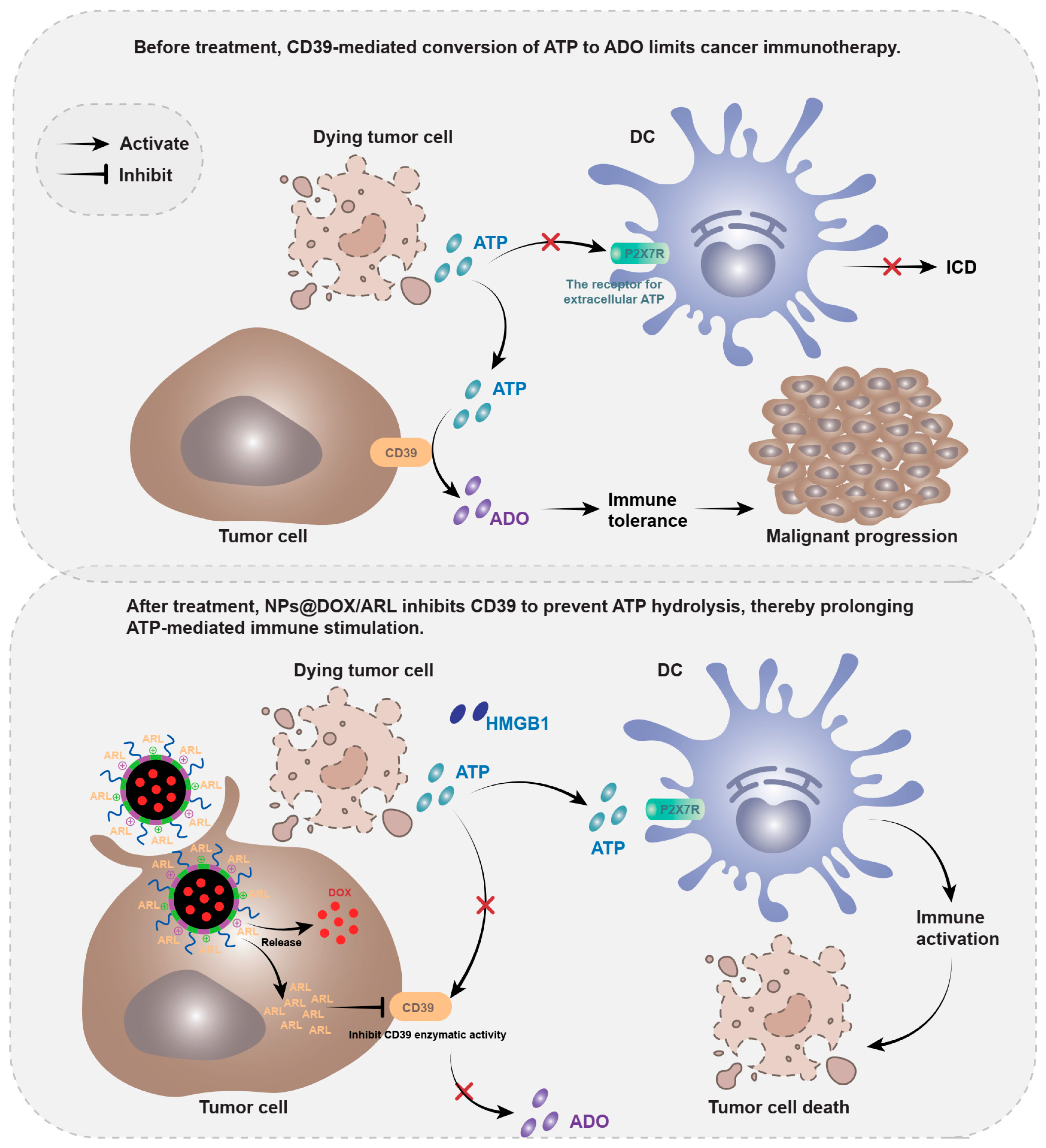
In this study, we aimed to develop a novel, nanoplatform for combined chemo-immunotherapy by co-delivering an ICD inducer and a CD39 inhibitor. We designed and synthesized a triblock amphiphilic copolymer, PEG2k-b-P(DMAEMA-co-DPAEMA)-b-PTDMAEMA (GDDM). This polymer was engineered to self-assemble into nanoparticles capable of encapsulating the hydrophobic ICD inducer DOX within its core and adsorbing the negatively charged CD39 inhibitor ARL67156 onto its cationic shell via electrostatic interactions. We hypothesized that upon accumulation in the acidic TME, the nanoparticles would disassemble, leading to the controlled release of both DOX and ARL67156. The released DOX would then induce ICD in cancer cells, triggering the release of immunostimulatory ATP. Concurrently, the released ARL67156 would inhibit the CD39 enzyme on the cell surface, preventing the rapid hydrolysis of this newly released ATP, thereby preserving its concentration and prolonging its immunostimulatory effect (
Figure 1). This study systematically investigates the synthesis and characterization of the GDDM polymer and its nanoparticles, evaluates their drug loading and release profiles, and assesses their combined anti-tumor efficacy and immune-activating potential both in vitro and in vivo, providing a comprehensive proof-of-concept for this targeted chemo-immunotherapy strategy.
2. Materials and Methods
2.1. Materials
Doxorubicin hydrochloride (DOX) was purchased from Dalian Meilun Biotechnology Co., Ltd, Dalian, China. ARL67156 was obtained from MedChemExpress, Monmouth Junction, NJ, USA. The monomers DMAEMA, DPAEMA, and TDMAEMA, as well as the chain transfer agent PEG-CTA, were synthesized or purchased from Sigma-Aldrich, Shanghai, China. All other reagents were of analytical grade and used without further purification. All animal experiments were approved by the institutional animal care and used committee of Xidian University (Approval Code: 20220311; Approval Date 1 January 2023). The experiments were conducted in strict accordance with the approved guidelines.
2.2. Synthesis of the Triblock Copolymer
The triblock copolymer PEG2k-b-P(DMAEMA-co-DPAEMA)-b-PTDMAEMA (GDDM) was synthesized via two-step reversible addition-fragmentation chain transfer (RAFT) polymerization. First, PEG-CTA was used as a macro-chain transfer agent to copolymerize DMAEMA and DPAEMA, yielding the intermediate diblock copolymer. Subsequently, TDMAEMA was polymerized using AIBN as an initiator to obtain the final triblock copolymer. The chemical structure was confirmed by 1H NMR spectroscopy.
2.3. Preparation and Characterization of Nanoparticles
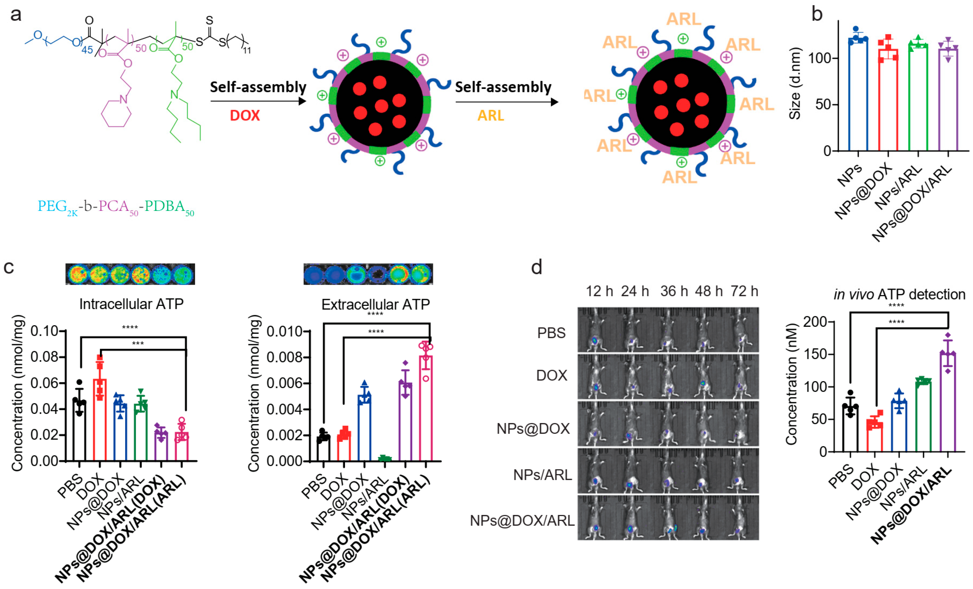
DOX-loaded nanoparticles (NPs@DOX) were prepared by a dialysis method. Briefly, the GDDM polymer and DOX were co-dissolved in DMF, followed by dialysis against deionized water to form self-assembled nanoparticles. For ARL adsorption, the DOX-loaded nanoparticles were mixed with ARL67156 solution under gentle stirring, allowing electrostatic adsorption of the negatively charged ARL onto the cationic shell to obtain NPs@DOX/ARL. Nanoparticle size and zeta potential were measured by dynamic light scattering (DLS) using a Malvern Zetasizer, Malvern, UK. The hydrodynamic diameter, polydispersity index (PDI), and zeta potential of the nanoparticles were determined using a Malvern Zetasizer, Malvern, UK at 25 °C. Samples were diluted with deionized water to an appropriate concentration before measurement. Each value was reported as the average of three independent measurements. The morphology of the nanoparticles was examined by transmission electron microscopy (TEM, JEM-2100, JEOL, Tokyo, Japan). Samples were prepared by placing a drop of nanoparticle suspension onto a carbon-coated copper grid, followed by negative staining with 2% uranyl acetate and air-drying. The encapsulation efficiency (EE) and drug loading content (LC) of DOX were determined by UV–Vis spectroscopy. Briefly, a known amount of nanoparticles was dissolved in DMF to release the encapsulated DOX, and the absorbance at 480 nm was measured against a standard calibration curve. EE and LC were calculated as follows: EE (%) = (amount of encapsulated DOX/total amount of DOX added) × 100%; LC (%) = (amount of encapsulated DOX/weight of nanoparticles) × 100%. For storage stability, nanoparticles were stored at 4 °C for 14 days, and the particle size and PDI were monitored at designated time points. For serum stability, nanoparticles were incubated in PBS containing 10% fetal bovine serum (FBS) at 37 °C for 7 days, and size changes were recorded. To confirm that ARL was surface-associated rather than loosely adsorbed, nanoparticles were subjected to salt-washing desorption studies. NPs@DOX/ARL were incubated with PBS containing increasing concentrations of NaCl (0.15 M, 0.5 M, and 1.0 M) for 30 min under gentle shaking. The suspension was then centrifuged, and the amount of ARL in the supernatant was determined by HPLC to calculate the desorption percentage.
2.4. Drug Release Study
In vitro drug release was studied using the dialysis method. NPs@DOX/ARL (equivalent to 1 mg DOX) were placed in dialysis bags (MWCO 10 kDa, Spectra/Por®, Waltham, MA, USA) and immersed in 50 mL of release media at pH 7.4, 6.5, or 5.0 (containing 0.1% Tween-80 to maintain sink conditions) at 37 °C with shaking at 100 rpm. At predetermined time points (4, 8, 12, 24, and 48 h), aliquots of the release medium were withdrawn and replaced with fresh buffer. The released DOX and ARL were quantified by UV–Vis spectroscopy at 480 nm and HPLC, respectively. All experiments were performed in triplicate.
To confirm that ARL was surface-associated rather than loosely adsorbed, nanoparticles were subjected to salt-washing desorption studies. NPs@DOX/ARL were incubated with PBS containing increasing concentrations of NaCl (0.15 M, 0.5 M, and 1.0 M) for 30 min under gentle shaking. The suspension was then centrifuged, and the amount of ARL in the supernatant was determined by HPLC to calculate the desorption percentage.
2.5. Cell Culture
B16 mouse melanoma cells were cultured in DMEM supplemented with 10% fetal bovine serum (FBS), 100 U/mL penicillin, and 100 μg/mL streptomycin at 37 °C in a 5% CO2 humidified atmosphere.
2.6. In Vitro Cytotoxicity Assay
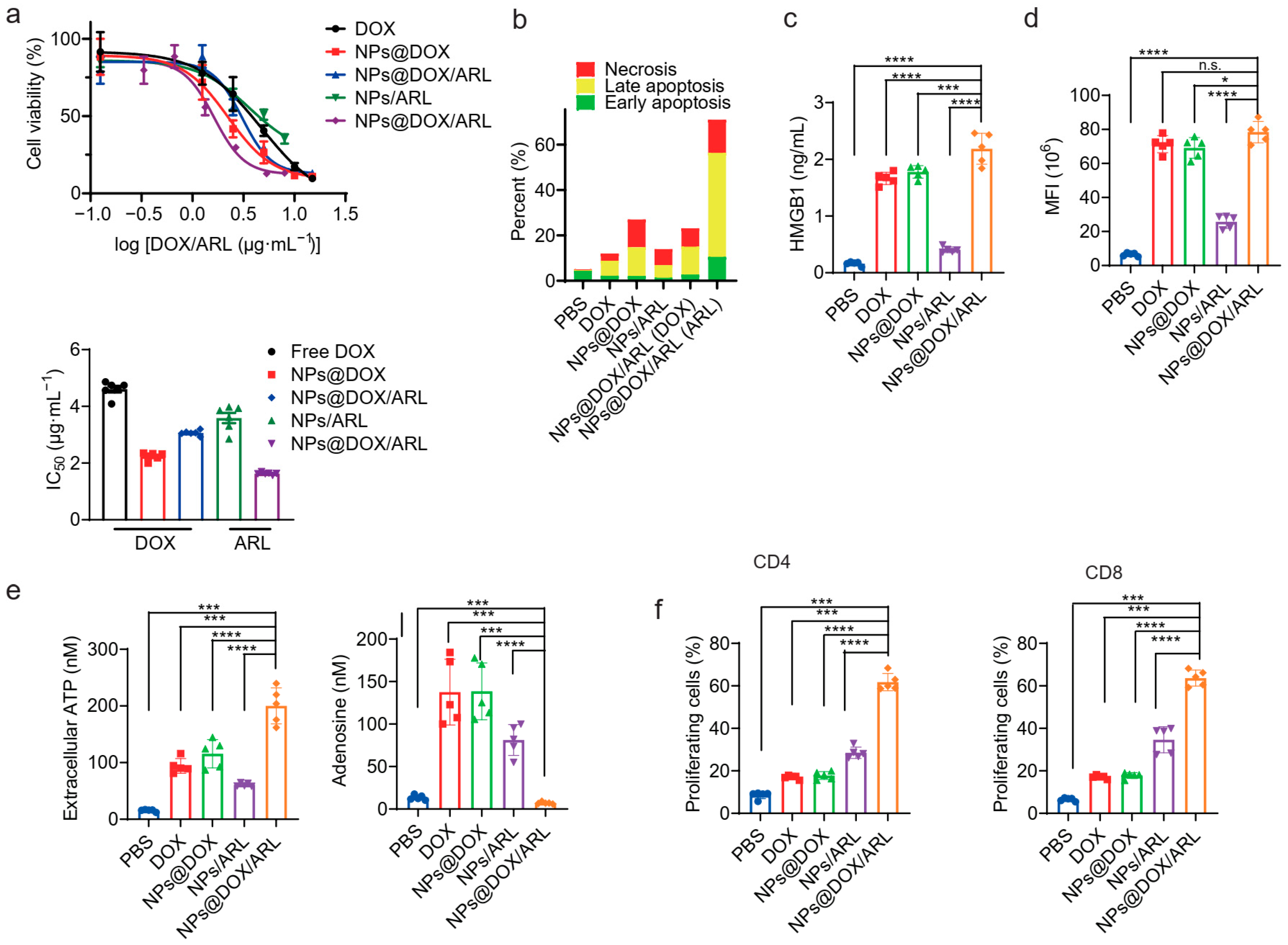
B16 cells were seeded at 5 × 10³ cells per well in 96-well plates and treated with various formulations (free DOX, NPs@DOX, NPs/ARL, and NPs@DOX/ARL) at different concentrations for 48 h. 10 μL of CCK-8 solution was added to each well and incubated for 2 h at 37 °C, followed by absorbance measurement at 450 nm using a microplate reader. Cell viability was assessed using the CCK-8 assay according to the manufacturer’s protocol.
2.7. Immunofluorescence Staining
B16 cells were seeded at 2 × 10⁴ cells per well on glass coverslips in 24-well plates and incubated overnight. Cells were then treated with different formulations for 24 h. After treatment, cells were washed twice with PBS, fixed with 4% paraformaldehyde for 15 min at room temperature, and permeabilized with 0.1% Triton X-100 in PBS for 10 min. Cells were blocked with 5% bovine serum albumin (BSA) in PBS for 1 h at room temperature and then incubated with anti-CD39 primary antibody (1:200, Abcam, Cambridge Science Park, UK) overnight at 4 °C. After washing with PBS, cells were incubated with Alexa Fluor 488-conjugated goat anti-rabbit IgG secondary antibody (1:500, Invitrogen) for 1 h at room temperature in the dark. Nuclei were counterstained with DAPI (1 μg/mL) for 5 min. Coverslips were mounted on glass slides using anti-fade mounting medium and imaged using a confocal laser scanning microscope.
2.8. Measurement of Extracellular ATP and Adenosine (ADO)
For extracellular ATP measurement, B16 cells were seeded at 1 × 10⁵ cells per well in 12-well plates and treated with different formulations for 24 h. Culture supernatants were collected and centrifuged at 12,000 rpm for 5 min to remove cell debris. ATP levels were measured using an ATP bioluminescence assay kit (ATP Determination Kit, Invitrogen, Waltham, MA, USA). Briefly, 100 μL of supernatant was mixed with 100 μL of luciferase-luciferin reagent, and luminescence was measured immediately using a GloMax 20/20 luminometer (Promega, Madison, WI, USA). ATP concentrations were calculated from a standard curve (0–10 μM) prepared in parallel. The detection limit was 10 nM.
For adenosine measurement, supernatants were deproteinized using a 10 kDa MWCO centrifugal filter (Millipore, Billerica, MA, USA) and assayed using a competitive adenosine ELISA kit (Adenosine ELISA Kit, Abcam, Cambridge Science Park, UK) according to the manufacturer’s instructions. Absorbance was measured at 450 nm using a microplate reader. The detection limit was 0.5 ng/mL.
For in vivo ATP imaging, ATP-Luc probe (50 μL, 1 mg/mL) was intratumorally injected 5 min before imaging. Bioluminescence signals were captured using an IVIS Spectrum imaging system (PerkinElmer, Waltham, MA, USA). For in vivo ADO measurement, tumor tissues were harvested, weighed, and homogenized in ice-cold PBS containing 10 μM dipyridamole and 10 μM EHNA. After centrifugation at 12,000 rpm for 10 min at 4 °C, the supernatant was deproteinized and analyzed by ELISA as described above. ADO concentrations were normalized to tissue weight (ng/mg tissue).In vivo ATP imaging.
For in vivo ATP detection, we utilized a ATP Bioluminescence Assay Kit (Basel, Switzerland), which consists of luciferase conjugated to a cell-penetrating peptide for intratumoral delivery. At 24 h post-injection of the nanoparticles, tumor-bearing mice were intratumorally injected with ATP-Luc (50 μL, 1 mg/mL). After 5 min, bioluminescence signals were captured using an in vivo imaging system (IVIS Spectrum, PerkinElmer, Waltham, MA, USA). The ATP concentration was correlated with the bioluminescence intensity and expressed as relative photon flux (photons/sec/cm2/sr). This method enables real-time monitoring of ATP levels in living tumors without tissue disruption.
2.9. Tumor Model
Female C57BL/6 mice (6–8 weeks old) were purchased from the Laboratory Animal Center. B16 tumor-bearing mice were established by subcutaneous injection of 5 × 10⁵ B16 cells into the right flank. When tumors reached a volume of approximately 100 mm3, mice were randomly assigned to different treatment groups.
2.10. In Vivo Imaging
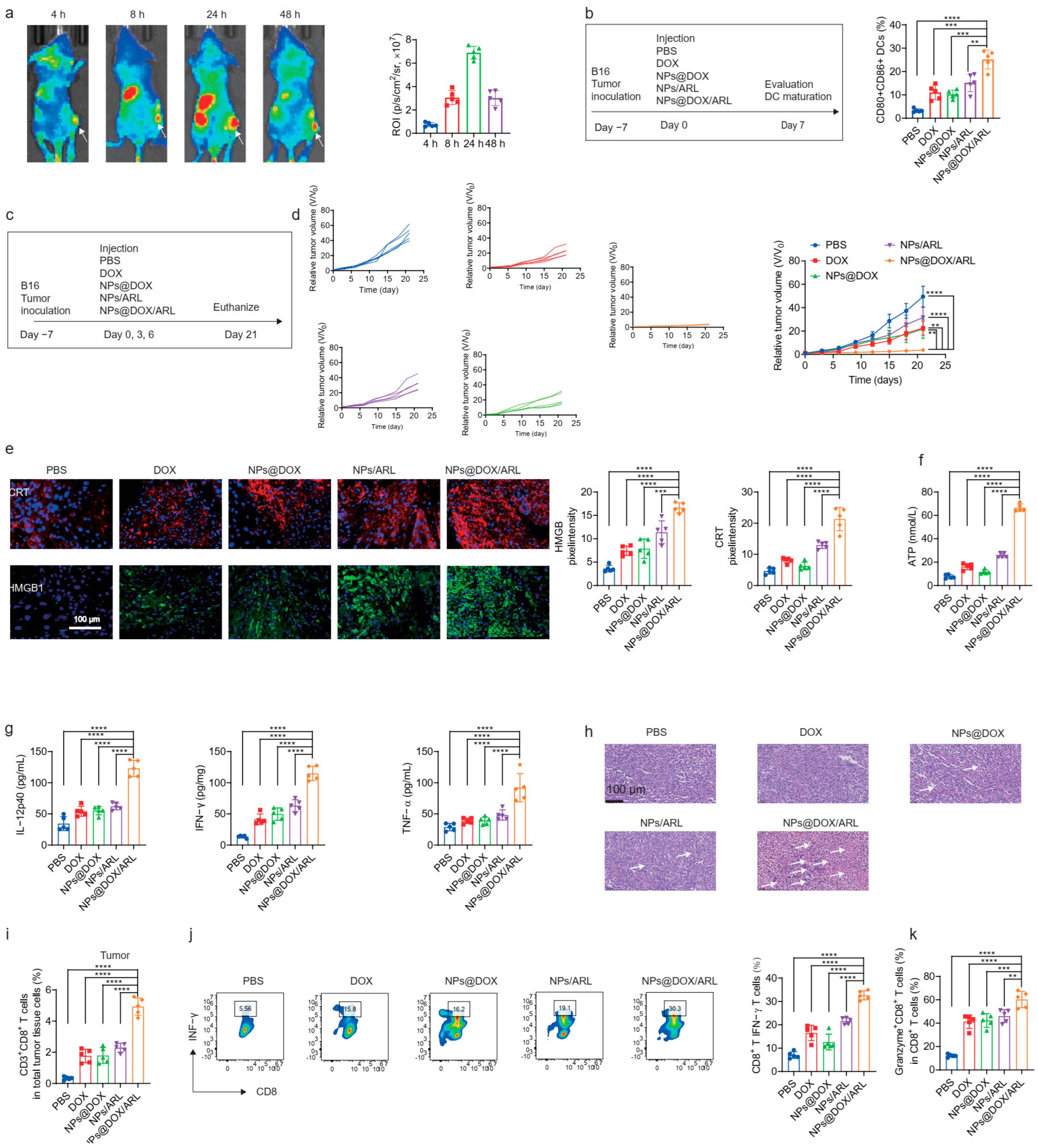
Cy5-labeled NPs@DOX/ARL were intravenously injected into B16 tumor-bearing mice. At different time points (0, 4, 8, 12, 24, 48, and 72 h), mice were anesthetized and imaged using an in vivo imaging system. Fluorescence intensity in the tumor region was quantified using the manufacturer’s software.
2.11. In Vivo Antitumor Efficacy
Mice bearing B16 tumors were intravenously injected with PBS, free DOX, NPs@DOX, NPs/ARL, or NPs@DOX/ARL on days 0, 3, and 6 (DOX dose: 5 mg/kg). Tumor volumes were measured every two days using a caliper and calculated as (length × width2)/2. On day 21, mice were euthanized, and tumors were excised, weighed, and processed for further analysis.
2.12. Histological and Immunofluorescence Analysis of Tumor Tissues
Excised tumors were fixed in 4% paraformaldehyde, embedded in paraffin, and sectioned. Hematoxylin and eosin (H & E) staining was performed to evaluate tumor necrosis. For immunofluorescence, sections were stained with anti-CRT and anti-HMGB1 antibodies, followed by fluorescent secondary antibodies. Nuclei were counterstained with DAPI.
2.13. DCs Maturation Assay
Bone marrow-derived dendritic cells (BMDCs) were generated from C57BL/6 mice according to standard protocols. Briefly, bone marrow cells were isolated from femurs and tibias, and red blood cells were lysed. Cells were cultured in RPMI 1640 medium supplemented with 10% FBS, 20 ng/mL GM-CSF, and 10 ng/mL IL-4 (PeproTech, Cranbury, NJ, USA) for 7 days. Fresh cytokines were added every 2 days. On day 7, non-adherent and loosely adherent cells were collected as immature BMDCs.
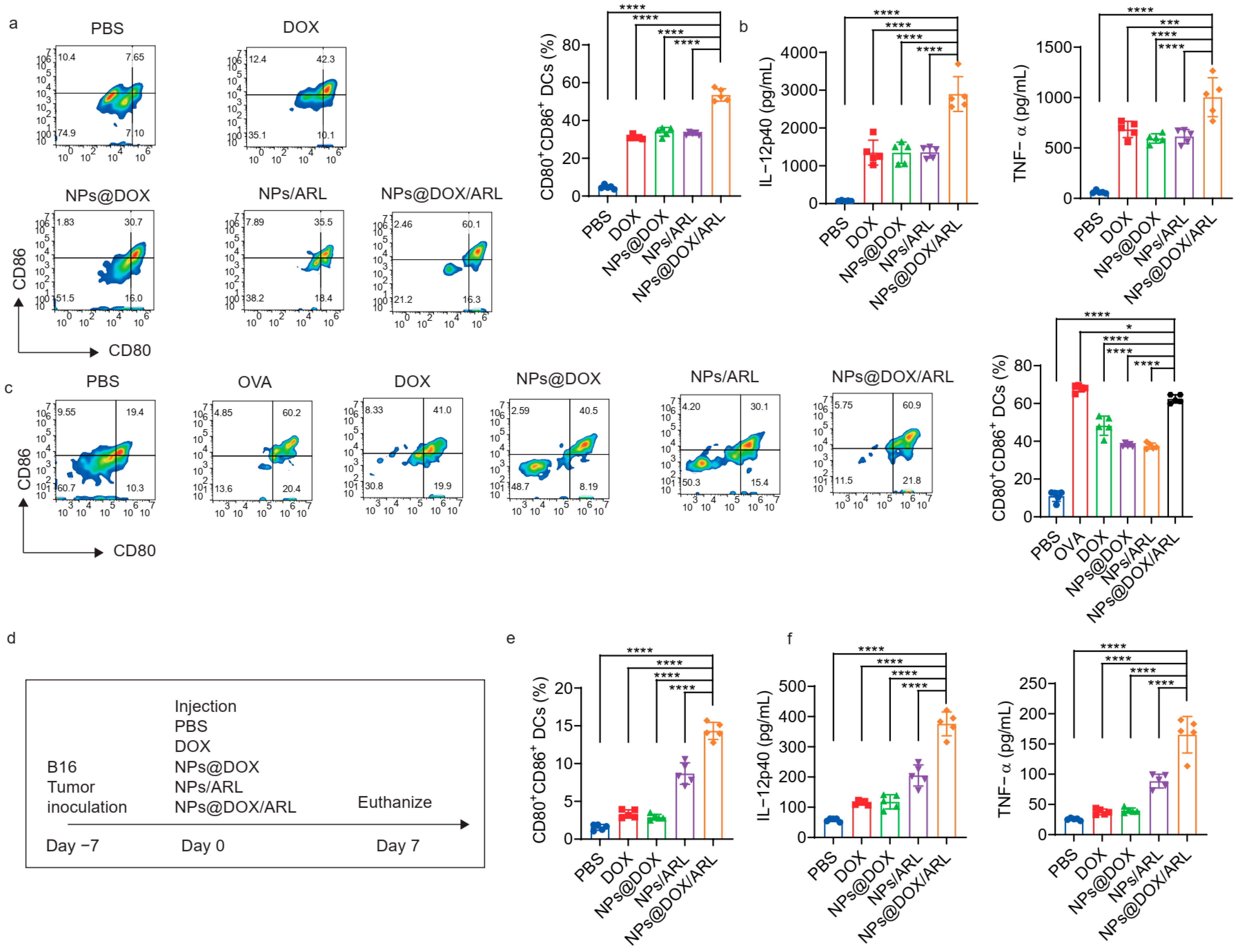
For in vitro DC maturation, B16 cells were pretreated with different formulations (DOX: 5 μM; ARL67156: 50 μM) for 12 h. BMDCs were then seeded at 2 × 10⁵ cells per well in 24-well plates and co-cultured with the pretreated B16 cells (ratio 1:1) for 24 h. Cells were harvested, washed with PBS, and stained with FITC-anti-CD80 (1:200) and PE-anti-CD86 (1:200) antibodies (BD Biosciences, Franklin Lake, NJ, USA) for 30 min at 4 °C in the dark. After washing, cells were analyzed on a BD FACSCanto II flow cytometer, and data were processed using FlowJo software (V10.8.1, BD Life Sciences, Franklin Lake, NJ, USA). Cytokine levels (IL-12p40 and TNF-α) in the supernatant were measured by ELISA (eBioscience, San Diego, CA, USA).
For in vivo DC maturation, B16 tumor-bearing mice were intravenously injected with the indicated formulations (DOX: 5 mg/kg; ARL67156: 2 mg/kg) on days 0, 3, and 6. On day 7, draining lymph nodes and tumors were collected, and single-cell suspensions were prepared. Cells were stained with FITC-anti-CD80 and PE-anti-CD86 antibodies and analyzed by flow cytometry.
2.14. T Cell Proliferation Assay
Splenocytes were collected from C57BL/6 mice, and T cells were isolated using a magnetic-activated cell sorting (MACS) kit (Miltenyi Biotec, Bergisch Gladbach, Germany) according to the manufacturer’s instructions. Isolated T cells were labeled with 5 μM CFSE (Invitrogen, Carlsbad, CA, USA) in PBS for 15 min at 37 °C. The reaction was quenched with an equal volume of FBS, and cells were washed twice with complete medium. Labeled T cells (2 × 10⁵ cells per well) were then co-cultured with supernatants from B16 cells treated with different nanoparticle formulations for 72 h. T cell proliferation was assessed by flow cytometry based on CFSE dilution, and proliferation index was calculated using ModFit LT software (v3.3, Verity Software House, Topsham, ME, USA).
2.15. Intratumoral CD8+ T Cell and Cytokine Analysis
Tumor tissues were dissociated into single-cell suspensions. Cells were surface-stained with anti-CD8α antibody, then fixed, permeabilized, and stained with anti-IFN-γ and anti-granzyme B antibodies for intracellular cytokine detection. Flow cytometry was performed on a BD FACSCanto II system and analyzed using FlowJo software. Serum cytokine levels (IL-12p40, TNF-α, IFN-γ) were measured by ELISA.
2.16. Statistical Analysis
All data are presented as mean ± SD from at least three independent experiments. Statistical comparisons were performed using one-way ANOVA followed by Tukey’s post hoc test or Student’s t-test, as appropriate. A p-value < 0.05 was considered statistically significant.
4. Discussion
In this study, we developed a pH-responsive nanoplatform (NPs@DOX/ARL) for synergistic cancer chemo-immunotherapy by co-delivering the ICD inducer doxorubicin (DOX) and the CD39 inhibitor ARL67156. Our results demonstrate that this strategy effectively preserves extracellular ATP by blocking its enzymatic hydrolysis to immunosuppressive adenosine, thereby amplifying the immunostimulatory cascade triggered by ICD and eliciting robust antitumor immunity. This discussion interprets our key findings, contextualizes them within the current literature, acknowledges the limitations of the present study, and outlines future directions for translational development.
The triblock copolymer GDDM was designed to self-assemble into nanoparticles with a DOX-loaded hydrophobic core and a cationic shell for electrostatic adsorption of the negatively charged ARL67156. Comprehensive physicochemical characterization confirmed that NPs@DOX/ARL exhibited favorable properties, including appropriate size (~100 nm), narrow polydispersity (PDI < 0.15), high drug encapsulation efficiency (78.6 ± 3.2%), and good colloidal stability under both storage and physiological conditions. The significant shift in zeta potential from +18.6 mV to –8.3 mV upon ARL adsorption confirmed successful surface loading. Importantly, the nanoparticles demonstrated pH-triggered drug release, with minimal leakage at physiological pH (7.4) and accelerated release under acidic conditions mimicking the tumor microenvironment (pH 6.5) and endosomal/lysosomal compartments (pH 5.0). Dialysis bags with a molecular weight cutoff (MWCO) of 10 kDa (Spectra/Por®, Waltham, MA, USA) were used. This stimuli-responsive behavior is critical for achieving localized drug delivery, minimizing systemic toxicity, and ensuring that both therapeutic agents are released preferentially at the tumor site in a temporally coordinated manner. These characterization data establish the structural robustness and translational potential of our nanoplatform.
A central finding of this study is the mechanistic validation of CD39 inhibition as a strategy to preserve ICD-released ATP. ARL67156, a selective non-hydrolysable ecto-ATPase inhibitor, effectively blocked CD39 enzymatic activity without downregulating its protein expression, as confirmed by direct enzymatic activity assays. Comprehensive metabolic profiling via HPLC-MS/MS revealed that NPs@DOX/ARL treatment significantly increased extracellular ATP levels while concomitantly reducing ADP, AMP, and adenosine accumulation. This dual effect—enhanced ATP release from DOX-induced ICD combined with reduced ATP degradation via CD39 inhibition—resulted in sustained high levels of immunostimulatory ATP both in vitro and in vivo. The specificity of this effect was further supported by CD39 siRNA knockdown experiments, which phenocopied the ATP preservation observed with ARL67156 treatment, confirming that the observed effects are specifically attributable to CD39 functional blockade rather than nonspecific nanoparticle-mediated changes. These findings underscore the critical role of CD39 as an immunological “switch” within the tumor microenvironment and validate our strategy of targeting this upstream enzyme to simultaneously enhance immunostimulation and relieve immunosuppression.
The immunological consequences of ATP preservation were evident at multiple levels. NPs@DOX/ARL treatment significantly promoted dendritic cell maturation, as demonstrated by increased frequencies of CD80+CD86+ mature DCs and elevated secretion of pro-inflammatory cytokines (IL-12p40 and TNF-α). This effect was superior to that observed with NPs@DOX alone, confirming that CD39 inhibition amplifies the ICD-driven immunostimulatory cascade. Furthermore, T cell proliferation assays revealed that supernatants from NPs@DOX/ARL-treated tumor cells induced substantially greater T cell expansion compared to controls, consistent with the reduced adenosine levels and preserved ATP in the culture medium. These in vitro findings were corroborated by in vivo immune profiling, which showed that NPs@DOX/ARL administration significantly enhanced intratumoral DC maturation, increased CD8+ T cell infiltration, and elevated frequencies of IFN-γ+ and granzyme B+ cytotoxic T lymphocytes. Importantly, we observed a comprehensive remodeling of the tumor immune microenvironment, including reduced frequencies of immunosuppressive Tregs and MDSCs, a shift in macrophage polarization from M2 to M1 phenotype, and decreased expression of exhaustion markers (PD-1 and TIM-3) on CD8+ T cells. Expanded cytokine profiling further confirmed a broad pro-inflammatory immune state, with elevated IL-2, IFN-γ, TNF-α, and IL-6, alongside reduced IL-10 and TGF-β. Collectively, these data demonstrate that our nanoplatform effectively reverses immunosuppression and establishes a pro-inflammatory tumor microenvironment conducive to antitumor immunity.
The in vivo antitumor efficacy of NPs@DOX/ARL was evaluated in the B16 melanoma model. Intravenous administration of the nanoplatform resulted in significant tumor accumulation, as confirmed by in vivo imaging, and led to substantial tumor growth inhibition, extensive tumor necrosis, and prolonged survival compared to control groups. The therapeutic efficacy was associated with enhanced intratumoral ATP levels, increased ICD-related factors (CRT exposure and HMGB1 release), and robust CD8+ T cell responses. Notably, the combination of DOX and ARL67156 in a single nanoplatform outperformed either agent alone, confirming the synergistic effect of ICD induction and CD39 blockade. These results align with the growing body of evidence supporting the combination of ICD inducers with adenosine-pathway inhibitors, and our study provides the first demonstration, to our knowledge, of a pH-responsive co-delivery system that achieves this synergy in a spatially and temporally coordinated manner.
Despite the promising results, several limitations of the present study should be acknowledged. First, the in vivo efficacy was evaluated in a single tumor model (B16 melanoma), and further validation in additional tumor models with distinct immunological backgrounds (e.g., CT26 colon carcinoma, 4T1 breast cancer, or B16-OVA) would be valuable to establish the broad applicability of this approach. Second, while our survival data demonstrate significant extension of median survival, long-term survival analysis and tumor rechallenge experiments are necessary to determine whether the observed antitumor effects translate into durable, memory-based protective immunity. Third, while we have demonstrated ATP preservation and downstream immune activation, direct mechanistic evaluation of P2X7R signaling was not performed in this study, and future investigations utilizing P2X7R-specific antagonists or knockout models will be required to definitively establish the contribution of this pathway. Fourth, detailed pharmacokinetic and pharmacodynamic studies, as well as comprehensive toxicity assessments in larger animal models, will be essential steps toward clinical translation.