Punctal and Intracanalicular Drug Delivery Systems for Ophthalmic Use: A Narrative Review of Technologies, Clinical Outcomes, and Critical Quality Attributes
 , , , , ,
Abstract
1. Introduction
2. Materials and Methods
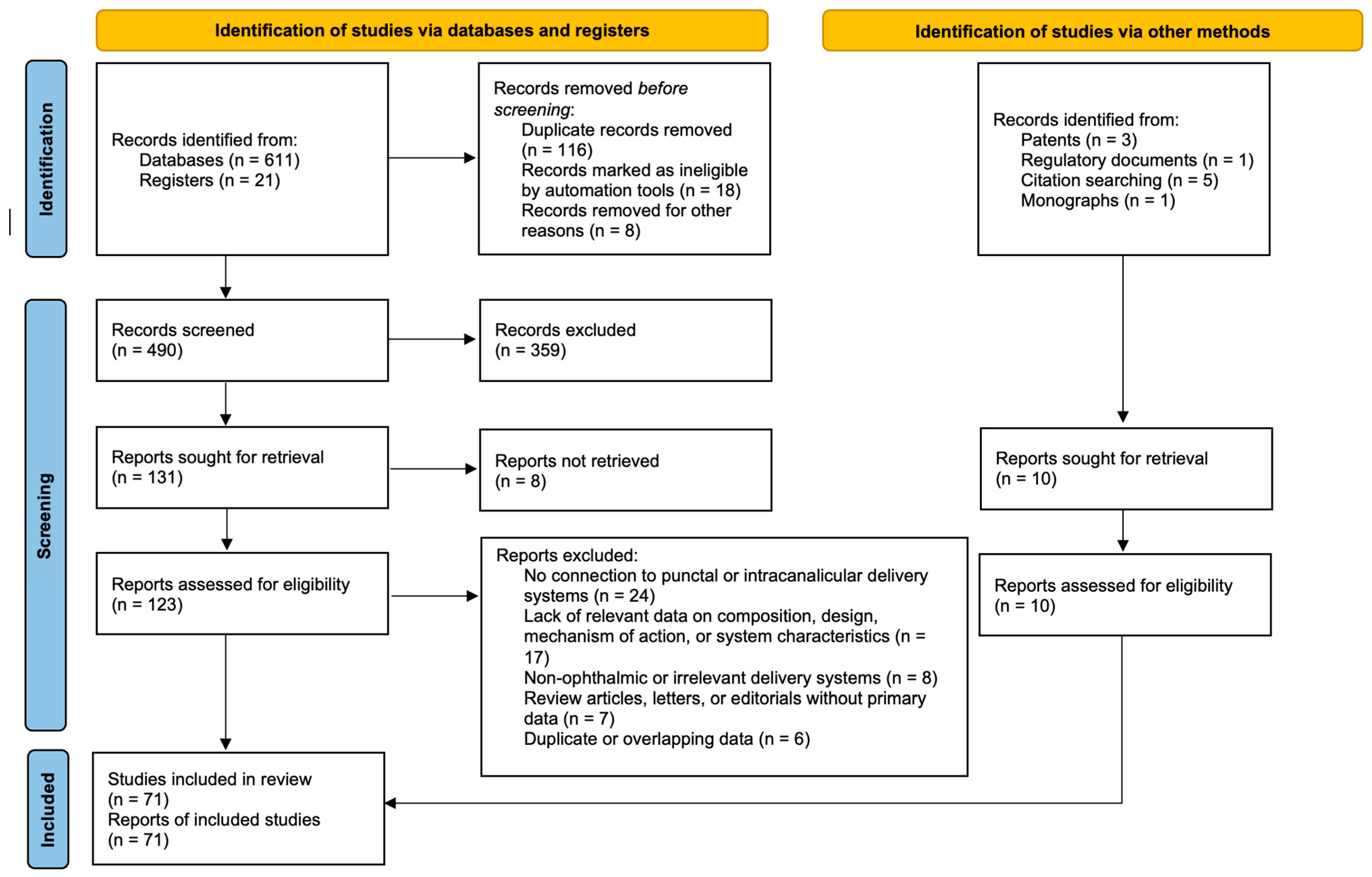
2.1. Literature Search Strategy
2.2. Inclusion and Exclusion Criteria
2.3. Source Selection and Systematization
2.4. Data Extraction and Synthesis
2.5. Evidence Level Assessment
2.6. Data Synthesis and Substantiation of Critical Quality Attributes
2.7. Use of Artificial Intelligence Tools for Figure Preparation
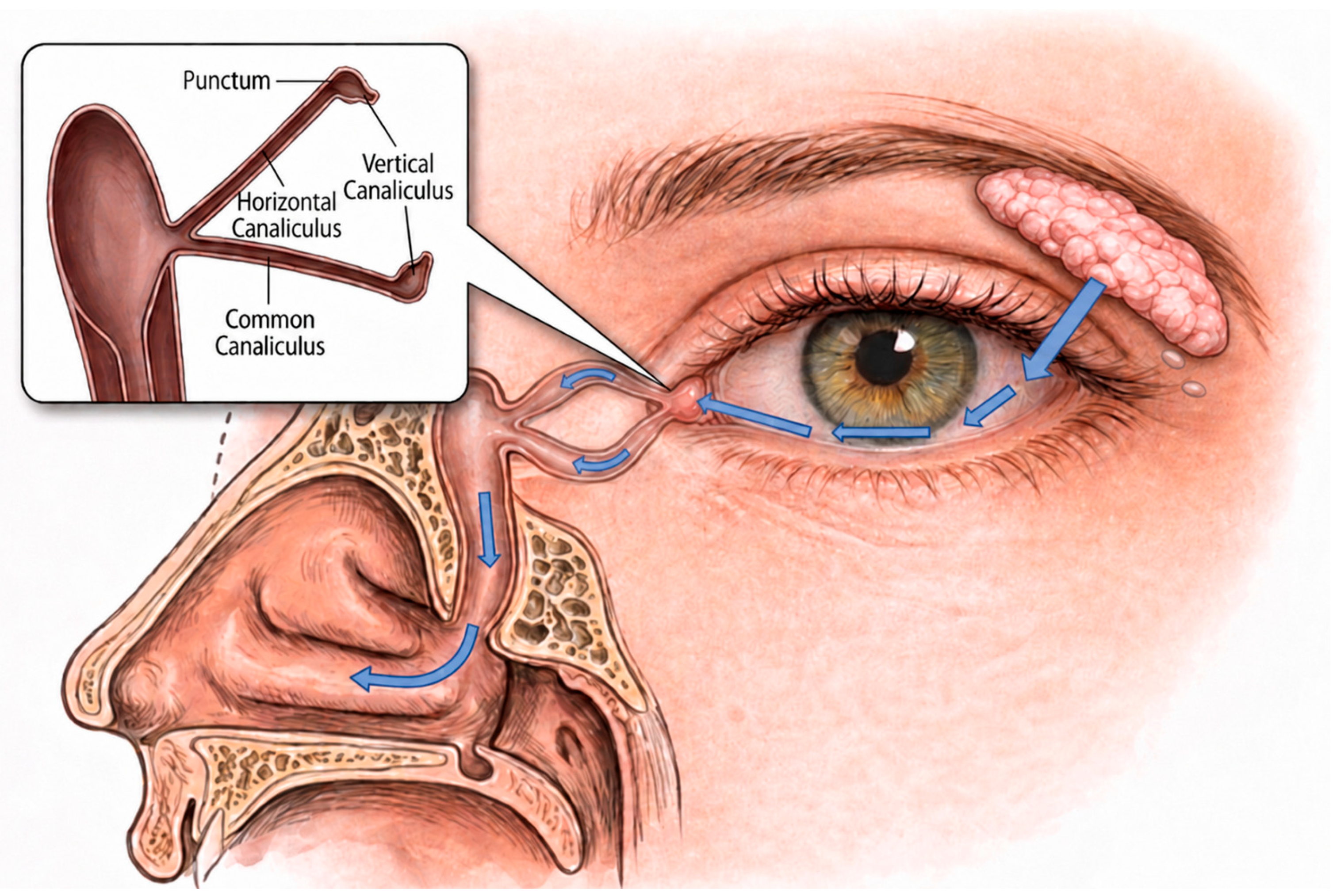
3. Anatomy and Physiology of the Lacrimal Drainage System

4. Occlusive Devices (Without API)
4.1. Punctal Plugs
4.2. Intracanalicular Plugs
5. Punctal Drug Delivery Systems
5.1. Evolute® Punctal Plug Delivery System (Mati Therapeutics, Austin, Texas, USA)
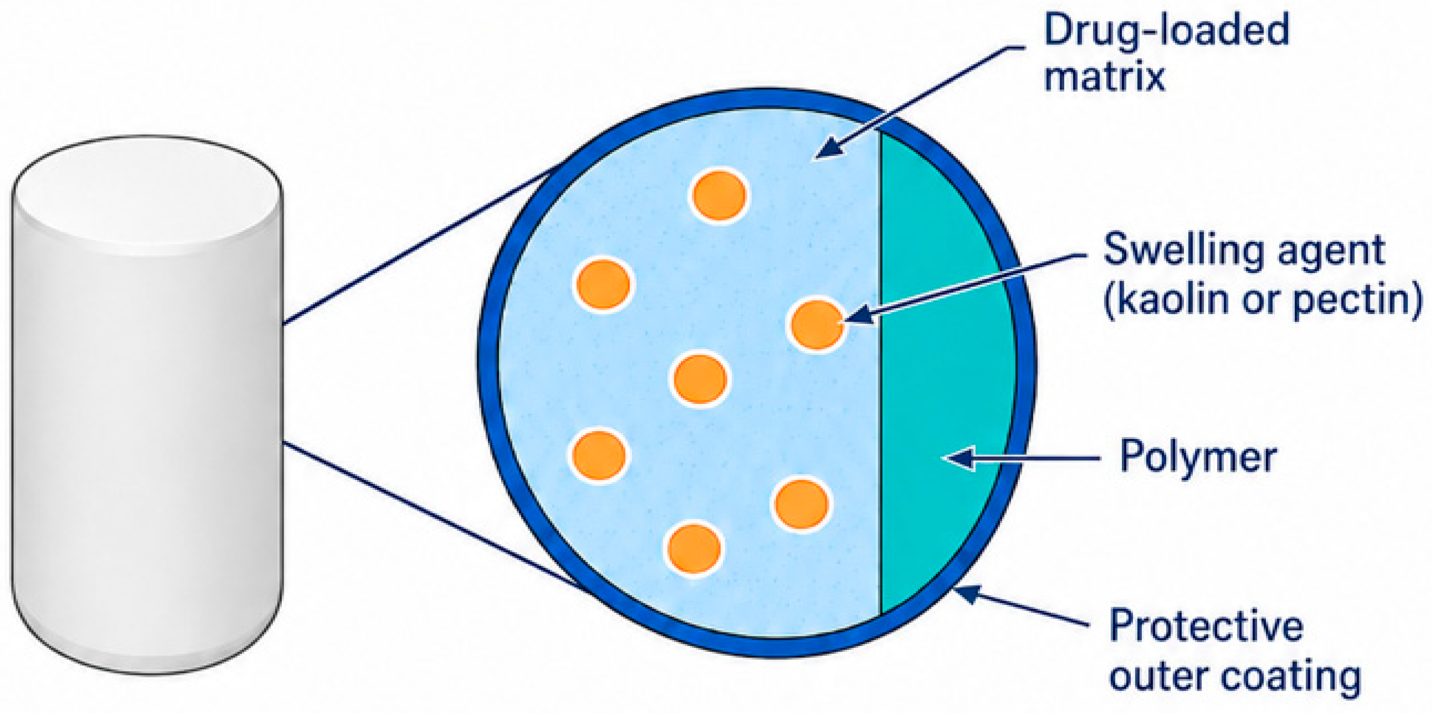
5.2. Eximore Pack and Release Ophthalmic Drug Delivery Platform

5.3. PEGDA-Based 3D-Printed Hydrogel Punctal Plugs
6. Intracanalicular Hydrogel Drug Delivery Systems
6.1. ELUTYX™ (Ocular Therapeutix™)
6.2. Photo-Crosslinked Hydrogel Drug Delivery System (Lin, Wang et al.)
7. In Situ-Forming Intracanalicular Systems
7.1. In Situ-Forming Occluders
7.1.1. Thermosensitive ‘Liquid’ Hydroxybutyl Chitosan Plug
7.1.2. In Situ-Forming Hydrogel Plug
7.2. In Situ-Forming Drug Delivery Systems
7.2.1. Phase-Inversion System—Injectable CsA Organogel In Situ
7.2.2. In Situ Lacrimal Duct Implant Based on a Biodegradable Thermoreversible Polymer Formulation
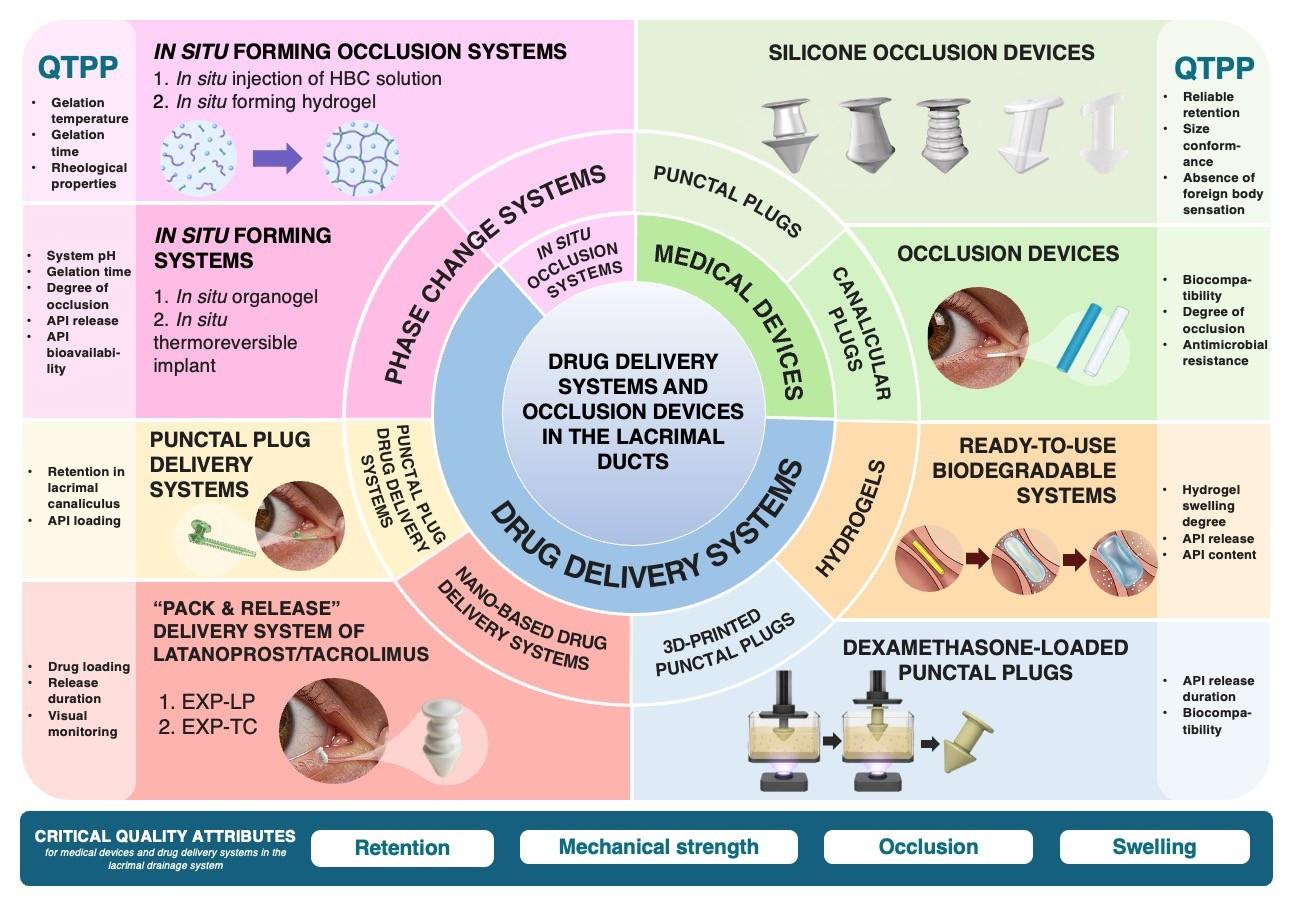
8. Target Product Quality Profile for Development of Products for Lacrimal Drainage System Placement
9. Limitations
9.1. Limitations of the Evidence
9.2. Limitations of the Review Process
10. Conclusions
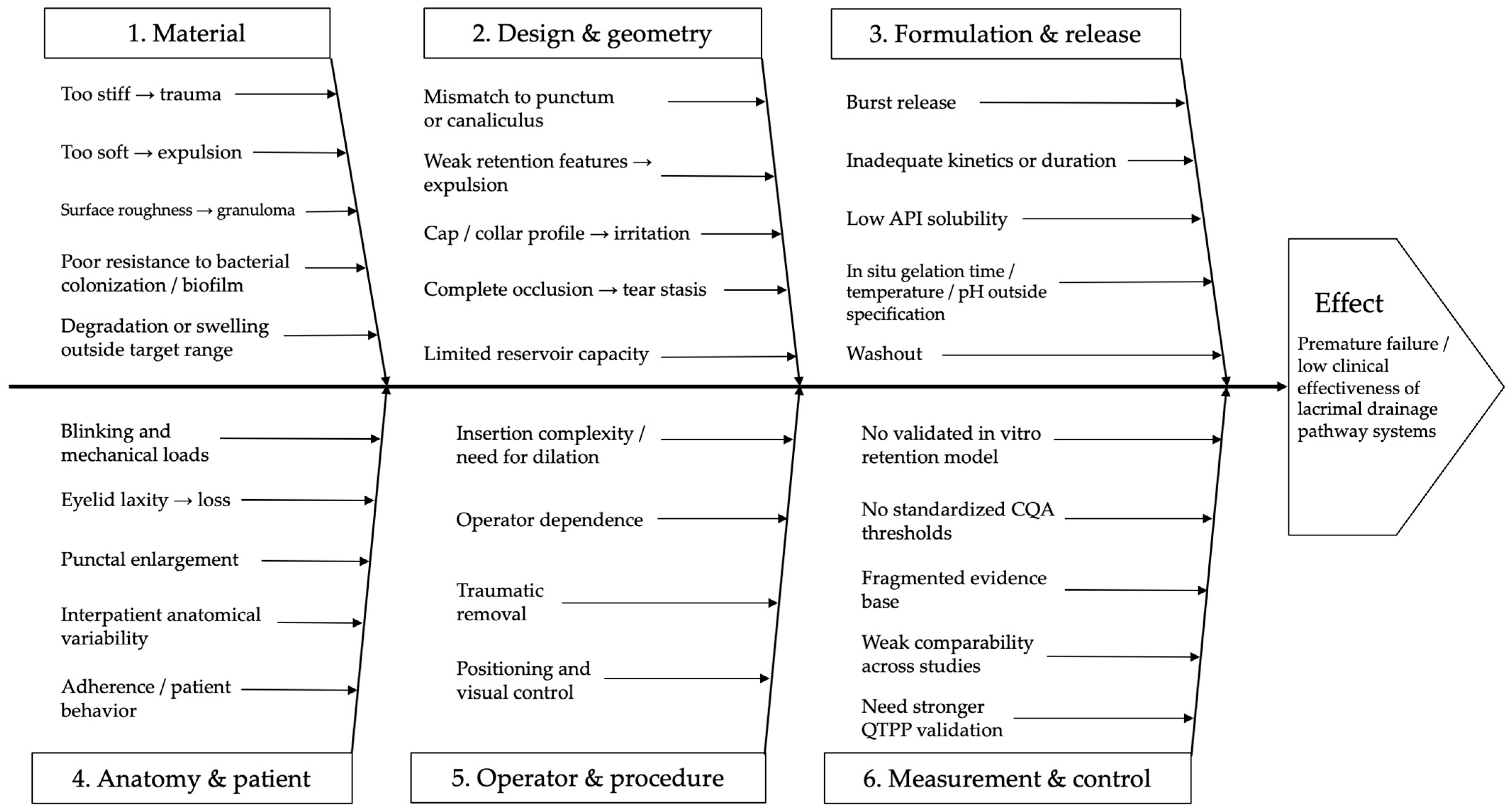
- Retention is at least as critical as drug release. The high rate of premature expulsion (up to 57.4% for some punctal plug designs) nullifies any advantages of the sustained release profile. Mechanical strength and anatomical adaptation (L-shaped design for punctal plugs, swelling capacity for intracanalicular systems) are mandatory critical quality attributes for any system involving this route of administration.
- Drug loading has strict anatomical constraints. The limited volume of the lacrimal punctum (diameter 0.2–0.3 mm) and canalicular lumen (0.5–0.6 mm) restricts total drug loading to approximately 500 µg, which may be insufficient for chronic diseases such as glaucoma. In the case of L-PPDS, limited drug-loading capacity may therefore represent one possible factor contributing to its underperformance; however, inadequate release kinetics at the ocular surface may also have played a role, and the precise mechanism has not been formally established.
- Thermosensitive and photocrosslinkable in situ systems eliminate the size-fitting problem but require strict control of gelation time (≤3 min), phase transition temperature, and pH to prevent premature washout or excessive occlusion. Additional complexity comes from the regulatory classification of such systems (pharmaceutical drug, medical device, or combination product), as well as low system stability and difficulties with scaling such solutions.
Research Perspectives
Supplementary Materials
Author Contributions
Funding
Institutional Review Board Statement
Informed Consent Statement
Data Availability Statement
Acknowledgments
Conflicts of Interest
Abbreviations
| API | Active pharmaceutical ingredient |
| AUC | Area under the curve |
| Cmax | Maximum concentration |
| CQA | Critical quality attribute |
| CQAs | Critical quality attributes |
| CsA | Cyclosporine A |
| DED | Dry eye disease |
| DLP | Digital light processing |
| FDA | Food and Drug Administration |
| FT-IR | Fourier-transform infrared spectroscopy |
| FTN | Fluorescent tracing nanoparticles |
| GRADE | Grading of Recommendations Assessment, Development and Evaluation |
| HAMA | Methacrylate-modified hyaluronic acid |
| HBC | Hydroxybutyl chitosan |
| HPLC | High-performance liquid chromatography |
| ICH | International Council for Harmonisation |
| IOP | Intraocular pressure |
| IVIS | In vivo imaging system |
| LAP | Lithium phenyl-2,4,6-trimethylbenzoylphosphinate |
| L-PPDS | Latanoprost punctal plug delivery system |
| N-PPDS | Nepafenac punctal plug delivery system |
| NMP | N-methyl-2-pyrrolidone |
| PEG | Polyethylene glycol |
| PEG 400 | Polyethylene glycol 400 |
| PEGDA | Polyethylene glycol diacrylate |
| PPDS | Punctal plug delivery system |
| PRISMA | Preferred Reporting Items for Systematic Reviews and Meta-Analyses |
| PVP | Polyvinylpyrrolidone |
| QbD | Quality by design |
| QTPP | Target product quality profile |
| SEM | Scanning electron microscopy |
| SFMA | Silk fibroin methacrylate |
| TEM | Transmission electron microscopy |
| UV | Ultraviolet |
References
- Lin, X.; Zhou, Y.; Lv, K.; Wu, W.; Chen, C. Nanomedicine-Based Ophthalmic Drug Delivery Systems for the Treatment of Ocular Diseases. Int. J. Nanomed. 2025, 20, 9221–9249. [Google Scholar] [CrossRef] [PubMed]
- Wang, Y.; Wang, C. Novel Eye Drop Delivery Systems: Advance on Formulation Design Strategies Targeting Anterior and Posterior Segments of the Eye. Pharmaceutics 2022, 14, 1150. [Google Scholar] [CrossRef] [PubMed]
- Jumelle, C.; Gholizadeh, S.; Annabi, N.; Dana, R. Advances and Limitations of Drug Delivery Systems Formulated as Eye Drops. J. Control. Release 2020, 321, 1–22. [Google Scholar] [CrossRef] [PubMed]
- Goldstein, M.H.; Silva, F.Q.; Blender, N.; Tran, T.; Vantipalli, S. Ocular Benzalkonium Chloride Exposure: Problems and Solutions. Eye 2022, 36, 361–368. [Google Scholar] [CrossRef] [PubMed]
- Zaharia, A.-C.; Dumitrescu, O.-M.; Radu, M.; Rogoz, R.-E. Adherence to Therapy in Glaucoma Treatment—A Review. J. Pers. Med. 2022, 12, 514. [Google Scholar] [CrossRef] [PubMed]
- Ibach, M.J.; Zimprich, L.; Wallin, D.D.; Olevson, C.; Puls-Boever, K.; Thompson, V. In Clinic Optometrist Insertion of Dextenza (Dexamethasone Ophthalmic Insert 0.4mg) Prior to Cataract Surgery: The PREPARE Study. Clin. Ophthalmol. 2022, 16, 2609–2615. [Google Scholar] [CrossRef] [PubMed]
- Lee, A.; Blair, H.A. Dexamethasone Intracanalicular Insert: A Review in Treating Post-Surgical Ocular Pain and Inflammation. Drugs 2020, 80, 1101–1108. [Google Scholar] [CrossRef] [PubMed]
- Anurova, M.N.; Bakhrushina, E.O.; Lapik, I.V.; Turaeva, A.R.; Demina, N.B.; Sysuev, B.B.; Krasnyuk, I.I. Biopharmaceutical Properties of New Mucoadhesive Dosage Form for Eye Degenerative Diseases Treatment. Drug Dev. Regist. 2023, 12, 41–48. [Google Scholar] [CrossRef]
- Weber, C.; Quintin, P.; Holz, F.G.; Fea, A.; Mercieca, K. Ocular Drug Delivery Systems: Glaucoma Patient Perceptions from a German University Hospital Eye Clinic. Graefes Arch. Clin. Exp. Ophthalmol. 2024, 262, 545–556. [Google Scholar] [CrossRef] [PubMed]
- Fea, A.M.; Vallino, V.; Cossu, M.; Marica, V.; Novarese, C.; Reibaldi, M.; Petrillo, F. Drug Delivery Systems for Glaucoma: A Narrative Review. Pharmaceuticals 2024, 17, 1163. [Google Scholar] [CrossRef] [PubMed]
- Chan, H.H.; Wong, T.T.; Lamoureux, E.; Perera, S. A Survey on the Preference of Sustained Glaucoma Drug Delivery Systems by Singaporean Chinese Patients: A Comparison Between Subconjunctival, Intracameral, and Punctal Plug Routes. J. Glaucoma 2015, 24, 485–492. [Google Scholar] [CrossRef] [PubMed]
- Shkolnik, S.F.; Shkolnik, G.S.; Krasnozhen, V.N.; Zakirova, A.M. Anatomy and Physiology of the Lacrimal Drainage System. In Application of New Technologies in the Treatment of Lacrimal Drainage System Pathology; Serebryanye Niti: Moscow, Russia, 2020; ISBN 978-5-89163-290-5. [Google Scholar]
- Maliborski, A.; Różycki, R. Diagnostic Imaging of the Nasolacrimal Drainage System. Part I. Radiological Anatomy of Lacrimal Pathways. Physiology of Tear Secretion and Tear Outflow. Med. Sci. Monit. 2014, 20, 628–638. [Google Scholar] [CrossRef] [PubMed]
- Tyson, S.L.; Campbell, P.; Biggins, J.; Driscoll, A.; Jarrett, P.; Gibson, A.; Vantipalli, S.; Metzinger, J.L.; Goldstein, M.H. Punctum and Canalicular Anatomy for Hydrogel-Based Intracanalicular Insert Technology. Ther. Deliv. 2020, 11, 173–182. [Google Scholar] [CrossRef] [PubMed]
- Xiao, B.; Guo, D.; Liu, R.; Tu, M.; Chen, Z.; Zheng, Y.; Liu, C.; Liang, L. Obstruction of the Tear Drainage Altered Lacrimal Gland Structure and Function. Investig. Ophthalmol. Vis. Sci. 2023, 64, 13. [Google Scholar] [CrossRef] [PubMed]
- Bhujel, B.; Oh, S.-H.; Kim, C.-M.; Yoon, Y.-J.; Chung, H.-S.; Ye, E.-A.; Lee, H.; Kim, J.-Y. Current Advances in Regenerative Strategies for Dry Eye Diseases: A Comprehensive Review. Bioengineering 2023, 11, 39. [Google Scholar] [CrossRef] [PubMed]
- Ervin, A.; Law, A.; Pucker, A.D. Punctal Occlusion for Dry Eye Syndrome. Cochrane Database Syst. Rev. 2017, 2017, CD006775. [Google Scholar] [CrossRef] [PubMed]
- Mansour, K.; Leonhardt, C.J.; Kalk, W.W.; Bootsma, H.; Bruin, K.J.; Blanksma, L.J.; Sjögren Workgroup. Lacrimal Punctum Occlusion in the Treatment of Severe Keratoconjunctivitis Sicca Caused by Sjögren Syndrome: A Uniocular Evaluation. Cornea 2007, 26, 147–150. [Google Scholar] [CrossRef] [PubMed]
- Chen, K.-Y.; Chan, H.-C.; Chan, C.-M. How Effective and Safe Are Punctal Plugs in Treating Dry Eye Disease? A Systematic Review and Meta-Analysis. Cont. Lens Anterior Eye 2025, 48, 102438. [Google Scholar] [CrossRef] [PubMed]
- Jung, I.; Yoon, J.S.; Ko, B.Y. Microbiologic Analysis of Removed Silicone Punctal Plugs in Dry Eye Patients. J. Clin. Med. 2022, 11, 2326. [Google Scholar] [CrossRef] [PubMed]
- Jehangir, N.; Bever, G.; Mahmood, S.M.J.; Moshirfar, M. Comprehensive Review of the Literature on Existing Punctal Plugs for the Management of Dry Eye Disease. J. Ophthalmol. 2016, 2016, 9312340. [Google Scholar] [CrossRef] [PubMed]
- Nasu, N.; Yokoi, N.; Nishii, M.; Komuro, A.; Inagaki, K.; Kinoshita, S. Clinical investigation of the extrusion rate and other complications of the new Super Flex Plug punctal plug and other plugs. Nippon. Ganka Gakkai Zasshi 2008, 112, 601–606. [Google Scholar] [PubMed]
- Sonomura, Y.; Yokoi, N.; Komuro, A.; Inagaki, K.; Kinoshita, S. Clinical investigation of the extrusion rate and other complications of the SuperEagle plug. Nippon. Ganka Gakkai Zasshi 2013, 117, 126–131. [Google Scholar] [PubMed]
- Horwath-Winter, J.; Thaci, A.; Gruber, A.; Boldin, I. Long-Term Retention Rates and Complications of Silicone Punctal Plugs in Dry Eye. Am. J. Ophthalmol. 2007, 144, 441–444. [Google Scholar] [CrossRef] [PubMed]
- Obata, H.; Ibaraki, N.; Tsuru, T. A Technique for Preventing Spontaneous Loss of Lacrimal Punctal Plugs. Am. J. Ophthalmol. 2006, 141, 567–569. [Google Scholar] [CrossRef] [PubMed]
- Sugita, J.; Yokoi, N.; Fullwood, N.J.; Quantock, A.J.; Takada, Y.; Nakamura, Y.; Kinoshita, S. The Detection of Bacteria and Bacterial Biofilms in Punctal Plug Holes. Cornea 2001, 20, 362–365. [Google Scholar] [CrossRef] [PubMed]
- Brissette, A.R.; Mednick, Z.D.; Schweitzer, K.D.; Bona, M.D.; Baxter, S.A. Punctal Plug Retention Rates for the Treatment of Moderate to Severe Dry Eye: A Randomized, Double-Masked, Controlled Clinical Trial. Am. J. Ophthalmol. 2015, 160, 238–242.e1. [Google Scholar] [CrossRef] [PubMed]
- Pastor-Pascual, F.; Aviñó-Martínez, J.; España-Gregori, E.; Alcocer-Yuste, P. Pyogenic granuloma following Smart Plug insertion. Arch. Soc. Esp. Oftalmol. 2007, 82, 653–655. [Google Scholar] [CrossRef] [PubMed]
- Khanna, T.; Akkara, J.D.; Bawa, V.; Sargunam, E.A. Designing and Making an Open Source, 3D-Printed, Punctal Plug with Drug Delivery System. Indian J. Ophthalmol. 2023, 71, 297–299. [Google Scholar] [CrossRef] [PubMed]
- Said, A.M.A.; Farag, M.E.; Abdulla, T.M.; Ziko, O.A.O.; Osman, W.M. Corneal Sensitivity, Ocular Surface Health and Tear Film Stability after Punctal Plug Therapy of Aqueous Deficient Dry Eye. Int. J. Ophthalmol. 2016, 9, 1598–1607. [Google Scholar] [CrossRef] [PubMed]
- Hill, R.H.; Norton, S.W.; Bersani, T.A. Prevalence of Canaliculitis Requiring Removal of SmartPlugs. Ophthalmic Plast. Reconstr. Surg. 2009, 25, 437–439. [Google Scholar] [CrossRef] [PubMed]
- Chen, S.X.; Lee, G.A. SmartPlug in the Management of Severe Dry Eye Syndrome. Cornea 2007, 26, 534–538. [Google Scholar] [CrossRef] [PubMed]
- SmartPlug Study Group Management of Complications after Insertion of the SmartPlug Punctal Plug: A Study of 28 Patients. Ophthalmology 2006, 113, 1859–1862.e2. [CrossRef] [PubMed]
- Jones, C.E.; Anklesaria, M.; Gordon, A.D.; Prouty, R.E.; Rashid, R.; Singla, R.K.; Schachet, J.L. Retrospective Safety Study of the Herrick Lacrimal Plug: A Device Used to Occlude the Lacrimal Canaliculus. CLAO J. 2002, 28, 206–210. [Google Scholar] [CrossRef] [PubMed]
- Joganathan, V.; Mehta, P.; Murray, A.; Durrani, O.M. Complications of Intracanalicular Plugs: A Case Series. Orbit 2010, 29, 271–273. [Google Scholar] [CrossRef] [PubMed]
- Gandhi, S.; Balas, M.; Rai, A. Sustainable Solutions for Eye Drop Plastic Waste: Challenges, Innovations, and Environmental Impact. J. Ophthalmol. 2025, 2025, 7375211. [Google Scholar] [CrossRef] [PubMed]
- Cvenkel, B.; Kolko, M. Implants to Treat Glaucoma: Promising or Not? Drugs Aging 2026, 43, 153–162. [Google Scholar] [CrossRef] [PubMed]
- Cadden, S.; Hao, Y.; Utkhede, D.; Rubinchik, V.; Kjellbotn, C.R. Sustained Release Delivery of Active Agents to Treat Glaucoma and Ocular Hypertension. U.S. Patent 9,610,271, 4 April 2017. [Google Scholar]
- Kompella, U.B.; Kadam, R.S.; Lee, V.H. Recent Advances in Ophthalmic Drug Delivery. Ther. Deliv. 2010, 1, 435–456. [Google Scholar] [CrossRef] [PubMed]
- Kompella, U.B.; Hartman, R.R.; Patil, M.A. Extraocular, Periocular, and Intraocular Routes for Sustained Drug Delivery for Glaucoma. Prog. Retin. Eye Res. 2021, 82, 100901. [Google Scholar] [CrossRef] [PubMed]
- Mati Therapeutics Inc. Clinical Study Evaluating Safety and Efficacy of a Nepafenac Punctal Plug Delivery System (N-PPDS) Compared with Placebo Punctal Plug Delivery System (p-PPDS) in Controlling Post-Operative Ocular Pain and Inflammation After Routine Unilateral Cataract Surgery. 2021. Available online: https://clinicaltrials.gov/study/NCT03496467 (accessed on 11 June 2026).
- Sheetrit, E.; Halahmi, I.; Attar, I. Devices and Methods for Delivering a Bio-Active Agent or Bio-Active Agents. U.S. Patent 12,318,394, 3 June 2025. [Google Scholar]
- Shaare Zedek Medical Center. Safety, Tolerability and Preliminary Efficacy of Unilateral Latanoprost-Loaded Punctual Plug in Patients with Open Angle Early Visual Field Defects Glaucoma or Ocular Hypertension in Comparison to Xalatan© Eye Drops in the Second Eye. 2017. Available online: https://clinicaltrials.gov/study/NCT03318146 (accessed on 10 June 2026).
- Eximore Ltd. Safety, Tolerability, Plug Retention and Preliminary Efficacy of Tacrolimus-Loaded Punctal Plug in Patients with Moderate to Severe Dry Eye Disease—Cohort B. 2023. Available online: https://clinicaltrials.gov/study/NCT05618730 (accessed on 11 June 2026).
- Xu, X.; Awwad, S.; Diaz-Gomez, L.; Alvarez-Lorenzo, C.; Brocchini, S.; Gaisford, S.; Goyanes, A.; Basit, A.W. 3D Printed Punctal Plugs for Controlled Ocular Drug Delivery. Pharmaceutics 2021, 13, 1421. [Google Scholar] [CrossRef] [PubMed]
- Cooper, R.C.; Yang, H. Hydrogel-Based Ocular Drug Delivery Systems: Emerging Fabrication Strategies, Applications, and Bench-to-Bedside Manufacturing Considerations. J. Control. Release 2019, 306, 29–39. [Google Scholar] [CrossRef] [PubMed]
- Lynch, C.R.; Kondiah, P.P.D.; Choonara, Y.E.; du Toit, L.C.; Ally, N.; Pillay, V. Hydrogel Biomaterials for Application in Ocular Drug Delivery. Front. Bioeng. Biotechnol. 2020, 8, 228. [Google Scholar] [CrossRef] [PubMed]
- Tsung, T.-H.; Chen, Y.-H.; Lu, D.-W. Updates on Biodegradable Formulations for Ocular Drug Delivery. Pharmaceutics 2023, 15, 734. [Google Scholar] [CrossRef] [PubMed]
- Sawhney, A.S.; Jarrett, P.; Bassett, M.; Blizzard, C. Drug Delivery Through Hydrogel Plugs. U.S. Patent 8,409,606, 2 April 2013. [Google Scholar]
- Singh, R.B.; Ichhpujani, P.; Thakur, S.; Jindal, S. Promising Therapeutic Drug Delivery Systems for Glaucoma: A Comprehensive Review. Ther. Adv. Ophthalmol. 2020, 12, 2515841420905740. [Google Scholar] [CrossRef] [PubMed]
- Perera, S.A.; Ting, D.S.; Nongpiur, M.E.; Chew, P.T.; Aquino, M.C.D.; Sng, C.C.; Ho, S.-W.; Aung, T. Feasibility Study of Sustained-Release Travoprost Punctum Plug for Intraocular Pressure Reduction in an Asian Population. Clin. Ophthalmol. 2016, 10, 757–764. [Google Scholar] [CrossRef] [PubMed]
- Kesav, N.P.; Young, C.E.C.; Ertel, M.K.; Seibold, L.K.; Kahook, M.Y. Sustained-Release Drug Delivery Systems for the Treatment of Glaucoma. Int. J. Ophthalmol. 2021, 14, 148–159. [Google Scholar] [CrossRef] [PubMed]
- Mohan, N.; Chakrabarti, A.; Nazm, N.; Mehta, R.; Edward, D.P. Newer Advances in Medical Management of Glaucoma. Indian J. Ophthalmol. 2022, 70, 1920–1930. [Google Scholar] [CrossRef] [PubMed]
- Ocular Therapeutix, Inc. A Randomized, Multi-Center, Double-Masked, Vehicle-Controlled, Phase 1/2 Study to Evaluate the Safety, Tolerability, and Efficacy of OTX-CSI (Cyclosporine Ophthalmic Insert) for Intracanalicular Use for the Treatment of Subjects with Dry Eye Disease (DED). 2023. Available online: https://clinicaltrials.gov/study/NCT04362670 (accessed on 11 June 2026).
- Ocular Therapeutix, Inc. A Randomized, Double-Masked, Vehicle-Controlled, Phase 2 Study to Evaluate the Efficacy and Safety of OTX-DED (Dexamethasone Intracanalicular Ophthalmic Insert) for the Short-Term Treatment of Signs and Symptoms of Dry Eye Disease (DED). 2023. Available online: https://clinicaltrials.gov/study/NCT04747977 (accessed on 11 June 2026).
- U.S. Food and Drug Administration. DEXTENZA (Dexamethasone Ophthalmic Insert) 0.4 Mg, for Intracanalicular Use: Prescribing Information; U.S. Food and Drug Administration: Silver Spring, MD, USA, 2025.
- McLaurin, E.B.; Evans, D.; Repke, C.S.; Sato, M.A.; Gomes, P.J.; Reilly, E.; Blender, N.; Silva, F.Q.; Vantipalli, S.; Metzinger, J.L.; et al. Phase 3 Randomized Study of Efficacy and Safety of a Dexamethasone Intracanalicular Insert in Patients With Allergic Conjunctivitis. Am. J. Ophthalmol. 2021, 229, 288–300. [Google Scholar] [CrossRef] [PubMed]
- Blizzard, C.; McLaurin, E.B.; Driscoll, A.; Silva, F.Q.; Vantipalli, S.; Metzinger, J.L.; Goldstein, M.H. Plasma Pharmacokinetic Parameters of Dexamethasone Following Administration of a Dexamethasone Intracanalicular Insert in Healthy Adults. Clin. Ophthalmol. 2021, 15, 2055–2061. [Google Scholar] [CrossRef] [PubMed]
- Walters, T.; Bafna, S. Efficacy and Safety of Sustained Release Dexamethasone for the Treatment of Ocular Pain and Inflammation after Cataract Surgery: Results from Two Phase 3 Studies. J. Clin. Exp. Ophthalmol. 2016, 7, 572. [Google Scholar] [CrossRef]
- Tyson, S.L.; Bafna, S.; Gira, J.P.; Goldberg, D.F.; Jones, J.J.; Jones, M.P.; Kim, J.K.; Martel, J.M.; Nordlund, M.L.; Piovanetti-Perez, I.K.; et al. Multicenter Randomized Phase 3 Study of a Sustained-Release Intracanalicular Dexamethasone Insert for Treatment of Ocular Inflammation and Pain after Cataract Surgery. J. Cataract. Refract. Surg. 2019, 45, 204–212. [Google Scholar] [CrossRef] [PubMed]
- Nijm, L.; Matossian, C.; Rhee, M.K.; Stephens, J.D.; Rosselson, M.E.; Majmudar, P.A.; Gollamudi, S.R.; Patel, R.H.; Bauskar, A.; Montieth, A.; et al. Early Real-World Patient and Staff Experience with an Intracanalicular Dexamethasone Insert. Clin. Ophthalmol. 2024, 18, 1391–1401. [Google Scholar] [CrossRef] [PubMed]
- Lin, T.; Wang, W.; Chen, T.; Bao, B.; Liu, T.; Zhao, H.; Feng, C.; Lin, Q.; Zhu, L.; Gong, L. A Lacrimal Duct Drug Delivery System Based on Photo-Induced Hydrogel for Dry Eye and Allergic Conjunctivitis Therapy. Compos. Part B Eng. 2023, 266, 111014. [Google Scholar] [CrossRef]
- Zhang, Q.; Yan, K.; Zheng, X.; Liu, Q.; Han, Y.; Liu, Z. Research Progress of Photo-Crosslink Hydrogels in Ophthalmology: A Comprehensive Review Focus on the Applications. Mater. Today Bio 2024, 26, 101082. [Google Scholar] [CrossRef] [PubMed]
- Ahmed, B.; Jaiswal, S.; Naryal, S.; Shah, R.M.; Alany, R.G.; Kaur, I.P. In Situ Gelling Systems for Ocular Drug Delivery. J. Control. Release 2024, 371, 67–84. [Google Scholar] [CrossRef] [PubMed]
- Bakhrushina, E.O.; Dubova, A.I.; Nikonenko, M.S.; Grikh, V.V.; Shumkova, M.M.; Korochkina, T.V.; Krasnyuk, I.I.; Krasnyuk, I.I. Thermosensitive Intravitreal In Situ Implant of Cefuroxime Based on Poloxamer 407 and Hyaluronic Acid. Gels 2023, 9, 693. [Google Scholar] [CrossRef] [PubMed]
- Lin, T.; Wang, W.; Lu, Y.; Gong, L. Treatment of Dry Eye With Intracanalicular Injection of Hydroxybutyl Chitosan: A Prospective Randomized Clinical Trial. Front. Med. 2021, 8, 769448. [Google Scholar] [CrossRef] [PubMed]
- Dai, M.; Xu, K.; Xiao, D.; Zheng, Y.; Zheng, Q.; Shen, J.; Qian, Y.; Chen, W. In Situ Forming Hydrogel as a Tracer and Degradable Lacrimal Plug for Dry Eye Treatment. Adv. Healthc. Mater. 2022, 11, 2200678. [Google Scholar] [CrossRef] [PubMed]
- Raut, S.; Azheruddin, M.; Kumar, R.; Singh, S.; Giram, P.S.; Datta, D. Lecithin Organogel: A Promising Carrier for the Treatment of Skin Diseases. ACS Omega 2024, 9, 9865–9885. [Google Scholar] [CrossRef] [PubMed]
- Cao, Z.; Chen, Y.; Bai, S.; Zheng, Z.; Liu, Y.; Gui, S.; Shan, S.; Wu, J.; He, N. In Situ Formation of Injectable Organogels for Punctal Occlusion and Sustained Release of Therapeutics: Design, Preparation, in Vitro and in Vivo Evaluation. Int. J. Pharm. 2023, 638, 122933. [Google Scholar] [CrossRef] [PubMed]
- Bakhrushina, E.O.; Ermachkova, M.A.; Dubova, A.I.; Demina, N.B.; Krasnyuk, I.I. Development of an in situ implant for the treatment of inflammatory diseases of the lacrimal duct using in vitro modeling techniques. Proc. Voronezh State Univ. Ser. Chem. Biol. Pharm. 2022, 4, 83–89. (In Russian) [Google Scholar]
- Mikhel, I.; Bakhrushina, E.; Abuelez, S.; Eremeeva, K.; Yang, X.; Svistushkin, V.; Stepanova, O.I.; Krasnyuk, I.I., Jr.; Zhemerikin, G.; Krasnyuk, I.I. 3D PRINTING TECHNOLOGIES IN THE DEVELOPMENT OF A BIORELEVANT IN VITRO MODEL OF THE NASAL CAVITY: NEW STEP OF INTRANASAL DRUGS QUALITY ASSESSMENT. Int. J. Appl. Pharm. 2025, 17, 66–76. [Google Scholar] [CrossRef]



| Plug Studied | Design | Device Characteristics | Results (Adverse Effects, Advantages) |
|---|---|---|---|
| Plug SuperEagle® (EagleVision®) |  | Soft material, thin non-protruding cap. Conical shape and wide curved nose designed to improve device retention [21] | 1. Premature expulsion rate 57.4%. 2. Mean time to premature expulsion was 82 days. 3. Granulation tissue formation observed in 34.5% of patients [22] |
| EaglePlug® (EagleVision®) |  | Conical shape aids plug retention. Simple insertion and removal [21] | 1. Punctum enlargement observed after premature expulsion. 2. Time to expulsion for this plug type was significantly shorter than that of other devices in the study [23] |
| EagleFlex Plug® (EagleVision®) |  | Conical ribbed shaft for improved retention and flexibility, providing 30% more surface area. Thin cap reduces risk of corneal contact [20] | |
| FCI® plugs: perforated punctal plug, FCI® plug F |  | Tilted cap provides better fit, matching natural eyelid anatomy and preventing displacement. Silicone perforated punctal plug coated with hydrophobic PVP (polyvinylpyrrolidone), allowing tear flow through the perforation (description adapted from the manufacturer’s official website). | 1. FCI® plug retention rate was 84.2% at three months, 69.5% after one year, and declined to 55.8% after 2 years [24] 2. Six-month retention rate for FCI® plug F was 70.4%. Spontaneous plug loss was associated with larger punctum size in patients [25] |
| FCI® Ready-Set |  | Ultra-thin occluders made from medical-grade silicone. Anatomically shaped to adapt to natural eyelid structure, providing a comfortable fit (description adapted from the manufacturer’s official website). | Biofilm formation and bacterial contamination were detected in 44% of material extracted from plug orifices. Staphylococcus epidermidis was most frequently isolated (75%), followed by Staphylococcus aureus (25%) [26] |
| SuperFlex® (EagleVision®) |  | Design based on a flexible ribbed shaft that compresses easily and adapts to lacrimal punctum anatomy. Elastic structure aids reliable retention. Low-profile rim reduces risk of corneal contact, minimizing foreign body sensation after insertion (description adapted from the manufacturer’s official website). | 1. Six-month retention rate for SuperFlex® was 32% [27] 2. Spontaneous plug loss was associated with eyelid laxity in elderly patients. Punctum enlargement was also observed [25]. |
| Parasol® Punctal Occluders |  | Hollow nose compresses during insertion, in most cases eliminating the need for punctal dilation. Design features a sharp-tipped end and smooth surface facilitating insertion (description adapted from the manufacturer’s official website). | 1. Pyogenic granuloma leading to plug extrusion in 4.2% of cases, possibly related to the irregular plug surface causing mucosal damage [21]. 2. Six-month retention rate for Parasol® was 68% [27]. |
| Plug Studied | Characteristics | Material | Results (Adverse Effects, Advantages) |
|---|---|---|---|
| Collagen plugs | Capable of expanding after insertion; swelling occurs approximately 60% [21] | Bovine collagen | 1. Good tolerability; only 3% of the population is allergic to bovine collagen. 2. Granulomatous reaction was observed among adverse effects [21] |
| SmartPlug® | Thermosensitive polymer from which the plug is made changes size and shape after insertion into the lacrimal canaliculus (description adapted from the manufacturer’s official website). | Thermosensitive acrylic material | 1. Compared with silicone plug, thermosensitive plug provided greater improvement in ocular surface condition [30] 2. Canaliculitis, dacryocystitis, conjunctivitis, epiphora, and pyogenic granuloma were observed [31,32,33]. 3. Premature expulsion was rare (2.2% of cases) [32]. |
| Herrick Lacrimal Plug® | While the collapsible design facilitates insertion, placement within the canaliculus may predispose to tear stasis and secondary infection [21] | Medical-grade silicone | Cases of ocular irritation, epiphora, and intracanalicular plug migration were reported [34]. |
| FORM FIT® plugs | Within 10 min, the hydrogel expands to match the shape of the lacrimal canaliculus, eliminating the need for size matching (description adapted from the manufacturer’s official website). | Hydrogel | Cases of canaliculitis development 6 months after device use and granulation tissue formation 5 years later have also been reported [35]. |
| Delivery System | Active Substance | Formulation Composition | Estimated Release Duration | Development Stage |
|---|---|---|---|---|
| L-PPDS | Latanoprost | API polymer matrix, medical-grade silicone, phosphatidylcholine | Approximately 30 days | Phase I |
| N-PPDS | Nepafenac | API polymer matrix, medical-grade silicone, phosphatidylcholine | No release duration data available | Phase II |
| EXP-LP | Latanoprost | Composite matrix based on porous carrier material; possible components: silica gel, activated charcoal, zeolite, kaolin/pectin, epoxy or polyurethane matrix, protective coating of parylene or butyral | Up to 3 months | 2019 clinical study |
| EXP-TC | Tacrolimus | Medical-grade composite with tacrolimus; punctal plug design with visible head and central shaft | Up to 3 months | Phase I |
| 3D-printed punctal plugs for controlled ocular delivery | Dexamethasone 10–20% | Irgacure® 819 2%, β-Carotene 1%, PEGDA 61.6–87%, PEG 400 15.4–17.4% (in D10PEG and D20PEG formulations) | 4–7 days (depending on formulation) | In vitro testing |
| Biodegradable Intracanalicular Delivery System | Active Substance | Development Stage | Release Duration | Key Results and Limitations |
|---|---|---|---|---|
| OTX-TP | Travoprost | Phase III | Up to 3 months (~90 days) | In an initial prospective study, IOP reduction reached up to 7.5 mmHg/28%; system retention declined: 100% on day 10 and 42% on day 30. However, Phase III did not demonstrate statistically significant superiority over placebo |
| OTX-CSI | Cyclosporine | Phase II | 3–4 months (~90–120 days) | At 0.36 mg dose and 16-week follow-up, Schirmer test improvement was 1.91–1.98 mm, while the placebo group showed 2.24–3.08 mm, indicating no pronounced advantage |
| OTX-DED | Dexamethasone | Phase II | 2–3 weeks (~14–21 days) | At doses of 0.2 mg/0.3 mg, hyperemia reduction was −0.51/−0.43 compared with −0.21 in the placebo group |
| DEXTENZA® | Dexamethasone | FDA-approved drug | 30 days | Absence of pain on day 8—up to 80.4%; absence of anterior chamber cells on day 14—52.3% vs. 31.1% placebo. Adverse effects included: IOP elevation up to 6%, anterior chamber inflammation 10%, reduced visual acuity 2% |
| Photocrosslinkable hydrogel | Cyclosporine A | In vitro and in vivo | In vitro: 60 days; in vivo: up to 90 days | Swelling increased from 33.9% to 49.5%; residual content on day 60 was CsA > 30%, FK506 > 80% |
| In Situ-Forming System | Composition | Selection Criteria | In Vivo and In Vitro Results |
|---|---|---|---|
| Thermosensitive ‘liquid’ hydroxybutyl chitosan plug | Hydroxybutyl chitosan (HBC) solution | Biocompatibility, antibacterial properties, mucoadhesion, solubility, hydrophilicity, non-toxicity, rapid phase transition capability, no size fitting required, reduced infection risk | Phase transition time at body temperature (37 °C) approximately 50 s; effect lasts at least 4 weeks |
| In situ-forming hydrogel plug | SFMA hydrogel matrix; FTN fluorescent nanoparticles; LAP photoinitiator. Optimized formulation: 8% SFMA, 0.02% FTN, 0.4% LAP | Rapid gelation under visible/blue light; conforming to lacrimal canaliculus shape; biodegradability; non-invasive position monitoring; biocompatibility; temporary lacrimal canaliculus occlusion | In vitro: gelation in 3.3–10.5 s; cytotoxicity not detected up to 800 µg/mL FTN; hemolysis < 3%. In vivo: subcutaneous administration in mice—no pronounced inflammation/fibrosis up to 56 days; insertion into rabbit lacrimal canaliculus—complete occlusion on day 1, partial restoration of permeability within 7 days, therapeutic effect for at least 1 month |
| Injectable CsA organogel in situ | API—cyclosporine A (CsA); stearic acid, injectable soybean oil, NMP at ratio 1.25:10:0.6 (w/v/v) | Rapid in situ gelation after injection, lacrimal canaliculus occlusion, sustained CsA release | In vitro: reduction in fluid flow to 52.78% at 2 min; in vivo: complete lacrimal pathway occlusion and significant reduction in tear drainage. Bioavailability enhancement: Cmax increased approximately 1.9-fold, AUC 2.15–2.49-fold in ocular tissues compared with eye drops |
| In situ lacrimal duct implant based on biodegradable thermoreversible polymer formulation | Optimal formulation: poloxamer 407—18.0%, poloxamer 188—3.0%; API—diclofenac 25.0 mg/mL, nitrofuralum at 40 mg per 10 mL polymer base | Preservation of thermoreversible properties, gelation temperature 38.5–40 °C, minimum gelation time, pH compliance with physiological values close to tear fluid pH 7.4 | Using a lacrimal duct and sac model at 1:8 scale, gelation temperature of the optimal formulation was 38.7 °C, and gelation time was 150 s |
| A. For All Punctal Plugs (Occlusive Devices and Drug Delivery Systems) | |||
|---|---|---|---|
| QTPP/Quality Parameter | Optimal/Target Value | How to Control/Method | Basis/Source of Derivation |
| Reliable retention | Minimal risk of premature expulsion/migration | In clinical studies—assessment of plug retention in the lacrimal punctum | Based on clinical retention/expulsion outcomes reported for commercial punctal plugs and punctal drug delivery systems, including SuperEagle®, FCI® plug, SuperFlex®, L-PPDS, and N-PPDS [22,24,25,27,39,41] |
| Size conformance | Diameter matches lacrimal punctum size | Pre-insertion size fitting | Derived from anatomical dimensions of the lacrimal punctum and canaliculus and from dimensions of punctal systems reported in the reviewed studies [12,13,14,38] |
| Absence of foreign body sensation | Minimal foreign body sensation | Primarily clinical assessment; indirectly controlled by shape, surface smoothness, material softness, and size | Based on clinical tolerability data and design-related factors described for punctal plugs (cap profile, shape, material softness, surface properties) [20,21,27,39] |
| Surface smoothness | Smooth surface without traumatizing irregularities | Visual inspection, microscopy/SEM | Proposed from reports linking irregular surface or traumatic interaction with irritation and granuloma formation; surface evaluation methods were used in experimental systems [21,28,45] |
| B. For punctal Drug Delivery Systems | |||
| QTPP/Quality Parameter | Optimal/Target Value | How to Control/Method | Basis/Source of Derivation |
| API release duration | Depending on the polymer system and required release profile—from 7 to 60 days | In vitro release profile | Derived from reported release durations: 4–7 days for PEGDA-based 3D-printed plugs, ~30 days for L-PPDS, and up to 3 months for EXP systems [37,38,39,40,41,42,43,44,45] |
| API loading/drug content | Based on system dimensions, limited to no more than 500 µg | HPLC after extraction | Based on reported loading ranges for punctal systems: L-PPDS (44–81 µg latanoprost) and EXP-LP (250–450 µg latanoprost), interpreted considering device dimensions and anatomical constraints [13,14,39,43] |
| Biocompatibility/cytocompatibility | Absence of pronounced cytotoxicity and irritation | Cytocompatibility testing | Derived from cytocompatibility results for PEGDA-based systems and clinical safety/tolerability data for punctal delivery systems [21,39,45] |
| A. For All Intracanalicular Plugs (Occlusive Devices and Drug Delivery Systems) | |||
|---|---|---|---|
| QTPP/Quality Parameter | Optimal/Target Value | How to Control/Method | Basis/Source of Derivation |
| Retention in lacrimal canaliculus | At least 80% over 4 weeks | In clinical studies—assessment of plug retention in the lacrimal punctum | Proposed as an expert-derived development target based on reported early retention values for lacrimal systems, including L-PPDS (78% at 4 weeks), N-PPDS (98% at 14 days), and OTX-TP retention profiles reported in different trials [39,41] |
| Hydrogel swelling degree | Should not be less than 30% and should not exceed 60% | In vitro studies in lacrimal duct model | Derived from reported swelling behavior of collagen plugs (~60%) and photocrosslinked hydrogel systems (33.9–49.5%) [21,63] |
| Biocompatibility | Absence of cytotoxicity | In vitro: cytocompatibility test using BALB/3T3 fibroblast cell line | Based on preclinical and clinical safety data for hydrogel-based intracanalicular systems [45,63] |
| Degree of occlusion | Controlled or partial occlusion | In vitro studies in lacrimal duct model | Derived from complication profiles associated with excessive tear stasis, canaliculitis, dacryocystitis, and epiphora in intracanalicular devices [31,32,33,34,35] |
| Absence of foreign body sensation | Minimal foreign body sensation | Primarily clinical assessment; indirectly controlled by shape, surface smoothness, material softness, and size | Based on tolerability findings reported for intracanalicular systems, including OTX-TP and dexamethasone inserts [53,56,61] |
| Antimicrobial resistance/infection risk | Material must be resistant to bacterial colonization | In vitro microbiological testing | Derived from reports of bacterial contamination, biofilm formation, canaliculitis, and dacryocystitis in lacrimal devices [26,31,33] |
| Mechanical strength | Compressive modulus range 91.25–133.4 kPa | Compression testing method | Proposed from the compression testing range reported for photocrosslinked lacrimal duct hydrogel systems [63] |
| B. For Intracanalicular Drug Delivery Systems | |||
| QTPP/Quality Parameter | Optimal/Target Value | How to Control/Method | Basis/Source of Derivation |
| API release | Depending on the polymer system and required release profile—from 7 to 60 days | In vitro/in vivo release profile | Derived from OTX-DED/DEXTENZA®-type dexamethasone systems (2–4 weeks) and OTX-CSI/CsA hydrogel systems (up to 3–4 months) [54,55,56,58,63] |
| API content | 0.2–0.4 mg; 10–30%—depending on the system | Quantitative API analysis | Based on reported values for OTX-DED (0.2–0.3 mg dexamethasone), DEXTENZA® (0.4 mg dexamethasone), and experimental CsA/FK506 hydrogels (10–30%) [55,56,63] |
| Release rate without burst release | No more than 40% released during the first quarter of the application period | In vitro dynamic flow model + HPLC | Proposed as an expert-derived target to avoid excessive burst release; informed by comparison between rapid early release in PEGDA-based systems and more sustained release in hydrogel/organogel platforms [45,63,69] |
| Matrix biodegradability | Predictable resorption without mandatory removal over the therapeutic action period | Clinical studies, in vivo retention studies | Based on the resorbable behavior of DEXTENZA® and controlled degradation reported for experimental hydrogel systems [56,58,63,67] |
| A. For All In Situ-Forming Systems (Occlusion Devices and Drug Delivery Systems) | |||
|---|---|---|---|
| QTPP/Quality Parameter | Optimal/Target Value | How to Control/Method | Basis/Source of Derivation |
| Gelation time | No more than 3 min | Visual gelation time recording/rheology | Derived from reported gelation times: ~50 s for hydroxybutyl chitosan, 3.3–10.5 s for SFMA/FTN hydrogel systems, and ~150 s for the thermoreversible lacrimal implant model [66,67,70] |
| Gelation temperature | Depending on nosology: up to 37 °C without inflammation, up to 39 °C—with inflammation | Thermostatted lacrimal duct model | Based on reported thermosensitive behavior of hydroxybutyl chitosan and the thermoreversible inflammatory lacrimal implant model (optimal 38.7 °C) [66,70] |
| System pH | Close to tear fluid pH—approximately 7.4 | pH measurement | Derived from formulation selection criteria used for the thermoreversible lacrimal implant [70] |
| Degree of occlusion | Complete or temporary lacrimal pathway occlusion; fluid flow reduction to 50% within 2 min | Fluid flow model/permeability test | Based on complete temporary occlusion reported for SFMA/FTN hydrogel plugs and fluid flow reduction to 52.78% at 2 min for CsA organogel [67,69] |
| Swelling and degradation | Controlled biodegradation; partial restoration of permeability within 7 days | In vitro studies in lacrimal duct model | Derived from the SFMA/FTN hydrogel plug model [67] |
| Rheological properties | Sufficient viscosity/strength after gelation for canalicular retention | Rheological analysis | Based on rheological characterization reported for SFMA/FTN hydrogel and thermoreversible systems [67,70] |
| Biocompatibility | Absence of pronounced cytotoxicity; hemolysis < 3%; no pronounced inflammation/fibrosis | Cell test, hemolytic test | Derived from cell, hemolysis, and in vivo biocompatibility studies of SFMA/FTN hydrogel plugs [67] |
| Duration of therapeutic effect | At least 4 weeks | Dynamic efficacy assessment in vitro/in vivo | Based on reported therapeutic duration for hydroxybutyl chitosan and therapeutic/monitoring duration for SFMA/FTN hydrogel systems [66,67] |
| B. For In Situ Drug Delivery Systems | |||
| QTPP/Quality Parameter | Optimal/Target Value | How To Control/Method | Basis/Source of Derivation |
| API release | Sustained API release while maintaining occlusion | In vitro release test | Derived from CsA organogel and related in situ lacrimal drug delivery systems [69,70] |
| API bioavailability | Target superiority over drops—observed improvement in pharmacokinetic parameters | Pharmacokinetic study | Based on CsA organogel pharmacokinetic data showing ~1.9-fold increase in Cmax and 2.15–2.49-fold increase in AUC compared with eye drops [69] |
| Critical Parameter | Comment |
|---|---|
| Retention in lacrimal canaliculus | Premature expulsion or device migration nullifies the therapeutic effect. Clinical studies demonstrate a decline in retention from 100% on day 10 to 42% on day 30 (OTX-TP). Target parameter: ≥80% retention over 4 weeks |
| Occlusion | The degree of lacrimal drainage system blockage determines both the mechanical effect (increased tear residence time) and the risk of complications (epiphora, dacryocystitis). Partial occlusion is preferable to complete occlusion to reduce fluid stasis. Target parameter: fluid flow reduction ≥ 50% within 2 min of introduction |
| Mechanical strength | The device must withstand compressive loads arising during blinking and eyelid movements without deformation, migration, or fragmentation. Excessively soft materials prematurely extrude; excessively rigid ones traumatize the mucosa. Target parameter: compressive modulus in the range of 91–133 kPa |
| Swelling | This characteristic is critical for hydrogel systems (intracanalicular plugs, in situ-forming implants). Swelling provides passive fixation without pre-sizing. Target range: 30–60% (based on reported hydrogel/collagen plug data). |
Disclaimer/Publisher’s Note: The statements, opinions and data contained in all publications are solely those of the individual author(s) and contributor(s) and not of MDPI and/or the editor(s). MDPI and/or the editor(s) disclaim responsibility for any injury to people or property resulting from any ideas, methods, instructions or products referred to in the content. |
© 2026 by the authors. Licensee MDPI, Basel, Switzerland. This article is an open access article distributed under the terms and conditions of the Creative Commons Attribution (CC BY) license.
Share and Cite
Bakhrushina, E.O.; Leonova, K.S.; Belyavsky, N.O.; Gegechkori, V.I.; Belyaev, V.V.; Sysuev, B.B.; Salakhetdinov, D.K.; Krasnyuk, I.I.; Atkova, E.L.; Yartsev, V.D. Punctal and Intracanalicular Drug Delivery Systems for Ophthalmic Use: A Narrative Review of Technologies, Clinical Outcomes, and Critical Quality Attributes. Pharmaceutics 2026, 18, 830. https://doi.org/10.3390/pharmaceutics18070830
Bakhrushina EO, Leonova KS, Belyavsky NO, Gegechkori VI, Belyaev VV, Sysuev BB, Salakhetdinov DK, Krasnyuk II, Atkova EL, Yartsev VD. Punctal and Intracanalicular Drug Delivery Systems for Ophthalmic Use: A Narrative Review of Technologies, Clinical Outcomes, and Critical Quality Attributes. Pharmaceutics. 2026; 18(7):830. https://doi.org/10.3390/pharmaceutics18070830
Chicago/Turabian StyleBakhrushina, Elena O., Kseniia S. Leonova, Nikita O. Belyavsky, Vladimir I. Gegechkori, Vasily V. Belyaev, Boris B. Sysuev, Damir K. Salakhetdinov, Ivan I. Krasnyuk, Eugenia L. Atkova, and Vasily D. Yartsev. 2026. "Punctal and Intracanalicular Drug Delivery Systems for Ophthalmic Use: A Narrative Review of Technologies, Clinical Outcomes, and Critical Quality Attributes" Pharmaceutics 18, no. 7: 830. https://doi.org/10.3390/pharmaceutics18070830
APA StyleBakhrushina, E. O., Leonova, K. S., Belyavsky, N. O., Gegechkori, V. I., Belyaev, V. V., Sysuev, B. B., Salakhetdinov, D. K., Krasnyuk, I. I., Atkova, E. L., & Yartsev, V. D. (2026). Punctal and Intracanalicular Drug Delivery Systems for Ophthalmic Use: A Narrative Review of Technologies, Clinical Outcomes, and Critical Quality Attributes. Pharmaceutics, 18(7), 830. https://doi.org/10.3390/pharmaceutics18070830