Review

Leaf Saponins of Quillaja brasiliensis as Powerful Vaccine Adjuvants

1
Unidad Académica de Desarrollo Biotecnológico, Instituto de Higiene, Facultad de Medicina, Universidad de la República (Udelar), Av. Alfredo Navarro 3051, Montevideo 11600, Uruguay
2
FACISA—Faculdade de Ciências da Saúde do Trairi, Universidade Federal do Rio Grande do Norte (UFRN), Av. Rio Branco, S/N, Santa Cruz 59200-000, Brazil
*
Author to whom correspondence should be addressed.
Pharmaceutics 2025, 17(8), 966; https://doi.org/10.3390/pharmaceutics17080966
Submission received: 13 June 2025 / Revised: 11 July 2025 / Accepted: 22 July 2025 / Published: 25 July 2025
(This article belongs to the Special Issue Advances in Vaccine Delivery and Vaccine Administration)

Abstract

Vaccine adjuvants are non-immunogenic agents that enhance or modulate immune responses to co-administered antigens and are essential to modern vaccines. Despite their importance, few are approved for human use. The rise of new pathogens and limited efficacy of some existing vaccines underscore the need for more advanced and effective formulations, particularly for vulnerable populations. Aluminum-based adjuvants are commonly used in vaccines and effectively promote humoral immunity. However, they mainly induce a Th2-biased response, making them suboptimal for diseases requiring cell-mediated immunity. In contrast, saponin-based adjuvants from the Quillajaceae family elicit a more balanced Th1/Th2 response and generate antigen-specific cytotoxic T cells (CTL). Due to ecological damage and limited availability caused by overharvesting Quillaja saponaria Molina barks, efforts have intensified to identify alternative plant-derived saponins with enhanced efficacy and lower toxicity. Quillaja brasiliensis (A.St.-Hil. and Tul.) Mart. (syn. Quillaja lancifolia D.Don), a related species native to South America, is considered a promising renewable source of Quillajaceae saponins. In this review, we highlight recent advances in vaccine adjuvant research, with a particular focus on saponins extracted from Q. brasiliensis leaves as a sustainable alternative to Q. saponaria saponins. These saponin fractions are structurally and functionally comparable, exhibiting similar adjuvant activity when they were formulated with different viral antigens. An alternative application involves formulating saponins into nanoparticles known as ISCOMs (immune-stimulating complexes) or ISCOM-matrices. These formulations significantly reduce hemolytic activity while preserving strong immunoadjuvant properties. Therefore, research advances using saponin-based adjuvants (SBA) derived from Q. brasiliensis and their incorporation into new vaccine platforms may represent a viable and sustainable solution for the development of more less reactogenic, safer, and effective vaccines, especially for diseases that require a robust cellular immunity.

1. Introduction

Vaccines developed in the two centuries since Jenner’s time have led to remarkable reductions in infection and disease wherever implemented [1]. Today, vaccination is regarded as one of the most effective medical interventions and plays a key role in reducing the burden of infectious diseases [2,3,4]. Effective vaccines and proper immunization schedules prevent millions of deaths worldwide each year. Notably, global vaccination efforts led to the eradication of smallpox, with no cases reported since 1978 [5]. Moreover, sustained and widespread vaccination efforts have led to the eradication of certain infectious diseases, such as poliomyelitis in the Americas and many other regions globally [6]. In the last pandemic of SARS-CoV-2, vaccines proved once again to be a powerful tool to control infection, and they contributed to the reduction of serious cases that lead, in many cases, to death [7,8,9]. Unfortunately, many vaccine antigens are poorly immunogenic, requiring adjuvants to elicit protective immune responses that correlates with the enhancement of the vaccine efficacy [10]. The addition of adjuvants (from Latin adjuvare, meaning “to help”), or “helper substances”, to vaccine formulations has significantly enhanced their effectiveness and the level of protection they provide. Immunological adjuvants were first described by Gaston Ramon [11] as “substances used in combination with a specific antigen that produce more immunity than the antigen alone”. In this sense, Ramon recognized that a variety of substances could increase antigen-specific antibody production when added to diphtheria and tetanus toxoids prior to vaccination [11].
Globally, vaccine adjuvants enhance efficacy by boosting, modulating, and extending the immune response [12,13]. They can also lower the required antigen dose, improving vaccine cost-effectiveness [14,15]. Adjuvants are now considered essential to modern vaccines, yet only a few are approved for human use despite advances in antigen isolation and production. These include aluminum salts, virus-like particles (VLP), MF59™, AS01™, AS03™, AS04™, and CpG [9,12].
Aluminum salts, developed almost a century ago, remain the most commonly used adjuvant for licensed human vaccines; they are found in toxoid vaccines like the tetanus vaccine and inactivated vaccines like the Salk polio vaccine and the Coronavac (SARS-CoV-2) vaccine, among others [8,16,17,18]. Due to the limitations of alum adjuvants, other compounds have been used in some vaccines. MF59™ was the second adjuvant introduced for human use [19,20,21,22]. MF59™ is an oil-in-water squalene emulsion used in flu vaccines across Europe [23]. Its adjuvant effect is linked to the early activation of innate immunity at the injection site [19,24]. While such emulsions promote a balanced immune response, they remain weak in inducing T-helper 1 (Th1) immunity and fail to effectively stimulate CTL [21]. The Adjuvant System (AS) is a proprietary formulation by GlaxoSmithKline (GSK) that combines known adjuvant compounds. AS01™, a liposome-based system containing QS-21 (a highly purified saponin from Q. saponaria) and 3-O-desacyl-4′-monophosphoryl lipid A (MPL), a potent adjuvant that has been used in two licensed vaccines, Shingrix™ for herpes zoster [25] and Mosquirix™ for malaria [26,27,28,29]. AS03™ is an oil-in-water emulsion included in the influenza vaccine [30], and finally, AS04™ is an MPL-absorbed aluminum salt used for hepatitis and papilloma virus vaccines [6,31]. AS incorporates immunostimulatory substances that tailor immune responses to specific pathogens, enhancing Th1 or CTL responses when needed. Current formulations combine molecules like MPL, QS-21, and CpG with classical adjuvants such as aluminum, liposomes, or oil-in-water emulsions [12,18,29,30,32,33,34,35].
QS-21 promotes Th1 cytokines (IL-2, IFN-γ), stimulates CTL responses to exogenous antigens, and induces IgG1, IgG2b, and IgG2a antibodies. These properties have made it a promising adjuvant for vaccines targeting intracellular pathogens and therapeutic cancer vaccines [34,36,37]; CTL responses are crucial for protection against intracellular pathogens like tuberculosis, malaria, and HIV. Developing effective vaccines requires inducing strong T cell immunity, a limitation of current approaches [3,38,39]. Adjuvants may be needed to enhance both antibody and effector T cell responses [40].
Despite decades of research, the immunological mechanisms of many adjuvants remain poorly understood [18,41]. It is established, however, that adjuvants primarily activate innate immune cells [12,13], often by prolonging antigen exposure to dendritic cells (DC) and promoting their maturation [42]. The discovery of Toll-like receptors (TLR) in the 1990s helped clarify the mechanisms of certain immunostimulants, such as flagellin, MPL, and CpG [43]. Despite advances, significant knowledge gaps remain for adjuvants that do not act via known receptors, such as mineral salts, microparticles, lipid-based carriers, SBA, and immunostimulant complexes (ISCOMs) [18,44,45,46,47,48]. Alum remains the most widely used adjuvant and is effective in promoting humoral immunity [49,50,51]. However, it primarily induces Th2-type responses in mice and is suboptimal for diseases requiring strong cell-mediated immunity [16,18,50,52,53,54].
Early adjuvants were designed mainly to boost antibody responses, which was adequate for many vaccines at the time. However, it is now clear that antibody enhancement alone is insufficient for many modern vaccine candidates [55]. Adjuvant selection should be guided by the specific immune response needed, such as Th1, Th2, antibody, or CTL-driven immunity [13,18]. As a result, there is an increasing demand for safe, non-toxic adjuvants capable of eliciting strong, durable protective immune responses [12,56,57].

2. Saponins

2.1. Characteristics, Structure, and Properties

Saponins are one of the most numerous and diverse plant secondary metabolites. Saponins are widely distributed in the plant Kingdom, operating as a chemical barrier or shield in the plant defense system to counter pathogens, parasites, and herbivores [58,59]. Therefore, it is found in plant tissues that are most vulnerable to fungal or bacterial attack or insect predation [60].
The name saponin is derived from the Latin word sapo, which means soap, due to the capacity to produce foaming water by the tree leaves and barks [61]. Saponins are secondary metabolites classified into triterpenoid and steroid glycosides, whose structures vary based on the number and position of attached sugar units [62]. These natural glucosides are amphipathic molecules composed of two distinct regions, a hydrophobic aglycone core and a hydrophilic sugar chain. The sugar moiety may include various units such as glucose, glucuronic acid, xylose, rhamnose, or methylpentose, which are connected to the aglycone via ether or ester linkages [62,63] (Figure 1A). These highly complex metabolites are present in a wide range of plant species and can be found in various plant parts including bark, leaves, stems, roots, and flowers [62,64,65]. This combination of polar and non-polar structural elements in their molecules explains their soap-like behavior in aqueous solutions [66,67] (Figure 1B).
Saponins are highly important in pharmacy, cosmetic, and food industries. Saponin metabolites are first isolated from soapwort (Saponaria officinalis L.) and have been traditionally employed as natural detergents in domestic applications [64,68]. In addition to their cleansing properties, they exhibit a wide range of biological activities, including immunoadjuvant, anti-inflammatory, antifungal, antiviral, molluscicide, hypoglycemic, hypocholesterolemia, and hemolytic effects [62,64,66,67]. The toxicity of certain saponins, closely associated with their hemolytic activity, is believed to stem from the aglycone’s high affinity for membrane cholesterol [64,66,69,70,71]. This interaction disrupts red blood cell membranes by forming pores through cholesterol complexation, leading to cell lysis [70,71,72,73].

2.2. Saponins as Immunoadjuvants

The adjuvant properties of these secondary metabolites were first noted in 1925, when it was shown that the addition of bread crumbs, tapioca, saponin, and “starch oil” to antigenic preparations greatly enhanced antibody responses to diphtheria or tetanus [11]. In 1951, Espinet used crude saponins to boost foot-and-mouth disease virus (FMDV) vaccine efficacy [74]. A major breakthrough came in 1974, when Dalsgaard successfully isolated Quil-A® from the barks of the South American tree Q. saponaria Molina, demonstrating its ability to stimulate both humoral and cellular immune responses and induce differential antibody isotypes [36,67,75,76,77,78]. Since then, Quil-A® has gained widespread use in veterinary vaccines and pre-clinical studies [67,79,80,81,82]. Additional studies showed its effects when co-formulated with aluminum salts, liposomes, and oil-in-water emulsions, with amphipathic proteins and lipids forming detergent/lipid/saponin complexes termed immune-stimulating complexes (ISCOMs) [83,84,85,86,87]. However, Quil-A® remains a heterogeneous mixture—containing up to 23 saponin fractions detectable by reverse-phase high performance liquid chromatography (RP-HPLC)—and its toxicity limits its use in human vaccines [34,67,77,88].
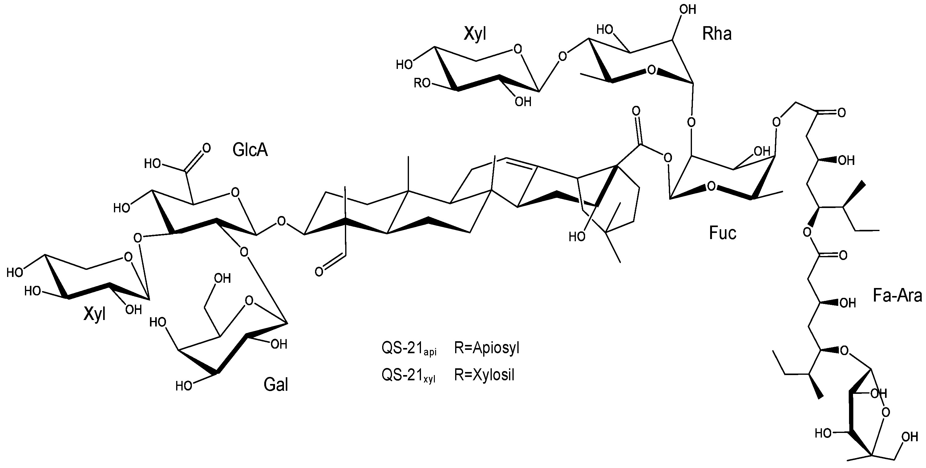
Afterwards, Kensil et al. purified several saponin fractions from Quil-A® using RP-HPLC, identifying QS-7, QS-17, QS-18, and QS-21 (Figure 2), all of them with potent adjuvant activity (Figure 2) [77]. Among these, toxicity varied significantly. QS-18 was highly toxic in mice, whereas QS-7 and QS-21 were much less so [34,77,89]. Saponins from Q. saponaria bark, especially QS-21, promote Th1-type cytokines (IL-2, IFN-γ), stimulate CTL against exogenous antigens, and enhance IgG1, IgG2b, and IgG2a antibody production. These properties have attracted attention for their use as adjuvants in vaccines targeting intracellular pathogens and in therapeutic cancer vaccines [14,36,37,67,90,91,92,93,94,95].
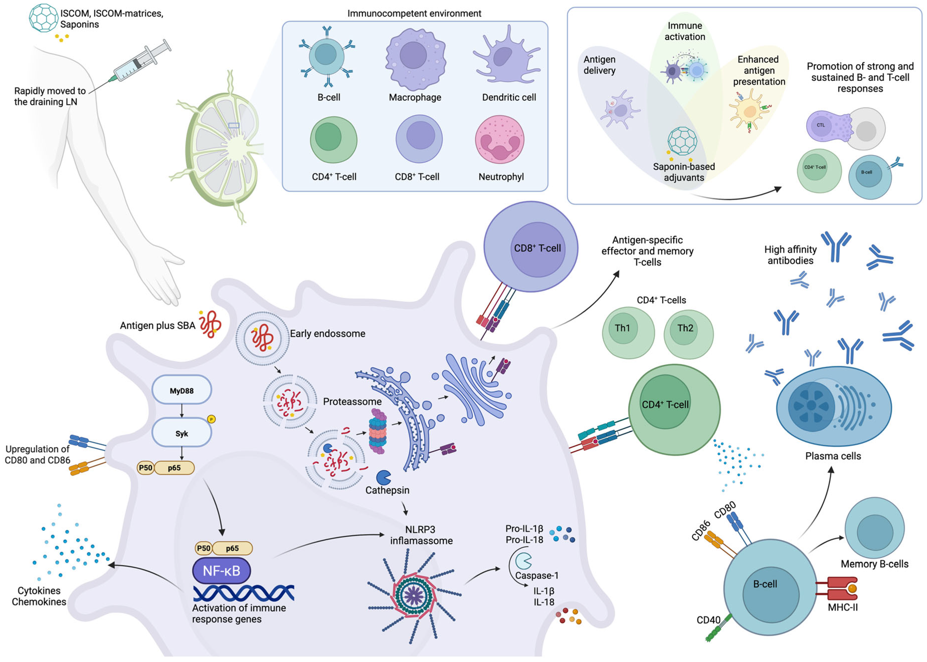
The exact mechanism by which saponins in general act as adjuvants remains unclear, though recent studies have begun to clarify their modes of action [96,97,98,99]. Saponins can induce both Th1/Th2 immune responses (Figure 3). Structure–activity relationship studies (SAR) suggest that imine-forming carbonyl groups are essential for T cell activation and the subsequent induction of Th1/Th2 responses [67,100,101,102,103].
While saponins with diverse triterpenoid aglycones and oligosaccharide chains can activate DC, the presence of fucopyranosyl residues may skew the response toward a Th2 profile via interaction with the DC-SIGN receptor [103,104]. Glycosides like QS-21 (Figure 2) interact with both T cells and DC; the aldehyde group co-stimulates T cells, while the triterpene and fucosyl components engage DC [104,105,106]. The mechanism of QS-21 involves inducing CD8+ [67,107,108,109], partly due to a unique aldehyde group at the triterpene C4 position, believed to stimulate T lymphocytes and enhance Th1 responses [34,101,103,110,111,112]. Its lipophilic moiety further facilitates antigen delivery to APC, promoting endogenous processing and amplifying CTL production [48,103,111,113].
According to Lacaille-Dubois, QS-21 stimulates both Th2 (humoral) and Th1 (cell-mediated) immune responses through actions on antigen-presenting cells and T cells. It also activates the NLRP3 inflammasome, promoting the release of caspase-1-dependent cytokines IL-1β and IL-18 [99,108,112,114]. Ongoing research continues to reveal the complex signaling pathways involved in the immunomodulatory effects of saponins.
Figure 3. Schematic representation of the mechanisms underlying the adjuvant activity of Quillajaceae saponins. SBA induces the transient recruitment and activation of innate immune cells, facilitating antigen uptake and transport to draining lymph nodes. There, enhanced antigen presentation promotes the activation of CD4+ and CD8+ T cells. CD4+ T cells differentiate into Th1 and Th2 subsets and support B cell maturation, the development of long-lived antibody responses with high affinity. Simultaneously, SBA promotes the generation of memory T and B cells. After uptake by antigen-presenting cells, SBA components and antigens localize to lysosomes, where saponins disrupt the lysosomal membrane, enabling cytosolic translocation of the antigen. This supports the following dual antigen presentation: via MHC class II to CD4+ T cells and via cross-presentation on MHC class I to CD8+ T cells. SBA also induces co-stimulatory molecule expression (e.g., CD80/CD86) and activates the NLRP3 inflammasome, leading to the release of IL-1β and IL-18, along with other pro-inflammatory cytokines [96,97,99,112,115,116,117]. Created with BioRender.com.
Pharmaceutics 17 00966 g003
QS-21 is currently used in combination with other immunostimulants in adjuvant systems such as AS01 and AS02 [12,29,30,32,33,34]. AS01, designed to enhance CD8+ T cell responses, is used in two licensed human vaccines, Shingrix™ (herpes zoster) and Mosquirix™ (malaria), and in candidate vaccines for HIV and tuberculosis [29,32,108,118,119,120]. AS02, meanwhile, has been evaluated in vaccines targeting pathogens that require strong T cell and humoral responses, including hepatitis B, malaria, and HIV [121,122].
Despite its promise, QS-21 faces several limitations, including low yield, complex purification, limited natural availability, dose-limiting toxicity, and chemical instability [34,68,123,124,125]. To address this, chemical synthesis has enabled the production of pure, homogeneous QS-21 analogues with improved pharmacological profiles [125,126]. Initial semi-synthetic QS-21 variants were designed with stable amide linkages in the acyl chain domain, maintaining in vivo adjuvanticity while reducing toxicity [125,126].
Another strategy to facing these limitations involves mixing saponins with sterols, like cholesterol, to form micellar aggregates. Cholesterol is included in liposomes to bind and “detoxify” saponins, preventing pore formation in lipid bilayers, such as in erythrocytes [26,29,32]. More recently, Nuvaxovid™ (NVX-CoV2373), a recombinant spike protein COVID-19 vaccine, was approved for human use, and it employs Matrix-MTM, an ISCOM-like adjuvant composed of Q. saponaria saponin fractions [8,97,127]. Table 1 summarizes adjuvants derived from Q. saponaria that have been approved for human use, highlighting their formulations and applications in licensed vaccines.
Table 1. Q. saponaria-based adjuvants approved for human use.
AS01AS02Matrix-MTM
Composition and delivery systemQS-21 + MPL in liposomes [27]QS-21 + MPL in oil-in-water emulsion [27]Saponin fraction from Q. saponaria in ISCOM-like nanoparticles [97]
Immune responseTh1 dominant response with CD8+ T cell activation and Ab responses [97]Balanced T cell (Th1/Th2) and Ab responses [27]Th1 dominant response with CD8+ T cell activation and Ab responses [97,118]
Clinical applicationsShingrix™ (Herpes Zoster); Mosquirix™ (Malaria); HIV, TB (candidate vaccines) [128]Malaria, HIV, Hepatitis B (clinical trials) [128]Nuvaxovid™ (NVX-CoV2373, COVID-19) [129]; Malaria [130]
AdvantagesEfficient CD8+ activation; reduced QS-21 toxicity via liposomesBroad immune stimulation; suitable for complex pathogensEfficient CD8+ activation; self-assembling; reduced saponin toxicity via ISCOM-matrices
LimitationsLimited QS-21 supply; requires liposome formulationLimited QS-21 supply; emulsion stability; potential reactogenicityLimited saponin supply; formulation complexity

2.3. Quillajaceae Saponin-Based Nanoparticulated Adjuvants

Nanoparticle (NP)-based formulations have been used in medicine for nearly three decades, offering the passive targeting of APC by mimicking pathogens in size and shape, thereby enhancing antigen uptake, processing, and cross-presentation [117,131,132,133,134]. Various NPs (typically 20–200 nm in size) serve as vaccine antigen carriers, protecting antigens from degradation and promoting APC uptake. Common NP systems include VLP, self-assembled proteins, micelles, liposomes, ISCOMs, inorganic NP, and polymers [133,134,135,136,137].
ISCOMs are 40 nm cage-like complexes of Quil-A®, antigen, cholesterol, and phospholipids, designed to reduce saponin toxicity while preserving immunogenicity [138]. ISCOMATRIX™ (IMX) is a similar adjuvant, but it is assembled without antigen [83,84,139]. The goal of this saponin-based colloidal adjuvant formulation is to reduce the hemolytic effect inherent in saponins, retaining an effective antigen delivery system and adjuvant effect. Both are under clinical evaluation for influenza and HPV vaccines due to their ability to elicit strong antibody and T cell responses with reduced toxicity compared to pure QS-21 [87,97,116,130,140,141].
ISCOMs and IMX have been evaluated across multiple species, including small and large animals [82,83,96,142], and more recently in human clinical trials [116,130,140,143,144,145,146,147,148,149]. IMX can be performed or combined with antigens during formulation, offering flexibility in vaccine design. A key advantage of ISCOMs is their stability, retaining integrity for over a year at 4 °C. Although their exact mechanism remains unclear [150], clinical studies have shown ISCOM-based vaccines to be both safe and highly immunogenic [87,97,130,139,140,148,151,152,153]. Multiple preclinical and clinical studies confirm that ISCOMs and IMX elicit strong humoral, cellular, and innate immune responses across various antigens [97,142]. These adjuvants offer dual functionality, efficient antigen delivery and immune stimulation, resulting in robust, long-lasting effector and memory antibody and cellular responses [145,151].
Notably, unlike many adjuvants, IMX induces a mixed Th1/Th2 cytokine profile, supporting diverse antibody isotypes [154]. It rapidly, though transiently, stimulates serum levels of interferon-γ (IFN-γ) and interleukin-5 (IL-5), leading to strong antibody and CD8+ T cell responses [118]. High-frequency antigen-specific CD8+ T cell induction is a consistent hallmark of IMX-adjuvanted vaccines in both animal and human studies [97,102,112,116,149,155,156,157].
In 2011, Duewell et al. showed that Ovalbumin (OVA) adjuvanted with IMX triggers early inflammasome activation and a mixed Th1/Th2 response [156]. They also confirmed that IL-1β secretion in vitro is caspase-1-dependent. However, Wilson et al. reported distinct in vitro and in vivo mechanisms of IMX-induced inflammasome activation [98]. In vitro, IMX promoted IL-1β production in LPS-primed, thioglycolate-elicited peritoneal macrophages via NLRP3, ASC, and caspase-1/11, implicating lysosomal destabilization [98]. In contrast, in vivo NK cell activation by IMX depended on IL-18R but was independent of NLRP3 and IL-1R1. Adaptive immunity also relied on the IL-18 pathway, as IL-18/ and IL-18R/ mice showed reduced CD8+ T cell and IgG2c responses, whereas NLRP3 and IL-1R1 deficiencies had no such effect. TNF-α may serve as a natural priming signal compensating for the absence of a TLR agonist in IMX [98]. These findings highlight both inflammasome-dependent and -independent pathways of IMX-induced immunity and suggest that IL-18, possibly released through APC cell death at the site of injection or processed by alternative proteases like caspase-8, contributes to adaptive responses independently of NLRP3 and caspase-1 [114,158].

3. Saponins from Q. brasiliensis Leaves

3.1. Methods of Isolation, Structural Similarity, and Toxicity Concerns Among Q. saponaria Bark Saponins and Q. brasiliensis Leaves Saponins
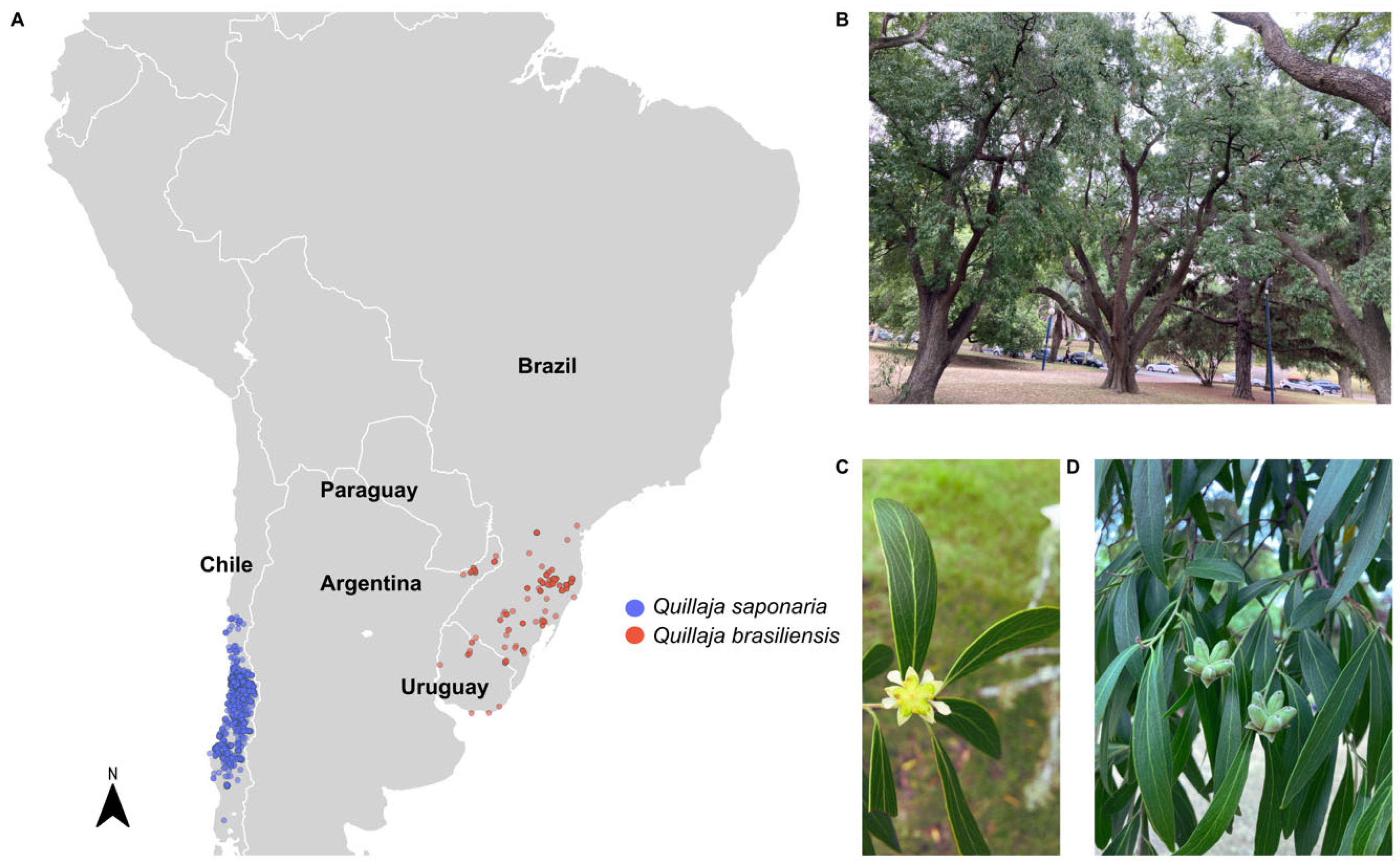
The genus Quillaja exhibits a striking geographic separation in South America due to the Andes Mountains chain. Q. saponaria is native to Chile, while Q. brasiliensis thrives in the Araucaria forests of southern Brazil, Uruguay, northeastern Argentina, and eastern Paraguay [159,160] (Figure 4A). Due to the overharvesting of Q. saponaria barks in Chilean forests, which has led to significant ecological damage and supply [161,162], extensive efforts have been made in recent decades to discover new saponins with enhanced adjuvant activity and lower toxicity [67,163]. A promising alternative is Q. brasiliensis, now recognized as Q. lancifolia, a native species of southern Brazil known as “pau-sabão” (soap tree) due to the capacity of its leaves and bark to produce abundant foam in water [159,160,164]. It is considered a great tool of renewable alternative resource saponins, decreasing the pressure on Q. saponaria exploitation [162,165,165,166,167]. Figure 4B–D illustrates the trees, flowers, fruits, and leaves of Q. brasiliensis.
One method for obtaining saponins from Q. brasiliensis leaves, bark, and branches involves aqueous extraction followed by lyophilization. The resulting leaf extract can be further purified using reversed-phase silica gel (RP-C18) chromatography with a methanol gradient. A notable saponin fraction obtained via this method, eluted at 90% methanol, is known as QB-90 [166,168,169,170]. Recently, a detailed method for purifying saponins from Q. brasiliensis leaves using RP-C18 chromatography was reported, describing the preparation of aqueous extracts and the isolation of QB-90 [171].
Building on these advances, several studies have isolated and studied the raw aqueous extract (AE) and RP-C18 fractions, including the saponin-enriched fraction QB-90, QB-80, Fraction B, and Fraction 3 [166,172]. The QB-80 fraction, a saponin-enriched fraction, shares structural similarities with QB-90 and was obtained using the same purification protocol [14], but eluted at 90% of methanol. Subsequently, two alternative methods were developed for isolating bioactive saponins. Fraction B was obtained from the AE of air-dried leaves via solid-phase extraction on C-18 reversed-phase columns, using a stepwise methanol–water gradient. Fraction 3 was isolated from leaf AE through a combination of ultrafiltration and ion-exchange chromatography [172].
Initial studies on Q. brasiliensis leaf constituents were conducted by Kauffman et al. (2004) [173], who reported the isolation of a new abietane diterpene (9-O-beta-D-glucopyranoside of 16-hydroxylambertic acid), a prosapogenin (3-O-beta-D-glucuronopyranosyl-quillaic acid), and two flavonoids (quercetin and rutin). They also described the preparation of aqueous extracts from leaves, bark, and branches and isolated the QB-90 fraction from leaves, characterizing its structure through hydrolysis and NMR analyses [168,173]. 1H NMR spectra of QB-90 and aqueous extracts from leaves, barks, and branches closely resembled those of Quil-A®, highlighting notable structural similarities between the saponins of Q. brasiliensis and Q. saponaria [168]. Further studies by Cibulski et al. using MALDI-ToF–MS (matrix-assisted laser desorption/ionization time-of-flight mass spectrometry) [14,174] and DI-ESI-MS [175] confirmed these similarities in saponin composition between the two species. More recently, Wallace et al. identified 75 distinct saponins in Q. brasiliensis. From these, 21 were from leaves, and 54 were from bark [176]. Among these, a novel triterpene saponin, Qb1 (an unreported isomer of QS-21), was identified and elucidated by MS and NMR techniques [174,176].
Figure 4. Geographic distribution of Quillajaceae and morphological aspects of Q. brasiliensis. (A) Occurrence records of Q. saponaria (blue) and Q. brasiliensis (red) in South America. Distribution data were retrieved from the Global Biodiversity Information Facility (GBIF, https://www.gbif.org, accessed on 1 July 2025), and the map was generated using Kauffman et al. 2004 [173]. Country borders are shown in white over a light gray political map. (B) Adult trees of Q. brasiliensis located in Parque Batlle, Montevideo, Uruguay. (C) Flower of Q. brasiliensis. (D) Fruits and leaves of Q. brasiliensis (Photos: F. Silveira).
Pharmaceutics 17 00966 g004
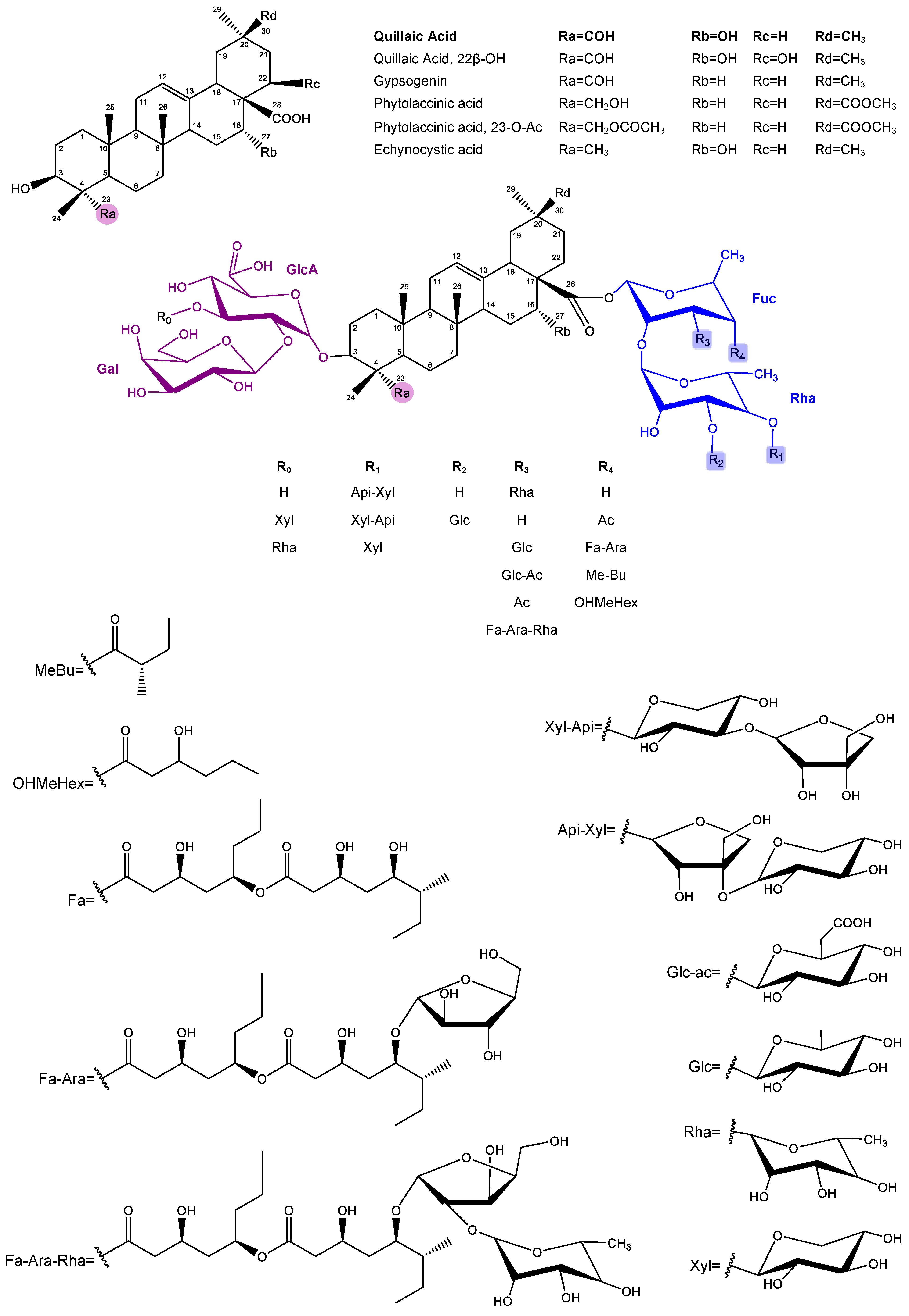
The saponins isolated from the leaves and bark of Q. brasiliensis present complex triterpene structures, which demonstrate remarkable similarity with the classical saponins of Q. saponaria, especially regarding the nature of the aglycones and the organization of the sugar residues. Both species share bidesmosidic saponins, with oligosaccharides linked at the C-3 and C-28 positions of the aglycone, usually quillaic acid, phytolaccinic acid, or their derivatives. In particular, the substitution pattern observed in the sugar residues, such as rhamnose, fucose, xylose, glucuronic acid, and acylations with groups such as Fa-Ara, reinforces the structural similarity between the saponins of both species. Mass spectrometry analysis (LC-ESI-IT-MSⁿ) showed that the fragment ions (“A ions”) of the saponins from Q. brasiliensis present the same fragmentation profiles already described for Q. saponaria, indicating the conservation of the basic architecture of the compounds. Additionally, many of the compounds identified in Q. brasiliensis have molecular masses identical or very close to those of the known saponins from Q. saponaria and are considered homologous or have variants with specific modifications such as substitutions of sugars or acyl groups. These structural similarities explain the functional equivalence observed between the extracts of the two species, especially in the context of their immunoadjuvant activity. Notably, the QS-21 m/z ion was detected in nanoparticles formulated with raw aqueous extracts (AE) [174] as well as with QB-80 [175] and Fraction B [177], all derived from Q. brasiliensis leaves. In mass spectrometry, m/z denotes the mass-to-charge ratio of an ion, defined as the ion’s mass (m) divided by its net charge (z), and serves as the principal variable by which ions are separated and detected within the mass analyzer. The general structures of the Quillajaceae saponins are described in Figure 5.
The primary limitation of saponins as adjuvants is their potential toxicity of amphiphilic molecules due to their ability to interact with cholesterol in mammalian cell membranes, disrupting membrane integrity and function [77,178], which is commonly assessed through a range of in vitro and in vivo assays [178]. A standard initial evaluation involves measuring hemolytic activity against red blood cell suspensions. Silveira et al. (2011) reported that saponins extracted from Q. brasiliensis plants in Uruguay (designated as QB-90U) exhibited lower hemolytic activity than Quil-A® [14,91]. Further cytotoxicity assessments were conducted on Vero cells using MTT viability and lactate dehydrogenase (LDH) release assays [86,91], aligning well with previous in vivo toxicity data in mice [14,86,168]. Additionally, Cibulski et al. (2016) demonstrated that both QB-90 and aqueous extracts (AE) from Q. brasiliensis exhibited significantly lower hemolytic activity (higher HD50 values) than Quil-A® [14,90].
Injection site reactions are the most frequently reported manifestation of vaccine-associated toxicity in animal models. These local responses, common with parenteral vaccines, can range from pain and swelling to granulomas, sterile abscesses, and ulceration [179]. Consistent with this, the in vivo toxicity of Q. brasiliensis saponins has been evaluated in several studies. Fleck et al. (2006) conducted the first acute toxicity assay using the raw AE and QB-90 in CF-1 female Swiss mice via subcutaneous (s.c.) administration [168]. No lethality, swelling, or hair loss effects were observed [168]. However, bark extracts caused local swelling at doses above 400 µg/dose following a second administration.
Cibulski et al. (2016) [86] further assessed acute toxicity in CD-1 male mice. At the lowest tested dose (31.25 µg), QB-90 induced no signs of local toxicity or lethality, while Quil-A® caused 40% lethality. At the highest dose (125 µg), mortality reached 60% with QB-90 and 100% with Quil-A®, reinforcing that QB-90 is significantly less toxic than Quil-A®, both in vitro and in vivo.
In summary, semi-purified aqueous extracts and saponin-enriched fractions from Q. brasiliensis demonstrate lower toxicity compared to Quil-A®, despite their structural similarity. These differences may be attributed to subtle structural variations and differences in extraction and purification methods [91].

3.2. Q. Brasiliensis Saponins: A Potent Natural Immunoadjuvant

A descriptive analysis of the immune responses elicited by diverse SBA derived from Q. brasiliensis was conducted using data compiled from studies involving different viral antigens and administration routes. These data are shown in Table 2. The adjuvants assessed include AE, QB-90, and QB-80 and its nanoparticulate formulations, as well as other purified saponin fractions. These were evaluated primarily through subcutaneous and intranasal immunization routes in murine models, using diverse inactivated or recombinant antigens such as herpesvirus (BoHV1 and BoHV5), poliovirus, pestivirus (BVDV), rabies virus, Zika virus, and influenza virus A.
Table 2. Immunoadjuvant activity of Q. brasiliensis saponins.
Adjuvant/Route (μg/Dose)AntigenIgGIgG1IgG2a/2cIgG2bIgG3IgANAb/HAIDTHOther AssaysRef.
AE-sc (400)Herpesvirus (BoHV1, inactivated virus)*** [168]
QB-90-sc (200, 150, 100 and 50)***
QB-90 a-sc (100)Herpesvirus (BoHV5, inactivated virus)*********** ***Upregulation of IFN-γ and IL-2 mRNA in splenocytes[91]
AE-sc (400)Poliovirus (Sabin 1, inactivated virus)****** * **Upregulation of IFN-γ and IL-2 mRNA in splenocytes[92]
QB-90-sc (50)****** * **
AE-sc (400, 200 and 100)Pestivirus (BVDV, inactivated virus)********* ***Splenocyte proliferation (including CD8+ T cells) and IFN-γ, IL-2, TNF, IL-10, and IL-17 cytokines secretion[14,90]
QB-90-sc (100, 50 and 10)********* ***
QB-90-sc (10)Ovalbumin (OVA)*****- -Splenocyte proliferation; IFN-γ, IL-2 secretion in IQB-90 vaccines[86]
IQB-90-sc (10)******** ***
QB-90-in (2)--- - -
IQB-90-in (2)******- */*** b -
AE-sc (400)Rabies virus (inactivated virus)*** *Protection in a challenge assay provided by the adjuvanted anti-rabies vaccines[172]
QB-80-sc (100 and 50)****
QB-90-sc (100 and 50)****
Fraction B-sc (50)***
Fraction 3-sc (50)***
AE-sc (400 and 200)Pestivirus (BVDV, inactivated virus)*********** ****Antigen-specific IFN-γ production in CD4+ and CD8+ T cells; dose sparing effect[14]
QB-80-sc (100, 50 and 10)************ ***
IMXQB-80-sc (2.5)Zika virus (inactivated virus)********** *** No differences between IMXQB-80 (2.5 μg) and QB-80 (10 μg) immunized animals[175]
QB-80-sc (10)*********** ***
IQB80-sc (10 and 2)Zika virus (recombinant E protein)*************** *** Antigen-specific splenocyte proliferation and antibody avidity increase[94]
IQB90-sc (5)Influenza (seasonal split vaccine)**************-******Protection in a challenge test provided by adjuvanted vaccines; dose sparing effect[93]
IQB90-in (5)************************ b****
IMXQB-sc (5)Influenza (seasonal split vaccine)***************** * Protection in a challenge test provided by adjuvanted vaccines; high levels of NAb and improved serum HI[180]
IMXQB-in (2.5)*********************
IMXQB-old mice sc (5)Influenza (seasonal split vaccine)***- - Protection in a challenge test provided by adjuvanted vaccines; IgG1, IgG2a, and IgA are maintained 120 days after priming[181]
IMXQB-old mice in (2.5)****** *******
IQB: vaccines as ISCOM, IMXQB: vaccines adjuvanted with ISCOM-matrices, sc subcutaneous, in intranasal, a QB-90 derived from Uruguayan specimens. b Seric IgA. Nab/HAI (neutralizing antibodies) DTH (delayed-type hypersensitivity), - not significant, * p < 0.05, ** p < 0.01, *** p < 0.001, **** p < 0.0001 (against group of animals vaccinated with unadjuvanted formulation).
The results across studies emphasize the versatility and efficacy of SBA derived from leaves of Q. brasiliensis in eliciting balanced and protective immune responses across different viral vaccine candidates. Their effectiveness via mucosal routes and in aged models further supports their potential for application in both human and veterinary vaccine development

3.2.1. Humoral Response

Fleck et al. (2006) [168] were the first to assess the adjuvant potential of Q. brasiliensis saponin-enriched fractions, which closely resemble those from Q. saponaria. A purified leaf-derived saponin fraction (QB-90) effectively stimulated both Th1 and Th2 responses, as well as cytotoxic T cell activity against herpesviruses 1 and 5 in mice, with a response comparable to Q. saponaria saponins (Quil-A®) [91,168]. Similarly, QB-90 from Uruguayan plants (QB-90U) induced strong IgG responses with neutralizing antibodies against BoHV-5 [91]. The immunoglobulin isotype profile elicited by the Uruguayan QB-90U fraction was comparable to that induced by Quil-A®, with a Th1-biased response characterized by elevated levels of IgG2a and IgG3, alongside significant IgG1 production, indicating concurrent Th2 activation [182]. AE from bark, leaves, and branches also showed adjuvant activity on par with Quil-A® in experimental BoHV-1 vaccines [168]. Additionally, AE from Q. brasiliensis leaves has been tested with human poliovirus, bovine viral diarrhea virus (BVDV), and inactivated rabies virus in mice [14,90,92,168,172].
Recent investigations have demonstrated that novel saponin fractions from Q. brasiliensis, such as QB-80 and Fraction 3, exhibit potent immunostimulant properties when co-administered with inactivated rabies virus antigens, eliciting robust antibody responses in murine models [172]. Cibulski et al. (2016) further reported that both AE and QB-90 fractions significantly enhanced the humoral response against BVDV, primarily by increasing total IgG levels via increments of IgG1 (Th2-associated) and IgG2a (Th1-associated) isotypes [90].
Chemically, both QB-80 and AE display notable similarities to the commercial saponin Quil-A®, though with reduced toxicity. These fractions not only induced sustained IgG1 and IgG2a responses with high avidity but also promoted delayed-type hypersensitivity (DTH) reactions and increased IFN-γ secretion in both CD4+ and CD8+ T cell populations [14]. As well as due to its low cost and ease production, AE emerges as a promising candidate for industrial-scale applications, including nanoparticle-based vaccine formulations [174].
In the context of Zika virus immunizations, both QB-80 and its nanoparticle-based formulation (IMXQB-80) significantly elevated anti-Zika virus IgG subtypes (IgG1, IgG2b, IgG2c) and neutralizing antibody titers, outperforming unadjuvanted vaccines [175]. Notably, IMXQB-80 achieved comparable immunogenicity with fourfold less saponin content and reduced toxicity. Mice immunized with the IQB80-zEDIII formulation exhibited enhanced IgG subclass responses, strong neutralizing activity, and robust splenocyte proliferation, supporting its candidacy for Zika virus vaccine development [94].
In studies on influenza, an ISCOM-like nanoparticle formulation incorporating QB-90 (IQB90) demonstrated superior protective efficacy compared to unadjuvanted vaccines, whether delivered subcutaneously or intranasally. The IQB90 formulation not only induced higher hemagglutination inhibition titers but also exhibited a tenfold dose-sparing effect, an attribute of significant relevance for pandemic preparedness strategies [93,180].

3.2.2. Cell-Mediated Immune Response Induced by Q. brasiliensis Saponins

DTH is a classical in vivo indicator of cell-mediated immunity, marked by the recruitment and activation of macrophages and T lymphocytes, particularly CD4+ and CD8+ cells, along with the release of pro-inflammatory cytokines such as IL-1, IL-2, IFN-γ, and TNF-α [183,184,185,186]. In mice, DTH reactions typically emerge 16–24 h post-antigen challenge, manifesting as mononuclear and granulocytic infiltration, increased vascular permeability, and edema [184].
Experimental vaccines adjuvanted with Q. brasiliensis saponin fractions (QB-90, QB-80, and the raw AE) induced significantly stronger DTH responses compared to control formulations (Alum or Quil-A®), particularly in response to bovine herpesvirus and human poliovirus antigens [91,92]. These results suggest the activation of memory Th1 CD4+ T cells [184], consistent with the immunostimulatory profile of Quil-A® [187].
In further studies, vaccines formulated with QB-90, QB-80, or AE, in combination with rabies (Pasteur strain) or BVDV antigens, also produced pronounced DTH responses relative to unadjuvanted controls [90,172]. Notably, the IQB90-Flu nanoparticle vaccine elicited robust DTH reactions at 24 h in both wild-type and caspase-1/11 knockout mice, while no DTH responses were observed in animals receiving the unadjuvanted influenza vaccine [93].
The cytokine profile induced by QB-90U paralleled that of Quil-A®, favoring a Th1-biased response characterized by elevated IgG2a and IgG3 isotypes, as well as increased IFN-γ and IL-2 mRNA expression in splenocytes [90,91,92]. Additional analysis using a Th1/Th2/Th17 bead array showed that BVDV-stimulated splenocytes from mice immunized with QB-90-adjuvanted vaccines secreted high levels of IFN-γ, TNF-α, and IL-2, corroborating the Th1-polarized humoral response. QB-90 also promoted IL-10 and moderate levels of IL-4 and IL-6, indicating a mixed Th1/Th2 cytokine milieu [90]. De Costa et al. confirmed these findings through qPCR, demonstrating nearly tenfold upregulation of IFN-γ transcripts in splenocytes from mice immunized with QB-90 or AE compared to non-adjuvanted controls, matching levels induced by Quil-A® [92].
Cell-mediated immunity, particularly through CD8+ T cells, plays a critical role in viral clearance by recognizing viral peptides presented via MHC I and initiating cytolytic activity or cytokine-mediated inhibition [188,189,190,191]. Mice immunized with BVDV antigens adjuvanted with AE or QB-90 exhibited significantly enhanced BVDV-specific CD8+ T cell proliferation and increased frequencies of IFN-γ+ CD4+ and CD8+ T cells, as assessed by flow cytometry [90]. These findings suggest that QB-90 enhances antigen cross-presentation, potentially via the chemotactic recruitment of dendritic cells to the immunization site [90].

3.2.3. Mucosal Immunity Induced by Q. brasiliensis Saponins

Mucosal surfaces, lining the gastrointestinal, respiratory, and urogenital tracts, represent major entry points for pathogens. Despite this, most vaccines are delivered parenterally, with few licensed for mucosal administration due to the absence of effective delivery systems and mucosal adjuvants [192,193,194]. Current adjuvants show limited efficacy in enhancing mucosal immune responses.
Intranasal vaccination promotes local immune responses, including secretory IgA and the induction of resident memory B and T cells, reducing infection rates and viral shedding [192,195]. Intranasal immunization with adjuvanted inactivated vaccines effectively induces both mucosal IgA, offering strong cross-protection against variant viruses in the upper respiratory tract, and serum IgG, providing protection in the lower tract [196]. Compared to injectable vaccines, nasal vaccines elicit higher local IgA levels and stronger mucosal cell-mediated immunity, albeit with lower systemic antibody titers. Nevertheless, both routes demonstrate comparable efficacy (70–90%) in healthy individuals when the vaccine and epidemic virus are well-matched antigenically [197].
Studies using Q. brasiliensis-derived saponins, such as QB-90, demonstrated enhanced mucosal immunity. De Costa et al. showed that intranasal immunization with the QB-90-adjuvanted poliovirus antigen induced high IgA titers in bile, feces, and vaginal washings, comparable or superior to Quil-A® [92]. Additionally, particulate formulations like IQB-90 promoted both mucosal IgA and systemic IgG responses across multiple sites, including the nasal passages, large intestine, and vaginal lumen [86].
Recent findings revealed that intranasal delivery of an inactivated seasonal influenza vaccine adjuvanted with IQB-90 enhanced IgM, IgG, and IgA levels 28 days post-priming. This experimental formulation also induced higher IgG1, IgG2a, IgG2b, and IgG3 levels and elevated hemagglutination inhibition titers compared to unadjuvanted commercial vaccines [93].

3.2.4. ISCOMs and ISCOM-Matrices Based on Q. brasiliensis Saponins

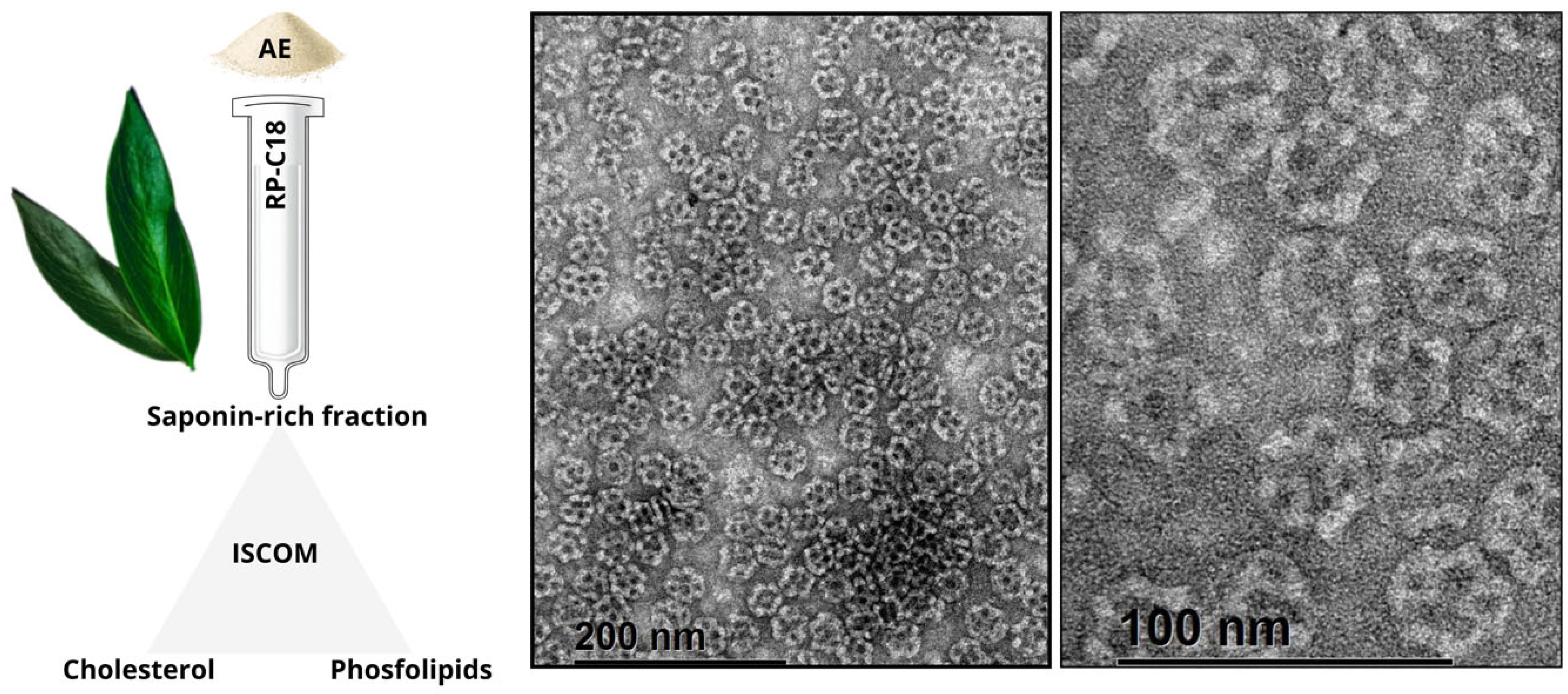
An improvement to the use of saponins as adjuvants was introduced by the development of immune stimulating complexes. These have been used as antigen delivery systems that proved to exert powerful immune stimulating activities yet displaying reduced toxicity in several animal models. Prior to the study by Cibulski et al. [86], there were no reports of ISCOMs production using saponins extracted from any plant other than Q. saponaria. In the study, for the very first time, ISCOM formulations were constructed by replacing the Quil-A® component by QB-90. These saponins extracted from Q. brasiliensis, successfully formulated ISCOMs, preserve their adjuvant activity while enhancing stability and reducing toxicity, notably by eliminating hemolytic effects [86,94,175]. Figure 6 provides a detailed illustrative scheme of the process for obtaining Q. brasiliensis-derived ISCOM-matrices, from the extraction of the raw aqueous extract (AE) to the final formulation and visualization by transmission electron microscopy (TEM).
The immunogenicity of ISCOMs prepared with the QB-90 fraction (IQB-90) was comparable to classical Quil-A®-based ISCOMs (IQA). IQB-90 nanoparticles (~40–50 nm, spherical, cage-like particles) comprise QB-90, cholesterol, phospholipids, and antigen (e.g., ovalbumin). These nanoparticles were efficiently internalized in vitro by murine bone marrow-derived DC and, upon subcutaneous administration, elicited strong serum IgG1 and IgG2a responses, DTH reactions, T cell proliferation, and Th1 cytokine production (IFN-γ, IL-2). Intranasally delivered IQB-90 also induced serum IgG/IgG1 and mucosal IgA responses across distant mucosal sites [86].
Mechanistic studies on ISCOM-matrices adjuvant based on QB-90 (IMXQB-90) revealed rapid cytosolic antigen delivery in dendritic cells, broad cytokine and chemokine induction, and the bridging of innate and adaptive immunity via a MyD88-dependent, Toll-like receptor-independent pathway [115,116,199]. Furthermore, QB-90 and IMXQB-90 promoted immune cell recruitment to draining lymph nodes and spleen, stimulated IL-1β secretion through caspase-1/11 and MyD88 pathways, implying canonical inflammasome activation, and induced the upregulation of cytokine and chemokine gene expression [99].
Cibulski et al. demonstrated that replacing Quil-A® with QB-90 in ISCOM and ISCOM-matrices formulations (IQB-90 and IMXQB-90) maintained potent immunogenicity, including robust antibody production, myeloid and lymphoid cell activation, and Th1-type cytokine secretion (IFN-γ, TNF-α) [86,99]. In vitro, both formulations triggered caspase-1-dependent IL-1β production in bone marrow-derived dendritic cells. However, the role of inflammasome activation in vivo remains to be fully elucidated [158].
Recent studies employing Zika virus antigens confirmed the efficacy of QB-90-based adjuvants. Both a classic inactivated vaccine and a recombinant antigen formulation adjuvanted with IMXQB-80 or IQB80-zEDIII induced significantly higher anti-Zika virus IgG titers and neutralizing antibodies compared to unadjuvanted controls [94,175]. Additionally, a split-inactivated influenza vaccine adjuvanted with IQB-90 elicited stronger protective responses than a commercial unadjuvanted vaccine when administered via either subcutaneous or intranasal routes [93].
In line with these findings, Silveira et al. [180] demonstrated that a trivalent influenza vaccine (TIV) formulated with Q. brasiliensis-based IMXQB nanoparticles induced potent humoral and cellular immune responses when delivered subcutaneously or intranasally, including high levels of IgG1/IgG2a, Th1/Th2 cytokines, and effector T cells. Notably, intranasal immunization with TIV-IMXQB conferred full protection against a lethal viral challenge, preventing weight loss, lung viral replication, and mortality.
Further supporting these observations, a subsequent study in aged mice revealed that IMXQB-adjuvanted TIV significantly improved both systemic and mucosal immunity when administered subcutaneously or intranasally [181]. The adjuvanted vaccine elicited sustained IgM, IgG, and IgA responses, enhanced hemagglutination inhibition (HAI) titers, and accelerated recovery following viral challenge. These findings highlight the potential of Q. brasiliensis-derived IMXQB as a versatile and effective mucosal and systemic adjuvant platform, particularly relevant for vulnerable populations such as the elderly.

4. Conclusions

Saponins are powerful adjuvants that enhance cellular immunity, making them particularly valuable for vaccines against intracellular pathogens, viruses, and cancers [36,67,75,76,77,78]. While Q. saponaria bark extract semi- or purified fractions have been widely used in both veterinary and human vaccines [97,98,116,118], their overharvesting has caused ecological damage in Chilean forests [162,165]. This has driven the search for sustainable alternatives with comparable immunostimulatory properties but reduced toxicity [67,163].
Q. brasiliensis, native to South America, has emerged as an ecologically sustainable solution [166,167]. Its leaf-derived saponins share structural and functional similarities with Q. saponaria saponins while offering several advantages [168]. Fractions like QB-90, QB-80, and the aqueous extract demonstrate potent adjuvant activity, inducing balanced humoral (IgG1, IgG2a) and cellular (Th1/Th2/Th17, CTL) responses with improved safety profiles. When incorporated into nanoparticle platforms (ISCOMs/ISCOM-matrices), Q. brasiliensis saponins show enhanced efficacy against diverse pathogens, including influenza, Zika, and rabies, while minimizing hemolytic effects. Their mechanism of action involves NLRP3 inflammasome activation, dendritic cell recruitment, and cytokine production (IFN-γ, IL-1β) and can be particularly valuable for adjuvant vaccines to diseases requiring strong cellular immunity (e.g., malaria, tuberculosis, HIV).
From an economic standpoint, the sourcing and production of saponins present significant challenges and opportunities for vaccine development. Currently, the market price of Quil-A® exceeds USD 500 per gram, while highly purified QS-21 can cost over USD 300 per milligram, making these adjuvants among the most expensive components of vaccine formulations. In contrast, Q. brasiliensis saponins can be sustainably harvested from leaves, avoiding bark extraction and thus minimizing ecological impact. Moreover, leaf collection allows for more frequent and scalable harvesting cycles. Although no commercial suppliers of Q. brasiliensis saponins currently exist and market pricing has yet to be established, preliminary studies indicate that its saponin fractions exhibit immunoadjuvant properties comparable to those of Q. saponaria [168]. As such, Q. brasiliensis represents a promising avenue for future development of accessible, sustainable, and economically viable vaccine adjuvants, particularly in settings where cost is a critical factor.
The sustainable leaf-harvesting approach for Q. brasiliensis addresses both ecological concerns and supply chain limitations, positioning it as a strategic resource for next-generation vaccine development. Ongoing preclinical and clinical studies will be crucial to fully realize its potential in human and veterinary applications, offering a promising combination of immunogenicity, safety, and environmental protection.

Author Contributions

V.M.: conceptualization, visualization, writing—original draft, writing—review and editing. N.S.: writing—original draft, writing—review and editing. S.C.: conceptualization, writing—original draft, writing—review and editing, F.S.: conceptualization, writing—original draft, writing—review and editing. All authors have read and agreed to the published version of the manuscript.

Funding

This research received no external funding.

Acknowledgments

The authors thank Thalisson Amorim and Genil Oliveira for their support in ChemDraw 23.0. V.M., N.S., and F.S. are members of the Sistema Nacional de Investigadores (SNI) of the Agencia Nacional de Investigación e Innovación (ANII).

Conflicts of Interest

The authors declare that they do not have any commercial or associative interests that represent any conflicts of interest in connection with the work submitted.

References

  1. Plotkin, S.A. Vaccines: Past, present and future. Nat. Med. 2005, 11, S5. [Google Scholar] [CrossRef]
  2. Rueckert, C.; Guzmán, C.A. Vaccines: From empirical development to rational design. PLoS Pathog. 2012, 8, e1003001. [Google Scholar] [CrossRef]
  3. Finco, O.; Rappuoli, R. Designing vaccines for the twenty-first century society. Front. Immunol. 2014, 5, 12. [Google Scholar] [CrossRef]
  4. De Gregorio, E.; Rappuoli, R. From empiricism to rational design: A personal perspective of the evolution of vaccine development. Nat. Rev. Immunol. 2014, 14, 505–514. [Google Scholar] [CrossRef]
  5. Meyer, H.; Ehmann, R.; Smith, G.L. Smallpox in the post-eradication Era. Viruses 2020, 12, 138. [Google Scholar] [CrossRef]
  6. Garçon, N.; Segal, L.; Tavares, F.; Van Mechelen, M. The safety evaluation of adjuvants during vaccine development: The AS04 experience. Vaccine 2011, 29, 4453–4459. [Google Scholar] [CrossRef]
  7. Ura, T.; Yamashita, A.; Mizuki, N.; Okuda, K.; Shimada, M. New vaccine production platforms used in developing SARS-CoV-2 vaccine candidates. Vaccine 2021, 39, 197–201. [Google Scholar] [CrossRef] [PubMed]
  8. Krammer, F. SARS-CoV-2 vaccines in development. Nature 2020, 586, 516–527. [Google Scholar] [CrossRef] [PubMed]
  9. Rappuoli, R.; Alter, G.; Pulendran, B. Transforming vaccinology. Cell 2024, 187, 5171–5194. [Google Scholar] [CrossRef] [PubMed]
  10. O’Hagan, D.T.; Valiante, N.M. Recent advances in the discovery and delivery of vaccine adjuvants. Nat. Rev. Drug Discov. 2003, 2, 727–735. [Google Scholar] [CrossRef]
  11. Ramon, G. Sur L’augmentation anormale de L’antitoxine chez les chevaux producteurs de serum antidiphtherique. Bull. Soc. Centr. Med. Vet. 1925, 101, 227–234. [Google Scholar]
  12. Schijns, V.; Fernández-Tejada, A.; Barjaktarović, Ž.; Bouzalas, I.; Brimnes, J.; Chernysh, S.; Gizurarson, S.; Gursel, I.; Jakopin, Ž.; Lawrenz, M.; et al. Modulation of immune responses using adjuvants to facilitate therapeutic vaccination. Immunol. Rev. 2020, 296, 169–190. [Google Scholar] [CrossRef]
  13. Roth, G.A.; Picece, V.C.T.M.; Ou, B.S.; Luo, W.; Pulendran, B.; Appel, E.A. Designing spatial and temporal control of vaccine responses. Nat. Rev. Mater. 2022, 7, 174–195. [Google Scholar] [CrossRef] [PubMed]
  14. Cibulski, S.; Rivera-Patron, M.; Suárez, N.; Pirez, M.; Rossi, S.; Yendo, A.C.; de Costa, F.; Gosmann, G.; Fett-Neto, A.; Roehe, P.M.; et al. Leaf saponins of Quillaja brasiliensis enhance long-term specific immune responses and promote dose-sparing effect in BVDV experimental vaccines. Vaccine 2018, 36, 55–65. [Google Scholar] [CrossRef]
  15. Hawken, J.; Troy, S.B. Adjuvants and inactivated polio vaccine: A systematic review. Vaccine 2012, 30, 6971–6979. [Google Scholar] [CrossRef] [PubMed]
  16. Wen, Y.; Shi, Y. Alum: An old dog with new tricks. Emerg. Microbes Infect. 2016, 5, e25. [Google Scholar] [CrossRef]
  17. Orr, M.T.; Khandhar, A.P.; Seydoux, E.; Liang, H.; Gage, E.; Mikasa, T.; Beebe, E.L.; Rintala, N.D.; Persson, K.H.; Ahniyaz, A.; et al. Reprogramming the adjuvant properties of aluminum oxyhydroxide with nanoparticle technology. npj Vaccines 2019, 4, 1. [Google Scholar] [CrossRef]
  18. Pulendran, B.; S. Arunachalam, P.; O’Hagan, D.T. Emerging concepts in the science of vaccine adjuvants. Nat. Rev. Drug Discov. 2021, 20, 454–475. [Google Scholar] [CrossRef]
  19. Kommareddy, S.; Sing, M.; O’Hagan, D.T. MF59: A Safe and Potent Adjuvant for Human Use. In Immunopotentiators in Modern Vaccines; Schijns, V.E.J.C., O’Hagan, D.T., Eds.; Academic Press: London, UK, 2016; pp. 249–263. ISBN 978-0-12-804019-5. [Google Scholar]
  20. O’Hagan, D.T.; Ott, G.S.; De Gregorio, E.; Seubert, A. The mechanism of action of MF59-An innately attractive adjuvant formulation. Vaccine 2012, 30, 4341–4348. [Google Scholar] [CrossRef] [PubMed]
  21. O’Hagan, D.T.; Ott, G.S.; Nest, G.V.; Rappuoli, R.; Giudice, G. Del The history of MF59® adjuvant: A phoenix that arose from the ashes. Expert Rev. Vaccines 2013, 12, 13–30. [Google Scholar] [CrossRef]
  22. Zheng, D.; Gao, F.; Zhao, C.; Ding, Y.; Cao, Y.; Yang, T.; Xu, X.; Chen, Z. Comparative effectiveness of H7N9 vaccines in healthy individuals. Hum. Vaccines Immunother. 2019, 15, 80–90. [Google Scholar] [CrossRef]
  23. O’Hagan, D.T. MF59 is a safe and potent vaccine adjuvant that enhances protection against influenza virus infection. Expert Rev. Vaccines 2007, 6, 699–710. [Google Scholar] [CrossRef]
  24. Mosca, F.; Tritto, E.; Muzzi, A.; Monaci, E.; Bagnoli, F.; Iavarone, C.; O’Hagan, D.; Rappuoli, R.; De Gregorio, E. Molecular and cellular signatures of human vaccine adjuvants. Proc. Natl. Acad. Sci. USA 2008, 105, 10501–10506. [Google Scholar] [CrossRef]
  25. Cunningham, A.L.; Sandgren, K.J.; Taylor, J. Current status of immunisation for herpes zoster. Hum. Vaccin. Immunother. 2025, 21, 2445384. [Google Scholar] [CrossRef]
  26. Alving, C.R.; Peachman, K.; Rao, M.; Reed, S.G. Adjuvants for human vaccines. Curr. Opin. Immunol. 2012, 24, 310–315. [Google Scholar] [CrossRef]
  27. Garçon, N.; Chomez, P.; Van Mechelen, M. GlaxoSmithKline Adjuvant Systems in vaccines: Concepts, achievements and perspectives. Expert Rev. Vaccines 2007, 6, 723–739. [Google Scholar] [CrossRef] [PubMed]
  28. Didierlaurent, A.M.; Laupèze, B.; Di Pasquale, A.; Hergli, N.; Collignon, C.; Garçon, N. Adjuvant system AS01: Helping to overcome the challenges of modern vaccines. Expert Rev. Vaccines 2017, 16, 55–63. [Google Scholar] [CrossRef]
  29. Roman, F.; Burny, W.; Ceregido, A.; Laupèze, B.; Temmerman, S.T.; Warter, L.; Coccia, M. Adjuvant system AS01: From mode of action to effective vaccines. Expert Rev. Vaccines 2024, 23, 715–729. [Google Scholar] [CrossRef] [PubMed]
  30. Morel, S.; Didierlaurent, A.; Bourguignon, P.; Delhaye, S.; Baras, B.; Jacob, V.; Planty, C.; Elouahabi, A.; Harvengt, P.; Carlsen, H.; et al. Adjuvant System AS03 containing α-tocopherol modulates innate immune response and leads to improved adaptive immunity. Vaccine 2011, 29, 2461–2473. [Google Scholar] [CrossRef] [PubMed]
  31. Loos, C.; Coccia, M.; Didierlaurent, A.M.; Essaghir, A.; Fallon, J.K.; Lauffenburger, D.; Luedemann, C.; Michell, A.; van der Most, R.; Zhu, A.L.; et al. Systems serology-based comparison of antibody effector functions induced by adjuvanted vaccines to guide vaccine design. npj Vaccines 2023, 8, 34. [Google Scholar] [CrossRef]
  32. Garçon, N.; Di Pasquale, A. From discovery to licensure, the Adjuvant System story. Hum. Vaccines Immunother. 2017, 13, 19–33. [Google Scholar] [CrossRef]
  33. Del Giudice, G.; Rappuoli, R.; Didierlaurent, A.M. Correlates of adjuvanticity: A review on adjuvants in licensed vaccines. Semin. Immunol. 2018, 39, 14–21. [Google Scholar] [CrossRef] [PubMed]
  34. Zhu, D.; Tuo, W. QS-21: A Potent Vaccine Adjuvant. Nat. Prod. Chem. Res. 2016, 3, 3–4. [Google Scholar] [CrossRef]
  35. Atmar, R.L.; El Sahly, H.M. The Promise of New Vaccines Against Respiratory Viruses. Pediatrics 2024, 153, e2023065328. [Google Scholar] [CrossRef] [PubMed]
  36. Kensil, C.R. Saponins as vaccine adjuvants. Crit. Rev. Ther. Drug Carrier Syst. 1996, 13, 1–55. [Google Scholar]
  37. Villacres-Eriksson, M.; Behboudi, S.; Morgan, A.J.; Trinchieri, G.; Morein, B. Immunomodulation by Quillaja saponaria adjuvant formulations: In vivo stimulation of interleukin 12 and its effects on the antibody response. Cytokine 1997, 9, 73–82. [Google Scholar] [CrossRef]
  38. Amanat, F.; Krammer, F. SARS-CoV-2 Vaccines: Status Report. Immunity 2020, 52, 583–589. [Google Scholar] [CrossRef] [PubMed]
  39. Mascola, J.R.; Fauci, A.S. Novel vaccine technologies for the 21st century. Nat. Rev. Immunol. 2020, 20, 87–88. [Google Scholar] [CrossRef]
  40. Coffman, R.L.; Sher, A.; Seder, R.A. Vaccine adjuvants: Putting innate immunity to work. Immunity 2010, 33, 492–503. [Google Scholar] [CrossRef]
  41. Turley, J.L.; Lavelle, E.C. Resolving adjuvant mode of action to enhance vaccine efficacy. Curr. Opin. Immunol. 2022, 77, 102229. [Google Scholar] [CrossRef]
  42. Ho, N.I.; Huis In ’t Veld, L.G.M.; Raaijmakers, T.K.; Adema, G.J. Adjuvants Enhancing Cross-Presentation by Dendritic Cells: The Key to More Effective Vaccines? Front. Immunol. 2018, 9, 2874. [Google Scholar] [CrossRef] [PubMed]
  43. Kawasaki, T.; Kawai, T. Toll-like receptor signaling pathways. Front. Immunol. 2014, 5, 461. [Google Scholar] [CrossRef] [PubMed]
  44. Awate, S.; Babiuk, L.A.; Mutwiri, G. Mechanisms of action of adjuvants. Front. Immunol. 2013, 4, 114. [Google Scholar] [CrossRef] [PubMed]
  45. De Gregorio, E.; Caproni, E.; Ulmer, J.B. Vaccine adjuvants: Mode of action. Front. Immunol. 2013, 4, 214. [Google Scholar] [CrossRef]
  46. O’Hagan, D.T.; Fox, C.B. Are we entering a new age for human vaccine adjuvants? Expert Rev. Vaccines 2015, 14, 909–911. [Google Scholar] [CrossRef][Green Version]
  47. Sanders, M.T.; Brown, L.E.; Deliyannis, G.; Pearse, M.J. ISCOMTM-based vaccines: The second decade. Immunol. Cell Biol. 2005, 83, 119–128. [Google Scholar] [CrossRef]
  48. Den Brok, M.H.; Büll, C.; Wassink, M.; De Graaf, A.M.; Wagenaars, J.A.; Minderman, M.; Thakur, M.; Amigorena, S.; Rijke, E.O.; Schrier, C.C.; et al. Saponin-based adjuvants induce cross-presentation in dendritic cells by intracellular lipid body formation. Nat. Commun. 2016, 7, 13324. [Google Scholar] [CrossRef]
  49. Oyston, P.; Robinson, K. The current challenges for vaccine development. J. Med. Microbiol. 2012, 61, 889–894. [Google Scholar] [CrossRef]
  50. Kool, M.; Fierens, K.; Lambrecht, B.N. Alum adjuvant: Some of the tricks of the oldest adjuvant. J. Med. Microbiol. 2012, 61, 927–934. [Google Scholar] [CrossRef]
  51. Petrovsky, N. Comparative Safety of Vaccine Adjuvants: A Summary of Current Evidence and Future Needs. Drug Saf. 2015, 38, 1059–1074. [Google Scholar] [CrossRef]
  52. Schijns, V.E.J.C.; Lavelle, E.C. Trends in vaccine adjuvants. Expert Rev. Vaccines 2011, 10, 539–550. [Google Scholar] [CrossRef] [PubMed]
  53. Pasquale, A.; Preiss, S.; Silva, F.; Garçon, N. Vaccine Adjuvants: From 1920 to 2015 and Beyond. Vaccines 2015, 3, 320–343. [Google Scholar] [CrossRef]
  54. Shah, R.R.; Hassett, K.J.; Brito, L.A. Overview of Vaccine Adjuvants: Introduction, History, and Current Status. In Vaccine Adjuvants: Methods and Protocols; Fox, C.B., Ed.; Methods in Molecular Biology; Springer Science + Business Media: New York, NY, USA, 2017; Volume 1494, pp. 1–13. ISBN 978-1-4939-6443-7. [Google Scholar]
  55. Garçon, N.; Hem, S.; Friede, M. Evolution of Adjuvants Across the Centuries. In Vaccine, 6th ed.; Plotkin, S., Orenstein, W., Offit, P., Eds.; Elsevier Inc.: Amsterdam, The Netherlands, 2012; ISBN 9781455700905. [Google Scholar]
  56. Maisonneuve, C.; Bertholet, S.; Philpott, D.J.; De Gregorio, E. Unleashing the potential of NOD- and Toll-like agonists as vaccine adjuvants. Proc. Natl. Acad. Sci. USA 2014, 111, 12294–12299. [Google Scholar] [CrossRef] [PubMed]
  57. Brito, L.A.; Malyala, P.; O’Hagan, D.T. Vaccine adjuvant formulations: A pharmaceutical perspective. Semin. Immunol. 2013, 25, 130–145. [Google Scholar] [CrossRef]
  58. Yendo, A.C.A.; Colling, L.C.; Matsuura, H.N.; Vargas, L.R.B.; Martinelli, J.A.; Chitolina, G.Z.; Vainstein, M.H.; Fett-Neto, A.G. Quillaja lancifolia Immunoadjuvant Saponins Show Toxicity to Herbivores and Pathogenic Fungi. Plants 2025, 14, 1252. [Google Scholar] [CrossRef]
  59. Augustin, J.M.; Kuzina, V.; Andersen, S.B.; Bak, S. Molecular activities, biosynthesis and evolution of triterpenoid saponins. Phytochemistry 2011, 72, 435–457. [Google Scholar] [CrossRef] [PubMed]
  60. Wina, E.; Muetzel, S.; Becker, K. The impact of saponins or saponin-containing plant materials on ruminant production—A review. J. Agric. Food Chem. 2005, 53, 8093–8105. [Google Scholar] [CrossRef]
  61. Vincken, J.P.; Heng, L.; de Groot, A.; Gruppen, H. Saponins, classification and occurrence in the plant kingdom. Phytochemistry 2007, 68, 275–297. [Google Scholar] [CrossRef]
  62. Hosttetmann, K.; Marston, A. Saponins, 1995th ed.; Hostettmann, K., Marston, A., Eds.; Cambridge University Press: Cambridge, UK, 2005. [Google Scholar]
  63. Oleszek, W.A. Chromatographic determination of plant saponins. J. Chromatogr. A 2002, 967, 147–162. [Google Scholar] [CrossRef]
  64. Sparg, S.G.; Light, M.E.; Van Staden, J. Biological activities and distribution of plant saponins. J. Ethnopharmacol. 2004, 94, 219–243. [Google Scholar] [CrossRef] [PubMed]
  65. Podolak, I.; Galanty, A.; Sobolewska, D. Saponins as cytotoxic agents: A review. Phytochem. Rev. 2010, 9, 425–474. [Google Scholar] [CrossRef] [PubMed]
  66. Lorent, J.H.; Quetin-Leclercq, J.; Mingeot-Leclercq, M.-P. The amphiphilic nature of saponins and their effects on artificial and biological membranes and potential consequences for red blood and cancer cells. Org. Biomol. Chem. 2014, 12, 8803–8822. [Google Scholar] [CrossRef]
  67. Sun, H.X.; Xie, Y.; Ye, Y.P. Advances in saponin-based adjuvants. Vaccine 2009, 27, 1787–1796. [Google Scholar] [CrossRef]
  68. Cheok, C.Y.; Salman, H.A.K.; Sulaiman, R. Extraction and quantification of saponins: A review. Food Res. Int. 2014, 59, 16–40. [Google Scholar] [CrossRef]
  69. Glauert, A.; Dingle, J.; Lucy, J. Action of saponin on biological cell membranes. Nature 1962, 195, 698. [Google Scholar] [CrossRef]
  70. De Groot, C.; Müller-Goymann, C.C. Saponin Interactions with Model Membrane Systems—Langmuir Monolayer Studies, Hemolysis and Formation of ISCOMs. Planta Med. 2016, 82, 1496–1512. [Google Scholar] [CrossRef]
  71. Böttger, S.; Hofmann, K.; Melzig, M.F. Saponins can perturb biologic membranes and reduce the surface tension of aqueous solutions: A correlation? Bioorg. Med. Chem. 2012, 20, 2822–2828. [Google Scholar] [CrossRef] [PubMed]
  72. Gauthier, C.; Legault, J.; Girard-Lalancette, K.; Mshvildadze, V.; Pichette, A. Haemolytic activity, cytotoxicity and membrane cell permeabilization of semi-synthetic and natural lupane- and oleanane-type saponins. Bioorg. Med. Chem. 2009, 17, 2002–2008. [Google Scholar] [CrossRef]
  73. Press, J.B.; Reynolds, R.C.; May, R.D.; Marciani, D.J. Structure/function relationships of emmunostimulating saponins. Stud. Nat. Prod. Chem. 2000, 24, 131–174. [Google Scholar]
  74. Espinet, R.G. Nouveau vaccin antiaphteux a complexe glucoviral. Gac. Vet. 1951, 13, 268. [Google Scholar]
  75. Dalsgaard, K. Saponin adjuvants. 3. Isolation of a substance from Quillaja saponaria Molina with adjuvant activity in food-and-mouth disease vaccines. Arch. Gesamte Virusforsch. 1974, 44, 243–254. [Google Scholar] [CrossRef] [PubMed]
  76. Dalsgaard, K. A study of the isolation and characterization of the saponin quil A: Evaluation of its adjuvant activity, with a special reference to the application in the vaccination of cattle against foot-and-mouth disease. Acta Vet. Scand. Suppl. 1978, 69, 7–40. [Google Scholar]
  77. Kensil, C.R.; Patel, U.; Lennick, M.; Marciani, D. Separation And Characterization Of Saponins with Adjuvant Activity From Quillaja saponaria Molina Cortex. J. Immunol. 1991, 146, 431–437. [Google Scholar] [CrossRef]
  78. Song, X.; Hu, S. Adjuvant activities of saponins from traditional Chinese medicinal herbs. Vaccine 2009, 27, 4883–4890. [Google Scholar] [CrossRef]
  79. Rajput, Z.I.; Hu, S.; Xiao, C.; Arijo, A.G. Adjuvant effects of saponins on animal immune responses. J. Zhejiang Univ. Sci. B 2007, 8, 153–161. [Google Scholar] [CrossRef] [PubMed]
  80. Santos, W.R.; De Lima, V.M.F.; De Souza, E.P.; Bernardo, R.R.; Palatnik, M.; De Sousa, C.B.P. Saponins, IL12 and BCG adjuvant in the FML-vaccine formulation against murine visceral leishmaniasis. Vaccine 2002, 21, 30–43. [Google Scholar] [CrossRef]
  81. Kensil, C.R. QS-21 Adjuvant. In Vaccine Adjuvants Preparation Methods and Research Protocols; O’Hagan, D.T., Ed.; Humana Press Inc.: Totowa, NJ, USA, 2000; Volume 42, pp. 259–271. [Google Scholar]
  82. Morein, B.; Hu, K.-F.; Abusugra, I. Current status and potential application of ISCOMs in veterinary medicine. Adv. Drug Deliv. Rev. 2004, 56, 1367–1382. [Google Scholar] [CrossRef]
  83. Sun, H.X.; Xie, Y.; Ye, Y.P. ISCOMs and ISCOMATRIXTM. Vaccine 2009, 27, 4388–4401. [Google Scholar] [CrossRef]
  84. Sjölander, A.; Cox, J.C.; Barr, I.G. ISCOMs: An adjuvant with multiple functions. J. Leukoc. Biol. 1998, 64, 713–723. [Google Scholar] [CrossRef]
  85. Morein, B.; Bengtsson, K.L. Immunomodulation by iscoms, immune stimulating complexes. Methods 1999, 19, 94–102. [Google Scholar] [CrossRef] [PubMed]
  86. Cibulski, S.P.; Mourglia-Ettlin, G.; Teixeira, T.F.; Quirici, L.; Roehe, P.M.; Ferreira, F.; Silveira, F. Novel ISCOMs from Quillaja brasiliensis saponins induce mucosal and systemic antibody production, T-cell responses and improved antigen uptake. Vaccine 2016, 34, 1162–1171. [Google Scholar] [CrossRef] [PubMed]
  87. McKenzie, A.; Watt, M.; Gittleson, C. ISCOMATRIX vaccines: Safety in human clinical studies. Hum. Vaccin. 2010, 6, 237–246. [Google Scholar] [CrossRef] [PubMed]
  88. Aguilar, J.C.; Rodríguez, E.G. Vaccine adjuvants revisited. Vaccine 2007, 25, 3752–3762. [Google Scholar] [CrossRef]
  89. Kensil, C.R.; Wu, J.Y.; Anderson, C.A.; Wheeler, D.A.; Amsden, J. QS-21 and QS-7: Purified saponin adjuvants. Dev. Biol. Stand. 1998, 92, 41–47. [Google Scholar]
  90. Cibulski, S.P.; Silveira, F.; Mourglia-Ettlin, G.; Teixeira, T.F.; dos Santos, H.F.; Yendo, A.C.; de Costa, F.; Fett-Neto, A.G.; Gosmann, G.; Roehe, P.M. Quillaja brasiliensis saponins induce robust humoral and cellular responses in a bovine viral diarrhea virus vaccine in mice. Comp. Immunol. Microbiol. Infect. Dis. 2016, 45, 1–8. [Google Scholar] [CrossRef]
  91. Silveira, F.; Cibulski, S.P.; Varela, A.P.; Marqués, J.M.; Chabalgoity, A.; de Costa, F.; Yendo, A.C.A.; Gosmann, G.; Roehe, P.M.; Fernández, C.; et al. Quillaja brasiliensis saponins are less toxic than Quil A and have similar properties when used as an adjuvant for a viral antigen preparation. Vaccine 2011, 29, 9177–9182. [Google Scholar] [CrossRef]
  92. De Costa, F.; Yendo, A.C.A.; Cibulski, S.P.; Fleck, J.D.; Roehe, P.M.; Spilki, F.R.; Gosmann, G.; Fett-Neto, A.G. Alternative inactivated poliovirus vaccines adjuvanted with Quillaja brasiliensis or Quil-A saponins are equally effective in inducing specific immune responses. PLoS ONE 2014, 9, e105374. [Google Scholar] [CrossRef]
  93. Rivera-patron, M.; Baz, M.; Roehe, P.M.; Cibulski, S.P.; Silveira, F. ISCOM-Like Nanoparticles Formulated with Quillaja brasiliensis Saponins Are Promising Adjuvants for Seasonal Influenza. Vaccines 2021, 9, 1350. [Google Scholar] [CrossRef]
  94. Cibulski, S.; Varela, A.P.M.; Teixeira, T.F.; Cancela, M.P.; Sesterheim, P.; Souza, D.O.; Roehe, P.M.; Silveira, F. Zika Virus Envelope Domain III Recombinant Protein Delivered With Saponin-Based Nanoadjuvant From Quillaja brasiliensis Enhances Anti-Zika Immune Responses, Including Neutralizing Antibodies and Splenocyte Proliferation. Front. Immunol. 2021, 12, 632714. [Google Scholar] [CrossRef]
  95. Kensil, C.; Kammer, R. QS-21: A water-soluble triterpene glycoside adjuvant. Expert Opin. Investig. Drugs 1998, 7, 1475–1482. [Google Scholar] [CrossRef] [PubMed]
  96. Carnrot, C.; Carow, B.; Palm, A.-K.E.; Akpinar, E.; Helgesson, P.-H.; Osterman, I.L.; Bringeland, E.; Foreman, B.; Patel, N.; Bankefors, J.; et al. Biodistribution of the Saponin-Based Adjuvant Matrix-MTM Following Intramuscular Injection in Mice. Front. Drug Deliv. 2023, 3, 1279710. [Google Scholar] [CrossRef]
  97. Stertman, L.; Palm, A.-K.E.; Zarnegar, B.; Carow, B.; Lunderius Andersson, C.; Magnusson, S.E.; Carnrot, C.; Shinde, V.; Smith, G.; Glenn, G.; et al. The Matrix-MTM adjuvant: A critical component of vaccines for the 21st century. Hum. Vaccin. Immunother. 2023, 19, 2189885. [Google Scholar] [CrossRef]
  98. Wilson, N.S.; Duewell, P.; Yang, B.; Li, Y.; Marsters, S.; Koernig, S.; Latz, E.; Maraskovsky, E.; Morelli, A.B.; Schnurr, M.; et al. Inflammasome-Dependent and -Independent IL-18 Production Mediates Immunity to the ISCOMATRIX Adjuvant. J. Immunol. 2014, 192, 3259–3268. [Google Scholar] [CrossRef]
  99. Cibulski, S.P.; Rivera-Patron, M.; Mourglia-Ettlin, G.; Casaravilla, C.; Yendo, A.C.A.; Fett-Neto, A.G.; Chabalgoity, J.A.; Moreno, M.; Roehe, P.M.; Silveira, F. Quillaja brasiliensis saponin-based nanoparticulate adjuvants are capable of triggering early immune responses. Sci. Rep. 2018, 8, 13582. [Google Scholar] [CrossRef]
  100. Wang, P. Natural and Synthetic Saponins as Vaccine Adjuvants. Vaccines 2021, 9, 222. [Google Scholar] [CrossRef]
  101. Ghirardello, M.; Ruiz-de-Angulo, A.; Sacristan, N.; Barriales, D.; Jiménez-Barbero, J.; Poveda, A.; Corzana, F.; Anguita, J.; Fernández-Tejeda, A. Exploiting structure–activity relationships of QS-21 in the design and synthesis of streamlined saponin vaccine adjuvants. Chem.Comm. 2019, 1, 3–6. [Google Scholar] [CrossRef]
  102. Huis in ’t Veld, L.G.; Cornelissen, L.A.; van den Bogaard, L.; Ansems, M.; Ho, N.I.; Adema, G.J. Saponin-based adjuvant uptake and induction of antigen cross-presentation by CD11b+ dendritic cells and macrophages. npj Vaccines 2025, 10, 15. [Google Scholar] [CrossRef]
  103. Marciani, D.J. Elucidating the Mechanisms of Action of Saponin-Derived Adjuvants. Trends Pharmacol. Sci. 2018, 39, 573–585. [Google Scholar] [CrossRef] [PubMed]
  104. Marciani, D.J. Vaccine Adjuvants: From Empirical to a More Rational Drug Design. Explor. Res. Hypothesis Med. 2024, 9, 199–208. [Google Scholar] [CrossRef]
  105. Martin, L.B.B.; Kikuchi, S.; Rejzek, M.; Owen, C.; Reed, J.; Orme, A.; Misra, R.C.; El-Demerdash, A.; Hill, L.; Hodgson, H.; et al. Complete biosynthesis of the potent vaccine adjuvant QS-21. Nat. Chem. Biol. 2024, 20, 493–502. [Google Scholar] [CrossRef]
  106. Soltysik, S.; Wu, J.; Recchia, J.; Wheeler, D.A.; Newman-f, M.J.; Coughlin, R.T.; Kensil, C.R. Structure/function studies of QS-21 adjuvant: Assessment of triterpene aldehyde and glucuronic acid roles in adjuvant function. Vaccine 1995, 13, 1403–1410. [Google Scholar] [CrossRef] [PubMed]
  107. Newman, M.J.; Wu, J.Y.; Gardner, B.H.; Anderson, C.A.; Kensil, C.R.; Recchia, J.; Coughlin, R.T.; Powell, M.F. Induction of cross-reactive cytotoxic T-lymphocyte responses specific for HIV-1 gp120 using saponin adjuvant (QS-21) supplemented subunit vaccine formulations. Vaccine 1997, 15, 1001–1007. [Google Scholar] [CrossRef]
  108. Lacaille-Dubois, M.-D. Updated insights into the mechanism of action and clinical profile of the immunoadjuvant QS-21: A review. Phytomedicine 2019, 60, 152905. [Google Scholar] [CrossRef] [PubMed]
  109. Detienne, S.; Welsby, I.; Collignon, C.; Wouters, S.; Coccia, M.; Delhaye, S.; Van Maele, L.; Thomas, S.; Swertvaegher, M.; Detavernier, A.; et al. Central Role of CD169(+) Lymph Node Resident Macrophages in the Adjuvanticity of the QS-21 Component of AS01. Sci. Rep. 2016, 6, 39475. [Google Scholar] [CrossRef] [PubMed]
  110. Rhodes, J. Covalent chemical events in immune induction: Fundamental and therapeutic aspects. Immunol. Today 1996, 17, 436–441. [Google Scholar] [CrossRef]
  111. Marciani, D.J. Vaccine adjuvants: Role and mechanisms of action in vaccine immunogenicity. Drug Discov. Today 2003, 8, 934–943. [Google Scholar] [CrossRef]
  112. Pifferi, C.; Fuentes, R.; Fernández-Tejada, A. Natural and synthetic carbohydrate-based vaccine adjuvants and their mechanisms of action. Nat. Rev. Chem. 2021, 5, 197–216. [Google Scholar] [CrossRef]
  113. Behboudi, S.; Morein, B.; Villacres-Eriksson, M. In vitro activation of antigen-presenting cells (APC) by defined composition of Quillaja saponaria Molina triterpenoids. Clin. Exp. Immunol. 1996, 105, 26–30. [Google Scholar] [CrossRef]
  114. Marty-Roix, R.; Vladimer, G.I.; Pouliot, K.; Weng, D.; Buglione-Corbett, R.; West, K.; MacMicking, J.D.; Chee, J.D.; Wang, S.; Lu, S.; et al. Identification of QS-21 as an inflammasome-activating molecular component of saponin adjuvants. J. Biol. Chem. 2016, 291, 1123–1136. [Google Scholar] [CrossRef]
  115. Wilson, N.S.; Yang, B.; Morelli, A.B.; Koernig, S.; Yang, A.; Loeser, S.; Airey, D.; Provan, L.; Hass, P.; Braley, H.; et al. ISCOMATRIX vaccines mediate CD8 T-cell cross-priming by a MyD88-dependent signaling pathway. Immunol. Cell Biol. 2012, 90, 540–552. [Google Scholar] [CrossRef]
  116. Morelli, A.B.; Becher, D.; Koernig, S.; Silva, A.; Drane, D.; Maraskovsky, E. ISCOMATRIX: A novel adjuvant for use in prophylactic and therapeutic vaccines against infectious diseases. J. Med. Microbiol. 2012, 61, 935–943. [Google Scholar] [CrossRef] [PubMed]
  117. Zhao, T.; Cai, Y.; Jiang, Y.; He, X.; Wei, Y.; Yu, Y.; Tian, X. Vaccine adjuvants: Mechanisms and platforms. Signal Transduct. Target. Ther. 2023, 8, 283. [Google Scholar] [CrossRef]
  118. Olotu, A.; Fegan, G.; Wambua, J.; Nyangweso, G.; Leach, A.; Lievens, M.; Kaslow, D.C.; Njuguna, P.; Marsh, K.; Bejon, P. Seven-Year Efficacy of RTS,S/AS01 Malaria Vaccine among Young African Children. N. Engl. J. Med. 2016, 374, 2519–2529. [Google Scholar] [CrossRef]
  119. Kumarasamy, N.; Poongulali, S.; Bollaerts, A.; Moris, P.; Beulah, F.E.; Ayuk, L.N.; Demoitie, M.A.; Jongert, E.; Ofori-Anyinam, O. A randomized, controlled safety, and immunogenicity trial of the M72/AS01 candidate tuberculosis vaccine in HIV-positive indian adults. Medicine 2016, 95, e2459. [Google Scholar] [CrossRef]
  120. Kumar, A.; Sharma, A.; Tirpude, N.V.; Padwad, Y.; Hallan, V.; Kumar, S. Plant-derived immuno-adjuvants in vaccines formulation: A promising avenue for improving vaccines efficacy against SARS-CoV-2 virus. Pharmacol. Rep. 2022, 74, 1238–1254. [Google Scholar] [CrossRef]
  121. Leroux-Roels, G.; Van Belle, P.; Vandepapeliere, P.; Horsmans, Y.; Janssens, M.; Carletti, I.; Garçon, N.; Wettendorff, M.; Van Mechelen, M. Vaccine Adjuvant Systems containing monophosphoryl lipid A and QS-21 induce strong humoral and cellular immune responses against hepatitis B surface antigen which persist for at least 4 years after vaccination. Vaccine 2015, 33, 1084–1091. [Google Scholar] [CrossRef]
  122. Bonam, S.R.; Partidos, C.D.; Halmuthur, S.K.M.; Muller, S. An Overview of Novel Adjuvants Designed for Improving Vaccine Efficacy. Trends Pharmacol. Sci. 2017, 38, 771–793. [Google Scholar] [CrossRef] [PubMed]
  123. Gilabert-Oriol, R.; Weng, A.; Von Mallinckrodt, B.; Stöshel, A.; Nissi, L.; Melzig, M.F.; Fuchs, H.; Thakur, M. Electrophoretic mobility as a tool to separate immune adjuvant saponins from Quillaja saponaria Molina. Int. J. Pharm. 2015, 487, 39–48. [Google Scholar] [CrossRef] [PubMed]
  124. Ragupathi, G.; Gardner, J.R.; Livingston, P.O.; Gin, D.Y. Natural and synthetic saponin adjuvant QS-21 for vaccines against cancer. Expert Rev. Vaccines 2011, 10, 463–470. [Google Scholar] [CrossRef] [PubMed]
  125. Fernández-Tejada, A.; Tan, D.S.; Gin, D.Y. Development of Improved Vaccine Adjuvants Based on the Saponin Natural Product QS-21 through Chemical Synthesis. Acc. Chem. Res. 2016, 49, 1741–1756. [Google Scholar] [CrossRef]
  126. Fernández-Tejada, A.; Walkowicz, W.E.; Tan, D.S.; Gin, D.Y. Semisynthesis of analogues of the saponin immunoadjuvant QS-21. In Methods in Molecular Biology; Humana Press Inc.: Totowa, NJ, USA, 2017; Volume 1494, pp. 45–71. [Google Scholar]
  127. Wilson, N.S.; Yang, B.; Morelli, A.B.; Koernig, S.; Yang, A.; Loeser, S.; Airey, D.; Albert, G.; Cho, I.; Robertson, A.; et al. Immune correlates analysis of the PREVENT-19 COVID-19 vaccine efficacy clinical trial. N. Engl. J. Med. 2020, 383, 2320–2332 NEJMoa2026920. [Google Scholar] [CrossRef]
  128. Yihunie, W.; Kebede, B.; Tegegne, A.; Getachew, M.; Abebe, D.; Aschale, Y.; Belew, H.; Bahiru, B.; Addis Tegegne, B. Systematic Review of Safety of RTS,S with AS01 and AS02 Adjuvant Systems Using Data from Randomized Controlled Trials in Infants, Children, and Adults. Clin. Pharmacol. 2023, 15, 21–32. [Google Scholar] [CrossRef] [PubMed]
  129. Parums, D.V. Editorial: First Approval of the Protein-Based Adjuvanted Nuvaxovid (NVX-CoV2373) Novavax Vaccine for SARS-CoV-2 Could Increase Vaccine Uptake and Provide Immune Protection from Viral Variants. Med. Sci. Monit. 2022, 28, 27–29. [Google Scholar] [CrossRef] [PubMed]
  130. Von Seidleinid, L. Roll out and prospects of the malaria vaccine R21/Matrix-M. PLOS Med. 2025, 22, e1004515. [Google Scholar] [CrossRef]
  131. Silva, A.L.; Peres, C.; Conniot, J.; Matos, A.; Moura, L.; Carreira, B.; Sainz, V.; Scomparin, A.; Satchi-Fainaro, R.; Préat, V.; et al. Nanoparticle impact on innate immune cell pattern-recognition receptors and inflammasomes activation. Semin. Immunol. 2017, 34, 3–24. [Google Scholar] [CrossRef]
  132. Zhao, L.; Seth, A.; Wibowo, N.; Zhao, C.X.; Mitter, N.; Yu, C.; Middelberg, A.P.J. Nanoparticle vaccines. Vaccine 2014, 32, 327–337. [Google Scholar] [CrossRef] [PubMed]
  133. Pati, R.; Shevtsov, M.; Sonawane, A. Nanoparticle Vaccines Against Infectious Diseases. Front. Immunol. 2018, 9, 2224. [Google Scholar] [CrossRef]
  134. Xu, S.; Sun, C.; Qian, T.; Chen, Y.; Dong, X.; Wang, A.; Zhang, Q.; Ji, Y.; Jin, Z.; Liu, C.; et al. Animal vaccine revolution: Nanoparticle adjuvants open the future of vaccinology. J. Control. Release 2025, 383, 113827. [Google Scholar] [CrossRef]
  135. Facciolà, A.; Visalli, G.; Laganà, P.; La Fauci, V.; Squeri, R.; Pellicanò, G.F.; Nunnari, G.; Trovato, M.; Di Pietro, A. The new era of vaccines: The “nanovaccinology. ” Eur. Rev. Med. Pharmacol. Sci. 2019, 23, 7163–7182. [Google Scholar] [CrossRef]
  136. Gregory, A.E.; Titball, R.; Williamson, D. Vaccine delivery using nanoparticles. Front. Cell. Infect. Microbiol. 2013, 3, 13. [Google Scholar] [CrossRef]
  137. Bezbaruah, R.; Chavda, V.P.; Nongrang, L.; Alom, S.; Deka, K.; Kalita, T.; Ali, F.; Bhattacharjee, B.; Vora, L. Nanoparticle-Based Delivery Systems for Vaccines. Vaccines 2022, 10, 1946. [Google Scholar] [CrossRef]
  138. Morein, B.; Sundquist, B.; Höglund, S.; Dalsgaard, K.; Osterhaus, A. Iscom, a novel structure for antigenic presentation of membrane proteins from enveloped viruses. Nature 1984, 308, 457–460. [Google Scholar] [CrossRef]
  139. Pearse, M.J.; Drane, D. ISCOMATRIXR adjuvant for antigen delivery. Adv. Drug Deliv. Rev. 2005, 57, 465–474. [Google Scholar] [CrossRef]
  140. Bigaeva, E.; Doorn, E.V.; Liu, H.; Hak, E. Meta-Analysis on Randomized Controlled Trials of Vaccines with QS-21 or ISCOMATRIX Adjuvant: Safety and Tolerability. PLoS ONE 2016, 11, e0154757. [Google Scholar] [CrossRef] [PubMed]
  141. Correia-Pinto, J.F.; Csaba, N.; Alonso, M.J. Vaccine delivery carriers: Insights and future perspectives. Int. J. Pharm. 2013, 440, 27–38. [Google Scholar] [CrossRef] [PubMed]
  142. Sjölander, A.; Drane, D.; Maraskovsky, E.; Scheerlinck, J.P.; Suhrbier, A.; Tennent, J.; Pearse, M. Immune responses to ISCOM® formulations in animal and primate models. Vaccine 2001, 19, 2661–2665. [Google Scholar] [CrossRef]
  143. Lövgren Bengtsson, K.; Morein, B.; Osterhaus, A.D. ISCOM technology-based Matrix MTM adjuvant: Success in future vaccines relies on formulation. Expert Rev. Vaccines 2011, 10, 401–403. [Google Scholar] [CrossRef] [PubMed]
  144. Buglione-Corbett, R.; Pouliot, K.; Marty-Roix, R.; Li, W.; West, K.; Wang, S.; Morelli, A.B.; Lien, E.; Lu, S. Reduced MyD88 dependency of ISCOMATRIXTM adjuvant in a DNA prime-protein boost HIV vaccine. Hum. Vaccines Immunother. 2014, 10, 1078–1090. [Google Scholar] [CrossRef][Green Version]
  145. Mount, A.; Koernig, S.; Silva, A.; Drane, D.; Maraskovsky, E.; Morelli, A.B. Combination of adjuvants: The future of vaccine design. Expert Rev. Vaccines 2013, 12, 733–746. [Google Scholar] [CrossRef]
  146. Dey, A.K.; Srivastava, I.K. Novel adjuvants and delivery systems for enhancing immune responses induced by immunogens. Expert Rev. Vaccines 2011, 10, 227–251. [Google Scholar] [CrossRef]
  147. Toback, S.; Marchese, A.M.; Warren, B.; Ayman, S.; Zarkovic, S.; ElTantawy, I.; Mallory, R.M.; Rousculp, M.; Almarzooqi, F.; Piechowski-Jozwiak, B.; et al. Safety and immunogenicity of the NVX-CoV2373 vaccine as a booster in adults previously vaccinated with the BBIBP-CorV vaccine. Vaccine 2024, 42, 1777–1784. [Google Scholar] [CrossRef]
  148. Heath, P.T.; Galiza, E.P.; Baxter, D.N.; Boffito, M.; Browne, D.; Burns, F.; Chadwick, D.R.; Clark, R.; Cosgrove, C.; Galloway, J.; et al. Safety and Efficacy of NVX-CoV2373 Covid-19 Vaccine. N. Engl. J. Med. 2021, 385, 1172–1183. [Google Scholar] [CrossRef]
  149. Tian, J.H.; Patel, N.; Haupt, R.; Zhou, H.; Weston, S.; Hammond, H.; Logue, J.; Portnoff, A.D.; Norton, J.; Guebre-Xabier, M.; et al. SARS-CoV-2 spike glycoprotein vaccine candidate NVX-CoV2373 immunogenicity in baboons and protection in mice. Nat. Commun. 2021, 12, 372. [Google Scholar] [CrossRef]
  150. Levast, B.T.; Awate, S.; Babiuk, L.; Mutwiri, G.; Gerdts, V.; Van, S.; Littel-Van Den Hurk, D. Vaccine Potentiation by Combination Adjuvants. Vaccines 2014, 2, 297–322. [Google Scholar] [CrossRef]
  151. Garcia, A.; Lema, D. An Updated Review of Iscoms TM and Iscomatrix TM Vaccines. Curr. Pharm. Des. 2016, 22, 6294–6299. [Google Scholar] [CrossRef]
  152. Morelli, A.B.; Maraskovsky, E. ISCOMATRIX Adjuvant in the Development of Prophylactic and Therapeutic Vaccines. In Immunopotentiators in Modern Vaccines; Schijns, V.E.J.C., O’Hagan, D.T., Eds.; Academic Press: London, UK, 2016; pp. 311–332. ISBN 978-0-12-804019-5. [Google Scholar]
  153. Keech, C.; Glenn, G.M.; Albert, G.; Cho, I.; Robertson, A.; Reed, P.; Neal, S.; Plested, J.S.; Zhu, M.; Cloney-Clark, S.; et al. First-in-Human Trial of a SARS-CoV-2 Recombinant Spike Protein Nanoparticle Vaccine. medRxiv 2020. [Google Scholar] [CrossRef]
  154. Sjölander, A.; van’t Land, B.; Lövgren Bengtsson, K. Iscoms containing purified Quillaja saponins upregulate both Th1-like and Th2-like immune responses. Cell. Immunol. 1997, 177, 69–76. [Google Scholar] [CrossRef] [PubMed]
  155. Huis in ’t Veld, L.G.M.; Ho, N.I.; Wassink, M.; den Brok, M.H.; Adema, G.J. Saponin-based adjuvant-induced dendritic cell cross-presentation is dependent on PERK activation. Cell. Mol. Life Sci. 2022, 79, 231. [Google Scholar] [CrossRef]
  156. Duewell, P.; Kisser, U.; Heckelsmiller, K.; Hoves, S.; Stoitzner, P.; Koernig, S.; Morelli, A.B.; Clausen, B.E.; Dauer, M.; Eigler, A.; et al. ISCOMATRIX Adjuvant Combines Immune Activation with Antigen Delivery to Dendritic Cells In Vivo Leading to Effective Cross-Priming of CD8+ T Cells. J. Immunol. 2011, 187, 55–63. [Google Scholar] [CrossRef]
  157. Davis, I.D.; Chen, W.; Jackson, H.; Parente, P.; Shackleton, M.; Hopkins, W.; Chen, Q.; Dimopoulos, N.; Luke, T.; Murphy, R.; et al. Recombinant NY-ESO-1 protein with ISCOMATRIX adjuvant induces broad integrated antibody and CD4(+) and CD8(+) T cell responses in humans. Proc. Natl. Acad. Sci. USA. 2004, 101, 10697–10702. [Google Scholar] [CrossRef] [PubMed]
  158. Reinke, S.; Thakur, A.; Gartlan, C.; Bezbradica, J.S.; Milicic, A. Inflammasome-Mediated Immunogenicity of Clinical and Experimental Vaccine Adjuvants. Vaccines 2020, 8, 554. [Google Scholar] [CrossRef] [PubMed]
  159. Gomes-da-Silva, J.; Filardi, F.L.R.; Barbosa, M.R.V.; Baumgratz, J.F.A.; Bicudo, C.E.M.; Cavalcanti, T.B.; Coelho, M.A.N.; Costa, A.F.; Costa, D.P.; Dalcin, E.C.; et al. Brazilian Flora 2020: Leveraging the power of a collaborative scientific network. Taxon 2022, 71, 178–198. [Google Scholar] [CrossRef]
  160. Luebert, F. Taxonomy and distribution of the genus Quillaja Molina (Quillajaceae). Feddes Repert. 2014, 124, 157–162. [Google Scholar] [CrossRef]
  161. San Martin, R.; Briones, R. Industrial Uses and Sustainable Supply of Quillaja saponaria (Roseaceae) saponins. Econ. Bot. 1999, 53, 302–311. [Google Scholar] [CrossRef]
  162. Schlotterbeck, T.; Castillo–Ruiz, M.; Cañon–Jones, H.; Martín, R.S. The Use ofLeaves from Young Trees ofQuillaja saponaria (Molina) Plantations as a New Source of Saponins. Econ. Bot. 2015, 69, 262–272. [Google Scholar] [CrossRef]
  163. Sander, V.A.; Corigliano, M.G.; Clemente, M. Promising Plant-Derived Adjuvants in the Development of Coccidial Vaccines. Front. Vet. Sci. 2019, 6, 20. [Google Scholar] [CrossRef]
  164. Snow, N. The Flowering Plants Handbook, A Practical Guide to Families and Genera of the World. (eBook edition). Syst. Bot. 2015, 40, 366–367. [Google Scholar] [CrossRef]
  165. de Costa, F.; Yendo, A.C.; Fleck, J.D.; Gosmann, G.; Fett-Neto, A. Immunoadjuvant and Anti-Inflammatory Plant Saponins: Characteristics and Biotechnological Approaches Towards Sustainable Production. Mini Rev. Med. Chem. 2011, 11, 857–880. [Google Scholar] [CrossRef] [PubMed]
  166. Magedans, Y.V.S.; Yendo, A.C.A.; Costa, F.D.; Gosmann, G.; Arthur, G. Foamy matters: An update on Quillaja saponins and their use as immunoadjuvants. Future Med. Chem. 2019, 11, 1485–1499. [Google Scholar] [CrossRef]
  167. Fleck, J.D.; Schwambach, J.; Almeida, M.E.; Yendo, A.C.A.; de Costa, F.; Gosmann, G.; Fett-Neto, A.G. Immunoadjuvant saponin production in seedlings and micropropagated plants of Quillaja brasiliensis. Vitr. Cell. Dev. Biol.-Plant 2009, 45, 715–720. [Google Scholar] [CrossRef]
  168. Fleck, J.D.; Kauffmann, C.; Spilki, F.; Lencina, C.L.; Roehe, P.M.; Gosmann, G. Adjuvant activity of Quillaja brasiliensis saponins on the immune responses to bovine herpesvirus type 1 in mice. Vaccine 2006, 24, 7129–7134. [Google Scholar] [CrossRef]
  169. Fleck, J.D.; de Costa, F.; Yendo, A.C.A.; Segalin, J.; Dalla Costa, T.C.T.; Fett-Neto, A.G.; Gosmann, G. Determination of new immunoadjuvant saponin named QB-90, and analysis of its organ-specific distribution in Quillaja brasiliensis by HPLC. Nat. Prod. Res. 2013, 27, 907–910. [Google Scholar] [CrossRef]
  170. Morais, V.; Suarez, N.; Silveira, F. Methods of saponin purification from Quillaja sp. for vaccine adjuvant production. Front. Nat. Prod. 2024, 3, 1524624. [Google Scholar] [CrossRef]
  171. Yendo, A.; de Costa, F.; Kauffmann, C.; Fleck, J.; Gosmann, G.; Fett-Neto, A. Purification of an Immunoadjuvant Saponin Fraction from Quillaja brasiliensis Leaves by Reversed-Phase Silica Gel Chromatography. In Vaccine Adjuvants; Fox, C.B., Ed.; Methods in Molecular Biology; Springer: New York, NY, USA, 2017; Volume 1494, pp. 87–93. ISBN 978-1-4939-6443-7. [Google Scholar]
  172. Yendo, A.C.A.; de Costa, F.; Cibulski, S.P.; Teixeira, T.F.; Colling, L.C.; Mastrogiovanni, M.; Soule, S.; Roehe, P.M.; Gosmann, G.; Ferreira, F.A.; et al. A rabies vaccine adjuvanted with saponins from leaves of the soap tree (Quillaja brasiliensis) induces specific immune responses and protects against lethal challenge. Vaccine 2016, 34, 2305–2311. [Google Scholar] [CrossRef] [PubMed]
  173. Kauffmann, C.; Machado, A.M.; Fleck, J.D.; Provensi, G.; Pires, V.S.; Guillaume, D.; Sonnet, P.; Reginatto, F.H.; Schenkel, E.P.; Gosmann, G. Constituents from leaves of Quillaja brasiliensis. Nat. Prod. Res. 2004, 18, 153–157. [Google Scholar] [CrossRef]
  174. Cibulski, S.; Amorim, T.; Joanda, D.S.; Raimundo, P.; Mangueira, Y.; Silva, L.; Norma, A.; Iris, S.; Paulo, M.; Roehe, M.; et al. ISCOM-Matrices Nanoformulation Using the Raw Aqueous Extract of Quillaja lancifolia (Q. brasiliensis). Bionanoscience 2022, 12, 1166–1171. [Google Scholar] [CrossRef] [PubMed]
  175. Cibulski, S.; Teixeira, T.F.; Varela, A.P.M.; de Lima, M.F.; Casanova, G.; Nascimento, Y.M.; Fechine Tavares, J.; da Silva, M.S.; Sesterheim, P.; Souza, D.O.; et al. IMXQB-80: A Quillaja brasiliensis saponin-based nanoadjuvant enhances Zika virus specific immune responses in mice. Vaccine 2020, 39, 571–579. [Google Scholar] [CrossRef] [PubMed]
  176. Wallace, F.; Bennadji, Z.; Ferreira, F.; Olivaro, C. Structural characterisation of new immunoadjuvant saponins from leaves and the first study of saponins from the bark of Quillaja brasiliensis by liquid chromatography electrospray ionisation ion trap mass spectrometry. Phytochem. Anal. 2019, 30, 644–652. [Google Scholar] [CrossRef]
  177. Wallace, F.; Fontana, C.; Ferreira, F. Structure Elucidation of Triterpenoid Saponins Found in an Immunoadjuvant Preparation of Quillaja brasiliensis Using. Molecules 2022, 27, 2402. [Google Scholar] [CrossRef]
  178. Oda, K.; Matsuda, H.; Murakami, T.; Katayama, S.; Ohgitani, T.; Yoshikawa, M. Adjuvant and haemolytic activities of 47 saponins derived from medicinal and food plants. Biol. Chem. 2000, 381, 67–74. [Google Scholar] [CrossRef]
  179. Batista-Duharte, A.; Portuondo, D.; Carlos, I.Z.; Pérez, O. An approach to local immunotoxicity induced by adjuvanted vaccines. Int. Immunopharmacol. 2013, 17, 526–536. [Google Scholar] [CrossRef]
  180. Silveira, F.; Rivera-Patron, M.; Deshpande, N.; Sienra, S.; Checa, J.; Moreno, M.; Chabalgoity, J.A.; Cibulski, S.P.; Baz, M. Quillaja brasiliensis nanoparticle adjuvant formulation improves the efficacy of an inactivated trivalent influenza vaccine in mice. Front. Immunol. 2023, 14, 1163858. [Google Scholar] [CrossRef] [PubMed]
  181. Silveira, F.; García, F.; García, G.; Chabalgoity, J.A.; Rossi, S.; Baz, M. Intranasal Delivery of Quillaja brasiliensis Saponin-Based Nanoadjuvants Improve Humoral Immune Response of Influenza Vaccine in Aged Mice. Vaccines 2024, 12, 902. [Google Scholar] [CrossRef] [PubMed]
  182. Ruterbusch, M.; Pruner, K.B.; Shehata, L.; Pepper, M. In Vivo CD4+ T Cell Differentiation and Function: Revisiting the Th1/Th2 Paradigm. Annu. Rev. Immunol. 2020, 38, 705–725. [Google Scholar] [CrossRef] [PubMed]
  183. Hernández, A.; Yager, J.A.; Wilkie, B.N.; Leslie, K.E.; Mallard, B.A. Evaluation of bovine cutaneous delayed-type hypersensitivity (DTH) to various test antigens and a mitogen using several adjuvants. Vet. Immunol. Immunopathol. 2005, 104, 45–58. [Google Scholar] [CrossRef]
  184. Cher, D.J.; Mosmann, T.R. Two types of murine helper T cell clone. II. Delayed-type hypersensitivity is mediated by TH1 clones. J. Imunol. 1987, 138, 3688–3694. [Google Scholar] [CrossRef]
  185. Gupta, R.K.; Relyveld, E.H.; Lindblad, E.B.; Bizzini, B.; Ben-Efraim, S.; Gupta, C.K. Adjuvants-A balance between toxicity and adjuvanticity. Vaccine 1993, 11, 293–306. [Google Scholar] [CrossRef]
  186. Schijns, V.E.J.C. Activation and programming of adaptive immune responses by vaccine adjuvants Immune effector mechanisms. Vet. Sci. Tomorrow 2001, 3, 1–7. [Google Scholar]
  187. Baldridge, J.R.; Vard, J.R. Effective adjuvants for the induction of antigen-specific delayed-type hypersensitivity. Vaccine 1997, 15, 395–401. [Google Scholar] [CrossRef]
  188. Sewell, A.K. Why must T cells be cross-reactive? Nat. Rev. Immunol. 2012, 12, 669–677. [Google Scholar] [CrossRef]
  189. Kägi, D.; Ledermann, B.; Bürki, K.; Zinkernagel, R.M.; Hengartner, H. Lymphocyte-mediated cytotoxicity in vitro and in vivo: Mechanisms and significance. Immunol. Rev. 1995, 146, 95–115. [Google Scholar] [CrossRef]
  190. Mosmann, T.R.; Li, L.; Sad, S. Functions of CD8 T-cell subsets secreting different cytokine patterns. Semin. Immunol. 1997, 9, 87–92. [Google Scholar] [CrossRef]
  191. Guidotti, L.G.; Chisari, F. V Cytokine-mediated control of viral infections. Virology 2000, 273, 221–227. [Google Scholar] [CrossRef]
  192. Zaman, M.; Chandrudu, S.; Toth, I. Strategies for intranasal delivery of vaccines. Drug Deliv. Transl. Res. 2013, 3, 100–109. [Google Scholar] [CrossRef]
  193. Lavelle, E.C.; Ward, R.W. Mucosal vaccines-Fortifying the frontiers. Nat. Rev. Immunol. 2021, 22, 236–250. [Google Scholar] [CrossRef]
  194. Otczyk, D.C.; Cripps, A.W. Mucosal immunization: A realistic alternative. Hum. Vaccin. 2010, 6, 978–1006. [Google Scholar] [CrossRef] [PubMed]
  195. Ng, S.; Nachbagauer, R.; Balmaseda, A.; Stadlbauer, D.; Ojeda, S.; Patel, M.; Rajabhathor, A.; Lopez, R.; Guglia, A.F.; Sanchez, N.; et al. Novel correlates of protection against pandemic H1N1 influenza A virus infection. Nat. Med. 2019, 25, 962–967. [Google Scholar] [CrossRef] [PubMed]
  196. Ito, R.; Ozaki, Y.A.; Yoshikawa, T.; Hasegawa, H.; Sato, Y.; Suzuki, Y.; Inoue, R.; Morishima, T.; Kondo, N.; Sata, T.; et al. Roles of anti-hemagglutinin IgA and IgG antibodies in different sites of the respiratory tract of vaccinated mice in preventing lethal influenza pneumonia. Vaccine 2003, 21, 2362–2371. [Google Scholar] [CrossRef]
  197. Holmgren, J.; Svennerholm, A.M. Vaccines against mucosal infections. Curr. Opin. Immunol. 2012, 24, 343–353. [Google Scholar] [CrossRef] [PubMed]
  198. Rivera-Patron, M.; Cibulski, S.P.; Miraballes, I.; Silveira, F. Formulation of IMXQB: Nanoparticles Based on Quillaja brasiliensis Saponins to be Used as Vaccine Adjuvants. In Plant Secondary Metabolism Engineering; Fett-Neto, A.G., Ed.; Methods in Molecular Biology; Humana: New York, NY, USA, 2022; Volume 2469, pp. 183–191. [Google Scholar]
  199. Drane, D.; Gittleson, C.; Boyle, J.; Maraskovsky, E. ISCOMATRIX adjuvant for prophylactic and therapeutic vaccines. Expert Rev. Vaccines 2007, 6, 761–772. [Google Scholar] [CrossRef]
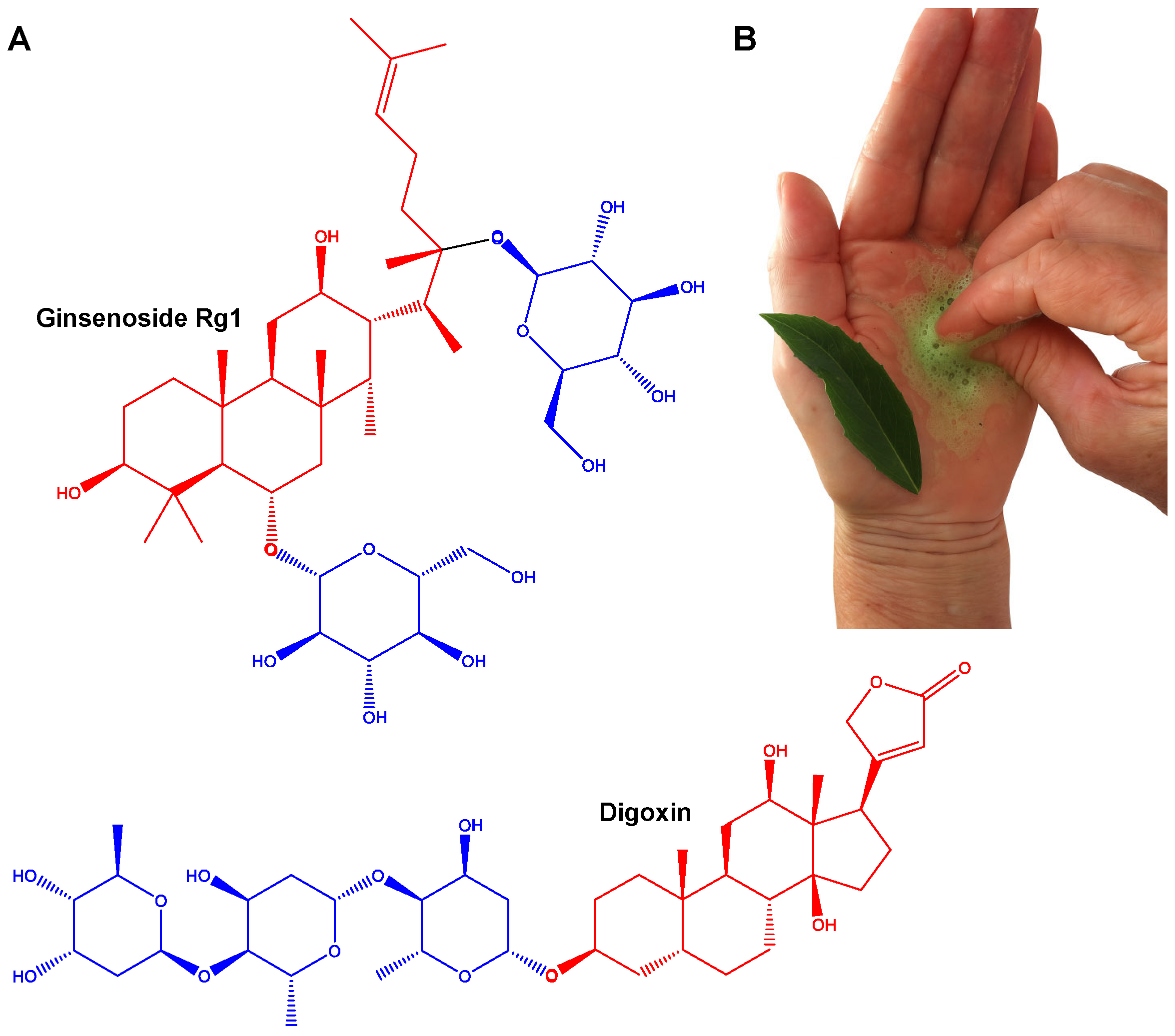
Figure 1. Chemical structures of representative triterpenoid and steroidal saponins. (A) The structural diversity of saponins, highlighting two main classes, triterpenoid saponins (e.g., ginsenoside Rg1) and steroidal saponins (e.g., digoxin). In each molecule, the aglycone (hydrophobic) is shown in red, while the sugar moieties (hydrophilic) are shown in blue. Digoxin is a cardiac glycoside derived from Digitalis lanata Ehrh. that is clinically used to treat heart failure and atrial fibrillation. Ginsenoside Rb1, isolated from Panax ginseng C.A.Mey., is a triterpenoid saponin commonly found in herbal formulations with reported immunomodulatory, neuroprotective, and anti-inflammatory effects. (B) Foam formed after crushing the leaves of Q. brasiliensis (“pau-de-sabão”, which means soap tree) in water (Photo: Iracema Rubas Cibulski).
Pharmaceutics 17 00966 g001
Figure 2. The QS-21 chemical structure. QS-21 is a complex triterpenoid saponin isolated from Quillaja saponaria bark that is characterized by four distinct structural domains. The molecule features a central lipophilic triterpene core (quillaic acid), which is flanked by a branched trisaccharide attached at the C3 position and a linear tetrasaccharide linked to the C28. Additionally, QS-21 contains a glycosylated pseudodimeric acyl chain (Fa-Ara) esterified to the fucose residue, contributing to its amphiphilic properties and adjuvant activity. QS-21 is as a mixture of two isomeric forms, QS-21-Api (65%) and QS-21-Xyl (35%), which differ only in the terminal sugar of the tetrasaccharide (apiose or xylose, respectively).
Pharmaceutics 17 00966 g002
Figure 5. General structure of saponins from Q. saponaria and Q. brasiliensis. Saponins of Quillajaceae contain a triterpenic aglycone, most frequently quillaic acid, and are glycosylated at the C-3 and C-28 positions of the aglycone.
Pharmaceutics 17 00966 g005
Figure 6. Preparation and ultrastructural visualization of Q. brasiliensis-derived ISCOM-matrices. On the left side, there is an illustrative scheme of the process involved in obtention of the raw aqueous extract (AE) of Q. brasiliensis leaves and the reverse-phase chromatography (RP-C18) used to obtain a saponin-rich fraction. The formulation of ISCOMs involves the combination of the saponin-rich fraction with cholesterol and phospholipids, resulting in nanostructures as visualized by transmission electron microscopy (TEM) in the images on the right. For these images, ISCOM-matrices were placed on formvar carbon grids (300 mesh) and negatively stained with 2% uranyl acetate and examined with a JEM-2100 TEM operated at an 200 kV accelerating voltage [198]. Scale bars: 200 nm (middle) and 100 nm (right).
Pharmaceutics 17 00966 g006
Disclaimer/Publisher’s Note: The statements, opinions and data contained in all publications are solely those of the individual author(s) and contributor(s) and not of MDPI and/or the editor(s). MDPI and/or the editor(s) disclaim responsibility for any injury to people or property resulting from any ideas, methods, instructions or products referred to in the content.