Article

Trans-p-Coumaryl Alcohol as a Bioactive Compound and Anti-Inflammatory Agent in Wannachawee Recipe for Psoriasis

by
Supreeya Tantipat
1,
Kongkiat Trisuwan
2,3,
Phraepakaporn Kunnaja
4,
Seewaboon Sireeratawong
5,6,
Surapol Natakankitkul
1,
Surasak Imiam
7 and
Sunee Chansakaow
1,6,*
1
Department of Pharmaceutical Sciences, Faculty of Pharmacy, Chiang Mai University, Chiang Mai 50200, Thailand
2
Department of Chemistry, Faculty of Science, Chiang Mai University, Chiang Mai 50200, Thailand
3
Center of Excellence for Innovation in Chemistry, Chiang Mai University, Chiang Mai 50200, Thailand
4
Department of Medical Technology, Faculty of Associated Medical Sciences, Chiang Mai University, Chiang Mai 50200, Thailand
5
Department of Pharmacology, Faculty of Medicine, Chiang Mai University, Chiang Mai 50200, Thailand
6
Clinical Research Center for Food and Herbal Product Trials and Development (CR-FAH), Faculty of Medicine, Chiang Mai University, Chiang Mai 50200, Thailand
7
Department of Thai Traditional Medicine, Phrapokklao Hospital, Chanthaburi 22000, Thailand
*
Author to whom correspondence should be addressed.
Pharmaceutics 2025, 17(7), 864; https://doi.org/10.3390/pharmaceutics17070864
Submission received: 20 April 2025 / Revised: 24 June 2025 / Accepted: 27 June 2025 / Published: 30 June 2025
(This article belongs to the Special Issue Natural Pharmaceuticals Focused on Anti-inflammatory Activities)

Abstract

Background/Objectives: Wannachawee recipe (WCR) has been listed in the Hospital Traditional Medicine Formulary and has been used as a Thai medicine to treat psoriasis in the Thai Traditional Medicine Clinic of Prapokklao Hospital since 2006. Previous reports have found that WCR demonstrates good results for the treatment of patients with psoriasis. Among 136 Thai psoriasis patients who received WCR, 92.80% responded well. Although WCR is effective, there is still a lack of scientific data, especially relating to the bioactive compound in WCR. Therefore, this study aims to evaluate the phytochemicals in WCR via bioassay-guided isolation. Methods: In this study, the WCR was extracted via decoction with water, in a process based on traditional Thai medicine. The water extract was concentrated and dried using a spray dryer. The crude water extract was isolated using the partition technique with organic solvents, namely petroleum ether and ethyl acetate. These fractions were then separated and tested for anti-inflammatory activity using the bioassay-guided fractionation method. Results: Two particular types of pro-inflammatory cytokines are involved in inflammation and are among the factors that cause psoriasis—TNF-α and IL-6. Thus, we evaluated the isolated samples in terms of anti-inflammatory activity. The isolation resulted in two pure compounds—p-coumaryl aldehyde and trans-p-coumaryl alcohol. In the efficacy test of the isolated compounds, compared to the standard indomethacin at the same concentration of 12.5 ug/mL, trans-p-coumaryl alcohol was found to have the best efficacy, inhibiting TNF-α by 29.28% and IL-6 by 36.75%, with the standard compound showing inhibitions rates of 15.80% for TNF-α and 27.44% for IL-6. Conclusions: This study is the first report to identify the bioactive compound of WCR as trans-p-coumaryl alcohol or 4-hydroxycinnamyl alcohol.

1. Introduction

Psoriasis is a persistent inflammatory autoimmune skin disorder that affects approximately 2–5% of the global population. It is increasingly recognized for its systemic impact and comorbidities [1,2]. The condition is characterized by erythematous plaques with silver-white scales and is associated with reduced quality of life and increased risk of cardiovascular diseases, malignancies, and depression [2,3] The large-scale global study assessed psoriasis prevalence, healthcare access, and quality of life impacts using consistent physician-confirmed diagnoses across 20 countries covering over 50% of the world’s population. It found that Global prevalence of psoriasis is 4.4%, with East Asia having the highest (5.7%) and Africa the lowest (1.7%). Contrary to prior reports, Asia shows higher prevalence, and globally, men have slightly higher prevalence than women. Many patients, especially in North America, do not consult healthcare professionals, with dermatologist visits being lowest in Australia and North America, likely due to higher healthcare costs. Younger patients report more embarrassment and lifestyle limitations due to psoriasis, including avoiding social and professional activities. [4]. Psoriasis is becoming an increasingly significant health issue in Thailand, with over 20,000 outpatient visits recorded in 2024, making it the country’s third most prevalent dermatological condition. An estimated 1.7 million individuals are affected, and about 30% of them experience severe symptoms that often require advanced therapeutic approaches beyond conventional treatments. The average treatment cost per visit is approximately 3100 THB [5]. Standard treatments include topical corticosteroids, phototherapy, and systemic immunomodulators; however, many of these therapies are limited by significant side effects and high costs [6]. As a result, there is a growing interest in traditional medicine-based therapies, such as the Wannachawee recipe (WCR), which has demonstrated high clinical response rates and is used in various Thai traditional medicine [7,8] Wannachawee (WCR), has been used to treat psoriasis since 2006 in the Thai Traditional Medicine Clinic at Prapokklao Hospital. In a study of 136 Thai patients, 92.8% showed improvement following WCR treatment. WCR is composed of eight Thai herbs including Kha (Alpinia galanga (L.) Willd.), Khao Yen Tai (Smilax glabra Wall.ex Roxb.), Khao Yen Nuea (Smilax corbularia Kunth.), Khao Yen Jeen (Smilax sp.), Hua Ta Pead (Stemona involuta Inthachub.), Non Tai Yak (Stemona collinsae Craib.), Thong Pan Chang (Rhinacanthus nasutus (L.) Kurz.), and Ngueak plaamo (Acanthus ilicifolius L.) [8]. The herbs in WCR possess pharmacological activities that align with psoriasis symptoms, including anti-inflammatory, anti-proliferative, anti-angiogenic, antioxidant, and anti-tumor activities [9,10,11]. The pathophysiology of psoriasis consists of two phases. In the early stages of psoriasis pathogenesis, an initial trigger such as skin injury, infection, stress, etc., activates innate immune cells, including plasmacytoid dendritic cells (pDCs), natural killer cells, and keratinocytes. These activated cells release cytokines like interferon-alpha (IFN-α), tumor necrosis factor-alpha (TNF-α), interleukin-1 beta (IL-1β), and interleukin-6 (IL-6) which stimulate myeloid dendritic cells (mDCs). The mDCs then migrate to regional lymph nodes, where they present antigens and secrete IL-12 and IL-23, promoting the differentiation of naive T cells into T helper 17 (Th17) and T helper 1 (Th1) cells. The effector T cells eventually migrate to the skin tissue, releasing cytokines such as IL-17A, IL-17F, IL-22, IFN-γ, and TNF-α that activate keratinocytes. The activated keratinocytes proliferate and produce antimicrobial peptides, pro-inflammatory cytokines (IL-1β, TNF-α, IL-6), and chemokines, perpetuating a feedback loop that sustains chronic inflammation and leads to plaque formation. This cycle involves additional interactions with fibroblasts and endothelial cells, contributing to the ongoing immunopathological progression of psoriasis [3,12,13]. Plaque psoriasis accounts for roughly 80% to 90% of all psoriasis cases [14]. It is marked by an inflammatory pathway involving TNF-α, IL-23, and Th17 cells. Cytokines like IL-6, IL-1β, and IL-23 are key in activating Th17 cells, which contribute to the development of chronic inflammation and autoimmunity [3]. Another study on IL-6 states that serum IL-6 levels are considered a marker of inflammatory activity in psoriasis and a useful indicator of treatment response [15]. IL-6 in particular has emerged as both a biomarker for disease activity and a potential therapeutic target [16]. Clinical studies have reported that patients with psoriasis exhibited significantly elevated serum levels of TNF-α, averaging 362.67 ± 456.9 pg/mL, compared to 14.64 ± 5.78 pg/mL in healthy controls [17]. In patients with psoriasis, serum IL-6 levels ranged from 7 to 183 pg/mL, with a mean of 61.26 ± 57.40 pg/mL. In contrast, healthy controls exhibited a mean IL-6 concentration of 2.38 pg/mL [17]. This substantial difference was found to be highly statistically significant (p < 0.001), underscoring TNF-α and IL-6′s role as key pro-inflammatory cytokines in the pathogenesis of psoriasis. Based on these data and available resources, we chose to investigate the anti-inflammatory activity against pro-inflammatory cytokines TNF-α and IL-6. Despite the widespread use of Wannachawee recipe (WCR) in psoriasis treatment, scientific evidence supporting its efficacy, especially in terms of identifying active anti-inflammatory compounds, remains limited. There is also a lack of standardized chemical quality control for this traditional formulation, which poses challenges for broader acceptance and consistent clinical application.

2. Materials and Methods

2.1. Chemicals

Acetonitrile of HPLC grade, chloroform, deionized water (HDPE), ethyl acetate, hexane, methanol, and sulfuric acid of AR grade were purchased from RCI Labscan (V.S. CHEM HOUSE, Bangkok, Thailand). Celite was obtained from the Fluka Company (SIGMA-ALDRICH, Singapore). TLC Silica gel 60 F254, the aluminum support, and silica gel were acquired from Merck (Merck KGaA, Darmstadt, Germany).

2.2. Plant Materials

All crude drugs included in WCR were sourced from Prapokklao Hospital in Chanthaburi Province, Thailand. The plant materials were identified by comparison with the authentic samples found in a botanical herbarium at the Faculty of Pharmacy, Chiang Mai University or referenced in official manuscripts/monographs/previous report [12]. Details are presented in Table 1.
The samples were then cleaned, reduced in size by coarsely grinding them, and dried at 50 °C in a hot air oven (Scientific Promotion, Bangkok, Thailand) for 2 h.

2.3. Sample Preparation

The extraction process utilized traditional methods, specifically boiling herbs in water [14]. The coarse powder of WCR was extracted through decoction for 30 min, using water as the solvent (640 g WCR extract per 5L water; temperature 80–100 °C). The resulting water extract was filtered via steel grating and was concentrated to achieve a Brix value of 3–5, followed by drying using a spray dryer (BUCHI, Bangkok, Thailand).

2.4. Bioassay-Guided Isolation with Anti-Inflammation Analysis

The extract of WCR was isolated by column chromatography (BECTHAI Bangkok Equipment & Chemical, Bangkok, Thailand). The bioactive compound(s) were investigated following bioassay-guided fractionation.

2.4.1. Separation Technique

Wannachawee extract was separated using column chromatography (CC) with silica gel as the stationary phase and a gradient mobile phase. The resulting fractions were examined by thin-layer chromatography (TLC) to identify and group similar chemical patterns. Subsequently, the anti-inflammatory activity of the fractions was tested. The active fractions were re-chromatographed by column chromatography and prep-TLC to obtain pure compound(s). A marker was selected from the bioactive compounds based on its high potency and significant concentration in the extract.

2.4.2. Anti-Inflammatory Activity Test [18,19]

  • Cell used and Cell Culture
The RAW 264.7 macrophage cell line was chosen as the primary in vitro model because it is widely used to study the inflammatory cascade, particularly in response to LPS stimulation. It produces pro-inflammatory cytokines such as TNF-α and IL-6, which are central to early-stage inflammation and are well-established biomarkers in initial anti-inflammatory screenings, including psoriasis-related studies [20]. The cells were maintained in Dulbecco’s Modified Eagle’s Medium (DMEM; GIBCO™, Thermo Fisher Scientific, MA, USA) supplemented with 10% heat-inactivated fetal bovine serum (GIBCO™, Thermo Fisher Scientific, Waltham, MA, USA), 100 units/mL penicillin, and 100 μg/mL streptomycin (GIBCO™, Thermo Fisher Scientific, MA, USA). Cultures were kept in a CO2 incubator at 37 °C with 5% CO2.
2.
Cytotoxicity Assay with Sulforhodamine B (SRB Assay)
RAW 264.7 cells (5 × 10⁴ cells/well) were seeded in a 96-well plate and incubated overnight at 37 °C in a 5% CO2 incubator. WCR extracts, prepared in various concentrations and dissolved in DMSO, were added to the cells which were then cultured for an additional 24 h. After this incubation, 10% trichloroacetic acid was added to fix the cells for 1 h at 4 °C. The cells were subsequently stained with 0.057% SRB for 30 min. The plate was washed with 1% acetic acid to remove excess dye and allowed to air dry at room temperature. To dissolve the protein-bound dye, Tris-base solution (10 mM) was added, and absorbance was measured at 510 nm using an ELISA plate reader. At least three concentrations were tested, and the final DMSO concentration was maintained below 0.1%. All experiments were conducted in triplicate and repeated in three independent experiments. Data were analyzed using GraphPad Prism version 9.0 to calculate means and standard errors.
3.
Studying the effect of the extracts on the inhibition of TNF-α and IL-6
RAW 264.7 cells were cultured in a 96-well plate (5 × 10⁴ per well). Cells were incubated at 37 °C for 24 h. Then, three concentrations of herbal extracts were added to the well and incubated for 1 h. Lipopolysaccharide (Sigma-Aldrich, St. Louis, MI, USA) was added at a final concentration of 1 µg/mL to stimulate the cells. The cells were incubated for another 24 h. At the end of the incubation, culture supernatants were aspirated to quantify TNF-α and IL-6 by using ELISA (BioLegend, San Diego, CA, USA), as specified in the test kit. In this trial, indomethacin was used as the reference drug.
4.
Statistical Analysis
All results are presented as the mean ± standard error of the mean (SEM) from three independent experiments. Statistical comparisons between groups were analyzed using one-way analysis of variance (ANOVA), followed by Tukey’s post hoc test for multiple comparisons. A p-value of less than 0.05 was considered statistically significant. The kurtosis statistic was used to assess the normality of the data distribution, where values within ±3 were considered acceptable for applying parametric tests. All analyses were performed using GraphPad Prism version 9.0.

2.5. Identification of Chemical Compounds in WCR Extract by HPLC

The Shimadzu Prominence High-Performance Liquid Chromatography system (SpectraLab Scientific Inc., Markham, ON, Canada), comprising a DGU-20A3R degasser, 2x LC-20AD liquid chromatographs, an SIL-20A auto-sampler, a CTD-20A column oven, and a SPD-20A UV-Visible detector, was used. The column used was a Mightysil, RP-C18 GP ( 250 × 4.6   m m , 5   µ m ) equipped with a guard column. The mobile phase was a gradient prepared from 10% (v/v) acetonitrile (component A) and formic acid in HPLC-grade water (component B), as detailed in Table 2, with a flow rate of 1.0 mL/min. The absorption wavelength was selected at 254 nm. The column was operated at 25 °C. The sample injection volume was 20 µ L.

2.6. Structure Elucidation by Spectroscopy

The structure of the isolated compound was elucidated using a Bruker NEOTM 500 MHz NMR spectrometer by measuring 1H NMR, 13C NMR, COSY, HMQC, and HMBC. Structure analysis was performed with MesReNova.

3. Results

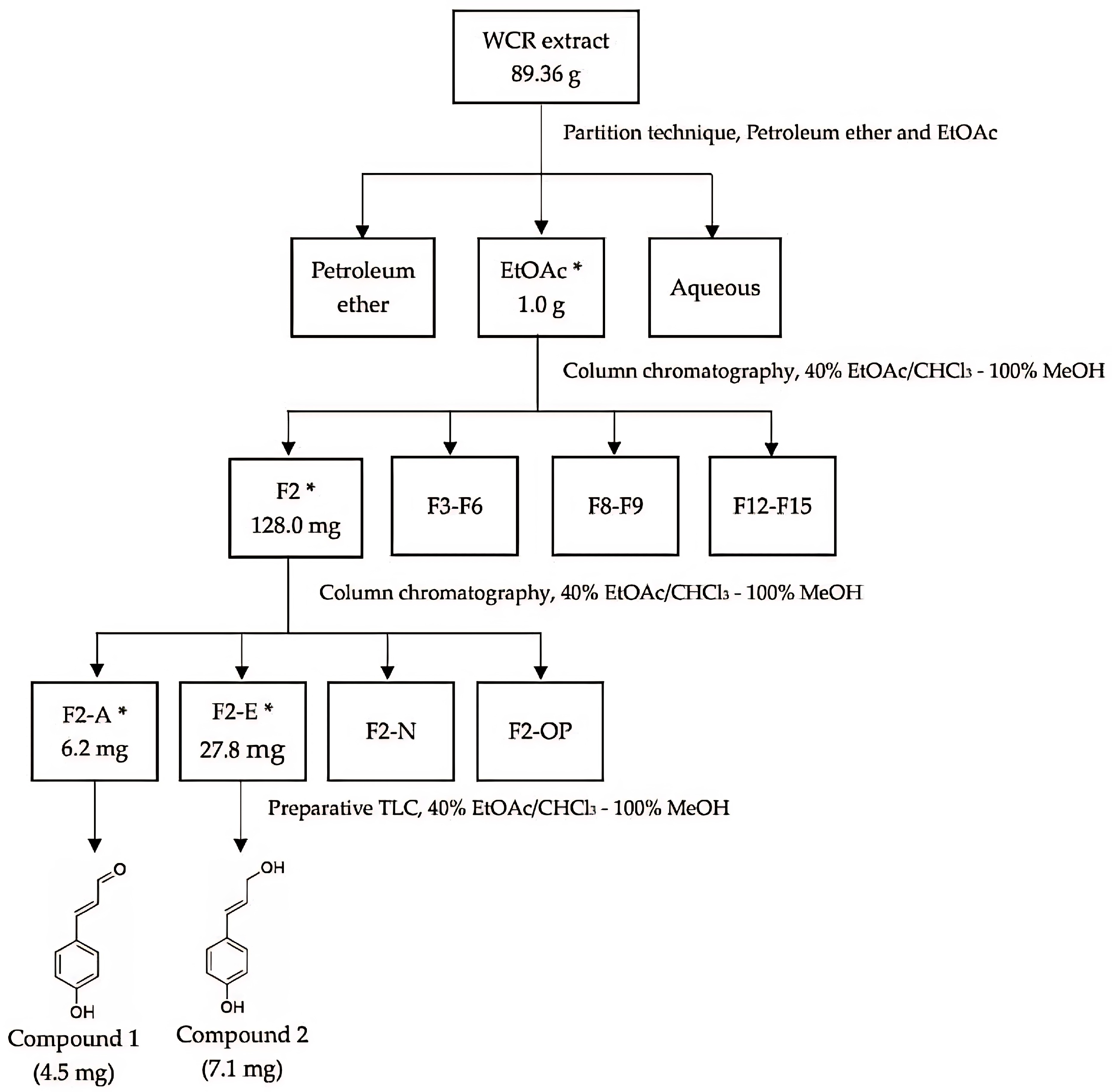
3.1. Preparation of the Extract

The extraction process followed the traditional method, which is known as decoction. The total volume obtained was subsequently processed using spray drying. The results are presented in Table 3.

3.2. Bioassay-Guided Fractionations

3.2.1. Separation Results

The crude extract of WCR was isolated using a partition technique, resulting in two fractions: ethyl acetate (EtOAc) and an aqueous fraction. The EtOAc fraction demonstrated the most potent activity. At a concentration of 3.125 µg/mL, the EtOAc fraction reduced the levels of TNF-α and IL-6 to 5555.34 ± 372.59 pg/mL pg/mL and 3765.62 ± 504.73 pg/mL, respectively. In comparison, the standard drug indomethacin, administered at a dose of 50 µg/mL, inhibited TNF-α to 4035.67 ± 191.57 pg/mL and IL-6 to 3543.95 ± 204.87 pg/mL. The EtOAc fraction was then subjected to column chromatography, yielding 15 subfractions. It was observed that F2 and F3 fractions, at the highest concentration, caused a slight decrease in TNF-α levels (Figure 1A). Meanwhile, F2 and F3 fractions at all concentrations resulted in a statistically significant decrease in IL-6 levels. Notably, cells incubated with the F2 fraction at all concentrations decreased the amount of IL-6 more effectively than those incubated with indomethacin (Figure 1B).
Then, F2 was re-chromatographed to afford 17 subfractions (F2A-F2P). At the highest concentrations of fractions F2A, F2E, and the EtOAc extract, the levels of TNF-α were measured at only 2215 ± 153 pg/mL, 2600 ± 363 pg/mL, and 2611 ± 446 pg/mL, respectively, after incubating the cells with the herbal extract. These results align with a corresponding decrease in IL-6 levels. It was observed that at the highest concentrations, F2A, F2E, and the EtOAc extract were the most effective in reducing IL-6 levels (Table 4).
Additionally, fractions F2A and F2E were separated using preparative thin-layer chromatography (Prep-TLC), resulting in two pure compounds, as shown in Figure 2, i.e., p-coumaryl aldehyde and trans-p-coumaryl alcohol.

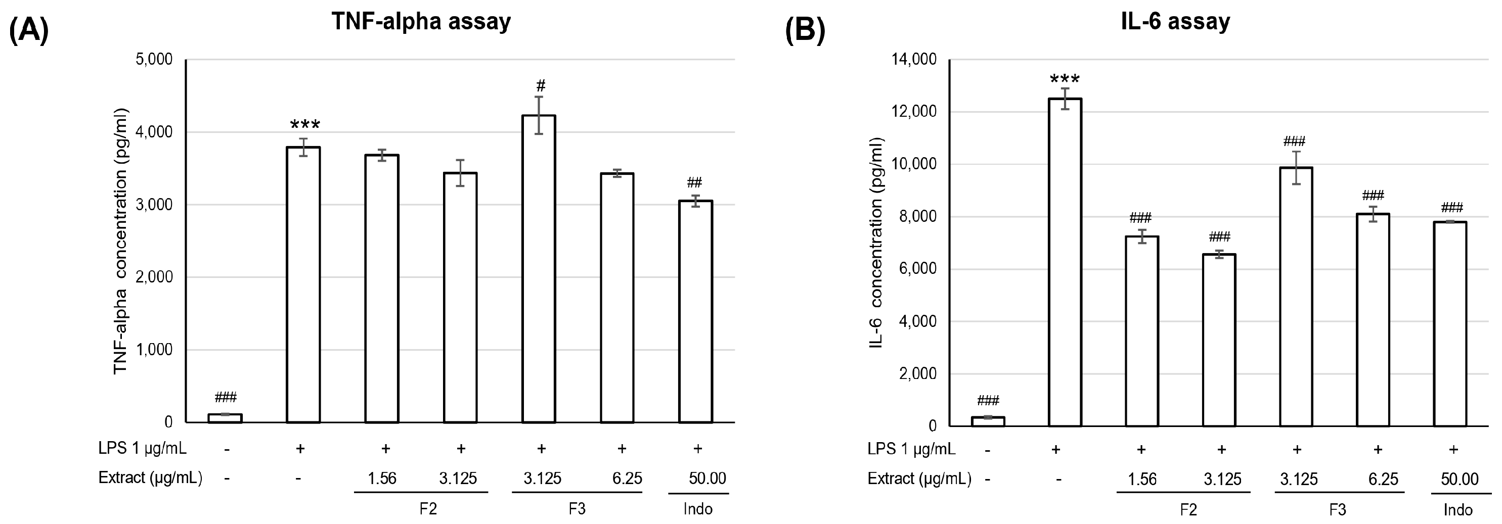
3.2.2. Anti-Inflammatory Activity
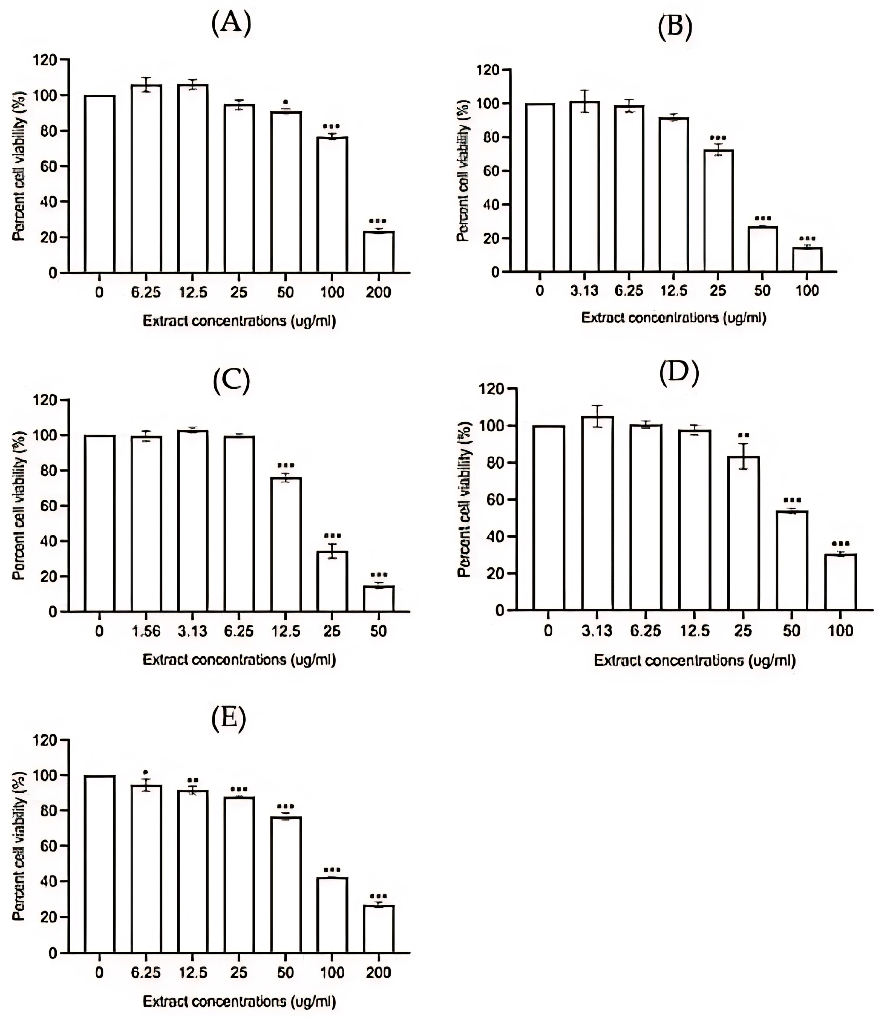
  • Cytotoxicity assay
The isolated compounds, the EtOAc fraction, and F2 were selected to assess their anti-inflammatory activity in RAW 264.7 cells. The cytotoxicity assay demonstrated significant cell death at 24 h after exposure to various concentrations of the test extracts. Noticeable cell death was observed at the following concentrations: compound 1 at 50–200 µg/mL (Figure 3A), compound 2 and the EtOAc extract at 25–100 µg/mL (Figure 3B,D), and F2 at 12.5–50 µg/mL (Figure 3C). For indomethacin, a standard anti-inflammatory drug, statistically significant cell death occurred at concentrations of 6.25 µg/mL or higher (Figure 3E). The cytotoxicity exhibited a clear concentration-dependent pattern.
2.
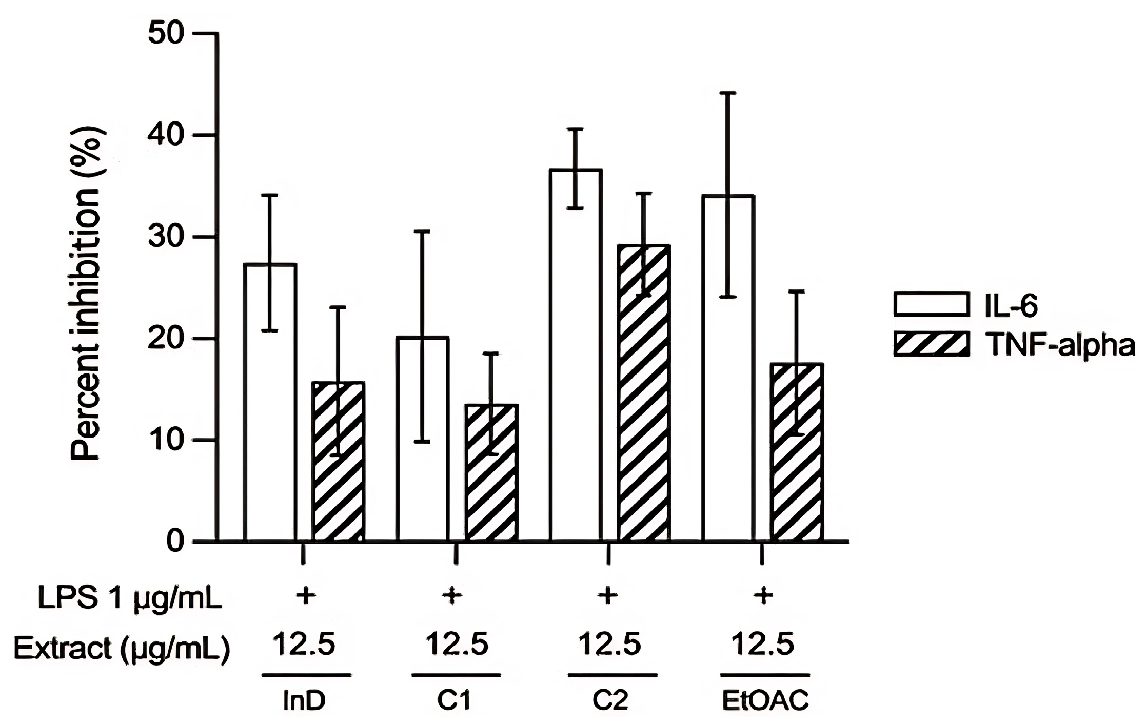
Effect of extracts on inhibition of TNF-α and IL-6
The anti-inflammatory activity of the pure compounds and fractions was assessed in LPS-stimulated RAW 264.7 cells by measuring the level of TNF-α and IL-6 using the ELISA kit. The results indicate that after LPS stimulation, cells produced significantly higher levels of TNF-α and IL-6, with concentrations of 19,200 ± 2402 and 42,257 ± 1623 pg/mL, respectively, compared to unstimulated cells. When the extracts were added at various concentrations to assess their anti-inflammatory activity, both pure compound 1 and pure compound 2 significantly reduced TNF-α levels, as shown in Figure 4A. Furthermore, pure compound 1, pure compound 2, and the EtOAc extract also significantly decreased IL-6 levels, as shown in Figure 4B. The anti-inflammatory effects of these test compounds were comparable to those of indomethacin, which is the positive control used in this study.
When comparing the anti-inflammatory activity of the test substances at the same concentration (12.5 µg/mL), it was found that pure compound 2 and the EtOAc extract exhibited better inhibition of inflammation than indomethacin, with consistent results for both TNF-α and IL-6 inhibition, as shown in Figure 5. The most effective extract in inhibiting inflammation was pure compound 2, which inhibited TNF-α by 29.28 ± 5.02% and IL-6 by 36.75 ± 3.88%. This was followed by EtOAc, which inhibited TNF-α by 17.59 ± 7.05% and IL-6 by 34.16 ± 10.04%, and then pure compound 1, which inhibited TNF-α by 13.59 ± 4.95% and IL-6 by 20.24 ± 10.34%.

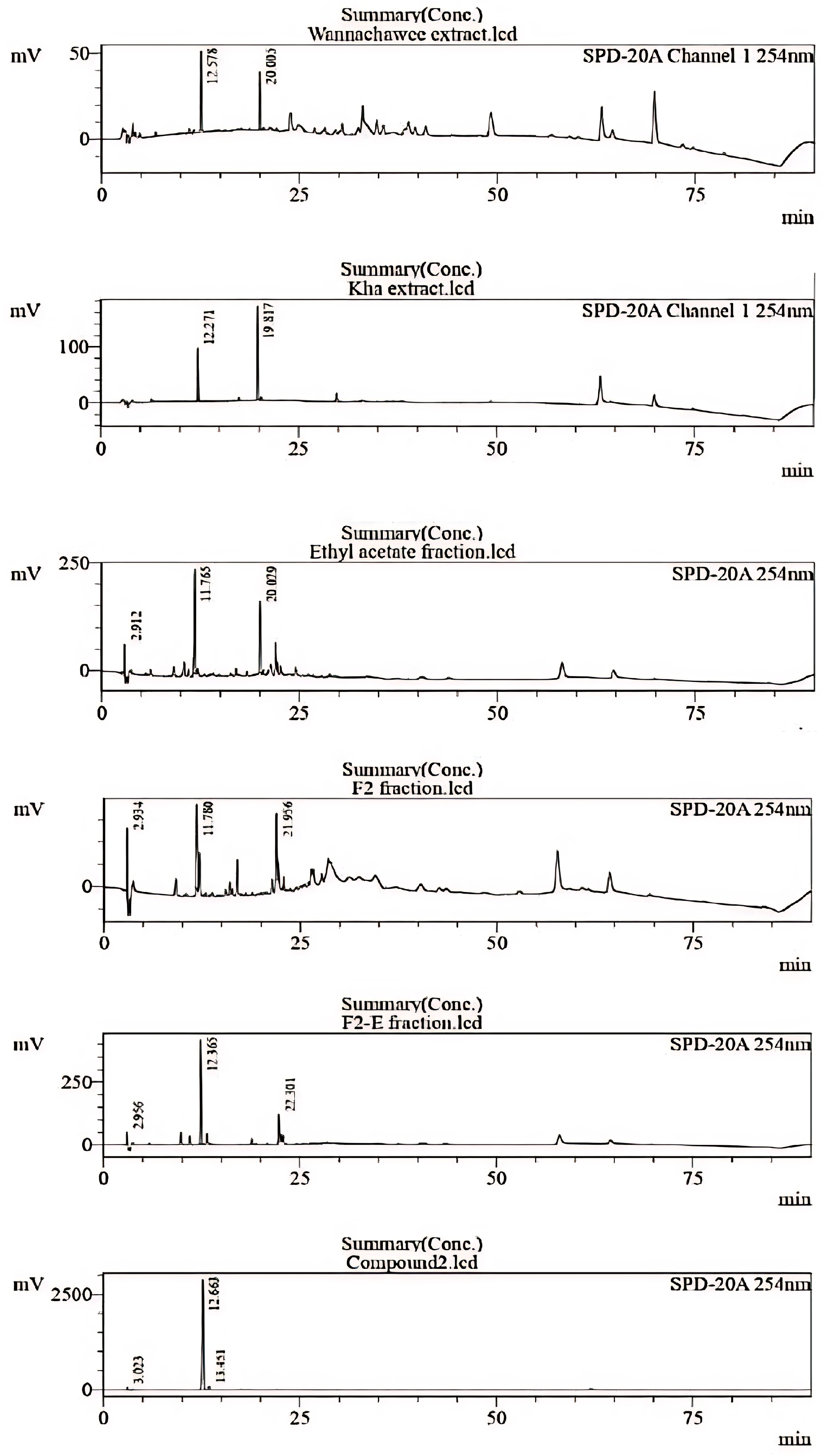
3.3. Chemical Identification of WCR Recipe by HPLC

HPLC is an effective method for ensuring the chemical quality of herbal extracts. Comparing the chemical constituents in the WCR extract with each herb and other bioactive fractions revealed interesting peaks at specific time points (Rt = 12, 20 min). The chromatograms of the Kha extract displayed the same Rt with the main compound in WCR recipe. Thus, this crude extract was separated. The chromatograms of the Kha (A. galanga) extract, EtOAc fraction, F2 fraction, F2-E fraction, and compound 2 are shown in Figure 6. The anti-inflammatory activity was primarily attributed to the ethyl acetate fraction. After purification, fraction F2 gave two pure compounds. One of these was identified as trans-p-coumaryl alcohol (Rt = 12 min), which demonstrated potent activity and was the predominant compound.

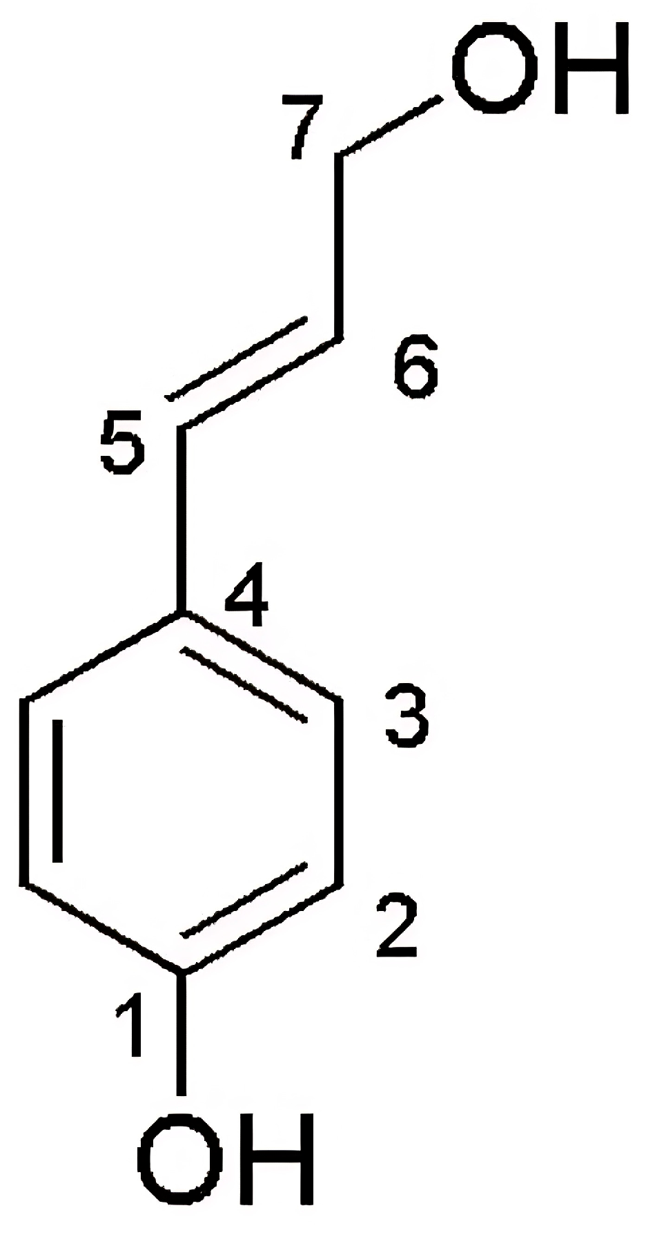
3.4. Results of Structure Elucidation

Regarding its physical characteristics, compound 1 (4.5 mg) is a brown powder. The structural elucidation of compound 1 was performed using a nuclear magnetic resonance spectrometer (NMR) for 1H NMR and 13C NMR data, as shown in Table 5 and Figure 7.
The 1H-NMR spectrum for compound 1 shows one signal of aldehyde proton [δ8.54 (s, 1H)], two signals of p-disubstituted aromatic protons [δ7.24 (d, 8.5 Hz, 2H)] and [δ6.73 (d, 8.5 Hz, 2H)], and two signals of trans-olefinic proton [δ 6.52 (d, 16.0 Hz, 1H)] and [δ 6.11 (dt, 15.5, 6.5 Hz, 1H)]. The 13C-NMR spectrum shows six signals of eight carbons including one signal of aldehyde carbon [δ200.08 (C-7)], two signals of quaternary carbon [δ180.3 (C-4) and 159.3 (C-1)], two signals of four aromatic methine carbons [δ128.8 (2C) and 116.4 (2C)], and two signals of olefinic methine carbon [δ133.9 (C-5) and 122.4 (C-6)]. According to the Scifinder database, compound 1 is a structurally reported substance called p-coumaryl aldehyde [22] or p-hydroxycinnamaldehyde [23].
Table 5. 1H and 13C-NMR data of compound 1 (Methanol-d4, 1H NMR 500 MHz, 13C NMR 125 MHz).
PositionCompound 1p-Coumaryl Aldehyde
δH (mult., J in Hz)δc, multδH (mult., J in Hz) of Ref. [23]
1-159.3, C-
26.73 (d, 8.5, 2H)116.4, CH6.72 (d, 8.5, 2H)
37.27 (d, 8.5, 2H)128.8, CH7.24 (d, 8.5, 2H)
4-180.3, C-
56.50 (d, 16.0, 1H)133.9, CH6.50 (d, 15.9, 1H)
66.11 (dt, 15.5, 6.5, 1H)122.4, CH6.16 (dt, 15.9, 6.0, 1H)
78.54 (s, 1H)200.08, CHO9.56 (d, 7.9, 1H)
s = singlet; d = doublet; dd = double of doublet; dt = double of triplet.
Regarding its physical characteristics, compound 2 (7.1 mg) is a brown powder. The structural elucidation of compound 2 was performed using a nuclear magnetic resonance spectrometer (NMR) for 1H NMR and 13C NMR data, as shown in Table 6 and Figure 8.
The 1H-NMR spectrum shows two signals of p-disubstituted aromatic protons [δ7.27 (d, 8.5 Hz, 2H)] and [δ6.72 (d, 8.5 Hz, 2H)], two signals of trans-olefinic proton [δ6.50 (d, 16.0 Hz, 1H)] and [δ6.16 (dt, 16.0, 6.0 Hz, 1H)], and one signal of methylene protons [δ4.18 (dd, 6.0, 1.5 Hz, 2H)]. The 13C-NMR spectrum data show that compound 2 has seven signals of nine carbons including two signals of quaternary carbon [δ158.2 (C-1) and 129.9 (C-4)], two signals of four aromatic methine carbons [δ128.6 (2C) and 116.3 (2C)], two signals of olefinic methine carbon [δ131.8 (C-5) and 126.6 (C-6)], and one signal of methylene carbon [δ63.9 (C-7)]. According to the Scifinder database, compound 2 is a structurally reported substance called trans p-coumarin alcohol [24] or 4-hydroxycinnamyl alcohol [25].

4. Discussion

Wannachawee recipe (WCR) is a traditional Thai medicine that has been used for generations to treat psoriasis. Since 2006, WCR has been utilized at the Thai Traditional Medicine Clinic of Prapokklao Hospital. This initiative is the result of collaboration between practitioners of modern medicine and those of traditional Thai medicine. In a study of 136 Thai psoriasis patients receiving WCR, 92.80% showed a good response [7,8]. Psoriasis is an immune-mediated chronic inflammatory skin disease [26]. There is evidence that inflammation drives immune and metabolic changes that exacerbate psoriasis [27]. One of the characteristics of chronic inflammation is the production of inflammatory cytokines [28]. TNF-α and IL-6 are the important cytokines involved in inflammation in psoriasis [3,14,15]. A previous report showed that TNF-α and IL-6 levels are higher in psoriasis patients compared to healthy individuals [17]. Therefore, the levels of these two cytokines are associated with psoriasis, making them reasonable markers for screening. Although WCR is continually used, there is still limited information available regarding chemical quality control. In our research, we aimed to study the anti-inflammatory properties of the bioactive compounds which can support biomarker selection in quality control processes and traditional Thai applications.
All crude drugs listed in WCR were obtained from Prapokklao Hospital in Chanthaburi Province, Thailand. The crude drugs used in this study are commonly utilized in hospitals. Identification was conducted by a Thai practitioner who specializes in Thai herbal medicine, as well as through comparison with authentic samples from a botanical herbarium or descriptions in manuscripts, monographs, or previous reports [29]. Traditional use of WCR involves extracting water through boiling. The decoction process is employed to align with traditional medicine, yielding 14.34% w/w. Bioassay-guided isolation was employed to identify the bioactive compound(s). Trans-p-coumaryl alcohol and p-coumaryl aldehyde were identified as the bioactive compounds.
Previous reports have indicated that these two compounds were isolated from the rhizomes and fruits of A. galanga in the ethyl acetate (EtOAc) fraction using reversed-phase ODS column chromatography [30,31,32,33]. Additionally, it was noted that p-coumaryl aldehyde was isolated from the 95% ethanol and hot water fractions of A. galanga using column chromatography [34]. In a study on the inhibition of melanogenesis in B16 melanoma 4A5 cells induced by theophylline, p-coumaryl aldehyde had an IC50 value of 18.3 µM, while trans-p-coumaryl alcohol had an IC50 value > 100 µM [34]. Both substances are effective neuroprotective agents due to their good permeability [34]. p-Coumaryl aldehyde has also been reported in Sarcophyta sanguinea [22]. It has also been reported to exhibit antimicrobial activity [22], while trans-p-coumaryl alcohol is a component of A. officinarum [33] and Malus domestica [35]. Previous research reports have shown that this compound demonstrates antioxidant activity by inhibiting the autoxidation of methyl linoleate in the bulk phase [35]. The two compounds identified in the extract of A. galanga align with the HPLC chromatograms. The peak for trans-p-coumaryl alcohol, which is a major component, appears at 12 min, corresponding to the A. galanga extract (see Figure 6). Additionally, p-coumaryl aldehyde is present as a minor compound, observed at 25 min. It has been established that trans-p-coumaryl alcohol is one of the major ingredients in the A. galanga water extract. In previous reports, three compounds from the genus Alpinia, namely (1E,6E)-1,7-Bis(4-hydroxy-3-methoxyphenyl)hepta-1,6 diene-3,5-dione, (−)-alphainanins A, and (−)-alphainanins C, exhibited anti-inflammatory mechanisms by inhibiting the production of TNF-α, IL-6, NO, IL-1β, and NO via the NF-kB signaling pathway in RAW264.7 cells upon inflammatory stimulation with LPS [33]. This indicates that the extract from A. galanga may also have anti-inflammation activity. Due to its potency and high concentration in the WCR extract, trans-p-coumaryl alcohol was chosen as a biomarker. The HPLC technique is an effective method for assessing the quality of extracts and products, including the monitoring of their stability through their biomarker.
In this study, the focus was on early-stage anti-inflammatory screening, specifically targeting immune-mediated pathways relevant to psoriasis pathogenesis. The RAW 264.7 macrophage cell line was selected as an appropriate in vitro model for this phase due to its well-established role in inflammation research. Upon stimulation with lipopolysaccharide (LPS), RAW 264.7 cells reliably produce key pro-inflammatory cytokines, making this model a widely accepted and reproducible platform for initial screening of anti-inflammatory activity. This study shows that trans-p-coumaryl alcohol and p-coumaryl aldehyde exerted anti-inflammatory effects. Trans-p-coumaryl alcohol exhibited anti-inflammatory properties by inhibiting TNF-α and IL-6 in RAW 264.7 macrophage cells. Trans-p-coumaryl alcohol demonstrated greater activity than the reference drug, indomethacin, while p-coumaryl aldehyde exhibited lower activity at the same concentration. Indomethacin was selected in this study as a standard reference due to its well-characterized anti-inflammatory properties. It is widely employed in in vitro inflammation models, particularly with LPS-stimulated RAW 264.7 macrophage cells, to benchmark the inhibition of pro-inflammatory cytokines such as TNF-α and IL-6. These cytokines play central roles in the pathogenesis of psoriasis [10]. Therefore, indomethacin was used as a positive control to compare the anti-inflammatory efficacy of WCR extracts and isolated compounds under standardized conditions.
To evaluate the therapeutic potential of the isolated compounds, we compared their cytotoxicity and anti-inflammatory efficacy. Trans-p-coumaryl alcohol significantly inhibited TNF-α and IL-6 production at 12.5 µg/mL, while cytotoxicity was observed at concentrations ≥25 µg/mL, with an estimated IC50 of approximately 50 µg/mL. This yields a preliminary therapeutic index (TI) of about 4, suggesting a favorable safety margin in this in vitro setting. A similar pattern was observed for the EtOAc fraction, whereas p-coumaryl aldehyde exhibited a narrower margin.
Trans-p-coumaryl alcohol and p-coumaryl aldehyde are phenylpropanoid compounds. Phenylpropanoids can inhibit inflammation through multiple mechanisms, such as suppression of TNF-α, IL-1β, and IL-6 production, suppression of NF-kB, reduced release of ROS, and prevention of histamine-induced bronchoconstriction [36]. In addition, p-coumaryl aldehyde and trans-p-coumaryl alcohol are also substances in the phenolic compound group which have a mechanism of action that inhibits mediators in the inflammation process, such as IL-1β, IL-6, IL-8, TNF-α, ROS, nitric oxide (NO), and prostaglandins [37]. Cytokines such as IL-17, IL-23, and IL-36 are well-established mediators in the pathogenesis of psoriasis and play critical roles in sustaining the chronic inflammatory environment characteristic of the disease [38,39]. These cytokines have also emerged as key therapeutic targets in current clinical strategies for psoriasis management [40,41]. The progression of psoriasis involves two major phases: an initial immune-mediated inflammatory phase followed by a keratinocyte hyperproliferation phase.
In the present study, we focused on the early inflammatory phase by assessing the inhibitory effects of the WCR extract on key pro-inflammatory cytokines, specifically TNF-α and IL-6. This approach provides initial mechanistic insight into the anti-inflammatory potential of the extract. While the LPS-stimulated RAW 264.7 macrophage model is widely utilized for preliminary anti-inflammatory screening, it primarily reflects an acute inflammatory response. Given that psoriasis is a chronic immune-mediated condition driven by the IL-23/Th17 axis, the use of LPS may not fully recapitulate its immunopathogenesis [42]. Therefore, future studies should include more disease-relevant models, such as IL-23-stimulated RAW 264.7 cells, to better represent the chronic inflammatory microenvironment and more accurately assess the therapeutic potential of the WCR extract in the context of psoriasis. For a more comprehensive understanding of WCR’s therapeutic potential, future studies should include evaluations of its anti-proliferative effects using relevant keratinocyte models such as HaCaT cells. Additionally, the investigation of a broader cytokine profile—including IL-17, IL-23, and IL-36—will be necessary to fully elucidate the underlying mechanisms and clinical relevance of WCR in psoriasis treatment.

5. Conclusions

This study successfully identified the bioactive compounds in Wannachawee recipe (WCR) using a bioassay-guided fractionation approach. Two phenylpropanoid compounds—trans-p-coumaryl alcohol and p-coumaryl aldehyde—were isolated from the ethyl acetate fraction of the WCR extract. Among them, trans-p-coumaryl alcohol demonstrated the most potent anti-inflammatory activity, significantly inhibiting the production of TNF-α (29.28%) and IL-6 (36.75%) in LPS-stimulated RAW 264.7 cells, surpassing the efficacy of the reference drug, indomethacin, at the same concentration.
These findings indicate that trans-p-coumaryl alcohol is a promising biomarker compound that can be used in relation to the quality control of WCR formulations. Moreover, this study provides scientific evidence supporting the traditional use of WCR for psoriasis treatment and lays the foundation for its standardization and further pharmacological validation.

Author Contributions

Conceptualization, S.C. and S.S.; methodology, S.C., S.S., S.T. and P.K.; formal analysis, S.T., S.C., K.T., P.K., S.S. and S.N.; investigation, S.T., S.C., S.S., K.T. and P.K.; resources, S.T., S.C., S.S., K.T. and P.K.; data curation, S.T.; writing—original draft preparation, S.T.; writing—review and editing, S.C., S.S., K.T., P.K., S.I. and S.N.; supervision, S.C. All authors have read and agreed to the published version of the manuscript.

Funding

This research project was supported by a grant from the Agricultural Research Development Agency (Public Organization) (R000026714), and the general supports from the Department of Pharmaceutical Sciences, Faculty of Pharmacy, and Graduate School, Chiang Mai University, Chiang Mai, Thailand.

Institutional Review Board Statement

Not applicable (This study does not involve research on humans or animals).

Informed Consent Statement

Not applicable (not involving humans).

Data Availability Statement

The original contributions presented in the study are included in the article, further inquiries can be directed to the corresponding author.

Conflicts of Interest

The authors declare no conflicts of interest.

References

  1. Pradhan, M.; Alexander, A.; Singh, M.R.; Singh, D.; Saraf, S.; Saraf, S.; Ajazuddin. Understanding the Prospective of Nano-Formulations towards the Treatment of Psoriasis. Biomed. Pharmacother. 2018, 107, 447–463. [Google Scholar] [PubMed]
  2. GBD 2021 Forecasting Collaborators. Burden of disease scenarios for 204 countries and territories, 2022–2050: A forecasting analysis for the Global Burden of Disease Study 2021. Lancet 2024, 403, 2204–2256. [Google Scholar]
  3. Orzan, O.A.; Tutunaru, C.V.; Ianosi, S.L. Understanding the Intricate Pathophysiology of Psoriasis and Related Skin Disorders. Int. J. Mol. Sci. 2025, 26, 749. [Google Scholar] [CrossRef] [PubMed]
  4. Skayem, C.; Taieb, C.; Halioua, B.; Baissac, C.; Aroman, M.S. Epidemiology of Psoriasis: A Worldwide Global Study. Acta Derm Venereol. 2025, 105, 42945. [Google Scholar] [CrossRef]
  5. Thailand Develops Herbal Patch Innovation for Psoriasis Treatment. Available online: https://thainews.prd.go.th/nbtworld/news/view/771386/?bid=1 (accessed on 19 June 2025).
  6. Lee, Y.G.; Jung, Y.; Choi, H.-K.; Lee, J.-I.; Lim, T.-G.; Lee, J. Natural Product-Derived Compounds Targeting Keratinocytes and Molecular Pathways in Psoriasis Therapeutics. Int. J. Mol. Sci. 2024, 25, 6068. [Google Scholar] [PubMed]
  7. Na Takuathung, M.; Wongnoppavich, A.; Panthong, A.; Khonsung, P.; Chiranthanut, N.; Soonthornchareonnon, N.; Sireeratawong, S. Antipsoriatic Effects of Wannachawee Recipe on Imiquimod-Induced Psoriasis-Like Dermatitis in BALB/c Mice. Evid. Based Complement. Alternat. Med. 2018, 2018, 7931031. [Google Scholar] [CrossRef]
  8. Na Takuathung, M.; Wongnoppavich, A.; Pitchakarn, P.; Panthong, A.; Khonsung, P.; Chiranthanut, N.; Soonthornchareonnon, N.; Sireeratawong, S. Effects of Wannachawee Recipe with Antipsoriatic Activity on Suppressing Inflammatory Cytokine Production in HaCaT Human Keratinocytes. Evid. Based Complement. Alternat. Med. 2017, 2017, 5906539. [Google Scholar] [CrossRef]
  9. Lu, C.L.; Zhu, W.; Wang, D.M.; Chen, W.L.; Hu, M.M.; Wang, M.; Xu, X.-J.; Lu, C.-J. Inhibitory Effects of Chemical Compounds Isolated from the Rhizome of Smilax glabra on Nitric Oxide and Tumor Necrosis Factor-alpha Production in Lipopolysaccharide-Induced RAW264.7 Cell. Evid. Based Complement. Alternat. Med. 2015, 2015, 602425. [Google Scholar] [CrossRef] [PubMed]
  10. Zhao, X.; Chen, R.; Shi, Y.; Zhang, X.; Tian, C.; Xia, D. Antioxidant and Anti-Inflammatory Activities of Six Flavonoids from Smilax glabra Roxb. Molecules 2020, 25, 5295. [Google Scholar] [CrossRef]
  11. Tewtrakul, S.; Tansakul, P.; Panichayupakaranant, P. Effects of Rhinacanthins from Rhinacanthus nasutus on Nitric Oxide, Prostaglandin E2 and Tumor Necrosis Factor-Alpha Releases Using RAW264.7 Macrophage Cells. Phytomedicine 2009, 16, 581–585. [Google Scholar] [CrossRef]
  12. Gaspari, A.; Tyring, S. New and emerging biologic therapies for moderate-to-severe plaque psoriasis: Mechanistic rationales and recent clinical data for IL-17 and IL-23 inhibitors. Dermatol. Ther. 2015, 28, 179–193. [Google Scholar] [CrossRef] [PubMed]
  13. Blauvelt, A.; Langley, R.G.; Branigan, P.J.; Liu, X.; Chen, Y.; DePrimo, S.; Ma, K.; Scott, B.; Campbell, K.; Muñoz-Elías, E.J.; et al. Guselkumab Reduces Disease- and Mechanism Related Biomarkers More Than Adalimumab in Patients with Psoriasis: A VOYAGE 1 Substudy. JID Innov. 2024, 4, 100287. [Google Scholar] [CrossRef] [PubMed]
  14. Armstrong, A.W. Pathophysiology, Clinical Presentation, and Treatment of Psoriasis A Review. JAMA 2020, 323, 1945–1960. [Google Scholar] [CrossRef] [PubMed]
  15. Saggini, A.; Chimenti, S.; Chiricozzi, A. IL-6 as a Druggable Target in Psoriasis: Focus on Pustular Variants. J. Immunol. Res. 2014, 2014, 964069. [Google Scholar] [CrossRef]
  16. Sieminska, I.; Pieniawska, M.; Grzywa, T.M. The Immunology of Psoriasis—Current Concepts in Pathogenesis. Clin. Rev. Allergy Immunol. 2024, 66, 164–191. [Google Scholar]
  17. Verghese, B.; Bhatnagar, S.; Tanwar, R.; Bhattacharjee, J. Serum Cytokine Profile in Psoriasis-A Case–Control Study in a Tertiary Care Hospital from Northern India. Indian J. Clin. Biochem. 2011, 26, 373–377. [Google Scholar] [CrossRef]
  18. Soonthornchareonnon, N. Research and Development of Crude Drug and Extractive of Wannachawee Recipes for Psoriasis Treatment; National Research Council of Thailand (NRCT): Bangkok, Thailand, 2014; pp. 1–69. [Google Scholar]
  19. Skehan, P.; Storeng, R.; Scudiero, D.; Monks, A.; McMahon, J.; Vistica, D.; Warren, J.T.; Bokesch, H.; Kenney, S.; Boyd, M.R. New Colorimetric Cytotoxicity Assay for Anticancer-Drug Screening. J. Natl. Cancer Inst. 1990, 82, 1107–1112. [Google Scholar] [CrossRef] [PubMed]
  20. Duarte, D.; Vale, N.; Trindade, T. RAW264.7 cell line as an in vitro model for screening anti-inflammatory and immunomodulatory compounds. Pharmaceuticals 2021, 14, 933. [Google Scholar]
  21. Ramanunny, A.K.; Wadhwa, S.; Gulati, M.; Gupta, S.; Porwal, O.; Jha, N.K.; Gupta, P.K.; Kumar, D.; Prasher, P.; Dua, K.; et al. Development and validation of RP-HPLC method for 1′-Acetoxychavicol acetate (ACA) and its application in optimizing the yield of ACA during its isolation from Alpinia galanga extract as well as its quantification in nanoemulsion. S. Afr. J. Bot. 2022, 149, 887–898. [Google Scholar] [CrossRef]
  22. Simon, A.; Nghiem, K.S.; Gampe, N.; Garadi, Z.; Boldizsar, I.; Backlund, A.; Darcsi, A.; Nedves, A.N.; Riethmüller, E. Stability Study of Alpinia galanga Constituents and Investigation of Their Membrane Permeability by ChemGPS-NP and the Parallel Artificial Membrane Permeability Assay. Pharmaceutics 2022, 14, 1967. [Google Scholar] [CrossRef]
  23. Leem, J.Y.; Jeong, I.J.; Park, K.T.; Park, H.Y. Isolation of p-hydroxycinnamaldehyde as an antibacterial substance from the saw fly, Acantholyda parki S. FEBS Lett. 1999, 442, 53–56. [Google Scholar] [CrossRef] [PubMed]
  24. Wang, C.; Qian, C.; Roman, M.; Glasser, W.G.; Esker, A.R. Surface-initiated dehydrogenative polymerization of monolignols: A quartz crystal microbalance with dissipation monitoring and atomic force microscopy study. Biomacromolecules 2013, 14, 3964–3972. [Google Scholar] [CrossRef] [PubMed]
  25. Bassanini, I.; Krejzova, J.; Panzeri, W.; Monti, D.; Kren, V.; Riva, S. A sustainable one-Pot, two-enzyme synthesis of naturally occurring arylalkyl glucosides. ChemSusChem 2017, 10, 2040–2045. [Google Scholar] [CrossRef]
  26. Lesuis, N.; Befrits, R.; Nyberg, F.; van Vollenhoven, R.F. Gender and the treatment of immune-mediated chronic inflammatory diseases: Rheumatoid arthritis, inflammatory bowel disease and psoriasis: An observational study. BMC Med. 2012, 10, 82. [Google Scholar] [CrossRef]
  27. Reich, K. The concept of psoriasis as a systemic inflammation: Implications for disease management. J. Eur. Acad. Dermatol. Venereol. 2011, 26, 3–11. [Google Scholar] [CrossRef]
  28. Chronic Inflammation. Available online: https://www.ncbi.nlm.nih.gov/books/NBK493173/ (accessed on 13 March 2025).
  29. Man, A.-M.; Orăsan, M.S.; Hoteiuc, O.-A.; Olănescu-Vaida-Voevod, M.-C.; Mocan, T. Inflammation and Psoriasis: A Comprehensive Review. Int. J. Mol. Sci. 2023, 24, 16095. [Google Scholar] [CrossRef]
  30. Chudiwal, A.K.; Jain, D.P.; Somani, R.S. Alpinia galanga Willd.—An overview on phyto-pharmacological properties. IJNPR 2010, 1, 143–149. [Google Scholar]
  31. Pengsook, A.; Puangsomchit, A.; Yooboon, T.; Bullangpoti, V.; Pluempanupat, W. Insecticidal activity of isolated phenylpropanoids from Alpinia galanga rhizomes against Spodoptera litura. Nat. Prod. Res. 2021, 35, 5261–5265. [Google Scholar] [CrossRef]
  32. Manse, Y.; Ninomiya, K.; Nishi, R.; Kamei, I.; Katsuyama, Y.; Imagawa, T.; Chaipech, S.; Muraoka, O.; Morikawa, T. Melanogenesis inhibitory activity of a 7-O-9’-linked neolignan from Alpinia galanga fruit. Bioorg. Med. Chem. 2016, 24, 6215–6224. [Google Scholar] [CrossRef]
  33. Lee, D.; Son, S.-R.; Qi, Y.; Kang, K.S.; Jang, D.S. (1S)-1-Acetoxyeugenol Acetate Enhances Glucose-Stimulated Insulin Secretion. Plants 2023, 12, 579. [Google Scholar] [CrossRef] [PubMed]
  34. Youn, I.; Han, A.-R.; Piao, D.; Lee, H.; Kwak, H.; Lee, Y.; Nam, J.-W.; Seo, E.K. Phytochemical and pharmacological properties of the genus Alpinia from 2016 to 2023. Nat. Prod. Rep. 2024, 41, 1346–1367. [Google Scholar] [CrossRef] [PubMed]
  35. Ly, T.N.; Shimoyamada, M.; Kato, K.; Yamauchi, R. Isolation and Characterization of Some Antioxidative Compounds from the Rhizome of Smaller Galanga (Alpinia officinarum Hance). J. Agric. Food Chem. 2003, 51, 4924–4929. [Google Scholar] [CrossRef]
  36. Da Silveira E Sá, R.D.C.; Andrade, L.N.; De Oliveira, R.D.R.B.; De Sousa, D.P. A Review on Anti-Inflammatory Activity of Phenylpropanoids Found in Essential Oils. Molecules 2014, 19, 1459–1480. [Google Scholar] [CrossRef]
  37. Ambriz-Pérez, D.L.; Leyva-López, N.; Gutierrez-Grijalva, E.P.; Heredia, J.B. Phenolic compounds: Natural alternative in inflammation treatment. A Review. Cogent Food Agric. 2016, 2, 1131412. [Google Scholar]
  38. Gaffen, S.L.; Jain, R.; Garg, A.V.; Cua, D.J. The IL-23-IL-17 immune axis: From mechanisms to the therapeutic testing. Nat. Rev. Immunol. 2014, 14, 585–600. [Google Scholar] [PubMed]
  39. Lowes, M.A.; Suarez-Farinas, M.; Krueger, J.G. Immunology of psoriasis. Annu. Rev. Immunol. 2014, 32, 227–255. [Google Scholar] [CrossRef] [PubMed]
  40. Blauvelt, A.; Chiricozzi, A. The immunologic role of IL-17 in psoriasis and psoriatic arthritis pathogenesis. Clin. Rev. Allergy Immunol. 2018, 55, 379–390. [Google Scholar] [PubMed]
  41. Di Cesare, A.; Di Meglio, P.; Nestle, F.O. The IL-23/Th17 axis in the immunopathogenesis of psoriasis. J. Investig. Dermatol. 2009, 129, 1339–1350. [Google Scholar] [CrossRef]
  42. Tey, H.L.; Bachelez, H. Psoriasis: Target cytokines and therapy. Br. J. Dermatol. 2016, 174, 502–513. [Google Scholar]
Figure 1. Effect of F2 and F3 of EtOAc fraction of WCR extract on inhibition of TNF-α (A) and IL-6 (B) at 24 h. Data are presented as mean ± SEM from three independent experiments. # Statistically significantly different from unstimulated cells, # p < 0.05, ## p < 0.01, ### p < 0.001. * Statistically significantly different from LPS-stimulated cells, *** p < 0.001.
Pharmaceutics 17 00864 g001
Figure 2. Bioassay-guided fractionation of crude Wannachawee (WCR) extract and its anti-inflammatory activity against TNF-α and IL-6 (* = active fraction).
Pharmaceutics 17 00864 g002
Figure 3. Effects of pure compound 1 (A), pure compound 2 (B), F2 (C), EtOAC fraction (D), and indomethacin (E) on growth of RAW 264.7 cells at 24 h. Data are presented as mean ± SEM from three independent experiments. Values are significantly different from control cells. * p < 0.05, ** p < 0.01, and *** p < 0.001.
Pharmaceutics 17 00864 g003
Figure 4. Effect of test compounds and fractions on the inhibition of TNF-α (A) and IL-6 (B) at 24 h. Data are presented as mean ± SEM from three independent experiments. # Statistically significantly different from unstimulated cells; ###: p < 0.001. * Statistically significantly different from LPS-stimulated cells; *: p < 0.05; **: p < 0.01; and ***: p < 0.001. InD: indomethacin.
Pharmaceutics 17 00864 g004
Figure 5. The effects of the test compounds and fractions at equal concentrations on anti-inflammatory activity at 24 h. The anti-inflammatory activity was directly proportional to the percentage of TNF-α and IL-6 inhibition. The data are presented as the mean ± SEM from 3 independent experiments. InD: indomethacin; C1: pure compound 1; C2: pure compound 2.
Pharmaceutics 17 00864 g005
Figure 6. Chromatogram of Kha extract, EtOAc fraction, F2 fraction, F2-E fraction, and compound 2 at 254 nm in WCR extract.
Pharmaceutics 17 00864 g006
Figure 7. The position and structure of compound 1 (p-coumaryl aldehyde).
Pharmaceutics 17 00864 g007
Figure 8. The position and structure of compound 2 (trans-p-coumaryl alcohol).
Pharmaceutics 17 00864 g008
Table 1. Medicinal plants contained in Wannachawee recipe.
Thai NameScientific NameFamilyPart UsedVoucher No.
KhaAlpinia galanga (L.) Willd.ZingiberaceaeRhizome0023406
Khao YenTaiSmilax glabra Wall.ex Roxb.SmilacaceaeRhizome0023412
Khao Yen NueaSmilax corbularia Kunth.SmilacaceaeRhizome0023411
Khao Yen JeenSmilax sp.SmilacaceaeRhizome0023413
Hua Ta PeadStemona involuta Inthachub.StemonaceaeRoot0023409
Non Tai YakStemona collinsae Craib.StemonaceaeRoot0023408
Thong Pan ChangRhinacanthus nasutus (L.) Kurz.AcanthaceaeAerial part0023410
Ngueak plaamoAcanthus ilicifolius L.AcanthaceaeAerial part0023407
Table 2. Gradient program for HPLC method (modified from [21]).
Time (min)A (per cent v/v)B (per cent v/v)
0.011090
20.004555
40.005050
45.005050
80.008020
85.001090
90.001090
Table 3. The results of preparing the water extract of WCR.
Volume of conc. Water Extract (mL)% BrixpHDried Water Extract (g)% Yield (w/w)
2900 *3 *5.17 *91.81 *14.34 *
* = single batch.
Table 4. Effect of F2 subfractions on levels of TNF-α and IL-6.
Extract Concentration (µg/mL)TNF-α
(pg/mL)
IL-6
(pg/mL)
Unstimulated cell162 ± 25285 ± 27
LPS-induced cell3661 ± 170 ###10,912 ± 856 ###
Ethyl acetate3.132796 ± 281 *6730 ± 956 ***
6.252611 ± 446 **5349 ± 1329 ***
F2-A1.562443 ± 167 **5055 ± 157 ***
3.132215 ± 153 ***3929 ± 128 ***
F2-E1.562692 ± 200 **5993 ± 557 ***
3.132600 ± 363 **5155 ± 524 ***
F2-N12.52607 ± 335 **5292 ± 1483 ***
252625 ± 48 **6041 ± 318 ***
F2-OP12.52673 ± 146 **6811 ± 123 ***
252649 ± 281 **6365 ± 454 ***
Indomethacin251851 ± 170 ***3910 ± 202 ***
501705 ± 160 ***3094 ± 195 ***
Data are presented as mean ± SEM from three independent experiments. # Statistically significantly different from unstimulated cells, ### p < 0.001. * Statistically significantly different from LPS-stimulated cells, * p < 0.05, ** p < 0.01, *** p < 0.001.
Table 6. 1H and 13C-NMR data of compound 2 (Methanol-d4, 1H NMR 500 MHz, 13C NMR 125 MHz).
PositionCompound 2trans-p-Coumaryl Alcohol
δH (mult., J in Hz)δc, multδH (mult., J in Hz) of Ref. [24]δc, (mult) of Ref. [23]
1-158.2, C-157.73, C
26.72 (d, 8.5, 2H)116.3, CH6.74 (d, 8.6, 2H)116.29, CH
37.27 (d, 8.5, 2H)128.6, CH7.25 (d, 8.5, 2H)128.31, CH
4-129.9, C 128.82, C
56.50 (d, 16.0, 1H)131.8, CH6.52 (d, 15.8, 1H)129.59, CH
66.16 (dt, 16.0, 6.0, 1H)126.6, CH6.18 (dt, 15.8, 6.0, 1H)128.08, CH
74.18 (dd, 6.0, 1.5, 2H)63.9, CH24.20 (dd, 6.0, 1.4, 2H)62.66, CH2
s = singlet; d = doublet; dd = double of doublet; dt = double of triplet.
Disclaimer/Publisher’s Note: The statements, opinions and data contained in all publications are solely those of the individual author(s) and contributor(s) and not of MDPI and/or the editor(s). MDPI and/or the editor(s) disclaim responsibility for any injury to people or property resulting from any ideas, methods, instructions or products referred to in the content.

