Article

Synthesis and In Vitro Evaluation of a Scandium-44 Radiolabeled Nanobody as a PD-L1 PET Imaging Probe

1
Department of Radiology, Mayo Clinic, Rochester, MN 55905, USA
2
Department of Urology, Mayo Clinic, Rochester, MN 55905, USA
3
Department of Immunology, Mayo Clinic, Rochester, MN 55905, USA
4
Department of Radiation Oncology, Mayo Clinic, Rochester, MN 55905, USA
5
Department of Pharmacology, Mayo Clinic, Rochester, MN 55905, USA
6
Mayo Clinic Comprehensive Cancer Center, Rochester, MN 55905, USA
*
Author to whom correspondence should be addressed.
Pharmaceutics 2025, 17(6), 796; https://doi.org/10.3390/pharmaceutics17060796
Submission received: 8 April 2025 / Revised: 23 May 2025 / Accepted: 13 June 2025 / Published: 19 June 2025
(This article belongs to the Section Nanomedicine and Nanotechnology)

Abstract

Background/Objective: Noninvasive PET imaging-based assessment of PD-L1 expression is of high clinical value for better patient selection and treatment response rates to PD-L1 immunotherapies. Due to their shorter biological half-life and faster clearance from the blood pool, radiolabeled antibody fragments are an attractive alternative for imaging than their full-length IgG counterpart. This work investigated the radiosynthesis and in vitro cell uptake of anti-PD-L1-B11-nanobody radiolabeled with 44Sc (t1/2 = 4.04 h) as an alternative to anti-PD-L1-B11-IgG, better suited for longer half-life radioisotopes such as 89Zr (t1/2 = 78.41 h). Methods: The proteins were conjugated with p-SCN-Bn-DTPA and radiolabeled at room temperature with 44Sc, achieving a radiochemical yield of a RCY of 94.8 ± 3.1% (n = 3) for [44Sc]Sc-B11-IgG and 73.6 ± 12.1% (n = 3) for [44Sc]Sc-B11-nanobody, before purification. Results: Significantly higher uptake in the PD-L1+ cells than PD-L1KO cells was observed for both probes. However, high non-specific uptake, particularly of the radiolabeled B11-nanobody, was also observed which may negatively impact its potential as a molecular imaging probe. Conclusions: Due to the high non-specific uptake in vitro, the 44Sc radiolabeled nanobody was not progressed to further in vivo evaluation. These results should, however, not discourage future evaluations of other nanobody based probes radiolabeled with 44Sc, due to their well-matched biological and physical half-life.

1. Introduction

Programmed death ligand-1 (PD-L1) is an immune inhibitory molecule, working as an antigen to programed death-1 (PD-1) receptors found on T-cells. The binding of this receptor has the function of deactivating the T-cell and preventing activation of new naïve T-cells [1,2,3]. On healthy cells, this expression is necessary for protecting against dysregulated immune response [4]. However, expression of PD-L1 on cancer cells hijacks this deactivation pathway to in turn shield them from T-cell induced cell death. One therapeutic approach against this is immune checkpoint inhibition therapy which uses anti-PD-L1 antibodies to inhibit the receptor-ligand interaction and subsequently promote anti-cancer T-cell-mediated immune response [5,6,7,8,9]. This form of treatment has been demonstrated to be highly effective, but only for small subsets of patients. Unsurprisingly, the responsiveness of patients to anti-PD-L1 immunotherapies is directly correlated to the level of PD-L1 expression [10]. The current gold standard method for quantifying PD-L1 is immunohistochemistry, but this method suffers from limitations such as sampling error due to tumor heterogeneity [11]. To this end, noninvasive positron emission tomography (PET) probes are emerging as promising alternatives to image quantitatively the whole tumor volume and better qualify patients for immunotherapies [12,13,14]. In this approach, anti-PD-L1 antibodies are radiolabeled with a positron-emitting radionuclide allowing for their biodistribution and uptake to be tracked in vivo and thereby enabling molecular tumor profiling [15]. Due to the long biological half-life of full-length monoclonal antibodies, it takes several days for optimal uptake, and they, therefore, must be radiolabeled with an appropriately long-lived radionuclide, warranting the use of zirconium-89 (89Zr) with a half-life of 78.41 h [16,17,18]. For immunoPET probes, it is of significant interest to develop antibody fragments which maintain their specificity and affinity for the target receptor while their reduced size allows for faster clearance from the blood pool [19,20]. Previously, our group showed promising in vivo results of the in-house developed [89Zr]Zr-DFO-B11-anti-PD-L1 (herein, [89Zr]Zr-B11-IgG) compound featuring a full-length humanized IgG antibody which was able to distinguish between PD-L1 expressing and PD-L1 knockout tumors in breast cancer and an melanoma animal model [21]. However, the ~150 kDa molecular weight antibody did not show optimal uptake and signal to background ratio (SBR) until several days post-administration. In a clinical setting, the logistical challenge of requiring the patient to return for the PET scan several days after administration would likely significantly stifle widespread adoption [22]. In this work, we present the radiosynthesis and pre-clinical evaluation of [44Sc]Sc-DTPA-B11-anti-PD-L1-nanobody (herein, [44Sc]Sc-B11-nanobody), a ~15 kDa antibody fragment of the full-length B11-IgG radiolabeled with the PET radionuclide scandium-44 (44Sc). 44Sc is gaining momentum towards clinical applications due to its favorable decay properties including a 94% positron emission intensity and ‘goldilocks’ half-life of 4.04 h—filling a significant market gap between 68Ga (t1/2 = 67.7 min) and 64Cu (t1/2 = 12.7 h) for use with peptides and antibody fragments [23,24,25]. Moreover, 44Sc forms a direct theranostic pair with 47Sc (β100%, t1/2 = 3.35 days) and is considered an excellent imaging surrogate for 177Lu targeted radionuclide therapy (TRT) [26,27]. The purpose of this work is to evaluate the radiosynthesis of [44Sc]Sc-B11-nanobody and its potential as a PD-L1 imaging probe. [44Sc]Sc-B11-nanobody and [44Sc]Sc-B11-IgG were investigated in parallel to compare the performance of the proteins while controlling for the potential effect of varying the metal-chelator complex on the performance of the antibody fragment.

2. Materials and Methods

2.1. Production and Purification of 44Sc

44Sc was produced via the 44Ca(p,n)44Sc reaction on a 16.5 MeV GE PETtrace cyclotron with the target at 30°, the beamline and beam energy degraded to ~11.7 MeV using a 0.3 mm Al degrader foil, calculated using SRIM-2013 software [28]. Two target materials, natCaCO3 and natCaO, were evaluated in pressed powder pellet form, with the natCaO showing better stability under increasing beam current (10–40 µA) and time (10 min–2 h). For subsequent radiolabeling studies, 100–150 mg natCaO target was irradiated for 30 min–1 h at 40 µA. Post-irradiation, the powder pellet was extracted and dissolved in 5.0 mL of 3 M HCl. For purification of 44Sc from the bulk Ca target, the dissolved target was passed through a two-column anion exchange purification system described previously by van der Meulen et al., [25]. Briefly, the first column contained 150 mg unbranched DGA pre-conditioned with 15.0 mL of 3 M HCl. The dissolved target was loaded, and bulk Ca2+ was washed from the column with 30.0 mL of 3 M HCl while the Sc3+ was retained on the resin. This was followed by 10.0 mL of 1 M HNO3 wash to remove trace metal impurities of Al and Fe, and finally elution of Sc3+ using 10.0 mL of 0.1 M HCl directly onto an SCX resin for preconcentration and fine purification. The SCX resin was used as purchased and 44Sc was eluted using 5 M NaCl/0.13 M HCl (1:1 v/v) in three fractions of 0.5 mL. The pH of the eluted 44Sc was adjusted to pH 5.5–6.0 for radiolabeling using 0.5 M NaOAc.

2.2. Quality Control of 44Sc

2.2.1. HPGe Analysis of Radioimpurities

The presence of radioimpurities was identified using a high-purity germanium (HPGe) detector (Mirion Technologies, Atlanta, GA, USA) using a 10 µL spot of the dissolved target solution at 10 cm above the detector surface. Samples were analyzed 30–60 min after end of bombardment (EOB), and for up to two weeks post-production to more easily identify low levels of longer-lived impurities.

2.2.2. MP-AES Analysis

The presence of Ca (317.933 nm), Al (396.152 nm) and Fe (371.993 nm) in the fractions eluted from the SCX column was analyzed using microwave plasma atomic emission spectroscopy (MP-AES) on an Agilent MP-AES-200 instrument equipped with SPS 4 autosampler. The measurement conditions are summarized in Table 1. Calibration curves from 0.1–10 ppm were generated through serial dilution of single-element standard solutions purchased from Agilent Technologies, Santa Clara, CA, USA. Final 44Sc elution fractions (3 × 0.5 mL per production) were diluted using 5% HNO3 and analyzed from three separate productions. Each measurement was taken in triplicate.
The authors note that in some publications related to MP-AES analysis of samples containing Ca, the use of an ionization suppressant (typically CsNO3) is recommend due to its low ionization energy [29,30]. This can have the effect of easily oversaturating the detector and suppressing the signal of atoms with higher ionization energy (resulting in an underestimation of their presence). During our method development, it was demonstrated the presence of Ca and Al from controlled samples prepared using standard solutions were accurately recovered when the Ca:Al ratio was between 1:1–1000:1 (1 ppm:1 ppm–100 ppm:0.1 ppm), with no added advantage of the ionization suppressant in this range (Supplementary Table S1). An ionization suppressant was, therefore, not used in the analysis of the final fractions, but would be recommended for analysis of trace metal impurities in, for example, fractions from the bulk Ca wash, where the presence of Ca is much higher.

2.3. Synthesis of (Anti-PD-L1) DTPA-B11-Nanobody and DTPA-B11-IgG

The full-length B11-IgG and B11-nanobody were conjugated to S-2-(4-Isothiocyanatobenzyl)-diethylenetriamine pentaacetic acid (p-SCN-Bn-DTPA) (Macrocylics, Plano, TX, USA), a bifunctional acyclic chelator for 44Sc, by adding a three-fold molar excess of chelator in 1X PBS at pH 9.0 and 37 °C for 30 and 60 min, respectively. The conjugation ratio was determined via matrix-assisted laser desorption ionization time-of-flight (MALDI-TOF) analysis, performed at the Mass Spectrometry Facility, School of Chemical Sciences, University of Illinois at Urbana Champaign. The conjugated antibodies/antibody fragments were purified and buffer exchanged in 0.1 M NaOAc pH 6.0 using Zeba spin desalting columns with a 7 kDa molecular weight cut-off (MWCO) (ThermoFisher Scientific, Waltham, MA, USA). The protein concentration of samples was analyzed via a Bradford assay [31] using a FLUOstar OPTIMA system (BMG Labtech, Ortenberg, Germany).

2.4. Radiosynthesis

For the radiosynthesis of [44Sc]Sc-B11-IgG, 100–150 µL (130–255 µg) of the DTPA-B11-IgG was added to 100 µL (8.1–9.2 MBq) of pH-adjusted 44Sc and topped with 100 µL 0.25 M NaOAc with a final pH of 5.7–6.0. For the radiosynthesis of [44Sc]Sc-B11-nanobody, 35–150 µL (17.5–30 µg) of the DTPA-B11-nanobody was added to 100 µL (8.2–9.3 MBq) of pH-adjusted 44Sc and topped with 100 µL 0.25 M NaOAc (pH 5.7–6.0). The radiolabeling reactions were performed at room temperature for 30 min. Where the radiolabeling yield was <99%, the compounds were purified using a G-25 Sephadex (PD-10) desalting column (Cytiva, Marlborough MA, USA), collecting the highest concentration fraction for further studies. The radiolabeling yield (RCY) and radiochemical purity (RCP) were determined via spotting 0.5 µL on iTLC-SG-strips in triplicate and assessed using a radio-TLC scanner (Eckert and Ziegler, Valencia, CA, USA) with 0.1 M sodium citrate, pH 5.0, as the mobile phase. The radiolabeled compound remained at the origin (Rf = 0) and free 44Sc traveled with the solvent front (Rf = 1).

2.5. SDS-PAGE

Sodium dodecyl sulfate–polyacrylamide gel electrophoresis (SDS-PAGE) and autoradiography were performed as per previously established protocol [32,33]. The radiolabeled and unconjugated versions of B11-IgG and B11-nanobody were diluted with 2× Laemmli sample buffer (1:1, v:v) (Bio-Rad laboratories, Hercules, CA, USA) with and without 10× NuPAGE sample reducing agent (Life Technologies Corporation, Carlsbad, CA, USA). For the reducing condition, the samples were reduced at 80 °C for 3 min. The reduced and non-reduced proteins were resolved by one-dimensional SDS-PAGE in 10.0% Mini-PROTEAN TGX Gel (Bio-Rad laboratories, Hercules, CA, USA) using 1× Tris–Glycine-SDS running buffer and stained using Coomassie G-250 stain (Bio-Rad laboratories, Hercules, CA, USA).

2.6. Autoradiography

To identify the radiolabeled proteins, autoradiography was performed on the SDS-PAGE gels after electrophoresis using a Cyclone Plus Storage Phosphor System (PerkinElmer Corporation, Waltham, MA, USA) and visualized using Image J 1.54g software.

2.7. Serum Stability Analysis

The stability of the radiolabeled compounds was assessed as formulated and in mouse and human sera in a 1:1 (v/v) ratio at 37 °C with light agitation (500 rpm) in a thermomixer and assessed using radio-iTLC at 2, 4, 6 and 8 h. The percentage of intact radiolabeled compound is calculated using the area under the curve (AUC) at the origin (Rf = 0) relative to the total AUC at Rf = 0 and Rf = 1 (free 44Sc).
%   I n t a c t = A U C R f = 0 A U C R f = 0 + A U C R f = 1 × 100

2.8. Cell Studies

The cell lines were developed as previously described [21,34]. Briefly, the E0771 mouse triple-negative breast cancer cell line was obtained from Robin L. Anderson at Olivia Newton-John Cancer Research Institute (Heidelberg, Australia). A PD-L1 knock-out version (E0771-PD-L1KO) was generated using CRISPR/Cas9 technology to obtain the PD-L1 negative control cell line. Human PD-L1 (hPD-L1) was then expressed in the E0771-PD-L1KO cell line to generate the hPD-L1 positive E0771 (E0771 PD-L1+) cell line. For the in vitro cell uptake study, E0771-PD-L1+ and E0771-PD-L1KO cells were plated in a 6-well plate at a concentration of ~1 × 10⁶ cells/well. After overnight culture, 5 and 30 pmol/well of [44Sc]Sc-B11-IgG and [44Sc]Sc-B11-nanobody were added to each well and incubated in a humidified CO2 incubator for 2 h at 37 °C. Post-incubation, the cells were washed three times with cold Hank’s buffered salt solution (HBSS), trypsinized, and counted using an automated 2480 Wizard2 gamma counter (PerkinElmer, Waltham, MA, USA). The uptake is reported as a % uptake per 1 × 10⁶ cells.

3. Results

3.1. Scadium-44 Production

In comparing the performance of the natCaO and natCaCO3 targets, the natCaO was successfully tested up to 40 µA for 2 h irradiation while the natCaCO3 target showed significant fragmentation even at 10 µA for 15 min (Supplementary Figure S1), consistent with previous reports by others [35]. For routine production and all radiolabeling experiments reported in this work, 44Sc was produced from a natCaO target. With this target, a maximum EOB activity of 943.13 ± 59.94 MBq (40 µA, 2 h, n = 3) was achieved (Table 2), with an average yield of 12.12 ± 1.70 MBq/µAh (n = 50).
The presence the of low-level radioimpurities 44mSc (t1/2 = 58.61 h, Ey = 271.35 keV), 47Sc (t1/2 = 3.35 days, Eβ-142.6, 203.9 keV), 48Sc t1/2 = 43.7 h, Ey = 175.4, 983.5, 1037.5, 1312.1 keV was picked up at later time points (>1 day post EOB) once the 44Sc activity (Eγ = 1157 keV) had decayed significantly (Supplementary Figure S2). Quantitative analysis of the radioimpurities is, therefore, not reported here, but is expected to be below 1% based on analysis at similar beam parameters reported by others [36]. The widespread adoption of any radiometal is inevitably linked to its ease of production at clinically relevant scales. CaO/CaCO3 pressed powder targets were selected as opposed to solid Ca metal targets as these are common materials for receiving the enriched 44Ca material, anticipated in future work. Based on our experimental data, the predicted yield for identical experiments with the enriched material (>98% 44Ca vs. 2% 44Ca in natCa) is expected to exceed 500 MBq/µAh.
Post-purification, the Ca, Al and Fe content in the eluted fractions was measured using MP-AES and is summarized in Table 3. As expected, the presence of Ca is higher than that of the other trace metals due to the remnant target material. Nonetheless, an average of ~10 ppm (0.01 mg/mL) Ca concentration is indicative of a highly effective bulk separation starting from ≥10,000 ppm (≥10 mg/mL).

3.2. Antibody and Antibody Fragment Conjugation

The nanobody fragment (B11-nanobody) of the humanized anti-PD-LI-B11 variant HC4LC4 was synthesized and characterized as previously reported [34]. Conjugation of the acyclic bi-functional chelator p-Bn-NCS-DTPA was performed as described in the methods section. The conjugation ratio (protein:chelator) was inferred from the increase in molecular weight as shown by MALDI-TOF analysis to be approximately 1:0.5 (DTPA-B11-nanobody, 60 min reaction) and ~1:0.9 (DTPA-B11-IgG, 30 min reaction) (Figure S3). The smaller size of the B11-nanobody fragment results in less available lysine groups for conjugation and a result requires a longer reaction time or higher molar excess of chelator to achieve a similar conjugation ratio as the full-length B11-IgG.

3.3. Radiolabeling

After 30 min of radiolabeling at room temperature, an RCY of 94.8 ± 3.1% (n = 3) for [44Sc]Sc-B11-IgG and 73.6 ± 12.1% (n = 3) for the [44Sc]Sc-B11-nanobody was achieved. Both compounds were purified further using a PD-10 column to achieve >99% RCP. The radiolabeled compound remained at the origin (Rf = 0) and free 44Sc traveled with the solvent front (Rf = 1), representative scans are included in Supplementary Figure S4. The purified [44Sc]Sc-B11-IgG and [44Sc]Sc-B11-nanobody had an apparent molar activity (Am) at end of synthesis of 6.3 ± 3.2 GBq/µmol and of 5.1 ± 2.8 MBq/µmol, respectively (n = 3)

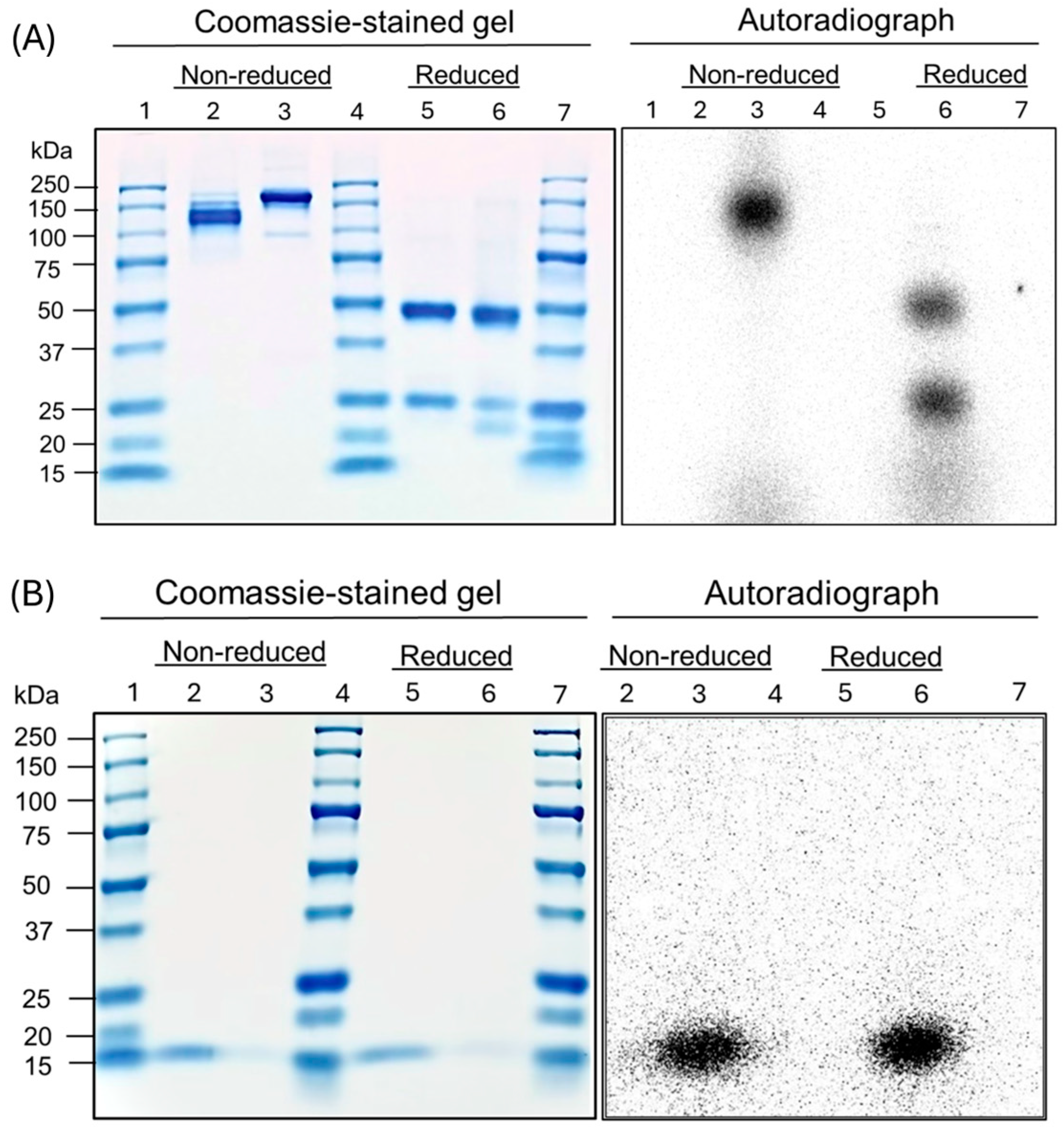
3.4. SDS-PAGE and Autoradiography

SDS-PAGE of the proteins and their radiolabeled conjugates demonstrated primary bands at the expected molecular weights at ~150 kDa for B11-IgG (Figure 1A) and ~15 kDa for B11-nanobody (Figure 1B). For the B11-IgG, a shift in the molecular weight of the primary band was noticeable, likely due to the addition of the DTPA chelator. A minor band is also visible at ~100 kDa, suggesting some possible degradation. Additionally, the autoradiography of [44Sc]Sc-B11-IgG demonstrated a radiolabeled impurity of ≤10 kDa (Figure 1A). Encouragingly, the [44Sc]Sc-B11-nanobody showed no radiolabeled impurities (Figure 1B).

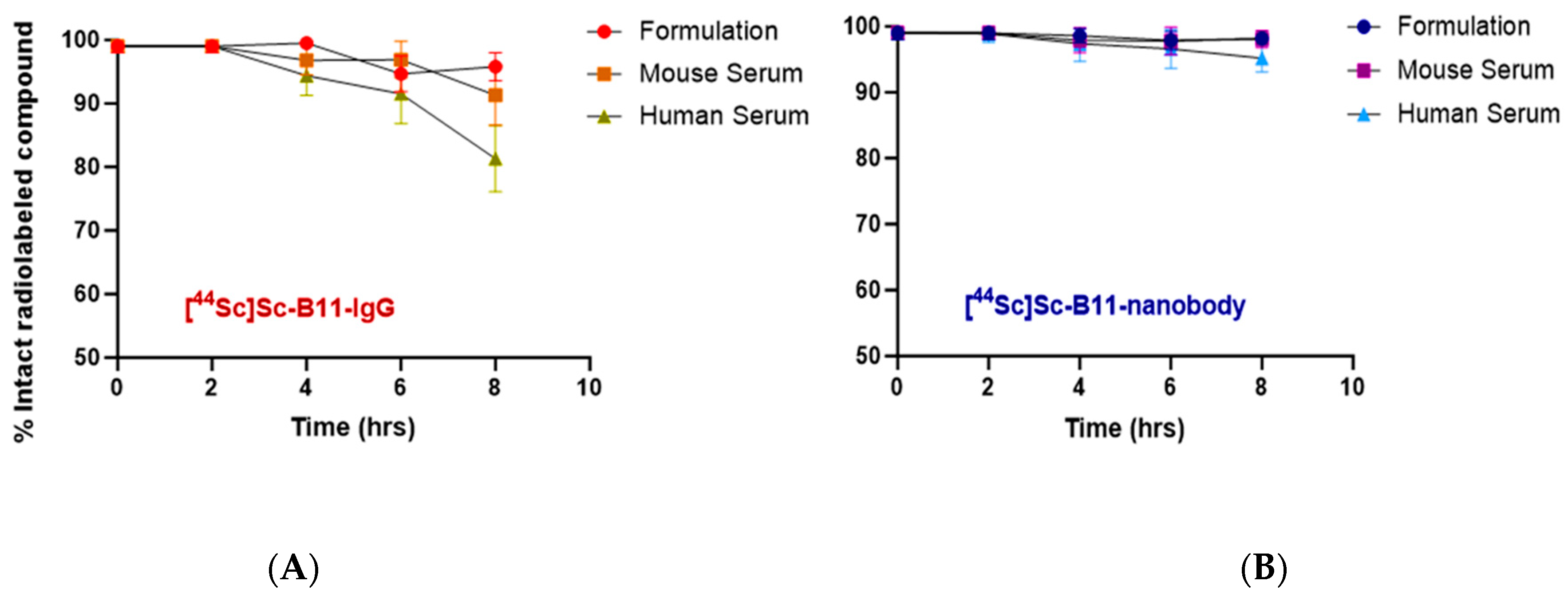
3.5. Stability of Radiotracers in Serum
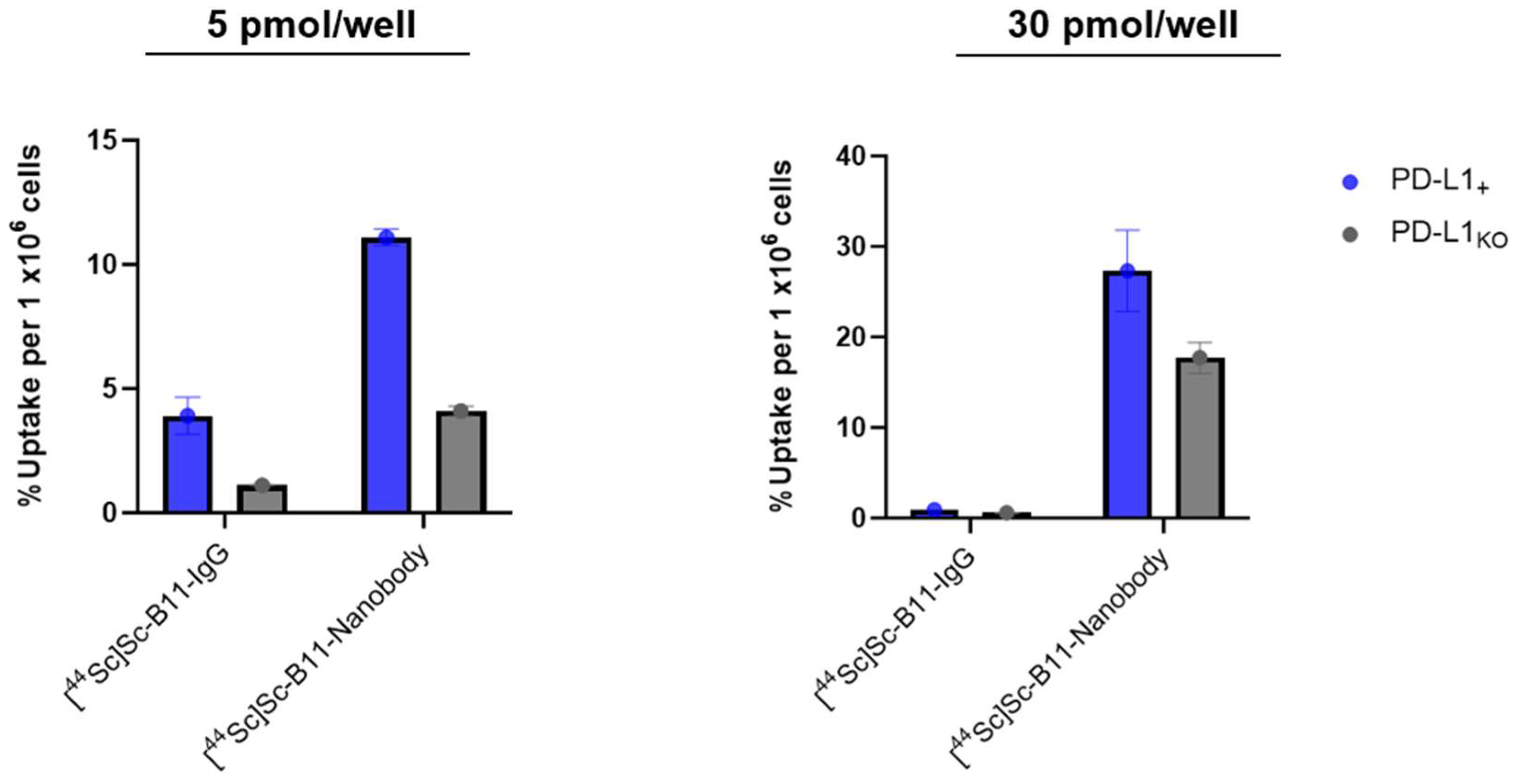
The stability of the radiolabeled full-length antibody and nanobody was assessed in its original formulation (0.25 NaOAc buffer, pH 5.7–6.0) and in mouse and human sera, at 37 °C with light agitation (500 rpm). As summarized in Figure 2, the percentage of intact [44Sc]Sc-B11-IgG after 8 h remained >95% in formulation but decreased to 91.3 ± 4.7% and 81.3 ± 8.3% in mouse and human sera, respectively, suggesting decomplexation or trans chelation in the presence of competing ions and/or natural ligands (Figure 2A, Table 4). Due to the larger molecular weight of the B11-IgG and thereby long plasma circulation time, stability over a longer period (>48 h) is of particular importance to allow for optimal uptake of the radioligand and clearance of background signal. For the [44Sc]Sc-B11-nanobody, the labeled compound was >98% intact after 8 h in formulation and >95% in human serum, encouraging further in vitro evaluation (Figure 2B, Table 4). The exact cause of the difference in stability between the radiolabeled B11-IgG and B11-nanobody, particularly in human serum, is unclear, but since both are conjugated to the same chelator and were synthesized with the same batch of 44Sc, this may reflect differences in the interactions of B11-IgG/B11-nanobody with serum proteins.

3.6. Cell Uptake

After PD-10 purification, [44Sc]Sc-B11-IgG and [44Sc]Sc-B11-nanobody (30 pmol/well) were added to E0771 PD-L1 positive (+) and knockout (KO) cells. Higher uptake was observed in the PD-L1+ than PD-L1KO cells for both the [44Sc]Sc-B11-IgG (0.91 ± 0.01 vs. 0.58 ± 0.05, p < 0.05) and [44Sc]Sc-B11-nanobody (27.33 ± 4.49 vs. 17.70 ± 1.71, p < 0.05) (Figure 3 and Table 5). Unexpectedly, the absolute uptake of [44Sc]Sc-B11-nanobody was significantly higher than the [44Sc]Sc-B11-IgG. However, there was also a significant amount of non-specific uptake in the PD-L1KO cells (Figure 3). The relative uptake ratio (PD-L1+/PD-L1KO) was slightly higher for the [44Sc]Sc-B11-IgG than [44Sc]Sc-B11-nanobody (1.6 vs 1.5). This effect was replicated when the study was repeated with 5 pmol/well. The absolute uptake in PD-L1+ cells vs. PD-L1KO was higher for [44Sc]Sc-B11-nanobody (11.1 ± 0.33 vs. 4.10 ± 0.19, p < 0.05), than the [44Sc]Sc-B11-IgG (3.91 ± 0.75 vs. 1.11 ± 0.02, p < 0.05) but the relative uptake (PD-L1+/PD-L1KO) was significantly higher for [44Sc]Sc-B11-IgG, due to high non-specific uptake of the [44Sc]Sc-B11-nanobody (Table 5).

4. Discussion

Noninvasive PET imaging-based assessment of PD-L1 expression is of high clinical value for better patient selection and treatment response rates to PD-L1 immunotherapies. Due to the slow clearance of full-length antibodies, smaller antibody fragments are attractive for use as radiolabeled molecular imaging probes if they maintain their selectivity and specificity. 44Sc has broad potential as a PET imaging radioisotope, particularly for use with peptides and antibody fragments. While 1,4,7,10-tetraazacyclododecane-1,4,7,10-tetraacetic (DOTA) is considered the gold standard for 44Sc chelation, this requires high temperatures (typically ≥ 80 °C). As such, acyclic chelators such as DTPA are better suited for use with heat-sensitive molecules such as antibodies/antibody fragments to allow for room-temperature radiolabeling. The stability of the [44Sc]Sc-B11-nanbody remained above 95% for up to 8 h, which encourages its use for further in vivo evaluation with 44Sc-labeled antibody fragments. Interestingly, the 44Sc-labeled B11-IgG showed poorer stability, remaining >95% intact in formulation up to 8 h but decreasing to ~80% in human serum. Since both the B11-nanobody and B11-IgG were conjugated with the same chelator and radiolabeled using the same batch of 44Sc and buffer/radiolabeling conditions, this could not be attributed to the complexation conditions. It is possible that the decomplexation is related to the degradation of the protein itself, but this was not probed further since the [44Sc]Sc-B11-IgG would not be a meaningful probe for in vivo evaluation due to the mismatch in the biological and physical half-life of the antibody and radiometal, respectively. The use of antibody fragments in place of a full-length IgG comes at the balanced trade-off between faster clearance but potential reduced specificity [20,34]. The radiolabeled B11-nanobody successfully showed significantly higher uptake in the PD-L1+ cells than the PD-L1KO. However, non-specific uptake was also observed, which negatively impacts its potential as a molecular imaging probe (potentially contributing to background signal and impacting the signal-to-noise ratio) and was, therefore, not advanced to further animal studies. The authors emphasize that these results are an evaluation of the particular nanobody studied and should not discourage future evaluation with other nanobodies for PD-L1 or otherwise. The suitability of the physical half-life of 44Sc with the biological half-life of nanobodies is highly encouraging for future evaluation with alternative nanobodies.

5. Conclusions

In this work,44Sc radiolabeled B11-nanobody and B11-IgG antibody were successfully synthesized with a final radiochemical purity > 99% and evaluated in an in vitro breast cancer model. The [44Sc]Sc-B11-nanobody was able to show higher uptake in PD-L1+ than PD-L1KO cells, but the effect of lowered specificity was apparent in the high non-specific binding and overall uptake in the PD-L1KO cells. For this reason, the 44Sc-labeled B11-nanobody was not advanced for further evaluation in an in vivo animal model. However, the high final radiochemical purity and stability data are encouraging for future evaluation of other nanobody-based PET probes radiolabeled with 44Sc.

Supplementary Materials

The following supporting information can be downloaded at: https://www.mdpi.com/article/10.3390/pharmaceutics17060796/s1, Figure S1: fragmentation of natCaCO3 and etching of the degrader foil at lo beam parameters (left) while natCaO target remained intact up to 40 µA for 2 hrs; Figure S2: Microimpurities present in natCaO target bombarded with 11.7 MeV protons to produce 44Sc (1157 keV). γ-spectrum shown two weeks post production (right) to clearly visualize radioimpurities of 44mSc, 43Sc, 47Sc, 48Sc; Figure S3: MALDI-TOF plots of (A) B11-IgG, (B) DTPA-B11-IgG, (C) B11-nanobody and (D) partially conjugated (~46%) DTPA-B11-nanobody; Figure S4: radio-TLC scans of the radiolabeled proteins before and after purification with PD-10 desalting column; Table S1: Analysis of standard solutions containing Ca and Al at varying ratios, with and without 1 mg/mL CsNO3.

Author Contributions

Conceptualization, M.K.P., S.S.P., H.D. and F.L.-M.; methodology, V.E.K., A.B., J.R.E., D.J.V., M.K.P., S.S.P., H.D. and F.L.-M.; formal analysis, V.E.K., A.B. and M.K.; investigation, V.E.K., A.B. and M.K.P.; resources, D.J.V., J.R.E., M.K.P., S.S.P., H.D. and F.L.-M.; data curation, V.E.K., A.B., M.K., D.J.V. and J.R.E.; writing—original draft preparation, V.E.K. and A.B.; writing—review and editing, All authors.; visualization, V.E.K. M.K., A.B. and M.K.P.; supervision, M.K.P.; project administration, M.K.P.; funding acquisition, M.K.P. and S.S.P., H.D. and F.L.-M. All authors have read and agreed to the published version of the manuscript.

Funding

Part of the research studies reported in the present publication was supported by the National Center for Advancing Translational Sciences of the National Institutes of Health of the USA (Award Numbers UL1TR002494 and UL1TR002377). Additional support was provided from the Minnesota Partnership for Biotechnology and Medical Genomics through the Translational Product Development Fund (TPDF) to MKP as a Principal Investigator.

Institutional Review Board Statement

Not applicable.

Informed Consent Statement

Not applicable.

Data Availability Statement

All data generated in this study are presented in the work.

Acknowledgments

The authors thank the Departments of Radiology, Urology, Immunology, Radiation Oncology, Pharmacology and Mayo Clinic Comprehensive Cancer Center for their support.

Conflicts of Interest

The authors declare no conflicts of interest. The funders had no role in the design of the study; in the collection, analyses, or interpretation of data; in the writing of the manuscript; or in the decision to publish the results.

Abbreviations

The following abbreviations are used in this manuscript:
PD-L1Programed Death Ligand 1
PD-1Programed Death 1
PETPositron Emission Tomography
TRTTargeted Radionuclide Therapy
SBRSignal to Background Ratio
MP-AESMicrowave Plasma Atomic Emission Spectroscopy
AUCArea Under the Curve
SDS-PAGESodium Dodecyl Sulfate—PolyacrylAmide Gel Electrophoresis
HBSSHanks Buffered Salt Solution

References

  1. Han, Y.; Liu, D.; Li, L. PD-1/PD-L1 pathway: Current researches in cancer. Am. J. Cancer Res. 2020, 10, 727. [Google Scholar] [PubMed]
  2. Blank, C.; Gajewski, T.F.; Mackensen, A. Interaction of PD-L1 on tumor cells with PD-1 on tumor-specific T cells as a mechanism of immune evasion: Implications for tumor immunotherapy. Cancer Immunol. Immunother. 2005, 54, 307–314. [Google Scholar] [CrossRef] [PubMed]
  3. Diskin, B.; Adam, S.; Cassini, M.F.; Sanchez, G.; Liria, M.; Aykut, B.; Buttar, C.; Li, E.; Sundberg, B.; Salas, R.D. PD-L1 engagement on T cells promotes self-tolerance and suppression of neighboring macrophages and effector T cells in cancer. Nat. Immunol. 2020, 21, 442–454. [Google Scholar] [CrossRef]
  4. Beenen, A.C.; Sauerer, T.; Schaft, N.; Dörrie, J. Beyond cancer: Regulation and function of PD-L1 in health and immune-related diseases. Int. J. Mol. Sci. 2022, 23, 8599. [Google Scholar] [CrossRef]
  5. Sunshine, J.; Taube, J.M. Pd-1/pd-l1 inhibitors. Curr. Opin. Pharmacol. 2015, 23, 32–38. [Google Scholar] [CrossRef]
  6. Hamanishi, J.; Mandai, M.; Matsumura, N.; Abiko, K.; Baba, T.; Konishi, I. PD-1/PD-L1 blockade in cancer treatment: Perspectives and issues. Int. J. Clin. Oncol. 2016, 21, 462–473. [Google Scholar] [CrossRef]
  7. Seliger, B. Basis of PD1/PD-L1 therapies. J. Clin. Med. 2019, 8, 2168. [Google Scholar] [CrossRef]
  8. Dong, H.; Strome, S.E.; Salomao, D.R.; Tamura, H.; Hirano, F.; Flies, D.B.; Roche, P.C.; Lu, J.; Zhu, G.; Tamada, K. Tumor-associated B7-H1 promotes T-cell apoptosis: A potential mechanism of immune evasion. Nat. Med. 2002, 8, 793–800. [Google Scholar] [CrossRef] [PubMed]
  9. Thompson, R.H.; Dong, H.; Kwon, E.D. Implications of B7-H1 expression in clear cell carcinoma of the kidney for prognostication and therapy. Clin. Cancer Res. 2007, 13, 709s–715s. [Google Scholar] [CrossRef]
  10. Gandini, S.; Massi, D.; Mandalà, M. PD-L1 expression in cancer patients receiving anti PD-1/PD-L1 antibodies: A systematic review and meta-analysis. Crit. Rev. Oncol./Hematol. 2016, 100, 88–98. [Google Scholar] [CrossRef]
  11. Vranic, S.; Gatalica, Z. PD-L1 testing by immunohistochemistry in immuno-oncology. Biomol Biomed 2023, 23, 15–25. [Google Scholar] [CrossRef] [PubMed]
  12. Niemeijer, A.; Leung, D.; Huisman, M.; Bahce, I.; Hoekstra, O.; Van Dongen, G.; Boellaard, R.; Du, S.; Hayes, W.; Smith, R. Whole body PD-1 and PD-L1 positron emission tomography in patients with non-small-cell lung cancer. Nat. Commun. 2018, 9, 4664. [Google Scholar] [CrossRef] [PubMed]
  13. Chatterjee, S.; Lesniak, W.G.; Nimmagadda, S. Noninvasive imaging of immune checkpoint ligand PD-L1 in tumors and metastases for guiding immunotherapy. Mol. Imaging 2017, 16, 1536012117718459. [Google Scholar] [CrossRef]
  14. Ehlerding, E.B.; England, C.G.; McNeel, D.G.; Cai, W. Molecular Imaging of Immunotherapy Targets in Cancer. J. Nucl. Med. 2016, 57, 1487–1492. [Google Scholar] [CrossRef] [PubMed]
  15. Wei, W.; Rosenkrans, Z.T.; Liu, J.; Huang, G.; Luo, Q.-Y.; Cai, W. ImmunoPET: Concept, design, and applications. Chem. Rev. 2020, 120, 3787–3851. [Google Scholar] [CrossRef]
  16. Kikuchi, M.; Clump, D.A.; Srivastava, R.M.; Sun, L.; Zeng, D.; Diaz-Perez, J.A.; Anderson, C.J.; Edwards, W.B.; Ferris, R.L. Preclinical immunoPET/CT imaging using Zr-89-labeled anti-PD-L1 monoclonal antibody for assessing radiation-induced PD-L1 upregulation in head and neck cancer and melanoma. Oncoimmunology 2017, 6, e1329071. [Google Scholar] [CrossRef]
  17. Jalilian, A.R.; Osso, J.A. Production, applications and status of zirconium-89 immunoPET agents. J. Radioanal. Nucl. Chem. 2017, 314, 7–21. [Google Scholar] [CrossRef]
  18. Van De Watering, F.C.; Rijpkema, M.; Perk, L.; Brinkmann, U.; Oyen, W.J.; Boerman, O.C. Zirconium-89 labeled antibodies: A new tool for molecular imaging in cancer patients. BioMed Res. Int. 2014, 2014, 203601. [Google Scholar] [CrossRef]
  19. Rashidian, M.; Ploegh, H. Nanobodies as non-invasive imaging tools. Immuno-Oncol. Technol. 2020, 7, 2–14. [Google Scholar] [CrossRef]
  20. Fu, R.; Carroll, L.; Yahioglu, G.; Aboagye, E.O.; Miller, P.W. Antibody Fragment and Affibody ImmunoPET Imaging Agents: Radiolabelling Strategies and Applications. ChemMedChem 2018, 13, 2466–2478. [Google Scholar] [CrossRef]
  21. Bansal, A.; Pandey, M.K.; Barham, W.; Liu, X.; Harrington, S.M.; Lucien, F.; Dong, H.; Park, S.S.; DeGrado, T.R. Non-invasive immunoPET imaging of PD-L1 using anti-PD-L1-B11 in breast cancer and melanoma tumor model. Nucl. Med. Biol. 2021, 100, 4–11. [Google Scholar] [CrossRef] [PubMed]
  22. Mohr, P.; van Sluis, J.; Lub-de Hooge, M.N.; Lammertsma, A.A.; Brouwers, A.H.; Tsoumpas, C. Advances and challenges in immunoPET methodology. Front. Nucl. Med. 2024, 4, 1360710. [Google Scholar] [CrossRef]
  23. Ferguson, S.; Jans, H.-S.; Wuest, M.; Riauka, T.; Wuest, F. Comparison of scandium-44 g with other PET radionuclides in pre-clinical PET phantom imaging. EJNMMI Phys. 2019, 6, 1–14. [Google Scholar] [CrossRef]
  24. Chernysheva, M.; Loveless, S.C.; Brossard, T.; Becker, K.; Cingoranelli, S.; Aluicio-Sarduy, E.; Song, J.; Ellison, P.; Nolen, J.; Rotsch, D.A.; et al. Accelerator Production of Scandium Radioisotopes: Sc-43, Sc-44, and Sc-47. Curr. Radiopharm. 2021, 14, 359–373. [Google Scholar] [CrossRef] [PubMed]
  25. van der Meulen, N.P.; Bunka, M.; Domnanich, K.A.; Müller, C.; Haller, S.; Vermeulen, C.; Türler, A.; Schibli, R. Cyclotron production of 44Sc: From bench to bedside. Nucl. Med. Biol. 2015, 42, 745–751. [Google Scholar] [CrossRef] [PubMed]
  26. Umbricht, C.A.; Benešová, M.; Schmid, R.M.; Türler, A.; Schibli, R.; van der Meulen, N.P.; Müller, C. 44Sc-PSMA-617 for radiotheragnostics in tandem with 177Lu-PSMA-617—Preclinical investigations in comparison with 68Ga-PSMA-11 and 68Ga-PSMA-617. EJNMMI Res. 2017, 7, 9. [Google Scholar] [CrossRef]
  27. Müller, C.; Bunka, M.; Haller, S.; Köster, U.; Groehn, V.; Bernhardt, P.; van der Meulen, N.; Türler, A.; Schibli, R. Promising Prospects for 44Sc-/47Sc-Based Theragnostics: Application of 47Sc for Radionuclide Tumor Therapy in Mice. J. Nucl. Med. 2014, 55, 1658–1664. [Google Scholar] [CrossRef]
  28. Ziegler, J.F.; Ziegler, M.D.; Biersack, J.P. SRIM—The stopping and range of ions in matter. Nucl. Instrum. Methods Phys. Res. Sect. B Beam Interact. Mater. At. 2010, 268, 1818–1823. [Google Scholar] [CrossRef]
  29. Karlsson, S.; Sjöberg, V.; Ogar, A. Comparison of MP AES and ICP-MS for analysis of principal and selected trace elements in nitric acid digests of sunflower (Helianthus annuus). Talanta 2015, 135, 124–132. [Google Scholar] [CrossRef]
  30. Guerin, A.; Steeg, U.; Abdelnour, Y. Determination of Exchangeable Cations in Soil Extracts Using the Agilent 4100 Microwave Plasma-Atomic Emission Spectrometer. 2012. Available online: https://www.agilent.com/cs/library/applications/5991-0048EN_AppNote_4100MP-AES_Agriculture_soil.pdf (accessed on 11 June 2025).
  31. Bradford, M.M. A rapid and sensitive method for the quantitation of microgram quantities of protein utilizing the principle of protein-dye binding. Anal. Biochem. 1976, 72, 248–254. [Google Scholar] [CrossRef]
  32. Schägger, H.; Von Jagow, G. Tricine-sodium dodecyl sulfate-polyacrylamide gel electrophoresis for the separation of proteins in the range from 1 to 100 kDa. Anal. Biochem. 1987, 166, 368–379. [Google Scholar] [CrossRef] [PubMed]
  33. Taubel, J.C.; Nelson, N.R.; Bansal, A.; Curran, G.L.; Wang, L.; Wang, Z.; Berg, H.M.; Vernon, C.J.; Min, H.-K.; Larson, N.B. Design, synthesis, and preliminary evaluation of [68Ga] Ga-NOTA-insulin as a PET probe in an Alzheimer’s disease mouse model. Bioconjugate Chem. 2022, 33, 892–906. [Google Scholar] [CrossRef] [PubMed]
  34. Bansal, A.; Lavoie, R.R.; Lucien, F.; Kethamreddy, M.; Wootla, B.; Dong, H.; Park, S.S.; Pandey, M.K. Synthesis and evaluation of anti-PD-L1-B11 antibody fragments for PET imaging of PD-L1 in breast cancer and melanoma tumor models. Sci. Rep. 2024, 14, 19561. [Google Scholar] [CrossRef] [PubMed]
  35. Becker, K.V.; Aluicio-Sarduy, E.; Bradshaw, T.; Hurley, S.A.; Olson, A.P.; Barrett, K.E.; Batterton, J.; Ellison, P.A.; Barnhart, T.E.; Pirasteh, A.; et al. Cyclotron production of 43Sc and 44gSc from enriched 42CaO, 43CaO, and 44CaO targets. Front. Chem. 2023, 11, 1167783. [Google Scholar] [CrossRef]
  36. Krajewski, S.; Cydzik, I.; Abbas, K.; Bulgheroni, A.; Simonelli, F.; Holzwarth, U.; Bilewicz, A. Cyclotron production of 44Sc for clinical application. Radiochim. Acta 2013, 101, 333–338. [Google Scholar] [CrossRef]
Figure 1. Coomassie-blue stained SDS-Page and autoradiography of non-radiolabeled and 44Sc-labeled (A) B11-Nanobody; where Lane 1: Protein marker marker (10-250 kDa, BioRad cat #1610373), Lane 2: B11-nanobody non-reduced, Lane 3: [44Sc]Sc-B11-nanobody non-reduced, Lane 4: Protein marker (2-250 kDa, BioRad cat #1610377), Lane 5: B11-nanobody reduced, Lane 6: [44Sc]Sc-B11-nanobody reduced, Lane 7: Protein marker (2-250 kDa, BioRad cat #1610377). (B) B11-IgG, where Lane 1: Protein Marker (2-250 kDa, BioRad cat #1610377), Lane 2: Lane 2: B11-IgG non-reduced, Lane 3: [44Sc]Sc-B11-IgG non-reduced, Lane 4: Protein marker marker (10-250 kDa, BioRad cat #1610373), Lane 5: B11-IgG reduced, Lane 6: [44Sc]Sc-B11-IgG reduced, Lane 7: Protein marker (10-250 kDa, BioRad cat #1610373).
Pharmaceutics 17 00796 g001
Figure 2. Stability of [44Sc]Sc-B11-nanobody (A) and [44Sc]Sc-B11-IgG (B), at 37 °C with light agitation (500 rpm) as formulated, and in mouse and human sera.
Pharmaceutics 17 00796 g002
Figure 3. Cell uptake of [44Sc]Sc-B1-IgG and [44Sc]Sc-B11-nanobody at 5 pmol/well (left) and 30 pmol/well (right), in E0771 PD-L1+ breast cancer cells and E0771 PD-L1KO cells.
Pharmaceutics 17 00796 g003
Table 1. MP-AES analysis conditions for Ca, Al and Fe analysis in final 44Sc elution fractions.
Element Wavelength (λ)Nebulizer Flow Rate (L/min)Viewing PositionPump SpeedStabilization Time
Ca (317.933 nm)0.60025 rpm15 s
Al (396.152 nm)0.95025 rpm15 s
Fe (371.993 nm)0.65025 rpm15 s
Table 2. Summary of production activities (A) and yields (Y) of 44Sc at end-of-bombardment (EOB) for the natCaO target.
Current (µA)Irradiation Time (min)AEOB (MBq)YEOB
(MBq/µAh)
151549.95 ± 1.48 (n = 2)9.99 ± 0.30
2030138.75 ± 1.49 (n = 3)13.87 ± 0.15
2060246.42 ± 5.92 (n = 3)12.32 ± 0.30
3060354.09 ± 8.51(n = 5)11.80 ± 0.28
4030257.15 ± 4.44 (n = 3)12.86 ± 0.22
4060483.22 ± 7.41 (n = 31)12.08 ± 0.19
40120943.13 ± 5.98 (n = 3)11.79 ± 0.07
Table 3. Analysis of remaining bulk Ca material and other trace metals (Fe, Al) in collected final elutions of 44Sc using a two-step DGA + SCX ion-exchange column set-up. Apparent specific activity reported for 30 min 40 µA productions at end of purification (no decay correction), assuming no other contaminants.
FractionCa (ppm)Fe (ppm)Al (ppm)Volume
(µL)
Activity
(GBq)
Apparent As (GBq/µg)
114.7 ± 5.62.2 ± 1.03.4 ± 1.6500109.8 ± 15.510.8 ± 7.7
28.7 ± 1.71.0 ± 0.31.4 ± 0.850031.3 ± 4.95.6 ± 3.7
36.0 ± 0.010.6 ± 0.01<0.0150010.1 ± 4.53.1 ± 2.2
Table 4. Percentage of intact [44Sc]Sc-B11-nanobody and [44Sc]Sc-B11-IgG over 8 h at 37 °C with light agitation (500 rpm) as formulated, and in mouse and human sera.
Stability0 h2 h4 h6 h8 h
[44Sc]Sc-B11-IgG (% intact)
Formulation>99>9999.5 ± 0.994.7 ± 2.895.8 ± 2.2
Mouse Serum>99>9996.8 ± 2.396.8 ± 2.991.3 ± 4.7
Human Serum>99>9994.4 ± 3.191.5 ± 4.781.3 ± 8.3
[44Sc]Sc-B11-Nanobody (% intact)
Formulation>99>9998.6 ± 1.197.9 ± 1.498.2 ± 0.9
Mouse Serum>99>9997.9 ± 1.997.8 ± 2.198.1 ± 1.3
Human Serum>9998.9 ± 1.397.4 ± 2.796.6 ± 2.995.2 ± 2.1
Table 5. Summary of cell uptake data comparing % uptake (per 1 × 10⁶ cells) in PD-L1+ and PD-L1KO cells and the relative uptake ratio.
CompoundApparent Molar Activity (Am) (GBq/µmol)% Uptake (PD-L1KO)% Uptake (PD-L1+)p-ValueUptake Ratio (+/KO)
5 pmol/ well
[44Sc]Sc-B11-IgG4.61.11 ± 0.023.91 ± 0.754 × 10⁻⁴~3.5
[44Sc]Sc-B11-nanobody2.44.10 ± 0.1911.1 ± 0.330.032.7
30 pmol/ well
[44Sc]Sc-B11-IgG2.30.58 ± 0.050.91 ± 0.010.003~1.6
[44Sc]Sc-B11-nanobody6.917.70 ± 1.7127.33 ± 4.496 × 10⁻⁶~1.5
Disclaimer/Publisher’s Note: The statements, opinions and data contained in all publications are solely those of the individual author(s) and contributor(s) and not of MDPI and/or the editor(s). MDPI and/or the editor(s) disclaim responsibility for any injury to people or property resulting from any ideas, methods, instructions or products referred to in the content.

