Article

Agmatine Abrogates Tacrolimus-Induced Testicular Injury in Rats

Department of Pharmacology and Toxicology, Faculty of Pharmacy, Mansoura University, Mansoura 35516, Egypt
*
Author to whom correspondence should be addressed.
Pharmaceutics 2025, 17(6), 703; https://doi.org/10.3390/pharmaceutics17060703
Submission received: 15 March 2025 / Revised: 26 April 2025 / Accepted: 28 April 2025 / Published: 27 May 2025
(This article belongs to the Section Clinical Pharmaceutics)

Abstract

Background/Objectives: Tacrolimus is an immunosuppressant drug widely used to prevent organ transplant rejection. Preclinical and clinical studies report that tacrolimus has destructive impacts on the male reproductive system owing to the induction of oxidative stress and inflammation. This study aimed at examining defensive impacts of agmatine against tacrolimus-induced testicular toxicity in rats. Methods: Male Wistar rats were randomly divided into six groups and treated based on the experimental design for 14 days. By the end of this study, blood samples were obtained to measure testosterone and luteinizing hormone. Also, both testes were removed for molecular analysis and histopathological examinations. Results: Agmatine administration increased serum levels of testosterone and luteinizing hormone and ameliorated all histopathological and toxicological changes induced by tacrolimus. Agmatine administration attenuated tacrolimus-induced oxidative stress as evidenced by the reduction of malondialdehyde content and inducible nitric oxide synthase expression and the elevation of reduced glutathione. This was parallel to the restoration of nuclear factor erythroid 2-related factor2 and hemeoxygenase-1 expression. Moreover, agmatine decreased the expressions of nuclear factor kappa B and interleukin-17. Agmatine also decreased the cell death revealed by decreased caspase-3 expression and increased expression of the antiapoptotic marker Bcl-2 in a dose-dependent manner. The antioxidant, anti-inflammatory, and antiapoptotic effects of agmatine were explained by increased expression of sirtuin-1. Conclusions: agmatine effectively attenuated testicular injuries induced by tacrolimus and enhanced spermatogenesis. This protective effect of agmatine might be mediated via the upregulation of sirtuin-1 expression that in turn restores oxidative status and regulates nuclear factor erythroid 2-related factor2/nuclear factor kappa B/Bcl-2 signaling.

1. Introduction

Tacrolimus (TAC), known as FK506, is a potent calcineurin inhibitor that is widely used to prevent organ rejection post-transplant and to treat severe atopic dermatitis [1,2]. The majority of studies have focused only on preventing toxic effects of TAC on the liver, kidney, and neurons, as well as the gastrointestinal tract [3,4]. Evidence reports that males on TAC therapy suffering from testicular injuries including a loss of epithelial thickness in seminiferous tubules, an increase in the number of spermatids and spermatozoa with damaged heads and tails, and a decrease in the number of spermatocytes [5,6]. However, very few studies have focused on the toxic effects of TAC on testicular damage and possible therapeutic tools that could help prevent the testicular toxicity associated with TAC therapy.
TAC exerts its immuno-suppressant activity through inhibiting interleukin (IL)-2 mRNA transcription via forming a pentameric complex with the intracellular receptor, FKBP12, Ca2+, calmodulin, and calcineurin [7]. FKBP12, which is a major pharmacological target molecule of TAC, has an important role in sperm motility and could be related to the TAC-induced decrease in sperm motility [8].
Reactive oxygen species (ROS) overproduction and diminished ATP production associated with impaired mitochondrial function have been proposed to be the main causes of TAC-associated organ toxicity [9]. Thus, blocking oxidative stress and maintaining energy homeostasis could be a potential mean of reducing the organ toxicity associated with TAC.
Agmatine (AGM) is an endogenous metabolite of L-arginine [10]. Preclinical and initial clinical evidence has shown that AGM could treat opioid addiction, neurodegenerative diseases, mood disorders, cognitive disorders, and neurotrauma [11,12]. AGM is known to have potent antioxidant and anti-inflammatory effects through inhibiting inducible nitric oxide synthase (iNOS) and activating endothelial nitric oxide synthase (eNOS) [13,14].
Study of Ommati, Farshad [15] demonstrated that AGM could alleviate hepatic injury in a rat model of obstructive jaundice via its antioxidant properties and its effects on mitochondrial indices [15]. Similarly, the antioxidant and anti-inflammatory potential of AGM has been demonstrated against hepatotoxicity induced by acetaminophen [16], D-galactosamine and lipopolysaccharide [17], and chlorpromazine [18]. Additionally, AGM may safeguard the gastrointestinal tract from the adverse effects of nonsteroidal anti-inflammatory drugs by reducing inflammation and oxidative damage [19]. AGM also counteracted the neurotoxicity associated with antipsychotic medications by diminishing neuroinflammation and oxidative stress, thereby potentially improving the safety profiles of these drugs [20]. Overall, the multifaceted protective effects of AGM highlight its potential as a therapeutic adjunct in managing drug-induced toxicity via restoring oxidative balance and inhibiting inflammation.
Collectively, we hypothesized that AGM could be a promising tool to prevent TAC-induced testicular toxicity in rats.

2. Materials and Methods

2.1. Animals

Thirty-six male Wistar rats, weighing 200–250 g (approximately 7–8 weeks old), were obtained from the “Medical Experimental Research Center”, Faculty of Medicine, Mansoura University, Egypt. The rats were placed in plastic cages at temperature of ~25 °C and allowed free access to food and water and were sustained on a 12 h light/dark cycle. Animal care and procedures were carried out in accordance with the National Institutes of Health (NIH) guidelines and approved by the “Animal Care and Use Committee”, Mansoura University, Egypt, as a part from research project with the code number MU-ACUC (PHARM.PhD.23.02.16).

2.2. Chemicals and Drugs

TAC was obtained as a Prograf® capsule from a local pharmaceutical company (Astellas Pharma Inc., Chuo-Ku, Japan) and was suspended in carboxymethyl cellulose (CMC) for intraperitoneal injection. Agmatine® powder was obtained from Nutricost (Vineyard, UT, USA) and was dissolved in distilled water for oral administration.

2.3. Experimental Design

Rats were randomly divided into 6 groups (6 rats each) as illustrated in Table 1:
The dose of TAC was selected based on previous studies [21,22,23] and the pilot study using three different regimens of TAC: (1) 5 mg/kg, oral; (2) 7 mg/kg, oral; (3) 5 mg/kg, i.p.; and the study indicated that 5 mg/kg i.p. is the most effective regimen to induce testicular injury.
On day 15, the rats were anaesthetized with thiopental sodium (40 mg/kg, i.p.). Blood samples were collected from the retro-orbital plexus and centrifuged at 4000 rpm for 15 min to collect serum for assessment of total testosterone (TT) and luteinizing hormone (LH). Then, the epididymes and testes were isolated, rinsed in ice-cold saline, dried, and weighed. The epididymes were collected for sperm analyses. The right testis was homogenized in phosphate-buffered saline (10% w/v) for biochemical and molecular assessment. The left testes were kept in Bouin’s fixative solution for histopathological and immunohistochemical analysis.

2.4. Semen Analysis

Cauda epididymis was dissected out, minced in physiological saline, and then incubated for 30 min at 37 °C to allow spermatozoa to leave the epididymal tubules for sperm analysis [24]. Sperm concentration and viability were analyzed for each sample. For sperm counting, every 0.5 mL of sperm sample was diluted with 9.5 mL of fixative (50 g of sodium bicarbonate and 10 mL of 35% (v/v) formalin (1:20)); then, the hemocytometer was loaded with diluted sample, allowing the spermatozoa to settle in a humid chamber, and then the spermatozoa were counted in the middle square using a 200x lens. The sperm count was presented as the concentration of million/mL. Sperm viability was calculated as a percent using the one step eosin-nigrosine procedure [25].

2.5. Assessment of Serum TT and LH Levels

Serum levels of TT (MyBioSource, San Diego, CA, USA, MBS282195) and LH (MyBioSource, San Diego, CA, USA, MBS590031) were determined according to the instructions of the manufacturer.

2.6. Assessment of Oxidative Stress Status

The testicular homogenate was used to estimate MDA content (MD 25 29) as well as GSH concentration (GR 25 11) using biodiagnostic assay kits (Giza, Egypt) according to the manufacturer’s instructions.

2.7. Assessment of Bcl-2, Inerleukin-17 (IL-17), Hemeoxygenase-1 (HO-1), Sirtuin1 (SIRT1), and iNOS Expressions

Levels of Bcl-2 (Cusabio, Houston, TX, USA, CSB-E08854r), IL-17 (Novus Biologicals, Centennial, CO, USA, CSB-E07451r), HO-1 (Cusabio, Houston, TX, USA, CSB-E08267r), SIRT1 (BT LAB, Beijing, China, E1145Ra), and iNOS (Assay Genie, Duplin, Ireland, RTFI00088) were measured in testicular homogenate as instructed by the manufacturer.

2.8. Histopathological Analyses

The left testicle was fixed in Bouin’s solution for histopathological analyses. After 24 h, all samples were dehydrated in ascending concentration of alcohol (70–100%). All tissues were then cleared in xylene, embedded in paraffin, and cut by the microtome at a thickness of 3 μm and stained with hematoxylin and eosin (H&E) for the evaluation of tissue injury. Histopathological analyses were carried out by the pathologist in a blinded manner.

2.9. Testicular Morphometry

Morphometric analysis of micrographs of the testes was performed as previously described [25]. About 15 transverse sections of seminiferous tubules were chosen randomly and measured for each animal. The seminiferous tubular diameter and height of the seminiferous tubule epithelium were measured at 100 × magnification using the software image J version 1.51r (NIH, Bethesda, MD, USA).

2.10. Immunohistochemical Analyses

Immunohistochemical analyses were used for the detection of caspase-3, nuclear factor kappa B (NF-κB), and nuclear factor erythroid 2-related factor2 (Nrf2) expression in testicular sections. Antigen retrieval was performed after deparaffinization and rehydration. Tissue sections were incubated overnight at 4 °C with rat polyclonal antibodies against caspase-3 (Servicebio, Wuhan, China), NF-κB (ABclonal, Woburn, MA, USA), and Nrf2 (Servicebio, China). After incubation, the sections were washed and treated with a goat anti-rat secondary antibody (Genemed Biotechnologies, Torrance, CA, USA) for 2 h at room temperature. Diaminobenzidine (DAB) staining was used for visualization, and a light microscope (Leica, Wetzlar, Germany) was used for the examination of stained sections. To minimize background staining and confirm specificity, appropriate control experiments were conducted [26]. Immunohistochemical evaluation was performed by the pathologist in a blinded manner. ImageJ software version 1.51r (NIH, USA) was used for semiquantification of positively stained regions in a blinded manner, and the mean of six readings from the left testicular sections was calculated for each rat.

2.11. Statistics

Data are presented as the mean ± standard deviation (SD). The Shapiro–Wilk test was used to test normal distribution of data. One-way analysis of variance (ANOVA) followed by Tukey–Kramer’s multiple comparison post hoc test was used to measure differences among groups. The significance level was set at p < 0.05. Statistical analyses and graphing were carried out using Graph Pad Prism software (V 8.4.2, Graph Pad Software Inc., La Jolla, CA, USA).

3. Results

There were no significant differences among the control, AGM10, and AGM40 group in all measured parameters.

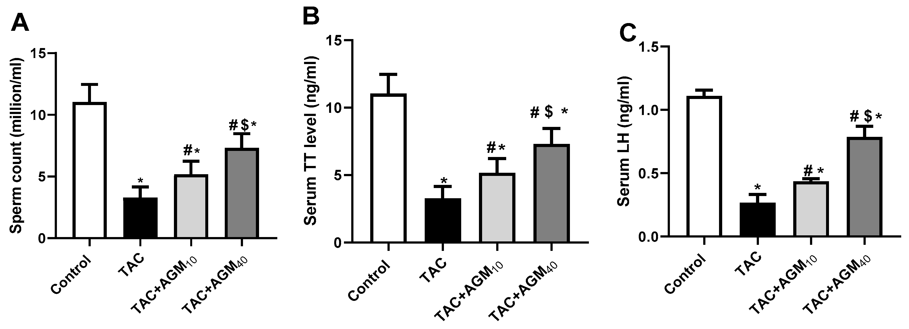
3.1. Impact of AGM10 and AGM40 on TAC-Induced Changes in Sperm Count, Sperm Viability, Serum TT, and LH Concentration

TAC administration exhibited a significant reduction in sperm viability after 1, 2, and 3 h by 45.4%, 50.0%, and 43.0%, respectively, relative to the control group (p < 0.05, Table 2). Administration of AGM10 along with TAC did not significantly affect sperm viability after 1, 2, and 3 h (p > 0.05, Table 1), whereas rats treated with AGM40 along with TAC showed a significant increase in sperm viability after 1, 2, and 3h (p < 0.05, Table 2) in comparison to the TAC group.
Administration of TAC for 14 days exhibited a significant decrease in sperm count by 70.3% compared to the control group (p < 0.05, Figure 1A). Rats treated with AGM10 or AGM40 along with TAC significantly increased sperm count 1.6- and 2.2-fold, respectively, relative to the TAC group (p < 0.05, Figure 1A). The elevation of sperm count was more significant in the TAC + AGM40 group than the TAC + AGM10 group (p < 0.05, Figure 1A).
TAC administration for 14 days significantly decreased serum TT level by 67.5% compared to the control group (p < 0.05, Figure 1B). Rats treated with AGM10 or AGM 40 along with TAC significantly increased serum TT level 1.2- and 1.8-fold, respectively, in comparison to the TAC group (p < 0.05, Figure 1B). The elevation of serum TT level was more significant in the TAC + AGM40 group than the TAC + AGM10 group (p < 0.05, Figure 1B).
TAC administration exhibited a significant reduction in LH levels by 76.4% relative to the control group (p < 0.05, Figure 1C). Rats treated with AGM10 or AGM40 significantly increased LH levels 1.9- and 2.9-fold, respectively, compared to the TAC group (p < 0.05, Figure 1C). However, the effect of TAC + AGM40 on LH levels was more pronounced than in TAC + AGM10 (p < 0.05, Figure 1C).

3.2. Impact of AGM10 and AGM40 on TAC-Induced Histopathological Changes

H&E micrographs of testicular sections showed normal features of the testis with an almost rounded seminiferous tubule lined by several layers of spermatogenic cells, and a lumen filled with sperm in the control group. On the other hand, TAC administration resulted in the loss of the seminiferous tubule’s normal shape, sloughing of the germinal epithelium, vacuolation with the pyknotic Leydig cell, and a decrease in the number of spermatocytes. Testicular sections from rats treated with AGM10 along with TAC showed a partial restoration of the seminiferous tubule’s normal shape, mild sloughing of the germinal epithelium, and necrosis of the Leydig cell, whereas testicular sections from rats treated with AGM40 along with TAC exhibited restoration of the seminiferous tubule’s normal shape, minimal sloughing of the germinal epithelium, and minimal vacuolation with the pyknotic Leydig cell (Figure 2).

3.3. Impact of AGM10 and AGM40 on TAC-Induced Changes in Morphometry of the Seminiferous Tubules

The height of the seminiferous epithelium was 70.6 ± 12.3 µm in the control group (Table 3). TAC administration significantly decreased height of the seminiferous epithelium compared to the height of the control group (p < 0.05). Rats treated with AGM10 or AGM40 along with TAC significantly increased the height of the seminiferous epithelium (p < 0.05).
The average diameter of the seminiferous tubules was 244.4± 16.8 µm in the control group (Table 3). TAC administration significantly decreased the diameter of the seminiferous tubules compared to the diameter in the control group (p < 0.05). Rats treated with AGM10 or AGM40 along with TAC significantly increased the diameter of the seminiferous tubules (p < 0.05).

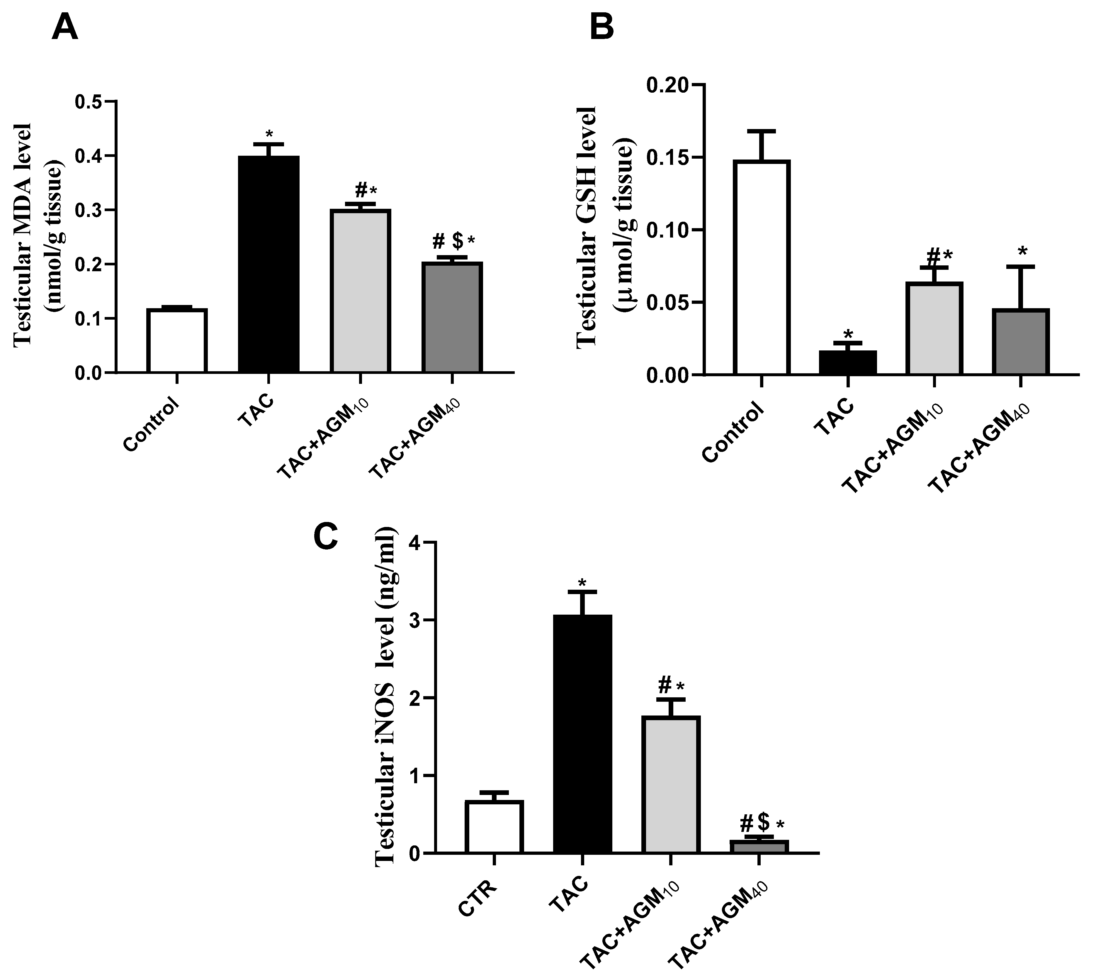
3.4. Impact of AGM10 and AGM40 on TAC-Induced Changes in Oxidative Stress Status

Administration of TAC for 14 days exhibited a 3.4-fold increase in MDA content relative to control group (p < 0.05, Figure 3A). Treatment with AGM10 and AGM40 along with TAC resulted in a significant reduction of MDA content by 24.3% and 48.7%, respectively, as compared to the TAC group (p < 0.05, Figure 3A). The effect was more significant in the TAC + AGM40 group than the TAC + AGM10 group (p < 0.05, Figure 3A).
TAC also significantly reduced GSH levels by 88.7% as compared to the control group (p < 0.05, Figure 3B). Rats treated with AGM10 and AGM40 along with TAC exhibited a significant increase in GSH levels by 3.8- and 2.7-fold, respectively, as compared to the TAC group (p < 0.05, Figure 3B).
Administration of TAC significantly increased the iNOS level 4.5-fold in comparison to the control group (p < 0.05, Figure 3C). Rats treated with AGM10 and AGM40 with TAC for 14 days significantly decreased iNOS levels by 42.3% and 94.3%, respectively, as compared to the TAC group (p < 0.05, Figure 3C). The impact was more significant in the TAC + AGM40 group than the TAC + AGM10 group (Figure 3C).

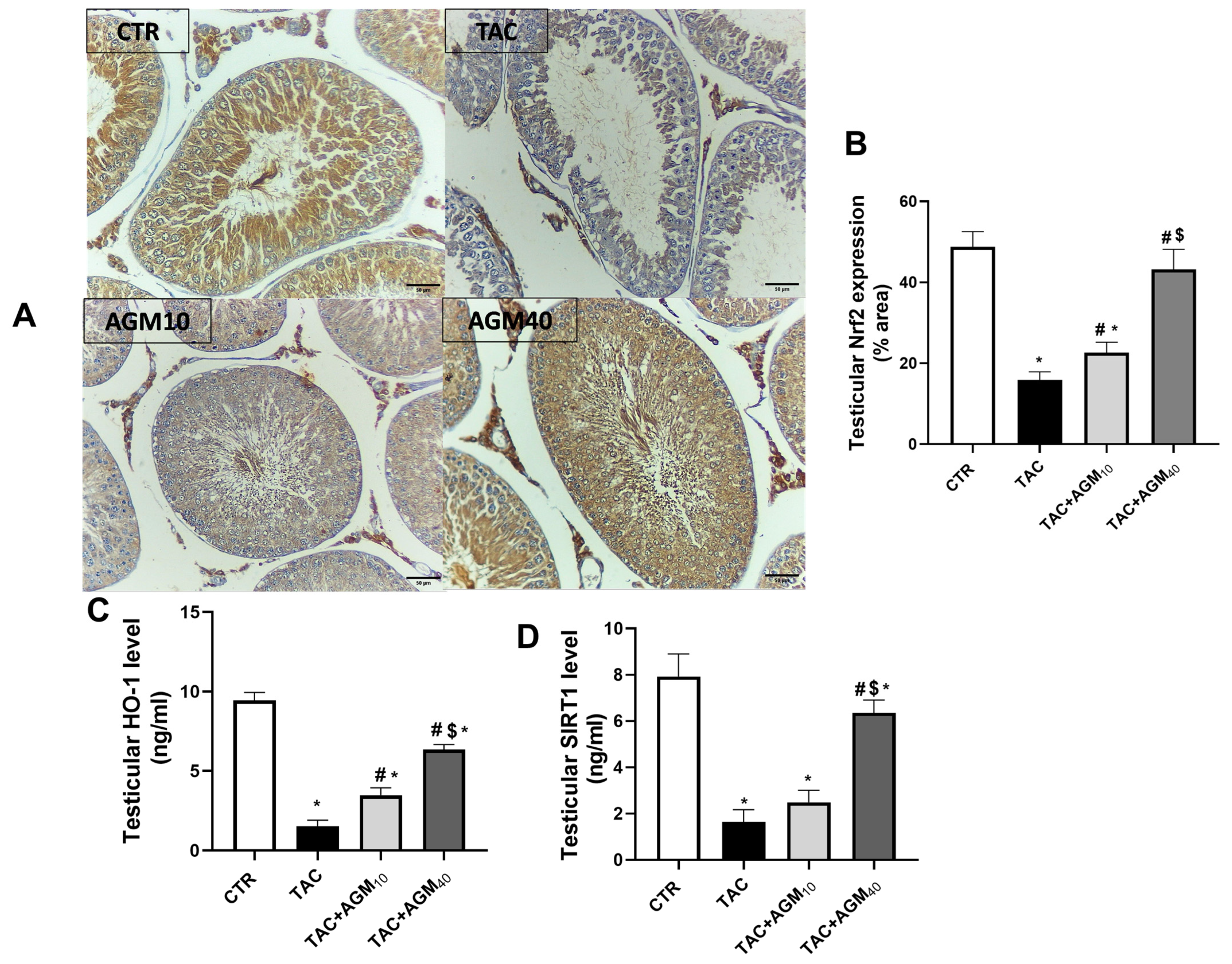
3.5. Impact of AGM10 and AGM40 on TAC-Induced Changes in Nrf2, HO-1, and SIRT1 Expression

Immunostaining of testicular sections against Nrf2 showed high immunoreactivity in spermatocytes, spermatogonia, spermatids, Sertoli cells, and Leydig cells in the control group (Figure 4A). However, the TAC group showed low immunoreactivity in the spermatocytes, spermatogonia, spermatids, and Leydig cells (Figure 4A). The TAC + AGM10 group displayed mild immunoreactivity in the spermatocytes and spermatids (Figure 4A). The TAC + AGM40 group showed moderate immunoreactivity in the spermatocytes, spermatogonia, and spermatids (Figure 4A).
Semiquantitative analysis showed that administration of TAC for 14 days significantly decreased Nrf2 expression by 76.3% compared to the control group (p < 0.05, Figure 4B). However, administration of AGM10 and AGM40 along with TAC significantly increased Nrf2 expression 1.4- and 1.6-fold, respectively, compared to the TAC group (p < 0.05, Figure 4B).
TAC administration also resulted in a significant decrease in HO-1 levels by 84% as compared to the control group (p < 0.05, Figure 4C). Administration of AGM10 or AGM40 along with TAC significantly increased HO-1 levels 2.3-fold and 4.2-fold, respectively, as compared to the TAC group (p < 0.05, Figure 4C). The impact was more significant in the TAC + AGM40 group than the TAC + AGM10 group (p < 0.05, Figure 4C).
TAC significantly decreased SIRT1 levels by 79.1% as compared to the control group (p < 0.05, Figure 4D). Rats treated with AGM10 along with TAC did not significantly increase SIRT1 levels compared to the TAC group (p < 0.05, Figure 4D). Administration of AGM40 along with TAC significantly increased SIRT1 levels 3.9-fold as compared to the TAC group (p < 0.05, Figure 4D). The impact was more significant in the TAC + AGM40 group than the TAC + AGM10 group (p < 0.05, Figure 4D)

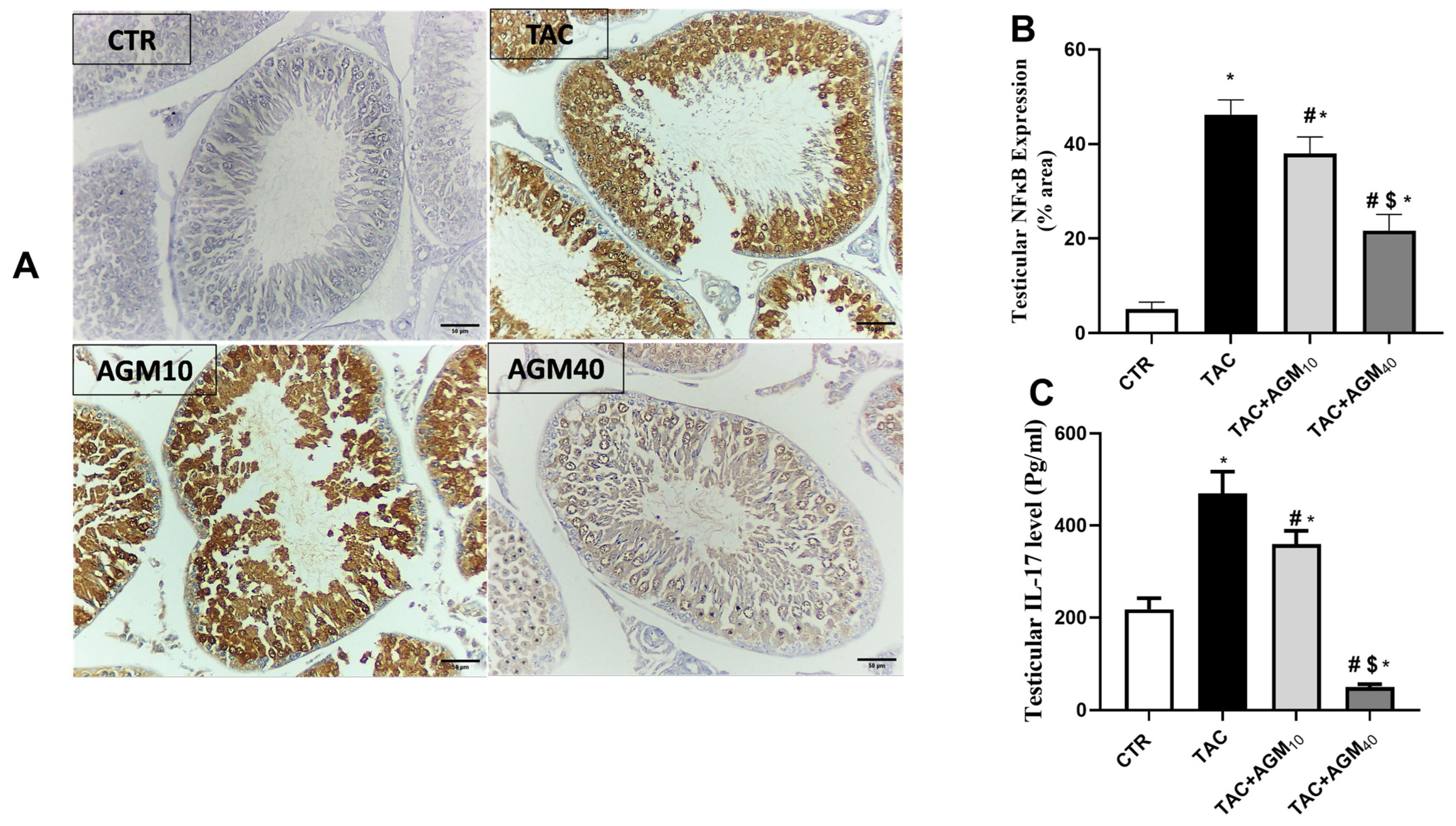
3.6. Impact of AGM10 and AGM40 on TAC-Induced Changes in NF-κB and IL-17 Expressions

Immunostaining of testicular sections against NF-κB showed mild immunoreactivity in spermatocytes, spermatogonia, or spermatids in the control group (Figure 5A). However, the TAC group showed high immunoreactivity in the spermatocytes, spermatogonia, spermatids, and Leydig cells (Figure 5A). Yet, the TAC + AGM10 group showed moderate immunoreactivity in the spermatocytes, spermatogonia, and spermatids (Figure 5A). The TAC + AGM40 group displayed mild immunoreactivity in the spermatocytes, spermatogonia, and spermatids (Figure 5A).
Semiquantitative analysis for NF-κB showed that TAC administration significantly increased NF-κB expression 9.1-fold compared to the control group (p < 0.05, Figure 5B). Yet, administration of AGM10 and AGM40 along with TAC significantly decreased NF-κB expression by 17.9% and 53%, respectively, compared to the TAC group (p < 0.05, Figure 5B). The effect was more so in the TAC + AGM40 group than the TAC + AGM10 group (p < 0.05, Figure 5B).
TAC also significantly increased IL-17 levels 2.1-fold compared to the control group (p < 0.05, Figure 5C). Yet, rats treated with AGM10 and AGM40 along with TAC exhibited significantly decreased IL-17 levels by 23.5% and 89.0%, respectively, as compared to the TAC group (p < 0.05, Figure 5C). The impact was more significant in the TAC + AGM40 group than the TAC + AGM10 group (p < 0.05, Figure 5C).

3.7. Impact of AGM10 and AGM40 on TAC-Induced Changes in Caspase-3 and Bcl-2 Expression

Immunostaining of testicular sections against caspase-3 showed minimal immunoreactivity in spermatocytes and spermatogonia of the control group (Figure 6A). However, the TAC group showed high immunoreactivity in the spermatocytes, spermatogonia, and spermatids (Figure 6A). Yet, the TAC + AGM10 group revealed moderate immunoreactivity in the spermatocytes, spermatogonia, and spermatids (Figure 6A). The TAC + AGM40 group revealed mild immunoreactivity in the spermatocytes and spermatids (Figure 6A).
Semiquantitative analysis for caspase-3 showed that TAC administration significantly increased caspase-3 expression 2.1-fold as compared to the control group (p < 0.05, Figure 6B). Nonetheless, administration of AGM10 and AGM40 along with TAC resulted in a significant decrease in caspase-3 expression by 24.5% and 64.8%, respectively, as compared to the TAC group (p < 0.05, Figure 6B). The effect was more so in the TAC + AGM40 group than the TAC + AGM10 group (p < 0.05, Figure 6B).
Administration of TAC for 14 days significantly decreased Bcl-2 levels by 93.6 % as compared to the control group (p < 0.05, Figure 6C). Yet, administration of AGM10 and AGM40 along with TAC significantly increased Bcl-2 levels 5.8- and 9.1-fold, respectively as compared to the control group (p < 0.05, Figure 6C). The impact was more significant in TAC + AGM40 group than the TAC + AGM10 group (p < 0.05, Figure 6C).

4. Discussion

Results of the current study demonstrated that AGM, a potent inhibitor of iNOS, could ameliorate TAC-induced testicular injury by restoring the oxidant/antioxidant balance, and this effect was associated with modulating the Nrf2/NF-κB/SIRT1/HO-1 pathway, resulting in a reduction in oxidative damage, inflammation, apoptosis, and autophagy by regulating IL-17, caspase-3, and Bcl-2 expression.
TAC treatment in this study resulted in a significant drop in serum testosterone, sperm viability, and sperm count, suggesting the impairment of the reproductive system. TAC had a significant impact on the hypothalamic–pituitary–gonadal axis, as demonstrated by a decline in the level of serum LH, causing spermatogenesis impairment. TAC-induced testicular injury was further confirmed by histopathology, showing sloughing of the germinal epithelium, a loss of the seminiferous tubule’s normal shape, vacuolation, and Leydig cells with pyknotic nuclei. The harmful effects of TAC on the male reproductive system have been investigated by few studies, and the findings include a decrease in testicular weight, a decrease in epithelial thickness in the seminiferous tubules [26,27], a drop in the number of spermatids, and an increase in the number of malformed spermatozoa [28], consistent with our findings.
Both low and high doses of AGM showed a dose-dependent increase in sperm viability, sperm count, LH concentrations, and testosterone levels, suggesting a possible protective role of AGM against TAC-induced testicular damage that was further assured by histopathological analysis.
Testicular injury by TAC is concomitant with a multifactional etiology; however, oxidative stress is regarded as the most common mechanism of TAC-induced testicular injury. Apoptotic cell death is closely related to TAC-induced oxidative stress [29]. A correlation between ROS and male infertility has been documented in some reports [30]. Consequently, several studies have reported that antioxidants could improve sperm parameters [31,32,33].
Studies have demonstrated that TAC administration caused a significant increase in MDA levels, an indicator of lipid peroxidation and a sign of oxidative stress in liver and kidney tissues [34,35,36]. Moreover, TAC also increased iNOS activity, leading to elevated production of nitric oxide and peroxynitrite [37]. GSH is an effective antioxidant present in cells that plays a role in protecting against oxidative stress and is involved in multiple metabolic processes [35] and whose expression has been proven to decrease post treatment with TAC [35,36]. In this study, the administration of TAC for 14 days resulted in a crucial elevation in MDA and iNOS content compared to the control group and a significant decrease in GSH, suggesting a marked oxidant/antioxidant imbalance. On the other hand, groups treated with AGM10 or AGM40 along with TAC exhibited a substantial decrease in MDA and iNOS and a significant increase in GSH levels in testicular tissues as compared to the TAC group, suggesting the antioxidant potential of AGM. These findings are consistent with previous studies that had supported that AGM shows clear therapeutic potential in numerous pathological circumstances by inhibiting oxidative stress and inflammation [38,39]. Thus, this would result in precluding the onset of oxidative stress.
SIRT1, a longevity gene, plays a vital role in regulating various biological processes, including cellular senescence, as well as regulating oxidative stress, inflammation, mitochondrial function, immune responses, cell apoptosis, and cellular differentiation and proliferation [35,40,41]. SIRT1 is a protein deacetylase that controls many survival activities by deacetylating histones and a number of transcription factors, including eNOS and Nrf2, which are involved in apoptosis and the generation of ROS [42]. Also, Vomund et al., 2017 proved that downregulation of SIRT1 under oxidative stress most likely leads to less deacetylation of Nrf2 [43]. SIRT1 enhances the activation of Nrf2, as well as its downstream key antioxidant gene HO-1, to protect cells against oxidative damage by deacetylating the transcription coactivator PPARγ coactivator-1α [35]. Nrf2 is the crucial regulator that counteracts ROS generation by activating antioxidant cascades [35]. Downstream of SIRT1, deacetylated PGC-1α in turn facilitates the upregulation of Nrf2 and antioxidant-related genes [35]. Normally, Keap1, the repressor of Nrf2, binds to Nrf2 in a complex, sequestering it in the cytoplasm [35,36]. Upon stimulation by oxidative and electrophilic chemical signals, Nrf2 is released from Keap1 and translocates into the nucleus, where it binds to antioxidant responsive elements (AREs) [35,36]. Many cellular protective genes, such as HO-1 and NAD(P)H quinine oxidoreductase-1(NQO1) [35,36], are target genes of nuclear Nrf2. Experimental evidence has shown that HO-1 plays an important role in host defense against ROS generation and oxidative injury [35,36]. It also contributes to the anti-inflammatory activity of cells and tissues [35]. Evidence indicates that the decreased expression of HO-1 and increased levels of thiobarbituric acid reactive substances observed in T2DM could be a consequence of a reduced expression of Nrf2 [44]. Lamiri et al., 2021 showed that linear regression analysis revealed a strong and positive correlation between the changes in the Nrf2 and HO-1 expression levels [44].
The SIRT1 and NF-κB signaling are evolutionarily conserved mechanisms in the maintenance of cellular homeostasis, and their interaction regulates the inflammatory response [45]. The nature of this relationship is antagonistic, so that SIRT1 is capable of inhibiting NF-κB signaling and vice versa [46]. Evidence reports that TAC-induced oxidative damage with the subsequent release of damage-associated molecular patterns in the mitochondrial and nuclear DNA provokes inflammatory cells, leading to stimulation of the transcription factor NF-κB and the generation and release of inflammatory mediators including IL-17. TAC is also reported to decrease the expression of Nrf2/HO-1, an essential pathway for shielding cells from inflammation and oxidative stress [47]. Also, previous studies have shown the important involvement of NF-κB in the inflammation, apoptosis, and oxidative damage induced by TAC. When TAC decreases Nrf2 and HO-1 expression, it leads to increased oxidative stress [48] and spermatocyte injury [49]. In addition, the absence of SIRT1 has been reported to contribute to failure of the HPG axis leading to reduced LH, therefore impeding the production of testosterone and estrogen. The reduction in sex hormones leads to compromised sexual behavior and aberrant spermatogenesis [50].
Therefore, we evaluated whether oxidant/antioxidant imbalance is related to imbalance in the SIRT1/Nrf2/NF-κB/Bcl-2 pathway. In this context, TAC administration in the present study markedly decreased SIRT1 and Nrf2 and increased NF-κB expression, which could explain, in part, the induction of oxidative stress. Also, NF-κB is considered as a main regulator of the inflammatory response owing to its ability to regulate the transcription of genes involved in the establishment of immune and inflammatory responses including IL-17 [51,52]. The overproduction of IL-17 leads to chronic inflammation and tissue damage [53,54]. IL-17 was reported to affect the blood–testicular barrier and thus might trigger an inflammatory cascade in the testis. IL17 could disrupt the Sertoli cell barrier. IL17 could enhance the recruitment of other immune cells, specifically macrophages, to the testes, and IL17 itself leads to harmful effect on germ cells and the seminiferous epithelium [44]. Numerous investigations have demonstrated that TAC markedly upregulated the expression of proinflammatory IL-10 and IL-17 while downregulating the expression of anti-inflammatory IL-4 and IFN-γ [55,56]. The findings of the current study revealed a substantial increase in the expression of IL-17 in the TAC-treated group.
Also, SIRT1 has a fundamental role in the regulation of apoptosis. It functions as a NAD+-dependent deacetylase, modulating the activity of various apoptotic factors. By deacetylating and activating key proteins such as p53, SIRT1 can suppress apoptosis in response to stress. It also promotes cell survival via enhancing the expression of anti-apoptotic proteins like Bcl-2 and inhibiting pro-apoptotic factors such as Bax [57,58]. The SIRT1-deficient testes revealed increased apoptosis in male germ cells and other testicular cells [59]. Male germ cell death in SIRT1−/− mice was related to higher p53 activity, as testicular apoptosis is dependent on acetylation-mediated p53 activity [50]. Overall, SIRT1 serves as a critical regulator of apoptosis, promoting cell resilience and longevity under stress conditions.
A previous study revealed that TAC greatly decreased the expression of antiapoptotic factors like Bcl-2 in spermatogenic cells, and significantly changed the expression of proteins linked to apoptosis, such as caspase-3, as compared to the control group [60]. Consistently, our study demonstrated an increase in caspase-3 expression in testicular sections in spermatocytes, spermatogonia, or spermatids in the TAC group along with the reduction in Bcl-2 expression.
The current study demonstrated that rats treated with AGM10 or AGM40 exhibited a significant upregulation of Nrf2, HO-1, SIRT1, and Bcl-2 expression and downregulation of NF-κB, IL-17, and caspase-3 in comparison to the TAC group. The increase in expression of HO-1 by AGM, as a potent iNOS inhibitor, could be a consequence of the increase in the expression of Nrf2 and/or decreased ROS levels as evidenced by lower MDA and iNOS and higher GSH levels. The inhibition of inflammation/oxidative stress observed with AGM through the modulation of Nrf2/HO-1 and NF-κB signaling could be a consequence of the upregulation of SIRT1. Chai et al., 2016 hypothesized that AGM inhibits the induction of ROS through AGM-mediated Nrf2/HO-1 expression [61]. Also, Freitas et al. 2015 proved that AGM protects against corticosterone effects in a hippocampal neuronal cell line by a mechanism that implicates Nrf2/HO-1 signaling [62]. These findings suggested that the antioxidant, anti-inflammatory, and antiapoptotic potential of AGM that are likely related to the inhibition of NF-κB expression and upregulation of SIRT1 and Nrf2 expression.
The limitation of this study is the focus on the acute phase of TAC-induced testicular toxicity, where longer time frame to assess the impact of AGM on chronic testicular damage needs to be considered. In addition, further validation and insight into the molecular mechanisms of AGM in the TAC-treated model is essential for future studies. Genetic approaches, such as using transgenic mice or gene knockdown/knockout techniques, should be employed to strengthen the findings and support the proposed mechanisms.

5. Conclusions

In conclusion, AGM effectively attenuated testicular injuries induced by TAC as evidenced by an elevation in sperm viability, sperm count, LH concentrations, and testosterone levels that was further assured by histopathological and morphometric analyses. This protective effect of AGM might be attributed to the modulation of the Nrf2/NF-κB/SIRT1/HO-1 pathway, resulting in reducing oxidative damage, inflammation, apoptosis, and autophagy by regulating IL-17, caspase-3, and Bcl-2 expression (Figure 7).

Author Contributions

N.A. conducted the experiment and wrote the manuscript draft; O.N., M.N.M. and M.N. analyzed data and edited and revised this manuscript. All authors have read and agreed to the published version of the manuscript.

Funding

This research received no external funding.

Institutional Review Board Statement

The animal study protocol was approved by the Animal Care and Use Committee of Mansoura University (Egypt) (PHARM.PhD.23.02.16, approved date: 7 February 2023).

Informed Consent Statement

Not applicable.

Data Availability Statement

Data are contained within the article.

Acknowledgments

The authors would like to acknowledge Osama Elkashty of Oral Pathology, Faculty of Dentistry, Mansoura University, for assistance with the histopathological and immunohistochemical experiments and Eman Saced Mokhtar Tawfeck, of Medical Histology and the Cell Biology, Faculty of Medicine, Mansoura University, for performing morphometric analysis of the testes.

Conflicts of Interest

The authors declare no conflicts of interest.

References

  1. Hart, A.; Lentine, K.; Smith, J.; Miller, J.; Skeans, M.; Prentice, M.; Robinson, A.; Foutz, J.; Booker, S.; Israni, A. OPTN/SRTR 2019 annual data report: Kidney. Am. J. Transplant. 2021, 21, 21–137. [Google Scholar] [CrossRef] [PubMed]
  2. Armstrong, V.W.; Oellerich, M. New developments in the immunosuppressive drug monitoring of cyclosporine, tacrolimus, and azathioprine. Clin. Biochem. 2001, 34, 9–16. [Google Scholar] [CrossRef] [PubMed]
  3. Ferjani, H.; El Arem, A.; Bouraoui, A.; Achour, A.; Abid, S.; Bacha, H.; Boussema-Ayed, I. Protective effect of mycophenolate mofetil against nephrotoxicity and hepatotoxicity induced by tacrolimus in Wistar rats. J. Physiol. Biochem. 2016, 72, 133–144. [Google Scholar] [CrossRef] [PubMed]
  4. Masuda, H.; Fujihira, S.; Ueno, H.; Kagawa, M.; Katsuoka, Y.; Mori, H. Ultrastructural study on cytotoxic effects of cyclosporine A in spermiogenesis in rats. Med. Electron. Microsc. 2003, 36, 183–191. [Google Scholar] [CrossRef]
  5. Seethalakshmi, L.; Flores, C.; Carboni, A.A.; Bala, R.A.M.; Diamond, D.A.; Menon, M. Cyclosporine: Its Effects on Testicular Function and Fertility in the Prepubertal Rat. J. Androl. 1990, 11, 17–24. [Google Scholar] [CrossRef]
  6. Hisatomi, A.; Fujihira, S.; Fujimoto, Y.; Fujii, T.; Mine, Y.; Ohara, K. Effect of Prograf (FK506) on spermatogenesis in rats. Toxicology 1996, 109, 75–83. [Google Scholar] [CrossRef]
  7. Walensky, L.D.; Dawson, T.M.; Steiner, J.P.; Sabatini, D.M.; Suarez, J.D.; Klinefelter, G.R.; Snyder, S.H. The 12 kD FK506 Binding Protein FKBP12 Is Released in the Male Reproductive Tract and Stimulates Sperm Motility. Mol. Med. 1998, 4, 502–514. [Google Scholar] [CrossRef]
  8. Annett, S.; Moore, G.; Robson, T. FK506 binding proteins and inflammation related signalling pathways; basic biology, current status and future prospects for pharmacological intervention. Pharmacol. Ther. 2020, 215, 107623. [Google Scholar] [CrossRef]
  9. Park, C.; Kwon, D.H.; Hwang, S.J.; Han, M.H.; Jeong, J.-W.; Hong, S.H.; Cha, H.-J.; Hong, S.-H.; Kim, G.-Y.; Lee, H.-J.; et al. Protective Effects of Nargenicin A1 against Tacrolimus-Induced Oxidative Stress in Hirame Natural Embryo Cells. Int. J. Environ. Res. Public Health 2019, 16, 1044. [Google Scholar] [CrossRef]
  10. El-Kashef, D.H.; El-Kenawi, A.E.; Rahim, M.A.; Suddek, G.M.; Salem, H.A. Agmatine improves renal function in gentamicin-induced nephrotoxicity in rats. Can. J. Physiol. Pharmacol. 2016, 94, 278–286. [Google Scholar] [CrossRef]
  11. Xu, W.; Gao, L.; Li, T.; Shao, A.; Zhang, J. Neuroprotective Role of Agmatine in Neurological Diseases. Curr. Neuropharmacol. 2018, 16, 1296–1305. [Google Scholar] [CrossRef] [PubMed]
  12. Akasaka, N.; Fujiwara, S. The therapeutic and nutraceutical potential of agmatine, and its enhanced production using Aspergillus oryzae. Amino Acids 2020, 52, 181–197. [Google Scholar] [CrossRef] [PubMed]
  13. Sharawy, M.H.; Abdelrahman, R.S.; El-Kashef, D.H. Agmatine attenuates rhabdomyolysis-induced acute kidney injury in rats in a dose dependent manner. Life Sci. 2018, 208, 79–86. [Google Scholar] [CrossRef] [PubMed]
  14. El-Sherbeeny, N.A.; Nader, M.A.; Attia, G.M.; Ateyya, H. Agmatine protects rat liver from nicotine-induced hepatic damage via antioxidative, antiapoptotic, and antifibrotic pathways. Naunyn-Schmiedeberg’s Arch. Pharmacol. 2016, 389, 1341–1351. [Google Scholar] [CrossRef]
  15. Ommati, M.M.; Farshad, O.; Mousavi, K.; Taghavi, R.; Farajvajari, S.; Azarpira, N.; Moezi, L.; Heidari, R. Agmatine alleviates hepatic and renal injury in a rat model of obstructive jaundice. PharmaNutrition 2020, 13, 100212. [Google Scholar] [CrossRef]
  16. Battaglia, V.; Rossi, C.A.; Colombatto, S.; Grillo, M.A.; Toninello, A. Different behavior of agmatine in liver mitochondria: Inducer of oxidative stress or scavenger of reactive oxygen species? Biochim. Biophys. Acta 2007, 1768, 1147–1153. [Google Scholar] [CrossRef]
  17. El-Agamy, D.S.; Makled, M.N.; Gamil, N.M. Protective effects of agmatine against D-galactosamine and lipopolysaccharide-induced fulminant hepatic failure in mice. Inflammopharmacology 2014, 22, 187–194. [Google Scholar] [CrossRef]
  18. Dejanovic, B.; Stevanovic, I.; Ninkovic, M.; Stojanovic, I.; Lavrnja, I.; Radicevic, T. Protective effects of agmatine against chlorpromazine-induced toxicity in the liver of wistar rats. Acta Fac. Med. Naissensis 2016, 33, 13. [Google Scholar] [CrossRef]
  19. Sari, Z.A.S. Protective effects of agmatine against gastric mucosal injury induced by nonsteroidal anti-inflammatory drugs: Role of nitric oxide and oxidative stress. J. Gastroenterol. Hepatol. 2020, 35, 1036–1044. [Google Scholar]
  20. Gonzalez, A.M. Agmatine protects against oxidative stress and inflammation in a model of neurodegeneration. J. Neuroinflammation 2018, 15, 239. [Google Scholar]
  21. Hisatomi, A.; Sakuma, S.; Fujiwara, M.; Seki, J. Effect of tacrolimus on the cauda epididymis in rats: Analysis of epididymal biochemical markers or antioxidant defense enzymes. Toxicology 2008, 243, 23–30. [Google Scholar] [CrossRef] [PubMed]
  22. Shigematsu, T.; Tajima, S.; Fu, R.; Zhang, M.; Itoyama, Y.; Tsuchimoto, A.; Egashira, N.; Ieiri, I. The mTOR inhibitor everolimus attenuates tacrolimus-induced renal interstitial fibrosis in rats. Life Sci. 2022, 288, 120150. [Google Scholar] [CrossRef] [PubMed]
  23. Dias, P.H.G.F.; Oliveira, G.A.; Dias, F.G.F.; Gomes, R.D.P.X.; Tambara Filho, R.; Fraga, R.D. Effects of immunosuppression with tacrolimus and mycophenolate mofetil on renal histology and function in single kidney rats submitted to ischemia and reperfusion. Acta Cir. Bras. 2015, 30, 127–133. [Google Scholar] [CrossRef] [PubMed]
  24. Kempinas, W.; Lamano-Carvalho, T.L. A method for estimating the concentration of spermatozoa in the rat cauda epididymidis. Lab. Anim. 1988, 22, 154–156. [Google Scholar] [CrossRef]
  25. Björndahl, L.; Söderlund, I.; Kvist, U. Evaluation of the one-step eosin-nigrosin staining technique for human sperm vitality assessment. Hum. Reprod. 2003, 18, 813–816. [Google Scholar] [CrossRef]
  26. Masuda, T.; Oikawa, H.; Sato, S.; Satodate, R.; Suzuki, K.; Sato, S. Distinguishing small lymph vessels in the portal tracts of human liver from portal veins by immunohistochemistry for α smooth muscle actin. Int. Hepatol. Commun. 1996, 4, 277–282. [Google Scholar] [CrossRef]
  27. Perez-Garcia, L.F.; Dolhain, R.J.E.M.; Vorstenbosch, S.; Bramer, W.; van Puijenbroek, E.; Hazes, J.M.W.; Te Winkel, B. The effect of paternal exposure to immunosuppressive drugs on sexual function, reproductive hormones, fertility, pregnancy and offspring outcomes: A systematic review. Hum. Reprod. Update 2020, 26, 961–1001. [Google Scholar] [CrossRef]
  28. Sasaki, J.C.; Chapin, R.E.; Hall, D.G.; Breslin, W.; Moffit, J.; Saldutti, L.; Enright, B.; Seger, M.; Jarvi, K.; Hixon, M.; et al. Incidence and nature of testicular toxicity findings in pharmaceutical development. Birth Defects Res. Part B Dev. Reprod. Toxicol. 2011, 92, 511–525. [Google Scholar] [CrossRef]
  29. Zheng, H.-L.; Zhang, H.-Y.; Zhu, C.-L.; Li, H.-Y.; Cui, S.; Jin, J.; Piao, S.-G.; Jiang, Y.-J.; Xuan, M.-Y.; Jin, J.-Z. L-Carnitine protects against tacrolimus-induced renal injury by attenuating programmed cell death via PI3K/AKT/PTEN signaling. Acta Pharmacol. Sin. 2021, 42, 77–87. [Google Scholar] [CrossRef]
  30. Takeshima, T.; Usui, K.; Mori, K.; Asai, T.; Yasuda, K.; Kuroda, S.; Yumura, Y. Oxidative stress and male infertility. Reprod. Med. Biol. 2021, 20, 41–52. [Google Scholar] [CrossRef]
  31. Ashour, M.A.; Fatima, W.; Imran, M.; Ghoneim, M.M.; Alshehri, S.; Shakeel, F. A review on the main phytoconstituents, traditional uses, inventions, and patent literature of gum Arabic emphasizing Acacia seyal. Molecules 2022, 27, 1171. [Google Scholar] [CrossRef] [PubMed]
  32. Dimitriadis, F.; Borgmann, H.; Struck, J.P.; Salem, J.; Kuru, T.H. Antioxidant Supplementation on Male Fertility-A Systematic Review. Antioxidants 2023, 12, 836. [Google Scholar] [CrossRef] [PubMed]
  33. Arafa, M.; Agarwal, A.; Majzoub, A.; Panner Selvam, M.K.; Baskaran, S.; Henkel, R.; Elbardisi, H. Efficacy of Antioxidant Supplementation on Conventional and Advanced Sperm Function Tests in Patients with Idiopathic Male Infertility. Antioxidants 2020, 9, 219. [Google Scholar] [CrossRef]
  34. Gonzales, H.M.; McGillicuddy, J.W.; Rohan, V.; Chandler, J.L.; Nadig, S.N.; Dubay, D.A.; Taber, D.J. A comprehensive review of the impact of tacrolimus intrapatient variability on clinical outcomes in kidney transplantation. Am. J. Transplant. Off. J. Am. Soc. Transplant. Am. Soc. Transpl. Surg. 2020, 20, 1969–1983. [Google Scholar] [CrossRef]
  35. Fatima, N.; Sheikh, N.; Satoskar, A.R.; Akhtar, T.; Tayyeb, A.; Ashfaq, I.; Ryan, N.; Ambreen, S.; Jha, B.K.; Oghumu, S. Effect of Short-Term Tacrolimus Exposure on Rat Liver: An Insight into Serum Antioxidant Status, Liver Lipid Peroxidation, and Inflammation. Mediat. Inflamm. 2021, 2021, 6613786. [Google Scholar] [CrossRef]
  36. Al-Harbi, N.O.; Imam, F.; Al-Harbi, M.M.; Iqbal, M.; Nadeem, A.; Sayed-Ahmed, M.M.; Alabidy, A.D.; Almukhallafi, A.F. Olmesartan attenuates tacrolimus-induced biochemical and ultrastructural changes in rat kidney tissue. BioMed Res. Int. 2014, 2014, 607246. [Google Scholar] [CrossRef]
  37. Park, Y.; Lee, H.; Eum, S.H.; Ko, E.J.; Min, J.W.; Yoon, S.-H.; Hwang, W.-M.; Yun, S.-R.; Yang, C.W.; Shin, J.; et al. Combined impact of the inter and intra-patient variability of tacrolimus blood level on allograft outcomes in kidney transplantation. Front. Immunol. 2022, 13, 1037566. [Google Scholar] [CrossRef]
  38. El-Awady, M.S.; Nader, M.A.; Sharawy, M.H. The inhibition of inducible nitric oxide synthase and oxidative stress by agmatine attenuates vascular dysfunction in rat acute endotoxemic model. Environ. Toxicol. Pharmacol. 2017, 55, 74–80. [Google Scholar] [CrossRef]
  39. Ferents, I.V.; Brodyak, I.V.; Lyuta, M.Y.; Burda, V.A.; Gavrylyshyn, G.S.; Sybirna, N.O. Effect of agmatine on the blood system parameters of rats under the condition of experimental diabetes mellitus. Stud. Biol. 2012, 6, 65–72. [Google Scholar] [CrossRef]
  40. Singh, V.; Ubaid, S. Role of Silent Information Regulator 1 (SIRT1) in Regulating Oxidative Stress and Inflammation. Inflammation 2020, 43, 1589–1598. [Google Scholar] [CrossRef]
  41. Luo, J.; Nikolaev, A.Y.; Imai, S.-I.; Chen, D.; Su, F.; Shiloh, A.; Guarente, L.; Gu, W. Negative Control of p53 by Sir2α Promotes Cell Survival under Stress. Cell 2001, 107, 137–148. [Google Scholar] [CrossRef]
  42. Alves-Fernandes, D.K.; Jasiulionis, M.G. The Role of SIRT1 on DNA Damage Response and Epigenetic Alterations in Cancer. Int. J. Mol. Sci. 2019, 20, 3153. [Google Scholar] [CrossRef]
  43. Vomund, S.; Schäfer, A.; Parnham, M.J.; Brüne, B.; von Knethen, A. Nrf2, the Master Regulator of Anti-Oxidative Responses. Int. J. Mol. Sci. 2017, 18, 2772. [Google Scholar] [CrossRef]
  44. Lamiri, W.M. Correlation Between Semen Interleukin-17 and Seminal Parameters in Infertile Males. ALEXMED ePosters 2021, 3, 23–24. [Google Scholar] [CrossRef]
  45. Kauppinen, A.; Suuronen, T.; Ojala, J.; Kaarniranta, K.; Salminen, A. Antagonistic crosstalk between NF-κB and SIRT1 in the regulation of inflammation and metabolic disorders. Cell. Signal. 2013, 25, 1939–1948. [Google Scholar] [CrossRef]
  46. de Gregorio, E.; Colell, A.; Morales, A.; Marí, M. Relevance of SIRT1-NF-κB Axis as Therapeutic Target to Ameliorate Inflammation in Liver Disease. Int. J. Mol. Sci. 2020, 21, 3858. [Google Scholar] [CrossRef]
  47. Borgia, F.; Li Pomi, F.; Vaccaro, M.; Alessandrello, C.; Papa, V.; Gangemi, S. Oxidative stress and phototherapy in atopic dermatitis: Mechanisms, role, and future perspectives. Biomolecules 2022, 12, 1904. [Google Scholar] [CrossRef]
  48. Nady, M.E.; El-Raouf, O.M.A.; El-Sayed, E.-S.M. Linagliptin ameliorates tacrolimus-induced renal injury: Role of Nrf2/HO-1 and HIF-1α/CTGF/PAI-1. Mol. Biol. Rep. 2024, 51, 608. [Google Scholar] [CrossRef]
  49. Yang, W.; Huang, J.; Xiao, B.; Liu, Y.; Zhu, Y.; Wang, F.; Sun, S. Taurine Protects Mouse Spermatocytes from Ionizing Radiation-Induced Damage Through Activation of Nrf2/HO-1 Signaling. Cell. Physiol. Biochem. 2017, 44, 1629–1639. [Google Scholar] [CrossRef]
  50. Khawar, M.B.; Sohail, A.M.; Li, W. SIRT1: A Key Player in Male Reproduction. Life 2022, 12, 318. [Google Scholar] [CrossRef]
  51. He, G.; Karin, M. NF-κB and STAT3—Key players in liver inflammation and cancer. Cell Res. 2011, 21, 159–168. [Google Scholar] [CrossRef]
  52. Liu, T.; Zhang, L.; Joo, D.; Sun, S.-C. NF-κB signaling in inflammation. Signal Transduct. Target. Ther. 2017, 2, 17023. [Google Scholar] [CrossRef]
  53. Rafael-Vidal, C.; Pérez, N.; Altabás, I.; Garcia, S.; Pego-Reigosa, J.M. Blocking IL-17: A Promising Strategy in the Treatment of Systemic Rheumatic Diseases. Int. J. Mol. Sci. 2020, 21, 7100. [Google Scholar] [CrossRef]
  54. von Vietinghoff, S.; Ley, K. Interleukin 17 in vascular inflammation. Cytokine Growth Factor Rev. 2010, 21, 463–469. [Google Scholar] [CrossRef]
  55. Bahrami-Asl, Z.; Farzadi, L.; Fattahi, A.; Yousefi, M.; Quinonero, A.; Hakimi, P.; Latifi, Z.; Nejabati, H.R.; Ghasemnejad, T.; Sadigh, A.R.; et al. Tacrolimus Improves the Implantation Rate in Patients with Elevated Th1/2 Helper Cell Ratio and Repeated Implantation Failure (RIF). Geburtshilfe Frauenheilkd. 2020, 80, 851–862. [Google Scholar] [CrossRef]
  56. Li, Y.; Guptill, J.T.; Russo, M.A.; Massey, J.M.; Juel, V.C.; Hobson-Webb, L.D.; Howard, J.F.; Chopra, M.; Liu, W.; Yi, J.S. Tacrolimus inhibits Th1 and Th17 responses in MuSK-antibody positive myasthenia gravis patients. Exp. Neurol. 2019, 312, 43–50. [Google Scholar] [CrossRef]
  57. Kim, M.G.; Kim, D.H.; Lee, H.R.; Lee, J.S.; Jin, S.J.; Lee, H.T. Sirtuin inhibition leads to autophagy and apoptosis in porcine preimplantation blastocysts. Biochem. Biophys. Res. Commun. 2017, 488, 603–608. [Google Scholar] [CrossRef] [PubMed]
  58. Kozako, T.; Aikawa, A.; Shoji, T.; Fujimoto, T.; Yoshimitsu, M.; Shirasawa, S.; Tanaka, H.; Honda, S.i.; Shimeno, H.; Arima, N.; et al. High expression of the longevity gene product SIRT1 and apoptosis induction by sirtinol in adult T-cell leukemia cells. Int. J. Cancer 2012, 131, 2044–2055. [Google Scholar] [CrossRef]
  59. Coussens, M.; Maresh, J.G.; Yanagimachi, R.; Maeda, G.; Allsopp, R. Sirt1 deficiency attenuates spermatogenesis and germ cell function. PLoS ONE 2008, 3, e1571. [Google Scholar] [CrossRef]
  60. Chung, Y.W.; Chung, M.W.; Choi, S.K.; Choi, S.J.; Choi, S.J.N.; Chung, S.Y. Tacrolimus-Induced Apoptosis is Mediated by Endoplasmic Reticulum–derived Calcium-dependent Caspases-3,-12 in Jurkat Cells. Transplant. Proc. 2018, 50, 1172–1177. [Google Scholar] [CrossRef]
  61. Chai, J.; Luo, L.; Hou, F.; Fan, X.; Yu, J.; Ma, W.; Tang, W.; Yang, X.; Zhu, J.; Kang, W.; et al. Agmatine Reduces Lipopolysaccharide-Mediated Oxidant Response via Activating PI3K/Akt Pathway and Up-Regulating Nrf2 and HO-1 Expression in Macrophages. PLoS ONE 2016, 11, e0163634. [Google Scholar] [CrossRef] [PubMed]
  62. Freitas, A.E.; Egea, J.; Buendía, I.; Navarro, E.; Rada, P.; Cuadrado, A.; Rodrigues, A.L.S.; López, M.G. Agmatine induces Nrf2 and protects against corticosterone effects in hippocampal neuronal cell line. Mol. Neurobiol. 2015, 51, 1504–1519. [Google Scholar] [CrossRef] [PubMed]
Figure 1. Impact of AGM10 and AGM40 on TAC-induced changes in sperm count, serum TT, and LH concentration. TAC was administered along with AGM for 14 days. (A) Sperm count, (B) serum TT, and (C) LH concentration. Data are expressed as means ± SD (n = 6 rats/group). Mean values were compared using one-way ANOVA followed by post hoc Tukey’s multiple comparison test. * indicates significance at p value of <0.05 vs. control group; # indicates significance at p value of <0.05 vs. TAC group; and $ indicates significance at p value of <0.05 vs. TAC + AGM10 group. AGM: agmatine; CTR: control; LH: luteinizing hormone; TAC: tacrolimus; TT: total testosterone.
Pharmaceutics 17 00703 g001
Figure 2. Impact of AGM10 and AGM40 on TAC-induced histopathological changes. Microscopic images of H&E-stained testicular sections. Upper panel: magnification 100x (scale bar 100 µm), and lower panel: magnification 200× (scale bar 50 µm). Black arrow: spermatogonia; red arrow: spermatocytes; blue arrow: rounded and longitudinal spermatids; yellow arrow: Leydig cell; black arrowhead: irregularly shaped seminiferous tubule; red arrowhead: sloughing of germinal epithelium; and yellow arrowhead: vacuolation with pyknotic or necrosis of Leydig cell. AGM: agmatine; CTR: control; TAC: tacrolimus.
Pharmaceutics 17 00703 g002
Figure 3. Impact of AGM10 and AGM40 on TAC-induced changes in oxidative status. (A) MDA content, (B) GSH level, and (C) iNOS level. TAC was administered along with AGM10 or AGM40 for 14 days. Data are expressed as means ± SD (n = 6 rats per group). Mean values were compared via one-way ANOVA followed by post hoc Tukey’s multiple comparison test. * indicates significance at p value of <0.05 vs. CTR group. # indicates significance at p value of <0.05 vs. TAC group and $ indicates significance at p value of <0.05 vs. TAC + AGM10 group. AGM: agmatine; CTR: control; GSH: reduced glutathione; iNOS: inducible nitric oxide; MDA: malondialdehyde; synthase; TAC: tacrolimus.
Pharmaceutics 17 00703 g003
Figure 4. Impact of AGM10 and AGM40 on TAC-induced changes in Nrf2 expression and HO-1 and SIRT1 levels. TAC was administered along with AGM10 or AGM40 for 14 days. (A) Photomicrograph of immunostained testicular sections against Nrf2 (magnification 200× and scale bar 50 µm), (B) semiquantification of Nrf2, (C) HO-1 level, and (D) SIRT1 level. Data are expressed as means ± SD (n = 6 rats per group). Mean values were compared via one-way ANOVA followed by post hoc Tukey’s multiple comparison test. * indicates significance at p value of <0.05 vs. CTR group, # indicates significance at p value of <0.05 vs. TAC group, and $ significance at p value of <0.05 vs. TAC + AGM10 group. AGM: agmatine; CTR: control; HO-1: Heme Oxygenase-1; Nrf2: nuclear factor erythroid 2; SIRT1: Nad-dependent protein deacetylase sirtuin 1; TAC: tacrolimus.
Pharmaceutics 17 00703 g004
Figure 5. Impact of AGM10 and AGM40 on TAC-induced changes in NF-κB and IL-17 levels. TAC was administered along with AGM10 or AGM40 for 14 days. Testes were collected to assess the following: (A) photomicrograph of immunostained testicular sections against NF-κB (magnification 200× and scale bar 50 µm), (B) NF-κB expression, and (C) IL-17 levels. Data are expressed as means ± SD (n = 6 rats/group). Mean values were compared via one-way ANOVA followed by post hoc Tukey’s multiple comparison test. * indicates significance at p value of <0.05 vs. CTR group. # indicates significance at p value of <0.05 vs. TAC group, and $ indicates significance at p value of <0.05 vs. TAC + AGM10 group. AGM: agmatine; CTR: control; IL-17: interleukin 17; NF-κB: nuclear factor kappa B; TAC: tacrolimus.
Pharmaceutics 17 00703 g005
Figure 6. Impact of AGM10 and AGM40 on TAC-induced changes in caspase 3 and Bcl-2 expression. TAC was administered along with AGM10 or AGM40 for 14 days. (A) Photomicrograph of immunostained testicular sections against caspase-3 (magnification 200× and scale bar 50 µm), (B) Caspase-3 expression, and (C) Bcl-2 levels. Data are expressed as means ± SD (n = 6 rats/group). Mean values were compared via one-way ANOVA followed by post hoc Tukey’s multiple comparison test. * indicates significance at p value of <0.05 vs. CTR group. # indicates significance at p value of <0.05 vs. TAC group, and $ indicates significance at p value of <0.05 vs. TAC + AGM10 group. AGM: agmatine; CTR: control; TAC: tacrolimus.
Pharmaceutics 17 00703 g006
Figure 7. The proposed protective mechanism of AGM on TAC-induced testicular toxicity. GSH: reduced glutathione; iNOS: inducible nitric oxide synthase; IL-17: interleukin 17; HO-1: heme oxygenase-1; NF-κB: nuclear factor kappa B; Nrf2: nuclear factor erythroid 2.
Pharmaceutics 17 00703 g007
Table 1. The experimental design.
1Control groupRats received vehicle only for 14 days
2AGM10 groupRats received AGM (10 mg/kg/day, oral) for 14 days [13]
3AGM40 groupRats received AGM (40 mg/kg/day, oral) for 14 days [13]
4TAC groupRats received TAC (5 mg/kg/day, i.p.) for 14 days
5TAC + AGM10 groupRats received TAC (5 mg/kg/day, i.p.) and AGM (10 mg/kg/day, oral) for 14 days simultaneously [13]
6TAC + AGM40 groupreceived TAC (5 mg/kg/day, i.p.) and AGM (40 mg/kg/day, oral) for 14 days simultaneously [13]
AGM: Agmatine; TAC: Tacrolimus.
Table 2. Impact of AGM10 and AGM40 on TAC-induced changes in sperm viability.
Sperm Viability %
After 1 hAfter 2 hAfter 3 h
Control52.50 ± 2.5047.50 ± 2.5042.50 ± 2.50
TAC28.75 ± 4.15 *23.75 ± 4.15 *24.25 ± 2.44 *
TAC + AGM1034.29 ± 4.3229.00 ± 4.1829.00 ± 9.62
TAC + AGM4044.50 ± 3.71 #39.00 ± 4.18 #34.00 ± 4.18 #
TAC was administered along with AGM for 14 days. Data were expressed as means ± SD (n = 6 rats/group). Mean values were compared using one-way ANOVA followed by post hoc Tukey’s multiple comparison test. * Significance at p value of <0.05 vs. control group; # significance at p value of <0.05 vs. TAC group. AGM: agmatine; CTR: control; TAC: tacrolimus.
Table 3. Impact of AGM10 and AGM40 on TAC-induced changes the morphometry of the seminiferous tubules.
The Height of the Seminiferous Epithelium (µm)The Diameter of the
Seminiferous Tubules (µm)
Control70.6 ± 12.3244.4 ± 16.8
TAC42.0 ± 2.0 *182.2 ± 6.8 *
TAC + AGM1059.4 ± 6.7 #198.4 ± 8.1
TAC + AGM4065.4 ± 4.6 #212.6 ± 3.6 #
TAC was administered along with AGM for 14 days. Data are expressed as means ± SD (n = 6 rats/group). Mean values were compared using one-way ANOVA followed by post hoc Tukey’s multiple comparison test. * indicates significance at p value of <0.05 vs. control group and # indicates significance at p value of <0.05 vs. TAC group. AGM: agmatine; CTR: control; TAC: tacrolimus.
Disclaimer/Publisher’s Note: The statements, opinions and data contained in all publications are solely those of the individual author(s) and contributor(s) and not of MDPI and/or the editor(s). MDPI and/or the editor(s) disclaim responsibility for any injury to people or property resulting from any ideas, methods, instructions or products referred to in the content.

