Article

Mechanistic Insights into Sphingomyelin Nanoemulsions as Drug Delivery Systems for Non-Small Cell Lung Cancer Therapy

by
Emma Ramos Docampo
1,2,3,4,†,
Jenifer García-Fernández
1,*,†,
Inés Mármol
5,
Irene Morín-Jiménez
6,
Maria Iglesias Baleato
1,4 and
María de la Fuente Freire
1,6,7
1
Nano-Oncology and Translational Therapeutics Unit, Health Research Institute of Santiago de Compostela (IDIS), University Hospital of Santiago de Compostela (CHUS), SERGAS, 15706 Santiago de Compostela, Spain
2
Molecular Imaging Group, Department of Radiology, Center for Research in Molecular Medicine and Chronic Diseases (CiMUS), 15782 Santiago de Compostela, Spain
3
Nuclear Medicine Department and Molecular Imaging Group, University Hospital CHUS-IDIS, 15782 Santiago de Compostela, Spain
4
Faculty of Pharmacy, Universidade de Santiago de Compostela (USC), 15782 Santiago de Compostela, Spain
5
Institute for Health Research Aragón (IIS Aragón), 50009 Zaragoza, Spain
6
DIVERSA Technologies SL, 15782 Santiago de Compostela, Spain
7
Cancer Network Research (CIBERONC), 28029 Madrid, Spain
*
Author to whom correspondence should be addressed.
These authors contributed equally to this paper.
Pharmaceutics 2025, 17(4), 461; https://doi.org/10.3390/pharmaceutics17040461
Submission received: 31 January 2025 / Revised: 24 March 2025 / Accepted: 26 March 2025 / Published: 2 April 2025
(This article belongs to the Special Issue New Nano-Systems for Imaging, Diagnostics, and Drug Delivery)

Abstract

:
Sphingomyelin nanoemulsions (SNs) are promising drug delivery systems with potential for treating challenging tumors, including non-small cell lung cancer (NSCLC), which has a poor prognosis and a 5-year survival rate below 5%. Understanding the toxicity mechanisms and intracellular behavior of SNs is crucial for optimizing their therapeutic application. This study aims to investigate the interaction between SNs and A549 lung adenocarcinoma cells, focusing on their cytotoxic effects and mechanisms of cellular toxicity. SNs were synthesized and characterized for size, surface charge, and stability. A549 cells were treated with varying concentrations of SNs, and cellular uptake pathways were assessed using inhibitors of energy-dependent processes. Cytotoxicity was evaluated through an alamarBlue assay to determine the IC50 value after 24 h. Mechanisms of toxicity, including lysosomal and mitochondrial involvement, were examined using co-localization studies, mitochondrial membrane potential assays, and markers of apoptosis. SNs exhibited rapid cellular uptake via energy-dependent pathways. The IC50 concentration for A549 cells was 0.89 ± 0.15 mg/mL, suggesting favorable cytocompatibility compared to other nanocarriers. At IC50, SNs induced apoptosis characterized by lysosomal damage, mitochondrial membrane permeabilization, and the release of apoptotic factors. These effects disrupted autophagic flux and contributed to cell death, demonstrating potential for overcoming drug resistance. Resveratrol-loaded SNs showed enhanced cytotoxicity, supporting their application as targeted drug delivery vehicles. This study highlights the potential of SNs as efficient drug delivery systems for NSCLC therapy, offering insights into their cellular interactions and toxicity mechanisms. These findings pave the way for the rational design of SN-based therapeutic platforms for cancer and other mitochondria-related diseases.

1. Introduction

The latest advances in nanomedicine have opened doors to a new era in developing innovative therapies. The use of nanocarriers enhances drug delivery to tumors, thus leading to a higher antitumor effect and a reduction in healthy tissue damage [1,2,3]. Among the great variety of materials for the development of nanocarriers [4,5,6], nanoemulsions made of biodegradable materials are easily manufactured and result in high biocompatibility. Nanoemulsions are finely dispersed systems where nanoscale droplets of one liquid are stabilized within another, typically comprising an oil-in-water or water-in-oil structure. Due to their size (typically under 200 nm), nanoemulsions enhance drug solubility, stability and bioavailability, which is crucial for effective drug delivery to tumours. Compared to other nanocarrier systems, such as liposomes and polymeric nanoparticles, nanoemulsions offer unique advantages, including greater stability, ease of manufacturing, and a high loading capacity for lipophilic drugs [7].
For metastatic lung cancer, nanoemulsions hold special promise [8]. Their small size allows them to penetrate deeply into tumour tissues and access metastatic cells that are often challenging to reach with conventional therapies. Additionally, their ability to encapsulate therapeutic molecules, minimizing their degradation and prolonging circulation time, enhances drug concentration at tumour sites and reduces systemic side effects [9,10]. Employing a nanoemulsion-based delivery system, like the sphingomyelin (SM) and vitamin E (VitE)-based formulations our group has developed (SNs), there is potential for improved therapeutic efficacy and patient outcomes in metastatic lung cancer. These nanoemulsions offer a versatile platform for delivering a variety of therapeutic agents, including hydrophobic drugs, biomolecules, oligonucleotides for gene therapy, and radiometals for diagnostic applications [9,11,12,13,14,15,16], which could provide targeted and sustained treatment options.
Assessing the toxicological profile of nanoparticles is crucial, even though nanostructures based on biodegradable organic materials tend to have low or no toxicity and do not accumulate in the body [17,18]. These specific SNs have been shown to exhibit low toxicity in previous assays reported in our research group. In vitro studies with SW480 and MiaPaCa-2 cells demonstrated cytotoxic concentration (IC50) values 10 to 20 times higher than the average values found in many other lipidic nanosystems [15]. Additional tests with stearylamine and DOTAP incorporation showed no toxic effects and confirmed the biocompatibility of the emulsions in SW480 colon cancer cells [9]. The cytotoxicity profile remained consistent when tested with SW620 and MCF7 cells, as well as with polyethylene glycol decoration [12,16]. In vivo studies with zebrafish and mice also showed very low toxicity and validated their potential for gene therapy development in cancer treatment [13,19].
The biocompatibility of SNs has already been established, as has the efficacy of SNs for targeting tumors, in particular breast [14,16,20] and lung cancer [21,22]. Lung cancer is the most diagnosed type of cancer and the leading cause of cancer-related death worldwide, with approximately 15% of patients surviving 5 years after diagnosis. In non-small cell lung cancer (NSCLC), highly metastatic locally and in distal organs, the 5-year mean survival is lower than 5% [23]. We have recently shown that SNs could be specifically delivered to disseminated lung cancer cells, opening new opportunities to treat metastasis [24,25].
This study aims to enhance our understanding of SNs’ interaction with cancer cells, particularly with lung cancer cells, and the potential underlying mechanisms that could induce toxicity. Our focus encompasses exploring the potential for damage to lysosomes or mitochondria, elucidating the intracellular trajectory of these nanocarriers, and delineating their potential applications in drug delivery.
Since toxicity is often associated with the production of reactive oxygen species (ROS), and interactions with cellular organelles such as the nucleus or endoplasmic reticulum may also be associated with increased toxicity [17], it is essential to investigate how biophysical properties including size, surface area/charge and aggregation state influence cellular interactions. Positively charged particles are typically more cytotoxic [26], while lipid-based nanoparticles exhibit lower toxicity. However, studies have reported that lipid nanocapsules can induce varying degrees of toxicity across different cancer cell lines by affecting signalling pathways and cell fate and inducing ROS and lipid peroxidation [27].
Examining cell interaction and death pathways is crucial when developing therapeutic nanocarriers such as SNs. Thus, identifying different signaling pathways activated by nanocarriers provides valuable insights into their biological effects [28]. Additionally, understanding how nanocarriers interact with cellular components, navigate intracellular pathways, and reach their mitochondrial targets can optimize their design and efficacy, ensuring efficient drug delivery while minimizing off-target effects and toxicity [29,30]. However, challenges arise in achieving intracellular drug accumulation, particularly in mitochondria, due to protective membranes and inner membrane negative potential.
To evaluate SNs’ potential in lung adenocarcinoma, we assessed their cytotoxicity and intracellular distribution in A549 cells, a model for lung adenocarcinoma where SNs have already demonstrated efficiency [21,22]. We compare the SNs-induced cytotoxicity between the A549 cell line and human embryonic kidney cells (HEK293) to assess if there is a cancer-specific cytotoxic effect. This investigation improves our understanding of nanocarrier toxicity profiles, enhancing their use and efficacy as targeted delivery strategies and expanding their therapeutic potential.
Beyond toxicity studies, we explored SNs as a delivery system for resveratrol (RSV), a naturally occurring polyphenolic compound significantly present in grapes and red wine, which has been shown to have beneficial effects in human health, including cardioprotective, antioxidant and anti-inflammatory activities and shows great promise in developing novel anticancer therapies [31,32]. Indeed preclinical studies have revealed that RSV is a potential anticancer agent due to its chemopreventive effects on three major stages of carcinogenesis, including initiation, promotion and progression [33]. RSV induces cell death through the mitochondrial apoptotic pathway, in which mitochondria play a central role in the release of pro-apoptotic factors [34]. However, there are present challenges for clinical applications in terms of poor water solubility, chemical instability, and low bioavailability due to intestinal metabolism [35].
By leveraging the oily core of SNs, which can dissolve large quantities of hydrophobic drugs, we aim to enhance RSV’s solubility and stability while protecting it from hydrolysis and enzymatic degradation. This work evaluates the potential of RSV-loaded SNs as mitochondria-targeting agents in A549 cells, further reinforcing SNs as an effective nanocarrier platform for cancer therapy.
This study provides novel insights into the intracellular behavior and toxicity mechanisms of SNs, highlighting their potential as promising drug delivery systems for NSCLC therapy. Moreover, the efficient encapsulation of RSV within SNs underscores their capability for targeted drug delivery. By elucidating these mechanisms, this work advances the rational design of SN-based therapeutic platforms, not only for NSCLC but also for other mitochondria-associated diseases.

2. Materials and Methods

2.1. Materials

Vitamin E (VitE) was obtained from Calbiochem (Merck-Milipore, Darmstadt, Germany). Sphingomyelin (SM) was purchased from Lipoid GmbH (Ludwigshafen, Germany). SM marked with Cy5 or TopFluor were obtained from Avanti Polar Lipids (Alabaster, AL, USA).
Roswell Park Memorial Institute (RPMI) medium and Dimethyl Sulfoxide (DMSO), Phosphate Buffer Saline (PBS), Trypsin, and Mowiol 4–88 were acquired from Sigma-Aldrich (Darmstadt, Germany). Fetal Bovine Serum (FBS) and Dulbecco’s Modified Eagle’s Medium (DMEM) without L-Glutamine or Phenol Red were obtained from Lomza (Basilea, Switzerland). Streptomycin and penicillin were acquired from Gibco (Waltham, MA, USA). Paraformaldehyde (PFA) 16% was obtained from Thermo Fisher (Waltham, MA, USA). Trypan blue was purchased from Gibco (Waltham, MA, USA).
Fluorescent probes, such as Hoechst 33342, MitoTracker Green FM, LysoTracker Green DND, MitoProbe DiIC1 (5) Assay Kit for Flow Cytometry, Annexin V, and propidium iodide (PI) for Flow Cytometry and alamarBlue Cell Viability Reagent, were obtained from ThermoFisher Scientific (Waltham, MA, USA). Autophagy Assay Kit MAK138 was purchased from Sigma-Aldrich, Merck (Darmstadt, Germany), and 7-Amino-Actinomycin D (7-AAD) was obtained from BD Bioscences (Franklin Lakes, NJ, USA). Resveratrol (RSV) was obtained from Cayman Chemicals (Ann Arbor, MI, USA).

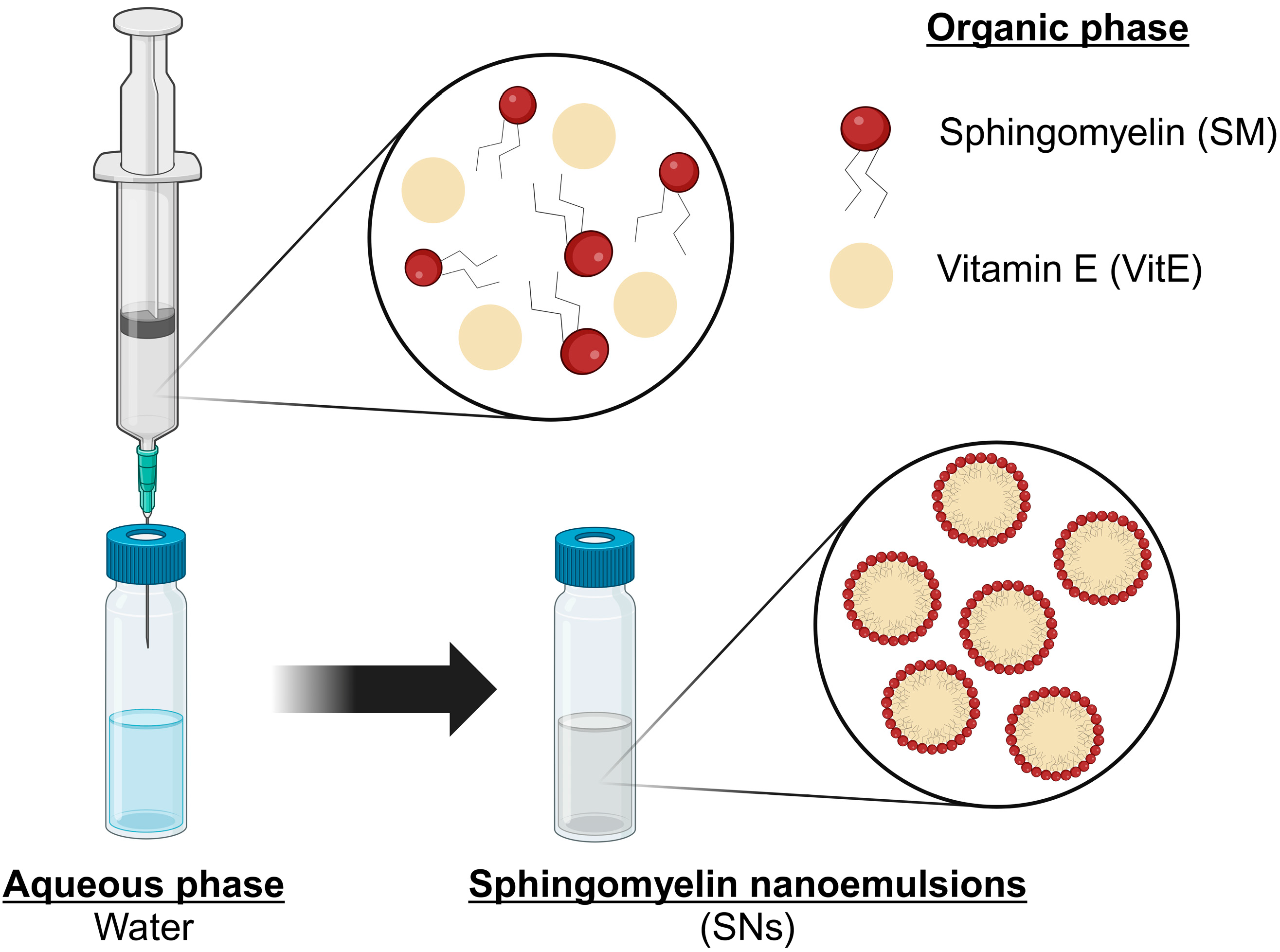
2.2. Preparation and Characterization of SNs

SNs made only of vitamin E and sphingomyelin, two natural components of cell membranes, were formulated by a simple and well-reproducible ethanol injection method, as previously described (Figure 1) [15,20]. One hundred microliters of the organic phase (composed of VitE, SM, and ethanol) were injected into one milliliter of ultrapure water at room temperature in a molar ratio of VitE/SM of 1:0.1 according to previous optimization and characterization studies [15].
The resulting nanosystems were characterized in terms of particle size, polydispersity index (PdI), and Z-Potential using a Zetasizer Nano ZS (Malvern Instruments Ltd., Worcestershire, UK) to measure Dynamic Light Scattering and Laser Doppler Anemometry. For size and polydispersity index measurements, samples were diluted 1:10 (v/v) with ultrapure water and were performed at 25 °C with a detection angle of 173°. For the determination of Z-potential, the formulations were further diluted 1:100 (v/v) in ultrapure water. Colloidal stability was assessed in short-term and long-term stability and relevant biological medium. Short-term stability was performed for early determination of immediate instability behaviour. Colloidal stability was also determined in a relevant biological medium, i.e. supplemented cell culture medium. Briefly, SNs were diluted 1:10 (v/v) with the correspondent medium for incubation (RPMI medium supplemented with 10% FBS) reaching a final concentration of 1 mg/mL. Nanosystems then were incubated at 37 °C under constant horizontal shaking for up to 24 h. For measuring purposes, the previous mixture was further diluted 1:10 (v/v) in water and analyzed by DLS.

2.3. Preparation of Fluorescent-Labeled SNs

Fluorescent-labeled nanosystems were prepared as described in the previous section, Section 2.2. SM-TopFluor was solubilized in ethanol, incorporated in the organic phase, and consequently injected into the aqueous phase for a final concentration of 4 µg/mL. In the case of C16 SM-Cy5-labeled SNs, the fluorophore was dissolved in chloroform, so it had to be evaporated before its incorporation into the ethanolic phase. Then, it was injected into 1 mL of ultrapure water to yield a final concentration of 2 µg/mL. SM-Cy5 and SM-TopFluor were incorporated into the lipidic structure of the nanosystem.

2.4. Preparation of RSV-Loaded SNs (RSV-SNs)

RSV-loaded SNs (RSV-SNs) were prepared analogously as previously described in Section 2.2 and Section 2.3. RSV was solubilized in ethanol at a concentration of 20 mg/mL in the stock solution and incorporated in the organic phase to consequently be injected into the aqueous phase for a final concentration of 100 µg/mL. RSV was incorporated into the lipidic structure of the nanosystem. RSV quantification was performed using High-Performance Liquid Chromatography (HPLC) using a Reverse Phase HPLC Column Poroshell 120-C18, 4.6 Å × 100 mm. Methanol + 2% 2-propanol and MiliQ H2O + 2% 2-propanol were used as mobile phases. The method was based on a 10 min run at 0.5 mL/min. A calibration curve was prepared for RSV ranging from 0 to 100 µg/mL in methanol, and absorbance signal was recorded at 303 nm. RSV quantification from the SNs was performed after ultrafiltration using Amicon 10 KDa filters (Amicon Ultra-0.5 Centrifugal Filter Unit, Merck Millipore, Darmstadt, Germany) under the conditions of 8000× g for 5 min at 15 °C. To calculate encapsulation efficiency (EE%), the following equation was used:
E E % = W i W t × 100
W i = Q u a n t i t y   o f   t h e   i n c o r p o r a t e d   d r u g   t o   t h e   S N s
W t = T o t a l   a m o u n t   o f   t h e   i n c o r p o r a t e d   d r u g   a d d e d   i n i t i a l l y

2.5. Cell Culture

The human lung adenocarcinoma cell line (A549) was purchased from American Type Cell Culture (ATCC) and maintained according to recommendations. Briefly, A549 cells were cultured in RPMI medium supplemented with 10% heat-inactivated FBS and penicillin (50 U/mL) and streptomycin (0.05 mg/mL). The cell line was maintained in a humidified incubator at 37 °C and 5% CO2.
The human embryonic kidney cell line (HEK293) was purchased from American Type Cell Culture (ATCC) and maintained according to recommendations. HEK293 cells were cultured in DMEM supplemented with 10% heat-inactivated FBS and penicillin (50 U/mL) and streptomycin (0.05 mg/mL). The cell line was maintained in a humidified incubator at 37 °C and 5% CO2.

2.6. Cell Viability Assay

Cells were seeded onto 96-well plates at a density of 10,000 cells per well and incubated overnight under standard culture conditions. Then, the cell medium was replaced by SNs and cell medium at the desired concentration. After incubation with SNs, the cell medium was removed, and cells were washed with PBS. Then, cell viability was measured with alamarBlue Cell Viability Reagent (ThermoFisher Scientific, Waltham, MA, USA) according to the manufacturer’s instructions. Briefly, the reagent was diluted in a non-supplemented RPMI medium at a final 1:10 (v/v) added to cells and incubated for 3 h at 37 °C protected from light. Fluorescence was measured at BMG Labtech Plate Reader at 530/590 nm (excitation/emission). Non-treated cells were considered the negative control (100% of viability) and cells treated with Triton X-100 were considered the positive control (0% of viability). For the remaining treated cells, viability was calculated accordingly. For IC50 calculation, cells were incubated for 24 h with the following range of lipid concentrations: 2.750, 1.375, 0.660, 0.330, and 0.165 mg/mL. This concentration range was established following pre-screening assays that identified the cytotoxicity effects of our lipid nanoemulsions within these concentrations. Similarly, other studies performed with lipid nanoparticles reported IC50 values between 1.5 and 3 mg/mL, reinforcing the range proposed here [36]. IC50 was calculated upon a semi-log dose–response curve. Analogously, HEK293 cells were incubated for 72 h with SNs at the same range of lipid concentrations (2.750–0.165 mg/mL), and IC50 was calculated upon a semi-log dose–response curve.
For IC50 of SNs-RSV, cells were incubated for 24 h with the following range of drug concentrations: 219.06, 131.43, 52.57, 26.29, and 13.14 µM. IC50 was calculated upon a semi-log dose–response curve.
For the determination of a non-cytotoxic range, A549 cells were incubated 24, 48, or 72 h with the following range of lipid concentrations: 0.220, 0.110, 0.055, 0.028, and 0.014 mg/mL.

2.7. Cell Death Study: Annexin V and Propidium Iodide (PI)

A549 cells were seeded onto 6-well plates at a density of 300,000 cells per well and incubated overnight under standard culture conditions. Cells were incubated for 24 h with SNs dispersed in complete growth medium at concentrations equivalent to their IC50 values. Cells were harvested and washed with PBS. Then, we re-centrifuged the washed cells. Alexa Fluor 488 annexin V and PI at a concentration of 100 μg/mL were added to each sample. Cells were incubated for 15 min at room temperature covered from light. Finally, samples were carefully mixed in 400 µL 1× annexin-binding buffer, and fluorescence was analyzed with BD FACSAria IIu sorter. Alexa Fluor 488 annexin V fluorescence was monitored using an excitation filter of 488 nm and an emission wavelength of 660 nm. PI fluorescence was monitored using an excitation filter of 488 nm and an emission wavelength of 530 nm.

2.8. Measurement of Caspases 3 and 7 Activation

A549 cells were seeded onto 6-well plates at a density of 300,000 cells per well and incubated overnight under standard culture conditions. Cells were incubated for 24 h with SNs dispersed in complete growth medium at concentrations equivalent to their IC50 values. Cells were harvested with trypsin. CellEvent Caspase-3/7 Green Detection Reagent at a concentration of 500 μM was added to each sample. Cells were incubated for 25 min at 37 °C protected from light and SYTOX AADvanced at a concentration of 1 mM was added to each sample. Cells were incubated for 5 min at 37 °C and protected from light. Finally, samples were carefully mixed, and fluorescence was analyzed with BD FACSAria IIu sorter. CellEvent Caspase 3/7 Green Detection Reagent fluorescence was monitored using an excitation filter of 511 and an emission wavelength of 533 nm. SYTOX AADvanced fluorescence was monitored using an excitation filter of 546 nm and an emission wavelength of 647 nm.

2.9. Determination of SNs Uptake by Confocal Microscopy

A549 cells were seeded onto an 8-well µ-chamber at a density of 70,000 cells per well and incubated overnight under standard culture conditions. The cell medium was replaced by 0.176 mg/mL of total lipid concentration of SN-labeled with TopFluor diluted in RPMI supplemented with 10% FBS and penicillin/streptomycin. Cells were incubated with the SNs for 30 min, 1 h, 2 h, and 4 h and then fixed with PFA 4% overnight. Samples were analyzed with a Multiphoton Microscope Leica TCS SP5 MP. TopFluor fluorescence was monitored using an excitation filter of 495 nm and an emission wavelength of 503 nm.

2.10. Determination of Lysosomal Co-Localization

A549 cells were seeded at FluoroDish at 300,000 cells per well and maintained overnight under standard culture conditions. Cells were incubated for 2 h protected from light with Cy5-labelled SNs dispersed in complete growth medium at concentrations equivalent to their IC50 values. Cells were washed once with PBS and nuclei were stained with Hoechst 33,342 (stock dilution 10 mg/mL was diluted 1:1000 in PBS). After 15 min incubation protected from light at room temperature, 1 μL LysoTracker Green DND 5 × 104 nM was added to each well and fluorescence was analyzed immediately with Multiphoton Microscope Leica TCS SP5 MP. Hoechst 33,342 fluorescence was monitored using an excitation filter of 460 nm and an emission wavelength of 350 nm. LysoTracker Green FM fluorescence was monitored using an excitation filter of 504 nm and an emission wavelength of 511 nm. Cy5 fluorescence was monitored using an excitation filter of 647 nm and an emission wavelength of 665 nm.

2.11. Analysis of Acidic Lysosome

A549 cells were seeded at 6-well plates at 300,000 cells per well and incubated overnight under standard culture conditions. Then, cells were treated with SNs at concentrations equivalent to their IC50 values for 24 h. Cells were harvested and washed with PBS. LysoTracker Green DND 5 × 104 nM was added to each sample. Samples were carefully mixed in 500 µL iced-cold PBS and fluorescence was analyzed with BD FACSAria IIu sorter. LysoTracker Green DND was monitored using an excitation filter of 504 nm and an emission wavelength of 511 nm.

2.12. Analysis of Autophagosome Formation

Cells were seeded onto 96-well plates at a density of 10,000 cells per well and incubated overnight under standard culture conditions. Then, cells were treated with SNs dispersed in complete growth medium at concentrations equivalent to their IC50 values for 24 h. Autophagosome Detection Reagent 500× was added to each sample. Cells were incubated for 30 min at 37 °C protected from light and washed twice with PBS. Finally, the fluorescence is read on the plate reader VICTOR Nivo using an excitation filter of 360 nm and an emission wavelength of 520 nm.

2.13. Measurement of Mitochondrial Co-Localization

A549 cells were seeded at FluoroDish at 300,000 cells per well and maintained overnight under standard culture conditions. Cells were incubated with Cy5-labelled SNs at concentrations equivalent to their IC50 values, previously determined. Treated cells were incubated for 2 h and protected from light. Cells were washed with PBS and nuclei were stained with Hoechst 33,342 (stock dilution 10 mg/mL was diluted 1:1000 in PBS). After 15 min incubation in the dark at room temperature, cells were washed with PBS and then MitoTracker Green FM solved in non-supplemented RPMI medium at 150 nM was added to each well. Cells were incubated for 20 min and fluorescence was analyzed with Multiphoton Microscope Leica TCS SP5 MP. Hoechst 33,342 fluorescence was monitored using an excitation filter of 460 nm and an emission wavelength of 350 nm. MitoTracker Green FM fluorescence was monitored using an excitation filter of 490 nm and an emission wavelength of 516 nm. Cy5 fluorescence was monitored using an excitation filter of 647 nm and an emission wavelength of 665 nm.

2.14. Analysis of Mitochondrial Membrane Potential (ψm) Integrity

A549 cells were seeded at 6-well plates at 300,000 cells per well and maintained overnight under standard culture conditions. Then, cells were treated with SNs at concentrations equivalent to their IC50 values for 24 h. Cells were harvested and washed with PBS. DiIC1 diluted in DMSO at a concentration of 10 µM was added to each sample. Cells were incubated for 30 min at 37 °C covered from light and washed twice with PBS. Finally, samples were carefully mixed in 500 µL iced-cold PBS and fluorescence was analyzed with BD FACSAria IIu sorter. DiIC1 fluorescence was monitored using an excitation filter of 638 nm and an emission wavelength of 658 nm.

2.15. Cell Uptake by Energy-Dependent Pathways Determination

A549 cells were seeded at two 6-well plates at a density of 300,000 cells per well and maintained overnight under standard culture conditions. Then, cells were incubated at 4 °C or 37 °C for 30 min. After cells were pre-incubated at 4 °C or 37 °C, fluorescent labeled-SNs were added at 0.08 mg/mL of lipid concentration and incubated for 2 h. After that, cells were harvested and washed twice with PBS. Untreated cells were maintained as a negative control in complete growth media without SNs. Finally, samples were analyzed with BD FACSAria IIu sorter, and the percentage of fluorescent cells—which would have internalized the SNs—was determined. TopFluor fluorescence was monitored using an excitation filter of 495 nm and an emission wavelength of 503 nm.

2.16. Statistical Analysis

Results were shown as mean ± SD. First, the Shapiro–Wilk test, a normality test for small samples (n < 30), was performed to observe whether the data followed a normal distribution or not. The significance of differences between two groups was determined via the Student’s t-test, and the significance of differences between more groups was determined via one-way analysis of variance (ANOVA). All statistics were conducted using GraphPad Prism 8.0 (Dotmatics, Boston, MA, USA), and differences were considered significant if p < 0.05 (*), p < 0.01 (**), p < 0.001 (***), p < 0.0001 (****).

3. Results

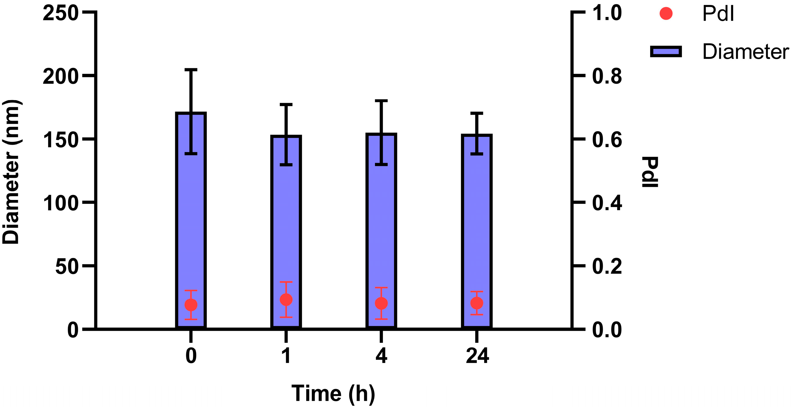
3.1. Preparation and Characterization of SNs

SNs were prepared and characterized as previously reported [22], and the results show a mean size of 113 ± 12 nm, a polydispersity index (PdI) of 0.10 ± 0.05, and a zeta potential of −11.8 ± 1.0 mV, in agreement with previously cited works [16]. Measurements were performed with n = 20 for size and PdI, and n = 6 for Z-potential. The PdI, a critical parameter reflecting nanoparticle uniformity, was 0.1 throughout the study, indicating a highly monodisperse nature (values ≤0.1 are considered optimal).
Next, the assessment of SNs stability was examined in RPMI medium supplemented with FBS. The results illustrated in Figure 2 confirm the stability of SNs in RPMI medium supplemented with FBS. No significant differences in size or PdI were observed, underscoring the robustness of the nanocarrier under the given conditions.

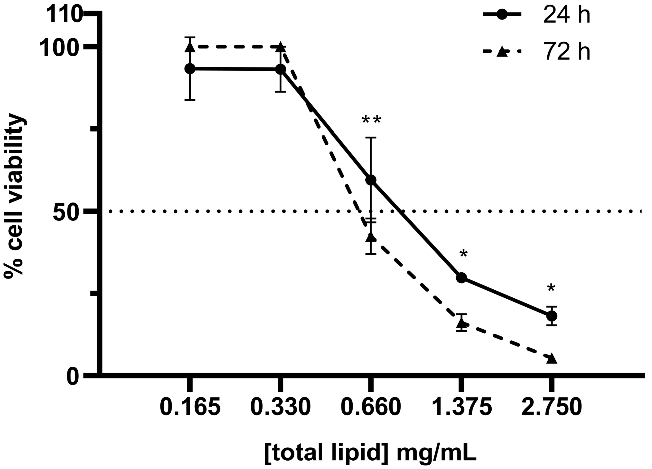
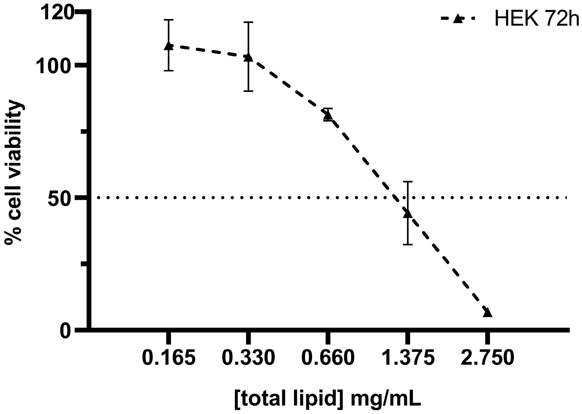
3.2. Cytotoxicity of SNs in the A549 Cell Line

The cytotoxic effect of SNs was assessed using the alamarBlue assay. After 24 and 72 h of incubation with SNs, the IC50 values for the A549 cell line were calculated as 0.89 mg/mL and 0.63 mg/mL, respectively (Figure 3). These results demonstrated a concentration- and time-dependent cytotoxicity. Comparatively, the IC50 values for the HEK293 cell line after 72 h of incubation (Figure 4) were 1.133 mg/mL. This value indicates that SNs are almost twice as cytotoxic to the tumor cell line compared to the normal cell line. To corroborate this, the selectivity index (SI) was calculated to quantify the selectivity of the SNs for cancer cells over normal cells using the equation:
S I = I C 50   f o r   n o r m a l   c e l l s   ( H E K 293 , 72 h ) I C 50   f o r   l u n g   c a n c e r   c e l l s   ( A 549 , 72 h )
A value of 1.8 was obtained, indicating greater selectivity for cancer cells.

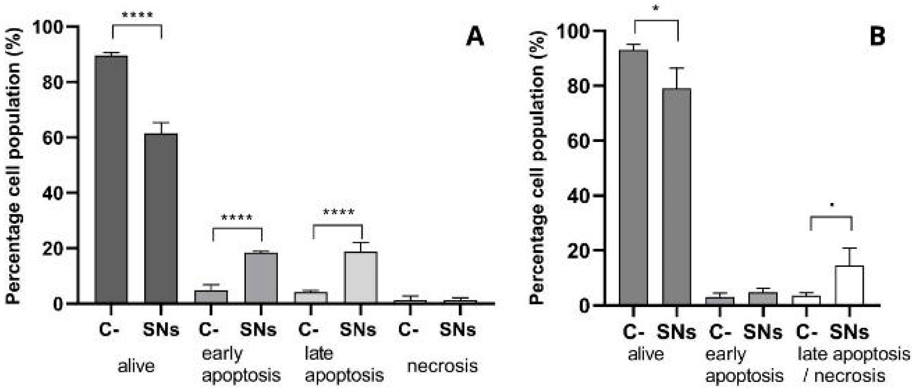
3.3. Mechanisms of Cell Death

Annexin V and PI assays revealed a significant increase in apoptotic A549 cells after 24 h of incubation with the SNs at the concentration equal to IC50 (Figure 5A). Around 20% of cells were detected in early and late apoptosis stages. Further validation using caspase-3/7 assays confirmed the activation of apoptotic pathways. The activation of caspases 3/7 was measured in cells treated with the concentration equal to IC50 of SNs after 24 h incubation (Figure 5B). The levels of caspase expression were compared with untreated cells (maintained in complete growth media without SNs). Although an 80% cell population was detected after treatment with SNs, this does not imply that 80% of cells are alive; rather, it suggests that these cells lack activated caspases 3 and 7.

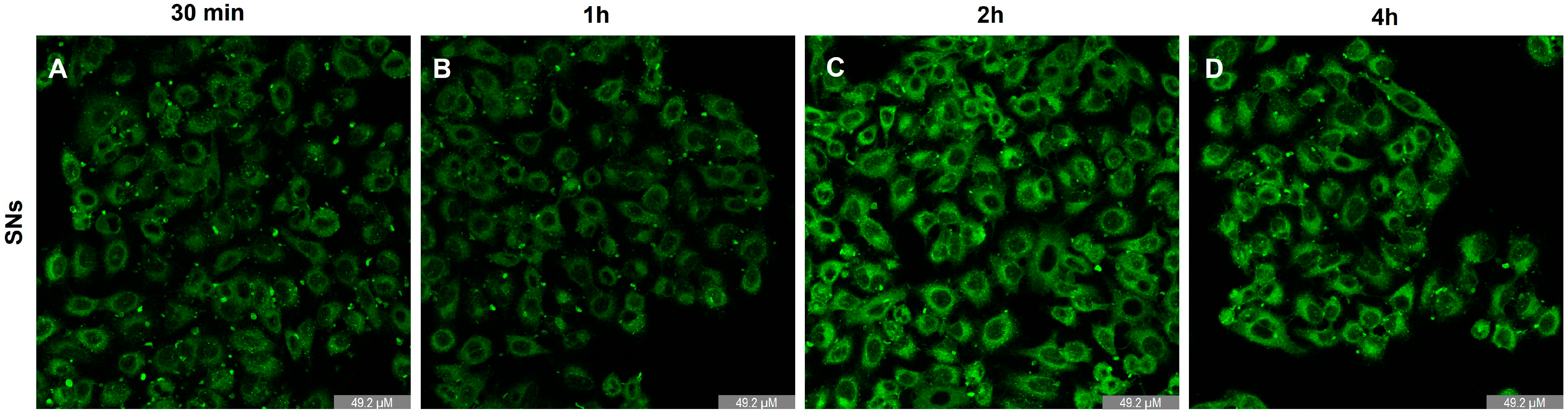
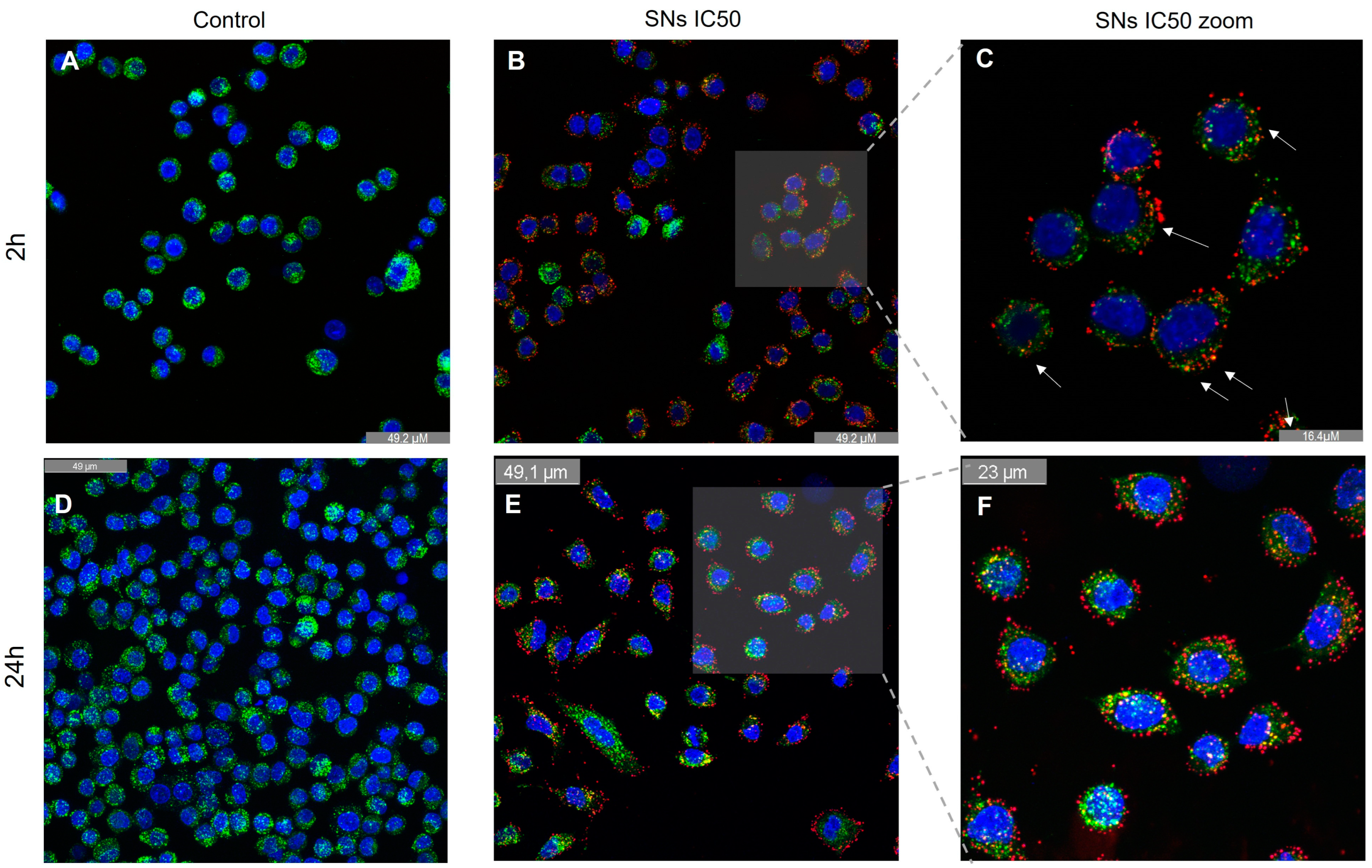
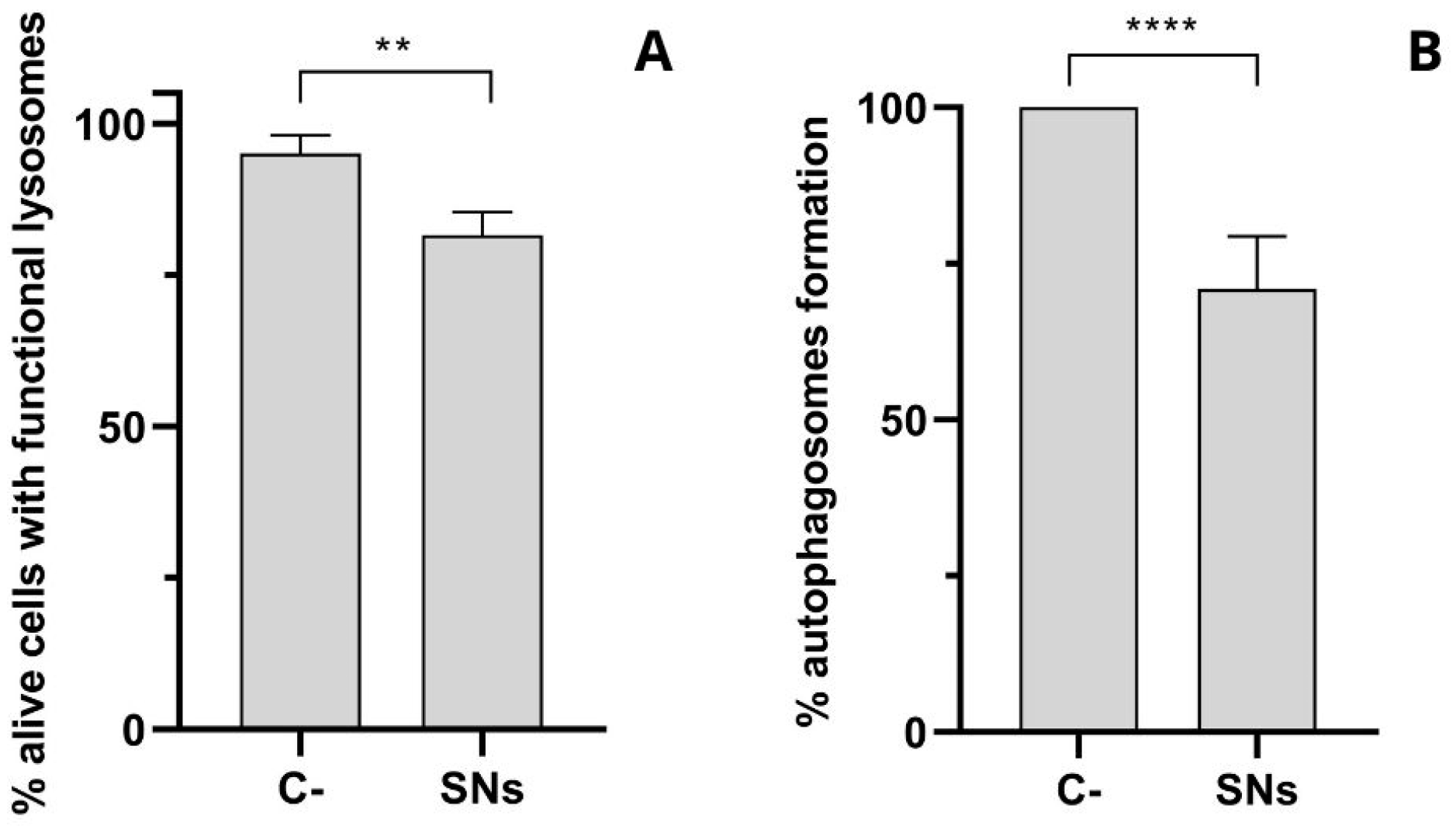
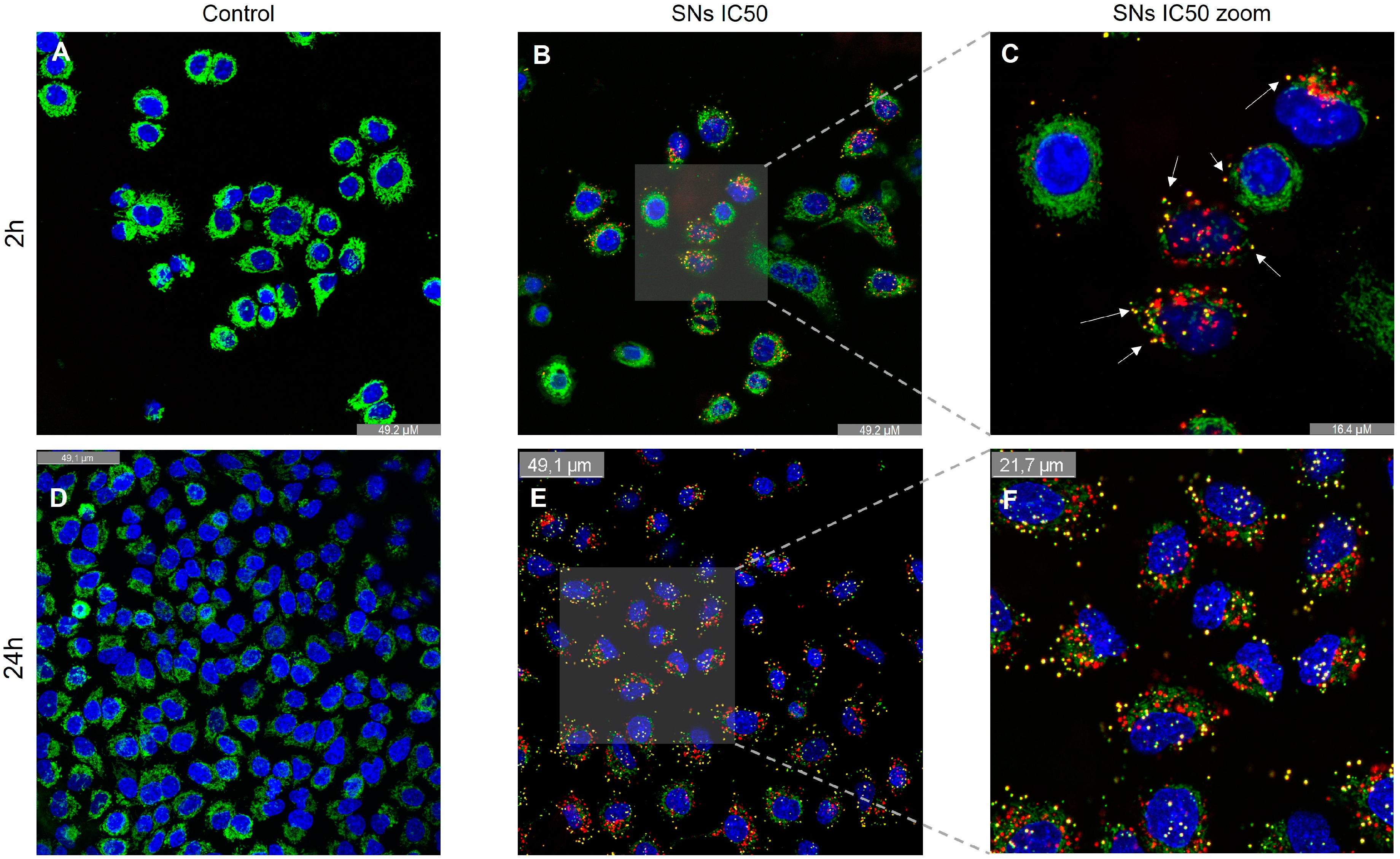
3.4. Intracellular Trafficking and Cell Uptake by Energy-Dependent Pathways Determination

Cellular uptake kinetics were analyzed by incubating A549 cells with TopFluor-labelled SNs for different time intervals (30 min, 1 h, 2 h, and 4 h) (Figure 6). Intracellular fluorescence was observed after just 30 min of incubation (Figure 6A), indicating rapid cellular uptake. To determine whether SN uptake was an energy-dependent process or not, cells were incubated at 4 °C or 37 °C for 30 min. As previously described by other authors [37,38] pre-incubation at 4 °C will impede the uptake of those nanoparticles that enter the cell through energy-dependent mechanisms. After cells were pre-incubated at 4 or 37 °C, fluorescent-labeled SNs were added and incubated for 2 h. Finally, cells were analyzed by flow cytometry, and the percentage of fluorescent cells—which would have internalized the SNs—was determined. Furthermore, SNs demonstrated active, energy-dependent entry into A549 cells, as shown by assays measuring the cell uptake of SNs after 30 min of incubation at 4 °C or 37 °C (Table 1).
To evaluate the presence of SNs in A549 lysosomes, fluorescence microscopy assays were conducted after 2 and 24 h of incubation, as shown in Figure 7A–F. The results indicate that SNs accumulate in lysosomes (Figure 7B,C,E,F). Image analysis using Pearson’s co-localization test, which assesses the correlation of intensity distribution between channels, yielded values of 0.39 after 2 h of incubation and 0.54 after 24 h (+1 indicate perfect co-localization, while values of -1 indicate reverse co-localization) [39].
Lysosomal integrity in response to incubation with the SNs was also analyzed. The loss of lysosomal acidification observed after treatment with SNs, as measured by the decrease in functional, acidic lysosomes using Lysotracker Green and 7-AAD after 24 h of incubation, suggests compromised lysosomal function. Autophagosome detection revealed a decrease in autophagic flux following lysosomal alterations (Figure 8).

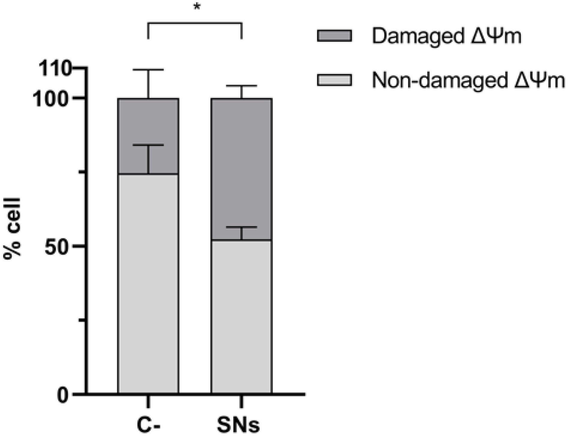
3.5. Mitochondrial Impact

We evaluated the capacity of these SNs to internalize into mitochondria after 2 and 24 h of incubation, as shown in Figure 9. Image analysis using Pearson’s co-localization test revealed a value of 0.53 in mitochondria after 2 h incubation and 0.75 after 24 h incubation, indicating approximately 75% co-localization in this organelle. Some yellow spots appearing outside the cells may correspond to nanoparticles that remained extracellular after washing or could be artefacts of the microscope itself. However, there is no definitive way to confirm this. We further analyzed mitochondrial membrane potential (ΔΨm), strongly related to mitochondrial integrity and function, to assess whether SNs internalization caused mitochondrial damage. As shown in Figure 10, significant changes in ΔΨm were observed after 24 h of incubation with IC50 of SNs, suggesting mitochondrial damage.

3.6. RSV Encapsulation in SNs

The encapsulation efficiency (EE%) of RSV in SNs was determined to be 82% according to Equation (1), based on the regression equation obtained from HPLC measurements after integrating the peak area absorbed at 303 nm: y = 0.653x – 0.266 (R2 = 0.9995). Encapsulated RSV showed improved cytotoxicity with a reduced IC50 value of 85.7 µg/mL compared to 118.5 µg/mL for the free drug (Figure 11, Table 2).

4. Discussion

The findings confirm that SNs exhibit promising characteristics as nanocarriers, including a highly monodisperse nature and stability in biologically relevant conditions. The stability observed in RPMI medium with FBS underscores the potential for SNs in drug delivery applications.
Although the cytotoxicity values expected for lipid nanoparticles considered non-toxic vary depending on the specific formulation and the cell type, these data show relatively low toxicity compared to other similar nanosystems. For example, a study on lipid-based nanocarriers, including liposomes and siRNA-loaded lipid nanoparticles (LNP-siRNA) showed toxicity in HL60 and A549 cells at concentrations above 128 and 16 µg/mL, respectively [40]. Another study on cationic solid lipid nanoparticles deemed them non-toxic with IC50 values from 300 to 900 µg/mL [41]. Additionally, research on lipid nanocapsules found that these nanoparticles could be toxic at high concentrations with cell viability dropping below 20% across all cell lines after incubation with 1 mg/mL for 2.5 h [27]. Another example using PEGylated lipid nanocapsules as anti-cancer drug delivery systems reported IC50 values of 10–20 µg/mL, depending on the cell line [42].
These examples demonstrate that the obtained cytotoxicity values for SNs are among the lowest in comparison to similar lipid nanoformulations considered safe and non-toxic carriers for drug delivery.
The cytotoxicity results highlight the selective toxicity of SNs, with a favorable SI of 1.8, suggesting preferential targeting of cancer cells over normal cells. This specificity may result from increased uptake of SNs in cancer cells due to their altered lipid metabolism, as supported by previous studies. This suggests that SNs preferentially induce cytotoxicity in lung cancer cells compared to normal cells, highlighting the potential of SNs to provide more efficient targeted treatment while minimizing harm to healthy tissues. A favourable SI value is generally considered to be greater than 1.0 [43].
This specific anti-cancer effect of SNs on human lung cancer (A549) could be due to a higher uptake of SNs in cancer cells than in normal ones. This is consistent with other studies reported for the same cell line, where A549 cells also showed higher induced cytotoxicity as a consequence of enhanced cellular uptake of hydroxyapatite nanoparticles than normal bronchial epithelial cells (16HBE) [44]. Further research is needed to investigate the underlying mechanisms of SN-induced selective cytotoxicity to achieve better outcomes for clinical applications.
Apoptosis plays a vital role in maintaining homeostasis and is characterized by several morphological and biochemical changes in cells. Mechanistic studies of cell death confirmed apoptosis as the primary cytotoxic pathway, with early and late apoptosis stages implicated. Early apoptosis is characterized by cell shrinkage, membrane blebbing, and the exposure of “eat-me” signals, facilitating apoptotic cell clearance. This mechanism is preferable for targeted drug delivery to eliminate cancer cells while minimizing inflammation and immune response. Late apoptosis, also known as secondary necrosis, occurs when the plasma membrane becomes permeabilized, leading to the release of immunostimulatory molecules and autoantigens to the immune system [45], which could trigger an immune response against cancer cells. In this case, approximately 20% of the cell population was detected in both early and late apoptosis stages in A549 cells treated in the concentration equal to IC50 of SNs after 24 h of exposure. However, care should be taken to ensure that Annexin V+ is indeed dying since some researchers have suggested that Annexin V can bind healthy cells that have the potential to continue proliferating. Conversely, some cells may not bind Annexin V when alive or dead. These potential issues can be overcome by following the assay to its completion (all cells PI+) or by measuring other features of cell death, such as caspase 3 activity [46]. Thus, these results were further validated with CellEvent Caspase-3/7 Green Detection Reagent and SYTOX AADvanced assay, which estimates the activation of executioner caspases 3 and 7 during the final stages of apoptosis.
Caspase-3 is a key mediator of apoptosis involved in the execution of cell death and has been shown to promote genetic instability and carcinogenesis when activated [47]. Additionally, caspase-3 activities can affect the survival, proliferation, and differentiation of both normal and malignant cells and tissues. Caspase-7, on the other hand, has distinct roles during apoptosis, including promoting ROS production and cell detachment, which may aid in the removal of apoptotic cells from the microenvironment [48]. Caspase activation underscores the potential for SNs to induce controlled apoptotic pathways, reducing inflammation and enhancing targeted effects.
Therefore, the activation of caspase-3 and caspase-7 by a nanocarrier may have different effects on cell fate and the surrounding microenvironment. While caspase-3 activation may lead to cell death and genetic instability, caspase-7 activation may contribute to ROS production and the detachment of apoptotic cells. The upregulation of these apoptotic mediators was observed after exposing A549 cells to SNs. Differences between the early apoptosis population were also detected between both assays, likely due to the limitations inherent to each assay. The Annexin V assay may encounter challenges in interpreting results after mechanical disaggregation of tissues, enzymatic or mechanical detachment of adherent cells, or cell transfection, which can influence phosphatidylserine (PS) flipping and introduce experimental bias. In contrast, the caspase-3/7 assay measures the activity of executioner proteases involved in apoptosis but does not directly measure the externalization of PS, a specific marker of early apoptosis detected by Annexin V.
Consequently, discrepancies arise between assays due to the different markers they detect and the complexities of the apoptotic process. While the direct effect of SNs on the activation of caspases 3 and 7 is not explicitly addressed in previous works, the broader context of caspase activation in cell death pathways and its potential modulation by sphingomyelin metabolism suggests a complex interplay that warrants further investigation in the specific context of SNs and their cytotoxic effects [49]. Overall, these findings point up the complexity of apoptosis and demonstrate that SNs incubation triggers apoptotic cell death in A549 cells, suggesting their potential for targeted cancer therapy while highlighting the necessity for further investigation into the specific pathways involved.
The mechanism by which nanostructures enter cells is largely determined by the physical and interfacial properties of nanoparticles, their interactions with the biological environment, and the properties of the cell membrane. The size, shape, and surface properties (particularly charge and hydrophobicity) of nanosystems can significantly influence cellular uptake pathways and determines their intracellular fate in terms of subcellular location and behaviour as drug nanocarriers [50,51]. In this study, we investigated the cellular uptake of the SNs in the A549 cell line. Intracellular trafficking results elucidated SNs’ rapid uptake and point out an enhancement in the bioavailability, cellular permeability, and cytotoxic effects of potential encapsulated drugs in the SNs. Therefore, the quick internalization within 30 min highlights SNs’ great potential for efficient drug delivery. The increasing fluorescence signal with longer incubation times indicates that cellular uptake is a time-dependent process, in agreement with previous reports in the field [14]. These results align well with previous data obtained by our group in other tumor cell lines, such as the SW620 metastatic colorectal cancer cell line [12] and the breast adenocarcinoma MCF-7 cell line [16]. This suggests that the formulation leads to a significant internalization capacity in cancer cells, irrespective of the time of the tumor. According to Carmona-Ule et al, the fast cell uptake observed might result from the enhanced lipid avidity of cancer cells due to their altered lipid metabolism, which increases the uptake of exogenous lipids [52]. Additionally, the lipidic nature of sphingomyelin, a component of cell membranes, may facilitate the interaction and uptake of these SNs by cells [53].
In addition, SNs’ energy-dependent entry mechanism (Table 1) influences the efficiency and kinetics of internalization, potentially affecting the pharmacokinetics and bioavailability of drugs or therapeutic agents delivered by SNs. This may have implications for the transport of therapeutic agents across biological barriers by influencing the mechanisms involved in cellular internalization.
Given the energy-dependent transport mechanisms through which SNs enter A549 cells, as discussed previously, it was anticipated that one or more endocytic pathways, mostly energy-dependent, could be involved. These pathways typically culminate in lysosomes, responsible for degrading endocytosed materials [54,55]. Consequently, the efficacy of nanoparticle-based therapies might be hampered by lysosomal enzyme degradation.
The lysosomal accumulation (39% of SNs co-localizing at 2 h and 54% after 24 h) indicates that SNs internalization into lysosomes is a time-dependent process. The Pearson correlation index can also be used to study lysosomal escape. As mentioned in the previous section (Section 3.4), the coefficient varies from 0, indicating complete lysosomal escape, to 1 indicating no lysosomal escape [56]. One implication is that it may affect the release of SNs (and their potential cargo) from the lysosomes into the cytoplasm. This could impact cellular processes regulated by sphingomyelin and related molecules, such as signalling pathways and membrane homeostasis. Additionally, the time-dependent nature of lysosomal escape may influence the extent of lysosomal damage and membrane permeabilization, which can in turn affect cell viability and function. The kinetics of lysosomal escape are crucial to be well understood for the design and optimization of SNs-based drug delivery systems. The loss of lysosomal acidification observed could be attributed to the presence of SNs in the lysosome, as nanoparticle accumulation in lysosomes has been reported to adversely affect their function [57]. Additionally, the composition of SNs, resembling cell membranes due to lipid content, may facilitate lysosomal escape [58] (Figure 8A). Our findings suggest that SNs impact lysosomal pH and function, potentially leading to release into the cytoplasm, facilitated by the membrane-like composition of SNs. This could result in enhanced drug release, as toxicity to lysosomes causing lysosomal membrane permeability can trigger the release of encapsulated drugs or therapeutic agents from nanoparticles, allowing for sustained and controlled release over time. Lysosomal integrity and function are also closely linked to autophagy. Lysosomal alterations might disrupt autophagic flux by inhibiting lysosome–autophagosome fusion, compromising autophagosome degradation [50,51]. This disruption has been associated with impaired cell function and cell death. Given our earlier findings of compromised lysosomal function (Figure 7E and Figure 8A), we proceeded to assess autophagosome formation after 24 h of incubation with Autophagosome Detection Reagent. The analysis revealed a significant decrease in autophagosome formation compared to the control (Figure 8B), suggesting an interruption in the autophagic flux. This interruption might be linked to SNs’ capacity to trigger apoptotic cell death (Figure 5), showing the role of dysfunctional autophagy in tumor progression and the potential for regulating this process to enhance the sensitivity and efficacy of cancer therapies [59,60].
Previous studies have suggested that sphingomyelin-containing liposomes can enter mitochondria via membrane fusion, primarily through micropinocytosis [61]. Once inside the cells, these liposomes escape from endosomes and subsequently penetrate mitochondria [62]. Given that SNs are lipid-based nanostructures enriched in sphingomyelin and considering the potential release of the nanoemulsion into the cytoplasm via endosomal escape due to lysosomal damage (Figure 7 and Figure 8A), we hypothesized that SNs might internalize into mitochondria. Moreover, evidence suggests that sphingomyelin accumulation in mitochondria is associated with reperfusion damage, and sphingolipid dysregulation interferes with mitochondrial regulation, leading to dysfunction and cell death [63]. By targeting mitochondrial metabolism, it is possible to disrupt energy production and biosynthetic pathways crucial for cancer cell survival, making mitochondrial targeting important in diseases like cancer or metabolic diseases [34,64]. By looking at the results (Figure 9), SNs’ entry into mitochondria was a time-dependent process and is consistent with lysosomal uptake observations (Figure 7). Image analysis using Pearson’s co-localization test revealed values of 53% co-localization after 2 h and 75% after 24 h of incubation in this organelle. Interestingly, only 54% of SNs co-localized in lysosomes after 24 h of incubation (Figure 7E). This could be attributed to lysosomal leakage caused by rupture of the lysosomal membrane at cytotoxic concentration, enabling SNs to internalize into mitochondria. These results suggest that SNs can accumulate in mitochondria in a time-dependent manner, indicating a dynamic interplay between mitochondria and lysosomes, and the potential accumulation of SNs in the mitochondria over time. This aligns with the literature indicating that sphingolipids, including sphingomyelin, can impact mitochondrial function and contribute to processes like mitochondrial dynamics, mitophagy, and cell death, as well as the dynamic inter-organelle transfer of metabolites between mitochondria and lysosomes in disease pathogenesis [65,66,67].
Concerning the significant changes in ΔΨm observed after 24 h of incubation with IC50 of SNs, this could indicate that SNs internalization into mitochondria may affect their function, consistent with similar findings reported in other studies with lipid nanoemulsions in A549 cells [68,69].
In summary, our results indicate that SNs can internalize or attach to mitochondria following endosomal escape, leading to modifications in mitochondrial membrane potential, indicative of dysfunction. These findings are significant as targeting mitochondria could offer potential therapeutic benefits, particularly in inhibiting cell proliferation in cancer cell lines [70] and emphasize the importance of endosomal escape and lysosomal interactions in optimizing SNs-based delivery systems. Understanding the impact of SNs on ΔΨm is relevant for the development of potential therapeutic applications.
The ability of SNs to penetrate mitochondria (Figure 9) suggests their potential as carriers for drugs targeting this organelle, such as the bioactive compound RSV [71,72]. Recently, several in vitro studies have reported the potential of RSV in managing NSCLC. Some researchers have even suggested that RSV could sensitize NSCLC cell models to conventional chemotherapeutic agents like paclitaxel [73] or etoposide [74]. Despite promising in vivo outcomes, achieving the concentrations required to induce apoptosis in vivo remains challenging [75]. Hence, encapsulating RSV in nanoparticles has emerged as a promising strategy to enhance its in vivo antitumor effect [76].
Using the equation outlined in the methodology section (Equation (1)), we determined an average EE% of 82% for SNs. These results align with previously reported encapsulation efficiencies for mesoporous silica nanoparticles, ranging from 67% to over 93% depending on the specific formulation and encapsulation method used [77,78].
We calculated the IC50 values of SNs-RSV and RSV alone, as shown in Table 2. Encapsulation reduced the IC50 from 118.5 µg/mL (free drug) to 85.7 µg/mL (encapsulated drug), indicating a decrease in the required dose. This reduction in the IC50 value of SNs-RSV compared to RSV alone can be attributed to the improved accumulation into mitochondria and the targeted delivery of RSV achieved through encapsulation in SNs. This facilitates an increased cellular uptake and improved intracellular release of RSV, thereby enhancing its pharmacological activity. Additionally, the presumably sustained release of RSV from SNs prolongs its exposure to target cells, further enhancing efficacy on mitochondrial delivery. This aligns with the increasing focus on mitochondrial targeting in cancer therapy, leveraging SNs’ lipid composition and bioavailability. Despite this improvement, the overall cytotoxic activity of SNs-RSV remains moderate, suggesting the need for further optimization to achieve a more pronounced therapeutic effect. One potential strategy is to enhance drug loading efficiency and optimize the lipid composition of SNs to further increase RSV intracellular availability. Another promising approach would be to explore combination therapies, co-encapsulating RSV with synergistic agents that could enhance its anti-cancer properties. Given RSV’s known multi-target action, its efficacy could be potentiated by combining it with metabolic inhibitors or sensitizers that enhance its apoptotic effects. Furthermore, refining the SNs’ release kinetics to achieve a more controlled and sustained drug release profile could improve therapeutic outcomes.
Future studies should delve deeper into the molecular mechanisms of SNs’ interactions with lysosomes and mitochondria, their impact on cellular metabolism, and their potential for the co-delivery of synergistic therapeutic agents, but these new mechanistic insights of SNs behaviour in healthy and cancer cells provide a valuable starting point. Understanding these pathways will be crucial for advancing SNs in clinical applications and maximizing their therapeutic efficacy. To further validate these in vitro findings, future animal studies will be essential to assess the biodistribution, pharmacokinetics, and therapeutic potential of SNs in vivo. Notably, some studies have already been conducted in mice, showing promising results in terms of targeted drug delivery, and are expected to be published soon [25].

5. Conclusions

The rapid and active penetration of cells by SNs through energy-dependent mechanisms has been demonstrated. Cytotoxicity studies indicate that SNs might have a cancer-specific cytotoxic effect on A549 lung cancer cells compared to normal cells and that cell death occurs primarily through apoptosis at cytotoxic concentrations. Intracellularly, SNs show co-localization with lysosomes and, to a greater extent, with mitochondria. Toxicity studies at cytotoxic concentrations reveal potential damage to both organelles, with lysosomal membrane permeability associated with autophagic flux blockage and mitochondrial outer membrane permeabilization.
Moreover, RSV, selected as an antitumor agent capable of stimulating apoptosis via the mitochondrial apoptotic pathway, has been successfully encapsulated in SNs. The reduction in IC50 value suggests that a lower dose of SNs-RSV is needed for the same therapeutic effect as RSV alone. This implies enhanced pharmacological activity of RSV when encapsulated in SNs, possibly due to improved bioavailability, increased cellular uptake, and targeted delivery.
In summary, the co-localization of SNs with lysosomes and mitochondria, coupled with their effects on lysosomal and mitochondrial permeability, along with their potential for encapsulating mitochondria-targeted drugs, like RSV, present new avenues for targeted drug delivery and the modulation of cellular processes for positive therapeutic outcomes. These findings contribute to the advancement of nanoparticle-based drug delivery systems and hold promises for more effective treatments across various medical applications. However, further research and testing are essential to validate these benefits and ensure the safety and efficacy of the nanoparticles in diverse clinical scenarios.

Author Contributions

Conceptualization, M.d.l.F.F., I.M. and J.G.-F.; methodology, I.M., J.G.-F. and M.d.l.F.F.; validation, E.R.D. and J.G.-F.; formal analysis, E.R.D.; investigation, E.R.D., I.M.-J. and M.I.B.; resources, M.d.l.F.F.; data curation, E.R.D.; writing—original draft preparation, E.R.D. and J.G.-F.; writing—review and editing, J.G.-F., M.I.B. and M.d.l.F.F.; visualization, E.R.D. and J.G.-F.; supervision, I.M., J.G.-F. and M.d.l.F.F.; project administration, M.d.l.F.F.; funding acquisition, M.d.l.F.F. All authors have read and agreed to the published version of the manuscript.

Funding

Authors thank the financial support given by Xunta de Galicia by Grupos de Potencial Crecemento (GPC IN607B2024/14) funded by Axencia Galega de Innovación (GAIN), Consellería de Economía, Emprego e Industria, and RTC2019-07227-1 (Ministry of Economy and Competitiveness, Spain), co-financed by the European Development Regional Fund “A way to achieve Europe” (ERDF). This study was also funded by the Instituto de Salud Carlos III, ISCIII (PI21/01262). E.R.D acknowledges Asociación Española Contra el Cáncer in A Coruña for funding.

Institutional Review Board Statement

Not applicable.

Data Availability Statement

All data underlying the results are available as part of the article and no additional source data is required.

Conflicts of Interest

M.D.L.F. is the co-founder and CEO of DIVERSA Technologies SL. Author I.M-J was employed by the company DIVERSA Technologies SL. Both authors are affiliated with the company DIVERSA but have no potential conflict of interest relationship. The remaining authors declare that the research was conducted in the absence of any commercial or financial relationships that could be construed as a potential conflict of interest.

References

  1. Wei, G.; Wang, Y.; Yang, G.; Wang, Y.; Ju, R. Recent Progress in Nanomedicine for Enhanced Cancer Chemotherapy. Theranostics 2021, 11, 6370–6392. [Google Scholar]
  2. Martins, J.P.; das Neves, J.; de la Fuente, M.; Celia, C.; Florindo, H.; Günday-Türeli, N.; Popat, A.; Santos, J.L.; Sousa, F.; Schmid, R.; et al. The Solid Progress of Nanomedicine. Drug Deliv. Transl. Res. 2020, 10, 726–729. [Google Scholar] [CrossRef]
  3. Shan, X.; Gong, X.; Li, J.; Wen, J.; Li, Y.; Zhang, Z. Current Approaches of Nanomedicines in the Market and Various Stage of Clinical Translation. Acta Pharm. Sin. B 2022, 12, 3028–3048. [Google Scholar] [CrossRef]
  4. Palazzolo, S.; Bayda, S.; Hadla, M.; Caligiuri, I.; Corona, G.; Toffoli, G.; Rizzolio, F. The Clinical Translation of Organic Nanomaterials for Cancer Therapy: A Focus on Polymeric Nanoparticles, Micelles, Liposomes and Exosomes. Curr. Med. Chem. 2018, 25, 4224–4268. [Google Scholar]
  5. Cheng, Z.; Li, M.; Dey, R.; Chen, Y. Nanomaterials for Cancer Therapy: Current Progress and Perspectives. J. Hematol. Oncol. 2021, 14, 85. [Google Scholar] [CrossRef]
  6. Graván, P.; Aguilera-Garrido, A.; Marchal, J.A.; Navarro-Marchal, S.A.; Galisteo-González, F. Lipid-Core Nanoparticles: Classification, Preparation Methods, Routes of Administration and Recent Advances in Cancer Treatment. Adv. Colloid. Interface Sci. 2023, 314, 102871. [Google Scholar] [CrossRef]
  7. Singh, Y.; Meher, J.G.; Raval, K.; Khan, F.A.; Chaurasia, M.; Jain, N.K.; Chourasia, M.K. Nanoemulsion: Concepts, Development and Applications in Drug Delivery. J. Control Release 2017, 252, 28–49. [Google Scholar] [CrossRef]
  8. Pahwa, R.; Sharma, G.; Chhabra, J.; Haider, T.; Anitha, K.; Mishra, N. Nanoemulsion Therapy: A Paradigm Shift in Lung Cancer Management. J. Drug Deliv. Sci. Technol. 2024, 101, 106227. [Google Scholar] [CrossRef]
  9. Nagachinta, S.; Bouzo, B.L.; Vazquez-Rios, A.J.; Lopez, R.; de la Fuente, M. Sphingomyelin-Based Nanosystems (SNs) for the Development of Anticancer MiRNA Therapeutics. Pharmaceutics 2020, 12, 189. [Google Scholar] [CrossRef]
  10. Díez-Villares, S.; Ramos-Docampo, M.A.; da Silva-Candal, A.; Hervella, P.; Vázquez-Ríos, A.J.; Dávila-Ibáñez, A.B.; López-López, R.; Iglesias-Rey, R.; Salgueiriño, V.; de la Fuente, M. Manganese Ferrite Nanoparticles Encapsulated into Vitamin E/Sphingomyelin Nanoemulsions as Contrast Agents for High-Sensitive Magnetic Resonance Imaging. Adv. Healthc. Mater. 2021, 10, e2101019. [Google Scholar] [CrossRef]
  11. Díez-Villares, S.; Pellico, J.; Gómez-Lado, N.; Grijalvo, S.; Alijas, S.; Eritja, R.; Herranz, F.; Aguiar, P.; de la Fuente, M. Biodistribution of 68/67Ga-Radiolabeled Sphingolipid Nanoemulsions by Pet and Spect Imaging. Int. J. Nanomed. 2021, 16, 5923–5935. [Google Scholar] [CrossRef] [PubMed]
  12. Bouzo, B.L.; Lores, S.; Jatal, R.; Alijas, S.; Alonso, M.J.; Conejos-Sánchez, I.; de la Fuente, M. Sphingomyelin Nanosystems Loaded with Uroguanylin and Etoposide for Treating Metastatic Colorectal Cancer. Sci. Rep. 2021, 11, 17213. [Google Scholar] [CrossRef]
  13. Lores, S.; Gámez-Chiachio, M.; Cascallar, M.; Ramos-Nebot, C.; Hurtado, P.; Alijas, S.; López López, R.; Piñeiro, R.; Moreno-Bueno, G.; de la Fuente, M. Effectiveness of a Novel Gene Nanotherapy Based on Putrescine for Cancer Treatment. Biomater. Sci. 2023, 11, 4210–4225. [Google Scholar] [CrossRef]
  14. García-Fernández, J.; Rivadulla Costa, L.; Pinto-Díez, C.; Elena Martín, M.; González, V.M.; de la Fuente Freire, M. Chemical Conjugation of Aptamer-Sphingomyelin Nanosystems and Their Potential as Inhibitors of Tumour Cell Proliferation in Breast Cancer Cells. Nanoscale 2023, 15, 19110–19127. [Google Scholar] [CrossRef]
  15. Bouzo, B.L.; Calvelo, M.; Martín-Pastor, M.; García-Fandiño, R.; De La Fuente, M. In Vitro- In Silico Modeling Approach to Rationally Designed Simple and Versatile Drug Delivery Systems. J. Phys. Chem. B 2020, 124, 5788–5800. [Google Scholar] [CrossRef]
  16. Jatal, R.; Mendes Saraiva, S.; Vázquez-Vázquez, C.; Lelievre, E.; Coqueret, O.; López-López, R.; de la Fuente, M. Sphingomyelin Nanosystems Decorated with TSP-1 Derived Peptide Targeting Senescent Cells. Int. J. Pharm. 2022, 617, 121618. [Google Scholar] [CrossRef]
  17. Strachan, J.B.; Dyett, B.P.; Nasa, Z.; Valery, C.; Conn, C.E. Toxicity and Cellular Uptake of Lipid Nanoparticles of Different Structure and Composition. J. Colloid. Interface Sci. 2020, 576, 241–251. [Google Scholar] [CrossRef]
  18. Najahi-Missaoui, W.; Arnold, R.D.; Cummings, B.S. Safe Nanoparticles: Are We There Yet? Int. J. Mol. Sci. 2021, 22, 385. [Google Scholar] [CrossRef]
  19. Cascallar, M.; Hurtado, P.; Lores, S.; Pensado-López, A.; Quelle-Regaldie, A.; Sánchez, L.; Piñeiro, R.; de la Fuente, M. Zebrafish as a Platform to Evaluate the Potential of Lipidic Nanoemulsions for Gene Therapy in Cancer. Front. Pharmacol. 2022, 13, 1007018. [Google Scholar] [CrossRef]
  20. Saraiva, S.M.; Gutiérrez-Lovera, C.; Martínez-Val, J.; Lores, S.; Bouzo, B.L.; Díez-Villares, S.; Alijas, S.; Pensado-López, A.; Vázquez-Ríos, A.J.; Sánchez, L.; et al. Edelfosine Nanoemulsions Inhibit Tumor Growth of Triple Negative Breast Cancer in Zebrafish Xenograft Model. Sci. Rep. 2021, 11, 1–13. [Google Scholar] [CrossRef]
  21. Vázquez-Ríos, A.J.; Molina-Crespo, Á.; Bouzo, B.L.; López-López, R.; Moreno-Bueno, G.; de la Fuente, M. Exosome-Mimetic Nanoplatforms for Targeted Cancer Drug Delivery. J. Nanobiotechnol. 2019, 17, 85. [Google Scholar] [CrossRef]
  22. De la Fuente Freire, M.; López López, R.; López Bouzo, B.; Vázquez Ríos, A.J.; Alonso Nocelo, M. Nanosystems as Selective Vehicles. European Patent Application Ep 3510995 A1, 19 July 2019. [Google Scholar]
  23. Bray, F.; Laversanne, M.; Sung, H.; Ferlay, J.; Siegel, R.L.; Soerjomataram, I.; Jemal, A. Global Cancer Statistics 2022: GLOBOCAN Estimates of Incidence and Mortality Worldwide for 36 Cancers in 185 Countries. CA Cancer J. Clin. 2024, 74, 229–263. [Google Scholar] [CrossRef] [PubMed]
  24. Vazquez Ríos, A.J.; Bertolini, G.; Alonso Nocelo, M.; Calabuig-Fañanas, S.; Groba de Antas, S.; Rivadulla Costa, L.; Alijas Pérez, S.; Centonze, G.; Murianni, F.; Milione, M.; et al. Taste Receptor Type 1 Member 3 (TAS1R3) Is Enriched in Lung Cancer Stem Cells and Involved in Metastatic Progression of Non-Small Cell Lung Cancer. Cancer Lett. 2025; Submitted. [Google Scholar]
  25. Garcia-Fernandez, J.; Diez-Villares, S.; Cascallar, M.; Rivadulla Costa, L.; Martins, A.S.; Groba de Antas, S.; Tarasco, M.C.; Pantano, E.; de la Fuente, M. Targeted Delivery of Therapeutics to Metastatic Lung Cancer Cells Using Aptamer-Conjugated Nanoemulsions. J. Control. Release, 2025; Submitted. [Google Scholar]
  26. Mitchell, M.J.; Billingsley, M.M.; Haley, R.M.; Wechsler, M.E.; Peppas, N.A.; Langer, R. Engineering Precision Nanoparticles for Drug Delivery. Nat. Rev. Drug Discov. 2021, 20, 101–124. [Google Scholar]
  27. Szwed, M.; Torgersen, M.L.; Kumari, R.V.; Yadava, S.K.; Pust, S.; Iversen, T.G.; Skotland, T.; Giri, J.; Sandvig, K. Biological Response and Cytotoxicity Induced by Lipid Nanocapsules. J. Nanobiotechnol. 2020, 18, 5. [Google Scholar] [CrossRef]
  28. Cummings, B.S.; Schnellmann, R.G. Measurement of Cell Death in Mammalian Cells. Curr. Protoc. Pharmacol. 2004, 25. [Google Scholar] [CrossRef]
  29. Ghosh, P.; Vidal, C.; Dey, S.; Zhang, L. Mitochondria Targeting as an Effective Strategy for Cancer Therapy. Int. J. Mol. Sci. 2020, 21, 3363. [Google Scholar] [CrossRef]
  30. Rodrigues, T.; Ferraz, L.S. Therapeutic Potential of Targeting Mitochondrial Dynamics in Cancer. Biochem. Pharmacol. 2020, 182, 114282. [Google Scholar] [CrossRef]
  31. Mirhadi, E.; Roufogalis, B.D.; Banach, M.; Barati, M.; Sahebkar, A. Resveratrol: Mechanistic and Therapeutic Perspectives in Pulmonary Arterial Hypertension. Pharmacol. Res. 2021, 163, 105287. [Google Scholar] [CrossRef]
  32. Farhan, M.; Rizvi, A. The Pharmacological Properties of Red Grape Polyphenol Resveratrol: Clinical Trials and Obstacles in Drug Development. Nutrients 2023, 15, 4486. [Google Scholar] [CrossRef]
  33. Jiang, L.; Yu, H.; Wang, C.; He, F.; Shi, Z.; Tu, H.; Ning, N.; Duan, S.; Zhao, Y. The Anti-Cancer Effects of Mitochondrial-Targeted Triphenylphosphonium–Resveratrol Conjugate on Breast Cancer Cells. Pharmaceuticals 2022, 15, 1271. [Google Scholar] [CrossRef] [PubMed]
  34. Tsujioka, T.; Sasaki, D.; Takeda, A.; Harashima, H.; Yamada, Y. Resveratrol-Encapsulated Mitochondria-Targeting Liposome Enhances Mitochondrial Respiratory Capacity in Myocardial Cells. Int. J. Mol. Sci. 2021, 23, 112. [Google Scholar] [CrossRef]
  35. Kang, J.H.; Ko, Y.T. Enhanced Subcellular Trafficking of Resveratrol Using Mitochondriotropic Liposomes in Cancer Cells. Pharmaceutics 2019, 11, 423. [Google Scholar] [CrossRef]
  36. Nassimi, M.; Schleh, C.; Lauenstein, H.D.; Hussein, R.; Lbbers, K.; Pohlmann, G.; Switalla, S.; Sewald, K.; Müller, M.; Krug, N.; et al. Low Cytotoxicity of Solid Lipid Nanoparticles in in Vitro and Ex Vivo Lung Models. Inhal. Toxicol. 2009, 21 (Suppl. 1), 104–109. [Google Scholar] [CrossRef]
  37. Kim, J.S.; Yoon, T.J.; Yu, K.N.; Mi, S.N.; Woo, M.; Kim, B.G.; Lee, K.H.; Sohn, B.H.; Park, S.B.; Lee, J.K.; et al. Cellular Uptake of Magnetic Nanoparticle Is Mediated through Energy-Dependent Endocytosis in A549 Cells. J. Vet. Sci. 2006, 7, 321. [Google Scholar] [CrossRef]
  38. dos Santos, T.; Varela, J.; Lynch, I.; Salvati, A.; Dawson, K.A. Effects of Transport Inhibitors on the Cellular Uptake of Carboxylated Polystyrene Nanoparticles in Different Cell Lines. PLoS ONE 2011, 6, e24438. [Google Scholar] [CrossRef]
  39. Adler, J.; Parmryd, I. Quantifying Colocalization by Correlation: The Pearson Correlation Coefficient Is Superior to the Mander’s Overlap Coefficient. Cytom. Part A 2010, 77, 733–742. [Google Scholar] [CrossRef]
  40. Syama, K.; Jakubek, Z.J.; Chen, S.; Zaifman, J.; Tam, Y.Y.C.; Zou, S. Development of Lipid Nanoparticles and Liposomes Reference Materials (II): Cytotoxic Profiles. Sci. Rep. 2022, 12, 18071. [Google Scholar] [CrossRef]
  41. Silva, A.M.; Martins-Gomes, C.; Coutinho, T.E.; Fangueiro, J.F.; Sanchez-Lopez, E.; Pashirova, T.N.; Andreani, T.; Souto, E.B. Soft Cationic Nanoparticles for Drug Delivery: Production and Cytotoxicity of Solid Lipid Nanoparticles (SLNs). Appl. Sci. 2019, 9, 4438. [Google Scholar] [CrossRef]
  42. Hervella, P.; Alonso-Sande, M.; Ledo, F.; Lucero, M.L.; Alonso, M.J.; Garcia-Fuentes, M.; T6d1, E.C.; Barcelona, A.; Vida, C. Pegylated Lipid Nanocapsules with Improved Drug Encapsulation and Controlled Release Properties. Curr. Top. Med. Chem. 2014, 14, 115–1123. [Google Scholar]
  43. Suffness, M. Assays Related to Cancer Drug Discovery. Methods PlantBiochem. Assays Bioactivity 1990, 6, 71–133. [Google Scholar]
  44. Sun, Y.; Chen, Y.; Ma, X.; Yuan, Y.; Liu, C.; Kohn, J.; Qian, J. Mitochondria-Targeted Hydroxyapatite Nanoparticles for Selective Growth Inhibition of Lung Cancer in Vitro and in Vivo. ACS Appl. Mater. Interfaces 2016, 8, 25680–25690. [Google Scholar] [CrossRef] [PubMed]
  45. Poon, I.K.H.; Hulett, M.D.; Parish, C.R. Molecular Mechanisms of Late Apoptotic/Necrotic Cell Clearance. Cell Death Differ. 2010, 17, 381–397. [Google Scholar]
  46. Crowley, L.C.; Marfell, B.J.; Scott, A.P.; Waterhouse, N.J. Quantitation of Apoptosis and Necrosis by Annexin V Binding, Propidium Iodide Uptake, and Flow Cytometry. Cold Spring Harb. Protoc. 2016, 2016, 953–957. [Google Scholar] [CrossRef]
  47. Eskandari, E.; Eaves, C.J. Paradoxical Roles of Caspase-3 in Regulating Cell Survival, Proliferation, and Tumorigenesis. J. Cell Biol. 2022, 221. [Google Scholar] [CrossRef]
  48. Brentnall, M.; Rodriguez-Menocal, L.; De Guevara, R.L.; Cepero, E.; Boise, L.H. Caspase-9, Caspase-3 and Caspase-7 Have Distinct Roles during Intrinsic Apoptosis. BMC Cell Biol. 2013, 14, 1–9. [Google Scholar] [CrossRef]
  49. Lafont, E.; Milhas, D.; Carpentier, S.; Garcia, V.; Jin, Z.X.; Umehara, H.; Okazaki, T.; Schulze-Osthoff, K.; Levade, T.; Benoist, H.; et al. Caspase-Mediated Inhibition of Sphingomyelin Synthesis Is Involved in FasL-Triggered Cell Death. Cell Death Differ. 2010, 17, 642–654. [Google Scholar] [CrossRef]
  50. Donahue, N.D.; Acar, H.; Wilhelm, S. Concepts of Nanoparticle Cellular Uptake, Intracellular Trafficking, and Kinetics in Nanomedicine. Adv. Drug Deliv. Rev. 2019, 143, 68–96. [Google Scholar]
  51. Behzadi, S.; Serpooshan, V.; Tao, W.; Hamaly, M.A.; Alkawareek, M.Y.; Dreaden, E.C.; Brown, D.; Alkilany, A.M.; Farokhzad, O.C.; Mahmoudi, M. Cellular Uptake of Nanoparticles: Journey inside the Cell. Chem. Soc. Rev. 2017, 46, 4218–4244. [Google Scholar]
  52. Carmona-Ule, N.; Abuín-Redondo, C.; Costa, C.; Piñeiro, R.; Pereira-Veiga, T.; Martínez-Pena, I.; Hurtado, P.; López-López, R.; de la Fuente, M.; Dávila-Ibáñez, A.B. Nanoemulsions to Support Ex Vivo Cell Culture of Breast Cancer Circulating Tumor Cells. Mater. Today Chem. 2020, 16. [Google Scholar] [CrossRef]
  53. Zhu, H.; Chen, H.-J.; Wen, H.-Y.; Wang, Z.-G.; Liu, S.-L. Engineered Lipidic Nanomaterials Inspired by Sphingomyelin Metabolism for Cancer Therapy. Molecules 2023, 28, 5366. [Google Scholar] [CrossRef] [PubMed]
  54. Patel, S.; Kim, J.; Herrera, M.; Mukherjee, A.; Kabanov, A.V.; Sahay, G. Brief Update on Endocytosis of Nanomedicines. Adv. Drug Deliv. Rev. 2019, 144, 90–111. [Google Scholar] [CrossRef]
  55. Kou, L.; Sun, J.; Zhai, Y.; He, Z. The Endocytosis and Intracellular Fate of Nanomedicines: Implication for Rational Design. Asian J. Pharm. Sci. 2013, 8, 1–10. [Google Scholar] [CrossRef]
  56. Torres-Vanegas, J.D.; Cifuentes, J.; Puentes, P.R.; Quezada, V.; Garcia-Brand, A.J.; Cruz, J.C.; Reyes, L.H. Assessing Cellular Internalization and Endosomal Escape Abilities of Novel BUFII-Graphene Oxide Nanobioconjugates. Front. Chem. 2022, 10, 974218. [Google Scholar] [CrossRef]
  57. Uzhytchak, M.; Smolková, B.; Lunova, M.; Frtús, A.; Jirsa, M.; Dejneka, A.; Lunov, O. Lysosomal Nanotoxicity: Impact of Nanomedicines on Lysosomal Function. Adv. Drug Deliv. Rev. 2023, 197, 114828. [Google Scholar] [CrossRef]
  58. Liu, C.G.; Han, Y.H.; Kankala, R.K.; Wang, S.B.; Chen, A.Z. Subcellular Performance of Nanoparticles in Cancer Therapy. Int. J. Nanomed. 2020, 15, 675–704. [Google Scholar]
  59. Elimam, H.; El-Say, K.M.; Cybulsky, A.V.; Khalil, H. Regulation of Autophagy Progress via Lysosomal Depletion by Fluvastatin Nanoparticle Treatment in Breast Cancer Cells. ACS Omega 2020, 5, 15476–15486. [Google Scholar] [CrossRef]
  60. Wang, C.; Li, Y.; Tian, Y.; Ma, W.; Sun, Y. Effects of Polymer Carriers on the Occurrence and Development of Autophagy in Drug Delivery. Nanoscale Adv. 2022, 4, 3676–3688. [Google Scholar]
  61. Allemailem, K.S.; Almatroudi, A.; Alsahli, M.A.; Aljaghwani, A.; El-Kady, A.M.; Rahmani, A.H.; Khan, A.A. Novel Strategies for Disrupting Cancer-Cell Functions with Mitochondria-Targeted Antitumor Drug–Loaded Nanoformulations. Int. J. Nanomed. 2021, 16, 3907. [Google Scholar] [CrossRef]
  62. Yamada, Y.; Akita, H.; Kamiya, H.; Kogure, K.; Yamamoto, T.; Shinohara, Y.; Yamashita, K.; Kobayashi, H.; Kikuchi, H.; Harashima, H. MITO-Porter: A Liposome-Based Carrier System for Delivery of Macromolecules into Mitochondria via Membrane Fusion. Biochim. Biophys. Acta Biomembr. 2008, 1778, 423–432. [Google Scholar] [CrossRef]
  63. Knupp, J.; Martinez-Montañés, F.; Van Den Bergh, F.; Cottier, S.; Schneiter, R.; Beard, D.; Chang, A. Sphingolipid Accumulation Causes Mitochondrial Dysregulation and Cell Death. Cell Death Differ. 2017, 24, 2044–2053. [Google Scholar] [CrossRef] [PubMed]
  64. Thuy, L.T.; Lee, S.; Dongquoc, V.; Choi, J.S. Nanoemulsion Composed of α-Tocopherol Succinate and Dequalinium Shows Mitochondria-Targeting and Anticancer Effects. Antioxidants 2023, 12, 437. [Google Scholar] [CrossRef]
  65. Chen, Q.; Fang, H.; Shao, X.; Tian, Z.; Geng, S.; Zhang, Y.; Fan, H.; Xiang, P.; Zhang, J.; Tian, X.; et al. A Dual-Labeling Probe to Track Functional Mitochondria–Lysosome Interactions in Live Cells. Nat. Commun. 2020, 11, 6290. [Google Scholar] [CrossRef]
  66. Wang, Y.; Wang, Y.F.; Li, X.; Wang, Y.; Huang, Q.; Ma, X.; Liang, X.J. Nanoparticle-Driven Controllable Mitochondrial Regulation through Lysosome-Mitochondria Interactome. ACS Nano 2022, 16, 12553–12568. [Google Scholar] [CrossRef]
  67. Sola, F.; Montanari, M.; Fiorani, M.; Barattini, C.; Ciacci, C.; Burattini, S.; Lopez, D.; Ventola, A.; Zamai, L.; Ortolani, C.; et al. Fluorescent Silica Nanoparticles Targeting Mitochondria: Trafficking in Myeloid Cells and Application as Doxorubicin Delivery System in Breast Cancer Cells. Int. J. Mol. Sci. 2022, 23, 3069. [Google Scholar] [CrossRef]
  68. Khan, I.; Bahuguna, A.; Kumar, P.; Bajpai, V.K.; Kang, S.C. In Vitro and in Vivo Antitumor Potential of Carvacrol Nanoemulsion against Human Lung Adenocarcinoma A549 Cells via Mitochondrial Mediated Apoptosis. Sci. Rep. 2018, 8, 144. [Google Scholar] [CrossRef]
  69. Meghani, N.; Patel, P.; Kansara, K.; Ranjan, S.; Dasgupta, N.; Ramalingam, C.; Kumar, A. Formulation of Vitamin D Encapsulated Cinnamon Oil Nanoemulsion: Its Potential Anti-Cancerous Activity in Human Alveolar Carcinoma Cells. Colloids Surf. B Biointerfaces 2018, 166, 349–357. [Google Scholar] [CrossRef]
  70. Fugio, L.B.; Coeli-Lacchini, F.B.; Leopoldino, A.M. Sphingolipids and Mitochondrial Dynamic. Cells 2020, 9, 581. [Google Scholar] [CrossRef]
  71. Li, J.; Fan, Y.; Zhang, Y.; Liu, Y.; Yu, Y.; Ma, M. Resveratrol Induces Autophagy and Apoptosis in Non-Small-Cell Lung Cancer Cells by Activating the NGFR-AMPK-MTOR Pathway. Nutrients 2022, 14, 2413. [Google Scholar] [CrossRef]
  72. Ashrafizadeh, M.; Javanmardi, S.; Moradi-Ozarlou, M.; Mohammadinejad, R.; Farkhondeh, T.; Samarghandian, S.; Garg, M. Natural Products and Phytochemical Nanoformulations Targeting Mitochondria in Oncotherapy: An Updated Review on Resveratrol. Biosci. Rep. 2020, 40, BSR20200257. [Google Scholar] [CrossRef]
  73. Kong, F.; Xie, C.; Zhao, X.; Zong, X.; Bu, L.; Zhang, B.; Tian, H.; Ma, S. Resveratrol Regulates PINK1/Parkin -Mediated Mitophagy via the LncRNA ZFAS1-MiR-150-5p-PINK1 Axis, and Enhances the Antitumor Activity of Paclitaxel against Non-Small Cell Lung Cancer. Toxicol. Res. 2022, 11, 962–974. [Google Scholar] [CrossRef] [PubMed]
  74. Ko, J.C.; Syu, J.J.; Chen, J.C.; Wang, T.J.; Chang, P.Y.; Chen, C.Y.; Jian, Y.T.; Jian, Y.J.; Lin, Y.W. Resveratrol Enhances Etoposide-Induced Cytotoxicity through Down-Regulating ERK1/2 and AKT-Mediated X-Ray Repair Cross-Complement Group 1 (XRCC1) Protein Expression in Human Non-Small-Cell Lung Cancer Cells. Basic. Clin. Pharmacol. Toxicol. 2015, 117, 383–391. [Google Scholar] [CrossRef] [PubMed]
  75. De Oliveira, M.R.; Nabavi, S.F.; Manayi, A.; Daglia, M.; Hajheydari, Z.; Nabavi, S.M. Resveratrol and the Mitochondria: From Triggering the Intrinsic Apoptotic Pathway to Inducing Mitochondrial Biogenesis, a Mechanistic View. Biochim. Biophys. Acta Gen. Subj. 2016, 1860, 727–745. [Google Scholar] [CrossRef]
  76. Yousef, M.; Vlachogiannis, I.A.; Tsiani, E. Effects of Resveratrol against Lung Cancer: In Vitro and In Vivo Studies. Nutrients 2017, 9, 1231. [Google Scholar] [CrossRef]
  77. Zhang, F.; Khan, M.A.; Cheng, H.; Liang, L. Co-Encapsulation of α-Tocopherol and Resveratrol within Zein Nanoparticles: Impact on Antioxidant Activity and Stability. J. Food Eng. 2019, 247, 9–18. [Google Scholar] [CrossRef]
  78. Marinheiro, D.; Ferreira, B.J.M.L.; Oskoei, P.; Oliveira, H.; Daniel-da-silva, A.L. Encapsulation and Enhanced Release of Resveratrol from Mesoporous Silica Nanoparticles for Melanoma Therapy. Materials 2021, 14, 1382. [Google Scholar] [CrossRef]
Figure 1. A schematic of the formulation method using ethanol injection. SNs are composed of vitamin E and sphingomyelin.
Pharmaceutics 17 00461 g001
Figure 2. The measurement of the stability of SNs in RPMI medium supplemented with 10% FBS and penicillin/streptomycin. Changes in diameter (nm) and PdI value over time are shown. Results are expressed against negative control (culture medium) as mean ± standard deviation of at least three independent assays.
Pharmaceutics 17 00461 g002
Figure 3. Percentage of A549 cell viability upon 24 or 72 h incubation with SNs at 2.750–0.165 mg/mL of total lipid. IC50 value at 24 h incubation is 0.89 ± 0.15 mg/mL; at 72 h, incubation is 0.63 ± 0.03 mg/mL. A dashed line indicates the 50% cell viability threshold, serving as a reference point for determining the effective concentration. Data are expressed as mean ± standard deviation of four independent assays. *: p < 0.05. **: p < 0.01.
Pharmaceutics 17 00461 g003
Figure 4. Percentage of HEK293 cell viability upon 72 h incubation with SNs at 2.750–0.165 mg/mL of total lipid. IC50 value at 72 h incubation is 1.133 ± 0.066 mg/mL. A dashed line indicates the 50% cell viability threshold, serving as a reference point for determining the effective concentration. Data are expressed as mean ± standard deviation of four independent assays.
Pharmaceutics 17 00461 g004
Figure 5. The percentage of the cell population of A549 after 24 h incubation with RPMI supplemented with 10% FBS and penicillin/streptomycin (C-), SNs at a concentration equivalent to their IC50. (A) Using Annexin V and PI assay by flow cytometry. The early apoptosis (Annexin V+, 7-PI), the late apoptosis (Annexin V+, PI+), the alive cells (Annexin V, PI), the necrosis (Annexin V, PI+). The results are expressed against a negative control (culture medium) as mean ± standard deviation of at least three independent assays. (B) Determined with CellEvent Caspase-3/7 Green Detection Reagent and SYTOX AADvanced assay by flow cytometry. The percentage of viable A549 cells in early apoptosis or late apoptosis/necrosis. ·: p < 0.1 *: p < 0.05. ****: p < 0.0001.
Pharmaceutics 17 00461 g005
Figure 6. A549 incubated with non-toxic concentrations (0.176 mg/mL of total lipid) of the SNs (green) for 30 min (A), 1 h (B), 2 h (C) or 4 h (D). The results of three independent assays.
Pharmaceutics 17 00461 g006
Figure 7. The effect of SNs on the lysosome of A549 cells after 2 and 24 h incubation. Confocal microscopy images (63×) showing the staining of nuclei in blue (Hoechst 33342) and lysosome in green (LysoTracker Green FM) of A549 cells incubated with the medium upon 2 h (A) and 24 h incubation (D) and with cytotoxic concentrations (IC50) of SNs in red (Cy5) after 2 h (B) and 24 h of incubation (E). Zoom ×3 after 2 h of incubation (C) and zoom ×1.6 after 24 h of incubation (F). The co-localization of lysosome and nanoparticles is marked with white arrows. The results of three independent assays.
Pharmaceutics 17 00461 g007
Figure 8. (A) The percentage of alive cells with functional lysosomes in A549 cells after 24 h incubation with SNs at a concentration equivalent to their IC50, determined with Lysotracker Green and 7-AAD assay by flow cytometry. The results are expressed against negative control (culture medium) as mean ± standard. **: p < 0.01. The results of five independent assays. (B) The percentage of autophagosome formation on A549 cells after 24 h incubation with SNs at a concentration equivalent to their IC50, determined with Autophagosome Detection Reagent assay by plate reader. The results are expressed against a negative control (culture medium) as mean ± standard. ****: p< 0.0001. The results of five independent assays.
Pharmaceutics 17 00461 g008
Figure 9. The internalization of SNs on the mitochondria of A549 cells. Confocal microscopy images (63×) showing the staining of nuclei in blue (Hoechst 33342) and mitochondria in green (MitoTracker Green FM) after incubation with culture medium upon 2 h (A) or 24 h (D) and with cytotoxic concentrations of SNs in red (Cy5) upon 2 h (B) and 24 h (E) of incubation. The co-localization of mitochondria and nanoparticles is marked with white arrows. Zoom ×3 after 2 h of incubation (C) and zoom ×1.7 after 24 h of incubation (F). The results of five independent assays.
Pharmaceutics 17 00461 g009
Figure 10. The effect of SNs on the mitochondrial membrane potential (ΔΨm) of A549 cells. The percentage of total cells with damaged and non-damaged ΔΨm upon 24 h incubation with cytotoxic concentrations (IC50) of SNs are shown. The results are as mean ± standard deviation of at least three independent assays. *: p < 0.05.
Pharmaceutics 17 00461 g010
Figure 11. Percentage of A549 cell viability upon 24-h incubation with SNs-RSV and free RSV at 0.003–0.05 mg/mL of total drug. Dash line represents the 50% cell viability threshold, serving as a reference point for determining the IC50 value.
Pharmaceutics 17 00461 g011
Table 1. SNs uptake by A549 cells was followed upon 30 min pre-incubation at 4 °C or 37 °C and 2 h further incubation with SNs (0.08 mg/mL) at 37 °C to determine whether SNs entrance was energy-dependent. Results are expressed as percentage of fluorescent cells, indicative of SNs entrance. Negative control: untreated cells. Data are expressed as mean ± standard deviation of three independent assays.
ConditionControl4 °C37 °C
Fluorescent cells (%)0.63 ± 0.420.53 ± 0.3545.37 ± 17.10
Table 2. IC50 values (µM) of the drug on A549 cell lines calculated upon 24 h incubation. Data are expressed as mean ± standard deviation of three independent assays.
ConditionRSV (Free Drug)SNs-RSV (Nanoencapsulated)
IC50 (µM)519.34 ± 22.52375.64 ± 17.57
Disclaimer/Publisher’s Note: The statements, opinions and data contained in all publications are solely those of the individual author(s) and contributor(s) and not of MDPI and/or the editor(s). MDPI and/or the editor(s) disclaim responsibility for any injury to people or property resulting from any ideas, methods, instructions or products referred to in the content.

