
Nanocrystallization Effectively Improves the Oral Efficacy of an Antileishmanial Chalcone

 , , , and
Abstract

1. Introduction
2. Materials and Methods
2.1. Materials
2.2. Preparation of nanoNAT22 Nanocrystals
2.3. Particle Size and Zeta Potential Analysis
2.4. Scanning Electron Microscope
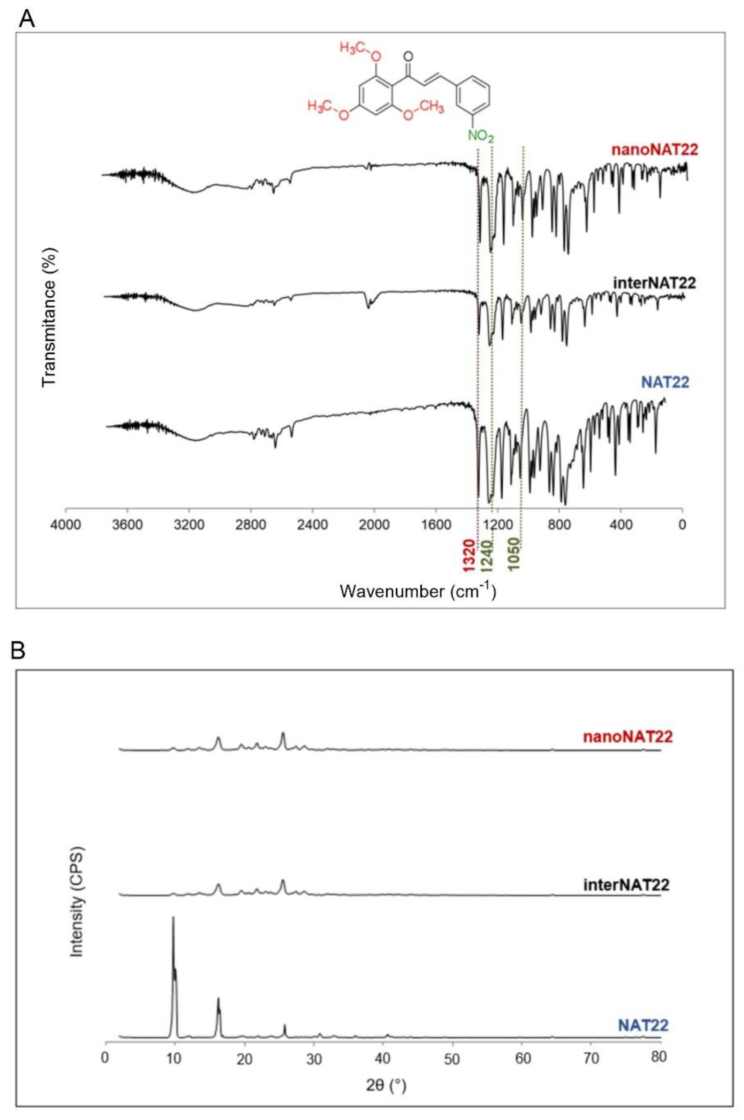
2.5. X-Ray Diffraction Analyses
2.6. Fourier-Transform Infrared Spectroscopy
2.7. Water Solubility
2.8. HPLC Analysis
2.9. Particle Size Stability
2.10. Particle Size Dispersibility
2.11. In Vitro Assays
2.11.1. Cell Culture
2.11.2. Antipromastigote Activity
2.11.3. Antiamastigote and Macrophage Cytotoxicity Assays
2.12. In Vivo Study
2.12.1. Animals
2.12.2. Efficacy Against CL
2.13. Statistical Analysis
3. Results and Discussion
3.1. Morphology and Size of NAT22 Nanocrystals
3.2. Chemical Stability and Crystallinity of NAT22 Chalcone
3.3. Solubility, Dispersibility, and Stability of NAT22 Crystals
3.4. Anti-Leishmania Activity of NAT22 Crystals
3.5. Oral Efficacy of nanoNAT22 in Murine Model of Cutaneous Leishmaniasis
4. Conclusions
Supplementary Materials
Author Contributions
Funding
Institutional Review Board Statement
Informed Consent Statement
Data Availability Statement
Conflicts of Interest
Abbreviations
| BMDM | Bone marrow-derived macrophages |
| CC50 | Half -maximal cytotoxic concentration |
| CL | Cutaneous leishmaniasis |
| DLS | Dynamic light scattering |
| DMSO | Dimethyl sulfoxide |
| DNDi | Drugs for Neglected Diseases Initiative |
| FTIR | Fourier-transform infrared spectroscopy |
| GFP | Green fluorescent protein |
| HIFCS | Heat-inactivated fetal calf serum |
| InterNAT22 | NAT22 intermediate crystals |
| IC50 | Half-maximal inhibitory concentration |
| LDA | Limiting dilution assay |
| LDH | Lactate dehydrogenase |
| NA | Not applicable |
| NanoNAT22 | NAT22 nanocrystals |
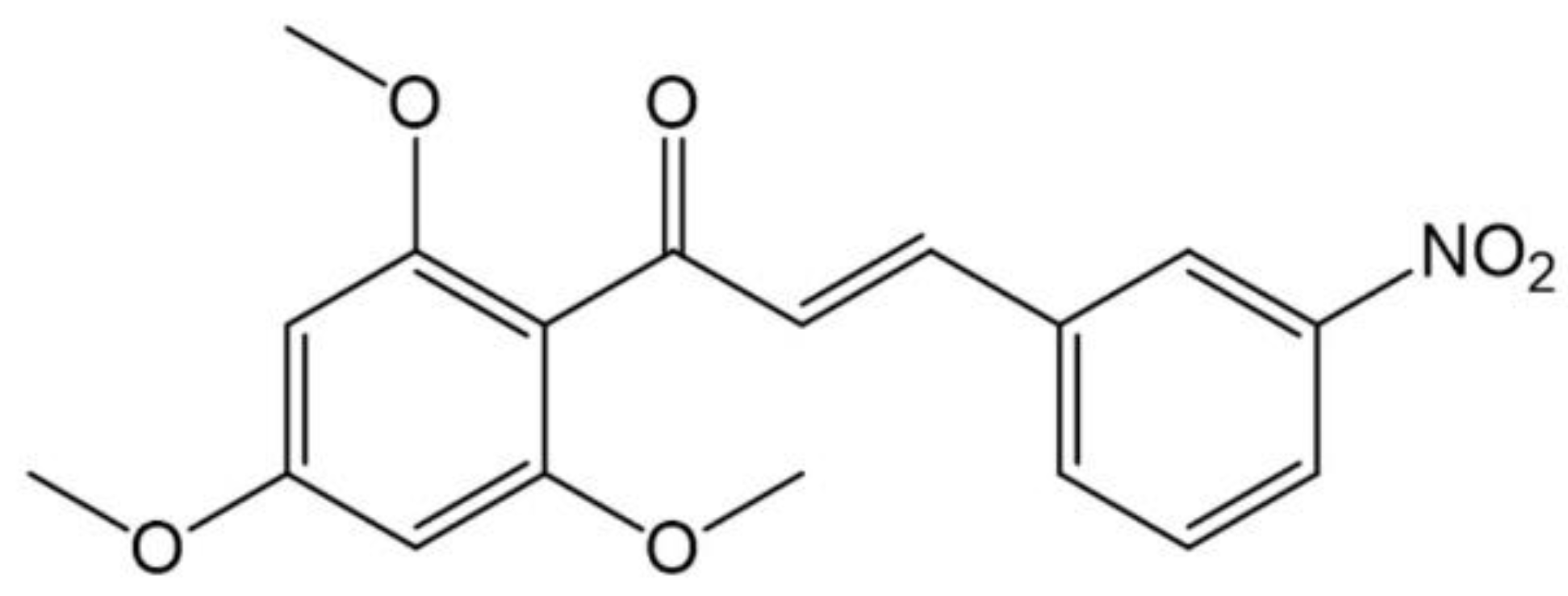
| NAT22 | 3-nitro-2′,4′,6′-trimethoxychalcone |
| ND | Not determined |
| OD | Optical density |
| SD | Standard deviation |
| PDI | Polydispersity index |
| pkCSM | Predicting small-molecule pharmacokinetic |
| PVP | Polyvinylpyrrolidone |
| SEM | Scanning electron microscopy |
| SEM | Standard error of the mean |
| SI | Selectivity index |
| UV-HPLC | High-Performance Liquid Chromatography with ultraviolet detector |
| ZP | Zeta potential |
References
- Bamorovat, M.; Sharifi, I.; Khosravi, A.; Aflatoonian, M.R.; Agha Kuchak Afshari, S.; Salarkia, E.; Sharifi, F.; Aflatoonian, B.; Gharachorloo, F.; Khamesipour, A.; et al. Global Dilemma and Needs Assessment Toward Achieving Sustainable Development Goals in Controlling Leishmaniasis. J. Epidemiol. Glob. Health 2024, 14, 22–34. [Google Scholar] [PubMed]
- Astman, N.; Arbel, C.; Katz, O.; Barzilai, A.; Solomon, M.; Schwartz, E. Tolerability and Safety of Miltefosine for the Treatment of Cutaneous Leishmaniasis. Trop. Med. Infect. Dis. 2024, 9, 218. [Google Scholar] [CrossRef]
- DNDI. Towards a New Generation of Treatments for Leishmaniasis. Available online: https://dndi.org/wp-content/uploads/2019/09/DNDi_Leishmaniasis_2019_POR.pdf (accessed on 10 January 2025).
- Chen, M.; Christensen, S.B.; Blom, J.; Lemmich, E.; Nadelmann, L.; Fich, K.; Theander, T.G.; Kharazmi, A. Licochalcone A, a novel antiparasitic agent with potent activity against human pathogenic protozoan species of Leishmania. Antimicrob. Agents Chemother. 1993, 37, 2550–2556. [Google Scholar]
- Yunes, R.; Chiaradia, L.D.; Leal, P.C.; Cechinel Filho, V.; Torres-Santos, E.C.; Falcão, C.A.B.; Rossi-Bergmann, B. Chalcones as new drug leads against leishmaniasis. Curr. Trends Med. Chem. 2006, 4, 47–56. [Google Scholar]
- Tajuddeen, N.; Isah, M.B.; Suleiman, M.A.; van Heerden, F.R.; Ibrahim, M.A. The chemotherapeutic potential of chalcones against leishmaniases: A review. Int. J. Antimicrob. Agents 2018, 51, 311–318. [Google Scholar]
- Barreto, T.S.A.; Santos, T.A.C.; Silva, A.R.S.T.; Costa, E.V.; Pinheiro, L.A.; Fernandes, R.P.M.; Scher, R.; Alves, P. Brominated chalcones as promising antileishmanial agentes. Bioorganic Med. Chem. Lett. 2025, 116, 130042. [Google Scholar]
- Torres-Santos, E.C.; Moreira, D.L.; Kaplan, M.A.; Meirelles, M.N.; Rossi-Bergmann, B. Selective effect of 2′,6′-dihydroxy-4′-methoxychalcone isolated from Piper aduncum on Leishmania amazonensis. Antimicrob. Agents Chemother. 1999, 43, 1234–1241. [Google Scholar]
- Boeck, P.; Falcão, C.A.B.; Leal, P.C.; Yunes, R.A.; Filho, V.C.; Torres-Santos, E.C.; Rossi-Bergmann, B. Synthesis of chalcone analogues with increased antileishmanial activity. Bioorganic Med. Chem. 2006, 14, 1538–1545. [Google Scholar]
- Sousa-Batista, A.J.; Arruda-Costa, N.; Escrivani-Oliveira, D.; Reynaud, F.; Steel, P.G.; Rossi-Bergmann, B. Single-dose treatment for cutaneous leishmaniasis with an easily synthesized chalcone entrapped in polymeric microparticles. Parasitology 2020, 147, 1032–1037. [Google Scholar]
- Escrivani-Oliveira, D.; Charlton, R.; Caruso, M.; Burle-Caldas, G.; Borsodi, M.P.G.; Zingali, R.; Mello, M.V.; Jesus, J.; Souza, A.; Abrahim-Viera, B.; et al. Chalcones identify cTXNPx as a potential antileishmanial drug target. PLoS Neglected Trop. Dis. 2021, 15, 11. [Google Scholar]
- Seo, S.; Kim, G.Y.; Kim, M.H.; Lee, K.W.; Kim, M.J.; Chaudhary, M.; Bikram, K.; Kim, T.; Choi, S.; Yang, H.; et al. Nanocrystal Formulation to Enhance Oral Absorption of Silybin: Preparation, In Vitro Evaluations, and Pharmacokinetic Evaluations in Rats and Healthy Human Subjects. Pharmaceutics 2024, 16, 1033. [Google Scholar] [CrossRef] [PubMed]
- Thipparaboina, R.; Chavan, R.B.; Shastri, N.R. Nanocrystals for Delivery of Therapeutic Agents. In Particulate Technology for Delivery of Therapeutics; Springer: Berlin/Heidelberg, Germany, 2017; pp. 291–316. [Google Scholar]
- Pardhi, V.P.; Verma, T.; Flora, S.J.S.; Chandasana, H.; Shukla, R. Nanocrystals: An Overview of Fabrication, Characterization and Therapeutic Applications in Drug Delivery. Curr. Pharm. Des. 2018, 24, 5129–5146. [Google Scholar] [CrossRef] [PubMed]
- Medarević, D.; Ibrić, S.; Vardaka, E.; Mitrić, M.; Nikolakakis, I.; Kachrimanis, K. Insight into the Formation of Glimepiride Nanocrystals by Wet Media Milling. Pharmaceutics 2020, 12, 53. [Google Scholar] [CrossRef]
- Jarvis, M.; Krishnan, V.; Mitragotri, S. Nanocrystals: A perspective on translational research and clinical studies. Bioeng. Transl. Med. 2019, 4, 5–16. [Google Scholar] [CrossRef] [PubMed]
- Amslinger, S.; Al-Rifai, N.; Winter, K.; Wörmann, K.; Scholz, R.; Baumeistera, P.; Wilda, M. Reactivity assessment of chalcones by a kinetic thiol assay. Org. Biomol. Chem. 2013, 11, 549. [Google Scholar] [CrossRef]
- Villaça, J.C.; Silva, L.C.R.P.; Locatelli, F.R.; Meireles, P.W.; Carmo, F.A.; Rodrigues, C.R.; Tavares, M.I.B.; Sousa, V.P.; Cabral, L.M. Full-factorial design for statistical planning of attritor milling parameters and evaluation of effects on particle size and structure of sodium-montmorillonite. Eng. Res. Express 2020, 2, 1. [Google Scholar] [CrossRef]
- Meireles, P.W.; de Souza, D.P.B.; Rezende, M.G.; Borsodi, M.P.G.; de Oliveira, D.E.; da Silva, L.C.R.P.; de Souza, A.M.T.; Viana, G.M.; Rodrigues, C.R.; do Carmo, F.A.; et al. Nanoparticles Loaded with a New Thiourea Derivative: Development and In vitro Evaluation Against Leishmania amazonensis. Curr. Drug Deliv. 2020, 17, 694–702. [Google Scholar] [CrossRef]
- Sousa-Batista, A.J.; Pacienza-Lima, W.; Arruda-Costa, N.; Falcão, C.A.B.; Ré, M.I.; Rossi-Bergmann, B. Depot Subcutaneous Injection with Chalcone CH8-Loaded Poly(Lactic-Co-Glycolic Acid) Microspheres as a Single-Dose Treatment of Cutaneous Leishmaniasis. Antimicrob. Agents Chemother. 2018, 62, e01822-17. [Google Scholar] [CrossRef]
- Lin, N.; Zhao, S.; Gan, L.; Chang, P.R.; Xia, T.; Huang, J. Preparation of fungus-derived chitin nanocrystals and their dispersion stability evaluation in aqueous media. Carbohydr. Polym. 2017, 173, 610–618. [Google Scholar] [CrossRef]
- Costa, S.S.; GOLIM, M.A.; Rossi-Bergmann, B.; Costa, F.T.M.; Giorgio, S. Use of In Vivo and In Vitro Systems to Select Leishmania amazonensis Expressing Green Fluorescent Protein. Korean J. Parasitol. 2011, 49, 357–364. [Google Scholar] [CrossRef]
- Rios, F.J.; Touyz, R.M.; Montezano, A.C. Isolation and Differentiation of Murine Macrophages. Methods Mol. Biol. 2017, 1527, 297–309. [Google Scholar] [PubMed]
- Demicheli, C.; Ochoa, R.; da Silva, J.B.B.; Falcão, C.A.B.; Rossi-Bergmann, B.; de Melo, A.L.; Sinisterra, R.D.; Frézard, F. Oral delivery of meglumine an-timoniate-beta-cyclodextrin complex for treatment of leishmaniasis. Antimicrob. Agents Chemother. 2004, 48, 100–103. [Google Scholar] [CrossRef] [PubMed]
- Lima, H.C.; Bleyenberg, J.A.; Titus, R.G. A simple method for quantifying Leishmania in tissues of infected animals. Parasitol. Today 1997, 13, 80–82. [Google Scholar]
- Gamboa, J.M.; Leong, K.W. In vitro and in vivo models for the study of oral delivery of nanoparticles. Adv. Drug Deliv. Rev. 2013, 65, 800–810. [Google Scholar]
- Bitterlich, A.; Mihorko, A.; Juhnke, M. Design Space and Control Strategy for the Manufacturing of Wet Media Milled Drug Nanocrystal Suspensions by Adopting Mechanistic Process Modeling. Pharmaceutics 2024, 16, 328. [Google Scholar] [CrossRef]
- Williams, H.D.; Trevaskis, N.L.; Charman, S.A.; Shanker, R.M.; Charman, W.N.; Pouton, C.W.; Porter, C.J. Strategies to address low drug solubility in discovery and development. Pharmacol. Rev. 2013, 65, 315–499. [Google Scholar] [PubMed]
- Kumar, D.; Worku, Z.A.; Gao, Y.; Kamaraju, V.K.; Glennon, B.; Babu, R.P.; Healy, A.M. Comparison of wet milling and dry milling routes for ibuprofen pharmaceutical crystals and their impact on pharmaceutical and biopharmaceutical properties. Powder Technol. 2018, 330, 228–238. [Google Scholar]
- Loh, Z.H.; Samanta, A.K.; Sia Heng, P.W. Overview of milling techniques for improving the solubility of poorly water-soluble drugs. Asian J. Pharm. Sci. 2015, 10, 255–274. [Google Scholar]
- Merisko-Liversidge, E.; Liversidge, G.G. Nanosizing for oral and parenteral drug delivery: A perspective on formulating poorly-water soluble compounds using wet media milling technology. Adv. Drug Deliv. Rev. 2011, 63, 6. [Google Scholar]
- Jahangir, M.A.; Imam, S.S.; Muheem, A.; Chettupalli, A.; Al-Abbasi, F.A.; Nadeem, M.S.; Kazmi, I.; Afzal, M.; Alshehri, S. Nanocrystals: Characterization Overview, Applications in Drug Delivery, and Their Toxicity Concerns. J. Pharm. Innov. 2020, 17, 237–248. [Google Scholar]
- Merck. IR Spectrum Table & Chart. Available online: https://www.sigmaaldrich.com/technical-documents/articles/biology/ir-spectrum-table.html#ir-table-by-compound (accessed on 15 January 2025).
- Budziak, I.; Arczewska, M.; Kaminski, D.M. Formation of Prenylated Chalcone Xanthohumol Cocrystals: Single Crystal X-Ray Diffraction, Vibrational Spectroscopic Study Coupled with Multivariate Analysis. Molecules 2019, 24, 4245. [Google Scholar] [CrossRef] [PubMed]
- Shah, S.M.; Ullah, F.; Khan, S.; Shah, S.M.; de Matas, M.; Hussain, Z.; Minhas, M.U.; AbdEl-Salam, N.M.; Assi, K.H.; Isreb, M. Smart Nanocrystals of Artemether: Fabrication, Characterization, and Comparative in Vitro and in Vivo Antimalarial Evaluation. Drug Design, Development and Therapy. Drug Des. Dev. Ther. 2016, 10, 3837–3850. [Google Scholar] [CrossRef] [PubMed]
- Liu, C.Z.; Chang, J.H.; Zhang, L.; Xue, H.F.; Liu, X.G.; Liu, P.; Fu, Q. Preparation and Evaluation of Diosgenin Nanocrystals to Improve Oral Bioavailability. AAPS PharmSciTech. 2017, 18, 2067–2076. [Google Scholar] [CrossRef] [PubMed]
- Yang, H.; Teng, F.; Wang, P.; Tian, B.; Lin, X.; Hu, X.; Zhang, L.; Zhang, K.; Zhang, Y.; Tang, X. Investigation of a Nanosuspension Stabilized by Soluplus® to Improve Bioavailability. Int. J. Pharm. 2014, 477, 88–95. [Google Scholar] [CrossRef]
- Malamatari, M.; Taylor, K.M.G.; Malamataris, S.; Douroumis, D.; Kachrimanis, K. Pharmaceutical nanocrystals: Production by wet milling and applications. Drug Discov. Today 2018, 23, 534–547. [Google Scholar] [CrossRef]
- Pireddu, R.; Caddeo, C.; Valenti, D.; Marongiu, F.; Scano, A.; Ennas, G.; Lai, F.; Fadda, A.M.; Sinico, C. Diclofenac acid nanocrystals as an effective strategy to reduce in vivo skin inflammation by improving dermal drug bioavailability. Colloids Surf. B Biointerfaces 2016, 143, 64–70. [Google Scholar] [CrossRef]
- Pires, D.E.V.; Blundell, T.L.; Ascher, D.B. pkCSM: Predicting Small-Molecule Pharmacokinetic and Toxicity Properties Using Graph-Based Signatures. J. Med. Chem. 2015, 58, 4066–4072. [Google Scholar] [CrossRef]
- Sugimoto, T. Chapter 4—Recrystallization. In Monodispersed Particles, 2nd ed.; Elsevier: Amsterdam, The Netherlands, 2019; pp. 167–179. [Google Scholar]
- Don, R.; Ioset, J.R. Screening strategies to identify new chemical diversity for drug development to treat kinetoplastid infections. Parasitology 2014, 141, 140–146. [Google Scholar] [CrossRef]
- Scarim, C.B.; de Souza, A.; Marins, D.S.S.; Santos, E.G.d.; de Figueiredo Diniz Castro, L.; Caldas, I.S.; Espuri, P.F.; Marques, M.J.; Ferreira, E.I.; Bou-Chacra, N.A.; et al. Synthesis, Characterization, and Activity of Hydroxymethylnitrofurazone Nanocrystals against Trypanosoma cruzi and Leishmania spp. Drugs Drug Candidates 2022, 1, 43–55. [Google Scholar] [CrossRef]
- Kayser, O.; Olbrich, C.; Yardley, V.; Kiderlen, A.F.; Croft, S.L. Formulation of Amphotericin B as Nanosuspension for Oral Administration. Int. J. Pharm. 2003, 254, 73–75. [Google Scholar] [CrossRef]


| Sample | Particle Size (µM) | PDI | Span | ZP (mV) |
|---|---|---|---|---|
| NAT22 | 225 ± 65 | NA * | 2.2 ± 0.2 | −2.3 ± 3.3 |
| interNAT22 | 0.701 ± 0.05 | 0.5 ± 0.03 | NA * | −7.5 ± 2.1 |
| nanoNAT22 | 0.257 ± 0.01 | 0.3 ± 0.04 | NA * | −24.6 ± 2.9 |
| Drug | Culture Medium | IC50 (µM) | CC50 (µM) | SI | |
|---|---|---|---|---|---|
| Promastigotes | Amastigotes | ||||
| NAT22 | Medium | 13.0 ± 14.0 | ND | ND | ND |
| nanoNAT22 | Medium | 0.7 ± 0.5 | 0.6 ± 0.1 | 12.3 ± 1 | 20 |
| NAT22 (DMSO) | 1% DMSO | 0.4 ± 0.2 | 0.6 ± 0.1 | 7.7 ± 0.9 | 13 |
| Pentamidine (DMSO) | 1% DMSO | 0.2 ± 0.0 | ND | ND | ND |
| Glucantime® | Medium | ND | 28.9 ± 0.1 | 197 * | 7 |
Disclaimer/Publisher’s Note: The statements, opinions and data contained in all publications are solely those of the individual author(s) and contributor(s) and not of MDPI and/or the editor(s). MDPI and/or the editor(s) disclaim responsibility for any injury to people or property resulting from any ideas, methods, instructions or products referred to in the content. |
© 2025 by the authors. Licensee MDPI, Basel, Switzerland. This article is an open access article distributed under the terms and conditions of the Creative Commons Attribution (CC BY) license (https://creativecommons.org/licenses/by/4.0/).
Share and Cite
Borsodi, M.P.G.; Pacienza-Lima, W.; Menezes, J.C.V.; Escrivani-Oliveira, D.; Arruda-Costa, N.; Silva, A.J.M.d.; Cabral, L.M.; Steel, P.G.; Sousa-Batista, A.d.J.; Rossi-Bergmann, B. Nanocrystallization Effectively Improves the Oral Efficacy of an Antileishmanial Chalcone. Pharmaceutics 2025, 17, 399. https://doi.org/10.3390/pharmaceutics17040399
Borsodi MPG, Pacienza-Lima W, Menezes JCV, Escrivani-Oliveira D, Arruda-Costa N, Silva AJMd, Cabral LM, Steel PG, Sousa-Batista AdJ, Rossi-Bergmann B. Nanocrystallization Effectively Improves the Oral Efficacy of an Antileishmanial Chalcone. Pharmaceutics. 2025; 17(4):399. https://doi.org/10.3390/pharmaceutics17040399
Chicago/Turabian StyleBorsodi, Maria Paula Gonçalves, Wallace Pacienza-Lima, Jaqueline Correia Villaça Menezes, Douglas Escrivani-Oliveira, Natalia Arruda-Costa, Alcides José Monteiro da Silva, Lucio Mendes Cabral, Patrick G. Steel, Ariane de Jesus Sousa-Batista, and Bartira Rossi-Bergmann. 2025. "Nanocrystallization Effectively Improves the Oral Efficacy of an Antileishmanial Chalcone" Pharmaceutics 17, no. 4: 399. https://doi.org/10.3390/pharmaceutics17040399
APA StyleBorsodi, M. P. G., Pacienza-Lima, W., Menezes, J. C. V., Escrivani-Oliveira, D., Arruda-Costa, N., Silva, A. J. M. d., Cabral, L. M., Steel, P. G., Sousa-Batista, A. d. J., & Rossi-Bergmann, B. (2025). Nanocrystallization Effectively Improves the Oral Efficacy of an Antileishmanial Chalcone. Pharmaceutics, 17(4), 399. https://doi.org/10.3390/pharmaceutics17040399