Review

Emerging Approaches in Glioblastoma Treatment: Modulating the Extracellular Matrix Through Nanotechnology

by
Miguel Horta
1,2,3,
Paula Soares
1,2,3,
Catarina Leite Pereira
1,4,*,† and
Raquel T. Lima
1,2,3,*,†
1
i3S—Instituto de Investigação e Inovação em Saúde, University of Porto, Rua Alfredo Allen 208, 4200-135 Porto, Portugal
2
IPATIMUP—Instituto de Patologia e Imunologia Molecular, University of Porto, Rua Júlio Amaral de Carvalho 45, 4200-135 Porto, Portugal
3
FMUP—Faculty of Medicine, University of Porto, Alameda Prof. Hernâni Monteiro, 4200-319 Porto, Portugal
4
INEB—Instituto Nacional de Engenharia Biomédica, University of Porto, Rua Alfredo Allen 208, 4200-135 Porto, Portugal
*
Authors to whom correspondence should be addressed.
These authors contributed equally to this work.
Pharmaceutics 2025, 17(2), 142; https://doi.org/10.3390/pharmaceutics17020142
Submission received: 21 December 2024 / Revised: 10 January 2025 / Accepted: 16 January 2025 / Published: 21 January 2025
(This article belongs to the Special Issue Nano-Based Technology for Glioblastoma)

Abstract

:
Glioblastoma’s (GB) complex tumor microenvironment (TME) promotes its progression and resistance to therapy. A critical component of TME is the extracellular matrix (ECM), which plays a pivotal role in promoting the tumor’s invasive behavior and aggressiveness. Nanotechnology holds significant promise for GB treatment, with the potential to address challenges posed by both the blood-brain barrier and the GB ECM. By enabling targeted delivery of therapeutic and diagnostic agents, nanotechnology offers the prospect of improving treatment efficacy and diagnostic accuracy at the tumor site. This review provides a comprehensive exploration of GB, including its epidemiology, classification, and current treatment strategies, alongside the intricacies of its TME. It highlights nanotechnology-based strategies, focusing on nanoparticle formulations such as liposomes, polymeric nanoparticles, and gold nanoparticles, which have shown promise in GB therapy. Furthermore, it explores how different emerging nanotechnology strategies modulate the ECM to overcome the challenges posed by its high density, which restricts drug distribution within GB tumors. By emphasizing the intersection of nanotechnology and GB ECM, this review underscores an innovative approach to advancing GB treatment. It addresses the limitations of current therapies, identifies new research avenues, and emphasizes the potential of nanotechnology to improve patient outcomes.

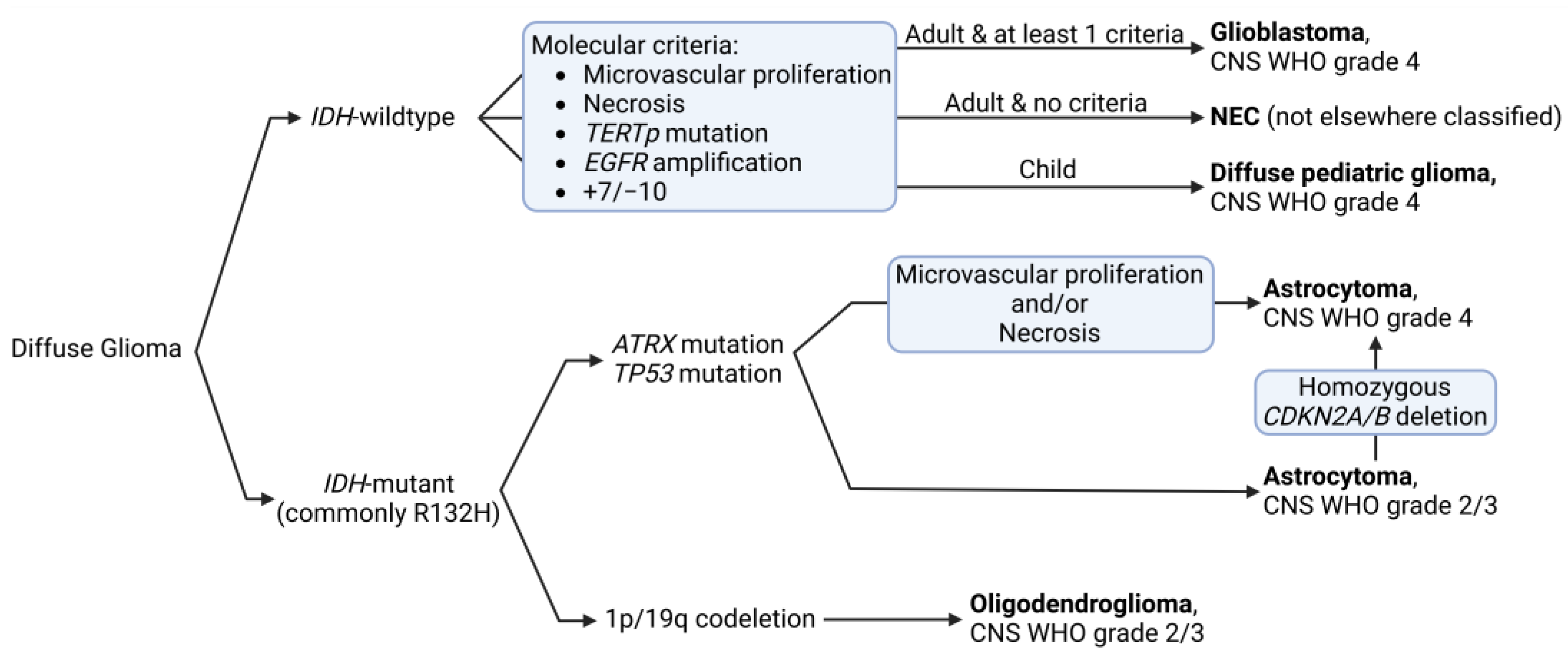
1. Glioblastoma: Epidemiology and Classification

Glioblastoma (GB) is the most aggressive and highly malignant brain tumor in adults, presenting a poor prognosis. Its incidence varies depending on the population studied, mostly ranging from 3 to 4.5 per 100,000 people annually [1,2,3], accounting for approximately 50% of all malignant central nervous system (CNS) tumors [4]. GB shows a slight male predominance, with a male to female ratio of about 1.6:1 [5]. Although the incidence increases with age, it can occur across all adult age groups [1]. While CNS tumors are common in children, according to the most recent World Health Organization (WHO) guidelines, GB is no longer a possible diagnosis in this age group; instead, it is considered a diffuse pediatric glioma [6].
As our knowledge of GB improves, so do our methods for diagnosing, classifying, and treating the disease [7]. Recent changes in tumor classification, driven by new insights from molecular profiling, have significantly influenced how to diagnose and treat GB, highlighting the clinical importance of these advancements (Figure 1) [8]. Under the most recent 2021 WHO classification of tumors of the CNS, which has significantly redefined GB, GB is strictly defined as an isocitrate dehydrogenase (IDH)-wildtype, WHO grade 4, tumor. This reclassification emphasizes the distinct biological behavior and more favorable prognosis of IDH-mutant tumors compared to their IDH-wildtype counterparts [1,6,9,10]. Furthermore, for the diagnosis of GB (IDH-wildtype WHO grade 4) at least one of the following criteria is now required: (i) microvascular proliferation or necrosis on histological examination; (ii) telomerase reverse transcriptase (TERT) gene promoter mutations; (iii) epithelial growth factor (EGF) receptor (EGFR) gene amplification; (iv) combined gain of chromosome 7 and loss of chromosome 10 (+7/−10) [11]. This integrated approach, combining histological and molecular features, provides a more accurate and clinically relevant diagnosis. Notably, this new classification no longer allows for the use of “not otherwise specified” (NOS) in GB diagnosis, further underscoring the importance of molecular testing in modern neuro-oncology [12]. Moreover, the “not elsewhere classified” (NEC) designation is now used when a tumor does not meet the specific GB criteria but still appears to be a diffuse astrocytic glioma without IDH mutation [13].
The prognosis for GB remains poor, with median survival varying based on molecular subtypes, which are defined by different transcription profiles [14]. The mesenchymal subtype is characterized by extensive necrosis and inflammation, exhibiting transcription profiles similar to mesenchymal tissues. The proneural subtype, considered milder, is characterized by the expression of genes involved in neurogenesis and oligodendrocyte development. The classical subtype, in between, shows features similar to astrocytes, hence the “classical” name reflecting typical GB characteristics [15]. The mesenchymal subtype has the worst prognosis, with a median survival of about 11.5 months, followed by the classical subtype at 14.7 months, and the proneural subtype at 17.0 months [16]. These survival differences highlight the clinical relevance of molecular subtyping in GB, which will be discussed in more detail later. Although there are several risk factors for GB, exposure to high-dose ionizing radiation and certain rare genetic conditions (e.g., Lynch syndrome, Neurofibromatosis type 1, Li-Fraumeni syndrome) have been associated with increased incidence [17,18]. However, the majority of GB cases occur sporadically without any known risk factors [19]. Anatomically, GBs are typically supratentorial, with rare cases in the cerebellum or spine [20,21]. They most commonly affect the frontal and temporal lobes, followed by the parietal and occipital lobes, with a slight preference for the right hemisphere [22]. Tumor location can significantly impact surgical resection and, consequently, patient outcomes [23,24].

2. Current Treatment Options

2.1. First-Line—Stupp Protocol

The “Stupp protocol”, introduced in 2005, is the standard of care for treating newly diagnosed GB cases [25]. Named after the Swiss oncologist Roger Stupp, this treatment regimen improved survival outcomes for GB patients compared to previous approaches. The protocol consists of maximal safe surgical resection followed by two main phases, concurrent chemoradiotherapy followed by adjuvant chemotherapy. During the first phase, patients receive radiotherapy (RT) with a total dose of 60 Gy, delivered in 30 fractions of 2 Gy each over 6 weeks (5 days per week). Simultaneously, patients take oral temozolomide (TMZ), an alkylating chemotherapy agent, at a dose of 75 mg/m2 of body surface area, daily, seven days a week throughout the RT period. During the second phase, after a 4-week break once completing RT, patients undergo six cycles of TMZ monotherapy. Each cycle lasts 28 days, with TMZ taken for the first 5 days at a dose of 150–200 mg/m2 [26].
The Stupp protocol demonstrated an improvement in survival compared to RT alone. In the original study, the median overall survival increased from 12.1 months with RT alone to 14.6 months with the combined approach. More importantly, the 2-year survival rate improved from 10.4% to 26.5% [27]. Despite these improvements, the prognosis for GB remains poor. Nevertheless, this protocol is most effective in patients under 70 years of age, with good performance status, and whose tumors exhibit methylguanine methyltransferase (MGMT) promoter methylation, a biomarker associated with better TMZ treatment response [28]. Moreover, side effects of the Stupp protocol can be significant, with the most common being fatigue, nausea, and myelosuppression, and the most severe complications including neutropenia, thrombocytopenia, lymphopenia, and increased risk of opportunistic infections. To reduce infection risks, patients with these complications often receive antibiotic prophylaxis during treatment [29]. While the Stupp protocol remains the backbone standard of care for GB, ongoing research seeks to further improve patient outcomes.

2.2. Second-Line Treatments

Following Stupp protocol failure, with frequent tumor relapse, there is no universally accepted standard for second-line treatment. The management of recurrent GB remains a therapeutic challenge, with several strategies being explored. Tumor reoperation is considered for patients with large, symptomatic tumors, but its benefits remain controversial and depend on patient selection [30,31,32]. Another strategy, reirradiation, presents options including stereotactic radiosurgery, hypofractionated stereotactic RT, conventionally fractioned external RT, or brachytherapy. Decision depends on factors such as time since initial radiation and tumor characteristics [33,34,35]. A third option is a TMZ rechallenge, which is considered for initial responders with prolonged treatment-free intervals. Various dosing schedules have been explored, but overall survival rarely exceeds 12 months [36,37,38,39,40]. Treatment with nitrosoureas, such as carmustine, lomustine, nimustine, and fotemustine, have also shown limited efficacy in recurrent GB, with median overall survival rarely exceeding 12 months [41,42,43,44]. Bevacizumab, a Food and Drug Administration (FDA)-approved anti-angiogenic antibody, has often been employed as a strategy combined with other agents, such as irinotecan or lomustine. While it has been shown to improve progression-free survival, impact on overall survival is still debated [45,46,47,48]. Nevertheless, recent research has indicated that high vascular endothelial growth factor (VEGF)-A expression is associated with improved progression-free survival in recurrent GB patients treated with bevacizumab, suggesting potential for a therapeutic improvement through biomarker-guided treatment selection [49]. The multi-kinase inhibitor Regorafenib has shown potential benefits in the REGOMA trial, but more comprehensive data are still needed due to its relatively recent introduction in clinical practice in 2020 [50,51,52]. Other tyrosine kinase inhibitors, such as cabozantinib, dasatinib, and entrectinib, are currently being evaluated, particularly in molecularly selected populations. However, their efficacy as monotherapies has been limited, mainly due to lack of tumor specificity and poor blood-brain barrier (BBB) permeability [53,54,55]. Over the last decade, immunotherapy, particularly checkpoint inhibitors, has emerged as a potential therapy for GB. While its efficacy has been limited in unselected GB populations, research continues to address its potential in patient specific subgroups [56,57,58,59]. Also, Tumor Treating Fields (TTFields) have emerged as a promising second-line treatment option in clinical practice. This device-based therapy delivers alternating electric fields to tumor sites, promoting cancer cell death by disrupting microtubule alignment during cell division [60,61,62]. TTFields have demonstrated safety and efficacy in clinical trials, leading to FDA approval for GB and malignant pleural mesothelioma. As TTFields continue to show promise, ongoing clinical trials are exploring its use in lung, ovarian, pancreatic, and other cancers, signaling a growing integration of this innovative approach into standard cancer care protocols [63].
GB treatment goals in the recurrent setting are often palliative, aiming to improve or maintain quality of life while potentially extending survival. The choice of second-line treatments for recurrent GB is highly individualized and depends on various factors, including the patient’s age, performance status, tumor molecular profile, response to initial therapy, and time to recurrence [64]. Despite the availability of the referred options, the prognosis for recurrent GB remains poor, with median overall survival typically ranging from 6 to 12 months in a clinical setting, with a very modest increase to approximately 15 months for clinical trial participants [65,66,67,68]. This underscores the urgent need for more effective therapies and highlights the importance of ongoing research in this challenging cancer type.

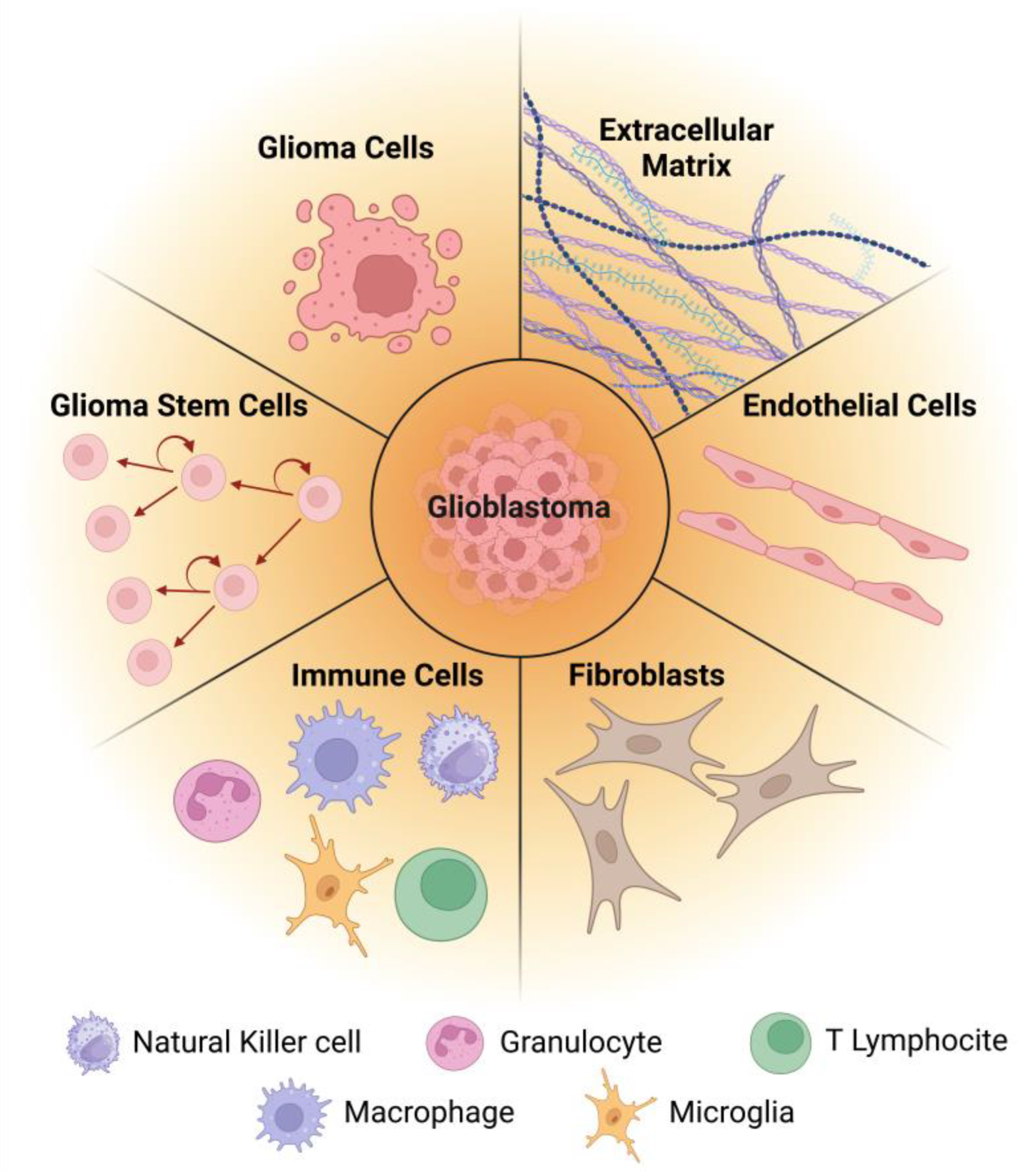
3. Glioblastoma and Its Microenvironment—Cellular and Molecular Features

Adding to its own cellular and molecular features, GB is characterized by a complex and dynamic tumor microenvironment (TME) that plays a crucial role in tumor progression, invasion, and therapy resistance [69]. The TME is composed of various cellular and molecular components (Figure 2) that intricately interact to support tumor growth and promote immune evasion [70]. These interactions are key to understanding the mechanisms of GB progression and therapy resistance. As such, gaining a deeper understanding of the TME is essential for developing effective therapeutic strategies.

3.1. Glioma Cells

The primary cellular component of GB tumors consists of malignant glial cells, which exhibit significant inter- and intra-tumoral heterogeneity [71]. This heterogeneity was classified into the previously referred three molecular subtypes, classical, mesenchymal, and proneural, based on transcriptomic analyses from The Cancer Genome Atlas (TCGA) [72]. A fourth one was initially proposed, the neural subtype, but later excluded as it was based on a misidentification caused by cellular cross contamination [16].
The classical subtype is the least common among GB tumors, accounting for approximately 20% of all GBs [73]. It is characterized by EGFR amplification in nearly all tumors (97%), and some harboring EGFR mutations [74,75]. Consistent genetic alterations included chromosome 7 amplification paired with chromosome 10 loss and focal 9p21.3 homozygous deletion targeting cyclin-dependent kinase inhibitor 2A (CDKN2A), which mostly co-occur with EGFR amplification [15]. Notably, tumor protein p53 (TP53) mutations are rare in the classical subtype despite being the most frequently mutated gene in GB overall [76]. Furthermore, the transcriptomic analyses showed an overexpression of Sonic-hedgehog (smoothened (SMO), growth arrest-specific protein 1 (GAS1), and GLI family zinc finger 2 (GLI2)) and Notch pathway (neurogenic locus notch homolog protein 3 (NOTCH3), jagged 1 (JAG1), and beta-1,3-N-acetylglucosaminyltransferase lunatic fringe (LFNG)) agents, along with high expression of the neural precursor marker nestin [77,78]. Patients with the classical subtype benefit from aggressive RT and chemotherapy [78].
The prevalence of the mesenchymal subtype ranges from 30% to 50% of all GB tumors depending on the population studied and the molecular classification methods used [73,79]. It frequently exhibits neurofibromin (NF1) mutations and deletions at 17q11.2 (further affecting NF1 expression), often co-occurring with phosphatase and tensin homolog (PTEN) mutations and phosphatidylinositol 3-kinase (PI3K)/AKT pathway enrichment [15,80]. Mesenchymal markers such as chitinase-3-like protein 1 (CHI3L1) and proto-oncogene tyrosine kinase MET are highly expressed, along with astrocytic markers like cluster of differentiation (CD) 44 and proto-oncogene tyrosine kinase MER, indicating potential epithelial-to-mesenchymal transition [15]. Gene expression analysis revealed activation of tumor necrosis factor and nuclear factor kappa-light-chain-enhancer of activated B cells (NF-κB) pathways, correlating with extensive necrosis and inflammatory infiltrates characteristic of this subtype [74,78]. Clinically, while responsive to aggressive RT and chemotherapy, patients with mesenchymal GB have the worst prognosis among all GB subtypes [14].
The proneural subtype, slightly less common than the mesenchymal subtype, accounts for approximately a third of GB tumors [73]. It is characterized by distinct genetic alterations, primarily involving the platelet-derived growth factor receptor A (PDGFRA) gene [78]. This subtype was associated with a gene expression profile linked to oligodendrocyte development and neurogenesis, reflecting its unique biological characteristics. The proneural subtype was also characterized by frequent TP53 mutations and loss of heterozygosity [15]. Clinically, patients with this subtype generally have a better prognosis compared to those with other subtypes, particularly in younger populations [14,76,77,80].
A more recent perspective on GB heterogeneity suggests that genetic factors alone do not fully account for the diverse glioma cell composition observed within these tumors. An international team of researchers proposed that somatic mutations play a crucial role in shaping the tumor’s cellular landscape by selectively promoting certain cellular states over others [81]. This process results in a dynamic equilibrium of different cell populations within the tumor, contributing to its heterogeneous nature. Neftel et al. conducted a computational analysis that identified gene signatures converging into four recurring cellular states across multiple GB tumors [81]. These states were present in varying combinations of two to four in each patient, again denoting the heterogeneity of GB, being classified as astrocyte-like (AC-like), mesenchymal-like (MES-like), oligodendrocyte progenitor-like (OPC-like), and neural progenitor-like (NPC-like). OPC-like states were enriched in tumors with PDGFRA alterations, NPC-like states in those with cyclin-dependent kinase 4 (CDK4) alterations, and AC-like states in tumors with EGFR alterations. MES-like states were characterized by NF1 alterations, extensive immune cell infiltration, and hypoxic conditions [81]. These cellular states aligned with the TCGA subtypes and corroborated findings from other studies suggesting that genomic aberrations alone do not fully explain GB heterogeneity [77,82]. Regardless, the heterogeneity of GB cell subtypes has significant clinical implications, with recent studies demonstrating that specific cellular compositions of tumors directly impact patient survival outcomes [83].

3.2. Glioma Stem Cells

A particularly concerning aspect of GB tumors is the presence of glioma stem cells (GSCs) in its TME, a subpopulation with self-renewal capacity that is thought to drive tumor initiation, recurrence, and therapy resistance [84]. Importantly, GSCs are now understood to represent a functional state rather than a distinct cell lineage [85]. This concept has been further supported by evidence showing that GB cells can dynamically acquire or lose stem-like characteristics in response to TME signals [86]. GSCs share features with normal neural stem cells, including stem cell marker expression such as nestin, sex determining region Y box 2 (SOX2), and CD133, as well as multi-lineage differentiation potential [85]. However, opposite to their normal counterpart, GSCs exhibit aberrant signaling pathway activation, enhancing survival, proliferation, and apoptosis resistance. These alterations confer GSCs a distinct advantage, enabling them to survive conventional therapies and contribute to tumor relapse, making them critical targets for developing more effective GB treatments [87]. In fact, evidence suggests that GSCs are more resistant to chemotherapy and RT than the bulk of tumor cells [85]. They possess enhanced deoxyribonucleic acid (DNA) repair mechanisms, activated survival pathways, and overexpress drug efflux transporters such as ATP-binding cassette (ABC) proteins [88]. Furthermore, GSCs often reside in specialized niches within the TME, which are characterized by hypoxia and supportive stromal cells. The niches provide protective signals that promote GSCs survival and maintenance, further shielding them from therapeutic interventions [89]. GSCs metabolism is regulated by several key signaling pathways, including Notch, Hedgehog, and Wnt. These pathways, essential for normal stem cell maintenance, are often dysregulated in cancer. Furthermore, transcription factors, such as SOX2, octamer-binding transcription factor 4 (OCT4), and c-Myc, are essential for maintaining GSCs pluripotency and proliferative capacity [90]. While GSCs are certainly influenced by TME cues, studies have also emphasized the reciprocal relationship between GSCs and their microenvironment, supporting and promoting glioma progression [91]. On one hand, TME-derived cytokines such as interleukin (IL)-6 and transforming growth factor beta (TGF-β) enhance GSCs self-renewal and survival [92]. On the other hand, GSCs have been shown to secrete factors that modulate the surrounding immune landscape, creating an immunosuppressive environment conducive to tumor growth. For example, GSCs can induce tumor-associated macrophages (TAMs) to adopt an M2-like phenotype, which suppresses immune responses and promotes tumor development [93]. This intricate interaction between GSCs and their microenvironment underscores the complexity of GB biology and highlights potential targets for therapeutic intervention.

3.3. Immune Cells

The immune component of GB represents approximately 50% of the tumor’s cellularity [94]. As previously mentioned, TAMs, which are differentiated from the resident microglia and peripheral monocytes, account for 30% of the tumor mass. These TAMs tend to adopt an M2 anti-inflammatory phenotype and have been shown to suppress CD8+ T cell activity, thereby promoting immune evasion [95]. TAMs are recruited by glioma-cell-derived factors, such as colony stimulating factor 1 (CSF-1), glial cell line-derived neurotrophic factor (GDNF), chemokine (CC motif) ligand 2 (CCL2), stromal cell-derived factor 1 (SFD-1), chemokine (CXC motif) ligand 1 (CXCL1), EGF, and granulocyte-macrophage colony-stimulating factor (GM-CSF) [96]. Once in the TME, TAMs secrete a range of molecules, including anti-inflammatory cytokines (IL4, IL10, TGFβ), angiogenesis factors (VEGF, IL8), and pro-tumorigenic growth factors (insulin-like growth factor 1 (IGF-1), EGF, and platelet-derived growth factor (PDGF)). These molecules significantly influence the GB TME and contribute to tumor progression and therapy resistance, ultimately leading to poorer prognosis for GB patients [94].
Another cell type within the GB TME is myeloid-derived suppressor cells (MDSCs), a diverse population of myeloid progenitor and precursor cells, including macrophages, granulocytes, and dendritic cells at various stages of differentiation [97]. Similar to TAMs, MDSCs impair the function of various T cell subsets, including natural killer T (NK) cells and cytotoxic T lymphocytes [98]. MDSCs also deplete essential amino acids from the TME, such as L-Arginine, and enhance the production of reactive oxygen species (ROS) to suppress T-cell activation and function, thereby contributing to the immune evasion of tumors. These changes in the TME create an unfavorable environment for T cells, leading to reduced immune responses against the tumor [99,100].
Lastly, NK cells, which are important to innate antitumor immunity through antigen-independent immune surveillance, were found to be the least abundant immune cell type in GB. Furthermore, their immune cytolytic activity is suppressed by major histocompatibility complex class I molecules expressed on GB cells [101].

3.4. Fibroblasts

Recent research has shed new light on the presence and significance of cancer-associated fibroblasts (CAFs) in GB, revealing their role in shaping the TME [102]. Being the primary extracellular matrix (ECM) producers, CAFs contribute significantly to the remodeling of the ECM in GB, promoting tumor growth and invasion. Their abundance correlates with higher tumor grades and activation of ECM remodeling pathways [102]. CAFs also secrete pro-tumorigenic factors, including fibronectin, which plays a critical role in promoting GB cell migration and invasion, thereby enhancing the tumor’s aggressive nature [102]. Furthermore, CAFs have been shown to promote resistance to TMZ through the secretion of CCL2, which activates the extracellular signal-regulated kinases (ERK) 1/2 signaling pathway in GB cells [103]. Interestingly, CAFs exhibit subtype-specific effects in GB [104]. They are more abundant in the mesenchymal subtype compared to other subtypes, while proneural GB cells appear to be more responsive to CAF signaling in terms of migratory and invasive behaviors. This heterogeneity in CAF distribution and influence across GB subtypes adds another layer of complexity to the TME [102]. CAFs have been shown to suppress anti-cancer immune responses in various cancers, highlighting their role in contributing to the immunosuppressive environment characteristic of GB [105].

3.5. Endothelial Cells

While not a part of the GB per se, endothelial cells, which encompass the perivascular niche, have been shown to be key components in the tumor’s aggressiveness, since angiogenesis is one of the hallmarks of GB recurrence, proliferation, and invasion [106]. In the GB TME, newly formed blood vessels are structurally fragile and prone to rupture, leading to a compromised BBB. This disruption causes an increased vascular permeability and edema, altering immune responses and the boundary between tumor and normal brain tissue, thus facilitating tumor invasiveness and presents challenges for effective treatment delivery [107].
The recruitment of endothelial cells to the GB site occurs through several mechanisms. Neoangiogenesis is the primary process, where GB cells secrete high levels of pro-angiogenic factors, particularly VEGF, stimulating the formation of new blood vessels from the sprouting of pre-existing ones. This process involves endothelial cell proliferation and migration towards the tumor [108]. Additionally, GBs recruit bone marrow-derived endothelial progenitor cells through vasculogenesis, which then differentiate into endothelial cells within the TME [109]. Lastly, GSCs also have the potential to transdifferentiate into endothelial cells, or to simply assemble into tubular-like structures mimicking blood vessels, further contributing to tumor vasculature [110].
Endothelial cells’ contribution to tumor progression is multifaceted. By forming new blood vessels, they ensure a steady supply of nutrients and oxygen to the rapidly growing tumor while enabling invasion through the vasculature [109]. Endothelial cells also create a specialized niche that promotes the self-renewal of GSCs by expressing NOTCH ligands that activate Notch signaling in adjacent GSCs, supporting their maintenance and proliferation [111]. Furthermore, endothelial cells facilitate bidirectional communication within the TME, exchanging extracellular vesicles with tumor cells, which can, in turn, promote angiogenesis, suppress the immune system, and confer drug resistance [108].

3.6. Extracellular Matrix

The non-cellular component of the GB TME is the ECM. The brain has an unique ECM composition, which accounts for approximately 20% of its volume [112]. It is comprised of various components, including, but not limited to: collagen IV; glycoproteins, such as fibronectin, laminins, and tenascins; glycosaminoglycans, such as hyaluronic acid (HA); and proteoglycans, such as the lectican family, phosphacan, and neuron-glial antigen 2 [113]. While sharing many components with the normal brain, the ECM of GB exhibits distinct characteristics that influence tumor behavior. Besides the fact that GB cells overexpress various ECM components, including HA, brevican, tenascin-C (TN-C), and fibronectin, they also show increased expression of specific integrins and receptors, which promote cell adhesion, migration, and invasion [114].
Notably, HA levels correlate with tumor malignancy, both increasing in parallel. HA, along with fibronectin, has been shown to promote the invasiveness of GB cells [115,116]. GB ECM is more condensed and less flexible compared to normal brain tissue due to overexpression of these ECM components, potentially hindering the diffusion of therapeutic agents and neuroactive molecules [117]. The tumor also induces altered protein synthesis in surrounding normal tissues, promoting ECM degradation at the invasive front while increasing ECM component production in nearby areas [118,119]. GB cells can actively migrate along blood vessels or axons through ECM interactions [120].
Research has revealed ECM-driven signals that shape tumor phenotypes and influence metabolic activity [121]. Moreover, the GB ECM is deficient in aggrecan, contains oncofetal proteins, and shows elevated levels of matrix metalloproteinases (MMPs), which increases ECM modulation, invasion, and angiogenesis [119,122]. The ECM’s significant role in cell survival, proliferation, and differentiation processes makes it an attractive target for novel therapeutic approaches, which could be particularly valuable given GB’s poor response to conventional treatments.

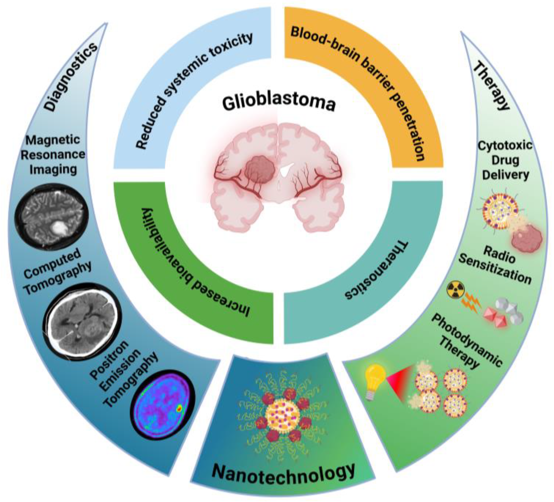
4. Nanotechnology in Glioblastoma

Among multiple alternative approaches to treat GB, nanotechnology has emerged as a particularly promising field to address the challenges posed by this tumor. Nanoparticle (NP)-based drug delivery systems offer the potential to overcome many of the challenges associated with treating brain tumors, including poor drug penetration across the BBB and the need for targeted delivery to tumor cells while minimizing toxicity to healthy tissue (Figure 3) [123].
Nanocarriers can be engineered to encapsulate various therapeutic agents, including chemotherapy drugs, small molecule inhibitors, nucleic acids for gene therapy, and even combinations of multiple agents for synergistic effects [126]. Their sizes typically range from 10 to 200 nm, with smaller sizes (10–50 nm) facilitating enhanced tumor penetration, cellular uptake, and TME diffusion, while larger sizes (100–200 nm) promote prolonged circulation, better retention at the tumor site, and reduced clearance through renal filtration [127].
Furthermore, the surface of nanoparticles can be modified with targeting ligands to facilitate transport across the BBB and to increase their specificity for GB cells [128]. As research in this area continues to evolve, nanotechnology-based approaches may offer new hope for improving the treatment outcomes for GB patients.
Despite all advances in this field, only a limited number of liposomal strategies have reached clinical trials in GB or other high grade gliomas, following a path similar to what was achieved in other cancers [129]. Among the main candidates studied, Caelyx™, a pegylated liposomal formulation of doxorubicin (DOX), underwent several phase I/II clinical trials, either alone or in a combination therapy (with TMZ, tamoxifen, RT, or a combination of these) [130,131,132,133,134]. However, these trials’ main conclusion was that the liposomal formulation did not show clinical benefits. More recently, a liposomal formulation of irinotecan (nal-IRI) was tested in two clinical trials, one as single agent and the other combined with TMZ; however, both trials ended due to lack of activity [135,136]. A new phase I clinical trial at Johns Hopkins University (NCT05768919) is currently recruiting patients to assess the efficacy of liposomal curcumin in combination with radiotherapy and TMZ; no study results have been released yet.
The limited efficacy observed thus far with these clinical trials suggests that while nanoparticle-based systems can improve drug delivery, further optimization in formulation and the development of new targeting strategies are necessary to enhance their clinical effectiveness in GB treatment. While nanotechnology offers promising advantages for GB treatment, its potential non-specific toxicity, both systemically and in brain tissue, remains a critical consideration. Ongoing research focuses on optimizing nanoparticle biocompatibility and biodegradability while minimizing inflammatory responses, aiming to optimize the balance between therapeutic efficacy and safety [137,138].

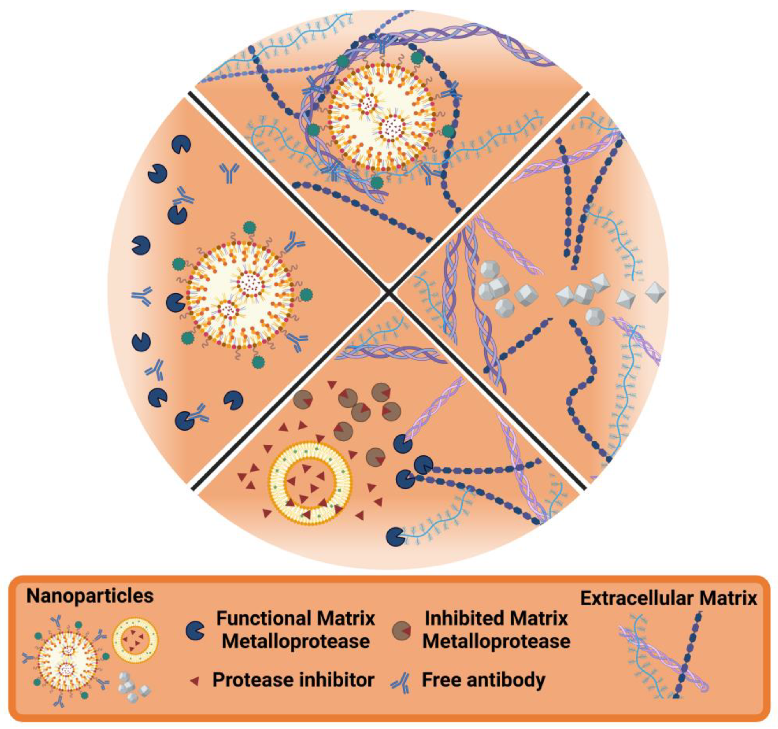
5. Nanotechnology and the ECM

While initial nanotechnological approaches focused primarily on targeting glioma cells, there is growing recognition of the ECM as a critical factor to tumor progression and malignancy [114], as previously discussed. Consequently, researchers are developing novel therapeutic strategies that specifically target the ECM in GB. These exploit the ubiquitous presence of ECM components within the TME, and thus offer significant advantages compared to targeting the glioma cells themselves. These ECM-focused strategies, often investigated in preclinical settings, include the inhibition of matrix-degrading metalloproteinases and integrins, ECM stiffening, and degradation [139,140]. Additionally, targeting structural ECM molecules can enhance other treatment modalities, such as immunotherapy, by increasing the infiltration of immune cells [141,142]. Nanotechnology approaches interacting with the GB ECM can be categorized into the following classes, as exemplified in Figure 4: (i) ECM as the target: nanoparticles designed to directly interact with or modify the ECM structural components; (ii) ECM-responsive nanoparticles: nanoparticles designed to respond to specific ECM characteristics or changes; (iii) Enhancing ECM degradation: nanoparticles that facilitate the breakdown of ECM components to enhance drug penetration or disrupt tumor structure; (iv) Preventing ECM degradation: nanoparticles that inhibit matrix-degrading enzymes like metalloproteinases to maintain ECM integrity and limit tumor invasion.
Table 1 summarizes the various nanotechnology strategies discussed in this section, highlighting their approach, key features, and potential applications in GB treatment. This overview provides a comprehensive snapshot of the diverse nanotechnology approaches being explored to reprogram the GB ECM.

5.1. ECM as the Target

As previously mentioned, ECM components are ubiquitously distributed throughout the tumor parenchyma, making them a more consistent and accessible target compared to heterogeneous tumor cell populations (Figure 5) [141].
Among these components, the extra domain B (EDB) of fibronectin has emerged as a highly promising target for cancer therapy, particularly in aggressive solid tumors such as GB [162]. This specific domain is prominently present in the perivascular space of many aggressive tumors, making it an excellent marker for tumor-associated blood vessels [163]. Its unique overexpression pattern in the TME, coupled with its absence in normal adult tissues, makes EDB a selective target for nanotechnological strategies [164]. Saw et al. developed a GB therapy using a fibronectin-targeted liposomal nanoplatform functionalized with an EDB-specific aptide to deliver cyclophilin A (CypA), small interfering ribonucleic acid (siRNA). In vitro studies showed improved cellular uptake and CypA silencing in GB cells compared to non-targeted liposomes, while in non-orthotopic GB-xenografted mice, this targeted system reduced tumor growth and increased survival rates [143].
Previous studies have demonstrated that GB is characterized by a leaky and hemorrhagic vasculature, which promotes thrombosis and fibrin accumulation [144]. This feature, absent in normal tissues, is hypothesized to result from the increased permeability in GB, allowing plasma proteins to infiltrate the tumor tissue, where fibrinogen is transformed into fibrin through the action of pro-coagulant factors [165]. Therefore, fibrin deposition in the ECM, a feature of both primary and metastatic brain tumors, could be a promising target for therapeutic intervention.
Taking this into consideration, Chung et al. [144] developed peptide-functionalized CREKA-micelle nanoparticles to target fibrin deposits in GB [166]. In an orthotopic mouse model of GB, both targeted and non-targeted micelles accumulated at the tumor site due to the enhanced permeability and retention (EPR) effect. However, CREKA-micelles showed enhanced tumor homing within one hour, indicating active fibrin targeting. Biodistribution studies revealed micelle accumulation in the liver and kidneys, suggesting clearance through renal filtration and the reticuloendothelial system. Importantly, no cytotoxicity or tissue damage was observed, supporting the nanoparticle system’s safety [144].
Another promising ECM target in GB for tumor-specific therapy is TN-C, a glycoprotein with certain splice isoforms uniquely expressed in tumors. Clinical trials have evaluated radiolabeled antibodies targeting TN-C domains A1 and D for glioma and lymphoma therapy [167].
Instead of antibodies, Kang et al. developed a dual-targeting peptide system, Ft-PLA-PTX, which combines paclitaxel (PTX) with polylactic acid (PLA) nanoparticles. The peptide Ft targets TN-C and neuropilin-1 (NRP-1), enhancing internalization in U87 glioma and HUVEC cells, as well as penetration in 3D glioma spheroids. In vivo studies in U87 glioma-bearing mice showed significant accumulation of Ft-PLA-PTX at tumor sites, leading to higher cytotoxicity and apoptosis rates than single-targeted nanoparticles. Treatment resulted in a 269% increase in median survival compared to saline, outperforming Taxol® and other formulations, indicating the effectiveness of this dual-targeting approach [145].
Lingasamy et al. studied the targeting of the same ECM molecules TN-C and NRP-1 using an 8-amino acid homing peptide, PL3, to enhance nanoparticle delivery to GB and prostate carcinoma. They found that PL3 effectively binds to both targets and functionalizing iron oxide nanoworms (NWs) and silver nanoparticles (AgNPs) with PL3 significantly improved their targeting ability in nude mice models, achieving an eight-fold increase in GB targeting compared to untargeted nanoparticles. Notably, PL3-guided NWs increased survival rates in glioma-bearing mice, while untargeted particles had no therapeutic effect. Furthermore, advanced imaging confirmed the accumulation of PL3-coated nanoparticles in TN-C and NRP-1 positive areas in clinical tumor samples, suggesting potential for future clinical applications [146]. Following their work with PL3, the authors investigated PL1, which targets the ECM components fibronectin EDB and TN-C. The PL1-functionalized AgNPs exhibited strong binding to these targets and facilitated cellular uptake in U87 cells via a macropinocytosis-like process. The binding affinity of PL1 was quantified, supporting its potential for delivering therapeutic agents intracellularly [147].
MMPs are key players in the ECM remodeling process that contributes to GB progression [168]. These zinc-dependent enzymes, essential for normal physiological processes, can contribute to malignancy when their regulation is disrupted [169]. In GB, several MMPs are overexpressed, with MMP-2 and MMP-9 being the most extensively studied [170,171,172]. These secreted gelatinases break down key ECM components like collagens, elastin, and fibronectin, creating space for tumor cell migration and invasion [173]. Additionally, they promote the release of pro-angiogenic factors such as VEGF, enhancing tumor resource availability and facilitating GB infiltration through newly formed blood vessels [174].
Another MMP found to be overexpressed in GB is the membrane-anchored MMP-14 (also known as MT1-MMP) [175]. It shares similar proteolytic, angiogenic, and immunomodulatory capabilities with the secreted gelatinases and can activate pro-MMP-2, further contributing to GB invasiveness [148,150]. MMP-14’s influence extends beyond ECM degradation, regulating numerous plasma membrane-anchored and extracellular proteins, thereby impacting both intercellular and cell-matrix communication [176]. In the GB TME, both cancer and stromal cells engage with the ECM through various adhesive structures [176]. This interaction often results in aggressive infiltration of adjacent brain tissue, including areas crucial for survival. To facilitate invasion, glioma cells secrete proteolytic enzymes that degrade the ECM and mediate the invasion process, and research has revealed that specific MMPs not only promote glioma cell invasion but also alter tumor cell behavior and stimulate cancer progression [118,177]. As the invasive nature of GB significantly contributes to its high mortality rate and poor prognosis, making it a critical area of study, understanding the complex interplay of various MMPs, in particular MMP-14, in GB is essential [175]. This deeper comprehension could lead to the development of new prognostic and predictive markers, improving patient outcomes. Moreover, exploring MMP interactions may pave the way for novel targeted therapies designed to counteract the invasive nature of GB.
Taking into consideration that MMP-14 is differentially expressed in GB, compared to normal tissues, Kasten et al. developed a dual-modality imaging agent targeting the membrane protease. Their peptide probe combines a near-infrared fluorescence (NIRF) dye linked to a quencher that is activated upon cleavage by MMP-14, and an MMP-14-binding peptide attached to a radionuclide chelate for positron emission tomography (PET). This approach enhanced tumor specificity and imaging contrast, as the NIRF signal activates only in the presence of MMP-14. In vitro and in vivo studies demonstrated the probe’s efficacy in detecting GB in cultured cell lines and patient-derived xenograft models, showing favorable tumor-to-background ratios. PET imaging confirmed significant localization of the probe in orthotopic GB models, and blocking experiments verified its specific targeting of MMP-14-expressing cells. Their strategy seemed to be effective for imaging GBs, providing valuable insights for both preoperative planning and real-time surgical guidance [148].
Finally, in terms of vascularization, GB stands out as one of the most highly angiogenic tumors, with VEGF playing a central role in this process [178]. The formation of new blood vessels in GB is driven by both hypoxia-dependent and independent mechanisms, leading to the development of unique vessels [109]. This angiogenic potential of GB is further enhanced by GSCs, which secrete significantly higher levels of VEGF compared to non-stem glioma cells [179]. Conditioned media from these stem cells have been shown to strongly enhance endothelial cell functions crucial for angiogenesis, including migration, proliferation, and tube formation. This heightened angiogenic activity, primarily driven by VEGF and exacerbated by GSCs, is a major contributor to the aggressive nature of GB, presenting both challenges and opportunities for therapeutic interventions targeting the tumor vasculature [180]. Taking advantage of the enhanced accumulation of VEGF in the ECM of GB tumors, Abakumov et al. developed iron oxide magnetic nanoparticles (MNPs) targeting VEGF for enhanced GB imaging [149]. The MNPs were coated with cross-linked bovine serum albumin (BSA) to improve biocompatibility and stability, then functionalized with monoclonal antibodies against VEGF, allowing specific binding to VEGF-positive GB cells. This targeted approach improved tumor visualization using magnetic resonance imaging (MRI), addressing limitations of existing contrast agents in specificity and retention [181]. In vitro studies confirmed the nanoparticles’ stability and cytocompatibility, while in vivo experiments in orthotopic C6-xenografted rat models demonstrated superior imaging of tumors and vasculature compared to non-targeted controls, with sustained contrast for 24 h post-injection [149].

5.2. ECM-Responsive Nanoparticles

The ECM provides essential mechanical and biochemical cues through its interactions with cell receptors, allowing it to function as both a structural scaffold and a regulator of cellular behaviors such as adhesion, proliferation, and differentiation [182]. The composition and architecture vary between tissue types, influenced by various factors including the cell type present, mechanical forces, and the availability of nutrients and oxygen in the local microenvironment. This dynamic nature of the ECM reflects changes in tissue status and disease conditions, with GB being no exception [183]. Nanosystems can be designed to modulate their effect once in contact with TME conditional changes, such as the increased presence of secreted and membrane MMPs, the acidic environment, and the lack of oxygenation (Figure 6).
As previously mentioned, MMP-14 is a proteolytic enzyme highly overexpressed in GBs and has been already applied as a microenvironment cue in GB therapy by Mohanty et al. Their work focused on creating a theranostic approach using cross-linked iron oxide nanoparticles, named CLIO-ICT, which combined therapeutic and diagnostic functions to target GB and GSCs. These nanoparticles consisted of cross-linked iron oxide particles conjugated to an azademethylcolchicine (ICT)-peptide prodrug that released the active drug upon cleavage by MMP-14. The study demonstrated that CLIO-ICT selectively targets GB cells and GSCs, disrupting tumor blood vessels and significantly inducing apoptosis while reducing GSCs populations. In vivo experiments in orthotopic xenografted mice revealed that CLIO-ICT, especially when combined with TMZ, improved survival rates and reduced tumor growth compared to TMZ alone. Additionally, the dual functionality of CLIO-ICT allowed for real-time tumor response monitoring via MRI. [150].
In a similar direction, Gu et al. [139] developed nanoparticles that utilize the upregulated expression of MMP-2 and MMP-9 in GB [168,184]. These nanoparticles were modified with an activatable cell-penetrating peptide, which is specifically cleaved by MMP-2 and MMP-9, enhancing internalization and enabling targeted PTX delivery within the GB microenvironment. After confirming successful conjugation of the peptide to polyethylene glycol-polycaprolactone (PEG-PCL) nanoparticles, in vitro studies showed significantly increased cellular uptake in GB cells and enhanced penetration in 3D GB spheroids. In vivo studies in intracranial GB xenograft models indicated that the peptide-modified nanoparticles accumulated in tumor tissues, leading to improved survival rates compared to conventional PTX formulations [139].
More recently, Fan et al. engineered MMP-2-activated nanoparticles that combine immunotherapy with ferroptosis induction for GB treatment [151]. These complex nanoparticles consist of bispecific antibodies targeting B7-H3 (associated with poor prognosis in GB) and CD3, along with a dimer of epigallocatechin-3-gallate (dEGCG) linked to HA via an MMP-2-cleavable sequence. dEGCG, on its own, has been shown to provide a myriad of antitumor effects, mainly associated with its production of intracellular ROS and subsequent induction of oxidative damage [185]. This design allowed the nanoparticles to cross the BBB, accumulate in GB tissue, and release their cargo upon MMP-2 cleavage. In vitro studies showed effective targeting of GB cells, T-cell activation, and cytokine release, leading to enhanced anti-tumor effects. In a mouse orthotopic U87 GB xenograft model, the nanoparticles demonstrated significantly higher intracranial accumulation compared to free antibodies or non-targeted formulations. Notably, the treatments resulted in 50% of mice surviving longer than 56 days, indicating a promising strategy for GB therapy that leverages both immune activation and ferroptosis [151].
In addition to MMP-responsiveness, tumor acidosis is a well-studied characteristic of most TMEs [186]. Rapidly proliferating tumor cells often deplete their own oxygen supply, thereby leading to hypoxia, where cells are unable to generate adenosine triphosphate (ATP) through oxidative phosphorylation. In response, the hypoxia-inducible factor (HIF) pathway shifts cellular metabolism towards anaerobic glycolysis, commonly referred to as the Warburg effect, allowing the tumor cells to continue ATP production despite the lack of oxygen. This metabolic adaptation results in the accumulation of lactate and protons as byproducts of fermentation, which alter not only the intracellular pH, but, once expelled, contribute to the characteristic acidification of the TME [94].
To take advantage of the acidic TME in GB, Zhao et al. developed a DOX-loaded liposome that responds to the surrounding pH by incorporating a conjugated peptide, which consists of a cell-penetrating sequence and a pH-sensitive trigger. The liposomes were produced through the common thin-film hydration method followed by the less common remote loading technique of DOX. In vitro experiments showed pH-triggered drug release and specific targeting of C6 and U87 GB cells under acidic conditions caused by the peptide modification. In vivo experiments in C6 subcutaneous and U87 orthotopic tumor-bearing mice demonstrated significant anti-tumor activity and antiangiogenic effects, indicating the liposomes’ potential for targeted drug delivery and vascular modulation [152].
In a simpler approach, Sathiyaseelan et al. utilized chitosan as the pH-responsive component in anti-nucleolin aptamer (AS1411)-functionalized gold nanoparticles, biosynthesized from Gynura procumbens, and loaded with both 5-fluorouracil (5-FU) and DOX. Chitosan, a cationic polysaccharide, becomes soluble at acidic pH, enhancing the release of its cargo in such environments. The study demonstrated higher drug release rates under acidic conditions that mimic the GB TME. In vitro experiments on LN229 GB cells showed that the dual-drug-loaded nanoparticles induced greater cell death than single-drug formulations, primarily through G0/G1 phase cell cycle arrest, with transmission electron microscopy (TEM) confirming their internalization in cell organelles [153].
More recently, our group has described an elegant functionalization design that utilizes an acid-cleavable angiopep-2 to functionally create two different nanosystems, a brain-targeting nanoparticle while in circulation that transforms into a GB-accumulating nanoparticle after BBB crossing [154]. In that sense, poly(lactic-co-glycolic acid) (PLGA) and PEG based NPs were designed to deliver docetaxel to GB tumors. The polymeric nanoparticles were dual-surface tailored with two key features: (i) a brain-targeted acid-responsive angiopep-2 moiety that triggers a structural rearrangement within BBB endosomal vesicles, and (ii) an L-histidine moiety that provides preferential accumulation into GB cells after BBB crossing. The angiopep-2 peptide targets the low-density lipoprotein receptor (LDLR) overexpressed on the BBB, while L-histidine targets the L-type amino acid transporter 1 (LAT1) overexpressed in GB cells. It is important to note that PEG with different molecular weights were used to create two levels of nanoparticle surface decoration, avoiding inter-steric hindrance. In vitro studies using tumor invasive margin patient cells showed that the stimuli-responsive multifunctional nanosystem effectively targeted GB cells, enhancing cell uptake 12-fold and inducing 3 times higher cytotoxicity in both 2D and 3D cell models compared to non-targeting formulations. In vitro BBB permeability was assessed with a hCMEC/D3 transwell model, with the nanosystem demonstrating a threefold increased BBB permeability. An in vivo biodistribution trial confirmed a threefold improvement of nanoparticle accumulation in the brain. The antitumor efficacy was validated in GB orthotopic models following both intratumoral and intravenous administration, with the median survival and number of long-term survivors increasing by 50% compared to control groups. The acid-cleavable properties of the nanoparticles ensure that the long-length PEG-angiopep-2 separates from the main structure after encountering the acidic pH of endosomes during BBB trafficking. This facilitates endosomal escape and nanoparticle expulsion towards the brain. Subsequently, the shorter PEG-L-histidine becomes exposed on the NP surface due to steric deprotection, allowing for GB cell targeting. This study represented the first time that a multi-ligand functionalized NP system was proposed to sequentially target the BBB and GB through the exploitation of a BBB-responsive transport mechanism [154].
Furthermore, as previously noted, hypoxia is a prevalent feature in cancer. The TME often stimulates neoangiogenesis, resulting in the development of aberrant and inefficient blood vessels. This process, paradoxically, intensifies the hypoxic conditions within the tumor. These irregular vessels restrict the delivery of oxygen, drugs, and immune cells, making tumors more resistant to treatment. Hypoxia also induces significant changes in tumor cells, activating pathways such as the previously mentioned HIF pathway, which enables cell adaptation, promoting invasion, therapy resistance, and immune evasion [187]. This is particularly relevant in GB, one of the most hypoxic tumors [188]. Very recently, Qi et al. developed a hypoxia-responsive liposomal system (AMVY@NPs) for GB treatment, designed for stepwise targeting and drug release. This system includes a polymetronidazole core that encapsulates the Yes-associated protein (YAP) inhibitor verteporfin (VP), a cationic lipid layer that adsorbs siRNA targeting YAP (siYAP), and an outer coating with the targeting peptide angiopep-2. The nanoparticles are engineered to cross the BBB, specifically target GB cells, and release their cargo in response to the hypoxic TME. In vitro and in vivo studies demonstrated that AMVY@NPs effectively released siYAP and VP under hypoxic conditions, leading to synergistic inhibition of GB cell growth and pluripotency. In an orthotopic U87 xenograft mouse model, these nanoparticles significantly inhibited tumor growth and improved survival without noticeable adverse effects [155].
TME hypoxia and acidosis are linked to ROS creation by oxidative stress [189]. In GB, elevated ROS levels are a hallmark of the TME, promoting tumor progression, therapy resistance, genomic instability, and invasion [190]. While therapies such as radiation aim to exploit ROS-induced cell death, GB cells often resist by enhancing antioxidant defenses [191]. Considering this, Yang et al. developed a hollow manganese dioxide (h-MnO2) nanosystem aimed at alleviating tumor hypoxia by degrading endogenous hydrogen peroxide (H2O2) into oxygen and water. The pH-responsive MnO2 breaks down in the acidic TME. This nanosystem, produced via a silica template method and functionalized with PEG for stability, enables targeted drug delivery, imaging, and TME modulation. It efficiently loaded chlorine e6 (Ce6), a photodynamic agent, and DOX, releasing them in the TME while generating Mn2+ ions to enhance MRI contrast. By producing oxygen, the system improves photodynamic therapy efficacy, which relies on oxygen to generate ROS. In vivo studies confirmed effective accumulation of Ce6/DOX-loaded nanoparticles in tumors and kidneys, demonstrating synergistic effects of chemotherapy and photodynamic therapy in reducing tumor mass [156].
Finally, regarding ECM-responsiveness, the interaction between glutathione (GSH) and disulfide (SS) bonds has been exploited [192]. This interaction relies on two characteristics: (i) there is a steep GSH gradient between the extracellular environment (micromolar) and the intracellular tumor environment (millimolar), and (ii) SS bonds can be cleaved by GSH. Therefore, drug delivery systems that incorporate SS bonds can rapidly release their therapeutic agents inside target cells, significantly improving drug efficacy [157,193].
This was explored by Tian et al., who developed a redox-sensitive nanocarrier system for targeted curcumin delivery to GB cells by conjugating HA with curcumin via disulfide bonds. This system takes advantage of the high glutathione levels in TMEs, where GSH can cleave disulfide bonds, leading to rapid drug release inside target cells. Their work explored how the molecular weight (MW) of HA affects the properties and performance of nanocarriers. The HA-curcumin conjugates formed nanoscale micelles, enhancing curcumin’s solubility and stability. Low (50 kDa) and medium (200–500 kDa) MW HA-based micelles showed GSH sensitivity and superior cytotoxicity and cellular uptake in G422 mouse GB cells compared to high MW micelles or free curcumin, indicating their effectiveness for intracellular drug delivery [157].

5.3. Enhancing ECM Degradation

The GB ECM, as previously discussed, is notably dense and complex, being comprised of overexpressed components, including HA, specific collagens, laminin, TN-C, and fibronectin, among others (Figure 2). These excessive ECM components make the TME more compact and less flexible, hindering the diffusion of therapeutic agents [194]. Additionally, this dense physical barrier impedes both T cell infiltration and drug or nanosystem penetration, contributing to immunosuppression and drug resistance [195]. Recent studies suggest that breaking down the tumor ECM or inhibiting its formation can enhance the penetration and accumulation of nanotherapeutics, thereby improving the efficacy of nanomedicines [140]. One approach to address this issue involves degrading key ECM components, which loosens the ECM structure and increases tumor tissue permeability, allowing immune cells to better migrate and infiltrate the tumor [196].
To optimize the ECM degradation strategy by targeting the most abundant components of GB ECM, Kiyokawa et al. investigated the role of HA degradation in enhancing the effect of oncolytic adenovirus immunotherapy for GB [158]. They used ICOVIR17, a hyaluronidase-expressing oncolytic adenovirus, to target the HA-rich ECM. In murine orthotopic GB models, ICOVIR17 increased tumor-infiltrating CD8+ T cells and macrophages, leading to prolonged animal survival compared to the control virus ICOVIR15 (no hyaluronidase). ICOVIR17 also upregulated programmed cell death protein (PD)-ligand 1 (PD-L1) expression on GB cells and macrophages. In vitro experiments showed that high MW HA inhibited adenovirus-induced NF-kB signaling in macrophages, linking HA degradation to macrophage activation. Combining ICOVIR17 with anti-PD-1 antibody therapy further extended survival in GB mice models, with some animals achieving long-term remission. Mechanistic studies revealed that CD4+ T cells, CD8+ T cells, and macrophages all contributed to the efficacy of the combination therapy. This treatment induced pro-inflammatory TAMs and enhanced tumor-specific T cell cytotoxicity both locally and systemically [158].
Similarly, Shukla et al. addressed ECM as barrier to nanoparticle penetration through the functionalization of human serum albumin nanoparticles (HSANPs) with collagenase. These collagenase-modified nanoparticles were loaded with gemcitabine (CG-HSANPs) to overcome the limitations of its short half-life and dose-dependent side effects. In vitro, CG-HSANPs demonstrated comparable cytotoxicity with uncoated NPs or free drug but exhibited improved tumor spheroid penetration. Also, CG-HSANPs induced higher levels of nuclear fragmentation, ROS generation, and mitochondrial membrane potential disruption compared to the native drug, emphasizing the importance of ECM degradation in enhancing drug delivery efficacy [159].

5.4. Preventing ECM Degradation

The invasive nature of GB is closely associated with its ECM, which facilitates tumor cell migration and invasion [176]. As previously elucidated, given the role of MMPs in GB progression, various approaches targeting their inhibition have been explored [197,198]. Synthetic MMP inhibitors such as Batimastat and Marimastat, which bind to the MMPs’ zinc atom, have shown promise in reducing MMP activity [199]. However, clinical studies have failed to demonstrate significant improvements in patient progression-free and overall survival, primarily due to the molecules’ low aqueous solubility and potential side effects [200]. As a result, research focusing on MMPs and ECM remodeling as actively targeted therapeutic strategies has been relatively limited [201]. Despite these past challenges, recent strategies have sought innovative ways to target MMPs. While traditional hydroxamate-based MMP inhibitors faced clinical setbacks, nanotechnology offers a promising alternative. By improving solubility and minimizing side effects, fields where nanoparticles excel, nanosystems have the potential to reinvigorate these inhibitors and enable more effective ECM-targeted therapies.
The scorpion venom-derived chlorotoxin (CTX) is a more modern and less synthetic alternative for the old hydroxamate-based MMP inhibitors [202] and has demonstrated active targeting of GB cells [203]. Agarwal et al. functionalized PLGA nanoparticles with CTX (with high affinity for MMP-2 and chloride channels overexpressed in GB cells) to deliver morusin, a chemotherapeutic agent with poor bioavailability. These CTX-functionalized nanoparticles (PLGA-MOR-CTX) effectively crossed the BBB and presented anti-cancer effects in U87 and GI-1 human GB cell lines through apoptosis induction, ROS generation, and MMP inhibition, with low toxicity to normal neuronal cells [160].
Islam et al. synthesized amphiphilic peptides containing a brain-targeting ligand (HAIYPRH or CKAPETALC) conjugated with an MMP-9 inhibiting peptide (CTTHWGFTLC), linked by glycine spacers and conjugated to cholesterol at the N-terminus [161]. The amphiphilic peptide-cholesterol (c-GGGCTTHWGFTLCHAIYPRH) formed nanoparticles able to cross an in vitro BBB hCMEC/D3 transwell model while showing low toxicity in HeLa cells. The authors also demonstrated an efficient inhibition of MMP-9 activity in an acellular in vitro setting [161].

6. Challenges and Future Perspectives

Nanotechnology offers significant promise to address the challenges of GB therapy, particularly in targeting its ECM. However, several hurdles remain to be addressed to achieve its full clinical potential.
The GB ECM is highly complex and heterogeneous, varying between patients and even within the same tumor. This makes it difficult to develop a one-size-fits-all nanoparticle approach. Additionally, the dense and rigid structure of the GB ECM poses a significant barrier to nanoparticle penetration and drug delivery. Current ECM degradation strategies need further enhancement. Additionally, the BBB presents a major obstacle. While some promising nanoparticle targeting strategies have shown promise in preclinical models, translating these results to humans remains challenging due to differences in BBB structure and function.
Current research is exploring innovative alternatives to address these issues. Multi-functional nanoparticles are being designed to simultaneously target GB cells and modulate ECM. For example, particles incorporating MMP inhibitors alongside chemotherapeutic agents aim to remodel the ECM and expose tumor cells to treatment. Stimuli-responsive systems, which activate in response to specific TME cues (e.g., pH, enzyme activity), are another promising avenue. This approach could improve target specificity by guaranteeing that the effect only occurs within the TME and reduce off-target effects. Biomimetic strategies, such as nanoparticles coated with cell membranes or engineered to mimic extracellular vesicles, offer potential to improve BBB crossing and tumor targeting. Combination therapies, like using nanoparticles in conjunction with other treatment modalities, such as focused ultrasound to temporarily disrupt the BBB, could enhance delivery to the tumor site.
Looking forward, several key areas of research are essential for advancing this field. Personalized approaches, such as tailoring NPs based on the ECM of each individual patient, could greatly improve therapeutic outcome by tailoring nanoparticle formulations accordingly. Advanced imaging techniques could enable real-time visualization of nanoparticle distribution and ECM interactions in vivo, optimizing delivery strategies. Robust predictive in vitro and in silico models of the GB ECM and BBB are needed to accelerate nanoparticle design and testing. High-throughput screening of NP formulations generated by these predictive models could identify the most feasible designs. Additionally, long-term safety studies are crucial to evaluate the potential long-term impact of ECM-modulating nanoparticles on healthy brain tissue and tumor recurrence. Ultimately, scalable manufacturing, by developing cost-effective and reproducible methods for large-scale production of complex nanoparticle formulations, will invariably have to be implemented.
In conclusion, nanotechnology offers promising avenues for advancing GB treatment by addressing key challenges such as BBB penetration, targeted drug delivery, and overcoming drug resistance. The ability to modulate the ECM through nanotechnological approaches presents a particularly exciting avenue for therapeutic strategies. While significant hurdles remain, including scale-up optimization and reproducibility, the rapid progress in this field provides cautious optimism for developing more effective GB therapies in the coming decade. As we continue to unravel the complexities of GB biology and refine nanotechnological tools, the potential for significant improvements in patient outcomes becomes ever more tangible.

Author Contributions

M.H., P.S., C.L.P. and R.T.L. contributed to the review conception and design. The original draft of the manuscript was written by M.H., C.L.P. and R.T.L., and all authors reviewed and edited previous versions of the manuscript. The work was supervised by C.L.P. and R.T.L. Funding and resources were obtained by P.S., C.L.P. and R.T.L. All authors have read and agreed to the published version of the manuscript.

Funding

This work was financed by Fundação para a Ciência e Tecnologia (FCT) with the PhD Grant 2020.10014.BD. This research was partly supported by the project “Institute for Research and Innovation in Health Sciences” (UID/BIM/04293/2019) and the project “The Porto Comprehensive Cancer Center” ref. NORTE-01-0145-FEDER-072678—Consórcio PORTO.CCC—Porto. Comprehensive Cancer Center Raquel Seruca. The funders had no role in the study design, data collection, analysis, publication decision, or manuscript preparation.

Institutional Review Board Statement

Not applicable.

Informed Consent Statement

Not applicable.

Data Availability Statement

No new data were created or analyzed in this study. Data sharing is not applicable to this article.

Conflicts of Interest

The authors declare no conflicts of interest.

Abbreviations

The following abbreviations are used in this manuscript:
GBGlioblastoma
CNSCentral Nervous System
WHOWorld Health Organization
IDHIsocitrate Dehydrogenase
TERTTelomerase Reverse Transcriptase
EGFEpithelial Growth Factor
EGFREpithelial Growth Factor Receptor
NOSNot Otherwise Specified
NECNot Elsewhere Classified
RTRadiotherapy
TMZTemozolomide
MGMTMethylguanine Methyltransferase
FDAFood and Drug Administration
VEGFVascular Endothelial Growth Factor
BBBBlood-Brain Barrier
TTFieldsTumor Treating Fields
TMETumor Microenvironment
TCGAThe Cancer Genome Atlas
CDKN2ACyclin-Dependent Kinase Inhibitor 2A
TP53Tumor Protein P53
SMOSmoothened
GAS1Growth Arrest-Specific Protein 1
GLI2GLI family zinc finger 2
NOTCH3Neurogenic Locus Notch Homolog Protein 3
JAG1Jagged 1
LFNGBeta-1,3-N-acetylglucosaminyltransferase Lunatic Fringe
NF1Neurofibromin
PTENPhosphatase and Tensin Homolog
PI3Kphosphatidylinositol 3-kinase
CHI3L1Chitinase-3-Like Protein 1
CDCluster of Differentiation
NF-κBNuclear Factor Kappa-Light-Chain-Enhancer of Activated B Cells
PDGFRAPlatelet-Derived Growth Factor Receptor A
AC-likeAstrocyte-like
MES-likeMesenchymal-like
OPC-likeOligodendrocyte Progenitor-like
NPC-likeNeural Progenitor-like
CDK4Cyclin-Dependent Kinase 4
GSCsGlioma Stem Cells
SOX2Sex Determining Region Y Box 2
DNADeoxyribonucleic Acid
ABCATP-Binding Cassette
OCT4 Octamer-Binding Transcription Factor 4
ILInterleukin
TGF-β Transforming Growth Factor Beta
TAMTumor-Associated Macrophage
CSF-1Colony Stimulating Factor 1
GDNFGlial Cell Line-Derived Neurotrophic Factor
CCL2 Chemokine (CC Motif) Ligand 2
SFD-1Stromal Cell-Derived Factor 1
CXCL1Chemokine (CXC Motif) Ligand 1
GM-CSFGranulocyte-Macrophage Colony-Stimulating Factor
IGF-1Insulin-like Growth Factor 1
PDGFPlatelet-Derived Growth Factor
MDSCMyeloid-Derived Suppressor Cells
NKNatural Killer
ROSReactive Oxygen Species
CAFCancer-Associated Fibroblast
ERKExtracellular Signal-Regulated Kinase
ECMExtracellular Matrix
HAHyaluronic Acid
TN-CTenascin-C
MMPMatrix Metalloproteinase
NPNanoparticle
DOXDoxorubicin
EDBExtra Domain B
CypACyclophilin A
siRNASmall Interfering Ribonucleic Acid
EPREnhanced Permeability and Retention
PTXPaclitaxel
PLAPolylactic Acid
NRP-1Neuropilin-1
NWNanoworm
AgNPSilver Nanoparticle
NIRFNear-Infrared Fluorescence
PETPositron Emission Tomography
MNPMagnetic Nanoparticle
BSABovine Serum Albumin
MRIMagnetic Resonance Imaging
CLIOCross-Linked Iron Oxide
ICTAzademethylcolchicine
PEGPolyethylene Glycol
PCLPolycaprolactone
dEGCGEpigallocatechin-3-Gallate Dimer
ATPAdenosine Triphosphate
HIFHypoxia-Inducible Factor
AS1411Anti-Nucleolin Aptamer
5-FU5-Fluorouracil
TEMTransmission Electron Microscopy
PLGAPoly(Lactic-Co-Glycolic Acid)
LDLRLow-Density Lipoprotein Receptor
LAT1L-type Amino Acid Transporter 1
YAPYes-Associated Protein
VPVerteporfin
h-MnO2Hollow Manganese Dioxide
H2O2Hydrogen Peroxide
Ce6Chlorine e6
GSHGlutathione
SSDisulfide Bonds
MW Molecular Weight
PDProgrammed Cell Death Protein
PD-L1Programmed Death-Ligand 1
CG-HSANPCollagenase-Modified Human Serum Albumin Nanoparticle
CTXChlorotoxin

References

  1. Grochans, S.; Cybulska, A.M.; Simińska, D.; Korbecki, J.; Kojder, K.; Chlubek, D.; Baranowska-Bosiacka, I. Epidemiology of Glioblastoma Multiforme-Literature Review. Cancers 2022, 14, 2412. [Google Scholar] [CrossRef] [PubMed]
  2. Gittleman, H.; Boscia, A.; Ostrom, Q.T.; Truitt, G.; Fritz, Y.; Kruchko, C.; Barnholtz-Sloan, J.S. Survivorship in adults with malignant brain and other central nervous system tumor from 2000–2014. Neuro-Oncology 2018, 20 (Suppl. 7), vii6–vii16. [Google Scholar] [CrossRef] [PubMed]
  3. Ostrom, Q.T.; Gittleman, H.; Truitt, G.; Boscia, A.; Kruchko, C.; Barnholtz-Sloan, J.S. CBTRUS Statistical Report: Primary Brain and Other Central Nervous System Tumors Diagnosed in the United States in 2011–2015. Neuro-Oncology 2018, 20 (Suppl. 4), iv1–iv86. [Google Scholar] [CrossRef] [PubMed]
  4. Reinders, A.N.; Koshy, M.; Korpics, M. The Patterns of Failure and Prognostic Impact of Tumor Location in Patients Undergoing Reirradiation for Glioblastoma. Cureus 2024, 16, e68820. [Google Scholar] [CrossRef]
  5. Carrano, A.; Juarez, J.J.; Incontri, D.; Ibarra, A.; Guerrero Cazares, H. Sex-Specific Differences in Glioblastoma. Cells 2021, 10, 1783. [Google Scholar] [CrossRef]
  6. Louis, D.N.; Perry, A.; Wesseling, P.; Brat, D.J.; Cree, I.A.; Figarella-Branger, D.; Hawkins, C.; Ng, H.K.; Pfister, S.M.; Reifenberger, G.; et al. The 2021 WHO Classification of Tumors of the Central Nervous System: A summary. Neuro-Oncology 2021, 23, 1231–1251. [Google Scholar] [CrossRef] [PubMed]
  7. Li, S.; Wang, C.; Chen, J.; Lan, Y.; Zhang, W.; Kang, Z.; Zheng, Y.; Zhang, R.; Yu, J.; Li, W. Signaling pathways in brain tumors and therapeutic interventions. Signal Transduct. Target. Ther. 2023, 8, 8. [Google Scholar] [CrossRef]
  8. Gue, R.; Lakhani, D.A. The 2021 World Health Organization Central Nervous System Tumor Classification: The Spectrum of Diffuse Gliomas. Biomedicines 2024, 12, 1349. [Google Scholar] [CrossRef]
  9. Han, S.; Liu, Y.; Cai, S.J.; Qian, M.; Ding, J.; Larion, M.; Gilbert, M.R.; Yang, C. IDH mutation in glioma: Molecular mechanisms and potential therapeutic targets. Br. J. Cancer 2020, 122, 1580–1589. [Google Scholar] [CrossRef] [PubMed]
  10. Whitfield, B.T.; Huse, J.T. Classification of adult-type diffuse gliomas: Impact of the World Health Organization 2021 update. Brain Pathol. 2022, 32, e13062. [Google Scholar] [CrossRef]
  11. Torp, S.H.; Solheim, O.; Skjulsvik, A.J. The WHO 2021 Classification of Central Nervous System tumours: A practical update on what neurosurgeons need to know—A minireview. Acta Neurochir. 2022, 164, 2453–2464. [Google Scholar] [CrossRef] [PubMed]
  12. Stoyanov, G.S.; Lyutfi, E.; Georgieva, R.; Georgiev, R.; Dzhenkov, D.L.; Petkova, L.; Ivanov, B.D.; Kaprelyan, A.; Ghenev, P. Reclassification of Glioblastoma Multiforme According to the 2021 World Health Organization Classification of Central Nervous System Tumors: A Single Institution Report and Practical Significance. Cureus 2022, 14, e21822. [Google Scholar] [CrossRef]
  13. Osborn, A.G.; Louis, D.N.; Poussaint, T.Y.; Linscott, L.L.; Salzman, K.L. The 2021 World Health Organization Classification of Tumors of the Central Nervous System: What Neuroradiologists Need to Know. Am. J. Neuroradiol. 2022, 43, 928–937. [Google Scholar] [CrossRef]
  14. Colman, H.; Zhang, L.; Sulman, E.P.; McDonald, J.M.; Shooshtari, N.L.; Rivera, A.; Popoff, S.; Nutt, C.L.; Louis, D.N.; Cairncross, J.G.; et al. A multigene predictor of outcome in glioblastoma. Neuro-Oncology 2010, 12, 49–57. [Google Scholar] [CrossRef]
  15. Verhaak, R.G.; Hoadley, K.A.; Purdom, E.; Wang, V.; Qi, Y.; Wilkerson, M.D.; Miller, C.R.; Ding, L.; Golub, T.; Mesirov, J.P.; et al. Integrated genomic analysis identifies clinically relevant subtypes of glioblastoma characterized by abnormalities in PDGFRA, IDH1, EGFR, and NF1. Cancer Cell 2010, 17, 98–110. [Google Scholar] [CrossRef] [PubMed]
  16. Wang, Q.; Hu, B.; Hu, X.; Kim, H.; Squatrito, M.; Scarpace, L.; deCarvalho, A.C.; Lyu, S.; Li, P.; Li, Y.; et al. Tumor Evolution of Glioma-Intrinsic Gene Expression Subtypes Associates with Immunological Changes in the Microenvironment. Cancer Cell 2017, 32, 42–56.e6. [Google Scholar] [CrossRef] [PubMed]
  17. Ostrom, Q.T.; Adel Fahmideh, M.; Cote, D.J.; Muskens, I.S.; Schraw, J.M.; Scheurer, M.E.; Bondy, M.L. Risk factors for childhood and adult primary brain tumors. Neuro-Oncology 2019, 21, 1357–1375. [Google Scholar] [CrossRef] [PubMed]
  18. Smith, C.J.; Perfetti, T.A.; Chokshi, C.; Venugopal, C.; Ashford, J.W.; Singh, S.K. Risk factors for glioblastoma are shared by other brain tumor types. Hum. Exp. Toxicol. 2024, 43, 09603271241241796. [Google Scholar] [CrossRef] [PubMed]
  19. Gunasegaran, B.; Ashley, C.L.; Marsh-Wakefield, F.; Guillemin, G.J.; Heng, B. Viruses in glioblastoma: An update on evidence and clinical trials. BJC Rep. 2024, 2, 33. [Google Scholar] [CrossRef] [PubMed]
  20. Tarev, I.; Cekov, A. Cerebellar Glioblastoma: A Literature Review and Case Analysis. Cureus 2024, 16, e55135. [Google Scholar] [CrossRef]
  21. Alharbi, B.; Alammar, H.; Alkhaibary, A.; Alharbi, A.; Khairy, S.; Alassiri, A.H.; AlSufiani, F.; Aloraidi, A.; Alkhani, A. Primary spinal cord glioblastoma: A rare cause of paraplegia. Surg. Neurol. Int. 2022, 13, 160. [Google Scholar] [CrossRef] [PubMed]
  22. Valenzuela-Fuenzalida, J.J.; Moyano-Valarezo, L.; Silva-Bravo, V.; Milos-Brandenberg, D.; Orellana-Donoso, M.; Nova-Baeza, P.; Suazo-Santibáñez, A.; Rodríguez-Luengo, M.; Oyanedel-Amaro, G.; Sanchis-Gimeno, J.; et al. Association between the Anatomical Location of Glioblastoma and Its Evaluation with Clinical Considerations: A Systematic Review and Meta-Analysis. J. Clin. Med. 2024, 13, 3460. [Google Scholar] [CrossRef] [PubMed]
  23. De Luca, C.; Virtuoso, A.; Papa, M.; Certo, F.; Barbagallo, G.M.V.; Altieri, R. Regional Development of Glioblastoma: The Anatomical Conundrum of Cancer Biology and Its Surgical Implication. Cells 2022, 11, 1349. [Google Scholar] [CrossRef] [PubMed]
  24. Fyllingen, E.H.; Bø, L.E.; Reinertsen, I.; Jakola, A.S.; Sagberg, L.M.; Berntsen, E.M.; Salvesen, Ø.; Solheim, O. Survival of glioblastoma in relation to tumor location: A statistical tumor atlas of a population-based cohort. Acta Neurochir. 2021, 163, 1895–1905. [Google Scholar] [CrossRef]
  25. Gilard, V.; Tebani, A.; Dabaj, I.; Laquerrière, A.; Fontanilles, M.; Derrey, S.; Marret, S.; Bekri, S. Diagnosis and Management of Glioblastoma: A Comprehensive Perspective. J. Pers. Med. 2021, 11, 258. [Google Scholar] [CrossRef]
  26. Rodgers, L.T.; Villano, J.L.; Hartz, A.M.S.; Bauer, B. Glioblastoma Standard of Care: Effects on Tumor Evolution and Reverse Translation in Preclinical Models. Cancers 2024, 16, 2638. [Google Scholar] [CrossRef]
  27. Stupp, R.; Mason, W.P.; van den Bent, M.J.; Weller, M.; Fisher, B.; Taphoorn, M.J.; Belanger, K.; Brandes, A.A.; Marosi, C.; Bogdahn, U.; et al. Radiotherapy plus concomitant and adjuvant temozolomide for glioblastoma. N. Engl. J. Med. 2005, 352, 987–996. [Google Scholar] [CrossRef] [PubMed]
  28. Rong, L.; Li, N.; Zhang, Z. Emerging therapies for glioblastoma: Current state and future directions. J. Exp. Clin. Cancer Res. 2022, 41, 142. [Google Scholar] [CrossRef] [PubMed]
  29. Jezierzański, M.; Nafalska, N.; Stopyra, M.; Furgoł, T.; Miciak, M.; Kabut, J.; Gisterek-Grocholska, I. Temozolomide (TMZ) in the Treatment of Glioblastoma Multiforme—A Literature Review and Clinical Outcomes. Curr. Oncol. 2024, 31, 3994–4002. [Google Scholar] [CrossRef] [PubMed]
  30. Furtak, J.; Kwiatkowski, A.; Śledzińska, P.; Bebyn, M.; Krajewski, S.; Szylberg, T.; Birski, M.; Druszcz, A.; Krystkiewicz, K.; Gasiński, P.; et al. Survival after reoperation for recurrent glioblastoma multiforme: A prospective study. Surg. Oncol. 2022, 42, 101771. [Google Scholar] [CrossRef] [PubMed]
  31. González, V.; Brell, M.; Fuster, J.; Moratinos, L.; Alegre, D.; López, S.; Ibáñez, J. Analyzing the role of reoperation in recurrent glioblastoma: A 15-year retrospective study in a single institution. World J. Surg. Oncol. 2022, 20, 384. [Google Scholar] [CrossRef] [PubMed]
  32. Zhao, Y.-H.; Wang, Z.-F.; Pan, Z.-Y.; Péus, D.; Delgado-Fernandez, J.; Pallud, J.; Li, Z.-Q. A Meta-Analysis of Survival Outcomes Following Reoperation in Recurrent Glioblastoma: Time to Consider the Timing of Reoperation. Front. Neurol. 2019, 10, 286. [Google Scholar] [CrossRef] [PubMed]
  33. Minniti, G.; Niyazi, M.; Alongi, F.; Navarria, P.; Belka, C. Current status and recent advances in reirradiation of glioblastoma. Radiat. Oncol. 2021, 16, 36. [Google Scholar] [CrossRef]
  34. Christ, S.M.; Youssef, G.; Tanguturi, S.K.; Cagney, D.; Shi, D.; McFaline-Figueroa, J.R.; Chukwueke, U.; Lee, E.Q.; Hertler, C.; Andratschke, N.; et al. Re-irradiation of recurrent IDH-wildtype glioblastoma in the bevacizumab and immunotherapy era: Target delineation, outcomes and patterns of recurrence. Clin. Transl. Radiat. Oncol. 2024, 44, 100697. [Google Scholar] [CrossRef] [PubMed]
  35. García-Cabezas, S.; Rivin Del Campo, E.; Solivera-Vela, J.; Palacios-Eito, A. Re-irradiation for high-grade gliomas: Has anything changed? World J. Clin. Oncol. 2021, 12, 767–786. [Google Scholar] [CrossRef] [PubMed]
  36. Bosio, A.; Cerretti, G.; Padovan, M.; Caccese, M.; Denaro, L.; Chioffi, F.; Della Puppa, A.; Aldegheri, V.; Guarneri, V.; Zagonel, V.; et al. Metronomic Temozolomide in Heavily Pretreated Patients with Recurrent Isocitrate Dehydrogenase Wild-type Glioblastoma: A Large Real-Life Mono-Institutional Study. Clin. Oncol. 2023, 35, e319–e327. [Google Scholar] [CrossRef] [PubMed]
  37. Brandes, A.A.; Tosoni, A.; Cavallo, G.; Bertorelle, R.; Gioia, V.; Franceschi, E.; Biscuola, M.; Blatt, V.; Crinò, L.; Ermani, M. Temozolomide 3 weeks on and 1 week off as first-line therapy for recurrent glioblastoma: Phase II study from gruppo italiano cooperativo di neuro-oncologia (GICNO). Br. J. Cancer 2006, 95, 1155–1160. [Google Scholar] [CrossRef] [PubMed]
  38. Franceschi, E.; Lamberti, G.; Visani, M.; Paccapelo, A.; Mura, A.; Tallini, G.; Pession, A.; De Biase, D.; Minichillo, S.; Tosoni, A.; et al. Temozolomide Rechallenge in Recurrent Glioblastoma: When is it Useful? Future Oncol. 2018, 14, 1063–1069. [Google Scholar] [CrossRef]
  39. Weller, M.; Tabatabai, G.; Kästner, B.; Felsberg, J.; Steinbach, J.P.; Wick, A.; Schnell, O.; Hau, P.; Herrlinger, U.; Sabel, M.C.; et al. MGMT Promoter Methylation Is a Strong Prognostic Biomarker for Benefit from Dose-Intensified Temozolomide Rechallenge in Progressive Glioblastoma: The DIRECTOR Trial. Clin. Cancer Res. 2015, 21, 2057–2064. [Google Scholar] [CrossRef] [PubMed]
  40. Wick, W.; Steinbach, J.P.; Küker, W.M.; Dichgans, J.; Bamberg, M.; Weller, M. One week on/one week off: A novel active regimen of temozolomide for recurrent glioblastoma. Neurology 2004, 62, 2113–2115. [Google Scholar] [CrossRef] [PubMed]
  41. Brandes, A.A.; Tosoni, A.; Amistà, P.; Nicolardi, L.; Grosso, D.; Berti, F.; Ermani, M. How effective is BCNU in recurrent glioblastoma in the modern era? A phase II trial. Neurology 2004, 63, 1281–1284. [Google Scholar] [CrossRef] [PubMed]
  42. Herrlinger, U.; Tzaridis, T.; Mack, F.; Steinbach, J.P.; Schlegel, U.; Sabel, M.; Hau, P.; Kortmann, R.-D.; Krex, D.; Grauer, O.; et al. Lomustine-temozolomide combination therapy versus standard temozolomide therapy in patients with newly diagnosed glioblastoma with methylated MGMT promoter (CeTeG/NOA–09): A randomised, open-label, phase 3 trial. Lancet 2019, 393, 678–688. [Google Scholar] [CrossRef] [PubMed]
  43. Brandes, A.A.; Tosoni, A.; Basso, U.; Reni, M.; Valduga, F.; Monfardini, S.; Amistà, P.; Nicolardi, L.; Sotti, G.; Ermani, M. Second-line chemotherapy with irinotecan plus carmustine in glioblastoma recurrent or progressive after first-line temozolomide chemotherapy: A phase II study of the Gruppo Italiano Cooperativo di Neuro-Oncologia (GICNO). J. Clin. Oncol. 2004, 22, 4779–4786. [Google Scholar] [CrossRef] [PubMed]
  44. Reithmeier, T.; Graf, E.; Piroth, T.; Trippel, M.; Pinsker, M.O.; Nikkhah, G. BCNU for recurrent glioblastoma multiforme: Efficacy, toxicity and prognostic factors. BMC Cancer 2010, 10, 30. [Google Scholar] [CrossRef]
  45. You, W.-C.; Lee, H.-D.; Pan, H.-C.; Chen, H.-C. Re-irradiation combined with bevacizumab for recurrent glioblastoma beyond bevacizumab failure: Survival outcomes and prognostic factors. Sci. Rep. 2023, 13, 9442. [Google Scholar] [CrossRef]
  46. Ameratunga, M.; Pavlakis, N.; Wheeler, H.; Grant, R.; Simes, J.; Khasraw, M. Anti-angiogenic therapy for high-grade glioma. Cochrane Database Syst. Rev. 2018, 11, CD008218. [Google Scholar] [CrossRef] [PubMed]
  47. Fu, M.; Zhou, Z.; Huang, X.; Chen, Z.; Zhang, L.; Zhang, J.; Hua, W.; Mao, Y. Use of Bevacizumab in recurrent glioblastoma: A scoping review and evidence map. BMC Cancer 2023, 23, 544. [Google Scholar] [CrossRef]
  48. Ranchor, R.; Ramos, M.J.R.G.D.L.; Romao, R.M.; Mendes, A.S.; Pichel, R.C.; Coelho, J.Q.; Rosendo, E.M.; Magalhaes, M.J.; Araújo, A.M.F. 3P Bevacizumab plus irinotecan as second-line treatment of glioblastoma: Real-world evidence. ESMO Open 2023, 8, 101015. [Google Scholar] [CrossRef]
  49. Alves, B.; Peixoto, J.; Macedo, S.; Pinheiro, J.; Carvalho, B.; Soares, P.; Lima, J.; Lima, R.T. High VEGFA Expression Is Associated with Improved Progression-Free Survival after Bevacizumab Treatment in Recurrent Glioblastoma. Cancers 2023, 15, 2196. [Google Scholar] [CrossRef] [PubMed]
  50. Chiesa, S.; Mangraviti, A.; Martini, M.; Cenci, T.; Mazzarella, C.; Gaudino, S.; Bracci, S.; Martino, A.; Della Pepa, G.M.; Offi, M.; et al. Clinical and NGS predictors of response to regorafenib in recurrent glioblastoma. Sci. Rep. 2022, 12, 16265. [Google Scholar] [CrossRef] [PubMed]
  51. Lombardi, G.; De Salvo, G.L.; Brandes, A.A.; Eoli, M.; Rudà, R.; Faedi, M.; Lolli, I.; Pace, A.; Daniele, B.; Pasqualetti, F.; et al. Regorafenib compared with lomustine in patients with relapsed glioblastoma (REGOMA): A multicentre, open-label, randomised, controlled, phase 2 trial. Lancet Oncol. 2019, 20, 110–119. [Google Scholar] [CrossRef]
  52. Mongiardi, M.P.; Pallini, R.; D’Alessandris, Q.G.; Levi, A.; Falchetti, M.L. Regorafenib and glioblastoma: A literature review of preclinical studies, molecular mechanisms and clinical effectiveness. Expert Rev. Mol. Med. 2024, 26, e5. [Google Scholar] [CrossRef] [PubMed]
  53. Pearson, J.R.D.; Regad, T. Targeting cellular pathways in glioblastoma multiforme. Signal Transduct. Target. Ther. 2017, 2, 17040. [Google Scholar] [CrossRef] [PubMed]
  54. Brar, H.K.; Jose, J.; Wu, Z.; Sharma, M. Tyrosine Kinase Inhibitors for Glioblastoma Multiforme: Challenges and Opportunities for Drug Delivery. Pharmaceutics 2022, 15, 59. [Google Scholar] [CrossRef]
  55. Rahban, M.; Joushi, S.; Bashiri, H.; Saso, L.; Sheibani, V. Characterization of prevalent tyrosine kinase inhibitors and their challenges in glioblastoma treatment. Front. Chem. 2024, 11, 1325214. [Google Scholar] [CrossRef] [PubMed]
  56. Arrieta, V.A.; Dmello, C.; McGrail, D.J.; Brat, D.J.; Lee-Chang, C.; Heimberger, A.B.; Chand, D.; Stupp, R.; Sonabend, A.M. Immune checkpoint blockade in glioblastoma: From tumor heterogeneity to personalized treatment. J. Clin. Investig. 2023, 133, e163447. [Google Scholar] [CrossRef]
  57. Ser, M.H.; Webb, M.J.; Sener, U.; Campian, J.L. Immune Checkpoint Inhibitors and Glioblastoma: A Review on Current State and Future Directions. J. Immunother. Precis. Oncol. 2024, 7, 97–110. [Google Scholar] [CrossRef]
  58. Wang, H.; Yang, J.; Li, X.; Zhao, H. Current state of immune checkpoints therapy for glioblastoma. Heliyon 2024, 10, e24729. [Google Scholar] [CrossRef] [PubMed]
  59. Schonfeld, E.; Choi, J.; Tran, A.; Kim, L.H.; Lim, M. The landscape of immune checkpoint inhibitor clinical trials in glioblastoma: A systematic review. Neurooncol. Adv. 2024, 6, vdae174. [Google Scholar] [CrossRef] [PubMed]
  60. Davis, M.E. Glioblastoma: Overview of Disease and Treatment. Clin. J. Oncol. Nurs. 2016, 20 (Suppl. 5), S2–S8. [Google Scholar] [CrossRef]
  61. Kirson, E.D.; Dbalý, V.; Tovarys, F.; Vymazal, J.; Soustiel, J.F.; Itzhaki, A.; Mordechovich, D.; Steinberg-Shapira, S.; Gurvich, Z.; Schneiderman, R.; et al. Alternating electric fields arrest cell proliferation in animal tumor models and human brain tumors. Proc. Natl. Acad. Sci. USA 2007, 104, 10152–10157. [Google Scholar] [CrossRef] [PubMed]
  62. Stupp, R.; Taphoorn, M.; Dirven, L.; Taillibert, S.; Honnorat, J.; Chen, T.; Sroubek, J.; Paek, S.H.; Bruna Escuder, J.; Easaw, J.; et al. Tumor Treating Fields (TTFields)—A novel cancer treatment modality: Translating preclinical evidence and engineering into a survival benefit with delayed decline in quality of life. Ann. Oncol. 2017, 28, v112. [Google Scholar] [CrossRef]
  63. Wang, Y.; Pandey, M.; Ballo, M.T. Integration of Tumor-Treating Fields into the Multidisciplinary Management of Patients with Solid Malignancies. Oncologist 2019, 24, e1426–e1436. [Google Scholar] [CrossRef] [PubMed]
  64. Tan, A.C.; Ashley, D.M.; López, G.Y.; Malinzak, M.; Friedman, H.S.; Khasraw, M. Management of glioblastoma: State of the art and future directions. CA Cancer J. Clin. 2020, 70, 299–312. [Google Scholar] [CrossRef]
  65. Chang, C.; Chavarro, V.S.; Gerstl, J.V.E.; Blitz, S.E.; Spanehl, L.; Dubinski, D.; Valdes, P.A.; Tran, L.N.; Gupta, S.; Esposito, L.; et al. Recurrent Glioblastoma—Molecular Underpinnings and Evolving Treatment Paradigms. Int. J. Mol. Sci. 2024, 25, 6733. [Google Scholar] [CrossRef]
  66. Birzu, C.; French, P.; Caccese, M.; Cerretti, G.; Idbaih, A.; Zagonel, V.; Lombardi, G. Recurrent Glioblastoma: From Molecular Landscape to New Treatment Perspectives. Cancers 2020, 13, 47. [Google Scholar] [CrossRef] [PubMed]
  67. Carpentier, A.; Stupp, R.; Sonabend, A.M.; Dufour, H.; Chinot, O.; Mathon, B.; Ducray, F.; Guyotat, J.; Baize, N.; Menei, P.; et al. Repeated blood–brain barrier opening with a nine-emitter implantable ultrasound device in combination with carboplatin in recurrent glioblastoma: A phase I/II clinical trial. Nat. Commun. 2024, 15, 1650. [Google Scholar] [CrossRef] [PubMed]
  68. Ling, A.L.; Solomon, I.H.; Landivar, A.M.; Nakashima, H.; Woods, J.K.; Santos, A.; Masud, N.; Fell, G.; Mo, X.; Yilmaz, A.S.; et al. Clinical trial links oncolytic immunoactivation to survival in glioblastoma. Nature 2023, 623, 157–166. [Google Scholar] [CrossRef] [PubMed]
  69. Barthel, L.; Hadamitzky, M.; Dammann, P.; Schedlowski, M.; Sure, U.; Thakur, B.K.; Hetze, S. Glioma: Molecular signature and crossroads with tumor microenvironment. Cancer Metastasis Rev. 2022, 41, 53–75. [Google Scholar] [CrossRef] [PubMed]
  70. Himes, B.T.; Geiger, P.A.; Ayasoufi, K.; Bhargav, A.G.; Brown, D.A.; Parney, I.F. Immunosuppression in Glioblastoma: Current Understanding and Therapeutic Implications. Front. Oncol. 2021, 11, 770561. [Google Scholar] [CrossRef] [PubMed]
  71. Eisenbarth, D.; Wang, Y.A. Glioblastoma heterogeneity at single cell resolution. Oncogene 2023, 42, 2155–2165. [Google Scholar] [CrossRef] [PubMed]
  72. Huse, J.T.; Phillips, H.S.; Brennan, C.W. Molecular subclassification of diffuse gliomas: Seeing order in the chaos. Glia 2011, 59, 1190–1199. [Google Scholar] [CrossRef]
  73. Lin, N.; Yan, W.; Gao, K.; Wang, Y.; Zhang, J.; You, Y. Prevalence and Clinicopathologic Characteristics of the Molecular Subtypes in Malignant Glioma: A Multi-Institutional Analysis of 941 Cases. PLoS ONE 2014, 9, e94871. [Google Scholar] [CrossRef]
  74. Chen, R.; Smith-Cohn, M.; Cohen, A.L.; Colman, H. Glioma Subclassifications and Their Clinical Significance. Neurotherapeutics 2017, 14, 284–297. [Google Scholar] [CrossRef] [PubMed]
  75. Rodriguez, S.M.B.; Kamel, A.; Ciubotaru, G.V.; Onose, G.; Sevastre, A.S.; Sfredel, V.; Danoiu, S.; Dricu, A.; Tataranu, L.G. An Overview of EGFR Mechanisms and Their Implications in Targeted Therapies for Glioblastoma. Int. J. Mol. Sci. 2023, 24, 11110. [Google Scholar] [CrossRef]
  76. Lee, Y.-J.; Seo, H.W.; Baek, J.-H.; Lim, S.H.; Hwang, S.-G.; Kim, E.H. Gene expression profiling of glioblastoma cell lines depending on TP53 status after tumor-treating fields (TTFields) treatment. Sci. Rep. 2020, 10, 12272. [Google Scholar] [CrossRef] [PubMed]
  77. Brennan, C.W.; Verhaak, R.G.; McKenna, A.; Campos, B.; Noushmehr, H.; Salama, S.R.; Zheng, S.; Chakravarty, D.; Sanborn, J.Z.; Berman, S.H.; et al. The Somatic Genomic Landscape of Glioblastoma. Cell 2013, 155, 462–477. [Google Scholar] [CrossRef] [PubMed]
  78. Zhang, P.; Xia, Q.; Liu, L.; Li, S.; Dong, L. Current Opinion on Molecular Characterization for GBM Classification in Guiding Clinical Diagnosis, Prognosis, and Therapy. Front. Mol. Biosci. 2020, 7, 562798. [Google Scholar] [CrossRef] [PubMed]
  79. Behnan, J.; Finocchiaro, G.; Hanna, G. The landscape of the mesenchymal signature in brain tumours. Brain 2019, 142, 847–866. [Google Scholar] [CrossRef] [PubMed]
  80. Azam, Z.; To, S.-S.T.; Tannous, B.A. Mesenchymal Transformation: The Rosetta Stone of Glioblastoma Pathogenesis and Therapy Resistance. Adv. Sci. 2020, 7, 2002015. [Google Scholar] [CrossRef] [PubMed]
  81. Neftel, C.; Laffy, J.; Filbin, M.G.; Hara, T.; Shore, M.E.; Rahme, G.J.; Richman, A.R.; Silverbush, D.; Shaw, M.L.; Hebert, C.M.; et al. An Integrative Model of Cellular States, Plasticity, and Genetics for Glioblastoma. Cell 2019, 178, 835–849.e21. [Google Scholar] [CrossRef]
  82. Couturier, C.P.; Ayyadhury, S.; Le, P.U.; Nadaf, J.; Monlong, J.; Riva, G.; Allache, R.; Baig, S.; Yan, X.; Bourgey, M.; et al. Single-cell RNA-seq reveals that glioblastoma recapitulates a normal neurodevelopmental hierarchy. Nat. Commun. 2020, 11, 3406. [Google Scholar] [CrossRef] [PubMed]
  83. Nikitin, P.V.; Musina, G.R.; Pekov, S.I.; Kuzin, A.A.; Popov, I.A.; Belyaev, A.Y.; Kobyakov, G.L.; Usachev, D.Y.; Nikolaev, V.N.; Mikhailov, V.P. Cell-Population Dynamics in Diffuse Gliomas during Gliomagenesis and Its Impact on Patient Survival. Cancers 2022, 15, 145. [Google Scholar] [CrossRef]
  84. Gimple, R.C.; Bhargava, S.; Dixit, D.; Rich, J.N. Glioblastoma stem cells: Lessons from the tumor hierarchy in a lethal cancer. Genes Dev. 2019, 33, 591–609. [Google Scholar] [CrossRef] [PubMed]
  85. Eckerdt, F.; Platanias, L.C. Emerging Role of Glioma Stem Cells in Mechanisms of Therapy Resistance. Cancers 2023, 15, 3458. [Google Scholar] [CrossRef]
  86. Almairac, F.; Turchi, L.; Sakakini, N.; Debruyne, D.N.; Elkeurti, S.; Gjernes, E.; Polo, B.; Bianchini, L.; Fontaine, D.; Paquis, P.; et al. ERK-Mediated Loss of miR-199a-3p and Induction of EGR1 Act as a “Toggle Switch” of GBM Cell Dedifferentiation into NANOG- and OCT4-Positive Cells. Cancer Res. 2020, 80, 3236–3250. [Google Scholar] [CrossRef] [PubMed]
  87. Ma, D.K.; Bonaguidi, M.A.; Ming, G.L.; Song, H. Adult neural stem cells in the mammalian central nervous system. Cell Res 2009, 19, 672–682. [Google Scholar] [CrossRef]
  88. Eramo, A.; Ricci-Vitiani, L.; Zeuner, A.; Pallini, R.; Lotti, F.; Sette, G.; Pilozzi, E.; Larocca, L.M.; Peschle, C.; De Maria, R. Chemotherapy resistance of glioblastoma stem cells. Cell Death Differ. 2006, 13, 1238–1241. [Google Scholar] [CrossRef] [PubMed]
  89. Auffinger, B.; Spencer, D.; Pytel, P.; Ahmed, A.U.; Lesniak, M.S. The role of glioma stem cells in chemotherapy resistance and glioblastoma multiforme recurrence. Expert Rev. Neurother. 2015, 15, 741–752. [Google Scholar] [CrossRef] [PubMed]
  90. Hersh, A.M.; Gaitsch, H.; Alomari, S.; Lubelski, D.; Tyler, B.M. Molecular Pathways and Genomic Landscape of Glioblastoma Stem Cells: Opportunities for Targeted Therapy. Cancers 2022, 14, 3743. [Google Scholar] [CrossRef]
  91. Crivii, C.-B.; Boșca, A.B.; Melincovici, C.S.; Constantin, A.-M.; Mărginean, M.; Dronca, E.; Suflețel, R.; Gonciar, D.; Bungărdean, M.; Șovrea, A. Glioblastoma Microenvironment and Cellular Interactions. Cancers 2022, 14, 1092. [Google Scholar] [CrossRef] [PubMed]
  92. Liu, S.; Zhang, C.; Wang, B.; Zhang, H.; Qin, G.; Li, C.; Cao, L.; Gao, Q.; Ping, Y.; Zhang, K.; et al. Regulatory T cells promote glioma cell stemness through TGF-β–NF-κB–IL6–STAT3 signaling. Cancer Immunol. Immunother. 2021, 70, 2601–2616. [Google Scholar] [CrossRef] [PubMed]
  93. Li, X.; Liu, M.; Zhao, J.; Ren, T.; Yan, X.; Zhang, L.; Wang, X. Research Progress About Glioma Stem Cells in the Immune Microenvironment of Glioma. Front. Pharmacol. 2021, 12, 750857. [Google Scholar] [CrossRef]
  94. Sharma, P.; Aaroe, A.; Liang, J.; Puduvalli, V.K. Tumor microenvironment in glioblastoma: Current and emerging concepts. Neurooncol. Adv. 2023, 5, vdad009. [Google Scholar] [CrossRef] [PubMed]
  95. Persico, P.; Lorenzi, E.; Dipasquale, A.; Pessina, F.; Navarria, P.; Politi, L.S.; Santoro, A.; Simonelli, M. Checkpoint Inhibitors as High-Grade Gliomas Treatment: State of the Art and Future Perspectives. J. Clin. Med. 2021, 10, 1367. [Google Scholar] [CrossRef]
  96. Roesch, S.; Rapp, C.; Dettling, S.; Herold-Mende, C. When Immune Cells Turn Bad-Tumor-Associated Microglia/Macrophages in Glioma. Int. J. Mol. Sci. 2018, 19, 436. [Google Scholar] [CrossRef]
  97. Mi, Y.; Guo, N.; Luan, J.; Cheng, J.; Hu, Z.; Jiang, P.; Jin, W.; Gao, X. The Emerging Role of Myeloid-Derived Suppressor Cells in the Glioma Immune Suppressive Microenvironment. Front. Immunol. 2020, 11, 737. [Google Scholar] [CrossRef]
  98. Alban, T.J.; Bayik, D.; Otvos, B.; Rabljenovic, A.; Leng, L.; Jia-Shiun, L.; Roversi, G.; Lauko, A.; Momin, A.A.; Mohammadi, A.M.; et al. Glioblastoma Myeloid-Derived Suppressor Cell Subsets Express Differential Macrophage Migration Inhibitory Factor Receptor Profiles That Can Be Targeted to Reduce Immune Suppression. Front. Immunol. 2020, 11, 1191. [Google Scholar] [CrossRef] [PubMed]
  99. Veglia, F.; Sanseviero, E.; Gabrilovich, D.I. Myeloid-derived suppressor cells in the era of increasing myeloid cell diversity. Nat. Rev. Immunol. 2021, 21, 485–498. [Google Scholar] [CrossRef]
  100. Gabrilovich, D.I. Myeloid-Derived Suppressor Cells. Cancer Immunol. Res. 2017, 5, 3–8. [Google Scholar] [CrossRef] [PubMed]
  101. Fares, J.; Davis, Z.B.; Rechberger, J.S.; Toll, S.A.; Schwartz, J.D.; Daniels, D.J.; Miller, J.S.; Khatua, S. Advances in NK cell therapy for brain tumors. npj Precis. Oncol. 2023, 7, 17. [Google Scholar] [CrossRef]
  102. Galbo, P.M., Jr.; Madsen, A.T.; Liu, Y.; Peng, M.; Wei, Y.; Ciesielski, M.J.; Fenstermaker, R.A.; Graff, S.; Montagna, C.; Segall, J.E.; et al. Functional Contribution and Clinical Implication of Cancer-Associated Fibroblasts in Glioblastoma. Clin. Cancer Res. 2024, 30, 865–876. [Google Scholar] [CrossRef] [PubMed]
  103. Zuo, M.; Zhang, S.; Chen, S.; Xiang, Y.; Yuan, Y.; Li, T.; Yang, W.; Wang, Z.; He, Y.; Li, W.; et al. Glioma-associated fibroblasts promote glioblastoma resistance to temozolomide through CCL2-CCR2 paracrine signaling. bioRxiv 2024. [Google Scholar] [CrossRef]
  104. Lootens, T.; Roman, B.I.; Stevens, C.V.; De Wever, O.; Raedt, R. Glioblastoma-Associated Mesenchymal Stem/Stromal Cells and Cancer-Associated Fibroblasts: Partners in Crime? Int. J. Mol. Sci. 2024, 25, 2285. [Google Scholar] [CrossRef] [PubMed]
  105. Zhang, C.; Fei, Y.; Wang, H.; Hu, S.; Liu, C.; Hu, R.; Du, Q. CAFs orchestrates tumor immune microenvironment—A new target in cancer therapy? Front. Pharmacol. 2023, 14, 1113378. [Google Scholar] [CrossRef] [PubMed]
  106. Randles, A.; Wirsching, H.G.; Dean, J.A.; Cheng, Y.K.; Emerson, S.; Pattwell, S.S.; Holland, E.C.; Michor, F. Computational modelling of perivascular-niche dynamics for the optimization of treatment schedules for glioblastoma. Nat. Biomed. Eng. 2021, 5, 346–359. [Google Scholar] [CrossRef] [PubMed]
  107. Ahir, B.K.; Engelhard, H.H.; Lakka, S.S. Tumor Development and Angiogenesis in Adult Brain Tumor: Glioblastoma. Mol. Neurobiol. 2020, 57, 2461–2478. [Google Scholar] [CrossRef]
  108. Hovis, G.; Chandra, N.; Kejriwal, N.; Hsieh, K.J.; Chu, A.; Yang, I.; Wadehra, M. Understanding the Role of Endothelial Cells in Glioblastoma: Mechanisms and Novel Treatments. Int. J. Mol. Sci. 2024, 25, 6118. [Google Scholar] [CrossRef] [PubMed]
  109. Pacheco, C.; Martins, C.; Monteiro, J.; Baltazar, F.; Costa, B.M.; Sarmento, B. Glioblastoma Vasculature: From its Critical Role in Tumor Survival to Relevant in Vitro Modelling. Front. Drug Deliv. 2022, 2, 823412. [Google Scholar] [CrossRef]
  110. Maddison, K.; Bowden, N.A.; Graves, M.C.; Tooney, P.A. Characteristics of vasculogenic mimicry and tumour to endothelial transdifferentiation in human glioblastoma: A systematic review. BMC Cancer 2023, 23, 185. [Google Scholar] [CrossRef] [PubMed]
  111. Zhu, T.S.; Costello, M.A.; Talsma, C.E.; Flack, C.G.; Crowley, J.G.; Hamm, L.L.; He, X.; Hervey-Jumper, S.L.; Heth, J.A.; Muraszko, K.M.; et al. Endothelial Cells Create a Stem Cell Niche in Glioblastoma by Providing NOTCH Ligands That Nurture Self-Renewal of Cancer Stem-Like Cells. Cancer Res. 2011, 71, 6061–6072. [Google Scholar] [CrossRef] [PubMed]
  112. Lam, D.; Enright, H.A.; Cadena, J.; Peters, S.K.G.; Sales, A.P.; Osburn, J.J.; Soscia, D.A.; Kulp, K.S.; Wheeler, E.K.; Fischer, N.O. Tissue-specific extracellular matrix accelerates the formation of neural networks and communities in a neuron-glia co-culture on a multi-electrode array. Sci. Rep. 2019, 9, 4159. [Google Scholar] [CrossRef] [PubMed]
  113. Lau, L.W.; Cua, R.; Keough, M.B.; Haylock-Jacobs, S.; Yong, V.W. Pathophysiology of the brain extracellular matrix: A new target for remyelination. Nat. Rev. Neurosci. 2013, 14, 722–729. [Google Scholar] [CrossRef] [PubMed]
  114. Marino, S.; Menna, G.; Di Bonaventura, R.; Lisi, L.; Mattogno, P.; Figà, F.; Bilgin, L.; D’Alessandris, Q.G.; Olivi, A.; Della Pepa, G.M. The Extracellular Matrix in Glioblastomas: A Glance at Its Structural Modifications in Shaping the Tumoral Microenvironment-A Systematic Review. Cancers 2023, 15, 1879. [Google Scholar] [CrossRef]
  115. Safarians, G.; Sohrabi, A.; Solomon, I.; Xiao, W.; Bastola, S.; Rajput, B.W.; Epperson, M.; Rosenzweig, I.; Tamura, K.; Singer, B.; et al. Glioblastoma Spheroid Invasion through Soft, Brain-Like Matrices Depends on Hyaluronic Acid–CD44 Interactions. Adv. Healthc. Mater. 2023, 12, 2203143. [Google Scholar] [CrossRef] [PubMed]
  116. Serres, E.; Debarbieux, F.; Stanchi, F.; Maggiorella, L.; Grall, D.; Turchi, L.; Burel-Vandenbos, F.; Figarella-Branger, D.; Virolle, T.; Rougon, G.; et al. Fibronectin expression in glioblastomas promotes cell cohesion, collective invasion of basement membrane in vitro and orthotopic tumor growth in mice. Oncogene 2014, 33, 3451–3462. [Google Scholar] [CrossRef] [PubMed]
  117. Wang, C.; Sinha, S.; Jiang, X.; Murphy, L.; Fitch, S.; Wilson, C.; Grant, G.; Yang, F. Matrix Stiffness Modulates Patient-Derived Glioblastoma Cell Fates in Three-Dimensional Hydrogels. Tissue Eng. Part A 2021, 27, 390–401. [Google Scholar] [CrossRef] [PubMed]
  118. Erices, J.I.; Bizama, C.; Niechi, I.; Uribe, D.; Rosales, A.; Fabres, K.; Navarro-Martínez, G.; Torres, Á.; San Martín, R.; Roa, J.C.; et al. Glioblastoma Microenvironment and Invasiveness: New Insights and Therapeutic Targets. Int. J. Mol. Sci. 2023, 24, 7047. [Google Scholar] [CrossRef]
  119. Belousov, A.; Titov, S.; Shved, N.; Garbuz, M.; Malykin, G.; Gulaia, V.; Kagansky, A.; Kumeiko, V. The Extracellular Matrix and Biocompatible Materials in Glioblastoma Treatment. Front. Bioeng. Biotechnol. 2019, 7, 341. [Google Scholar] [CrossRef]
  120. Gupta, R.K.; Niklasson, M.; Bergström, T.; Segerman, A.; Betsholtz, C.; Westermark, B. Tumor-specific migration routes of xenotransplanted human glioblastoma cells in mouse brain. Sci. Rep. 2024, 14, 864. [Google Scholar] [CrossRef] [PubMed]
  121. Sood, D.; Tang-Schomer, M.; Pouli, D.; Mizzoni, C.; Raia, N.; Tai, A.; Arkun, K.; Wu, J.; Black, L.D., III; Scheffler, B.; et al. 3D extracellular matrix microenvironment in bioengineered tissue models of primary pediatric and adult brain tumors. Nat. Commun. 2019, 10, 4529. [Google Scholar] [CrossRef] [PubMed]
  122. Ganguly, K.; Adhikary, K.; Acharjee, A.; Acharjee, P.; Trigun, S.K.; Mutlaq, A.S.; Ashique, S.; Yasmin, S.; Alshahrani, A.M.; Ansari, M.Y. Biological significance and pathophysiological role of Matrix Metalloproteinases in the Central Nervous System. Int. J. Biol. Macromol. 2024, 280, 135967. [Google Scholar] [CrossRef]
  123. Wu, D.; Chen, Q.; Chen, X.; Han, F.; Chen, Z.; Wang, Y. The blood–brain barrier: Structure, regulation and drug delivery. Signal Transduct. Target. Ther. 2023, 8, 217. [Google Scholar] [CrossRef] [PubMed]
  124. Mărginean, L.; Ștefan, P.A.; Lebovici, A.; Opincariu, I.; Csutak, C.; Lupean, R.A.; Coroian, P.A.; Suciu, B.A. CT in the Differentiation of Gliomas from Brain Metastases: The Radiomics Analysis of the Peritumoral Zone. Brain Sci. 2022, 12, 109. [Google Scholar] [CrossRef]
  125. Manzarbeitia-Arroba, B.; Hodolic, M.; Pichler, R.; Osipova, O.; Soriano-Castrejón, Á.M.; García-Vicente, A.M. 18F-Fluoroethyl-L Tyrosine Positron Emission Tomography Radiomics in the Differentiation of Treatment-Related Changes from Disease Progression in Patients with Glioblastoma. Cancers 2024, 16, 195. [Google Scholar] [CrossRef] [PubMed]
  126. Yusuf, A.; Almotairy, A.R.Z.; Henidi, H.; Alshehri, O.Y.; Aldughaim, M.S. Nanoparticles as Drug Delivery Systems: A Review of the Implication of Nanoparticles’ Physicochemical Properties on Responses in Biological Systems. Polymers 2023, 15, 1596. [Google Scholar] [CrossRef] [PubMed]
  127. Yu, W.; Liu, R.; Zhou, Y.; Gao, H. Size-Tunable Strategies for a Tumor Targeted Drug Delivery System. ACS Cent. Sci. 2020, 6, 100–116. [Google Scholar] [CrossRef] [PubMed]
  128. Ruiz-Molina, D.; Mao, X.; Alfonso-Triguero, P.; Lorenzo, J.; Bruna, J.; Yuste, V.J.; Candiota, A.P.; Novio, F. Advances in Preclinical/Clinical Glioblastoma Treatment: Can Nanoparticles Be of Help? Cancers 2022, 14, 4960. [Google Scholar] [CrossRef]
  129. Lai, G.; Wu, H.; Yang, K.; Hu, K.; Zhou, Y.; Chen, X.; Fu, F.; Li, J.; Xie, G.; Wang, H.-F.; et al. Progress of nanoparticle drug delivery system for the treatment of glioma. Front. Bioeng. Biotechnol. 2024, 12, 1403511. [Google Scholar] [CrossRef] [PubMed]
  130. Fabel, K.; Dietrich, J.; Hau, P.; Wismeth, C.; Winner, B.; Przywara, S.; Steinbrecher, A.; Ullrich, W.; Bogdahn, U. Long-term stabilization in patients with malignant glioma after treatment with liposomal doxorubicin. Cancer 2001, 92, 1936–1942. [Google Scholar] [CrossRef] [PubMed]
  131. Chua, S.L.; Rosenthal, M.A.; Wong, S.S.; Ashley, D.M.; Woods, A.M.; Dowling, A.; Cher, L.M. Phase 2 study of temozolomide and Caelyx in patients with recurrent glioblastoma multiforme. Neuro-Oncology 2004, 6, 38–43. [Google Scholar] [CrossRef]
  132. Hau, P.; Fabel, K.; Baumgart, U.; Rümmele, P.; Grauer, O.; Bock, A.; Dietmaier, C.; Dietmaier, W.; Dietrich, J.; Dudel, C.; et al. Pegylated liposomal doxorubicin-efficacy in patients with recurrent high-grade glioma. Cancer 2004, 100, 1199–1207. [Google Scholar] [CrossRef] [PubMed]
  133. Beier, C.P.; Schmid, C.; Gorlia, T.; Kleinletzenberger, C.; Beier, D.; Grauer, O.; Steinbrecher, A.; Hirschmann, B.; Brawanski, A.; Dietmaier, C.; et al. RNOP-09: Pegylated liposomal doxorubicine and prolonged temozolomide in addition to radiotherapy in newly diagnosed glioblastoma—A phase II study. BMC Cancer 2009, 9, 308. [Google Scholar] [CrossRef] [PubMed]
  134. Ananda, S.; Nowak, A.K.; Cher, L.; Dowling, A.; Brown, C.; Simes, J.; Rosenthal, M.A. Phase 2 trial of temozolomide and pegylated liposomal doxorubicin in the treatment of patients with glioblastoma multiforme following concurrent radiotherapy and chemotherapy. J. Clin. Neurosci. 2011, 18, 1444–1448. [Google Scholar] [CrossRef] [PubMed]
  135. Clarke, J.L.; Molinaro, A.M.; DeSilva, A.A.; Rabbitt, J.E.; Drummond, D.C.; Chang, S.M.; Butowski, N.A.; Prados, M. A phase I trial of intravenous liposomal irinotecan in patients with recurrent high-grade gliomas. J. Clin. Oncol. 2015, 33 (Suppl. 15), 2029. [Google Scholar] [CrossRef]
  136. Elinzano, H.; Toms, S.; Robison, J.; Mohler, A.; Carcieri, A.; Cielo, D.; Donnelly, J.; Disano, D.; Vatketich, J.; Baekey, J.; et al. Nanoliposomal Irinotecan and Metronomic Temozolomide for Patients with Recurrent Glioblastoma: BrUOG329, A Phase I Brown University Oncology Research Group Trial. Am. J. Clin. Oncol. 2021, 44, 49–52. [Google Scholar] [CrossRef] [PubMed]
  137. Roque, D.; Cruz, N.; Ferreira, H.A.; Reis, C.P.; Matela, N.; Herculano-Carvalho, M.; Cascão, R.; Faria, C.C. Nanoparticle-Based Treatment in Glioblastoma. J. Pers. Med. 2023, 13, 1328. [Google Scholar] [CrossRef]
  138. Horta, M.; Soares, P.; Sarmento, B.; Leite Pereira, C.; Lima, R.T. Nanostructured lipid carriers for enhanced batimastat delivery across the blood–brain barrier: An in vitro study for glioblastoma treatment. Drug Deliv. Transl. Res. 2025. online ahead of print. [Google Scholar] [CrossRef]
  139. Gu, G.; Xia, H.; Hu, Q.; Liu, Z.; Jiang, M.; Kang, T.; Miao, D.; Tu, Y.; Pang, Z.; Song, Q.; et al. PEG-co-PCL nanoparticles modified with MMP-2/9 activatable low molecular weight protamine for enhanced targeted glioblastoma therapy. Biomaterials 2013, 34, 196–208. [Google Scholar] [CrossRef]
  140. Xu, X.; Wu, Y.; Qian, X.; Wang, Y.; Wang, J.; Li, J.; Li, Y.; Zhang, Z. Nanomedicine Strategies to Circumvent Intratumor Extracellular Matrix Barriers for Cancer Therapy. Adv. Healthc. Mater. 2022, 11, 2101428. [Google Scholar] [CrossRef] [PubMed]
  141. Mohiuddin, E.; Wakimoto, H. Extracellular matrix in glioblastoma: Opportunities for emerging therapeutic approaches. Am. J. Cancer Res. 2021, 11, 3742–3754. [Google Scholar]
  142. Mai, Z.; Lin, Y.; Lin, P.; Zhao, X.; Cui, L. Modulating extracellular matrix stiffness: A strategic approach to boost cancer immunotherapy. Cell Death Dis. 2024, 15, 307. [Google Scholar] [CrossRef]
  143. Saw, P.E.; Zhang, A.; Nie, Y.; Zhang, L.; Xu, Y.; Xu, X. Tumor-Associated Fibronectin Targeted Liposomal Nanoplatform for Cyclophilin A siRNA Delivery and Targeted Malignant Glioblastoma Therapy. Front. Pharmacol. 2018, 9, 1194. [Google Scholar] [CrossRef] [PubMed]
  144. Chung, E.J.; Cheng, Y.; Morshed, R.; Nord, K.; Han, Y.; Wegscheid, M.L.; Auffinger, B.; Wainwright, D.A.; Lesniak, M.S.; Tirrell, M.V. Fibrin-binding, peptide amphiphile micelles for targeting glioblastoma. Biomaterials 2014, 35, 1249–1256. [Google Scholar] [CrossRef] [PubMed]
  145. Kang, T.; Zhu, Q.; Jiang, D.; Feng, X.; Feng, J.; Jiang, T.; Yao, J.; Jing, Y.; Song, Q.; Jiang, X.; et al. Synergistic targeting tenascin C and neuropilin-1 for specific penetration of nanoparticles for anti-glioblastoma treatment. Biomaterials 2016, 101, 60–75. [Google Scholar] [CrossRef]
  146. Lingasamy, P.; Tobi, A.; Kurm, K.; Kopanchuk, S.; Sudakov, A.; Salumäe, M.; Rätsep, T.; Asser, T.; Bjerkvig, R.; Teesalu, T. Tumor-penetrating peptide for systemic targeting of Tenascin-C. Sci. Rep. 2020, 10, 5809. [Google Scholar] [CrossRef] [PubMed]
  147. Lingasamy, P.; Põšnograjeva, K.; Kopanchuk, S.; Tobi, A.; Rinken, A.; General, I.J.; Asciutto, E.K.; Teesalu, T. PL1 Peptide Engages Acidic Surfaces on Tumor-Associated Fibronectin and Tenascin Isoforms to Trigger Cellular Uptake. Pharmaceutics 2021, 13, 1998. [Google Scholar] [CrossRef] [PubMed]
  148. Kasten, B.B.; Jiang, K.; Cole, D.; Jani, A.; Udayakumar, N.; Gillespie, G.Y.; Lu, G.; Dai, T.; Rosenthal, E.L.; Markert, J.M.; et al. Targeting MMP-14 for dual PET and fluorescence imaging of glioma in preclinical models. Eur. J. Nucl. Med. Mol. Imaging 2020, 47, 1412–1426. [Google Scholar] [CrossRef] [PubMed]
  149. Abakumov, M.A.; Nukolova, N.V.; Sokolsky-Papkov, M.; Shein, S.A.; Sandalova, T.O.; Vishwasrao, H.M.; Grinenko, N.F.; Gubsky, I.L.; Abakumov, A.M.; Kabanov, A.V.; et al. VEGF-targeted magnetic nanoparticles for MRI visualization of brain tumor. Nanomed. Nanotechnol. Biol. Med. 2015, 11, 825–833. [Google Scholar] [CrossRef]
  150. Mohanty, S.; Chen, Z.; Li, K.; Morais, G.R.; Klockow, J.; Yerneni, K.; Pisani, L.; Chin, F.T.; Mitra, S.; Cheshier, S.; et al. A Novel Theranostic Strategy for MMP-14-Expressing Glioblastomas Impacts Survival. Mol. Cancer Ther. 2017, 16, 1909–1921. [Google Scholar] [CrossRef] [PubMed]
  151. Fan, R.; Chen, C.; Mu, M.; Chuan, D.; Liu, H.; Hou, H.; Huang, J.; Tong, A.; Guo, G.; Xu, J. Engineering MMP-2 Activated Nanoparticles Carrying B7-H3 Bispecific Antibodies for Ferroptosis-Enhanced Glioblastoma Immunotherapy. ACS Nano 2023, 17, 9126–9139. [Google Scholar] [CrossRef]
  152. Zhao, Y.; Ren, W.; Zhong, T.; Zhang, S.; Huang, D.; Guo, Y.; Yao, X.; Wang, C.; Zhang, W.-Q.; Zhang, X.; et al. Tumor-specific pH-responsive peptide-modified pH-sensitive liposomes containing doxorubicin for enhancing glioma targeting and anti-tumor activity. J. Control. Release 2016, 222, 56–66. [Google Scholar] [CrossRef]
  153. Sathiyaseelan, A.; Saravanakumar, K.; Mariadoss, A.V.A.; Wang, M.-H. pH-controlled nucleolin targeted release of dual drug from chitosan-gold based aptamer functionalized nano drug delivery system for improved glioblastoma treatment. Carbohydr. Polym. 2021, 262, 117907. [Google Scholar] [CrossRef]
  154. Martins, C.; Araujo, M.; Malfanti, A.; Pacheco, C.; Smith, S.J.; Ucakar, B.; Rahman, R.; Aylott, J.W.; Preat, V.; Sarmento, B. Stimuli-Responsive Multifunctional Nanomedicine for Enhanced Glioblastoma Chemotherapy Augments Multistage Blood-to-Brain Trafficking and Tumor Targeting. Small 2023, 19, e2300029. [Google Scholar] [CrossRef] [PubMed]
  155. Qi, J.; Zhang, L.; Ren, Z.; Yuan, Y.; Yu, J.; Zhang, Y.; Gu, L.; Wang, X.; Wang, Y.; Xu, H.; et al. Stepwise-targeting and hypoxia-responsive liposome AMVY@NPs carrying siYAP and verteporfin for glioblastoma therapy. J. Nanobiotechnol. 2024, 22, 495. [Google Scholar] [CrossRef]
  156. Yang, G.; Xu, L.; Chao, Y.; Xu, J.; Sun, X.; Wu, Y.; Peng, R.; Liu, Z. Hollow MnO2 as a tumor-microenvironment-responsive biodegradable nano-platform for combination therapy favoring antitumor immune responses. Nat. Commun. 2017, 8, 902. [Google Scholar] [CrossRef]
  157. Tian, C.; Asghar, S.; Xu, Y.; Chen, Z.; Zhang, M.; Huang, L.; Ye, J.; Ping, Q.; Xiao, Y. The effect of the molecular weight of hyaluronic acid on the physicochemical characterization of hyaluronic acid-curcumin conjugates and in vitro evaluation in glioma cells. Colloids Surf. B Biointerfaces 2018, 165, 45–55. [Google Scholar] [CrossRef]
  158. Kiyokawa, J.; Kawamura, Y.; Ghouse, S.M.; Acar, S.; Barçın, E.; Martínez-Quintanilla, J.; Martuza, R.L.; Alemany, R.; Rabkin, S.D.; Shah, K.; et al. Modification of Extracellular Matrix Enhances Oncolytic Adenovirus Immunotherapy in Glioblastoma. Clin. Cancer Res. 2021, 27, 889–902. [Google Scholar] [CrossRef] [PubMed]
  159. Shukla, M.K.; Behera, C.; Chakraborty, S.; Sandha, K.K.; Goswami, A.; Gupta, P.N. Tumor micro-environment targeted collagenase-modified albumin nanoparticles for improved drug delivery. J. Drug Deliv. Sci. Technol. 2022, 71, 103366. [Google Scholar] [CrossRef]
  160. Agarwal, S.; Mohamed, M.S.; Mizuki, T.; Maekawa, T.; Sakthi Kumar, D. Chlorotoxin modified morusin–PLGA nanoparticles for targeted glioblastoma therapy. J. Mater. Chem. B 2019, 7, 5896–5919. [Google Scholar] [CrossRef] [PubMed]
  161. Islam, Y.; Khalid, A.; Pluchino, S.; Sivakumaran, M.; Teixidò, M.; Leach, A.; Fatokun, A.A.; Downing, J.; Coxon, C.; Ehtezazi, T. Development of Brain Targeting Peptide Based MMP-9 Inhibiting Nanoparticles for the Treatment of Brain Diseases with Elevated MMP-9 Activity. J. Pharm. Sci. 2020, 109, 3134–3144. [Google Scholar] [CrossRef]
  162. Ventura, E.; Weller, M.; Macnair, W.; Eschbach, K.; Beisel, C.; Cordazzo, C.; Claassen, M.; Zardi, L.; Burghardt, I. TGF-β induces oncofetal fibronectin that, in turn, modulates TGF-β superfamily signaling in endothelial cells. J. Cell Sci. 2018, 131, jcs209619. [Google Scholar] [CrossRef]
  163. Lui, B.G.; Salomon, N.; Wüstehube-Lausch, J.; Daneschdar, M.; Schmoldt, H.-U.; Türeci, Ö.; Sahin, U. Targeting the tumor vasculature with engineered cystine-knot miniproteins. Nat. Commun. 2020, 11, 295. [Google Scholar] [CrossRef]
  164. Spaeth, N.; Wyss, M.T.; Pahnke, J.; Biollaz, G.; Trachsel, E.; Drandarov, K.; Treyer, V.; Weber, B.; Neri, D.; Buck, A. Radioimmunotherapy targeting the extra domain B of fibronectin in C6 rat gliomas: A preliminary study about the therapeutic efficacy of iodine-131-labeled SIP(L19). Nucl. Med. Biol. 2006, 33, 661–666. [Google Scholar] [CrossRef] [PubMed]
  165. Dzikowski, L.; Mirzaei, R.; Sarkar, S.; Kumar, M.; Bose, P.; Bellail, A.; Hao, C.; Yong, V.W. Fibrinogen in the glioblastoma microenvironment contributes to the invasiveness of brain tumor-initiating cells. Brain Pathol. 2021, 31, e12947. [Google Scholar] [CrossRef]
  166. Zhang, N.; Ru, B.; Hu, J.; Xu, L.; Wan, Q.; Liu, W.; Cai, W.; Zhu, T.; Ji, Z.; Guo, R.; et al. Recent advances of CREKA peptide-based nanoplatforms in biomedical applications. J. Nanobiotechnol. 2023, 21, 77. [Google Scholar] [CrossRef] [PubMed]
  167. Brack, S.S.; Silacci, M.; Birchler, M.; Neri, D. Tumor-Targeting Properties of Novel Antibodies Specific to the Large Isoform of Tenascin-C. Clin. Cancer Res. 2006, 12, 3200–3208. [Google Scholar] [CrossRef] [PubMed]
  168. Zhou, W.; Yu, X.; Sun, S.; Zhang, X.; Yang, W.; Zhang, J.; Zhang, X.; Jiang, Z. Increased expression of MMP-2 and MMP-9 indicates poor prognosis in glioma recurrence. Biomed. Pharmacother. 2019, 118, 109369. [Google Scholar] [CrossRef] [PubMed]
  169. de Almeida, L.G.N.; Thode, H.; Eslambolchi, Y.; Chopra, S.; Young, D.; Gill, S.; Devel, L.; Dufour, A. Matrix Metalloproteinases: From Molecular Mechanisms to Physiology, Pathophysiology, and Pharmacology. Pharmacol. Rev. 2022, 74, 714–770. [Google Scholar] [CrossRef] [PubMed]
  170. Ramachandran, R.K.; Sorensen, M.D.; Aaberg-Jessen, C.; Hermansen, S.K.; Kristensen, B.W. Expression and prognostic impact of matrix metalloproteinase-2 (MMP-2) in astrocytomas. PLoS ONE 2017, 12, e0172234. [Google Scholar] [CrossRef] [PubMed]
  171. Xue, Q.; Cao, L.; Chen, X.Y.; Zhao, J.; Gao, L.; Li, S.Z.; Fei, Z. High expression of MMP9 in glioma affects cell proliferation and is associated with patient survival rates. Oncol. Lett. 2017, 13, 1325–1330. [Google Scholar] [CrossRef] [PubMed]
  172. Rao, J.S. Molecular mechanisms of glioma invasiveness: The role of proteases. Nat. Rev. Cancer 2003, 3, 489–501. [Google Scholar] [CrossRef] [PubMed]
  173. Conlon, G.A.; Murray, G.I. Recent advances in understanding the roles of matrix metalloproteinases in tumour invasion and metastasis. J. Pathol. 2019, 247, 629–640. [Google Scholar] [CrossRef]
  174. Munaut, C.; Noel, A.; Hougrand, O.; Foidart, J.M.; Boniver, J.; Deprez, M. Vascular endothelial growth factor expression correlates with matrix metalloproteinases MT1-MMP, MMP-2 and MMP-9 in human glioblastomas. Int. J. Cancer 2003, 106, 848–855. [Google Scholar] [CrossRef]
  175. Ulasov, I.; Yi, R.; Guo, D.; Sarvaiya, P.; Cobbs, C. The emerging role of MMP14 in brain tumorigenesis and future therapeutics. Biochim. Biophys. Acta (BBA) Rev. Cancer 2014, 1846, 113–120. [Google Scholar] [CrossRef]
  176. Niland, S.; Riscanevo, A.X.; Eble, J.A. Matrix Metalloproteinases Shape the Tumor Microenvironment in Cancer Progression. Int. J. Mol. Sci. 2021, 23, 146. [Google Scholar] [CrossRef]
  177. Hatoum, A.; Mohammed, R.; Zakieh, O. The unique invasiveness of glioblastoma and possible drug targets on extracellular matrix. Cancer Manag. Res. 2019, 11, 1843–1855. [Google Scholar] [CrossRef] [PubMed]
  178. Zhang, A.B.; Mozaffari, K.; Aguirre, B.; Li, V.; Kubba, R.; Desai, N.C.; Wei, D.; Yang, I.; Wadehra, M. Exploring the Past, Present, and Future of Anti-Angiogenic Therapy in Glioblastoma. Cancers 2023, 15, 830. [Google Scholar] [CrossRef] [PubMed]
  179. Codrici, E.; Enciu, A.-M.; Popescu, I.-D.; Mihai, S.; Tanase, C. Glioma Stem Cells and Their Microenvironments: Providers of Challenging Therapeutic Targets. Stem Cells Int. 2016, 2016, 5728438. [Google Scholar] [CrossRef]
  180. Reardon, D.A.; Turner, S.; Peters, K.B.; Desjardins, A.; Gururangan, S.; Sampson, J.H.; McLendon, R.E.; Herndon, J.E., II; Jones, L.W.; Kirkpatrick, J.P.; et al. A review of VEGF/VEGFR-targeted therapeutics for recurrent glioblastoma. J. Natl. Compr. Cancer Netw. 2011, 9, 414–427. [Google Scholar] [CrossRef]
  181. Wu, Z.; Dai, L.; Tang, K.; Ma, Y.; Song, B.; Zhang, Y.; Li, J.; Lui, S.; Gong, Q.; Wu, M. Advances in magnetic resonance imaging contrast agents for glioblastoma-targeting theranostics. Regen. Biomater. 2021, 8, rbab062. [Google Scholar] [CrossRef] [PubMed]
  182. Karamanos, N.K.; Theocharis, A.D.; Piperigkou, Z.; Manou, D.; Passi, A.; Skandalis, S.S.; Vynios, D.H.; Orian-Rousseau, V.; Ricard-Blum, S.; Schmelzer, C.E.H.; et al. A guide to the composition and functions of the extracellular matrix. FEBS J. 2021, 288, 6850–6912. [Google Scholar] [CrossRef] [PubMed]
  183. Hogan, K.J.; Perez, M.R.; Mikos, A.G. Extracellular matrix component-derived nanoparticles for drug delivery and tissue engineering. J. Control. Release 2023, 360, 888–912. [Google Scholar] [CrossRef] [PubMed]
  184. Karimi, N.; Kheiri, H.; Zarrinpour, V.; Forghanifard, M.M. Bioinformatic analysis of MMP family members in GBM. Inform. Med. Unlocked 2023, 39, 101240. [Google Scholar] [CrossRef]
  185. Talib, W.H.; Awajan, D.; Alqudah, A.; Alsawwaf, R.; Althunibat, R.; Abu AlRoos, M.; Al Safadi, A.a.; Abu Asab, S.; Hadi, R.W.; Al Kury, L.T. Targeting Cancer Hallmarks with Epigallocatechin Gallate (EGCG): Mechanistic Basis and Therapeutic Targets. Molecules 2024, 29, 1373. [Google Scholar] [CrossRef] [PubMed]
  186. Bogdanov, A.; Bogdanov, A.; Chubenko, V.; Volkov, N.; Moiseenko, F.; Moiseyenko, V. Tumor acidity: From hallmark of cancer to target of treatment. Front. Oncol. 2022, 12, 979154. [Google Scholar] [CrossRef] [PubMed]
  187. Chen, Z.; Han, F.; Du, Y.; Shi, H.; Zhou, W. Hypoxic microenvironment in cancer: Molecular mechanisms and therapeutic interventions. Signal Transduct. Target. Ther. 2023, 8, 70. [Google Scholar] [CrossRef]
  188. Park, J.H.; Lee, H.K. Current Understanding of Hypoxia in Glioblastoma Multiforme and Its Response to Immunotherapy. Cancers 2022, 14, 1176. [Google Scholar] [CrossRef]
  189. Kuo, C.-L.; Ponneri Babuharisankar, A.; Lin, Y.-C.; Lien, H.-W.; Lo, Y.K.; Chou, H.-Y.; Tangeda, V.; Cheng, L.-C.; Cheng, A.N.; Lee, A.Y.-L. Mitochondrial oxidative stress in the tumor microenvironment and cancer immunoescape: Foe or friend? J. Biomed. Sci. 2022, 29, 74. [Google Scholar] [CrossRef]
  190. Aggarwal, V.; Tuli, H.S.; Varol, A.; Thakral, F.; Yerer, M.B.; Sak, K.; Varol, M.; Jain, A.; Khan, M.A.; Sethi, G. Role of Reactive Oxygen Species in Cancer Progression: Molecular Mechanisms and Recent Advancements. Biomolecules 2019, 9, 735. [Google Scholar] [CrossRef]
  191. Olivier, C.; Oliver, L.; Lalier, L.; Vallette, F.M. Drug Resistance in Glioblastoma: The Two Faces of Oxidative Stress. Front. Mol. Biosci. 2020, 7, 620677. [Google Scholar] [CrossRef] [PubMed]
  192. Ma, W.; Wang, X.; Zhang, D.; Mu, X. Research Progress of Disulfide Bond Based Tumor Microenvironment Targeted Drug Delivery System. Int. J. Nanomed. 2024, 19, 7547–7566. [Google Scholar] [CrossRef] [PubMed]
  193. Li, J.; Huo, M.; Wang, J.; Zhou, J.; Mohammad, J.M.; Zhang, Y.; Zhu, Q.; Waddad, A.Y.; Zhang, Q. Redox-sensitive micelles self-assembled from amphiphilic hyaluronic acid-deoxycholic acid conjugates for targeted intracellular delivery of paclitaxel. Biomaterials 2012, 33, 2310–2320. [Google Scholar] [CrossRef]
  194. Henke, E.; Nandigama, R.; Ergün, S. Extracellular Matrix in the Tumor Microenvironment and Its Impact on Cancer Therapy. Front. Mol. Biosci. 2020, 6, 160. [Google Scholar] [CrossRef]
  195. Abyaneh, H.S.; Regenold, M.; McKee, T.D.; Allen, C.; Gauthier, M.A. Towards extracellular matrix normalization for improved treatment of solid tumors. Theranostics 2020, 10, 1960–1980. [Google Scholar] [CrossRef] [PubMed]
  196. Zhou, Y.; Chen, X.; Cao, J.; Gao, H. Overcoming the biological barriers in the tumor microenvironment for improving drug delivery and efficacy. J. Mater. Chem. B 2020, 8, 6765–6781. [Google Scholar] [CrossRef]
  197. Parsons, S.L.; Watson, S.A.; Steele, R.J. Phase I/II trial of batimastat, a matrix metalloproteinase inhibitor, in patients with malignant ascites. Eur. J. Surg. Oncol. 1997, 23, 526–531. [Google Scholar] [CrossRef]
  198. Groves, M.D.; Puduvalli, V.K.; Hess, K.R.; Jaeckle, K.A.; Peterson, P.; Yung, W.K.; Levin, V.A. Phase II trial of temozolomide plus the matrix metalloproteinase inhibitor, marimastat, in recurrent and progressive glioblastoma multiforme. J. Clin. Oncol. 2002, 20, 1383–1388. [Google Scholar] [CrossRef]
  199. Boguszewska-Czubara, A.; Budzynska, B.; Skalicka-Wozniak, K.; Kurzepa, J. Perspectives and New Aspects of Metalloproteinases’ Inhibitors in the Therapy of CNS Disorders: From Chemistry to Medicine. Curr. Med. Chem. 2019, 26, 3208–3224. [Google Scholar] [CrossRef] [PubMed]
  200. Cathcart, J.; Pulkoski-Gross, A.; Cao, J. Targeting Matrix Metalloproteinases in Cancer: Bringing New Life to Old Ideas. Genes Dis. 2015, 2, 26–34. [Google Scholar] [CrossRef]
  201. Sehedic, D.; Chourpa, I.; Tetaud, C.; Griveau, A.; Loussouarn, C.; Avril, S.; Legendre, C.; Lepareur, N.; Wion, D.; Hindre, F.; et al. Locoregional Confinement and Major Clinical Benefit of 188Re-Loaded CXCR4-Targeted Nanocarriers in an Orthotopic Human to Mouse Model of Glioblastoma. Theranostics 2017, 7, 4517–4536. [Google Scholar] [CrossRef] [PubMed]
  202. Fields, G.B. Mechanisms of Action of Novel Drugs Targeting Angiogenesis-Promoting Matrix Metalloproteinases. Front. Immunol. 2019, 10, 1278. [Google Scholar] [CrossRef] [PubMed]
  203. Farkas, S.; Cioca, D.; Murányi, J.; Hornyák, P.; Brunyánszki, A.; Szekér, P.; Boros, E.; Horváth, P.; Hujber, Z.; Rácz, G.Z.; et al. Chlorotoxin binds to both matrix metalloproteinase 2 and neuropilin 1. J. Biol. Chem. 2023, 299, 104998. [Google Scholar] [CrossRef]
Figure 1. Glioma classification according to the WHO 2021 classification. CNS WHO grades: (1) Generally indolent tumors with favorable prognosis, (2) Low-grade tumors with potential for recurrence, (3) Malignant tumors with increased mitotic activity, (4) Highly malignant tumors with rapid progression.
Pharmaceutics 17 00142 g001
Figure 2. Cellular and non-cellular components of GB and its microenvironment.
Pharmaceutics 17 00142 g002
Figure 3. Nanotechnology benefits and examples of approaches in GB diagnostics and therapy. The brain images included in this figure were adapted from Gue, R. et al. [8], Mărginean, L. et al. [124], and Manzarbeitia-Arroba, B. et al. [125], and available under the Creative Commons Attribution (CC BY) license.
Pharmaceutics 17 00142 g003
Figure 4. Nanotechnology strategies interacting with the ECM. Top: Nanoparticles targeting the ECM; Left: Nanoparticles responding to ECM cues (e.g., increased MMP concentration in the TME); Right: Nanoparticles degrading the ECM; Bottom: Nanoparticles preventing ECM degradation.
Pharmaceutics 17 00142 g004
Figure 5. ECM targeting nanotechnology strategy. (a) Schematic overview illustrating nanoparticles interacting with ECM components to locally exert their effect; (b) Detailed depiction of nanoparticles avoiding cell receptors and specifically targeting ECM proteins such as collagen and fibronectin.
Pharmaceutics 17 00142 g005
Figure 6. Utilizing ECM physicochemical cues as a nanotechnological strategy. (a) Nanoparticles in circulation; (b) Nanoparticles upon sensing TME conditional alterations. From top to bottom, an increase in secreted and membrane matrix metalloproteases, an increase in acidity, an increase in hypoxia accompanied by an increase in ROS.
Pharmaceutics 17 00142 g006
Table 1. Overview of nanotechnology approaches for modulating the ECM in GB.
StrategyApproachKey FeaturesOutcomeRef.
ECM as the targetFibronectin-targeted liposomal nanoplatformFunctionalized with EDB-specific aptide to deliver CypA siRNAImproved cellular uptake and CypA silencing in GB cells; reduced tumor growth and increased survival rates in vivo[143]
CREKA-micelle nanoparticlesTargets fibrin deposits in GBEnhanced tumor homing; potential for targeted drug delivery[144]
Ft-PLA-PTX dual-targeting peptide systemTargets TN-C and NRP-1Enhanced internalization in glioma cells and penetration in 3D spheroids; Increased median survival compared to saline[145]
PL3-functionalized iron oxide nanoworms and silver nanoparticlesTargets TN-C and NRP-1Increased GB targeting; increased survival rates in glioma-bearing mice[146]
PL1-functionalized silver nanoparticlesTargets fibronectin EDB and TN-CStrong binding to targets and facilitated cellular uptake[147]
MMP-14 targeting dual-modality imaging agentCombines NIRF dye and PET radionuclideEnhanced tumor specificity and imaging contrast for GB detection; potential for preoperative planning and real-time surgical guidance[148]
VEGF-targeting iron oxide magnetic nanoparticlesCoated with cross-linked BSA and functionalized with anti-VEGF antibodiesImproved tumor visualization using MRI; sustained contrast for 24 h post-injection[149]
ECM-responsive nanoparticlesCLIO-ICT nanoparticlesMMP-14-activated prodrug
release system
Selective targeting of GB cells and GSCs; disrupted tumor blood vessels; induced apoptosis and reduced GSCs populations; real-time tumor response monitoring via MRI[150]
Activatable cell-penetrating peptide-modified nanoparticlesMMP-2 and MMP-9 responsive designEnhanced internalization and targeted PTX delivery within GB microenvironment; improved penetration in 3D GB spheroids; increased survival rates compared to conventional PTX formulations[139]
MMP-2-activated nanoparticles with bispecific antibodiesCombines immunotherapy
with ferroptosis induction
Targeted B7-H3 and CD3; crossed BBB; accumulated in GB tissue; released cargo upon MMP-2 cleavage; activated T-cells and induced cytokine release[151]
pH-responsive DOX-loaded liposomesIncorporates pH-sensitive peptide triggerpH-triggered drug release; specific targeting of GB cells under acidic conditions; anti-tumor activity and anti-angiogenic effects[152]
Chitosan-based pH-responsive gold nanoparticlesFunctionalized with anti-nucleolin aptamer (AS1411)Enhanced drug release in acidic environments; dual-drug delivery (5-FU and DOX); induced greater cell death than single-drug formulations[153]
Acid-cleavable angiopep-2 functionalized nanoparticlesDual-surface tailored PLGA and PEG based NPsBrain-targeting in circulation; transformed into GB-accumulating after BBB crossing; enhanced cell uptake and cytotoxicity; improved BBB permeability[154]
Hypoxia-responsive liposomal system (AMVY@NPs)Stepwise targeting and drug releaseCrossed BBB; targeted GB cells; released cargo in hypoxic conditions; synergistic inhibition of GB cell growth and pluripotency[155]
Hollow manganese dioxide (h-MnO2) nanosystempH-responsive designAlleviated tumor hypoxia; enabled targeted drug delivery, imaging, and TME modulation; enhanced photodynamic therapy efficacy[156]
Redox-sensitive HA-curcumin conjugate nanocarriersGSH-responsive disulfide bondsRapid drug release in high GSH environments; enhanced curcumin solubility and stability; superior cytotoxicity and cellular uptake in GB cells[157]
Enhancing ECM degradationHyaluronidase-expressing oncolytic adenovirus (ICOVIR17)Targets HA-rich ECM; increases tumor-infiltrating CD8+ T cells and macrophagesProlonged survival in GB models; potential combination with anti-PD-1 therapies for enhanced efficacy[158]
Collagenase-modified human serum albumin nanoparticles (CG-HSANPs)Loaded with gemcitabine; improved tumor spheroid penetrationOvercame limitations of gemcitabine’s short half-life; enhanced drug delivery efficacy and induced higher levels of nuclear fragmentation and ROS generation[159]
Preventing ECM degradationPLGA nanoparticles functionalized with chlorotoxin (CTX)Targets MMP-2 and chloride channels overexpressed in GB cells; delivers morusinEffectively crossed BBB; induced apoptosis and ROS generation in GB cells with low toxicity to normal cells[160]
Amphiphilic peptide nanoparticlesConjugated with MMP-9 inhibiting peptide; brain-targeting ligand includedEfficiently inhibited MMP-9 activity; demonstrated low toxicity while crossing the BBB[161]
Disclaimer/Publisher’s Note: The statements, opinions and data contained in all publications are solely those of the individual author(s) and contributor(s) and not of MDPI and/or the editor(s). MDPI and/or the editor(s) disclaim responsibility for any injury to people or property resulting from any ideas, methods, instructions or products referred to in the content.

