Article

Inhibition of Aβ Aggregation by Cholesterol-End-Modified PEG Vesicles and Micelles

Department of Applied Chemistry, Tokyo Metropolitan University, Tokyo 192-0397, Japan
*
Author to whom correspondence should be addressed.
Pharmaceutics 2025, 17(1), 1; https://doi.org/10.3390/pharmaceutics17010001
Submission received: 21 November 2024 / Revised: 17 December 2024 / Accepted: 19 December 2024 / Published: 24 December 2024
(This article belongs to the Section Drug Delivery and Controlled Release)

Abstract

:
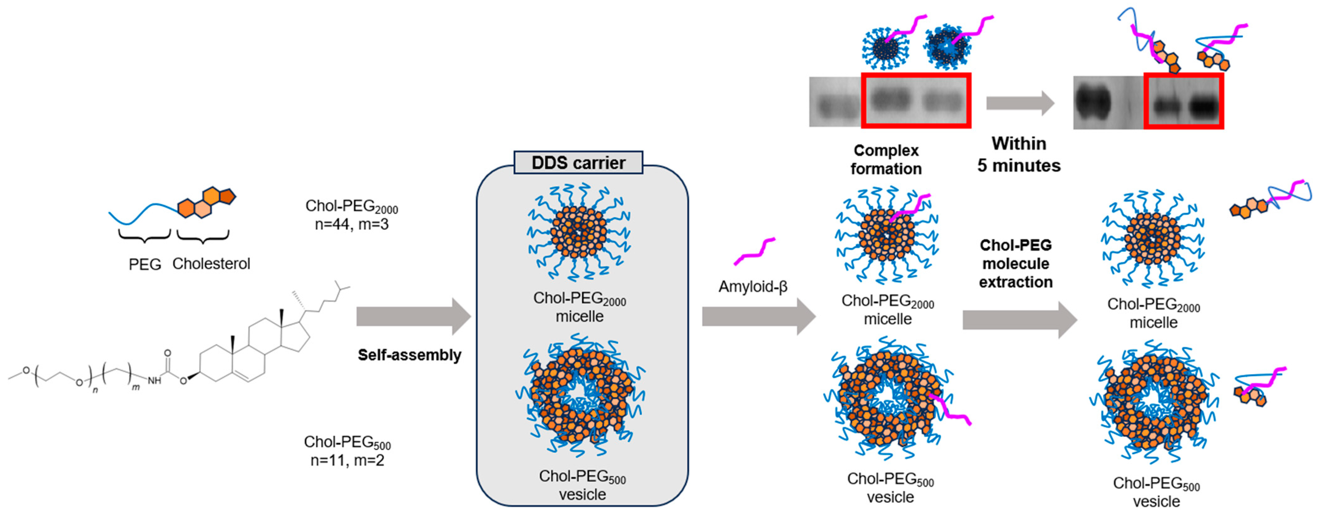
Background/Objectives: This study aimed to design and evaluate Chol-PEG2000 micelles and Chol-PEG500 vesicles as drug delivery system (DDS) carriers and inhibitors of amyloid-β (Aβ) aggregation, a key factor in Alzheimer’s disease (AD). Methods: The physical properties of Chol-PEG assemblies were characterized using dynamic light scattering (DLS), electrophoretic light scattering (ELS), and transmission electron microscopy (TEM). Inhibitory effects on Aβ aggregation were assessed via thioflavin T (ThT) assay, circular dichroism (CD) spectroscopy, and native polyacrylamide gel electrophoresis (native-PAGE). Results: Chol-PEG2000 micelles and Chol-PEG500 vesicles were found to exhibit diameters of 20–30 nm and 70–80 nm, respectively, with neutral surface charges and those physical properties indicated the high affinity for Aβ. At a 10-fold molar ratio, thioflavin T (ThT) assay revealed that Chol-PEG2000 delayed Aβ fibril elongation by 20 hours, while Chol-PEG500 delayed it by 40 hours against Aβ peptide. At a 50-fold molar ratio, both Chol-PEG2000 and Chol-PEG500 significantly inhibited Aβ aggregation, as indicated by minimal fluorescence intensity increases over 48 hours. CD spectroscopy indicated that Aβ maintained its random coil structure in the presence of Chol-PEG assemblies at a 50-fold molar ratio. Native-PAGE analysis demonstrated a retardation in Aβ migration immediately after mixing with Chol-PEG assemblies, suggesting complex formation. However, this retardation disappeared within 5 min, implying rapid dissociation of the complexes. Conclusions: This study demonstrated that Chol-PEG500 vesicles more effectively inhibit Aβ aggregation than Chol-PEG2000 micelles. Chol-PEG assemblies perform as DDS carriers to be capable of inhibiting Aβ aggregation. Chol-PEG assemblies can deliver additional therapeutics targeting other aspects of AD pathology. This dual-function platform shows promise as both a DDS carrier and a therapeutic agent, potentially contributing to a fundamental cure for AD.

Graphical Abstract

1. Introduction

In the brains of Alzheimer’s disease (AD) patients, amyloid-beta (Aβ) peptides are produced by sequential proteolytic cleavages of a membrane-bound protein called amyloid-beta precursor protein (APP) by proteases known as β-secretase and γ-secretase [1,2]. These Aβ peptides, consisting of around 40 amino acid residues, are known for their high propensity to form β-sheets and aggregate. Aggregated Aβ peptides have been reported in numerous studies to form amyloid plaques, which induce neuronal cell death [3,4]. The neuronal cell death leads to brain atrophy and cognitive impairment, ultimately resulting in the onset of AD [5,6]. Such diseases triggered by conformational changes in peptides or proteins are collectively referred to as “conformational diseases” [7]. Therefore, controlling peptide conformation is crucial for the treatment of AD and other conformational diseases. Consequently, significant efforts have been made to develop AD therapeutics targeting Aβ. Various agents, such as secretase inhibitors, anti-Aβ peptides, and anti-Aβ antibodies have been developed [8,9]. Recently, antibody drugs named aducanumab, lecanemab, and donanemab have been approved by regulatory authorities [10]; however, issues such as adverse effects, including ARIA (amyloid-related imaging abnormalities), remain unresolved, and the therapeutic effects are limited [11]. The low transfer rate of conventional antibody drugs to the brain, approximately 0.1% of the serum antibody concentration, is considered to be a significant factor limiting their therapeutic efficacy [12]. To facilitate drug delivery to the brain, it is essential for the drug to cross the blood-brain barrier (BBB). The BBB plays a critical role in protecting the central nervous system (CNS) from potentially harmful blood-borne substances; however, it selectively permits the passage of certain molecules, presenting a major obstacle to effective brain-targeted drug delivery [13]. Recently, intranasal administration has gained attention as a non-invasive method for drug delivery that bypasses the BBB [14,15]. Although the precise mechanisms remain incompletely understood, studies have shown that drugs administered via the intranasal route can reach the brain through transport via the olfactory and trigeminal nerves [16]. Additionally, the high cost of antibody drugs makes widespread application among the large population of AD patients impractical.
The alteration in cholesterol levels in the brains of AD patients has been reported [17,18,19,20], suggesting a potential link between cholesterol metabolism disorders and AD. Although the brain constitutes only 2% of body weight, it contains approximately one quarter of the body’s total cholesterol [21]. Furthermore, numerous studies have demonstrated that Aβ interacts with lipid membranes, with a particular preference for binding to cholesterol-containing lipid membranes [21,22]. C. Duyckaerts’s research group observed the binding of cholesterol and Aβ in AD senile plaques [23], J. Fantini and co-workers have reported that cholesterol in a lipid membrane strongly bound to Aβ peptide [24], and J. R. Harris has confirmed that micelles of Chol-PEG also interact with Aβ peptides and fibrils by TEM observation [25]. However, whether such an interaction between Aβ and cholesterol inserted into lipid membranes inhibits or promotes the aggregation of Aβ is still under discussion due to the complex interplay of various factors, including cholesterol content, surface charge, and membrane fluidity [26,27].
So far, our laboratory has developed a cholesterol-end-modified PEG, as a drug delivery carrier and bioinert surface coating, which was found to spontaneously form micelles and vesicles (assemblies) in water [28,29]. We successfully obtained not only micelles but also vesicles from cholesterol-end-modified PEG with molecular weights of 2000 and 500, respectively. These assemblies involving cholesterol, which can bind to Aβ, are expected to be used not only as a drug carrier but also as an inhibitor of Aβ aggregation. Additionally, it has been reported that PEG-modified nanoparticles, such as these assemblies, enhance the efficiency of brain delivery via intranasal administration [30,31]. Although the optimal characteristics of nanocarriers for intranasal administration have yet to be fully elucidated, PEGylated nanoparticles (NPs) with a diameter of less than 200 nm and a surface charge close to neutral have been employed for efficient drug delivery and may be particularly suitable for intranasal administration [30,31,32].
Here, we investigated an inhibitory effect of cholesterol-end-modified PEG assemblies on aggregation of Aβ peptides, particularly Aβ40, which consists of 40 amino acids. It is well known that Aβ42, consisting of 42 amino acids, has a higher propensity for aggregation compared to Aβ40 [33], while it is possible that such an Aβ42 aggregation can be controlled by a decrease in Aβ40 aggregation [34]. In this study, we examined the effects on Aβ40 aggregation, because Aβ40 is a good therapeutic target to be produced at nearly ten times the rate of Aβ42 [35].

2. Materials and Methods

2.1. Materials

mPEG-NH2 (molecular weight of 2000, SUNBRIGHT® ME-020EA) was purchased from NOF Corporation (Tokyo, Japan). mPEG-NH2 (molecular weight of 500, 767565) and Thioflavin T (ThT) were purchased from Sigma-Aldrich Co. LLC (St. Louis, MO, USA). Cholesterol chloroformate was purchased from TCI Co., Ltd. (Tokyo, Japan). Amyloid β-Protein (Human, 1–40) [HCl Form] (Aβ40) was purchased from PEPTIDE INSTITUTE, INC. (Osaka, Japan). All other chemicals of a special grade were used without further purification.

2.2. Preparation of Chol-PEG Assemblies

Chol-PEG assemblies (Chol-PEG2000 micelles and Chol-PEG500 vesicles) were self-assembled solely by dissolving them in potassium phosphate buffer, without stirring or ultrasonication.

2.3. Particle Size and Zeta Potential Measurement

A dynamic light scattering (DLS) method by an electrophoresis light-scattering spectrophotometer (ELS-Z2, Otsuka Electronics Co., Ltd., Tokyo, Japan) determined the size of the Chol-PEG assembles with each concentration in potassium phosphate buffer at room temperature. The zeta potential of the resulting sample was measured at room temperature by ELS with electrodes.

2.4. Transmission Electron Microscopy (TEM) Observation

Chol-PEG2000 and Chol-PEG500 assemblies were prepared in potassium phosphate buffer to 9.4 μmol and used as sample solutions for TEM observation. A TEM grid (Nisshin EM Co., Tokyo, Japan) was dipped into the sample solution for a few seconds. The excess solution was blotted away by filter paper. The sample was stained by 2% phosphotungstic acid solution. The samples on grids were observed by a JEM-1400 (JEOL Ltd., Tokyo, Japan) at an acceleration voltage of 120 kV. Chol-PEG500, prepared in deionized water at a concentration of 5.5 mM, was examined using a JEM-2100F (JEOL Ltd., Tokyo, Japan) electron microscope at an acceleration voltage of 200 kV with uranyl acetate as the staining agent.

2.5. Thioflavin T (ThT) Assay

40 was dissolved in a 0.1% aqueous ammonia solution to a concentration of 4.7 × 10² μM. For each well of a 96-well black PP plate (Greiner Bio-One, Co., Ltd., Frickenhausen, Germany), 2 μL of this solution was used. ThT was mixed to a final concentration of 9.4 × 10⁻³ μM, and Chol-PEGs were added at molar ratios of 10 and 50 relative to Aβ40. The total volume was adjusted to 100 μL per well. Each sample was prepared in triplicate (n = 3). As controls, NH2-mPEGs with the same molar concentrations as Chol-PEGs were prepared for each PEG molecular weight. Fluorescence intensities were measured every 15 min while incubating the prepared samples at 37 °C using a SpectraMaxTM mini (Molecular Devices LLC., US). A pre-shake was set for 60 s before each measurement. The excitation wavelength was 485 nm, and the emission wavelength was 535 nm. The dissociation of Aβ40 aggregation was evaluated under the same concentration conditions as the ThT assay described above. A sample containing Aβ40 alone was prepared following the same procedure as described previously, and aggregation was induced at 37 °C for 72 h. Subsequently, Chol-PEG500 was added at a molar ratio of 50 relative to Aβ40, and the sample was incubated again at 37 °C. Fluorescence intensity was measured hourly during the incubation.

2.6. Circular Dichroism (CD) Measurement

All samples were adjusted to the same concentration as in the ThT assay. The CD spectra from 200 nm to 250 nm of the resulting sample were measured with a J-820 spectropolarimeter (JASCO Corporation, Tokyo, Japan).

2.7. Naive Polyacrylamide Gel Electrophoresis (Native-PAGE)

The preparation of Aβ40 and Chol-PEGs was adjusted so that the final concentration and total amount matched those used in the ThT assay. The prepared samples were incubated at 37 °C, and gel electrophoresis was performed at 0 and 72 h. A 7.2 μL aliquot from each sample was taken and mixed with 0.8 μL of 10× loading buffer, then loaded onto an 8% polyacrylamide gel. The prepared gel was run using a buffer (pH 8.3) comprising 50 mM tris (hydroxymethyl)aminomethane (Tris) and 38 mM glycine. Electrophoresis was performed at room temperature for 10 min, and the electric current was maintained at 24 W using a WSE-1010 Compact PAGE Ace (ATTO Co., Tokyo, Japan). After electrophoresis, the gel was shaken in a fixing solution (methanol: water: acetic acid = 4: 5: 1) for 30 min, followed by staining with Coomassie brilliant blue (CBB) for 30 min. The gel was then imaged using a GelDoc Go Imaging System (Bio-Rad Laboratories Inc., Hercules, CA, USA). The captured images were adjusted for brightness (+40%) using Microsoft PowerPoint.

3. Results

3.1. Physical Properties of Chol-PEG Assemblies

Cholesterol-end-modified PEGs were synthesized as reported previously [28,29]. Briefly, the synthesis was carried out by SN2 reaction between cholesteryl chloroformate and methoxy poly (ethylene glycol) amine (mPEG-NH2). Hereafter, cholesterol-end-modified PEGs using PEGs with molecular weights of 2000 and 500 are referred to as Chol-PEG2000 and Chol-PEG500, respectively. In this paper, we prepared Chol-PEG assemblies with two different concentrations, 94 µM and 470 µM, above the CAC reported previously [28,29] to study the effect of Chol-PEG concentration in Aβ40 inhibition experiments that follow. Figure 1 shows the particle size and zeta potential of the Chol-PEG assemblies in buffer (potassium phosphate buffer of 50 mM, pH 7.4) using dynamic light scattering (DLS) and electrophoretic light scattering (ELS). As previously reported [28,29], Chol-PEG2000 exhibited a size of approximately 20–30 nm in diameter, while Chol-PEG500 showed a size of approximately 70–80 nm in diameter at each concentration. The zeta potential was nearly neutral in both cases (Figure 1). The concentration did not affect the size and surface charge of assemblies. Figure 2A–D present the TEM images of the Chol-PEG assemblies in potassium phosphate buffer, which will be used in later Aβ experiments. Because phosphates in the buffer interacted with the staining solution, the images could not be taken clearly, so images taken using water as the solvent are shown in Figure S1. TEM images showed that Chol-PEG2000 formed a uniform micelle and its particle diameter was consistent with the DLS results (Figure 1 and Figure S2). On the other hand, Chol-PEG500 self-assembled into a uniform hollow vesicle with the uniform size of 70–80 nm, which corresponds to DLS results (Figure 1 and Figure S2). At 4.7 × 10⁻⁵ μM below the CAC, no particles were observed in either Chol-PEG2000 or Chol-PEG500 (Figure 2C,D). As a result, the micelles and vesicles with neutral surface charges, which have been reported to interact with Aβ [21,22], were successfully obtained.

3.2. ThT Assay of Aβ40/Chol-PEG Assemblies

To investigate the aggregation inhibitory effect of Chol-PEGs on Aβ, a ThT assay [36,37] was performed. ThT is a fluorescent reagent known to specifically bind to β-sheets, resulting in increased fluorescence intensity. Since Aβ forms a β-sheet structure upon aggregation, the fluorescence intensity derived from ThT increases with the aggregation of Aβ40. The graph showing the fluorescence intensity of each sample over time is presented in Figure 3. The figure with error bars was complicated, so it is shown in Figure S3. In the sample of Aβ40 alone, the fluorescence intensity began to increase at around 3 h and stabilized at around 6–7 h, confirming that aggregation was progressing. This is a typical result of a ThT assay showing Aβ aggregation [38]. When Aβ40 was incubated with Chol-PEG assemblies at high concentrations (50 times the molar ratio relative to Aβ40, 470 µM), neither Chol-PEG2000 micelles nor Chol-PEG500 vesicles showed a significant increase in fluorescence intensity throughout the measurement period. At low concentrations (10 times the molar ratio relative to Aβ40, 94 µM), the mixture of Aβ40 and Chol-PEG assemblies delayed the onset time of increase in fluorescence intensity, which indicates that Chol-PEG delayed a nucleation of Aβ [39] (Figure 3A,B). In detail, the mixture of Aβ40 and Chol-PEG2000 showed an increase of fluorescence intensity after 20 h, while the increase of fluorescence intensity was observed after 40 h in the mixture of Aβ40 and Chol-PEG500. These results indicate that Chol-PEG500 delayed nucleation longer than Chol-PEG2000. Furthermore, when low concentrations of Chol-PEG2000 were added, the value at which the fluorescence intensity plateaued was approximately half that of Aβ40 alone (Figure S4A). It has been reported that increasing fluorescence intensity of ThT means fibril elongation [40]. This suggests that fibril elongation is stopped midway. Further, an incubation up to 72 h showed that when 10 equivalents of Chol-PEG500 were added to Aβ40, the fluorescence intensity at the plateau was 30–40% lower than that of Aβ40 alone and lower than that of the mixture with Chol-PEG2000. (Figure S4B). The fluorescence intensities measured at the end of the experiment were 3.2 × 10⁶ for Aβ alone (FI), 1.1 × 10⁶ for the mixture with Chol-PEG2000 (FI2000), and 5.9 × 10⁵ for the mixture with Chol-PEG500 (FI500). The binding ratio of Aβ40 to each Chol-PEG can be estimated from (FI − FI2000)/FI and (FI − FI500)/FI, assuming that Aβ40 not bound to Chol-PEGs forms fibrils. Applying this formula, the binding ratio of Chol-PEGs to Aβ40 is calculated to be 64% for Chol-PEG2000 [(FI − FI2000)/FI × 100] and 84% for Chol-PEG500 [(FI − FI500)/FI × 100], respectively. Namely, Chol-PEG500 exhibited 1.3 times the binding affinity for Aβ40 compared to Chol-PEG2000. These results indicate that Chol-PEG2000 and Chol-PEG500 suppressed the Aβ40 aggregation at a higher molar ratio than 50 and 10 against Aβ40, respectively. So far, some reports have shown that cholesterol promotes Aβ aggregation [41,42], but our results were the opposite, inhibiting Aβ aggregation. In the ThT assay, it is known that the period from when the fluorescence intensity begins to increase until it plateaus reflects the elongation of amyloid fibrils [40]. In this study, no significant changes in elongation rate were observed at high concentrations of Chol-PEG2000 and at all concentrations of Chol-PEG500, suggesting that Chol-PEGs might suppress nucleation before Aβ monomers form oligomers. Next, ThT assays were performed by adding mPEG-NH2 of two molecular weights to Aβ40 as controls. The fluorescence intensity with mPEG2000-NH2 was reduced by approximately –30% for PEG2000 compared to Aβ40 alone (Figure 3C). It has been reported that the presence of polymers such as PEG can inhibit the binding of Aβ to ThT [43,44]. However, such an inhibitory effect of PEG on the binding of Aβ40 to ThT had minimal impact on the results in our case. The mixture of Chol-PEG alone and ThT (without Aβ40) showed no increase in fluorescence intensity (Figure 3C,D). This means that both the cholesterol moiety and the PEG chain play an important role in inhibiting Aβ40 fibril formation, and the importance of Chol-PEG, a covalent conjugate of cholesterol and PEG, has been proved.

3.3. Evaluation of β-Sheet Formation with CD Measurement

CD spectroscopy was performed to confirm the secondary structure of Aβ40 before and after incubation with Chol-PEG assemblies. Aβ40 was prepared at the same concentrations as in the ThT assay. Immediately after mixing Aβ40 and Chol-PEG assembly (0 h), Aβ40 exhibited a random coil conformation, and the spectrum was consistent with previously reported results [45,46] (Figure 4A,B). The Chol-PEG assemblies themselves are optically active, exhibiting a spectrum with a minimum around 205 nm (Figure S5). Therefore, to facilitate comparison, the ellipticity of Chol-PEG alone was subtracted from the CD ellipticities obtained for all Aβ40 and Chol-PEG mixtures, and the difference was plotted as [Aβ/Chol-PEG]–[Chol-PEG]. Each sample was incubated at 37 °C for 48 h, after which CD spectra were measured under the same conditions. After 48 h, the Aβ40-only sample showed an increase in the short wavelength region of 200–220 nm, which initially showed negative values at 0 h and shifted to near-zero values (Figure 4C,D). It is known that Aβ40 forms β-sheets from naked random coils as they aggregate, with a positive maximum around 197 nm and a cross-over at 203–206 nm [47]. In both Chol-PEG2000 and Chol-PEG500, Aβ40 mixed with Chol-PEGs exhibited a random coil-like spectrum similar to that of Aβ40 alone at 0 h. Even at 48 h, the Aβ40/Chol-PEG mixtures maintained a random coil-like spectrum for both Chol-PEG2000 and Chol-PEG500. In contrast, control samples of only PEG chains, or mPEG2000-NH2 or mPEG500-NH2 mixed with Aβ40, showed spectra indicative of a β-sheet structure similar to that of Aβ40 alone. These results suggest that in the presence of Chol-PEG, Aβ40 maintained its secondary structure as a random coil.

3.4. Aggregation Evaluation Using Native-PAGE

The aggregation inhibition effect of Chol-PEG assemblies and the formation of complexes between Aβ40 and Chol-PEG assemblies were evaluated using polyacrylamide gel electrophoresis (Native-PAGE). As shown in Figure 5A, in the presence of Chol-PEG assemblies, Aβ40 led to a noticeable retardation compared to the migration profile of Aβ40 alone. The retardation means an increase in molecular weight. Results from multiple repeated experiments are presented in Figure 5C for clarity. In these images, the retardation reproduced in the case of mixture of Aβ40 and Chol-PEG assemblies. These retardations reflect the binding of Chol-PEG assemblies to Aβ40, leading to the formation of complexes. Electrophoresis results after 48 h of incubation under aggregation conditions are shown in Figure 5B. An incubation of Aβ40 alone resulted in no band which means insolubilization by aggregation (lane i). Notably, the bands of Aβ40 mixed with Chol-PEG assemblies appeared at the same position (no retardation) as the naked Aβ40 after the 48 h incubation. While retardation was observed immediately after mixing (Figure 5A), no retardation after 48 h of incubation indicates a complex dissociation for inhibition of Aβ40 aggregation. For more detailed study, Native-PAGE was performed after 5 min of incubation (Figure S6). Interestingly, no retardation was observed even after 5 min.
The mixture of mPEG-NH2 and Aβ40 showed a band identical to that of Aβ40 alone (Figure S7A), which disappeared following incubation (Figure S7B). Additionally, no bands were observed in individual polymers alone (lanes iv–vii).

3.5. Disaggregation Effect of Chol-PEG Assembly on Aggregated Aβ

Finally, the disaggregation effect of Chol-PEG500 vesicles was evaluated using the ThT assay by addition of 50 equivalents of Chol-PEG500 vesicles to aggregated Aβ40 (Figure S8). Aggregated Aβ40 was prepared by incubating at 37 °C for 72 h using the same method as the ThT assay in Section 3.2. The fluorescence intensity was maintained during incubation for 3 days, which indicates that Aβ40 kept aggregation form and was not disaggregated by adding Chol-PEG500 vesicles.

4. Discussion

Native-PAGE results showed that retardation was observed immediately after mixing Aβ40 and Chol-PEG assemblies, but not after 48 h of incubation. Although no retardation was observed after 48 h, it is unlikely that the complexes completely dissociate from each other because the ThT assay and CD spectra showed that the aggregation was inhibited from 0 h to 48 h. For a more detailed study of the mechanism, Native-PAGE was performed 5 min after mixing (Figure S6). The results showed that no retardation was observed even after 5 min. Although it is difficult to perform Native-PAGE at shorter time points due to the experimental manipulations, at least within 5 min, the complexes of Aβ40 and Chol-PEG micelles or vesicles are dissociated. According to a report by Hashemi M et al., aggregation of Aβ bound to lipid bilayers was detected after 1 h. Since binding to Chol-PEG assemblies was resolved within 5 min, it is suggested that aggregation was not enhanced [41]. If the complex formation had completely dissociated, it would not be possible to explain the suppression of aggregation over a 48 h period. Therefore, we propose a mechanism by which Aβ40 pulls out Chol-PEG molecules from Chol-PEG micelles and vesicles (Figure 6).
PEGs can remove hydration from biopharmaceuticals and is used as a precipitant. Therefore, as the molecular weight of PEG increases, the dehydration of peptides intensifies, potentially triggering aggregation [48,49]. Moreover, it has been reported that a higher PEG molecular weight leads to increased steric hindrance, reducing interactions between the hydrophobic group and the peptide [50]. The surface of Chol-PEG assemblies is covered by hydrophilic PEG chains, and it was considered that Aβ40 was more likely to interact with cholesterol in Chol-PEG500, which has shorter PEG chains. Therefore, in this study, it is considered that Chol-PEG500, with its shorter PEG chains, inhibited aggregation more effectively compared to Chol-PEG2000. These binding and aggregation inhibitory effects in this study were contributed by several interactions reported in the literature [48,49,51,52,53,54,55,56].
In addition, we evaluated whether the addition of 50 equivalents of Chol-PEG500 to aggregated Aβ40 would cause disaggregation using the ThT assay. As a result, no decrease in fluorescence intensity was observed, and it was found that there was no disaggregation effect on already aggregated Aβ40 (Figure S8). When Aβ40 aggregates, hydrophobic amino acids (e.g., Phe-19, Phe-20, etc.) that bind to cholesterol in the vesicle contribute to the formation of the core and are hidden inside [57,58]. Therefore, the interaction with cholesterol-containing vesicles consisting of phospholipids is known to be reduced, as compared to that with naked Aβ. This suggests that Aβ40 and Chol-PEGs are bound to hydrophobic amino acids that are known to easily bind to cholesterol. It is well known that vesicles containing free (non-covalently bound) cholesterol and Aβ bind to each other, accelerating aggregation [59,60]. In this study, Chol-PEG is a covalently bonded cholesterol and PEG, and even after the dissociation complex between Aβ and Chol-PEG, the binding to the amphiphilic Chol-PEG molecules remained, so it is thought that aggregation was suppressed. While Aβ has been a primary focus in this research, other factors, such as tau protein and APOEε4, among others, are also known to contribute to its pathogenesis [61]. As previously reported, Chol-PEG assemblies can encapsulate therapeutic agents [28,29]. Therefore, by encapsulating additional AD-related factors, these assemblies offer potential for dual-targeted AD therapy via intranasal administration.

5. Conclusions

In this study, we found that the vesicle and micelle composed of Chol-PEG had aggregation-inhibiting effects on Aβ40, the causative agent of AD. These Chol-PEG assemblies with a high affinity for Aβ40 result in the absorption of Aβ40 into the Chol-PEG assemblies and suppress the aggregation of Aβ40. The reasons for this were expected to be that the surface PEG layer of Chol-PEG500 vesicles was thinner than that of Chol-PEG2000 micelles, making Aβ40 more likely to interact with cholesterol, and the too-long PEG chain length of Chol-PEG2000 causes dehydration [50] of Aβ40, inducing Aβ40 aggregation. Furthermore, the suppression of aggregation was considered to be due to hydrogen bonding with the urethane group of Chol-PEGs, the exclusion volume effect of PEG, and its high hydrophilicity The resulting suppression effect of Chol-PEG assemblies on Aβ40 aggregation is promoted at higher concentrations of the dispersion of the Chol-PEG assemblies. Although general cholesterol-containing micelles [20] and vesicles consisting of phospholipids are known to promote Aβ aggregation [51,52], our Chol-PEG assemblies used in this study proved to have moderate binding strength that rather inhibited aggregation. This provides an idea for the development of therapeutic agents in the study of interaction between Aβ and vesicles. Moreover, Chol-PEG500 vesicles, as well as Chol-PEG2000 micelles, can encapsulate hydrophilic and hydrophobic drugs due to their hollow shape with internal water phase and hydrophobic membrane. These Chol-PEG assemblies are designed to both inhibit Aβ aggregation and simultaneously deliver additional therapeutic agents via intranasal administration, offering a dual-functional approach for effective AD treatment.

Supplementary Materials

The following supporting information can be downloaded at: https://www.mdpi.com/article/10.3390/pharmaceutics17010001/s1, Figure S1: TEM image of Chol-PEG500 aqueous solution; Figure S2: Particle sizes of Chol-PEG assemblies measured from TEM images; Figure S3: ThT assay results of Aβ40 aggregation in the presence of each concentration of Chol-PEGs with error bars; Figure S4: ThT assay results of Aβ40 aggregation in the presence of each concentration of Chol-PEGs for 72 h with error bars; Figure S5: CD spectra of Chol-PEG alone; Figure S6: Polyacrylamide gel electrophoresis of Aβ40 incubated with Chol-PEGs 5 min after mixing Aβ and Chol-PEG assemblies; Figure S7: Polyacrylamide gel electrophoresis of each polymer incubated with or without Aβ40 as controls; Figure S8: Results of the ThT assay for aggregated Aβ40.

Author Contributions

Conceptualization, S.W. and S.A.; methodology, S.W.; validation, S.W., M.U., and S.A.; investigation, S.W.; data curation, S.W.; writing—original draft preparation, S.W.; writing—review and editing, M.U. and S.A.; visualization, S.W.; supervision, M.U. and S.A.; project administration, S.A.; funding acquisition, S.A. All authors have read and agreed to the published version of the manuscript.

Funding

This work was supported by Grant-in-Aid for Challenging Research (Exploratory) (JSPS KAKENHI grant No. 21K19921).

Institutional Review Board Statement

Not applicable.

Informed Consent Statement

Not applicable.

Data Availability Statement

All the data supporting the reported results can be found in the main article and in the Supplementary Materials files.

Acknowledgments

A part of this work was conducted in the Institute for Molecular Science, supported by “Advanced Research Infrastructure for Materials and Nanotechnology in Japan (ARIM)” of the Ministry of Education, Culture, Sports, Science and Technology (MEXT). Project Issue Number 23UT0090.

Conflicts of Interest

The authors declare no conflicts of interest.

References

  1. Beshir, S.A.; Hussain, N.; Menon, V.B.; Al Haddad, A.H.I.; Al Zeer, R.A.K.; Elnour, A.A. Advancements and Challenges in Antiamyloid Therapy for Alzheimer’s Disease: A Comprehensive Review. Int. J. Alzheimer’s Dis. 2024, 2024, 2052142. [Google Scholar] [CrossRef] [PubMed]
  2. Hur, J.-Y. γ-Secretase in Alzheimer’s disease. Exp. Mol. Med. 2022, 54, 433–446. [Google Scholar] [CrossRef] [PubMed]
  3. Grøntvedt, G.R.; Schröder, T.N.; Sando, S.B.; White, L.; Bråthen, G.; Doeller, C.F. Alzheimer’s disease. Curr. Biol. 2018, 28, R645–R649. [Google Scholar] [CrossRef]
  4. Aleksis, R.; Oleskovs, F.; Jaudzems, K.; Pahnke, J.; Biverstål, H. Structural studies of amyloid-β peptides: Unlocking the mechanism of aggregation and the associated toxicity. Biochimie 2017, 140, 176–192. [Google Scholar] [CrossRef]
  5. Leong, Y.Q.; Ng, K.Y.; Chye, S.M.; Ling, A.P.K.; Koh, R.Y. Mechanisms of action of amyloid-beta and its precursor protein in neuronal cell death. Metab. Brain Dis. 2019, 35, 11–30. [Google Scholar] [CrossRef]
  6. Choi, H.; Kim, C.; Song, H.; Cha, M.; Cho, H.J.; Son, S.M.; Kim, H.J.; Mook-Jung, I. Amyloid β-induced elevation of O-GlcNAcylated c-Fos promotes neuronal cell death. Aging Cell 2018, 18, e12872. [Google Scholar] [CrossRef]
  7. Carrell, R.W.; Lomas, D.A. Conformational disease. Lancet 1997, 350, 134–138. [Google Scholar] [CrossRef]
  8. Yamin, G.; Ono, K.; Inayathullah, M.; Teplow, D.B. Amyloid β -Protein Assembly as a Therapeutic Target of Alzheimers Disease. Curr. Pharm. Des. 2008, 14, 3231–3246. [Google Scholar] [CrossRef]
  9. Kapadia, A.; Sharma, K.K.; Maurya, I.K.; Singh, V.; Khullar, M.; Jain, R. Structural and mechanistic insights into the inhibition of amyloid-β aggregation by Aβ39-42 fragment derived synthetic peptides. Eur. J. Med. Chem. 2020, 212, 113126. [Google Scholar] [CrossRef]
  10. Loeffler, D.A. Antibody-Mediated Clearance of Brain Amyloid-β: Mechanisms of Action, Effects of Natural and Monoclonal Anti-Aβ Antibodies, and Downstream Effects. J. Alzheimer’s Dis. Rep. 2023, 7, 873–899. [Google Scholar] [CrossRef]
  11. Amano, A.; Sanjo, N.; Araki, W.; Anraku, Y.; Nakakido, M.; Matsubara, E.; Tomiyama, T.; Nagata, T.; Tsumoto, K.; Kataoka, K.; et al. Peripheral administration of nanomicelle-encapsulated anti-Aβ oligomer fragment antibody reduces various toxic Aβ species in the brain. J. Nanobiotechnol. 2023, 21, 36. [Google Scholar] [CrossRef] [PubMed]
  12. Niazi, S.K.; Mariam, Z.; Magoola, M. Engineered Antibodies to Improve Efficacy against Neurodegenerative Disorders. Int. J. Mol. Sci. 2024, 25, 6683. [Google Scholar] [CrossRef]
  13. Xie, J.; Shen, Z.; Anraku, Y.; Kataoka, K.; Chen, X. Nanomaterial-based blood-brain-barrier (BBB) crossing strategies. Biomaterials 2019, 224, 119491. [Google Scholar] [CrossRef]
  14. Lee, D.; Minko, T. Nanotherapeutics for Nose-to-Brain Drug Delivery: An Approach to Bypass the Blood Brain Barrier. Pharmaceutics 2021, 13, 2049. [Google Scholar] [CrossRef]
  15. Wang, Z.; Xiong, G.; Tsang, W.C.; Schätzlein, A.G.; Uchegbu, I.F.; Tsang, A. Nose-to-Brain Delivery. J. Pharmacol. Exp. Ther. 2019, 370, 593–601. [Google Scholar] [CrossRef]
  16. Adscheid, S.A.; Türeli, A.E.; Günday-Türeli, N.; Schneider, M. Nanotechnological approaches for efficient N2B delivery: From small-molecule drugs to biopharmaceuticals. Beilstein J. Nanotechnol. 2024, 15, 1400–1414. [Google Scholar] [CrossRef]
  17. Xiong, H.; Callaghan, D.; Jones, A.; Walker, D.G.; Lue, L.-F.; Beach, T.G.; Sue, L.I.; Woulfe, J.; Xu, H.; Stanimirovic, D.B.; et al. Cholesterol retention in Alzheimer’s brain is responsible for high β- and γ-secretase activities and Aβ production. Neurobiol. Dis. 2008, 29, 422–437. [Google Scholar] [CrossRef]
  18. Lazar, A.N.; Bich, C.; Panchal, M.; Desbenoit, N.; Petit, V.W.; Touboul, D.; Dauphinot, L.; Marquer, C.; Laprévote, O.; Brunelle, A.; et al. Time-of-flight secondary ion mass spectrometry (TOF-SIMS) imaging reveals cholesterol overload in the cerebral cortex of Alzheimer disease patients. Acta Neuropathol. 2013, 125, 133–144. [Google Scholar] [CrossRef]
  19. Ledesma, M.D.; Abad-Rodriguez, J.; Galvan, C.; Biondi, E.; Navarro, P.; Delacourte, A.; Dingwall, C.; Dotti, C.G. Raft disorganization leads to reduced plasmin activity in Alzheimer’s disease brains. EMBO Rep. 2003, 4, 1190–1196. [Google Scholar] [CrossRef]
  20. Egawa, J.; Pearn, M.L.; Lemkuil, B.P.; Patel, P.M.; Head, B.P. Membrane lipid rafts and neurobiology: Age-related changes in membrane lipids and loss of neuronal function. J. Physiol. 2016, 594, 4565–4579. [Google Scholar] [CrossRef]
  21. Rushworth, J.V.; Hooper, N.M. Lipid Rafts: Linking Alzheimer′s Amyloid-β Production, Aggregation, and Toxicity at Neuronal Membranes. Int. J. Alzheimer’s Dis. 2010, 2011, 603052. [Google Scholar] [CrossRef] [PubMed]
  22. Avdulov, N.A.; Chochina, S.V.; Igbavboa, U.; Warden, C.S.; Vassiliev, A.V.; Wood, W.G. Lipid Binding to Amyloid β-Peptide Aggregates: Preferential Binding of Cholesterol as Compared with Phosphatidylcholine and Fatty Acids. J. Neurochem. 1997, 69, 1746–1752. [Google Scholar] [CrossRef] [PubMed]
  23. Panchal, M.; Loeper, J.; Cossec, J.-C.; Perruchini, C.; Lazar, A.; Pompon, D.; Duyckaerts, C. Enrichment of cholesterol in microdissected Alzheimer’s disease senile plaques as assessed by mass spectrometry. J. Lipid Res. 2010, 51, 598–605. [Google Scholar] [CrossRef] [PubMed]
  24. Di Scala, C.; Chahinian, H.; Yahi, N.; Garmy, N.; Fantini, J. Interaction of Alzheimer’s β-Amyloid Peptides with Cholesterol: Mechanistic Insights into Amyloid Pore Formation. Biochemistry 2014, 53, 4489–4502. [Google Scholar] [CrossRef] [PubMed]
  25. Harris, J.R. Cholesterol binding to amyloid-β fibrils: A TEM study. Micron 2008, 39, 1192–1196. [Google Scholar] [CrossRef]
  26. Martínez-Senac, M.d.M.; Villalaín, J.; Gómez-Fernández, J.C. Structure of the Alzheimer β-amyloid peptide (25–35) and its interaction with negatively charged phospholipid vesicles. Eur. J. Biochem. 1999, 265, 744–753. [Google Scholar] [CrossRef]
  27. Ahyayauch, H.; Masserini, M.E.; Alonso, A.; Goñi, F.M. Understanding Aβ Peptide Binding to Lipid Membranes: A Biophysical Perspective. Int. J. Mol. Sci. 2024, 25, 6401. [Google Scholar] [CrossRef]
  28. Asayama, S.; Nagashima, K.; Negishi, Y.; Kawakami, H. Byproduct-Free Intact Modification of Insulin by Cholesterol End-Modified Poly(ethylene glycol) for in Vivo Protein Delivery. Bioconjugate Chem. 2018, 29, 67–73. [Google Scholar] [CrossRef]
  29. Watanabe, S.; Asayama, S. Cholesterol-end-modified–PEG vesicle: DDS carrier with long-term stability to encapsulate a drug model by facile preparation. Chem. Lett. 2024, 53, upae166. [Google Scholar] [CrossRef]
  30. Kurano, T.; Kanazawa, T.; Ooba, A.; Masuyama, Y.; Maruhana, N.; Yamada, M.; Iioka, S.; Ibaraki, H.; Kosuge, Y.; Kondo, H.; et al. Nose-to-brain/spinal cord delivery kinetics of liposomes with different surface properties. J. Control. Release 2022, 344, 225–234. [Google Scholar] [CrossRef]
  31. Zhang, Q.-Z.; Zha, L.-S.; Zhang, Y.; Jiang, W.-M.; Lu, W.; Shi, Z.-Q.; Jiang, X.-G.; Fu, S.-K. The brain targeting efficiency following nasally applied MPEG-PLA nanoparticles in rats. J. Drug Target. 2006, 14, 281–290. [Google Scholar] [CrossRef] [PubMed]
  32. Huang, Q.; Chen, X.; Yu, S.; Gong, G.; Shu, H. Research progress in brain-targeted nasal drug delivery. Front. Aging Neurosci. 2024, 15, 1341295. [Google Scholar] [CrossRef]
  33. Jarrett, J.T.; Berger, E.P.; Lansbury, P.T. The carboxy terminus of the .beta. amyloid protein is critical for the seeding of amyloid formation: Implications for the pathogenesis of Alzheimer’s disease. Biochemistry 1993, 32, 4693–4697. [Google Scholar] [CrossRef]
  34. Cukalevski, R.; Yang, X.; Meisl, G.; Weininger, U.; Bernfur, K.; Frohm, B.; Knowles, T.P.J.; Linse, S. The Aβ40 and Aβ42 peptides self-assemble into separate homomolecular fibrils in binary mixtures but cross-react during primary nucleation. Chem. Sci. 2015, 6, 4215–4233. [Google Scholar] [CrossRef]
  35. Wang, L.; Eom, K.; Kwon, T. Different Aggregation Pathways and Structures for Aβ40 and Aβ42 Peptides. Biomolecules 2021, 11, 198. [Google Scholar] [CrossRef]
  36. Groenning, M. Binding mode of Thioflavin T and other molecular probes in the context of amyloid fibrils—Current status. J. Chem. Biol. 2009, 3, 1–18. [Google Scholar] [CrossRef]
  37. LeVine, H., 3rd. Quantification of β-sheet amyloid fibril structures with thioflavin T. Methods En Mol. 1999, 309, 274. [Google Scholar] [CrossRef]
  38. Batzli, K.M.; Love, B.J. Agitation of amyloid proteins to speed aggregation measured by ThT fluorescence: A call for standardization. Mater. Sci. Eng. C 2015, 48, 359–364. [Google Scholar] [CrossRef]
  39. Tikader, B.; Maji, S.K.; Kar, S. A generic approach to decipher the mechanistic pathway of heterogeneous protein aggregation kinetics. Chem. Sci. 2021, 12, 13530. [Google Scholar] [CrossRef]
  40. Arosio, P.; Knowles, T.P.J.; Linse, S. On the lag phase in amyloid fibril formation. Phys. Chem. Chem. Phys. 2015, 17, 7606–7618. [Google Scholar] [CrossRef]
  41. Hashemi, M.; Banerjee, S.; Lyubchenko, Y.L. Free Cholesterol Accelerates Aβ Self-Assembly on Membranes at Physiological Concentration. Int. J. Mol. Sci. 2022, 23, 2803. [Google Scholar] [CrossRef] [PubMed]
  42. Yanagisawa, K. Cholesterol and Aß Aggregation. Pharmacopsychiatry 2003, 36, 127–129. [Google Scholar] [CrossRef]
  43. Biancalana, M.; Koide, S. Molecular mechanism of Thioflavin-T binding to amyloid fibrils. Biochim. Biophys Acta 2010, 1804, 1405–1412. [Google Scholar] [CrossRef]
  44. Tsuchie, Y.; Kusuda, S.; Kawabe, H.; Mori, W.; Lindgren, M.; Watanabe, Y.; Zako, T. Polyallylamine Binds to Aβ Amyloid and Inhibits Antibody Recognition. Int. J. Mol. Sci. 2024, 25, 3112. [Google Scholar] [CrossRef]
  45. Kawahara, M.; Muramoto, K.; Kobayashi, K.; Mori, H.; Kuroda, Y. Aluminum Promotes the Aggregation of Alzheimer′s Amyloid β-Protein in Vitro. Biochem. Biophys. Res. Commun. 1994, 198, 531. [Google Scholar] [CrossRef]
  46. Nakano, H.; Hamaguchi, T.; Ikeda, T.; Watanabe-Nakayama, T.; Ono, K.; Yamada, M. Inactivation of seeding activity of amyloid β-protein aggregates in vitro. J. Neurochem. 2022, 160, 499–516. [Google Scholar] [CrossRef]
  47. Pachahara, S.K.; Adicherla, H.; Nagaraj, R. Self-Assembly of Aβ40, Aβ42 and Aβ43 Peptides in Aqueous Mixtures of Fluorinated Alcohols. PLoS ONE 2015, 10, e0136567. [Google Scholar] [CrossRef]
  48. Cheung, D.L. Surface Hydrophobicity Strongly Influences Adsorption and Conformation of Amyloid Beta Derived Peptides. Molecules 2024, 29, 3634. [Google Scholar] [CrossRef]
  49. Qu, L.; Fudo, S.; Matsuzaki, K.; Hoshino, T. Computational Study on the Assembly of Amyloid β-Peptides in the Hydrophobic Environment. Chem. Pharm. Bull. 2019, 67, 959–965. [Google Scholar] [CrossRef]
  50. Mueller, C.; Capelle, M.A.; Seyrek, E.; Martel, S.; Carrupt, P.-A.; Arvinte, T.; Borchard, G. Noncovalent PEGylation: Different Effects of Dansyl-, l-Tryptophan–, Phenylbutylamino-, Benzyl- and Cholesteryl-PEGs on the Aggregation of Salmon Calcitonin and Lysozyme. J. Pharm. Sci. 2012, 101, 1995–2008. [Google Scholar] [CrossRef]
  51. Ahyayauch, H.; García-Arribas, A.B.; Masserini, M.E.; Pantano, S.; Goñi, F.M.; Alonso, A. β-Amyloid (1–42) peptide adsorbs but does not insert into ganglioside-containing phospholipid membranes in the liquid-disordered state: Modelling and experimental studies. Int. J. Biol. Macromol. 2020, 164, 2651–2658. [Google Scholar] [CrossRef] [PubMed]
  52. Lockhart, C.; Klimov, D.K. The Alzheimer’s disease A β peptide binds to the anionic DMPS lipid bilayer. Biochim. Biophys. Acta (BBA)—Biomembr. 2016, 1858, 1118–1128. [Google Scholar] [CrossRef] [PubMed]
  53. Dey, C.; Roy, M.; Ghosh, R.; Pal, P.; Roy, D.; Dey, S.G. Active Site Environment and Reactivity of Copper-Aβ in Membrane Mimetic SDS Micellar Environment. Chem.—Eur. J. 2024, 30, e202401531. [Google Scholar] [CrossRef]
  54. Salahuddin, P.; Fatima, M.T.; Abdelhameed, A.S.; Nusrat, S.; Khan, R.H. Structure of amyloid oligomers and their mechanisms of toxicities: Targeting amyloid oligomers using novel therapeutic approaches. Eur. J. Med. Chem. 2016, 114, 41. [Google Scholar] [CrossRef]
  55. Pozzi, D.; Colapicchioni, V.; Caracciolo, G.; Piovesana, S.; Capriotti, A.L.; Palchetti, S.; De Grossi, S.; Riccioli, A.; Amenitsch, H.; Laganà, A. Effect of polyethyleneglycol (PEG) chain length on the bio–nano-interactions between PEGylated lipid nanoparticles and biological fluids: From nanostructure to uptake in cancer cells. Nanoscale 2014, 6, 2782–2792. [Google Scholar] [CrossRef]
  56. Yu, X.; Zheng, J. Cholesterol promotes the interaction of Alzheimer β-amyloid monomer with lipid bilayer. J. Mol. Biol. 2012, 421, 561–571. [Google Scholar] [CrossRef]
  57. Wälti, M.A.; Ravotti, F.; Arai, H.; Glabe, C.G.; Wall, J.S.; Böckmann, A.; Güntert, P.; Meier, B.H.; Riek, R. Atomic-resolution structure of a disease-relevant Aβ(1–42) amyloid fibril. Proc. Natl. Acad. Sci. USA 2016, 113, E4976–E4984. [Google Scholar] [CrossRef]
  58. van Gils, J.H.M.; van Dijk, E.; Peduzzo, A.; Hofmann, A.; Vettore, N.; Schützmann, M.P.; Groth, G.; Mouhib, H.; Otzen, D.E.; Buell, A.K.; et al. The hydrophobic effect characterises the thermodynamic signature of amyloid fibril growth. PLOS Comput. Biol. 2020, 16, e1007767. [Google Scholar] [CrossRef]
  59. Banerjee, S.; Hashemi, M.; Zagorski, K.; Lyubchenko, Y.L. Cholesterol in Membranes Facilitates Aggregation of Amyloid β Protein at Physiologically Relevant Concentrations. ACS Chem. Neurosci. 2021, 12, 506–516. [Google Scholar] [CrossRef]
  60. Elbassal, E.A.; Liu, H.; Morris, C.; Wojcikiewicz, E.P.; Du, D. Effects of Charged Cholesterol Derivatives on Aβ40 Amyloid Formation. J. Phys. Chem. B 2016, 120, 59–68. [Google Scholar] [CrossRef]
  61. Breijyeh, Z.; Karaman, R. Comprehensive Review on Alzheimer’s Disease: Causes and Treatment. Molecules 2020, 25, 5789. [Google Scholar] [CrossRef] [PubMed]
Figure 1. Particle sizes and zeta potentials of Chol-PEGs assemblies at each concentration used in later Aβ experiments. All concentrations of Chol-PEGs assemblies were above the critical aggregation concentration (CAC).
Pharmaceutics 17 00001 g001
Figure 2. TEM images of assembly of (A,C) Chol-PEG2000 and (B,D) Chol-PEG500 at the concentration of (A,B) 470 μM above CAC and (C,D) 4.7 × 10⁻⁵ μM below CAC.
Pharmaceutics 17 00001 g002
Figure 3. ThT assay results of Aβ40 aggregation in the presence of each concentration of Chol-PEGs. (A) Aβ40 incubated with Chol-PEG2000, (B) Aβ40 incubated with Chol-PEG500, (C) Aβ40 incubated with mPEG2000-NH2 and Chol-PEG2000 alone (without Aβ40), (D) Aβ40 incubated with mPEG500-NH2 and Chol-PEG500 alone (without Aβ40). The final concentration of Chol-PEGs at Chol-PEG/Aβm = 10 and Chol-PEG/Aβ = 50 is 94 µM and 470 µM, respectively.
Pharmaceutics 17 00001 g003
Figure 4. CD spectra of Aβ40 and Chol-PEG mixtures. (A,B) CD spectra immediately after mixing (0 h) of Aβ40 with (A) Chol-PEG2000 or (B) Chol-PEG500, and their respective controls, mPEG2000-NH2 and mPEG500-NH2. (C,D) CD spectra after 48 h of incubation (48 h) of Aβ40 with (C) Chol-PEG2000 or (D) Chol-PEG500, and their respective controls.
Pharmaceutics 17 00001 g004
Figure 5. Polyacrylamide gel electrophoresis of Aβ40 incubated with Chol-PEGs (A) just after mixing Aβ40 and Chol-PEG assemblies, (B) after incubation for 48 h. (C) Magnified results of repeating the experiment in (A) five times, (D) magnified results of repeating the experiment in (B) five times.
Pharmaceutics 17 00001 g005
Figure 6. Hypothesis on the mechanism by which Chol-PEG inhibits Aβ40 aggregation.
Pharmaceutics 17 00001 g006
Disclaimer/Publisher’s Note: The statements, opinions and data contained in all publications are solely those of the individual author(s) and contributor(s) and not of MDPI and/or the editor(s). MDPI and/or the editor(s) disclaim responsibility for any injury to people or property resulting from any ideas, methods, instructions or products referred to in the content.