Abstract
Most antiviral and anticancer nucleosides are prodrugs that require stepwise phosphorylation to their triphosphate nucleotide form for biological activity. Monophosphorylation may be rate-limiting, and the nucleotides may be unstable and poorly internalized by target cells. Effective targeting and delivery systems for nucleoside drugs, including oligonucleotides used in molecular therapeutics, could augment their efficacy. The development of a carrier designed to effect selective transmembrane internalization of nucleotides via the asialoglycoprotein receptor (ASGPr) is now reported. In this work, the polycationic, polygalactosyl drug delivery carrier heptakis[6-amino-6-deoxy-2-O-(3-(1-thio-β-D-galactopyranosyl)-propyl)]-β-cyclodextrin hepta-acetate salt (GCyDAc), potentially a bifunctional carrier of (poly)nucleotides, was modeled by molecular docking in silico as an ASGPr-ligand, then synthesized for testing. The antivirals arabinosyl adenine (araA, vidarabine, an early generation antiviral nucleoside), arabinosyl adenine 5′-monophosphate (araAMP), and 12-mer-araAMP (p-araAMP) were selected for individual formulation with GCyDAc to develop this concept. Experimentally, beta cyclodextrin was decorated with seven protonated amino substituents on the primary face, and seven thiogalactose residues on its secondary face. AraA, araAMP, and p-araAMP were individually complexed with GCyDAc and complex formation for each drug was confirmed by differential scanning calorimetry (DSC). Finally, the free drugs and their GCyDAc complexes were evaluated for antiviral activity using ASGPr-expressing HepAD38 cells in cell culture. In this model, araA, araAMP, and p-araAMP showed relative antiviral potencies of 1.0, 1.1, and 1.2, respectively. In comparison, GCyDAc-complexes of araA, araAMP, and p-araAMP were 2.5, 1.3, and 1.2 times more effective than non-complexed araA in suppressing viral DNA production. The antiviral potencies of these complexes were minimally supportive of the hypothesis that ASGPr-targeted, CyD-based charge-association complexation of nucleosides and nucleotides could effectively enhance antiviral efficacy. GCyDAc was non-toxic to mammalian cells in cell culture, as determined using the MTS proliferation assay.
Keywords:
antiviral nucleosides; arabinosyl adenine (araA, vidarabine); arabinosyl adenine 5′-monophosphate (araAMP); asialoglycoprotein receptor (ASGPr); beta-cyclodextrin (CyD); endocytic receptors; HepAD38 cells; molecular docking; nucleotide delivery; polycationic cyclodextrins; targeted drug delivery 1. Introduction
Many antiviral and anticancer nucleosides, like arabinosyl adenine (araA; vidarabine), are prodrugs. Their antiviral activity requires sequential molecular bioactivation to their respective 5′-mono-, 5′-di-, and 5′-tri-phosphate nucleotide anabolites [,]. AraA is an early generation antiviral and anticancer therapeutic nucleoside [] that is no longer in clinical use because it has been superseded by more specifically targeted and more potent drugs []. Conversion of araA to araA-triphosphate (araATP) is a typical, essential metabolic elaboration [] that does not require viral enzymes at any of the three phosphorylation steps for its antiviral action. AraATP competitively inhibits DNA polymerases, thereby preventing DNA elongation when incorporated into terminal positions of both host and viral DNA. Because viral DNA synthesis is sensitive to lower doses of the drug, there is a selective antiviral effect, such that araATP is up to 40 times more toxic to some DNA viruses than to their host cells, although large doses of araA are also cytotoxic to dividing host cells [,,,].
Passage of araA across cell membranes is promoted by at least three transporter proteins, classified as nitrobenzylthioinosine sensitive (es), nitrobenzylthioinosine insensitive (ei), and concentrative (N1) [,]. AraA has a low aqueous solubility and susceptibility to metabolic degradation in vivo [], together with its reported rate-limiting intracellular monophosphorylation []. Historically, this provided justification for the pursuit of therapy using araAMP rather than araA [,], because araAMP is inactivated relatively slowly in humans, is highly water soluble, and may therefore be a good substrate for vesicular nucleotide transport []. Unfortunately, most monophosphates (e.g., araAMP) are not metabolically stable in vivo. Monophosphate nucleotides in general cannot diffuse across cellular membranes because of their double negative charge at physiological pH [,], although nucleotides may be transported by nucleotide transporters []. Poly/oligo-nucleotide transport proteins appear not to exist in mammalian systems, but the use of carriers, especially those exploiting vesicular transmembrane transport, represents an important approach to promote delivery of poly/oligo-nucleotides into target cells [,,].
Cyclodextrins (CyDs; cyclic α-(1,4) linked glucose oligosaccharides) are non-toxic, slowly metabolized ‘molecular buckets’ with wide-end internal cavity diameters of 4.9, 6.2, and 7.9 Å for α-, β-, and γ-CyDs, respectively. These dimensions are ideal for complexing low molecular weight lipophilic drugs (guests), forming inclusion complexes that alter the solubility, in vivo stability, toxicity, and bioavailability of the inclusion guest. The stability of guest/host inclusion complexes is governed by several association/dissociation principles that can be selectively modified via chemical manipulation to modulate drug release parameters. The literature is replete with discussions and reviews of the principles and challenges of engineering carriers that pose specific demands [,,].
Research into nucleoside delivery using naturally occurring cyclodextrins was first reported in 1970 []. A detailed study of araA complexes formed with β-cyclodextrin (β-CyD) and hydroxypropyl-β-CD was reported in 1990 [], but to our knowledge, the clinical application of CyDs as nucleoside delivery devices has not been extended to the delivery of partially activated nucleoside phosphates (e.g., monophosphates). Polynucleotide delivery is particularly important because of the therapeutic potential of antisense, mRNA, and other oligonucleotide therapeutics. Common approaches to oligonucleotide delivery involve chemical modification to mimic other drugs, covalent conjugation to cell-targeting and/or cell-penetrating moieties, nanoparticle formulation with endogenous vesicles, spherical nucleic acids, DNA cages, and smart materials [,]. Challenges to the development of effective and targeted carriers for nucleotides in general are related in part to the metabolic and transport complexities of nucleosides and nucleotides [].
The utilization of charged CyDs for delivery of oppositely charged araA nucleosides has been proposed as a medium for araAMP delivery into target cells []. Earlier modeling of mononucleotide binding has demonstrated the impact of a single oppositely charged (protonated amino) group on either the primary or secondary face of β-CyD [] and on per-aminated β-CyD [].
The current manuscript extends the concept of charge-association between nucleotides and cationic β-CyDs to include targeting the asialoglycoprotein receptor (ASGPr) for site-specific uptake of the drug/host complex. The synthesis of GCyDAc (8, Figure 1), targeted to ASGPr for cellular delivery of antiviral (poly)nucleotide-CyD complexes to hepatocytes, is now reported. The influence of complexation of 8 with araA, araAMP, and p-araAMP on their anti-HBV potencies was estimated using a mammalian hepatocyte model [,]. Cytotoxicity of GCyDAc to mammalian cells was determined by assay using a commercial MTS assay.
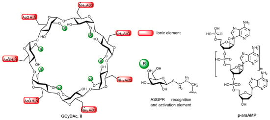
Figure 1.
Schematic representations of GCyDAc (8; left) and p-araAMP (right), the synthetic 12-mer oligonucleotide deoxyadenylyl-(5′-3′)-(arabinoadenylic)10-5′-arabinoadenosine monophosphate. The ionic substituents are on the primary face and the 1(thio-β-D-galactopyranosyl)propyl-ASGPr binding elements (R, center) are on the secondary face of the rigid 7-glucose toroidal CyD backbone.
2. Chemistry, Formulation and Molecular Docking
β-Cyclodextrin (β-CyD), arabinosyl adenine (araA), and arabinosyl adenine monophosphate (araAMP), purchased from Sigma-Aldrich Canada, (Oakville, ON, Canada), were dried over P2O5 at room temperature for 24–48 h before use. Other chemicals and solvents were purchased from Sigma-Aldrich Co. and Fisher Scientific Company (Ottawa, ON, Canada). Solvents were dried according to literature procedures. The solvents used for synthesis were reagent grade unless specified otherwise, and other chemicals and solvents were of analytical grade. The 12-mer oligonucleotide deoxyadenylyl-(5′-3′)-(arabinoadenylic)10-(5′-3′)-arabinoadenosine monophosphate (p-araAMP; Figure 1) was used as provided under contract by Nucleic Acid Protein Service (UBC, Vancouver, BC, Canada). Additional electrophoretic analyses of p-araAMP purity are described in the Supplemental Data file.
Thin-layer chromatography (TLC) was carried out on Kieselgel 60 F254 (Merck, Darmstadt, Germany) and visualization was accomplished by charring with 5% methanolic sulfuric acid. Column chromatography was performed using silica gel 60 (70-230 Mesh ASTM, 0.063–0.2 mm, Rose Scientific Ltd., Edmonton, Canada). A Labconco Freeze Dryer (Kansas City, MO, USA) was used for lyophilization. Differential scanning calorimetry (DSC) was performed on a DSC 120 (SEIKO SII, model SSC/5200) calorimeter. NMR spectra were recorded on either a Bruker AM-300 or a Varian VXR-500 spectrometer (300 and 500 MHz, respectively, for 1H; 75.4, and 125.74 MHz for 13C). Chemical shifts are quoted in ppm, referenced to residual CHCl3 at δ 7.27 for CDCl3 solutions or HOD at δ 4.82 (25 °C) for D2O solutions. Coupling constants (J) are reported in Hertz. 13C-NMR spectral assignments were aided by the J-MOD spin-echo technique in which methyl and methine carbon resonances appear as positive peaks, methylene and quaternary carbon resonances appear as negative peaks. MALDI mass spectra were obtained using an Applied BioSystems Voyager Elite/N2 laser instrument.
Commercially available 2 and 3 (Scheme 1) were prepared from 1 using literature procedures [,]. Briefly, heptakis(6-bromo-6-deoxy)-β-cyclodextrin 2 was prepared by direct halogenation of unprotected β-CyD 1 with (C6H5)3P/Br2/DMF; the 13C-NMR (75.5 MHz, D2O) spectrum of the crude product showed an upfield shift for C-6 to δ 34.4 ppm from δ 60.9 ppm for starting material (β-CyD). The crude bromide was subjected to a nucleophilic displacement reaction with azide to yield the corresponding heptakis(6-azido-6-deoxy)-β-CyD (90%) as a tan-colored solid. Due to the poor solubility of the crude azide, it was converted to fully acetylated compound upon treatment with acetic anhydride and pyridine to facilitate the column purification. The structure of the acetylated compound was confirmed with 1H-NMR and IR spectroscopy. Zemplen deacetylation yielded the pure azide, heptakis(6-azido-6-deoxy)-β-cyclodextrin 3. Details are presented in the Supplementary Data file.
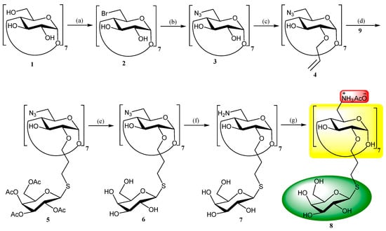
Scheme 1.
Chemical elaboration of β-CyD 1 to heptakis[6-amino-6-deoxy-2-O-(3-(1′-thio-β-D-galactopyranosyl)-propyl)]-β-cyclodextrin acetate 8 (GCyDAc). Reagents and conditions: (a) (C6H5)3P/Br2/DMF, 1 h; (b) NaN3, DMF, KI, 80 °C, 12 h, 60%; (c) NaH, AllylBr, DMF, 48 h, 40%; (d) 9, AIBN, CH3CN, 70 ºC, 7 h, 61%; (e) NaOCH3- CH3OH, CH3OH, 30 h, 93%; (f) Ph3P/NH4OH, DMF, 48 h, 88%; (g) HOAc.
Heptakis(6-azido-6-deoxy-2-O-allyl)-β-cyclodextrin 4: Sodium hydride powder (60%, 1.27 g, 31.63 mmol) was added to a solution of 3 (5.18 g, 3.95 mmol) in DMF (240 mL) under an atmosphere of argon at 0 °C. The reaction mixture was stirred at 0 °C for 1.5 h and then stirred overnight at room temperature. Allyl bromide (3.87 g, 32 mmol) was added dropwise, stirred at 0 °C for 1 h and then stirred overnight at room temperature. The solution was then cautiously poured into ice water (500 mL) and the precipitate was collected by filtration, then purified by column chromatography (hexane/EtOAc, 1:1) to afford 4 (2.56 g, 40%) as a white solid: 1H-NMR (CDCl3, 300 MHz): δ = 3.31 (t, 7H; H-2), 3.42–3.47 (dd, 7H; H-4), 3.52–3.59 (dd, 7H; H-5), 3.71–3.75 (m, 14H; H-6), 3.88–3.94 (t, 7H; H-3), 4.21–4.27 (dd, 7H; OCHa), 4.45–4.51 (dd, 7H; OCHb), 4.83 (d, 7H; H-1), 4.91 (s, 7H; OH), 5.24–5.35 (m, 14H; CH=CH2), 5.87–6.00 ppm (m, 7H; CH=CH2); 13C-NMR (CDCl3, 75 MHz): δ = 51.5 (C-6), 70.2, 73.0, 78.7, 84.7 (C-2, C-3, C-4, C-5), 73.6 (OCH2), 101.8 (C-1), 119.2 (CH2=CH), 134.3 ppm (CH2=CH); MS (MALDI) calcd for C63H91N21O28 m/z [M + Na]+ 1613.51, found 1613; elemental analysis calcd for C63H91N21O28 C, 47.57; H, 5.77; N, 18.49, found C, 47.65; H, 5.66; N, 18.19.
Heptakis[6-azido-6-deoxy-2-O-(3-(2′,3′,4′,6′-tetra-O-acetyl-β-D-thiogalacto-pyranosyl)propyl)]-β-cyclodextrin 5: Cyclodextrin 4 (100 mg, 0.063 mmol) and 9 (280 mg, 0.77 mmol) were dissolved in CH3CN (40 mL). Azobisisobutyronitrile (AIBN; 30 mg, 0.18 mmol) was added under argon at 70 °C, and after stirring for 7 h, the solvent was removed under vacuum (water aspirator), and the residue was purified by column chromatograph (hexane/EtOAc/CH3OH, 5:5:1) to afford 5 (0.16 g, 0.0386 mmol; 61%) as a white foam: 1H-NMR (CDCl3, 500 MHz): δ = 1.87–1.94 (m, 14H; SCH2CH2), 1.97, 2.04, 2.06, 2.15 (4s, 84H; 28 × CH3CO), 2.58–2.87 (m, 14H; SCH2), 3.28 (t, 7H; H-2), 3.34–3.37 (m, 7H; H-4), 3.52–3.56 (m, 7H; H-6a), 3.67–3.84 (m, 28H), 3.95–4.17 (m, 28H), 4.52–4.58 (m, 7H; H-1′), 4.78 (s, 7H; OH), 4.90 (s, 7H; H-1), 5.06 (dd, 7H), 5.19 (m, 7H; H-2′), 5.43 (m, 7H; H-3′); 13C-NMR (CDCl3, 125 MHz): δ = 20.66, (28 × CH3CO), 27.12 (SCH2CH2), 29.98 (SCH2), 51.45, 60.37, 61.21, 67.20, 67.30, 68.0, 68.05, 70.35, 71.49, 71.78, 72.82, 74.25, 80.37, 84.50, 84.62, 101.65, 169.54, 170.02, 170.19, 170.30 ppm (28 × CH3CO); MS MALDI calcd for C161H231N21O91S7 m/z [M + Na]+ 4164.08, found 4164. Elemental anal calcd for C161H231N21O91S7: C, 46.70; H, 5.62; N, 7.10, found C, 46.44; H, 5.47; N, 6.96.
Heptakis[6-azido-6-deoxy-2-O-(3-(1′-thio-β-D-galactopyranosyl)propyl)]-β-cyclodextrin 6: NaOCH3 (1 M, 18 mL) was added to a stirred solution of 5 (1.8 g, 435 mmol) in dry CH3OH (250 mL), and the reaction mixture was stirred for 30 h at RT. Water (30 mL) was added, and the reaction was stirred for 1 h to dissolve precipitates. The reaction was neutralized with Amberlite IR-120 H+ ion-exchange resin and filtered. The solution was concentrated under vacuum (water aspirator) and the resultant glassy residue was dissolved in H2O and freeze-dried to afford 6 as a white fluff (1.2 g, 93%). Selected NMR data,1H-NMR (D2O, 300 MHz): δ = 1.90–1.96 (m, 14H; CH2), 2.77–2.80 (m, 7H; SCH2), 4.47 (d, 7H; H-1′), 5.14 ppm (s, 7H; H-1); MS (MALDI) calcd for C105 H175 N21 O63 S7 m/z [M + Na]+ 2987.05.08, found 2987.
Heptakis[6-amino-6-deoxy-2-O-(3-(1′-thio-β-D-galactopyranosyl)propyl)]-β-cyclodextrin 7: Azido CyD 6 (145 mg, 0.049 mmol) and triphenylphosphine (0.27g, 1.03 mmol) were dissolved in DMF (3 mL) and the mixture was stirred at RT for 30 min. Aqueous ammonia (3 mL) was added and stirring continued at RT for 48 h. The mixture was diluted with CH3OH and evaporated under vacuum. The residue was dissolved in H2O (40 mL) and washed with CH2Cl2 until no UV-active material was present in the organic layer. The aqueous layer was lyophilized to afford 7 (121 mg, 88%) as a solid. Selected NMR data, 1H-NMR (D2O, 300 MHz): δ = 1.84–1.93 (m, 14H; CH2), 2.68–3.10 (m, 14H; SCH2), 4.46 (d, 7H; H-1′), 5.19 ppm (s, 7H; H-1); 13C-NMR (D2O, 75 MHz): δ = 28.71, 28.76, 28.96, 43.52, 60.15, 60.9, 63.54, 70.37, 71.25, 71.62, 72.13, 72.68, 73.96, 74.7, 75.2, 76.44, 81.37, 82.17, 82.26, 82.29, 88.17, 88.3, 102.31 ppm; MS (MALDI) calcd for C105 H189 N7 O63 S7 m/z [M + Na]+ 2805.07, found 2804.
Heptakis[6-amino-6-deoxy-2-O-(3-(1′-thio-β-D-galactopyranosyl)propyl)]-β-cyclodextrin acetate 8 (GCyDAc). CyD 7 (86 mg, 0.031 mmol) was treated with aqueous HOAc (0.113 M, 1.92 mL), and the solution was lyophilized to give 84 mg of 8 as a solid. Selected NMR data, 1H-NMR (D2O, 300 MHz): δ = 1.84–2.14 (m, 35H; CH3CO and CH2), 2.68–2.91 (m, 14H; SCH2), 4.47 (d, 7H; H-1′), 5.27 ppm (s, 7H; H-1).
2,3,4,6-Tetra-O-acetyl-1-thio-β-D-galactopyranose 9 (O-Ac-TG). Thioacetamide (0.4 g, 5.4 mmol) was mixed with 2,3,4,6-tetra-O-acetyl-β-D-galactopyranosyl bromide (2.1 g, 5 mmol), and heated at 120 °C under argon for 10 min. The reaction mixture was allowed to cool to RT, then CH3OH (40 mL) was added to dissolve the solid. Solvent was removed under vacuum (water aspirator), and the residue was purified using column chromatography (hexane/EtOAc, 1:1) to afford the title compound 9 (1.33 g, 73%); 1H-NMR (CDCl3, 300 MHz): δ = 1.98, 2.04, 2.05, 2.09 (4s, 12H; 4Ac), 2.37 (d, 1H, J =10.1 Hz; SH), 3.92–3.97 (m, 1H; H-5), 4.03–4.17 (m, 1H; H-6a), 4.51–4.63 (m, 1H; H-6b), 4.54 (t, 1H; H-1), 4.99–5.03 (dd, 1H, J = 10.1; H-2), 5.15–5.30 (m, 2H; H-4 and H-3); 13C-NMR (CDCl3, 75 MHz): δ = 20.6, 20.7, 20.7, 20.8, 61.5, 67.2, 71.5, 74.9, 76.7, 79.1, 169.7, 169.9, 170.0, 170.9 ppm; MS (ESI) calcd for C14H20O9S m/z [M + Na]+: 387.37, found 387.1.
Formulation: Test nucleosides and nucleotides were formulated with GCyDAc 8 in 1:1 molar ratios except for p-araAMP, which was formulated in a 2:1 = 8:p-araAMP molar ratio. The complexes were prepared by the solvent evaporation method, first dissolving 8 in distilled water, then adding the test agent, sonicating (1 min), filtering (0.22 μ), and finally freeze drying. Weighed amounts (3~5 mg) of samples were sealed in aluminum pans for DSC analysis using a Seiko DSC 120 instrument. DSC scans were performed over a 20° to 300 °C range at a heating rate of 10 °C/min. Representative thermograms are presented in Figure 2.
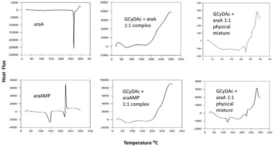
Figure 2.
DSC thermograms of araA (upper left) and araAMP (lower left) are characterized by a sharp endothermic peak and a sharp exothermic peak, respectively. These features remain visible for the 1:1 GCyDAc:araA and GCyDAc:araAMP physical mixtures (upper right and lower right, respectively), but are essentially absent for 1:1 complexes with GCyDAc of araA and araAMP (upper and lower center frames, respectively).
Cell toxicity and antiviral activity of GCyDAc formulations: Cytotoxicity was estimated using the MTS assay (Promega North America, Madison, WI, USA) of metabolic activity in four mammalian cell lines grown in cell culture: EMT-6 (murine mammary carcinoma), KBALB (murine, virus-transformed embryonic Kirsten sarcoma), 143B (human osteosarcoma), and HepG2 (human hepatoma). Antiviral studies were performed under contract by the Katholieke Universiteit Leuven, Rega Institute for Medical Research, Belgium, using a procedure developed by Ladner et al. [,]. Briefly, growth of a tetracycline-responsive HepAD38 cell line, stably transfected with a cDNA copy of the pregenomic RNA of wild-type HBV virus, was initiated by tetracycline withdrawal, with or without test substance in the culture medium. The antiviral effect was quantified by measuring levels of viral DNA at day 4 post-induction, by a real-time quantitative PCR (Q-PCR), and analyzed using an SDS 7000 (Applied Biosystems, Foster City, CA, USA). A plasmid containing the full-length insert of the HBV genome was used to prepare a standard curve, and the amount of viral DNA produced in treated cultures was expressed as a percentage of the mock-treated samples.
Molecular Docking: Ligand preparation: the ligand, GCyDAc, was constructed using the 3D model kit of Spartan16 Parallel Suit [,] and preliminarily minimized using the program’s comprehensive molecular mechanics tool. Geometry optimization and energy minimization was then performed using the intuitive graphical user interface (GUI) of Spartan 16 Parallel Suit. Protein: the x-ray crystallographic structure of α-lactose complexed with ASGPr, 5jpv.pdb, was used to construct a 3D computational model for binding study []. Docking: both ligand and protein files required for the binding study were prepared and followed by molecular docking using AutoDock Vina [,].
3. Results
Heptakis[6-amino-6-deoxy-2-O-(3-(1′-thio-ß-D-galactopyranosyl)propyl)]-β-cyclodextrin acetate 8 (GCyDAc) was synthesized from heptakis(6-azido-6-deoxy)-β-cyclodextrin 3 in five steps, with an overall yield of 20%. Reactions were not optimized for yield.
Differential scanning calorimetry (DSC) studies were performed on araA, araAMP, and p-araAMP, alone, as physical mixtures and as formulated complexes with GCyDAc. Thermograms shown in Figure 2 and Figure 3 depict substantial differences among the thermographs of 8, araA, araAMP, and p-araAMP, and among thermographs of their physical mixtures and complexes with 8.
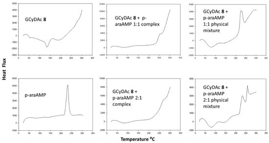
Figure 3.
DSC thermograms of p-araAMP (lower left) and GCyDAc (upper left) alone, as complexed (center frames) and as physical mixtures (left frames). The strong exothermic peak of p-araAMP (lower left) is evident in the physical mixtures at both GCyDAc concentration ratios (1:1 upper right and 2:1 lower right), whereas it is suppressed by complexation at both ratios (1:1 upper center and 2:1 lower center). The thermal fluctuations at lower temperatures seen with GCyDAc alone (upper left) are evident in the physical mixtures (upper right and upper left) but are not present in the complexes (upper and lower center).
GCyDAc was complexed with p-araAMP in both 1:1 and 2:1 ratios. The respective thermographs (Figure 3) show diminution of the nucleotide’s exothermal transition as the GCyDAc:p-araAMP ratio increased from 1:1 to 2:1.
GCyDAc was tested for toxicity against four mammalian cell lines, using a colorimetric antiproliferation assay. There was no evidence of toxicity towards these cell lines in culture, with an IC50 of approximately 0.5 millimolar for each cell line (Figure 4).
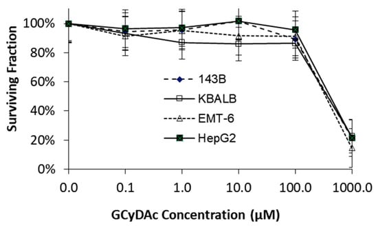
Figure 4.
Surviving fractions of mammalian cell lines, as determined by MTT assay and expressed as a function of GCyDAc concentration.
The antiviral activities of araA, araAMP, p-araAMP, and their respective GCyDAc formulations, are presented in Table 1. Data are expressed in molar-equivalent potencies; lamivudine (LV; 3TC; (-)-L-2′,3′-dideoxy-3′-thiacytidine) and PMPA (tenofovir; (9-[9(R)-2-(phosphonomethoxy)propyl]adenine) were used as positive controls for the antiviral assay. These data show that araA, araAMP, and p-araAMP had similar potencies, and that GCyDAc effected only minimal increases in antiviral potency. GCyDAc slightly decreased the potency of LV (reference antiviral nucleoside).

Table 1.
Antiviral potencies (EC50) of araA, araAMP, p-araAMP, two active anti-HBV antivirals, and their GCyDAc formulations, against hepatoma HepAD38 cells in cell culture.
In silico docking calculations using AutoDock Vina demonstrated the possible interaction of two galactose units of the ligand GCyDAc with the two sugar binding sites of ASGPr H1 domain (Figure 5B,C). This modeled binding is similar to the molecular interaction of two α-lactose molecules with ASGPr H1 domain (Figure 5A, 5jpv.pdb, []). The shallow sugar-binding pockets showed interaction, requiring calcium ions, with both hydroxyls of C-3 and C-4 of β-D-galactose. Moreover, the remaining galactose units of GCyDAc showed additional binding except for one unit, which was oriented away from the receptor-binding site, Figure 5D.
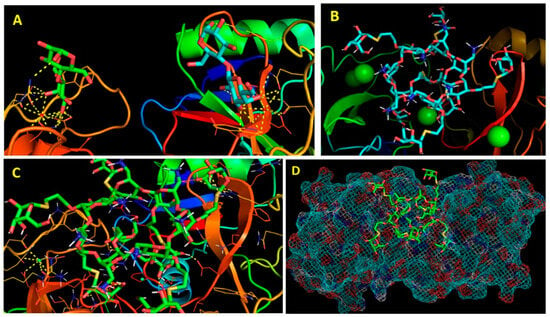
Figure 5.
(A) shows the molecular interaction between lactose and ASGPr H1 domain (5jpv.pdb; [], showing involvement of calcium ions (red dots). (B,C) show the mode of molecular binding of GCyDAc to ASGPr H1 domain (5jpv.pdb; []), showing involvement of calcium ions (green spheres). (D) shows a top view of the modeled binding of GCyDAc to ASGPr H1 domain, 5jpv.pdb.
4. Discussion
Interest in the synthesis of substituted cyclodextrins for drug delivery remains strong after more than five decades of research []. The rigid 3D molecular structure of CyDs is particularly attractive for chemical elaboration because it offers two geometric surfaces for independent stereoselective derivatization [] to precisely accommodate multiple enhancements including targeting ligands and therapeutic motifs. The immediate benefit of this spatial arrangement is augmented by the chemical reactivity of hydroxyl substituents on the two faces, with the one face projecting primary, more nucleophilic, hydroxyl groups at C-6, and the other face projecting less reactive, more acidic, secondary hydroxyl groups at C-2 and C-3, all of which can be exploited via their respective differences in chemical reactivity [,,,]. This approach has been utilized for targeted co-delivery of a range of therapeutic moieties including lectin-binding galactose motifs and therapeutic molecules such as SiRNA [,,]. However, despite the promising characteristics of these delivery systems, their application in commercial products remains disappointing.
The current report addresses the exploitation of two very different functions, one targeting cellular membrane ligands, and the second based on charge-associated complexation involving alkyl amines at C-6 []. The influence of electrostatic interactions between host-guest, and their impact on classical bonding drivers including hydrophobic and van der Waals forces, and hydrogen-bonding, are complex and currently unpredictable. However, they have been recognized for their contributions to charge-association bonding []. Positively charged guanidino- and alkylamine-derivatized cyclodextrins [] have been shown to strongly bind nucleotides through both cavity inclusion and ionic interactions in which the deoxyribose moiety, driven by phosphate-guanidino group interactions, lodges inside the cavity of per(6-guanidino-6-deoxy)-β CyD and then readily enters cancer cells to deliver monophosphorylated gemcitabine [,]. Overall, however, the development of charged CyD carriers has lagged behind other approaches in building, understanding, and exploiting these structures. The current manuscript extends the C-6 (primary face) amine modification to include C-2 (secondary face) hydroxyls decorated with ASGPR-reactive thio-β-D-galactopyranosylpropyl substituents.
The synthesis of GCyDAc 8 was designed to avoid lengthy synthesis methods, tedious protection–deprotection steps, and indiscriminate (sledgehammer) reactions with lengthy purification procedures []. The first two intermediates in the synthetic sequence (2, 3; both commercially available) were prepared from β-CyD by classical methods [,], affording the per-azido CyD 3 in 90% yield. An acetylation–deacetylation step, introduced to facilitate purification of the poorly soluble per-azido CyD 3 on silica gel, reduced the overall recovered yield to 60%. Other syntheses have been reported, including a mechanochemical (ball mill) procedure [] and alternative quench procedures for the initial halogenation reaction [], both of which have the advantage of easy scale-up.
Addition of the galactose component at C-2 was achieved in two steps from per-azido-CyD 3 by first introducing a linker arm 4 and then coupling with protected thiogalactose 9 to afford 5. Other approaches to CyD glycosylation have been reported, including the synthesis of multivalent lactose or galactose-decorated CyD via heptakis-C-6 amino-CyD []. Although the latter approach could have been modified to meet current needs (galactosylation at C-2), it was deemed unsuitable because it afforded a mixture of tri-, tetra-, penta-, hexa- and hepta-lactose-substituted CyDs in an overall yield of 25%, which would mitigate extensive purification to obtain fully glycosylated CyD []. A versatile approach to prepare per-glycosylated CyDs in 30–90% yield from a range of derivatized heptakis(6-azido-6-deoxy)-CyDs via click reactions and microwave irradiation [] could possibly be adapted to prepare compounds that could fit within the objectives of the current project.
Aminocyclodextrins have been shown to bind nucleoside monophosphates (i.e., mononucleotides) in a geometric relationship in which the phosphate protrudes through the CyD cavity to associate with the amino group, and the sugar moiety lies within the CyD cavity [,,]. Adenine-based nucleotides are bound preferentially to CyD compared to nucleotides with other nucleobases [], but the impact of heptakis-2-O-substitution on these properties is not known. In the case of p-araAMP, classical inclusion complex formation from the secondary side of GCyDAc may be difficult because per-alkyl-thiogalactosylation at C-2 creates a barrier approximately 9.8 Å deep that protects the secondary face []. The current docking study showed that three of the C-2 galactoses are oriented downward, which may result in blocking access to the CyD cavity from the secondary face. Conversely, C-6 amino groups are oriented upward, forming a crown of positive charges on top of the primary face which could facilitate the interaction with the phosphate groups. Charge association for araAMP by approach from the secondary face of GCyDAc may still be possible, with the phosphate moiety penetrating through the CyD cavity and protruding from the primary face, ionically stabilized by C-6 amino groups as per the Suzuki model []. Additionally, in the case of p-araAMP, ionic-associated stabilization could also occur, through either terminal or backbone phosphate groups associating directly with CyD primary face alkylamino substituents, without the classical host-guest geometry, and/or by penetrating the torus from the secondary side as in the Suzuki model []. This could coincide with intramolecular ‘wrap-around’ or intramolecular charge associations involving additional GCyDAc molecules. A preliminary high-field NMR (800 MHz 1H) titration study (GCyDAc vs. p-araAMP) revealed concentration-dependent changes consistent with the latter concept, but the exceedingly complex spectra (not shown) precluded analysis. This work was not pursued further because resolution of these spectra would have required inclusion of an isotopic marker in the GCyDAc structure to identify the resonance of at least one participating sugar moiety.
Receptor-mediated endocytosis has long been identified as a mechanism to improve the delivery of membrane-impermeable drugs including nucleotides [], including the delivery of RNAi using a cyclodextrin-containing polymer targeted to the transferrin receptor by human transferrin []. Performance-limiting properties of ASPGr as a drug delivery mechanism include its capacity constraints [], variability of expression density in cell membranes [], turnover (on-off and binding-internalization) times [], and endosomal escape of transported payloads [,,]. The interrelationship among these parameters is not known and no data are available specifically for araA, araAMP, and p-araAMP. However, slow receptor regeneration rates alone may create plateau/saturation kinetics that could limit the delivery and efficacy of drugs by the ASGPr receptor []. ASGPr expression density in the HepAD38 cell line used in the current studies, at 17,000 receptors per cell [], is substantially lower than the 76,000 per cell reported estimate for the more commonly used HepG2 hepatic cell lines [,].
The ASGPr transport process starts with the formation of coated pits upon trivalent contact between the substrate and the receptor. Once triggered, membrane invagination and the formation of endosomes inside the cell proceed. The classical model for endosome processing includes a recycling component to regenerate plasma membrane and a degradative sequence that destroys entrapped ligand complexes. Early endosomes have a near neutral pH (>6), but as they mature, they become more acidic, so that the pH of late endosomes may be below 5. Drug-ligand complexes are destroyed in late endosomes, either directly by hydrolases, or following fusion with lysosomes [,,]. Mechanisms of drug release from endosomes are substrate specific, complex, and largely unresolved [,].
The current research was based on two concepts: (1) that the fully substituted cationic CyD (GCyDAc, 8) would form reasonably stable complexes with (oppositely charged) nucleotides (i.e., araAMP and p-araAMP), and (2) that fully galactosylated CyD (8) would target, bind, and activate endocytosis of the ion-stabilized complex (GCyDAc:araAMP or GCyDAC:p-araAMP). Release of the mononucleotide araAMP from the endosome would increase the antiviral effectiveness of the dose by by-passing the rate-limiting activation of the nucleoside araA. The current molecular docking model depicts only two strong binding sites between GCyDAc and ASGPr, and although other weak/cross molecular associations are possible, the inclusion of seven ‘binding sugars’ on the secondary face of GCyDAc may mitigate against full receptor activation seen with ternary binding.
Successful complexation between GCyDAc and either araA, araAMP, or p-araAMP was demonstrated by DSC. In each case (Figure 2 and Figure 3), the respective thermographs depict clear differences among the carrier, the drug, and the various physical mixtures and the complexes of the drug and carrier. The primary endothermic or exothermic transitions characteristic of thermographs of pure test substances and GCyDAc:test-substance-mixtures are absent from, or very faint in, the thermograms of the complexes. It has been shown previously by HPLC that the GCyDAc complexed with hexachlorofluorescein-tagged p-araAMP (HEX-p-araAMP) was stable in certain buffer solutions and in water, and that the 1:1 formulation contained ~90% free HEX-p-araAMP, whereas the 2:1 formulation contained <25% free HEX-p-araAMP []. As with other charged CyD:phosphate ester complexes [], hydrolysis GCyDAc:HEX-p-araAMP varied by buffer, with complete dissociation within a 4 h incubation in borate buffer. Guthrie [] further reported that their analytical method (capillary electrophoresis and laser-induced fluorescence) did not detect any complex formation between HEX-p-araAMP and the neutral CyD analog (6), which underscores the importance of ionic association in the complexation of p-araAMP. However, the HEX-p-araAMP findings may not translate into similar effects for p-araAMP, given the presence of the bulky (MW 741), hydrophobic HEX fluorophore that may substantially alter the association with GCyDAc, despite attachment of HEX at the terminal adenine-NH2.
Interaction of GCyDAc with membrane ASPGr and subsequent internalization via membrane invagination and endosome formation is a core premise of the current research. The interaction of small-molecule terminal galactose moieties with ASPGr is established in the literature, but research with sugar-derivatized CyDs and small-molecule drugs does not always support this as a major/effective drug delivery mechanism. For example, 5′-O-lactosyl-araA reached higher concentrations than araA in liver of mice following IV injection or in hepatocytes in cell culture [], but arabinosylated-2-nitroimidazole exhibited higher in vivo uptake in liver than its corresponding galactosyl compound []. That is, the expected outcomes were met by lactosyl-araA but not by the galactosyl-nitroimidazole. In the case of lactosylated or galactosylated β-CyD carriers, elevated drug delivery across the blood–brain barrier and elevated hepatic drug uptake has been observed, and although endocytosis was the rationalized mechanism, the sugar density on the membrane ligand was not specified [].
A number of plausible metabolic events may have prevented the GCyDAc complexes from providing higher antiviral activity compared to the respective non-complexed antivirals (Table 1). Plateau/saturation kinetics and slow receptor regeneration rates [,] could have limited the delivery of the antivirals via the ASGPr, thereby preventing cytostatic drug concentrations from being reached in HepAD38 cells. It is also possible that low antiviral activity could be due to the failure of the GCyDAc complexes to initiate internalization of the receptor. A third scenario is that even if ASPGr internalization and endosome formation did occur, araAMP was degraded prior to release into the cytoplasm. For example, endosomal nucleases may have hydrolyzed p-araAMP to the 3‘-O- rather than the 5′-O-monophosphates. This would prevent up-phosphorylation to the trinucleotide, since the 3′-O monophosphate may not be readily converted to active trinucleotide 5′-O-araATP. Production of the 3′-O- vs. the 5′-O-monophosphates is known to be nuclease-dependent and substrate (polynucleotide)-dependent; for example, DNA and RNA are hydrolyzed to deoxyribomonophosphates and ribo-3′-monophosphates, respectively [,]. To our knowledge, the products of chemical hydrolysis or enzymatic hydrolysis products of poly-arabinosyl nucleotides are not reported. It is also possible that degradation processes other than hydrolysis effected inactivation of endosomal GCyDAc:p-araAMP. Deaminases, for example, could limit antiviral activity of GCyDAc-complexed araAMP and p-araAMP via metabolic deamination to their corresponding, inactive, inosine metabolites, which would parallel a major metabolic pathway active in the degradation of adenosine and its mononucleotide [,].
5. Summary
The objectives of the current research were to develop a receptor-targeted delivery agent to by-pass the rate-limiting cellular activation (5′-monophosphorylation) of antiviral/anticancer nucleosides (i.e., araA) and to simultaneously improve the delivery of their respective mononucleotides (i.e., araAMP) and polynucleotides (p-araAMP). AraA was selected for this demonstration because, like many antiviral nucleosides, it requires three-step anabolic phosphorylation to its triphosphate (i.e., araATP) in order to effect inhibition of viral DNA synthesis, its main mechanism of antiviral action. Since the first step, monophosphorylation, is rate-limiting, delivery of araAMP as a drug or as a metabolite of p-araAMP could effectively by-pass rate-limiting monophosphorylation.
GCyDAc was chosen as the nucleotide carrier, offering seven primary-side charge associations per carrier molecule, and at the same time, targeting the ASGPr ligand on hepatocyte membranes via its seven secondary side galactose arms. The ASGPr targeting/transport system was selected to deliver the nucleotides via its endosome-forming mechanism, with the goal of delivering the nucleotides and releasing activated drug intermediates (e.g., araAMP) into the hepatocyte. The efficacy of this process would enhance viral antiviral potency as determined by cell cytotoxicity in the HepAD38 cell line in cell culture.
It has been stated that CyD drug delivery research today needs novel cyclodextrins with demonstrated therapeutic effectiveness in addition to those currently approved []. The current report describes a high-density charge-association binding mechanism that could potentially supplement fundamental CyD inclusion complexation forces, and simultaneously targets cell membrane ligands to initiate payload importation via endosome formation. This approach supplements contemporary understanding of cyclodextrin-based drug delivery [].
Supplementary Materials
The following supporting information can be downloaded at: https://www.mdpi.com/article/10.3390/pharmaceutics16030323/s1, Figure S1: p-araAMP gels and CE.
Author Contributions
Conceptualization and methodology, L.I.W. and J.D.; software and modeling, H.E.; data curation, analysis, and validation, L.I.W., J.D. and H.E.; investigation, J.-H.R., W.Z. and X.-H.Y.; writing—original draft preparation, L.I.W.; writing—review and editing, L.I.W., J.D. and H.E.; visualization, L.I.W., J.D. and H.E.; supervision, L.I.W. and J.D.; project administration, L.I.W.; funding acquisition, L.I.W. All authors have read and agreed to the published version of the manuscript.
Funding
Aspects of this work were funded in part through operating grants from the Alberta Cancer Board (RI-14) and the Canadian Institutes for Health Research (13480).
Institutional Review Board Statement
No institutional approvals or statements required.
Informed Consent Statement
No informed consent required.
Data Availability Statement
Requests for data access should be addressed to the corresponding author.
Acknowledgments
The authors thank Rudy Sedlak and Knut Woltjen for assistance with chemical syntheses. We recognize the advice and support offered by N.A.P.S., UBC, Vancouver, B.C., for the contract synthesis of p-araAMP, and thank Rega Institute for Medical Research for the antiviral evaluation of our novel substances and formulations on contract through the Katholieke Universiteit Leuven, Belgium. Finally, we thank the National High Field Nuclear Magnetic Resonance Centre (NANUC) staff for conducting GCyDAc:p-araAMP titration experiments and for their expert advice on NMR studies of the GCyDAc:p-araAMP complexation process.
Conflicts of Interest
All authors declare that they have no conflicts of interest regarding this work.
Abbreviations
AMP (adenosine-5′-monophosphate); araA (9-arabinosyl adenine, 9-β-D-arabinofuranosyl adenine; vidarabine); araAMP (arabinosyl adenine-5′-monophosphate); ASGPr (asialoglycoprotein receptor); CyD (cyclodextrin); DSC (differential scanning calorimetry); GCyDAc (8; heptakis[6-amino-6-deoxy-2-O-(3-(1′-thio-β-D-galactopyranosyl)propyl)]-β-cyclodextrin acetate); OAc-TG (9; 2,3,4,6-tetra-O-acetyl-1-thio-β-D-galacto-pyranose); p-araAMP [12-mer-polyaraAMP; 5′-phosphodeoxyadenlyl(5′-3′)(araAMP)11].
References
- LePage, G.A.; Naik, S.R.; Katakkar, S.B.; Khaliq, A. 9-Beta-D-arabinofuranosyladenine 5’-Phosphate Metabolism and Excretion in Humans. Cancer Res. 1975, 35, 3036–3040. [Google Scholar] [PubMed]
- Holzer, S.; Rzechorzek, N.J.; Short, I.R.; Jenkyn-Bedford, M.; Pellegrini, L.; Kilkenny, M.L. Structural Basis for Inhibition of Human Primase by Arabinofuranosyl Nucleoside Analogues Fludarabine and Vidarabine. ACS Chem. Biol. 2019, 14, 1904–1912. [Google Scholar] [CrossRef] [PubMed]
- Whitley, R.J.; Ch’ien, L.T.; Dolin, R.; Galasso, G.J.; Alford, C.A., Jr. Adenine Arabinoside Therapy of Herpes Zoster in the Immunosuppressed—NIAID Collaborative Antiviral Study. N. Engl. J. Med. 1976, 294, 1193–1199. [Google Scholar] [CrossRef] [PubMed]
- Sagar, S.; Kaur, M.; Minneman, K.P. Antiviral Lead Compounds from Marine Sponges. Mar. Drugs 2010, 8, 2619–2638. [Google Scholar] [CrossRef] [PubMed]
- Balzarini, J.; De Clercq, E. 9-Beta-D-arabinofuranosyladenine 5′-monophosphate (araAMP) is Converted Directly to its Antivirally Active 5′-triphosphate Form by 5-Phosphoribosyl-1-pyrophosphate (PRPP) Synthtase. Biochem. Biophys. Res. Commun. 1990, 173, 781–787. [Google Scholar] [CrossRef] [PubMed]
- York, J.L.; LePage, G.A. A Proposed Mechanism for the Action of 9-Beta-D-arabinofuranosyladenine as an Inhibitor of the Growth of Some Ascites Cells. Can. J. Biochem. Physiol. 1966, 44, 19–28. [Google Scholar] [CrossRef]
- Furth, J.J.; Cohen, S.S. Inhibition of Mammalian DNA Polymerase by the 5′-Triphosphate of 1-Beta-D-Arabinofuranosylcytosine and the 5′-Triphosphate of 9-Beta-D-arabinofuranosyladenineoxyladenine. Cancer Res. 1968, 60, 2061–2067. [Google Scholar]
- Derse, D.; Cheng, Y.C. Herpes Simplex Virus Type I DNA Polymerase. Kinetic Properties of the Associated 3′-5′ Exonuclease Activity and its Role in araAMP incorporation. J. Biol. Chem. 1981, 256, 8525–8530. [Google Scholar] [CrossRef]
- Bijsterbosch, M.K.; Ying, C.; de Vrueh, R.L.A.; de Clercq, E.; Biessen, E.A.L.; Neyts, J.; van Berkel, T.J.C. Carrier-mediated Delivery Improves the Efficacy of 9-(2-Phosphonylmethoxyethyl)adenine against Hepatitis B Virus. Mol. Pharmac. 2001, 60, 521–527. [Google Scholar]
- Cass, C.E. Nucleoside transport. In Drug Transport in Antimicrobial and Anticancer Chemotherapy; Georgopapadakou, N.H., Ed.; Marcel Dekker, Inc.: New York, NY, USA, 1995; pp. 408–451. ISBN 10978-0824793999. [Google Scholar]
- Young, J.D.; Yao, S.Y.M.; Sun, L.; Cass, C.E.; Baldwin, S.A. Human Equilibrative Nucleoside Transporter (ENT) Family of Nucleoside and Nucleobase Transporter Proteins. Xenobiotica 2008, 38, 995–1021. [Google Scholar] [CrossRef]
- LePage, G.A.; Khaliq, A.; Gottlieb, J.A. Studies of 9-Beta-D-arabinofuranosyladenine in Man. Drug Metab. Disp. 1973, 1, 756–759. [Google Scholar]
- Van Rompay, A.; Johansson, M.; Karlsson, A. Phosphorylation of Nucleosides and Nucleoside Analogs by Mammalian Nucleoside Monophosphate Kinases. Pharmacol. Therapeut. 2000, 87, 189–198. [Google Scholar] [CrossRef] [PubMed]
- Pavan-Langston, D.; North, R.D.; Geary, P.A. Ara AMP- a Highly Soluble New Antiviral Drug. Ann. Ophthalmol. 1976, 8, 571–579. [Google Scholar] [PubMed]
- LePage, G.A.; Lin, Y.T.; Orth, R.E.; Gottlieb, J.A. 5′-Nucleotides as Potential Formulations for Administering Nucleoside Analogs in Man. Cancer Res. 1972, 32, 2441–2444. [Google Scholar] [PubMed]
- Sawada, K.; Echigo, N.; Juge, N.; Miyaji, T.; Otsuka, M.; Omote, H.; Yamamoto, A.; Moriyama, Y. Identification of a Vesicular Nucleotide Transporter. Proc. Natl. Acad. Sci. USA 2008, 105, 5683–5686. [Google Scholar] [CrossRef] [PubMed]
- Alexander, S.P.H.; Kelly, E.; Mathie, A.; Peters, J.A.; Veale, E.L.; Armstrong, J.F.; Faccenda, E.; Harding, S.D.; Pawson, A.J.; Sharman, J.L.; et al. The Concise Guide to PHARMACOLOGY 2019/20: Transporters. Br. J. Pharmacol. 2019, 176, S397–S493. [Google Scholar] [CrossRef]
- Kim, J.; Chou, T.-F.; Griesgraber, G.W.; Wagner, C.R. Direct Measurement of Nucleoside Monophosphate Delivery from a Phosphoramidate Pronucleotide by Stable Isotope Labeling and LC−ESI(-)-MS/MS. Mol. Pharmaceut. 2004, 1, 102–111. [Google Scholar] [CrossRef]
- Ahuja, S.; Whorton, M.R. Structural Basis for Mammalian Nucleotide Sugar Transport. eLife 2019, 8, e45221. [Google Scholar] [CrossRef]
- Markov, O.V.; Filatov, A.V.; Kupryushkin, M.S.; Chernikov, I.V.; Patutina, O.A.; Strunov, A.A.; Chernolovskaya, E.L.; Vlassov, V.V.; Pyshnyi, D.V.; Zenkova, M.A. Transport Oligonucleotides—A Novel System for Intracellular Delivery of Antisense Therapeutics. Molecules 2020, 25, 3663. [Google Scholar] [CrossRef]
- Hiasa, M.; Togawa, N.; Moriyama, Y. Vesicular Nucleotide Transport: A Brief History and the Vesicular Nucleotide Transporter as a Target for Drug Development. Curr. Pharm. Des. 2014, 20, 2745–2749. [Google Scholar] [CrossRef]
- Bender, M.L.; Komiyama, M. Cyclodextrin Chemistry. Reactivity and Structure: Concepts in Organic Chemistry; Springer: Berlin/Heidelberg, Germany, 1978; Volume 6. [Google Scholar] [CrossRef]
- Szejtli, J.; Osa, T. Cyclodextrins. In Comprehensive Supramolecular Chemistry; Lehn, J.M., Atwood, J.L., Davies, J.E.D., Eds.; Pergamon: New York, NY, USA, 1999; Volume 3, ISBN 9780080427157. [Google Scholar]
- Muankaew, C.; Loftsson, T. Cyclodextrin-based Formulations: A Non-invasive Platform for Targeted Drug Delivery. Basic Clin. Pharmacol. Toxicol. 2018, 122, 46–55. [Google Scholar] [CrossRef]
- Hoffman, J.L.; Bock, R.M. The Interaction of Cyclodextrins with Nucleic Acids. A Study of Secondary Structure in Three Transfer Ribonucleic Acids. Biochemistry 1970, 9, 3542–3550. [Google Scholar] [CrossRef]
- Xiang, T.-X.; Anderson, B.D. Inclusion Complexes of Purine Nucleosides with Cyclodextrins. II. Investigation of Inclusion Complex Geometry and Cavity Microenvironment. Int. J. Pharmaceut. 1990, 59, 45–55. [Google Scholar] [CrossRef]
- Juliano, R.L. The Delivery of Therapeutic Oligonucleotides. Nucleic Acids Res. 2016, 44, 6518–6548. [Google Scholar] [CrossRef] [PubMed]
- Roberts, T.C.; Langer, R.; Wood, M.J.A. Advances in Oligonucleotide Drug Delivery. Nat. Rev. Drug Discov. 2020, 19, 673–694. [Google Scholar] [CrossRef] [PubMed]
- Hermann, R.; Krajcsi, P.; Fluck, M.; Seithel-Keuth, A.; Bytyqi, A.; Galazka, A.; Munafo, A. Review of Transporter Substrate, Inhibitor, and Inducer Characteristics of Cladribine. Clin. Pharmacokinet. 2021, 60, 1509–1535. [Google Scholar] [CrossRef] [PubMed]
- Guthrie, J.W.; Ryu, J.-H.; Le, X.C.; Wiebe, L.I. Characterization of a Cyclodextrin-Oligonucleotide Complex by Capillary Electrophoresis using Laser-Induced Fluorescence. J. Pharm. Pharmaceut. Sci. 2007, 10, 246–255. [Google Scholar]
- Suzuki, I.; Miura, T.; Anzai, J.-I. Superiority of Secondary Hydroxy Side Modification in Cyclodextrin Complexation for Highly Hydrophilic Adenine Nucleotides. J. Supramol. Chem. 2001, 1, 283–288. [Google Scholar] [CrossRef]
- Eliseev, A.V.; Schneider, H.-J. Molecular Recognition of Nucleotides, Nucleosides, and Sugars by Aminocyclodextrins. J. Am. Chem. Soc. 1994, 116, 6081–6088. [Google Scholar] [CrossRef]
- Ladner, S.K.; Otto, M.J.; Barker, C.S.; Zaifert, K.; Wang, G.H.; Guo, J.T.; Seeger, C.; King, R.W. Inducible Expression of Human Hepatitis B Virus (HBV) in Stably Transfected Hepatoblastoma Cells: A Novel System for Screening Potential Inhibitors of HBV Replication. Antimicrob. Agents Chemother. 1997, 41, 1715–1720. [Google Scholar] [CrossRef]
- King, R.W.; Ladner, S.K. Hep AD38 Assay: A High-Throughput, Cell-Based Screen for the Evaluation of Compounds against Hepatitis B Virus. In Antiviral Methods and Protocols. Methods in Molecular Medicine™; Kinchington, D., Schinazi, R.F., Eds.; Humana Press Inc.: Totowa, NJ, USA, 2001; Volume 24, pp. 43–50. [Google Scholar] [CrossRef]
- Szurmai, Z.; Liptak, A.; Szejtli, J. Halogen Azide Displacement to Prepare Some Symmetrically Substituted β-Cyclodextrin Derivatives. Starch 1990, 42, 447–449. [Google Scholar] [CrossRef]
- Parrot-Lopez, H.; Ling, C.C.; Zhang, P.; Baszkin, A.; Albrecht, G.; de Rango, C.; Coleman, A.W. Self-Assembling Systems of the Amphiphilic Cationic Per-6-amino-β-cyclodextrin 2,3-di-O-alkyl ethers. J. Am. Chem. Soc. 1992, 114, 5479–5480. [Google Scholar] [CrossRef]
- Shao, Y.; Molnar, L.F.; Jung, Y.; Kussmann, J.; Ochsenfeld, C.; Brown, S.T.; Gilbert, A.T.B.; Slipchenko, L.V.; Levchenko, S.V.; O’Neill, D.P.; et al. Advances in Methods and Algorithms in a Modern Quantum Chemistry Program Package. Phys. Chem. Chem. Phys. 2006, 8, 3172–3191. [Google Scholar] [CrossRef] [PubMed]
- Shao, Y.; Gan, Z.; Epifanovsky, E.; Gilbert, A.T.B.; Wormit, M.; Kussmann, J.; Lange, A.W.; Behn, A.; Deng, J.; Feng, X.; et al. Advances in Molecular Quantum Chemistry Contained in the Q-Chem 4 Program Package. Mol. Phys. 2015, 113, 184–215. [Google Scholar] [CrossRef]
- Mamidyala, S.K.; Dutta, S.; Chrunyk, B.A.; Preville, C.; Wang, H.; Withka, J.M.; McColl, A.; Subashi, T.A.; Hawrylik, S.J.; Griffor, M.C.; et al. Glycomimetic Ligands for the Human Asialoglycoprotein Receptor. J. Am. Chem. Soc. 2012, 134, 1978–1981. [Google Scholar] [CrossRef] [PubMed]
- Eberhardt, J.; Santos-Martins, D.; Tillack, A.F.; Forli, S. AutoDock Vina 1.2.0: New Docking Methods, Expanded Force Field, and Python Bindings. J. Chem. Inf. Model. 2021, 61, 3891–3898. [Google Scholar] [CrossRef] [PubMed]
- Trott, O.; Olson, A.J. AutoDock Vina: Improving the Speed and Accuracy of Docking with a New Scoring Function, Efficient Optimization and Multithreading. J. Comput. Chem. 2010, 31, 455–461. [Google Scholar] [CrossRef]
- Sanhueza, C.A.; Baksh, M.M.; Thuma, B.; Roy, M.D.; Dutta, S.; Preville, C.; Chrunyk, B.A.; Beaumont, K.; Dullea, R.; Ammirati, M.; et al. Efficient Liver Targeting by Polyvalent Display of a Compact Ligand for the Asialoglycoprotein Receptor. J. Am. Chem. Soc. 2017, 139, 3528–3536. [Google Scholar] [CrossRef]
- Huang, J.-R.; Zhuang, H.-N.; Jin, Z.-Y. Chapter 1: Introduction. In Cyclodextrin Chemistry Preparation and Application; Jin, Z.-Y., Ed.; World Scientific Publishing Co. Pte Ltd.: Singapore, 2013; pp. 1–18. ISBN 978-981-4436-79-3. [Google Scholar] [CrossRef]
- Sugihara, J.M. Relative reactivities of hydroxyl groups of carbohydrates. In Advances in Carbohydrate Chemistry; Hudson, C.S., Wolfrom, M.L., Eds.; Academic Press: Cambridge, MA, USA, 1953; Volume 8, pp. 1–44. [Google Scholar] [CrossRef]
- Khan, A.R.; Forgo, P.; Stine, K.J.; D’Souza, V.T. Methods for Selective Modifications of Cyclodextrins. Chem. Rev. 1998, 98, 1977–1996. [Google Scholar] [CrossRef]
- Przybyla, M.A.; Yilmaz, G.; Becer, C.R. Natural Cyclodextrins and their Derivatives for Polymer Synthesis. Polym. Chem. 2020, 11, 7582–7602. [Google Scholar] [CrossRef]
- Zhang, J.; Ma, P.X. Cyclodextrin-based Supramolecular Systems for Drug Delivery: Recent Progress and Future Perspective. Adv. Drug Deliv. Rev. 2013, 65, 1215–1233. [Google Scholar] [CrossRef]
- Haley, R.M.; Gottardi, R.; Langer, R.; Mitchell, M.J. Cyclodextrins in Drug Delivery: Applications in Gene and Combination Therapy. Drug Deliv. Transl. Res. 2020, 10, 661–677. [Google Scholar] [CrossRef]
- Mazzaglia, A.; Forde, D.; Garozzo, D.; Malvagna, P.; Ravoo, B.J.; Darcy, R. Multivalent Binding of Galactosylated Cyclodextrin Vesicles to Lectin. Org. Biomol. Chem. 2004, 2, 957–960. [Google Scholar] [CrossRef]
- Malhotra, M.; Gooding, M.; Evans, J.C.; O’Driscoll, D.; Darcy, R.; O’Driscoll, C.M. Cyclodextrin-siRNA Conjugates as Versatile Gene Silencing Agents. Eur. J. Pharmaceut. Sci. 2018, 114, 30–37. [Google Scholar] [CrossRef] [PubMed]
- Rekharsky, M.V.; Inoue, Y. Complexation and Chiral Recognition Thermodynamics of 6-Amino-6-deoxy-β-cyclodextrin with Anionic, Cationic, and Neutral Chiral Guests: Counterbalance Between van der Waals and Coulombic Interactions. J. Am. Chem. Soc. 2002, 124, 813–826. [Google Scholar] [CrossRef] [PubMed]
- Aggelidou, C.; Mavridis, I.M.; Yannakopoulou, K. Binding of Nucleotides and Nucleosides to Per(6-guanidino-6-deoxy)-cyclodextrins in Solution. Eur. J. Org. Chem. 2009, 14, 2299–2305. [Google Scholar] [CrossRef]
- Rodriguez-Ruiz, V.; Maksimenko, A.; Salzano, G.; Lampropoulou, M.; Lazarou, Y.G.; Agostoni, V.; Couvreur, P.; Gref, R.; Yannakopoulou, K. Positively Charged Cyclodextrins as Effective Molecular Transporters of Active Phosphorylated Forms of Gemcitabine into Cancer Cells. Sci. Rep. 2017, 7, 8353. [Google Scholar] [CrossRef] [PubMed]
- Jicsinszky, L.; Caporaso, M.; Martina, K.; Calcio Gaudino, E.; Cravotto, G. Efficient Mechanochemical Synthesis of Regioselective Persubstituted Cyclodextrins. Beilstein J. Org. Chem. 2016, 12, 2364–2371. [Google Scholar] [CrossRef] [PubMed]
- Zultanski, S.L.; Kuhl, N.; Zhong, W.; Cohen, R.D.; Reibarkh, M.; Jurica, J.; Kim, J.; Weisel, L.; Ekkati, A.R.; Klapars, A.; et al. Mechanistic Understanding of a Robust and Scalable Synthesis of Per(6-deoxy-6-halo)cyclodextrins, Versatile Intermediates for Cyclodextrin Modification. Org. Process Res. Dev. 2020, 25, 597–607. [Google Scholar] [CrossRef]
- Motoyama, K.; Nishiyama, R.; Maeda, Y.; Higashi, T.; Ishitsuka, Y.; Kondo, Y.; Irie, T.; Era, T.; Arima, H. Synthesis of Multi-lactose-appended β-Cyclodextrin and its Cholesterol-lowering Effects in Niemann–Pick Type C Disease-like HepG2 Cells. Beilstein J. Org. Chem. 2017, 13, 10–18. [Google Scholar] [CrossRef]
- Alali, U.; Vallin, A.; Bil, A.; Khanchouche, T.; Mathiron, D.; Przybylski, C.; Beaulieu, R.; Kovensky, J.; Benazza, M.; Bonnet, V. The Uncommon Strong Inhibition of α-Glucosidase by Multivalent Glycoclusters Based on Cyclodextrin Scaffolds. Org. Biomol. Chem. 2019, 17, 7228–7237. [Google Scholar] [CrossRef]
- Eliseev, A.V.; Schneider, H.-J. Aminocyclodextrins as Selective Hosts with Several Binding Sites for Nucleotides. Angew. Chem. Int. Ed. Engl. 1993, 32, 1331–1333. [Google Scholar] [CrossRef]
- Zhang, D.; Liu, J.; Wang, T.; Sun, L. Why Does β-Cyclodextrin Prefer to Bind Nucleotides with an Adenine Base Rather than Other 2′-Deoxyribonucleoside 5′-Monophosphates? J. Mol. Model. 2017, 23, 149. [Google Scholar] [CrossRef] [PubMed]
- Fallon, R.J.; Schwartz, A.L. Receptor–Mediated Endocytosis and Targeted Drug Delivery. Hepatology 1985, 5, 899–901. [Google Scholar] [CrossRef] [PubMed]
- Davis, M.E.; Zuckerman, J.E.; Choi, C.H.J.; Seligson, D.; Tolcher, A.; Alabi, C.A.; Yen, Y.; Heidel, J.D.; Ribas, A. Evidence of RNAi in Humans from Systemically Administered siRNA via Targeted Nanoparticles. Nature 2010, 464, 1067–1070. [Google Scholar] [CrossRef] [PubMed]
- Bon, C.; Hofer, T.; Bousquet-Mélou, A.; Davies, M.R.; Krippendorff, B.-F. Capacity Limits of Asialoglycoprotein Receptor-mediated Liver Targeting. mAbs 2017, 9, 1360–1369. [Google Scholar] [CrossRef] [PubMed]
- Li, Y.; Huang, G.; Diakur, J.; Wiebe, L.I. Targeted Delivery of Macromolecular Drugs: Asialoglycoprotein Receptor (ASGPr) Expression by Selected Hepatoma Cell Lines Used in Antiviral Drug Development. Curr. Drug Deliv. 2008, 5, 299–302. [Google Scholar] [CrossRef] [PubMed]
- Schwartz, A.L.; Fridovich, S.E.; Knowles, B.B.; Lodish, H.F. Characterization of the Asialoglycoprotein Receptor in a Continuous Hepatoma Line. J. Biol. Chem. 1981, 256, 8878–8881. [Google Scholar] [CrossRef] [PubMed]
- Schwartz, A.L.; Geuze, H.J.; Lodish, H.F. Recycling of the Asialoglycoprotein Receptor: Biochemical and Immunocytochemical Evidence. Philos. Trans. R. Soc. Lond. B Biol. Sci. 1982, 300, 229–235. [Google Scholar] [CrossRef]
- Pei, D.; Buyanova, M. Overcoming Endosomal Entrapment in Drug Delivery. Bioconjugate Chem. 2019, 30, 273–283. [Google Scholar] [CrossRef]
- Li, C.; Cao, X.-W.; Zhao, J.; Wang, F.-J. Effective Therapeutic Drug Delivery by GALA3, an Endosomal Escape Peptide with Reduced Hydrophobicity. J. Membr. Biol. 2020, 253, 139–152. [Google Scholar] [CrossRef]
- Du Rietz, H.; Hedlund, H.; Wilhelmson, S.; Nordenfelt, P.; Wittrup, A. Imaging Small Molecule-induced Endosomal Escape of siRNA. Nat. Commun. 2020, 11, 1809. [Google Scholar] [CrossRef] [PubMed]
- Huotari, J.; Helenius, A. Endosome Maturation. EMBO J. 2011, 30, 3481–3500. [Google Scholar] [CrossRef] [PubMed]
- Fujiwara, Y.; Wada, K.; Kabuta, T. Lysosomal Degradation of Intracellular Nucleic Acids—Multiple Autophagic Pathways. J. Biochem. 2017, 161, 145–154. [Google Scholar] [CrossRef]
- Ghosh, M.; Zhang, R.; Lawler, R.G.; Seto, C.T. The Effects of Buffers on the Thermodynamics and Kinetics of Binding between Positively-Charged Cyclodextrins and Phosphate Ester Guests. J. Org. Chem. 2000, 65, 735–741. [Google Scholar] [CrossRef]
- Awumey, E.M.K.; Somayaji, V.V.; Wiebe, L.I.; Tyrrell, D.L.; Paterson, A.R.P. Synthesis, Hepatocyte Uptake and In Vivo Biodistribution of Lactosyl-9-β-D-arabinofuranosyl Adenine (Lactosyl-araA), a Proposed Prodrug for Targeting the Delivery of 9-β-D-Arabinofuranosyl Adenine (araA) to Liver. Pharmaceut. Sci. Comm. 1993, 4, 59–67. [Google Scholar]
- Mercer, J.R.; Mannan, R.H.; Somayaji, V.V.; Lee, J.; Chapman, J.D.; Wiebe, L.I. Sugar-coupled 2-Nitroimidazoles: Novel In Vivo Markers for Hypoxic Tissue. In Advances in Radiopharmacology; Maddalena, D.J., Snowden, G.M., Boniface, G.R., Eds.; Wollongong University Printery Services: Wollongong, Australia, 1990; pp. 104–113. [Google Scholar]
- Yokoyama, R.; Taharabaru, T.; Nishida, T.; Ohno, Y.; Maeda, Y.; Sato, M.; Ishikura, K.; Yanagihara, K.; Takagi, H.; Nakamura, T.; et al. Lactose-appended β-Cyclodextrin as an Effective Nanocarrier for Brain Delivery. J. Control Release 2020, 328, 722–735. [Google Scholar] [CrossRef]
- Yang, W. Nucleases: Diversity of Structure, Function and Mechanism. Q. Rev. Biophys. 2011, 44, 1–93. [Google Scholar] [CrossRef]
- Mikkola, S.; Lönnberg, T.; Lönnberg, H. Phosphodiester Models for Cleavage of Nucleic Acids. Beilstein J. Org. Chem. 2018, 14, 803–837. [Google Scholar] [CrossRef]
- Smith, L.D.; Kizer, D.E. Purification and Properties of Rat Liver AMP Deaminase. Biochim. Biophys. Acta 1969, 191, 415–424. [Google Scholar] [CrossRef]
- Connor, J.D.; Sweetman, L.; Carey, S.; Stuckey, M.A.; Buchanan, R. Effect of Adenosine Deaminase upon the Antiviral Activity In Vitro of Adenine Arabinoside for Vaccinia Virus. Antimicrob. Agents Chemother. 1974, 6, 630–636. [Google Scholar] [CrossRef] [PubMed][Green Version]
- Jicsinszky, L.; Martina, K.; Cravotto, G. Cyclodextrins in the Antiviral Therapy. J. Drug. Deliv. Sci. Technol. 2021, 64, 102589. [Google Scholar] [CrossRef] [PubMed]
- Carneiro, S.B.; Costa Duarte, F.Í.; Heimfarth, L.; Siqueira Quintans, J.d.S.; Quintans-Júnior, L.J.; Veiga Júnior, V.F.d.; Neves de Lima, Á.A. Cyclodextrin-Drug Inclusion Complexes: In Vivo and In Vitro Approaches. Int. J. Mol. Sci. 2019, 20, 642. [Google Scholar] [CrossRef] [PubMed]
Disclaimer/Publisher’s Note: The statements, opinions and data contained in all publications are solely those of the individual author(s) and contributor(s) and not of MDPI and/or the editor(s). MDPI and/or the editor(s) disclaim responsibility for any injury to people or property resulting from any ideas, methods, instructions or products referred to in the content. |
© 2024 by the authors. Licensee MDPI, Basel, Switzerland. This article is an open access article distributed under the terms and conditions of the Creative Commons Attribution (CC BY) license (https://creativecommons.org/licenses/by/4.0/).