
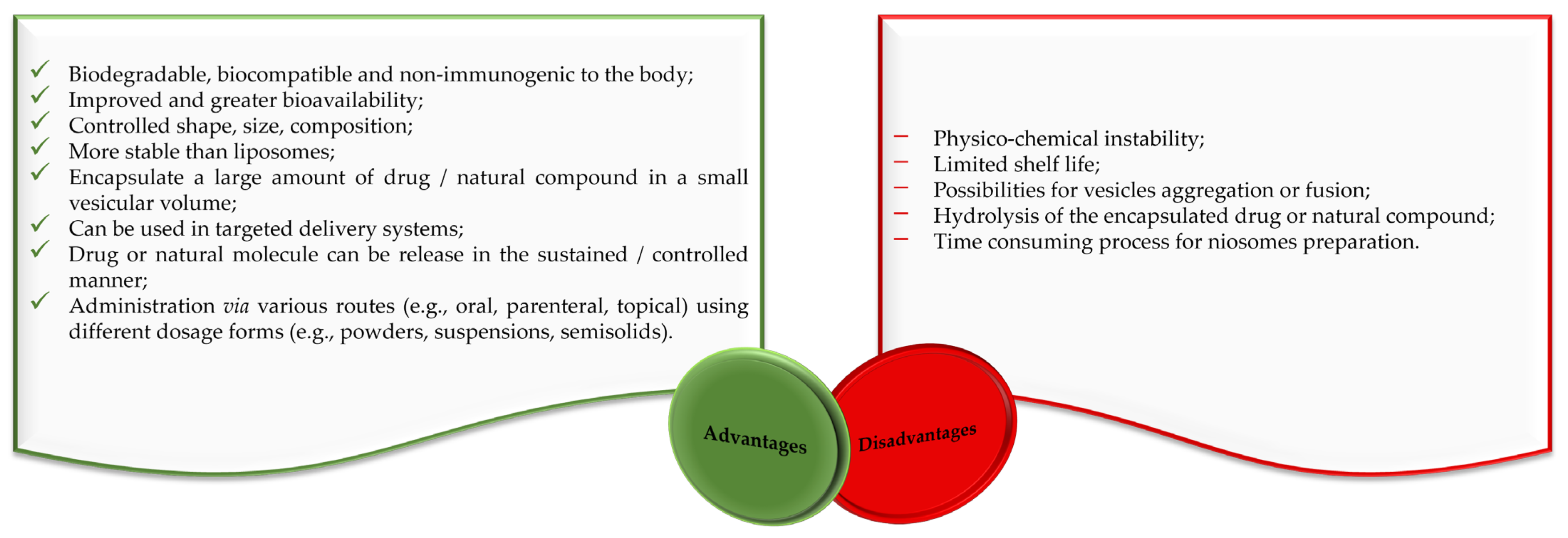
Niosomes: Composition, Formulation Techniques, and Recent Progress as Delivery Systems in Cancer Therapy


Abstract
1. Introduction
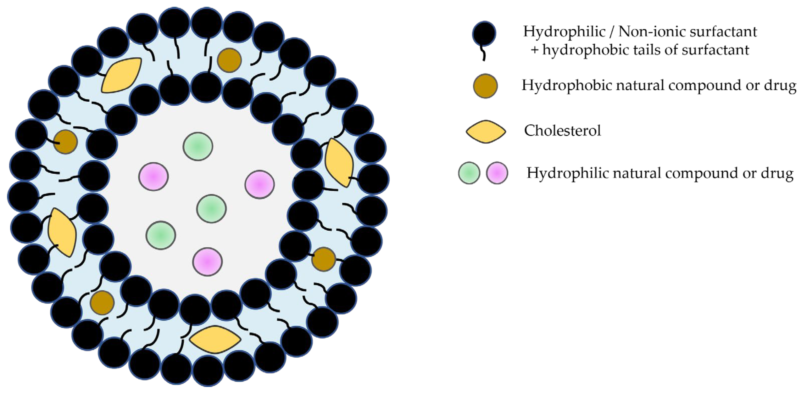
2. Composition of Niosomes
3. Classification and Formulation Techniques of Niosomes
3.1. Preparation Methods for Small Unilamellar Vesicles
3.1.1. Micro-Fluidization Technique
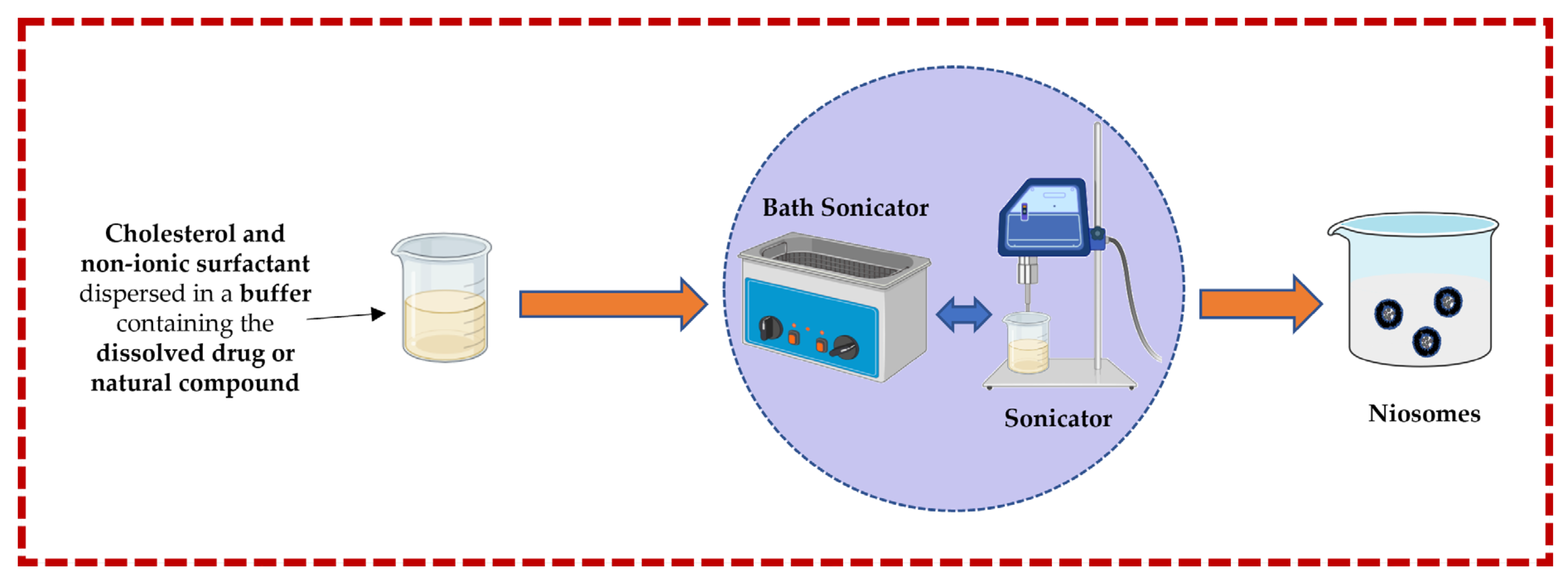
3.1.2. Sonication Technique
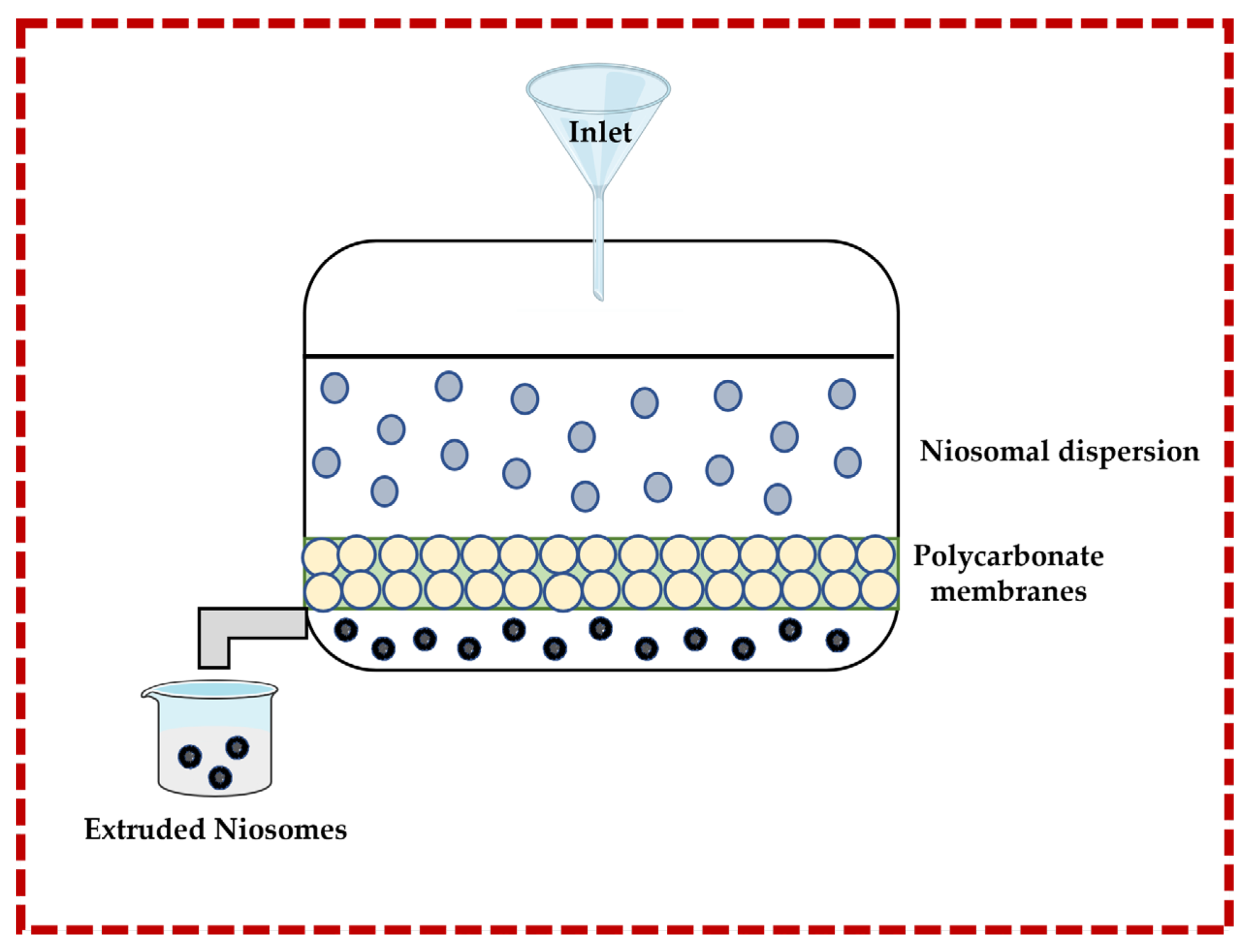
3.1.3. Multiple Membrane Extrusion Technique
3.2. Preparation Methods for Large Unilamellar Vesicle Niosomes
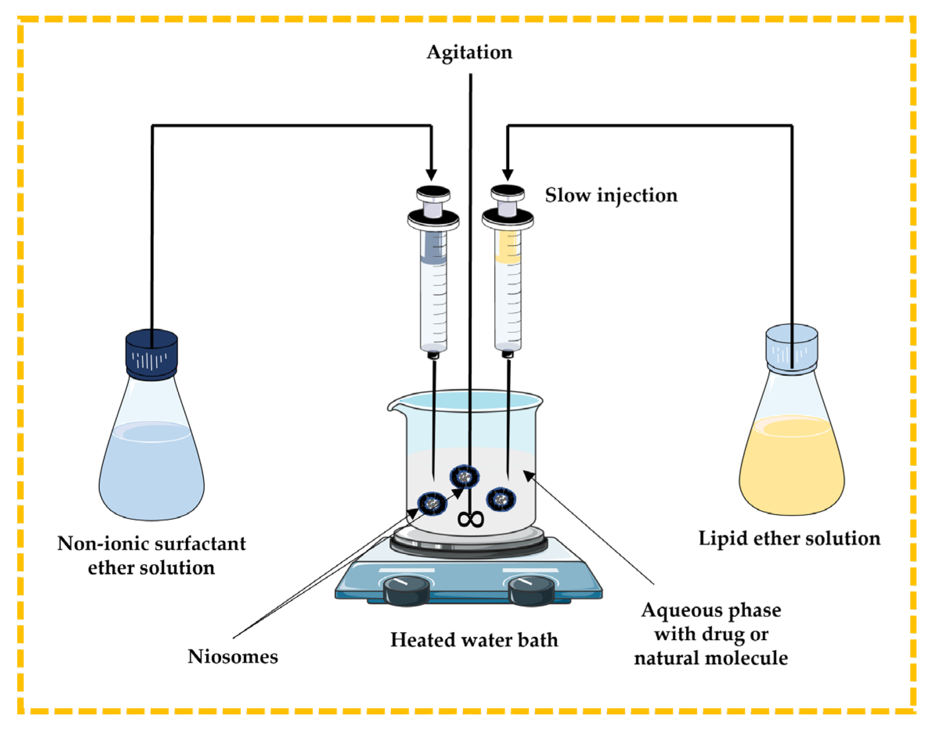
3.2.1. Ether Injection Technique
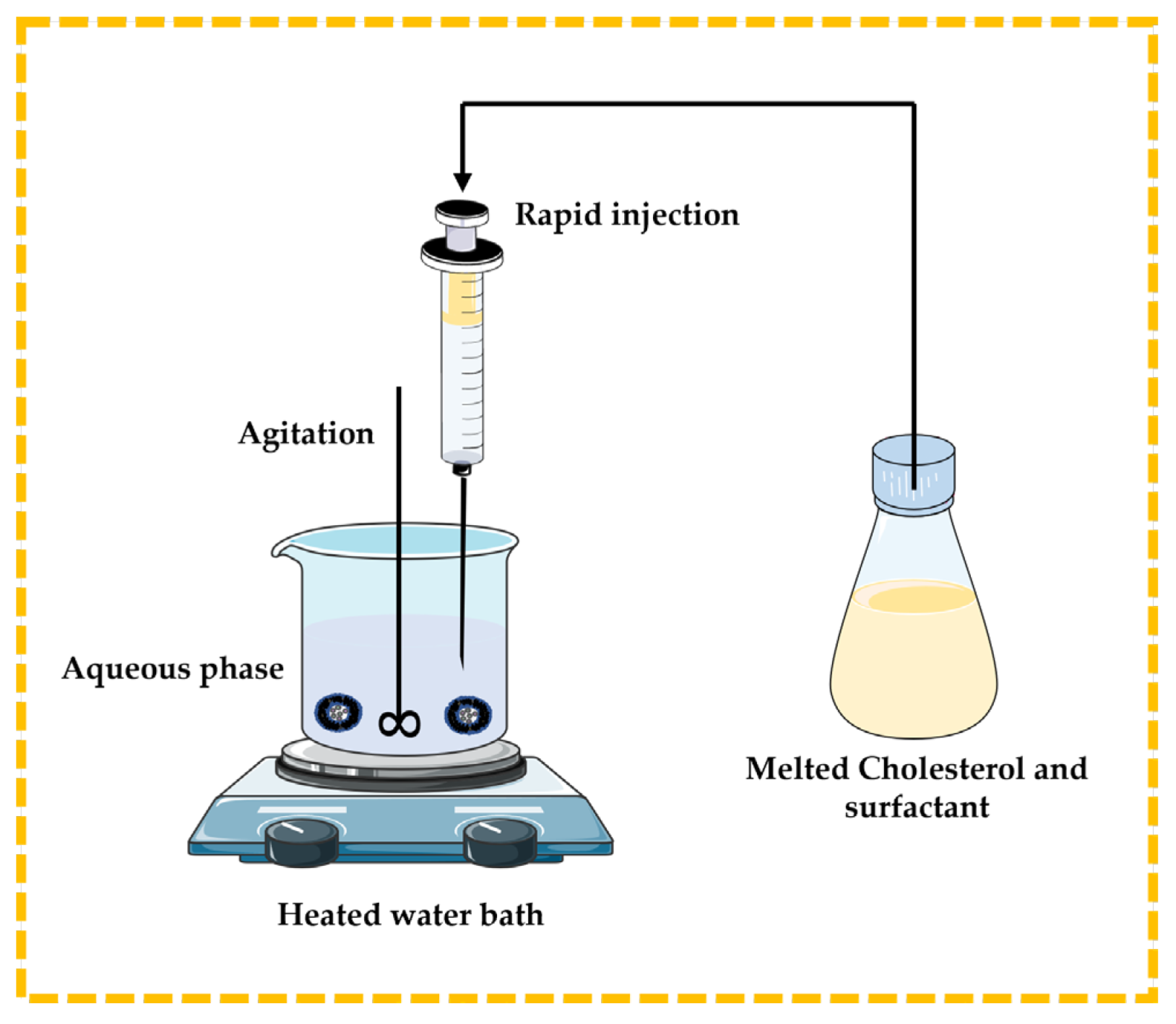
3.2.2. Lipid Injection Technique
3.2.3. Bubble Technique
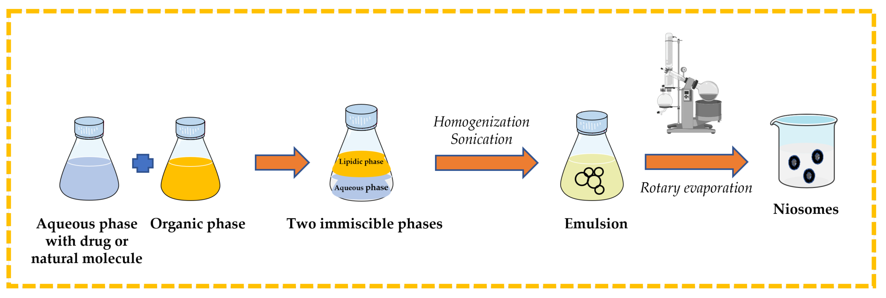
3.2.4. Reverse-Phase Evaporation Technique
3.3. Preparation Methods for Multilamellar Vesicle Niosomes
3.3.1. Trans-Membrane pH Gradient Technique
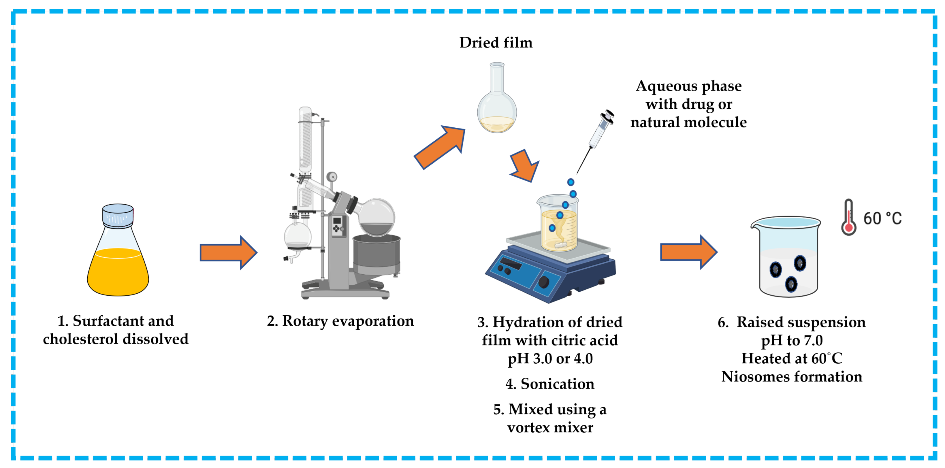
3.3.2. Thin-Film/Thin-Layer Hydration Technique
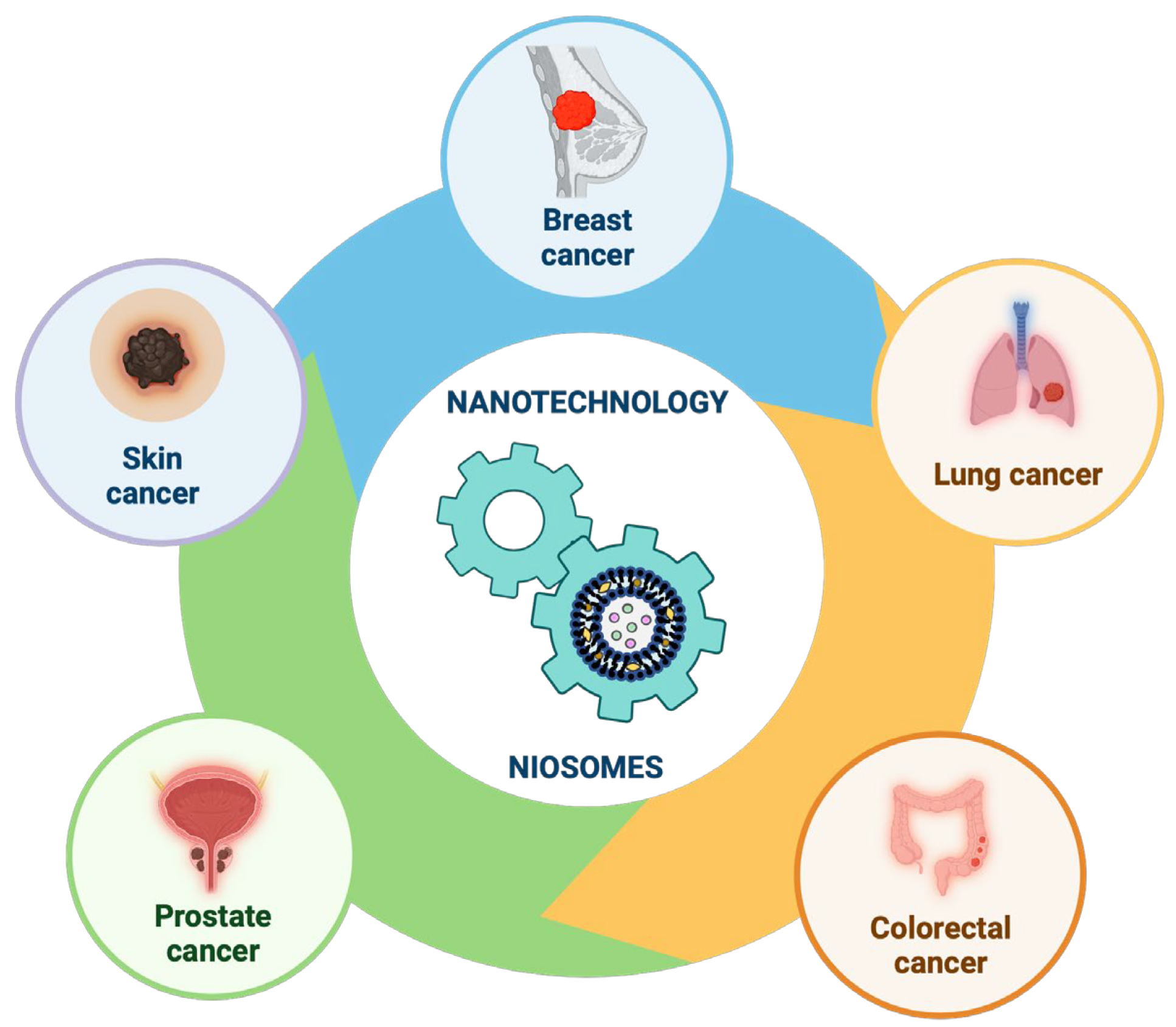
4. Recent Progress in Niosomes as Delivery Systems in Cancer Therapy
4.1. Recent Progress in the Development of Niosomal Formulations for Drug/Natural Molecules Delivery in Different Types of Cancer
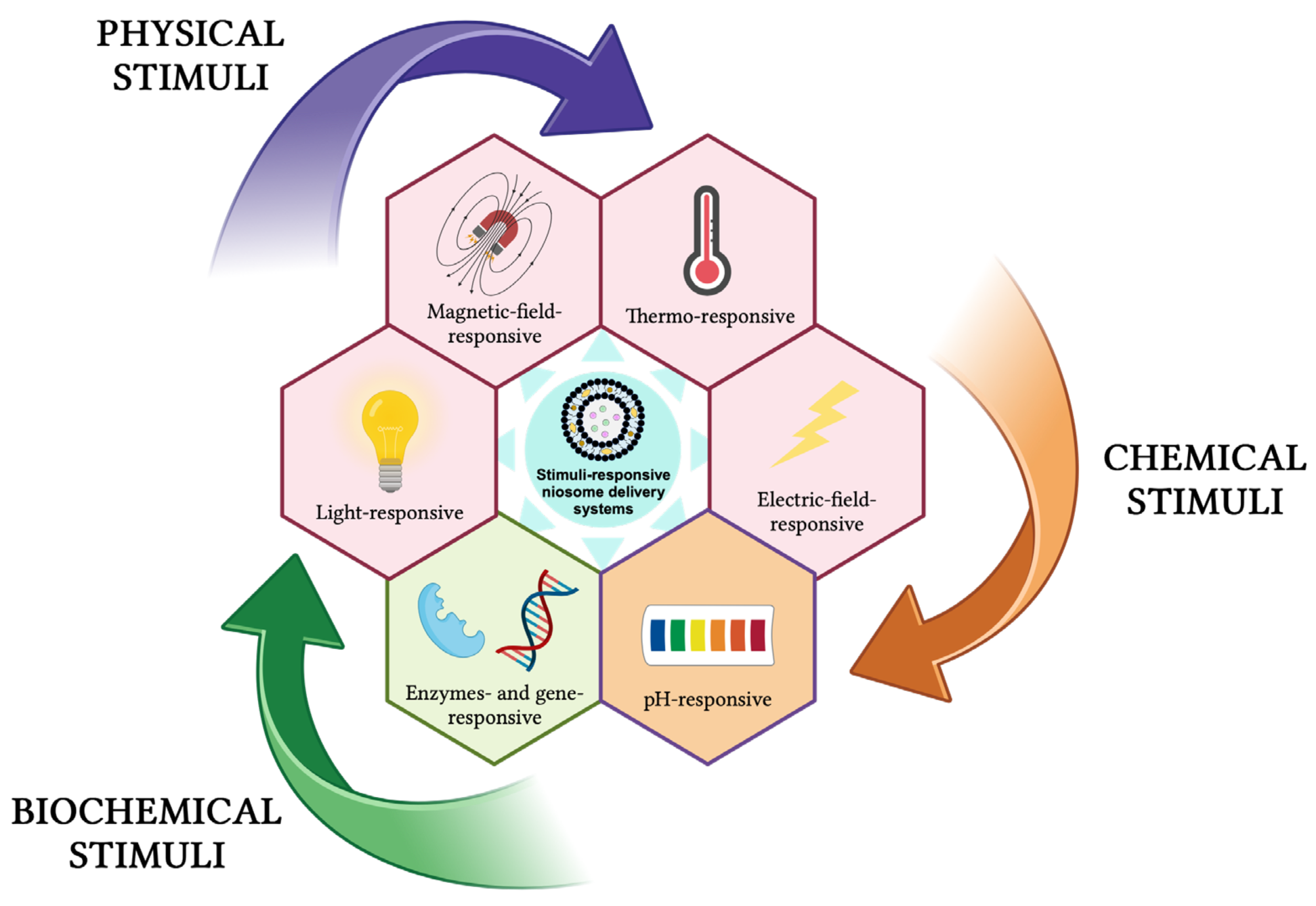
4.2. Recent Approaches for Elaboration of Specialized Niosomes as Delivery Systems
5. Future Perspectives
Author Contributions
Funding
Institutional Review Board Statement
Informed Consent Statement
Data Availability Statement
Conflicts of Interest
References
- Mazayen, Z.M.; Ghoneim, A.M.; Elbatanony, R.S.; Basalious, E.B.; Bendas, E.R. Pharmaceutical nanotechnology: From the bench to the market. Future J. Pharm. Sci. 2022, 8, 12. [Google Scholar] [CrossRef] [PubMed]
- Sahani, S.; Sharma, Y.C. Advancements in applications of nanotechnology in global food industry. Food Chem. 2021, 342, 128318. [Google Scholar] [CrossRef] [PubMed]
- Thatyana, M.; Dube, N.P.; Kemboi, D.; Manicum, A.-L.E.; Mokgalaka-Fleischmann, N.S.; Tembu, J.V. Advances in Phytonanotechnology: A Plant-Mediated Green Synthesis of Metal Nanoparticles Using Phyllanthus Plant Extracts and Their Antimicrobial and Anticancer Applications. Nanomaterials 2023, 13, 2616. [Google Scholar] [CrossRef] [PubMed]
- Bayda, S.; Adeel, M.; Tuccinardi, T.; Cordani, M.; Rizzolio, F. The History of Nanoscience and Nanotechnology: From Chemical–Physical Applications to Nanomedicine. Molecules 2020, 25, 112. [Google Scholar] [CrossRef] [PubMed]
- Soni, R.A.; Rizwan, M.A.; Singh, S. Opportunities and potential of green chemistry in nanotechnology. Nanotechnol. Environ. Eng. 2022, 7, 661–673. [Google Scholar] [CrossRef]
- Kanwar, R.; Rathee, J.; Salunke, D.B.; Mehta, S.K. Green Nanotechnology-Driven Drug Delivery Assemblies. ACS Omega 2019, 4, 8804–8815. [Google Scholar] [CrossRef] [PubMed]
- Malik, S.; Muhammad, K.; Waheed, Y. Emerging Applications of Nanotechnology in Healthcare and Medicine. Molecules 2023, 28, 6624. [Google Scholar] [CrossRef]
- Kántor, I.; Dreavă, D.; Todea, A.; Péter, F.; May, Z.; Biró, E.; Babos, G.; Feczkó, T. Co-Entrapment of Sorafenib and Cisplatin Drugs and iRGD Tumour Homing Peptide by Poly [ε-caprolactone-co-(12-hydroxystearate)] Copolymer. Biomedicines 2022, 10, 43. [Google Scholar] [CrossRef]
- Mbunge, E.; Muchemwa, B.; Jiyane, S.E.; Batani, J. Sensors and healthcare 5.0: Transformative shift in virtual care through emerging digital health technologies. Glob. Health J. 2021, 5, 169–177. [Google Scholar] [CrossRef]
- Anjum, S.; Ishaque, S.; Fatima, H.; Farooq, W.; Hano, C.; Abbasi, B.H.; Anjum, I. Emerging Applications of Nanotechnology in Healthcare Systems: Grand Challenges and Perspectives. Pharmaceuticals 2021, 14, 707. [Google Scholar] [CrossRef]
- Alshawwa, S.Z.; Kassem, A.A.; Farid, R.M.; Mostafa, S.K.; Labib, G.S. Nanocarrier Drug Delivery Systems: Characterization, Limitations, Future Perspectives and Implementation of Artificial Intelligence. Pharmaceutics 2022, 14, 883. [Google Scholar] [CrossRef] [PubMed]
- Pires, P.C.; Paiva-Santos, A.C.; Veiga, F. Liposome-Derived Nanosystems for the Treatment of Behavioral and Neurodegenerative Diseases: The Promise of Niosomes, Transfersomes, and Ethosomes for Increased Brain Drug Bioavailability. Pharmaceuticals 2023, 16, 1424. [Google Scholar] [CrossRef] [PubMed]
- Yasamineh, S.; Yasamineh, P.; Ghafouri Kalajahi, H.; Gholizadeh, O.; Yekanipour, Z.; Afkhami, H.; Eslami, M.; Hossein Kheirkhah, A.; Taghizadeh, M.; Yazdani, Y.; et al. A state-of-the-art review on the recent advances of niosomes as a targeted drug delivery system. Int. J. Pharm. 2022, 624, 121878. [Google Scholar] [CrossRef]
- Paliwal, H.; Parihar, A.; Prajapati, B.G. Current State-of-the-Art and New Trends in Self-Assembled Nanocarriers as Drug Delivery Systems. Front. Nanotechnol. 2022, 4, 836674. [Google Scholar] [CrossRef]
- Dhiman, N.; Awasthi, R.; Sharma, B.; Kharkwal, H.; Kulkarni, G.T. Lipid Nanoparticles as Carriers for Bioactive Delivery. Front. Chem. 2021, 9, 580118. [Google Scholar] [CrossRef] [PubMed]
- Marianecci, C.; Di Marzio, L.; Rinaldi, F.; Celia, C.; Paolino, D.; Alhaique, F.; Esposito, S.; Carafa, M. Niosomes from 80s to present: The state of the art. Adv. Colloid Interface Sci. 2014, 205, 187–206. [Google Scholar] [CrossRef]
- Bhardwaj, P.; Tripathi, P.; Gupta, R.; Pandey, S. Niosomes: A review on niosomal research in the last decade. J. Drug Deliv. Sci. Technol. 2020, 56, 101581. [Google Scholar] [CrossRef]
- Adnan, M.; Akhter, M.H.; Afzal, O.; Altamimi, A.S.A.; Ahmad, I.; Alossaimi, M.A.; Jaremko, M.; Emwas, A.-H.; Haider, T.; Haider, M.F. Exploring Nanocarriers as Treatment Modalities for Skin Cancer. Molecules 2023, 28, 5905. [Google Scholar] [CrossRef]
- Mawazi, S.M.; Ann, T.J.; Widodo, R.T. Application of Niosomes in Cosmetics: A Systematic Review. Cosmetics 2022, 9, 127. [Google Scholar] [CrossRef]
- Sguizzato, M.; Esposito, E.; Cortesi, R. Lipid-Based Nanosystems as a Tool to Overcome Skin Barrier. Int. J. Mol. Sci. 2021, 22, 8319. [Google Scholar] [CrossRef]
- Umbarkar, M.G. Niosome as a Novel Pharmaceutical Drug Delivery: A Brief Review Highlighting Formulation, Types, Composition and Application. Indian J. Pharm. Educ. Res. 2021, 55, s11–s28. [Google Scholar] [CrossRef]
- Arumugam, K.; Payal, B.; Jitendra, S.; Salonee, C. Niosomes: A Novel Carrier Drug Delivery System. J. Drug Deliv. Ther. 2021, 11, 162–170. [Google Scholar] [CrossRef]
- Akbarzadeh, A.; Rezaei-Sadabady, R.; Davaran, S.; Joo, S.W.; Zarghami, N.; Hanifehpour, Y.; Samiei, M.; Kouhi, M.; Nejati-Koshki, K. Liposome: Classification, preparation, and applications. Nanoscale Res. Lett. 2013, 8, 102. [Google Scholar] [CrossRef]
- Leitgeb, M.; Knez, Ž.; Primožič, M. Sustainable technologies for liposome preparation. J. Supercrit. Fluids 2020, 165, 104984. [Google Scholar] [CrossRef]
- Liu, P.; Chen, G.; Zhang, J. A Review of Liposomes as a Drug Delivery System: Current Status of Approved Products, Regulatory Environments, and Future Perspectives. Molecules 2022, 27, 1372. [Google Scholar] [CrossRef] [PubMed]
- Azeem, A.; Anwer, M.K.; Talegaonkar, S. Niosomes in sustained and targeted drug delivery: Some recent advances. J. Drug Target. 2009, 17, 671–689. [Google Scholar] [CrossRef] [PubMed]
- Bartelds, R.; Nematollahi, M.H.; Pols, T.; Stuart, M.C.A.; Pardakhty, A.; Asadikaram, G.; Poolman, B. Niosomes, an alternative for liposomal delivery. PLoS ONE 2018, 13, e0194179. [Google Scholar] [CrossRef] [PubMed]
- Aparajay, P.; Dev, A. Functionalized niosomes as a smart delivery device in cancer and fungal infection. Eur. J. Pharm. Sci. 2022, 168, 106052. [Google Scholar] [CrossRef]
- Gharbavi, M.; Amani, J.; Kheiri-Manjili, H.; Danafar, H.; Sharafi, A. Niosome: A Promising Nanocarrier for Natural Drug Delivery through Blood-Brain Barrier. Adv. Pharmacol. Sci. 2018, 2018, 6847971. [Google Scholar] [CrossRef]
- Momekova, D.B.; Gugleva, V.E.; Petrov, P.D. Nanoarchitectonics of Multifunctional Niosomes for Advanced Drug Delivery. ACS Omega 2021, 6, 33265–33273. [Google Scholar] [CrossRef]
- Abdelkader, H.; Alani, A.W.G.; Alany, R.G. Recent advances in non-ionic surfactant vesicles (niosomes): Self-assembly, fabrication, characterization, drug delivery applications and limitations. Drug Deliv. 2014, 21, 87–100. [Google Scholar] [CrossRef] [PubMed]
- Izhar, M.P.; Hafeez, A.; Kushwaha, P.; Simrah. Drug Delivery through Niosomes: A Comprehensive Review with Therapeutic Applications. J. Clust. Sci. 2023, 34, 2257–2273. [Google Scholar] [CrossRef]
- Shah, N.; Prajapati, R.; Gohil, D.; Sadhu, P.; Patel, S. Niosomes: A Promising Novel Nano Carrier for Drug Delivery. J. Pharm. Res. Int. 2021, 33, 53–66. [Google Scholar] [CrossRef]
- Chen, S.; Hanning, S.; Falconer, J.; Locke, M.; Wen, J. Recent advances in non-ionic surfactant vesicles (niosomes): Fabrication, characterization, pharmaceutical and cosmetic applications. Eur. J. Pharm. Biopharm. 2019, 144, 18–39. [Google Scholar] [CrossRef]
- Somjid, S.; Krongsuk, S.; Johns, J.R. Cholesterol concentration effect on the bilayer properties and phase formation of niosome bilayers: A molecular dynamics simulation study. J. Mol. Liq. 2018, 256, 591–598. [Google Scholar] [CrossRef]
- Ge, X.; Wei, M.; He, S.; Yuan, W.-E. Advances of Non-Ionic Surfactant Vesicles (Niosomes) and Their Application in Drug Delivery. Pharmaceutics 2019, 11, 55. [Google Scholar] [CrossRef]
- Junyaprasert, V.B.; Teeranachaideekul, V.; Supaperm, T. Effect of Charged and Non-ionic Membrane Additives on Physicochemical Properties and Stability of Niosomes. AAPS PharmSciTech 2008, 9, 851–859. [Google Scholar] [CrossRef]
- Kumar, G.P.; Rajeshwarrao, P. Nonionic surfactant vesicular systems for effective drug delivery—An overview. Acta Pharm. Sin. B 2011, 1, 208–219. [Google Scholar] [CrossRef]
- Witika, B.A.; Bassey, K.E.; Demana, P.H.; Siwe-Noundou, X.; Poka, M.S. Current Advances in Specialised Niosomal Drug Delivery: Manufacture, Characterization and Drug Delivery Applications. Int. J. Mol. Sci. 2022, 23, 9668. [Google Scholar] [CrossRef]
- García-Manrique, P.; Machado, N.D.; Fernández, M.A.; Blanco-López, M.C.; Matos, M.; Gutiérrez, G. Effect of drug molecular weight on niosomes size and encapsulation efficiency. Colloids Surf. B Biointerfaces 2020, 186, 110711. [Google Scholar] [CrossRef]
- Bashkeran, T.; Harun, A.; Ngo, T.X.; Suda, K.; Umakoshi, H.; Watanabe, N.; Nadzir, M.M. Niosomes in cancer treatment: A focus on curcumin encapsulation. Heliyon 2023, 9, e18710. [Google Scholar] [CrossRef]
- Ag Seleci, D.; Seleci, M.; Walter, J.-G.; Stahl, F.; Scheper, T. Niosomes as Nanoparticular Drug Carriers: Fundamentals and Recent Applications. J. Nanomater. 2016, 2016, 7372306. [Google Scholar] [CrossRef]
- Durga Bhavani, G.; Veera Lakshmi, P. Recent advances of non-ionic surfactant-based nano-vesicles (niosomes and proniosomes): A brief review of these in enhancing transdermal delivery of drug. Future J. Pharm. Sci. 2020, 6, 100. [Google Scholar] [CrossRef]
- Moghtaderi, M.; Sedaghatnia, K.; Bourbour, M.; Fatemizadeh, M.; Salehi Moghaddam, Z.; Hejabi, F.; Heidari, F.; Quazi, S.; Farasati Far, B. Niosomes: A novel targeted drug delivery system for cancer. Med. Oncol. 2022, 39, 240. [Google Scholar] [CrossRef]
- Fenske, D.B.; Cullis, P.R. Encapsulation of Drugs within Liposomes by pH-Gradient Techniques. In Liposome Technology; Gregoriadis, G., Ed.; CRC Press: Boca Raton, FL, USA, 2016; pp. 27–50. [Google Scholar]
- Choi, C.-H.; Kwak, Y.; Malhotra, R.; Chang, C.-H. Microfluidics for Two-Dimensional Nanosheets: A Mini Review. Processes 2020, 8, 1067. [Google Scholar] [CrossRef]
- Joshi, S.; White, R.; Sahu, R.; Dennis, V.A.; Singh, S.R. Comprehensive Screening of Drug Encapsulation and Co-Encapsulation into Niosomes Produced Using a Microfluidic Device. Processes 2020, 8, 535. [Google Scholar] [CrossRef]
- Kumar, A.; Dhiman, A.; Suhag, R.; Sehrawat, R.; Upadhyay, A.; McClements, D.J. Comprehensive review on potential applications of microfluidization in food processing. Food Sci. Biotechnol. 2022, 31, 17–36. [Google Scholar] [CrossRef] [PubMed]
- Obeid, M.A.; Ogah, C.A.; Ogah, C.O.; Ajala, O.S.; Aldea, M.R.; Gray, A.I.; Igoli, J.I.; Ferro, V.A. Formulation and evaluation of nanosized hippadine-loaded niosome: Extraction and isolation, physicochemical properties, and in vitro cytotoxicity against human ovarian and skin cancer cell lines. J. Drug Deliv. Sci. Technol. 2023, 87, 104766. [Google Scholar] [CrossRef]
- Radmard, A.; Saeedi, M.; Morteza-Semnani, K.; Hashemi, S.M.H.; Nokhodchi, A. An eco-friendly and green formulation in lipid nanotechnology for delivery of a hydrophilic agent to the skin in the treatment and management of hyperpigmentation complaints: Arbutin niosome (Arbusome). Colloids Surf. B Biointerfaces 2021, 201, 111616. [Google Scholar] [CrossRef] [PubMed]
- Siegel, R.L.; Miller, K.D.; Fuchs, H.E.; Jemal, A. Cancer Statistics, 2021. CA A Cancer J. Clin. 2021, 71, 7–33. [Google Scholar] [CrossRef]
- Siegel, R.L.; Miller, K.D.; Fuchs, H.E.; Jemal, A. Cancer statistics, 2022. CA A Cancer J. Clin. 2022, 72, 7–33. [Google Scholar] [CrossRef]
- Debela, D.T.; Muzazu, S.G.Y.; Heraro, K.D.; Ndalama, M.T.; Mesele, B.W.; Haile, D.C.; Kitui, S.K.; Manyazewal, T. New approaches and procedures for cancer treatment: Current perspectives. SAGE Open Med. 2021, 9, 20503121211034366. [Google Scholar] [CrossRef]
- Dessale, M.; Mengistu, G.; Mengist, H.M. Nanotechnology: A Promising Approach for Cancer Diagnosis, Therapeutics and Theragnosis. Int. J. Nanomed. 2022, 17, 3735–3749. [Google Scholar] [CrossRef]
- Chehelgerdi, M.; Chehelgerdi, M.; Allela, O.Q.B.; Pecho, R.D.C.; Jayasankar, N.; Rao, D.P.; Thamaraikani, T.; Vasanthan, M.; Viktor, P.; Lakshmaiya, N.; et al. Progressing nanotechnology to improve targeted cancer treatment: Overcoming hurdles in its clinical implementation. Mol. Cancer 2023, 22, 169. [Google Scholar] [CrossRef]
- Yao, Y.; Zhou, Y.; Liu, L.; Xu, Y.; Chen, Q.; Wang, Y.; Wu, S.; Deng, Y.; Zhang, J.; Shao, A. Nanoparticle-Based Drug Delivery in Cancer Therapy and Its Role in Overcoming Drug Resistance. Front. Mol. Biosci. 2020, 7, 193. [Google Scholar] [CrossRef]
- Zhong, L.; Li, Y.; Xiong, L.; Wang, W.; Wu, M.; Yuan, T.; Yang, W.; Tian, C.; Miao, Z.; Wang, T.; et al. Small molecules in targeted cancer therapy: Advances, challenges, and future perspectives. Signal Transduct. Target. Ther. 2021, 6, 201. [Google Scholar] [CrossRef]
- Zhu, R.; Zhang, F.; Peng, Y.; Xie, T.; Wang, Y.; Lan, Y. Current Progress in Cancer Treatment Using Nanomaterials. Front. Oncol. 2022, 12, 930125. [Google Scholar] [CrossRef]
- Giaquinto, A.N.; Sung, H.; Miller, K.D.; Kramer, J.L.; Newman, L.A.; Minihan, A.; Jemal, A.; Siegel, R.L. Breast Cancer Statistics, 2022. CA A Cancer J. Clin. 2022, 72, 524–541. [Google Scholar] [CrossRef]
- Arnold, M.; Morgan, E.; Rumgay, H.; Mafra, A.; Singh, D.; Laversanne, M.; Vignat, J.; Gralow, J.R.; Cardoso, F.; Siesling, S.; et al. Current and future burden of breast cancer: Global statistics for 2020 and 2040. Breast 2022, 66, 15–23. [Google Scholar] [CrossRef]
- Kumar, P.; Mangla, B.; Javed, S.; Ahsan, W.; Musyuni, P.; Sivadasan, D.; Alqahtani, S.S.; Aggarwal, G. A review of nanomaterials from synthetic and natural molecules for prospective breast cancer nanotherapy. Front. Pharmacol. 2023, 14, 1149554. [Google Scholar] [CrossRef]
- Marcolin, J.C.; Lichtenfels, M.; da Silva, C.A.; de Farias, C.B. Gynecologic and Breast Cancers: What’s New in Chemoresistance and Chemosensitivity Tests? Curr. Probl. Cancer 2023, 47, 100996. [Google Scholar] [CrossRef] [PubMed]
- Jain, V.; Kumar, H.; Anod, H.V.; Chand, P.; Gupta, N.V.; Dey, S.; Kesharwani, S.S. A review of nanotechnology-based approaches for breast cancer and triple-negative breast cancer. J. Control. Release 2020, 326, 628–647. [Google Scholar] [CrossRef] [PubMed]
- Zhang, C.; Zhou, X.; Zhang, H.; Han, X.; Li, B.; Yang, R.; Zhou, X. Recent Progress of Novel Nanotechnology Challenging the Multidrug Resistance of Cancer. Front. Pharmacol. 2022, 13, 776895. [Google Scholar] [CrossRef] [PubMed]
- Nicholson, A.G.; Tsao, M.S.; Beasley, M.B.; Borczuk, A.C.; Brambilla, E.; Cooper, W.A.; Dacic, S.; Jain, D.; Kerr, K.M.; Lantuejoul, S.; et al. The 2021 WHO Classification of Lung Tumors: Impact of Advances Since 2015. J. Thorac. Oncol. 2022, 17, 362–387. [Google Scholar] [CrossRef] [PubMed]
- Duan, Y.; Shen, C.; Zhang, Y.; Luo, Y. Advanced diagnostic and therapeutic strategies in nanotechnology for lung cancer. Front. Oncol. 2022, 12, 1031000. [Google Scholar] [CrossRef]
- Zulfiqar, B.; Farooq, A.; Kanwal, S.; Asghar, K. Immunotherapy and targeted therapy for lung cancer: Current status and future perspectives. Front. Pharmacol. 2022, 13, 1035171. [Google Scholar] [CrossRef]
- Xu, Y.; Liu, Y.; Ge, Y.; Li, H.; Zhang, Y.; Wang, L. Drug resistance mechanism and reversal strategy in lung cancer immunotherapy. Front. Pharmacol. 2023, 14, 1230824. [Google Scholar] [CrossRef]
- Holder, J.E.; Ferguson, C.; Oliveira, E.; Lodeiro, C.; Trim, C.M.; Byrne, L.J.; Bertolo, E.; Wilson, C.M. The use of nanoparticles for targeted drug delivery in non-small cell lung cancer. Front. Oncol. 2023, 13, 1154318. [Google Scholar] [CrossRef]
- Siegel, R.L.; Wagle, N.S.; Cercek, A.; Smith, R.A.; Jemal, A. Colorectal cancer statistics, 2023. CA A Cancer J. Clin. 2023, 73, 233–254. [Google Scholar] [CrossRef]
- Xi, Y.; Xu, P. Global colorectal cancer burden in 2020 and projections to 2040. Transl. Oncol. 2021, 14, 101174. [Google Scholar] [CrossRef]
- Johdi, N.A.; Sukor, N.F. Colorectal Cancer Immunotherapy: Options and Strategies. Front. Immunol. 2020, 11, 1624. [Google Scholar] [CrossRef]
- Weng, J.; Li, S.; Zhu, Z.; Liu, Q.; Zhang, R.; Yang, Y.; Li, X. Exploring immunotherapy in colorectal cancer. J. Hematol. Oncol. 2022, 15, 95. [Google Scholar] [CrossRef]
- Brar, B.; Ranjan, K.; Palria, A.; Kumar, R.; Ghosh, M.; Sihag, S.; Minakshi, P. Nanotechnology in Colorectal Cancer for Precision Diagnosis and Therapy. Front. Nanotechnol. 2021, 3, 699266. [Google Scholar] [CrossRef]
- Jain, A.; Bhattacharya, S. Recent advances in nanomedicine preparative methods and their therapeutic potential for colorectal cancer: A critical review. Front. Oncol. 2023, 13, 1211603. [Google Scholar] [CrossRef]
- Kasi, P.B.; Mallela, V.R.; Ambrozkiewicz, F.; Trailin, A.; Liška, V.; Hemminki, K. Theranostics Nanomedicine Applications for Colorectal Cancer and Metastasis: Recent Advances. Int. J. Mol. Sci. 2023, 24, 7922. [Google Scholar] [CrossRef] [PubMed]
- Moreira-Silva, F.; Henrique, R.; Jerónimo, C. From Therapy Resistance to Targeted Therapies in Prostate Cancer. Front. Oncol. 2022, 12, 877379. [Google Scholar] [CrossRef]
- Licitra, F.; Giovannelli, P.; Di Donato, M.; Monaco, A.; Galasso, G.; Migliaccio, A.; Castoria, G. New Insights and Emerging Therapeutic Approaches in Prostate Cancer. Front. Endocrinol. 2022, 13, 840787. [Google Scholar] [CrossRef]
- Chen, D.; Hu, Y. Approaches for boosting antitumor immunity in prostate cancer therapy: A comprehensive review on drugs, products, and nanoparticles. J. Drug Deliv. Sci. Technol. 2023, 89, 105048. [Google Scholar] [CrossRef]
- Belkahla, S.; Nahvi, I.; Biswas, S.; Nahvi, I.; Ben Amor, N. Advances and development of prostate cancer, treatment, and strategies: A systemic review. Front. Cell Dev. Biol. 2022, 10, 991330. [Google Scholar] [CrossRef]
- Vieira, I.R.; Tessaro, L.; Lima, A.K.; Velloso, I.P.; Conte-Junior, C.A. Recent Progress in Nanotechnology Improving the Therapeutic Potential of Polyphenols for Cancer. Nutrients 2023, 15, 3136. [Google Scholar] [CrossRef]
- Hasan, N.; Nadaf, A.; Imran, M.; Jiba, U.; Sheikh, A.; Almalki, W.H.; Almujri, S.S.; Mohammed, Y.H.; Kesharwani, P.; Ahmad, F.J. Skin cancer: Understanding the journey of transformation from conventional to advanced treatment approaches. Mol. Cancer 2023, 22, 168. [Google Scholar] [CrossRef] [PubMed]
- Abdalla, B.M.Z.; Abdalla, C.M.Z. Epidemiology of Skin Cancer. In Oncodermatology: An Evidence-Based, Multidisciplinary Approach to Best Practices; Abdalla, C.M.Z., Sanches, J.A., Munhoz, R.R., Belfort, F.A., Eds.; Springer International Publishing: Cham, Switzerland, 2023; pp. 29–35. [Google Scholar]
- Achatz, M.I.; Coloma, M.C.G.; de Albuquerque Cavalcanti Callegaro, E. Risk Factors for Skin Cancer. In Oncodermatology: An Evidence-Based, Multidisciplinary Approach to Best Practices; Abdalla, C.M.Z., Sanches, J.A., Munhoz, R.R., Belfort, F.A., Eds.; Springer International Publishing: Cham, Switzerland, 2023; pp. 37–55. [Google Scholar]
- Pashazadeh, A.; Boese, A.; Friebe, M. Radiation therapy techniques in the treatment of skin cancer: An overview of the current status and outlook. J. Dermatol. Treat. 2019, 30, 831–839. [Google Scholar] [CrossRef] [PubMed]
- Sun, J.; Zhao, H.; Fu, L.; Cui, J.; Yang, Y. Global Trends and Research Progress of Photodynamic Therapy in Skin Cancer: A Bibliometric Analysis and Literature Review. Clin. Cosmet. Investig. Dermatol. 2023, 16, 479–498. [Google Scholar] [CrossRef] [PubMed]
- Olszowy, M.; Nowak-Perlak, M.; Woźniak, M. Current Strategies in Photodynamic Therapy (PDT) and Photodynamic Diagnostics (PDD) and the Future Potential of Nanotechnology in Cancer Treatment. Pharmaceutics 2023, 15, 1712. [Google Scholar] [CrossRef]
- Malik, S.; Muhammad, K.; Waheed, Y. Nanotechnology: A Revolution in Modern Industry. Molecules 2023, 28, 661. [Google Scholar] [CrossRef]
- Zeng, L.; Gowda, B.H.J.; Ahmed, M.G.; Abourehab, M.A.S.; Chen, Z.-S.; Zhang, C.; Li, J.; Kesharwani, P. Advancements in nanoparticle-based treatment approaches for skin cancer therapy. Mol. Cancer 2023, 22, 10. [Google Scholar] [CrossRef]
- Prajapat, V.M.; Mahajan, S.; Paul, P.G.; Aalhate, M.; Mehandole, A.; Madan, J.; Dua, K.; Chellappan, D.K.; Singh, S.K.; Singh, P.K. Nanomedicine: A pragmatic approach for tackling melanoma skin cancer. J. Drug Deliv. Sci. Technol. 2023, 83, 104394. [Google Scholar] [CrossRef]
- Chandra, J.; Hasan, N.; Nasir, N.; Wahab, S.; Thanikachalam, P.V.; Sahebkar, A.; Ahmad, F.J.; Kesharwani, P. Nanotechnology-empowered strategies in treatment of skin cancer. Environ. Res. 2023, 235, 116649. [Google Scholar] [CrossRef]
- Diaz, M.J.; Natarelli, N.; Aflatooni, S.; Aleman, S.J.; Neelam, S.; Tran, J.T.; Taneja, K.; Lucke-Wold, B.; Forouzandeh, M. Nanoparticle-Based Treatment Approaches for Skin Cancer: A Systematic Review. Curr. Oncol. 2023, 30, 7112–7131. [Google Scholar] [CrossRef]
- Chang, J.; Yu, B.; Saltzman, W.M.; Girardi, M. Nanoparticles as a Therapeutic Delivery System for Skin Cancer Prevention and Treatment. JID Innov. 2023, 3, 100197. [Google Scholar] [CrossRef]
- Zhang, C.; Zhu, X.; Hou, S.; Pan, W.; Liao, W. Functionalization of Nanomaterials for Skin Cancer Theranostics. Front. Bioeng. Biotechnol. 2022, 10, 887548. [Google Scholar] [CrossRef]
- Gupta, N.; Gupta, G.D.; Singh, D. Localized topical drug delivery systems for skin cancer: Current approaches and future prospects. Front. Nanotechnol. 2022, 4, 1006628. [Google Scholar] [CrossRef]
- Akbarzadeh, I.; Farid, M.; Javidfar, M.; Zabet, N.; Shokoohian, B.; Arki, M.K.; Shpichka, A.; Noorbazargan, H.; Aghdaei, H.A.; Hossein-khannazer, N.; et al. The Optimized Formulation of Tamoxifen-Loaded Niosomes Efficiently Induced Apoptosis and Cell Cycle Arrest in Breast Cancer Cells. AAPS PharmSciTech 2022, 23, 57. [Google Scholar] [CrossRef]
- Gaikwad, D.S.; Chougale, R.D.; Patil, K.S.; Disouza, J.I.; Hajare, A.A. Design, development, and evaluation of docetaxel-loaded niosomes for the treatment of breast cancer. Future J. Pharm. Sci. 2023, 9, 43. [Google Scholar] [CrossRef]
- Basheer, H.A.; Alhusban, M.A.; Zaid Alkilani, A.; Alshishani, A.; Elsalem, L.; Afarinkia, K. Niosomal Delivery of Celecoxib and Metformin for Targeted Breast Cancer Treatment. Cancers 2023, 15, 5004. [Google Scholar] [CrossRef]
- Barani, M.; Hajinezhad, M.R.; Zargari, F.; Shahraki, S.; Davodabadi, F.; Mirinejad, S.; Sargazi, S.; Rahdar, A.; Díez-Pascual, A.M. Preparation, characterization, cytotoxicity and pharmacokinetics of niosomes containing gemcitabine: In vitro, in vivo, and simulation studies. J. Drug Deliv. Sci. Technol. 2023, 84, 104505. [Google Scholar] [CrossRef]
- Fahmy, S.A.; Nasr, S.; Ramzy, A.; Dawood, A.S.; Abdelnaser, A.; Azzazy, H.M.E.-S. Cytotoxic and Antioxidative Effects of Geranium Oil and Ascorbic Acid Coloaded in Niosomes against MCF-7 Breast Cancer Cells. ACS Omega 2023, 8, 22774–22782. [Google Scholar] [CrossRef]
- Akbarzadeh, I.; Shayan, M.; Bourbour, M.; Moghtaderi, M.; Noorbazargan, H.; Eshrati Yeganeh, F.; Saffar, S.; Tahriri, M. Preparation, Optimization and In-Vitro Evaluation of Curcumin-Loaded Niosome@calcium Alginate Nanocarrier as a New Approach for Breast Cancer Treatment. Biology 2021, 10, 173. [Google Scholar] [CrossRef]
- Moammeri, A.; Abbaspour, K.; Zafarian, A.; Jamshidifar, E.; Motasadizadeh, H.; Dabbagh Moghaddam, F.; Salehi, Z.; Makvandi, P.; Dinarvand, R. pH-Responsive, Adorned Nanoniosomes for Codelivery of Cisplatin and Epirubicin: Synergistic Treatment of Breast Cancer. ACS Appl. Bio Mater. 2022, 5, 675–690. [Google Scholar] [CrossRef]
- Honarvari, B.; Karimifard, S.; Akhtari, N.; Mehrarya, M.; Moghaddam, Z.S.; Ansari, M.J.; Jalil, A.T.; Matencio, A.; Trotta, F.; Yeganeh, F.E.; et al. Folate-Targeted Curcumin-Loaded Niosomes for Site-Specific Delivery in Breast Cancer Treatment: In Silico and In Vitro Study. Molecules 2022, 27, 4634. [Google Scholar] [CrossRef]
- Sahrayi, H.; Hosseini, E.; Karimifard, S.; Khayam, N.; Meybodi, S.M.; Amiri, S.; Bourbour, M.; Farasati Far, B.; Akbarzadeh, I.; Bhia, M.; et al. Co-Delivery of Letrozole and Cyclophosphamide via Folic Acid-Decorated Nanoniosomes for Breast Cancer Therapy: Synergic Effect, Augmentation of Cytotoxicity, and Apoptosis Gene Expression. Pharmaceuticals 2022, 15, 6. [Google Scholar] [CrossRef]
- Lalami, Z.A.; Tafvizi, F.; Naseh, V.; Salehipour, M. Characterization and optimization of co-delivery Farnesol-Gingerol Niosomal formulation to enhance anticancer activities against breast cancer cells. J. Drug Deliv. Sci. Technol. 2022, 72, 103371. [Google Scholar] [CrossRef]
- Zaer, M.; Moeinzadeh, A.; Abolhassani, H.; Rostami, N.; Tavakkoli Yaraki, M.; Seyedi, S.A.; Nabipoorashrafi, S.A.; Bashiri, Z.; Moeinabadi-Bidgoli, K.; Moradbeygi, F.; et al. Doxorubicin-loaded Niosomes functionalized with gelatine and alginate as pH-responsive drug delivery system: A 3D printing approach. Int. J. Biol. Macromol. 2023, 253, 126808. [Google Scholar] [CrossRef]
- Safari Sharafshadeh, M.; Tafvizi, F.; Khodarahmi, P.; Ehtesham, S. Preparation and physicochemical properties of cisplatin and doxorubicin encapsulated by niosome alginate nanocarrier for cancer therapy. Int. J. Biol. Macromol. 2023, 235, 123686. [Google Scholar] [CrossRef]
- Mansoori-Kermani, A.; Khalighi, S.; Akbarzadeh, I.; Niavol, F.R.; Motasadizadeh, H.; Mahdieh, A.; Jahed, V.; Abdinezhad, M.; Rahbariasr, N.; Hosseini, M.; et al. Engineered hyaluronic acid-decorated niosomal nanoparticles for controlled and targeted delivery of epirubicin to treat breast cancer. Mater. Today Bio 2022, 16, 100349. [Google Scholar] [CrossRef]
- Agarwal, S.; Mohamed, M.S.; Raveendran, S.; Rochani, A.K.; Maekawa, T.; Kumar, D.S. Formulation, characterization and evaluation of morusin loaded niosomes for potentiation of anticancer therapy. RSC Adv. 2018, 8, 32621–32636. [Google Scholar] [CrossRef]
- Dabbagh Moghaddam, F.; Akbarzadeh, I.; Marzbankia, E.; Farid, M.; Khaledi, L.; Reihani, A.H.; Javidfar, M.; Mortazavi, P. Delivery of melittin-loaded niosomes for breast cancer treatment: An in vitro and in vivo evaluation of anti-cancer effect. Cancer Nanotechnol. 2021, 12, 14. [Google Scholar] [CrossRef]
- Barani, M.; Hajinezhad, M.R.; Sargazi, S.; Rahdar, A.; Shahraki, S.; Lohrasbi-Nejad, A.; Baino, F. In vitro and in vivo anticancer effect of pH-responsive paclitaxel-loaded niosomes. J. Mater. Sci. Mater. Med. 2021, 32, 147. [Google Scholar] [CrossRef] [PubMed]
- Pourmoghadasiyan, B.; Tavakkoli, F.; Beram, F.M.; Badmasti, F.; Mirzaie, A.; Kazempour, R.; Rahimi, S.; Larijani, S.F.; Hejabi, F.; Sedaghatnia, K. Nanosized paclitaxel-loaded niosomes: Formulation, in vitro cytotoxicity, and apoptosis gene expression in breast cancer cell lines. Mol. Biol. Rep. 2022, 49, 3597–3608. [Google Scholar] [CrossRef] [PubMed]
- Abtahi, N.A.; Naghib, S.M.; Haghiralsadat, F.; Akbari Edgahi, M. Development of highly efficient niosomal systems for co-delivery of drugs and genes to treat breast cancer in vitro and in vivo. Cancer Nanotechnol. 2022, 13, 28. [Google Scholar] [CrossRef]
- Ghourchian, H.; Pecho, R.D.C.; Karimi-Dehkordi, M.; Mazandarani, A.; Ghajari, G.; Piri-Gharaghie, T. Novel Niosome-Encapsulated 2,5-Diketopiperazine (BHPPD): Synthesis, Formulation, and Anti-breast Cancer Activity. Appl. Biochem. Biotechnol. 2023, 1–22. [Google Scholar] [CrossRef]
- Hussein, M.M.A.; Abdelfattah-Hassan, A.; Eldoumani, H.; Essawi, W.M.; Alsahli, T.G.; Alharbi, K.S.; Alzarea, S.I.; Al-Hejaili, H.Y.; Gaafar, S.F. Evaluation of anti-cancer effects of carnosine and melittin-loaded niosomes in MCF-7 and MDA-MB-231 breast cancer cells. Front. Pharmacol. 2023, 14, 1258387. [Google Scholar] [CrossRef]
- Amiri, S.; Pashizeh, F.; Moeinabadi-Bidgoli, K.; Eyvazi, Y.; Akbari, T.; Salehi Moghaddam, Z.; Eskandarisani, M.; Farahmand, F.; Hafezi, Y.; Nouri Jevinani, H.; et al. Co-encapsulation of hydrophilic and hydrophobic drugs into niosomal nanocarrier for enhanced breast cancer therapy: In silico, and in vitro studies. Environ. Res. 2023, 239, 117292. [Google Scholar] [CrossRef]
- Saharkhiz, S.; Zarepour, A.; Zarrabi, A. Empowering Cancer Therapy: Comparing PEGylated and Non-PEGylated Niosomes Loaded with Curcumin and Doxorubicin on MCF-7 Cell Line. Bioengineering 2023, 10, 1159. [Google Scholar] [CrossRef]
- Pengnam, S.; Opanasopit, P.; Rojanarata, T.; Yingyongnarongkul, B.-E.; Thongbamrer, C.; Plianwong, S. Dual-Targeted Therapy in HER2-Overexpressing Breast Cancer with Trastuzumab and Novel Cholesterol-Based Nioplexes Silencing Mcl-1. Pharmaceutics 2023, 15, 2424. [Google Scholar] [CrossRef]
- Shukla, S.K.; Nguyen, V.; Goyal, M.; Gupta, V. Cationically modified inhalable nintedanib niosomes: Enhancing therapeutic activity against non-small-cell lung cancer. Nanomedicine 2022, 17, 935–958. [Google Scholar] [CrossRef]
- Shahbazi, R.; Jafari-Gharabaghlou, D.; Mirjafary, Z.; Saeidian, H.; Zarghami, N. Design and optimization various formulations of PEGylated niosomal nanoparticles loaded with phytochemical agents: Potential anti-cancer effects against human lung cancer cells. Pharmacol. Rep. 2023, 75, 442–455. [Google Scholar] [CrossRef]
- Salmani-Javan, E.; Jafari-Gharabaghlou, D.; Bonabi, E.; Zarghami, N. Fabricating niosomal-PEG nanoparticles co-loaded with metformin and silibinin for effective treatment of human lung cancer cells. Front. Oncol. 2023, 13, 1193708. [Google Scholar] [CrossRef] [PubMed]
- Dehghan, S.; Naghipour, A.; Anbaji, F.Z.; Golshanrad, P.; Mirazi, H.; Adelnia, H.; Bodaghi, M.; Far, B.F. Enhanced in vitro and in vivo anticancer activity through the development of Sunitinib-Loaded nanoniosomes with controlled release and improved uptake. Int. J. Pharm. 2023, 640, 122977. [Google Scholar] [CrossRef] [PubMed]
- Ugorji, O.L.; Umeh, O.N.C.; Agubata, C.O.; Adah, D.; Obitte, N.C.; Chukwu, A. The effect of niosome preparation methods in encapsulating 5-fluorouracil and real time cell assay against HCT-116 colon cancer cell line. Heliyon 2022, 8, e12369. [Google Scholar] [CrossRef] [PubMed]
- El-Far, S.W.; Abo El-Enin, H.A.; Abdou, E.M.; Nafea, O.E.; Abdelmonem, R. Targeting Colorectal Cancer Cells with Niosomes Systems Loaded with Two Anticancer Drugs Models; Comparative In Vitro and Anticancer Studies. Pharmaceuticals 2022, 15, 816. [Google Scholar] [CrossRef] [PubMed]
- Jadid, M.F.S.; Jafari-Gharabaghlou, D.; Bahrami, M.K.; Bonabi, E.; Zarghami, N. Enhanced anti-cancer effect of curcumin loaded-niosomal nanoparticles in combination with heat-killed Saccharomyces cerevisiae against human colon cancer cells. J. Drug Deliv. Sci. Technol. 2023, 80, 104167. [Google Scholar] [CrossRef]
- Shafiei, G.; Jafari-Gharabaghlou, D.; Farhoudi-Sefidan-Jadid, M.; Alizadeh, E.; Fathi, M.; Zarghami, N. Targeted delivery of silibinin via magnetic niosomal nanoparticles: Potential application in treatment of colon cancer cells. Front. Pharmacol. 2023, 14, 1174120. [Google Scholar] [CrossRef]
- Kusdemir, B.C.; Kozgus Guldu, O.; Yurt Kilcar, A.; Medine, E.I. Preparation and in vitro investigation of prostate-specific membrane antigen targeted lycopene loaded niosomes on prostate cancer cells. Int. J. Pharm. 2023, 640, 123013. [Google Scholar] [CrossRef]
- Shah, H.S.; Gotecha, A.; Jetha, D.; Rajput, A.; Bariya, A.; Panchal, S.; Butani, S. Gamma oryzanol niosomal gel for skin cancer: Formulation and optimization using quality by design (QbD) approach. AAPS Open 2021, 7, 9. [Google Scholar] [CrossRef]
- El-Ela, F.I.A.; Gamal, A.; Elbanna, H.A.; ElBanna, A.H.; Salem, H.F.; Tulbah, A.S. In Vitro and In Vivo Evaluation of the Effectiveness and Safety of Amygdalin as a Cancer Therapy. Pharmaceuticals 2022, 15, 1306. [Google Scholar] [CrossRef]
- Fahmy, S.A.; Ramzy, A.; Sawy, A.M.; Nabil, M.; Gad, M.Z.; El-Shazly, M.; Aboul-Soud, M.A.M.; Azzazy, H.M. Ozonated Olive Oil: Enhanced Cutaneous Delivery via Niosomal Nanovesicles for Melanoma Treatment. Antioxidants 2022, 11, 1318. [Google Scholar] [CrossRef]
- Rahim, M.A.; Jan, N.; Khan, S.; Shah, H.; Madni, A.; Khan, A.; Jabar, A.; Khan, S.; Elhissi, A.; Hussain, Z.; et al. Recent Advancements in Stimuli Responsive Drug Delivery Platforms for Active and Passive Cancer Targeting. Cancers 2021, 13, 670. [Google Scholar] [CrossRef]
- Husni, P.; Lim, C.; Oh, K.T. Tumor microenvironment stimuli-responsive lipid-drug conjugates for cancer treatment. Int. J. Pharm. 2023, 639, 122942. [Google Scholar] [CrossRef]
- Kolosnjaj-Tabi, J.; Gibot, L.; Fourquaux, I.; Golzio, M.; Rols, M.-P. Electric field-responsive nanoparticles and electric fields: Physical, chemical, biological mechanisms and therapeutic prospects. Adv. Drug Deliv. Rev. 2019, 138, 56–67. [Google Scholar] [CrossRef]
- Estelrich, J.; Escribano, E.; Queralt, J.; Busquets, M.A. Iron Oxide Nanoparticles for Magnetically-Guided and Magnetically-Responsive Drug Delivery. Int. J. Mol. Sci. 2015, 16, 8070–8101. [Google Scholar] [CrossRef] [PubMed]
- Lu, Y.; Sun, W.; Gu, Z. Stimuli-responsive nanomaterials for therapeutic protein delivery. J. Control. Release 2014, 194, 1–19. [Google Scholar] [CrossRef] [PubMed]
- Mura, S.; Nicolas, J.; Couvreur, P. Stimuli-responsive nanocarriers for drug delivery. Nat. Mater. 2013, 12, 991–1003. [Google Scholar] [CrossRef]
- Andresen, T.L.; Thompson, D.H.; Kaasgaard, T. Enzyme-triggered nanomedicine: Drug release strategies in cancer therapy (Invited Review). Mol. Membr. Biol. 2010, 27, 353–363. [Google Scholar] [CrossRef]
- Stuart, M.A.C.; Huck, W.T.S.; Genzer, J.; Müller, M.; Ober, C.; Stamm, M.; Sukhorukov, G.B.; Szleifer, I.; Tsukruk, V.V.; Urban, M.; et al. Emerging applications of stimuli-responsive polymer materials. Nat. Mater. 2010, 9, 101–113. [Google Scholar] [CrossRef] [PubMed]
- Abtahi, N.A.; Naghib, S.M.; Haghiralsadat, F.; Reza, J.Z.; Hakimian, F.; Yazdian, F.; Tofighi, D. Smart stimuli-responsive biofunctionalized niosomal nanocarriers for programmed release of bioactive compounds into cancer cells in vitro and in vivo. Nanotechnol. Rev. 2021, 10, 1895–1911. [Google Scholar] [CrossRef]
- Sargazi, S.; Hosseinikhah, S.M.; Zargari, F.; Chauhana, N.P.S.; Hassanisaadi, M.; Amani, S. pH-responsive cisplatin-loaded niosomes: Synthesis, characterization, cytotoxicity study and interaction analyses by simulation methodology. Nanofabrication 2021, 6, 1–15. [Google Scholar] [CrossRef]
- Taboada, P.; Sargazi, S.; Rhadar, A.; Barani, M.; Zargari, F.; Arshad Khan, R.; Elaissari, A.; Sharma, R. Preparation of Ph-Responsive Vesicular Doxorubicin: Evidence from In-Vitro and In Silico Evaluations. SSRN Electron. J. 2022, 31–48. [Google Scholar] [CrossRef]
- Nasri, N.; Saharkhiz, S.; Dini, G.; Yousefnia, S. Thermo- and pH-responsive targeted lipid-coated mesoporous nano silica platform for dual delivery of paclitaxel and gemcitabine to overcome HER2-positive breast cancer. Int. J. Pharm. 2023, 648, 123606. [Google Scholar] [CrossRef]


| Non-Ionic Surfactants | |
|---|---|
| Alkyl ethers | Alkyl glycerol ethers (e.g., hexadecyl diglycerol ether) |
| Polyoxyethylene alkyl ethers (Brij—30, 35, 52, 58, 72, 76, 92) | |
| Alkyl esters | Sorbitan fatty acid esters (Spans—20, 40, 60, 80, 85) |
Polyoxyethylene sorbitan fatty acid esters (Tweens—20, 40, 60, 80) | |
| Alkyl amides | Alkyl galactosydes (Octyl-decylpolyglucoside, Decylpolyglucoside) |
| Alkyl glucosides (C-Glycoside derivative surfactants) | |
| Block Copolymers | Poloxamer/Pluronic |
| Fatty Alcohols | 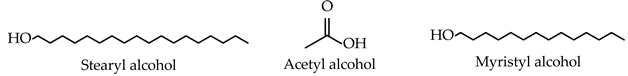 |
| Fatty acids | 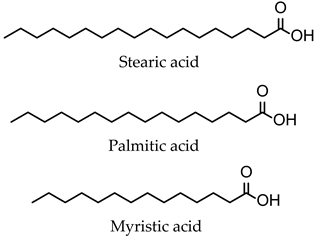 |
| Charged molecules | |
| Positive | 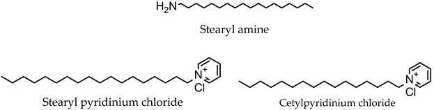 |
| Negative | Diacetyl phosphate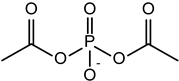 |
| Phosphatidic acid | |
Dihexadecyl phosphate | |
| Hydration medium | |
| Phosphate buffer | |
| Cationic/Helper Lipids | |
2,3-di(tetradecyloxy)propan-1-amine (chloride salt)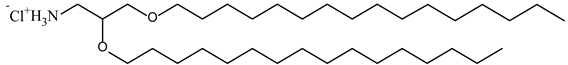 | |
N-(2,3-Dioleoyloxy-1-propyl)-trimethylammonium methyl sulfate (DOTAP methyl sulphate)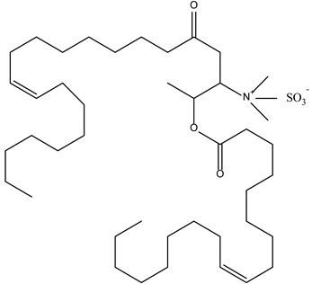 | |
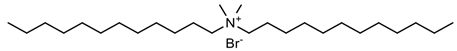 Dimethyl didecyl ammonium bromide | |
| Lipidic Components | |
Cholesterol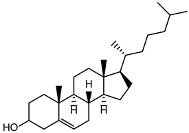 | |
| Type of Cancer | Formulation Method | Drug/Natural Molecules | Composition | Niosomal Formulation Results | References |
|---|---|---|---|---|---|
| Breast cancer | Thin-film hydration | Tamoxifen | Spans (20, 40, 60, 80), cholesterol | Inhibitory effects on cancerous lines: MDA-MB-231, SKBR3 cells; Less IC50 values; Significant downregulation of cyclin D, cyclin E, VEGFR-1, MMP-2, MMP-9 genes and upregulation of caspase-3, caspase-9 genes; Increase caspase activity and apoptosis induction in cancerous cells. | [96] |
| Docetaxel | Span 40, PF108 | AD = 244.9 nm; EE (%) = 97.43 ± 1.2%; PDI = 0.75; ZP = −10 mV; Niosomal formulation improved the Docetaxel stability; Sustainable release during an in vitro drug release study; MCF-7 cells significantly affected; | [97] | ||
| Metformin, Celecoxib | Span 60, cholesterol/ Span 60, cholesterol, Tween 80 | Metformin-loaded niosomes: AD = 110.6 ± 0.6 nm; EE (%) = 68.94 ± 1.28%; RD (%) = 89.2%; PDI = 0.139 ± 0.017; ZP = −44.42 ± 1.990 mV; Celecoxib-loaded niosomes: AD = 96.7 ± 0.7 nm; EE (%) = 94.44 ± 2.09%; RD (%) = 77.80%; PDI = 0.278 ± 0.003; ZP = −53.89 ± 5.680 mV; Metformin-loaded niosomes (62.44% viability) outperformed free Metformin (80.37% viability), showing significantly lower cell viability; free Celecoxib exhibited a viability of 3.18%, while Celecoxib-loaded niosomes showed 1.59% viability; In MDA-MB-231 cells, both Metformin-loaded niosomes and Celecoxib-loaded niosomes showed lower IC10 and IC20 values than their respective free drugs, non-lethal doses; Penetration rate of Metformin-loaded niosomes (85.26%) surpassing free Metformin (61.50%), and the penetration rate of Celecoxib-loaded niosomes (71.08%) compared to free Celecoxib (31.29%). | [98] | ||
| Gemcitabine | Cholesterol, Span 60, Tween 60 | AD = 205 nm; EE (%) = 89.9 ± 1.27%; PDI = 0.19 ± 0.03; RD (%) = 49.7 ± 1.3% after 48 h, while about RD (%) = 87% free Gemcitabine after 4 h; Anticancer activity is superior to free Gemcitabine in treating SH-SY5Y and MCF7 cells during the same incubation period (14.0 and 19.7 ng/mL, respectively); | [99] | ||
| Ascorbic acid, Geranium oil | Cholesterol, Span 60, Tween 60 | AD = 219.4 ± 44.5 nm; EE (%) = 98.3 ± 4.2% (ascorbic acid), 98.7 ± 3.1% (geranium oil); PDI = 0.23 ± 0.20; ZP = −11.1 ± 1.39; IC50 (μg/mL) = 7.69 ± 8; Significantly higher increase apoptotic effect on MCF-7 cells; Antioxidative activity. | [100] | ||
| Curcumin | Span 80, diacetyl phosphate, Cholesterol, Calcium alginate | AD = 167.1 nm; EE (%) = 94.949%; RD (%) = 61.7 ± 1.23%; Greater biocompatibility in cytotoxicity tests than particles without free Curcumin; Enhanced chemotherapy effect due to the alginate. | [101] | ||
| Cisplatin, Epirubicin | Spans, cholesterol, PEG | AD = 192.5 ± 8.9 nm; EE (%) = 91.24 ± 1.32 (Cisplatin), 71.93 ± 1.11% (Epirubicin); RD (%) = 36.78% (Cisplatin), 56.30% (Epirubicin); PDI = 0.142 ± 0.012; Improved stability for two months and continued release in physiological pH; Antitumor activity toward SKBR3 and 4T1 cancer cells; Exhibit lower cytotoxicity toward healthy cells; Significant inhibition of cancer cells’ migration and division than with free drugs. | [102] | ||
| Curcumin, Folic acid | Spans, diacetyl phosphate, cholesterol | AD = 187.13 ± 7.55 nm; EE (%) = 98.2517 ± 0.7851%; PDI = 0.160 ± 0.033; ZP = −8.1 mV; Exhibit higher cellular uptake efficiency in vitro; Induce high apoptosis rate in breast cancer cells (MCF7 and 4T1). | [103] | ||
| Letrozole, Cyclophosphamide, Folic Acid | Span 60, cholesterol | AD = 213.9 ± 3.2 nm; EE (%) = 94.10 ± 1.85% (Cyclophosphamide), 98.50 ± 1.88% (Letrozole); PDI = 0.143 ± 0.007; IC50 values (μg/mL) for MDA-MB-231 = 31.13 ± 1.35 (48 h) and 23.18 ± 1.07 (72 h); IC50 values (μg/mL) for SKBR3 cell = 24.92 ± 1.35 (48 h) and 20.94 ± 1.07 (72 h); Treatment led to a significantly higher increase in Caspase-3, Caspase-9 levels, and a more significant decrease in cyclin-D, Cyclin-E, MMP-2, and MMP-9 expression levels; Increase total apoptosis in treated cancer cell lines. | [104] | ||
| Farnesol, Gingerol | Tween 60, Span 60, cholesterol | AD = 224 ± 14.60 nm; EE (%) = 67.29 ± 1.46% (Gingerol), 92.63 ± 2.57% (Farnesol); PDI = 0.171; Controlled drug release at pH = 7.4; Excellent improved biocompatibility in comparison to free Farnesol and Gingerol; Show significant cytotoxicity toward MCF7, and SKBR3 breast cancer cells; Synergistic inhibitory effect of combined drugs improved chemotherapy; Induce apoptosis in both MCF7 and SKBR3 cell lines. | [105] | ||
| Doxorubicin | Span 60, cholesterol, gelatine, alginate | AD = 226.4 ± 7.95 nm; EE (%) = 73.69 ± 1.68%; PDI = 0.189 ± 0.011; ZP = −13.74 ± 1.49 mV; Excellent biocompatibility with non-tumorigenic breast cells (MCF-10A); High cytotoxicity against breast cancer cells (MCF-7). | [106] | ||
| Cisplatin, Doxorubicin | Span 60, cholesterol | AD = 313.0 ± 9.22 nm; EE (%) = 80.65 ± 1.80% (Doxorubicin), 65.54 ± 1.25% (Cisplatin); PDI = 0.261 ± 0.01; ZP = −30.65 ± 0.64 mV; Higher synergetic inhibitory effect of combined drugs; The caspase activity assay indicated that the cancer cells treated had significantly higher Caspase 3/7 activities compared to uncoated niosomes and free drugs; Higher effective apoptosis induction rate, and cell cycle arrest in cancer cells; | [107] | ||
| Epirubicin, Hyaluronic acid | Span 60, cholesterol | AD = 225.9 nm; EE (%) = 82.1%; PDI = 0.160; CD44-mediated internationalization into breast cancer cells; Improve Epirubicin impact on breast cancer cells, including an increase in cytotoxicity and apoptosis, as well as inhibition of metastasis. | [108] | ||
| Morusin | Span 60, cholesterol | AD = 479 nm; EE (%) = 97 ± 1.25%; PDI = 0.29; ZP = −19.8 mV; Inhibit the survival of MDA-MB-453; Cause considerable toxicity in the cells treated, leading to a decrease in the number of alive cells and an increase in dead cells. | [109] | ||
| Melittin | Span 60, Tween 60, cholesterol | Affects gene expression by downregulating the expression of Bcl2, MMP2, MMP9 genes while upregulating the expression of Bax, Caspase3, Caspase9; Enhanced the apoptosis rate and inhibited cell migration. | [110] | ||
| Paclitaxel | Tween 60, Span 60, ergosterol, cholesterol hemisuccinate | AD = 240 nm; EE (%) = 77.0 ± 2.3%; Show high efficacy against human cancers derived from cervix and breast tumors. | [111] | ||
| Paclitaxel | Span 60, cholesterol | AD = 192.73 ± 5.50 nm; EE (%) = 94.71 ± 1.56%; Significant cytotoxicity on breast cancer cell lines including MCF-7, T-47D, SkBr3, MDA-MB-231 in a time- and dose-dependent manner. | [112] | ||
| Curcumin | Tween 60, Tween 80, cholesterol | AD = 110 ± 0.45 nm; EE (%) = 78.34%; RD (24 h, 37 °C) = 19 ± 0.67%; PDI = 0.21 ± 0.16; ZP = −24 ± 0.34 mV; The presence of both positive charge and niosome promote cellular uptake via changing the penetration mechanism to endocytosis; Reduce the expression of NF-κB and improve the p53 better than their free states. | [113] | ||
| 2,5-Diketopiperazine | Span 60, Tween 60, cholesterol; Tween 40, Span 40, cholesterol | AD = 149.43 ± 3.2 nm; EE (%) = 70.22 ± 0.13%; PDI = 0.171 ± 0.025; Inhibit proliferation and invasion of MCF-7, MDA- MB-231, AU-565 malignant cells in vitro; Breast cancer cells’ proliferation is directly influenced by the presence of niosome-encapsulated BHPPD. | [114] | ||
| Carnosine, Melittin | Span 60, cholesterol | AD = 58 ± 0.50 nm (Carnosine), 163 ± 1.3 nm (Melittin); PDI = 0.16441 ± 0.04 (Carnosine), 0.0424 ± 0.1 (Melittin); ZP = −20 ± 0.3 mV (Carnosine), −86.6 ± 0.9 mV (Melittin); Melittin-loaded niosomes showed significantly greater anticancer activity on breast cancer cells compared to Carnosine-loaded niosomes; Carnosine-loaded niosomes inhibit the cells at the G2/M phase transition in MCF-7 cells and S phase at MDA-MB- 231 cells; Melittin-loaded niosomes inhibit both cells at the G0/1 phase transition and occur inhibition of cells at S phase. | [115] | ||
| Ascorbic acid, Curcumin | Tween 60, Span 60, cholesterol | AD = 224.30 ± 6.52 nm; EE (%) = 74.75 ± 1.35% (Ascorbic acid), 93.19 ± 1.88% (Curcumin); PDI = 0.084 ± 0.012; ZP = −23.7 ± 1.03 mV; Exhibit a higher apoptotic rate and enhance anticancer effects against breast cancer MCF-7 cells. | [116] | ||
| Doxorubicin, Curcumin | Tween 60, Span 60, cholesterol, PEG | AD = 273.1 ± 3.2 nm; PDI = 0.39 ± 0.08; EE (%) = 62.90 ± 1.1% (Doxorubicin), 96.50 ± 3.7% (Curcumin); ZP = −43.2 ± 1.0 mV; IC50 value (μg/mL) on the MCF-7 cell line = 20.7 ± 2.3; Show a more controllable release manner and enhance cytotoxicity on cancer cells after PEGylation. | [117] | ||
| Trastuzumab, Mcl-1 Nioplex | Span 20, cholesterol-based cationic lipids | Exhibit cell-growth inhibition in both HER2-positive and HER2-negative breast cancer cells; Decrease cell survival and promote apoptosis compared to single treatment in HER2-overexpression breast cancer cells. | [118] | ||
| Lung cancer | Thin-film hydration | Nintedanib | Span 60, cholesterol, 1,2-dioleoyl-3-trimethylammonium-propane (DOTAP) | AD = 246.2 ± 2.3 nm; EE (%) = 73.1 ± 2.7%; PDI = 0.19 ± 0.08; ZP = −20.5 ± 1.9 mV; IC50 values in different human non-small-cell lung cancer cell lines: 1.5 ± 0.8 (A549), 1.8 ± 0.3 (H2122), 2.1 ± 0.8 (H1299), 1.1 ± 0.4 (H358), 1.3 ± 0.5 (H460); Incorporation of cationically charged lipid increased drug encapsulation in niosomes along with optimum vesicle size and size distribution; Possess appropriate aerosolization properties for efficient pulmonary delivery; Significant inhibitory action on the metastatic property of NSCLC cells. | [119] |
| Artemisin, Metformin | Span 60, cholesterol | AD = 256 nm; EE (%) = 95%; PDI = 0.202; Increase Bax levels in a dose-dependent manner; Anticancer effect against A549 cancer cells. | [120] | ||
| Metformin, Silibinin | Span 60, cholesterol, PEG | AD = 162.5 ± 1.8 nm; EE (%) = 95%; PDI = 0.424; ZP = −17.7 ± 7 mV; Induce apoptosis and cell cycle arrest in the A549 lung cancer cell line; Significant reduction in expression of hTERT and BCL-2 genes. | [121] | ||
| Sunitinib | Span 60, cholesterol | Triggered apoptosis in in vitro experiments of lung cancer cell lines (A549); Caused downregulation or upregulation of genes associated with apoptosis; | [122] | ||
| Colorectal cancer | Various techniques formulation (thin-film hydration, reverse-phase evaporation, sonication, ethanol injection) | 5-fluorouracil | Span 60, Tween 60, cholesterol | 5-Flurouracil-loaded niosomes displayed a slight decrease in cell viability (reduced cell index) compared to the pure drug. | [123] |
| Thin-film hydration | Oxaliplatin, Paclitaxel | Span 60, Tween 80, TPGS | AD = 285.8 ± 23.5 nm (Oxaliplatin), 258.6 ± 13.3 nm (Paclitaxel); EE (%) = 91.03 ± 2.80% (Oxaliplatin), 93.31 ± 3.31% (Paclitaxel); PDI = 0.295 ± 0.07 (Oxaliplatin), 0.287 ± 0.09 (Paclitaxel); ZP = −33.25 ± 1.41 mV (Oxaliplatin), −32.99 ± 1.08 mV (Paclitaxel); Using vesicular niosomes to administer both drugs altered their release rate in comparison to their free counterparts, as they demonstrated extended drug release; Oxaliplatin and Paclitaxel’s cytotoxicity and apoptosis efficacy were significantly improved by encapsulation into niosome particles compared to the free drugs. | [124] | |
| Curcumin, Saccharomyces cerevisiae | Span 60, cholesterol, PEG | AD = 201 ± 9.94 nm; EE (%) = 88%; PDI = 0.193; ZP = − 17.14 ± 4.8 mV; Show favorable results compared to free curcumin in gene expression, cytotoxicity, apoptosis induction, cell cycle arrest, and invasion rate reduction tests. | [125] | ||
| Silibinin | Span 60, Tween 80, cholesterol | AD = 70 nm; PDI = 0.52; ZP = −19.0 mV; Cytotoxic effects on HT-29 colon cancer cells in a dose- and time- dependent manner; Show accelerated release rate in acidic pH in cancer cells compared to the neutral condition. | [126] | ||
| Prostate cancer | Thin-film hydration followed by bath sonication | Lycopene | Tween 60/Span 60, cholesterol | AD = 136.00 ± 8.83 nm; PDI = 0.460 ± 0.02; ZP = −36.0 ± 3.45 mV; Significantly reduce cell viability for PC-3 and LNCaP cells; Increase antiproliferative and apoptotic effects on PSMA + LNCaP cell; Increase cellular uptake. | [127] |
| Skin cancer | Microfluidic mixing | Hippadine | Span 60, cholesterol | AD = 138.40 ± 1.40 nm; EE (%) = 35.98 ± 0.99%; PDI = 0.15 ± 0.01; ZP = −32.80 ± 2.50 mV; Significantly improve the characteristics of hippadine by increasing its cytotoxic properties; Improve molecule solubility and enhance drug uptake by the cells at a higher rate. | [49] |
| Solvent injection method | Gamma- oryzanol | Span 60, dicetyl phosphate, Carbopol 940 | AD = 196.6 ± 0.9 nm; EE (%) = 78.31%; PDI = 0.268 ± 0.02; ZP = − 41.6 mV; pH niosomal gel = 7.3 ± 0.1; Reduce the frequency of drug administration. | [128] | |
| Thin-film hydration | Amygdalin | Cholesterol, Tween 60, DDP, Carbopol 934 | Show significant antitumor activity compared with oral Tamoxifen; Enhance permeation into deep skin layers. | [129] | |
| Ozonated olive oil | Cholesterol, Span 60, Tween 60 | AD = 125.34 ± 13.29 nm; EE (%) = 87.30 ± 4.95%; PDI = 0.24 ± 0.04; ZP = −11.34 ± 4.71 mV; Ensure sustained release behavior and improve skin permeation; Exert anticancer activity on A375 cells. | [130] |
Disclaimer/Publisher’s Note: The statements, opinions and data contained in all publications are solely those of the individual author(s) and contributor(s) and not of MDPI and/or the editor(s). MDPI and/or the editor(s) disclaim responsibility for any injury to people or property resulting from any ideas, methods, instructions or products referred to in the content. |
© 2024 by the authors. Licensee MDPI, Basel, Switzerland. This article is an open access article distributed under the terms and conditions of the Creative Commons Attribution (CC BY) license (https://creativecommons.org/licenses/by/4.0/).
Share and Cite
Liga, S.; Paul, C.; Moacă, E.-A.; Péter, F. Niosomes: Composition, Formulation Techniques, and Recent Progress as Delivery Systems in Cancer Therapy. Pharmaceutics 2024, 16, 223. https://doi.org/10.3390/pharmaceutics16020223
Liga S, Paul C, Moacă E-A, Péter F. Niosomes: Composition, Formulation Techniques, and Recent Progress as Delivery Systems in Cancer Therapy. Pharmaceutics. 2024; 16(2):223. https://doi.org/10.3390/pharmaceutics16020223
Chicago/Turabian StyleLiga, Sergio, Cristina Paul, Elena-Alina Moacă, and Francisc Péter. 2024. "Niosomes: Composition, Formulation Techniques, and Recent Progress as Delivery Systems in Cancer Therapy" Pharmaceutics 16, no. 2: 223. https://doi.org/10.3390/pharmaceutics16020223
APA StyleLiga, S., Paul, C., Moacă, E.-A., & Péter, F. (2024). Niosomes: Composition, Formulation Techniques, and Recent Progress as Delivery Systems in Cancer Therapy. Pharmaceutics, 16(2), 223. https://doi.org/10.3390/pharmaceutics16020223