
Exploring the Antioxidant Potential of Gellan and Guar Gums in Wound Healing


Abstract
:1. Introduction
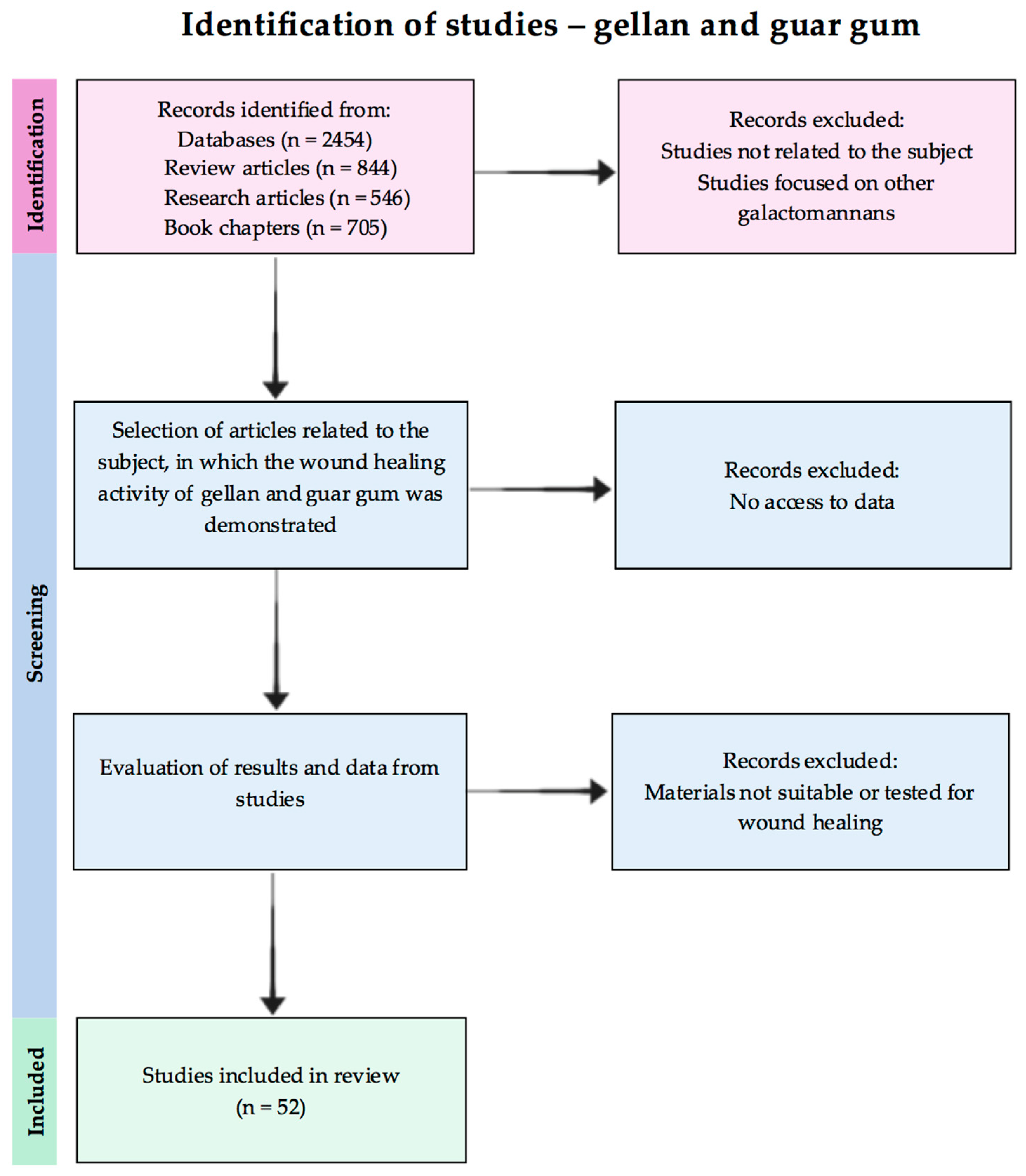
2. Methods
3. Antioxidant Activity Future in Wound Healing
- -
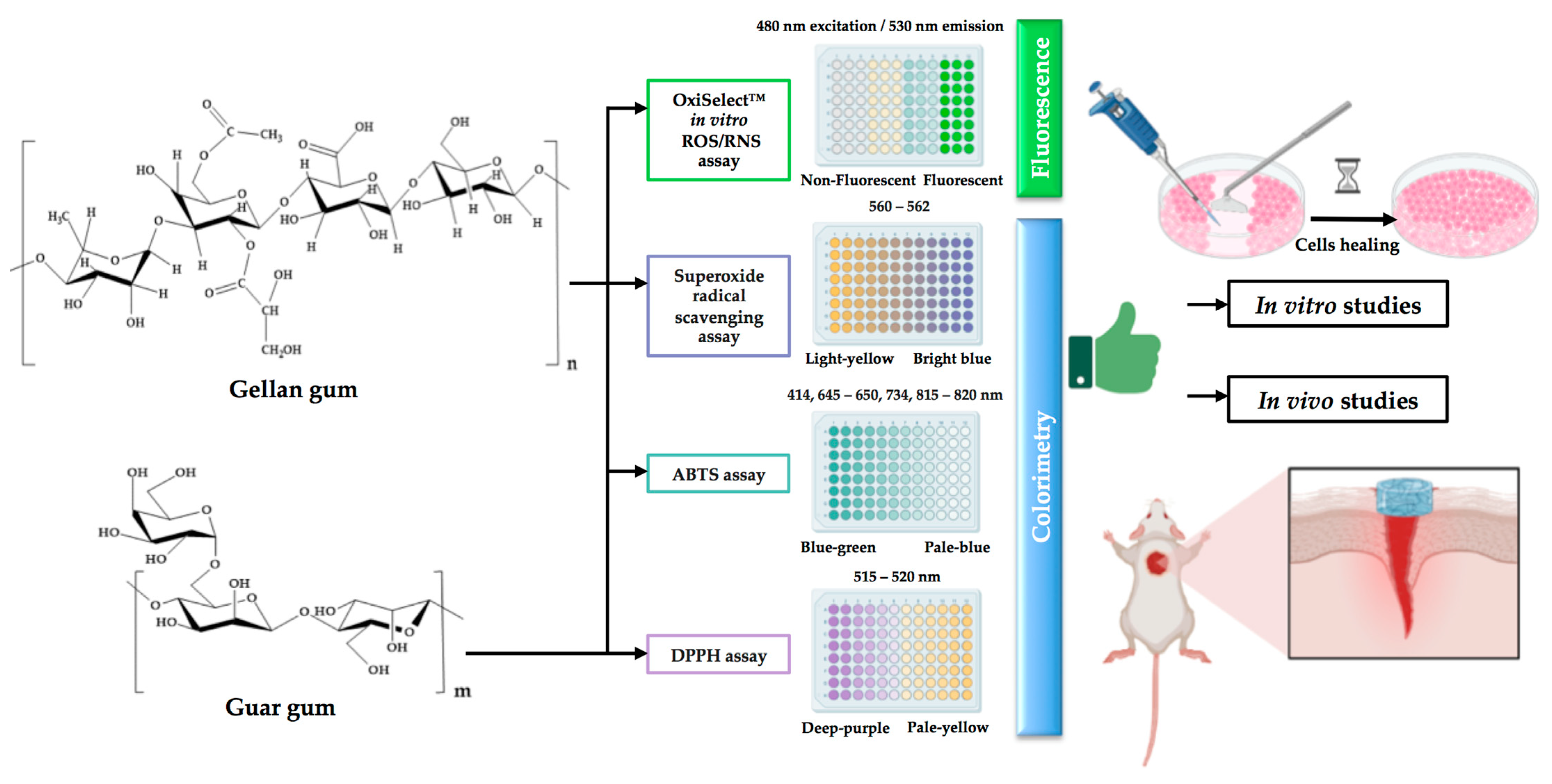
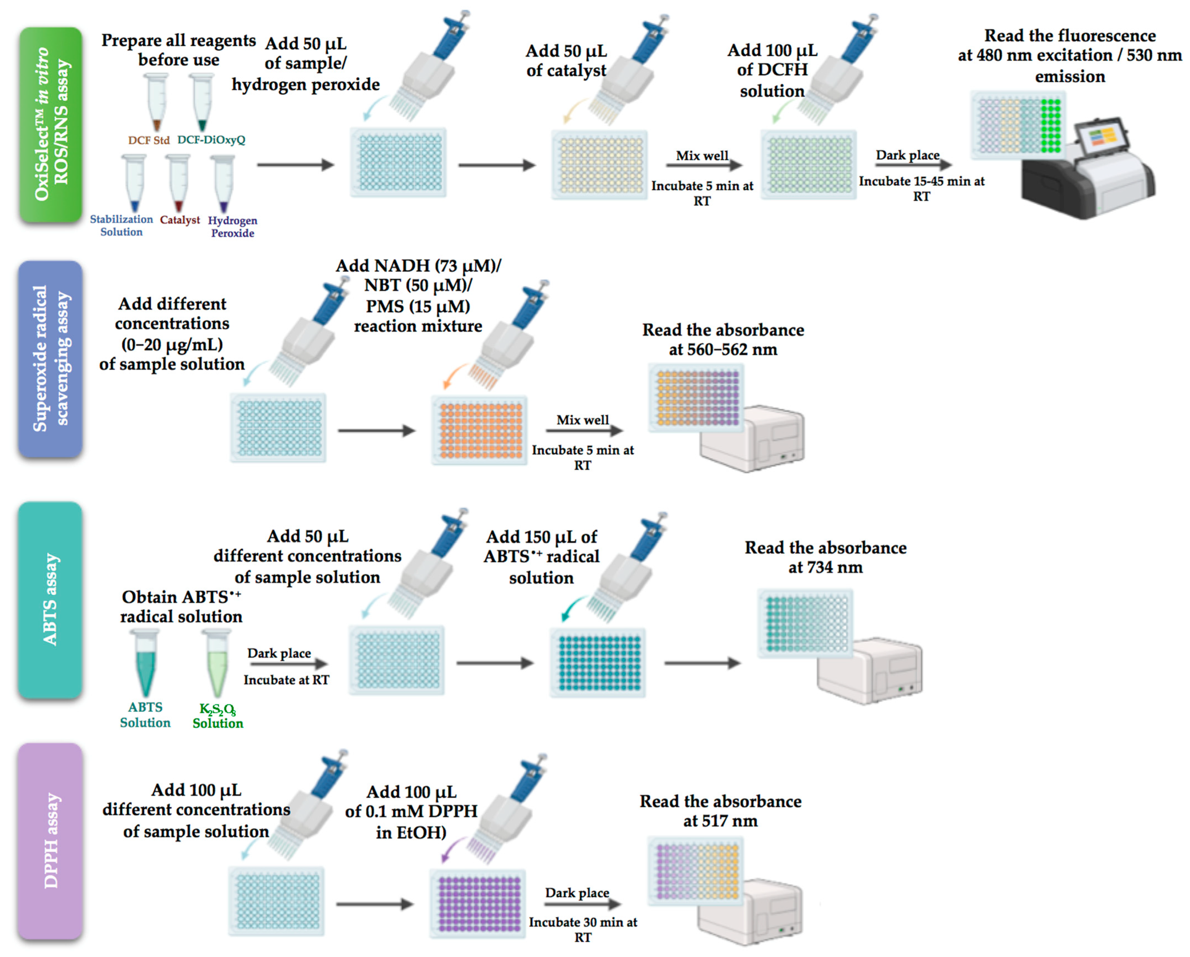
- A [2,2-di(4-tert-octylphenyl)-1-picrylhydrazyl] (DPPH) test measures the antioxidant reaction with free organic radicals, founded on the neutralisation of the DPPH radical due to donated electrons from the antioxidants, with colour change at 517 nm from purple to yellow; the DPPH radical scavenging activity is often expressed using the following equation [23]:where Acontrol and Asample are absorbance values of a control mixture without antioxidant and a mixture containing antioxidant, respectively.The EC50 (the efficient concentration of the antioxidant necessary to reduce the initial DPPH concentration by 50%) and TEC50 (the necessary time to reach the equilibrium state with EC50) values are also used to measure the antioxidant activity of the DPPH scavenging method.
- -
- A Folin–Ciocalteu assay is based on the reductive capacity of antioxidants to determine the TPC in an alkaline state; the Folin–Ciocalteu reagent contains a mixture of phosphomolybdate and phosphotungstate that are reduced to generate a blue colour chromophore when it reacts with phenols with the maximum absorption at 765 nm. This single electron transfer reaction-based method uses gallic acid, catechin, caffeic acid, chlorogenic acid, or ferulic acid as reference standards, and the TPC values are usually given, for example, as gallic acid equivalents (GAE) [24].
- -
- A cupric reducing antioxidant power (CUPRAC) test measures the TAC of the reduction of Cu (II) light blue to Cu (I) yellow-orange by antioxidants at 450 nm; the CUPRAC activity is expressed either as % inhibition or the equivalent of Trolox, gallic acid, ascorbic acid, quercetin, or α-tocopherol, used as standard compounds [25].
- -
- A ferric reducing antioxidant power (FRAP) test measures the antioxidant potential through the reduction of the colourless complex of ferric ions (Fe (III)) to the intensely blue ferrous complex (Fe (II)) with tripyridyltriazine or potassium ferricyanide by antioxidants at 593 nm in acid environments; the activity is expressed as micromolar equivalents of ferrous ions or in relation to a standard antioxidant [26].
- -
- A 2,2′-azinobis-(3-ethylbenzothiazoline-6-sulfonic acid) (ABTS) test measures the relative ability of antioxidants to neutralise the blue-green ABTS stable radical cation generated in the aqueous phase, and quantified at 734 nm; the antioxidant activity is dependent on the degree of discolouration of the blue-green colour [22].
- -
- Thiobarbituric acid reactive substance (TBARS) procedures are centered on the reactivity of malondialdehyde (MDA) with TBA to produce a red colour quantified at 535 nm; the technique first complexes ethylenediaminetetraacetic acid (EDTA) with Fe (II), then interacts with hydrogen peroxide to produce HO• radical, which reacts in the presence of ascorbic acid with the deoxyribose sugar to produce by-products. At low pH values, the mixture forms MDA in combination with TBA, followed by MDA-TBA chromogen. The scavenging activity is measured based on the inhibition of deoxyribose degradation [27].
- -
- A ferrous oxidation xylenol (FOX) test measures the levels of hydrogen peroxide in biological systems by the oxidation of Fe (II) to Fe (III), which is then reacted with xylenol orange (XO) to generate a ferric-XO complex, quantified at 560 nm with a blue-purple colour [28].
- -
- A ferric thiocyanate (FTC) assay also measures the levels of hydrogen peroxide as the ferric ion is converted by an oxidant from a ferrous ion, similar to FOX, but in this case, the ferrous ions are monitored by the thiocyanate complex, quantified at 500 nm with a blood-red colour; the test identifies and quantifies the total phenolic content, lipid oxidation, and flavonoids using mainly gallic acid as a standard [29].
- -
- A β-carotene bleaching assay quantifies pro-oxidants and antioxidant species by measuring the levels of peroxyl radicals that combine with β-carotene to form a stable carotene radical, thus reducing the concentration of β-carotene in a testing solution; if an antioxidant is present in the solution, β-carotene competes for the formation of the carotene radical, followed by the decolourization of the orange-yellow or dark-yellow carotene solution verified at 440 nm [30].
- -
- Superoxide radical scavenging and nitric oxide radical scavenging assays determine both the total oxidant scavenging capacity of antioxidants. First, one measures the bright blue colour of formazan at 560–562 nm produced by nitroblue tetrazolium (NBT) light-yellow salt after it is reduced by oxygen as the tetrazole ring is broken, triggering dismutation. In the second test, nitric oxide radicals are determined by aqueous sodium nitroprusside (SNP) solution, react with oxygen to form nitrite ions, and with sulfanilic acid for diazonium ion, then are linked with N-(1-naphthyl) ethylenediamine (NED) to produce a red azo dye [31].
- -
- The oxygen radical absorption capacity (ORAC) and the hydroxyl radical antioxidant capacity (HORAC) assays rely on the antioxidant reaction with peroxyl radicals induced by 2,2′-azobis-2-amidino-propane (AAPH) and quench OH radicals generated by a Co (II)-based Fenton-like system [32].
- -
- Hydrogen peroxide scavenging and peroxynitrite scavenging activity assays establish the total oxidant scavenging capacity of antioxidants in fluorescence, using horseradish peroxidase (HRP) enzyme and scopoletin or dihydrorhodamine to measure the fluorescence intensity or inhibition [33].
- -
- The total peroxyl radical trapping antioxidant parameter (TRAP) calculates the antioxidant capacity to scavenge luminol-derived radicals, generated from AAPH decomposition in chemiluminescence [34].
- -
- A catalase assay (CAT) demonstrates the presence of catalase, an enzyme that dissociates hydrogen peroxide into molecular oxygen and water; the catalase activity is calculated based on the rate of decomposition of hydrogen peroxide, proportional to the reduction in the absorbance at 240 nm [35].
- -
- Glutathione peroxidase (GSH-Px) catalyses the reduction of hydrogen peroxides with the aid of GSH to produce alcohol, oxidized glutathione, and water; the absorbance level measured by UV-VIS at 340 nm is directly proportional to the GPx activity in the sample [36].
- -
- Superoxide dismutase (SOD) converts the superoxide anion to H2O2, which is a substrate for CAT and GSH-Px; the SOD activity is determined as the inhibition or reduction of water-soluble formazan dye at 450 nm, for the detection of superoxide radicals generated by xanthine oxidase and hypoxanthine [37].
- -
- A GSH content assay involves oxidation of GSH by the sulfhydryl reagent 5,5′-dithio-bis (2-nitrobenzoic acid) to form the yellow derivative 5′-thio-2-nitrobenzoic acid, measurable at 412 nm [38].
4. Gums
4.1. Gellan Gum and Its Derivatives
4.2. Guar Gum and Its Derivatives
4.2.1. Films
4.2.2. Hydrogels and Matrices
4.2.3. Other Types of Formulations

| Formulation Type | Co-Products | Active Ingredient/Drug | Obtaining Method | Studies In Vitro/In Vivo | Properties | References |
|---|---|---|---|---|---|---|
| Film | Guar and gellan gums | Honey | Solvent casting method | DPPH assay, MTT, scratch test on skin fibroblasts | Antioxidant properties, higher honey concentration caused an increased degradation rate, a decreased swelling capacity, and a lower rate of water vapor permeability | [72] |
| Aldehyde guar gum, gelatin, bacterial nanocellulose | Honey | Blending | MTT Assay, Cell attachment study on NIH 3T3 fibroblast cells | Swelling behaviour influenced by the honey concentration, antibacterial activity, improved mechanical properties | [73] | |
| Carboxymethyl guar gum, gelatin | Curcumin | Non-covalent interaction method | MTT Assay on NIH 3T3 fibroblast cell | Hemocompatibility, antibacterial properties | [75] | |
| Film | Guar gum, polyhydroxyalkanoates (PHBV) | Curcumin | Solvent casting method | MTT Assay on NIH 3T3 fibroblast cell In vivo studies on Swiss-albino mice | Hemocompatibility, bactericidal activity, the PHBV content increased film’s porosity, improved mechanical properties | [76] |
| Aminated carboxymethyl guar gum, collagen | Ceftazidime | Cross-linking with ceftazidime | MTT and CalceinAM assays on NIH 3T3 fibroblast | Hemocompatibility and antimicrobial activity | [77] | |
| Carboxymethyl guar gum | Ciprofloxacin | Cross-linking with citric acid | In vivo studies on infected Wistar rats | Hemocompatibility, increasing curing temperature decreased the swelling capacity | [78] | |
| Hydrophobically modified guar gum, polyvinyl alcohol | Gentamicin sulphate/Amoxicillin | Microwave-assisted poly condensation with dimer acid | In vivo studies on Wistar albino rats | Antibacterial and antifungal properties, capability of absorbing wound exudates, biodegradability | [79] | |
| Hydroxypropyl-guar, hyaluronic acid, PEG and polyvinylpyrrolidone | - | Film casting method | In vivo studies on GVHD mouse model (C3H mice with bone marrow and T-cells transplated from BL/6 donor) | Mucoadhesivity, the mechanical properties of the film do not impact further surgery procedures | [80] | |
| Semi-interpenetrating hydrogel | Guar gum, collagen | - | Cross-linking with hydrophilic polyurethane | - | Superabsorbent behaviour, interconnected porosity, potential use as a wound dressing | [81] |
| Micro-porous interpenetrating network | Guar gum, Carrageenan | - | Microwave irradiation and film casting method | In vivo studies on mice | Hemocompatibility, degradability, honeycomb structure, good adhesion to stomach, large intestine, and small intestine | [82] |
| Dual-layer wound dressing | Guar gum, polyvinyl alcohol, hydrocolloid | Lactobacillus plantarum | Freeze-and-thaw method | In vivo studies on Male Sprague–Dawley rats | Antibacterial properties (eradicating P. aeruginosa), efficient in promoting complete re-epithelization | [83] |
| Hydrogel | Guar gum, chitosan, polyvinyl alcohol | Paracetamol | Cross-linking with tetra orthosilicate | - | Antibacterial properties, degradability, responsive to pH, potential use as a wound dressing | [84] |
| Oxidized quaternized guar gum, carboxymethyl chitosan | - | Schiff base reaction | MTT Assay on L929 fibroblast cells In vivo studies on male rats with S. aureus infected wounds | Hemocompatibility, hemostatic properties, injectability, self-healing properties, antibacterial properties | [85] | |
| Guar gum, arabinoxylan | Silver sulfadiazine | Cross-linking with tetraethyl orthosilicate | Neutral Red assay on MC3T3-E1 cell line | Degradability, antibacterial properties | [86] | |
| Cationic guar gum, poly (3,4-ethylenedioxythiophene), poly(styrenesulfonate) | - | Electrostatic interaction | CCK-8 assay on NIH 3T3, MDCK and HAF cell lines In vivo studies on Sprague Dawley rats | Hemolytic activity, self-healable, conductivity, injectability, suitable for wounds located in stretchable parts of the body | [87] | |
| Hydrogel-nanoparticle | Guar gum, curcumine-stabilisez silver nanoparticles | - | Gelation with guar gum, cross-linking with borax | Swelling degree, antimicrobial activity on E. coli, Pseudomonas aeruginosa, and Staphylococcus aureus | Improved degree of swelling, stronger inhibition against Gram-positive bacteria compared to Gram-negative bacteria | [88] |
| In vivo studies on Wistar rats | Superior wound healing and antibacterial action compared to commercial gels (by >40% and 60%, respectively) | [89] | ||||
| Nanobiomaterial matrix | Guar gum alkylamine, silver nanoparticles | - | In situ incorporation | In vivo studies on Wistar rats; primary skin irritation study conducted on albino rabbit | Promote wound healing by modulation of collagen deposition and regulation of keratinocytes and support re-epithelialization | [90] |
| Nanofibrous matrix | Guar gum, polyvinyl alcohol | Acalypha indica, Aristolochia bracteolata, Lawsonia inermis, and Thespesia populne | Electrospinning | MTT an Calcein AM assays on gingival mesenchymal stem cells, in vivo studies on Wistar rats | Exudate absorbability, tissue fully regenerated with minimal scarring | [91] |
| Nanofiber scaffold | Guar gum, reduced graphene oxide, polyvinyl alcohol | - | Scratch assay on 3T3-L1 fibroblast cells, In vivo studies on adult New Zealand rabbits | Oxygen transport to the cells facilitated by the porosity of the material; the moisture content of the wound site is maintained | [92] | |
| Sponge | Xanthan, guar gum/locust bean gum | - | Freeze drying | - | Solid, bioabsorbable | [93] |
| Nanoparticles | Guar gum | - | Acid hydrolysis technique | Scratch assay on NIH 3T3 fibroblast cell line In vivo studies on Balb/c mice with Oxazolon-induced atopic dermatitis | Efficient in alleviating symptoms of atopic dermatitis | [94,95] |
| Dietary fiber | Commercial partially hydrolyzed guar gum (Sunfiber R) | - | - | RhoA pull-down assays and western blot analysis on YAMC cell line, In vivo studies on C57B6 mice | Efficiency in activating ERK1/2 and Rho kinase, enhancing curing of colonic epithelial wounds | [96] |
5. Conclusions
Author Contributions
Funding
Institutional Review Board Statement
Informed Consent Statement
Data Availability Statement
Conflicts of Interest
Abbreviations
References
- Farahani, M.; Shafiee, A. Wound Healing: From Passive to Smart Dressings. Adv. Healthc. Mater. 2021, 10, 2100477. [Google Scholar] [CrossRef] [PubMed]
- Gardikiotis, I.; Cojocaru, F.-D.; Mihai, C.-T.; Balan, V.; Dodi, G. Borrowing the Features of Biopolymers for Emerging Wound Healing Dressings: A Review. Int. J. Mol. Sci. 2022, 23, 8778. [Google Scholar] [CrossRef] [PubMed]
- Nejaddehbashi, F.; Rafiee, Z.; Orazizadeh, M.; Bayati, V.; Hemmati, A.; Hashemitabar, M.; Makvandi, P. Antibacterial and Antioxidant Double-Layered Nanofibrous Mat Promotes Wound Healing in Diabetic Rats. Sci. Rep. 2023, 13, 3166. [Google Scholar] [CrossRef]
- Agyare, C.; Bekoe, E.O.; Boakye, Y.D.; Dapaah, S.O.; Appiah, T.; Bekoe, S.O. Medicinal Plants and Natural Products with Demonstrated Wound Healing Properties. In Wound Healing—New insights into Ancient Challenges; Alexandrescu, V.A., Ed.; InTech: Houston TX, USA, 2016; ISBN 978-953-51-2678-2. [Google Scholar]
- Brainina, K.; Stozhko, N.; Vidrevich, M. Antioxidants: Terminology, Methods, and Future Considerations. Antioxidants 2019, 8, 297. [Google Scholar] [CrossRef] [PubMed]
- Hajhashemi, V.; Vaseghi, G.; Pourfarzam, M.; Abdollahi, A. Are Antioxidants Helpful for Disease Prevention? Res. Pharm. Sci. 2010, 5, 1–8. [Google Scholar]
- Bardia, A.; Tleyjeh, I.M.; Cerhan, J.R.; Sood, A.K.; Limburg, P.J.; Erwin, P.J.; Montori, V.M. Efficacy of Antioxidant Supplementation in Reducing Primary Cancer Incidence and Mortality: Systematic Review and Meta-Analysis. Mayo Clin. Proc. 2008, 83, 23–34. [Google Scholar] [CrossRef]
- Sharifi-Rad, M.; Anil Kumar, N.V.; Zucca, P.; Varoni, E.M.; Dini, L.; Panzarini, E.; Rajkovic, J.; Tsouh Fokou, P.V.; Azzini, E.; Peluso, I.; et al. Lifestyle, Oxidative Stress, and Antioxidants: Back and Forth in the Pathophysiology of Chronic Diseases. Front. Physiol. 2020, 11, 694. [Google Scholar] [CrossRef]
- Rudrapal, M.; Khairnar, S.J.; Khan, J.; Dukhyil, A.B.; Ansari, M.A.; Alomary, M.N.; Alshabrmi, F.M.; Palai, S.; Deb, P.K.; Devi, R. Dietary Polyphenols and Their Role in Oxidative Stress-Induced Human Diseases: Insights into Protective Effects, Antioxidant Potentials and Mechanism(s) of Action. Front. Pharmacol. 2022, 13, 806470. [Google Scholar] [CrossRef]
- Tabriz, A.G.; Douroumis, D. Recent Advances in 3D Printing for Wound Healing: A Systematic Review. J. Drug Deliv. Sci. Technol. 2022, 74, 103564. [Google Scholar] [CrossRef]
- Hajialyani, M.; Tewari, D.; Sobarzo-Sánchez, E.; Nabavi, S.M.; Farzaei, M.H.; Abdollahi, M. Natural Product-Based Nanomedicines for Wound Healing Purposes: Therapeutic Targets and Drug Delivery Systems. IJN 2018, 13, 5023–5043. [Google Scholar] [CrossRef]
- LaVan, F.B.; Hunt, T.K. Oxygen and Wound Healing. Clin. Plast. Surg. 1990, 17, 463–472. [Google Scholar] [CrossRef]
- Hopf, H.W. Development of Subcutaneous Wound Oxygen Measurement in Humans: Contributions of Thomas, K. Hunt, MD. Wound Repair Regen 2003, 11, 424–430. [Google Scholar] [CrossRef]
- Gordillo, G.M.; Sen, C.K. Revisiting the Essential Role of Oxygen in Wound Healing. Am. J. Surg. 2003, 186, 259–263. [Google Scholar] [CrossRef]
- Dunnill, C.; Patton, T.; Brennan, J.; Barrett, J.; Dryden, M.; Cooke, J.; Leaper, D.; Georgopoulos, N.T. Reactive Oxygen Species (ROS) and Wound Healing: The Functional Role of ROS and Emerging ROS-Modulating Technologies for Augmentation of the Healing Process: Reactive Oxygen Species and Wound Healing. Int. Wound J. 2017, 14, 89–96. [Google Scholar] [CrossRef] [PubMed]
- Shen, H.-M.; Pervaiz, S. Reactive Oxygen Species in Cell Fate Decisions. In Essentials of Apoptosis; Dong, Z., Yin, X.-M., Eds.; Humana Press: Totowa, NJ, USA, 2009; pp. 199–221. ISBN 978-1-60327-380-0. [Google Scholar]
- Poljsak, B.; Šuput, D.; Milisav, I. Achieving the Balance between ROS and Antioxidants: When to Use the Synthetic Antioxidants. Oxidative Med. Cell. Longev. 2013, 2013, 956792. [Google Scholar] [CrossRef] [PubMed]
- Rodriguez, P.G.; Felix, F.N.; Woodley, D.T.; Shim, E.K. The Role of Oxygen in Wound Healing: A Review of the Literature. Dermatol. Surg. 2008, 34, 1159–1169. [Google Scholar] [CrossRef]
- Munteanu, I.G.; Apetrei, C. Analytical Methods Used in Determining Antioxidant Activity: A Review. Int. J. Mol. Sci. 2021, 22, 3380. [Google Scholar] [CrossRef] [PubMed]
- Christodoulou, M.C.; Orellana Palacios, J.C.; Hesami, G.; Jafarzadeh, S.; Lorenzo, J.M.; Domínguez, R.; Moreno, A.; Hadidi, M. Spectrophotometric Methods for Measurement of Antioxidant Activity in Food and Pharmaceuticals. Antioxidants 2022, 11, 2213. [Google Scholar] [CrossRef]
- Moharram, H.; Youssef, M. Methods for Determining the Antioxidant Activity: A Review. Alex. J. Fd. Sci. Technol. 2014, 11, 31–42. [Google Scholar]
- Carocho, M.; Ferreira, I.C.F.R. A Review on Antioxidants, Prooxidants and Related Controversy: Natural and Synthetic Compounds, Screening and Analysis Methodologies and Future Perspectives. Food Chem. Toxicol. 2013, 51, 15–25. [Google Scholar] [CrossRef] [PubMed]
- Si Trung, T.; Bao, H.N.D. Physicochemical Properties and Antioxidant Activity of Chitin and Chitosan Prepared from Pacific White Shrimp Waste. Int. J. Carbohydr. Chem. 2015, 2015, 706259. [Google Scholar] [CrossRef]
- Everette, J.D.; Bryant, Q.M.; Green, A.M.; Abbey, Y.A.; Wangila, G.W.; Walker, R.B. Thorough Study of Reactivity of Various Compound Classes toward the Folin−Ciocalteu Reagent. J. Agric. Food Chem. 2010, 58, 8139–8144. [Google Scholar] [CrossRef] [PubMed]
- Amoli, P.; Hadidi, M.; Hasiri, Z.; Rouhafza, A.; Jelyani, A.; Hadian, Z.; Khaneghah, A.; Lorenzo, J. Incorporation of Low Molecular Weight Chitosan in a Low-Fat Beef Burger: Assessment of Technological Quality and Oxidative Stability. Foods 2021, 10, 1959. [Google Scholar] [CrossRef] [PubMed]
- Gulcin, İ. Antioxidants and Antioxidant Methods: An Updated Overview. Arch Toxicol. 2020, 94, 651–715. [Google Scholar] [CrossRef]
- Aguilar Diaz De Leon, J.; Borges, C.R. Evaluation of Oxidative Stress in Biological Samples Using the Thiobarbituric Acid Reactive Substances Assay. JoVE 2020, 159, e61122. [Google Scholar] [CrossRef]
- del Carmen Pinto, M.; Tejeda, A.; Duque, A.L.; Macías, P. Determination of Lipoxygenase Activity in Plant Extracts Using a Modified Ferrous Oxidation−Xylenol Orange Assay. J. Agric. Food Chem. 2007, 55, 5956–5959. [Google Scholar] [CrossRef]
- Sharma, S.; Vig, A.P. Evaluation of in Vitro Antioxidant Properties of Methanol and Aqueous Extracts of Parkinsonia aculeata, L. Leaves. Sci. World J. 2013, 2013, 604865. [Google Scholar] [CrossRef]
- Moon, J.-K.; Shibamoto, T. Antioxidant Assays for Plant and Food Components. J. Agric. Food Chem. 2009, 57, 1655–1666. [Google Scholar] [CrossRef]
- Hazra, B.; Biswas, S.; Mandal, N. Antioxidant and Free Radical Scavenging Activity of Spondias Pinnata. BMC Complement. Altern. Med. 2008, 8, 63. [Google Scholar] [CrossRef]
- Santos-Sánchez, N.F.; Salas-Coronado, R.; Villanueva-Cañongo, C.; Hernández-Carlos, B.; Santos-Sánchez, N.F.; Salas-Coronado, R.; Villanueva-Cañongo, C.; Hernández-Carlos, B. Antioxidant Compounds and Their Antioxidant Mechanism. In Antioxidants; IntechOpen: Houston TX, USA, 2019; ISBN 978-1-78923-920-1. [Google Scholar]
- Bibi Sadeer, N.; Montesano, D.; Albrizio, S.; Zengin, G.; Mahomoodally, M.F. The Versatility of Antioxidant Assays in Food Science and Safety—Chemistry, Applications, Strengths, and Limitations. Antioxidants 2020, 9, 709. [Google Scholar] [CrossRef]
- Apak, R.; Capanoglu, E.; Shahidi, F. Measurement of Antioxidant Activity and Capacity: Recent Trends and Applications; Wiley: Hoboken, NJ, USA, 2017; p. 337. [Google Scholar]
- Li, Y.; Schellhorn, H.E. Rapid Kinetic Microassay for Catalase Activity. J. Biomol. Tech. 2007, 18, 185–187. [Google Scholar] [PubMed]
- Ursini, F.; Maiorino, M. Glutathione Peroxidases. In Encyclopedia of Biological Chemistry; Elsevier: Amsterdam, The Netherlands, 2013; pp. 399–404. ISBN 978-0-12-378631-9. [Google Scholar]
- Prasad, N.; Ramteke, P.; Dholia, N.; Yadav, U.C.S. Therapeutic Interventions to Block Oxidative Stress-Associated Pathologies. In Immunity and Inflammation in Health and Disease; Elsevier: Amsterdam, The Netherlands, 2018; pp. 341–362. ISBN 978-0-12-805417-8. [Google Scholar]
- Rahman, I.; Kode, A.; Biswas, S.K. Assay for Quantitative Determination of Glutathione and Glutathione Disulfide Levels Using Enzymatic Recycling Method. Nat. Protoc. 2006, 1, 3159–3165. [Google Scholar] [CrossRef] [PubMed]
- Rana, V.; Rai, P.; Tiwary, A.K.; Singh, R.S.; Kennedy, J.F.; Knill, C.J. Modified Gums: Approaches and Applications in Drug Delivery. Carbohydr. Polym. 2011, 83, 1031–1047. [Google Scholar] [CrossRef]
- Nagaraja, K.; Rao, K.M.; Krishna Rao, K.S.V.; Riazunnisa, K.; Suresh Reddy, K.V.N. Polysaccharides of Natural Gums-Based Biomedical Devices for Drug Delivery Application. In Smart Nanomaterials in Biomedical Applications; Nanotechnology in the Life Sciences; Kim, J.-C., Alle, M., Husen, A., Eds.; Springer International Publishing: Cham, Switzerland, 2021; pp. 507–554. ISBN 978-3-030-84261-1. [Google Scholar]
- Mano, J.F.; Silva, G.A.; Azevedo, H.S.; Malafaya, P.B.; Sousa, R.A.; Silva, S.S.; Boesel, L.F.; Oliveira, J.M.; Santos, T.C.; Marques, A.P.; et al. Natural Origin Biodegradable Systems in Tissue Engineering and Regenerative Medicine: Present Status and Some Moving Trends. J. R Soc. Interface 2007, 4, 999–1030. [Google Scholar] [CrossRef]
- Mohammadinejad, R.; Kumar, A.; Ranjbar-Mohammadi, M.; Ashrafizadeh, M.; Han, S.S.; Khang, G.; Roveimiab, Z. Recent Advances in Natural Gum-Based Biomaterials for Tissue Engineering and Regenerative Medicine: A Review. Polymers 2020, 12, 176. [Google Scholar] [CrossRef] [PubMed]
- Munir, H.; Bilal, M.; Khan, M.I.; Iqbal, H.M.N. Gums-Based Bionanostructures for Medical Applications. In Polysaccharides; Inamuddin, Ahamed, M.I., Boddula, R., Altalhi, T., Eds.; Wiley: Hoboken, NJ, USA, 2021; pp. 385–398. ISBN 978-1-119-71141-4. [Google Scholar]
- Giavasis, I.; Harvey, L.M.; McNeil, B. Gellan Gum. Crit. Rev. Biotechnol. 2000, 20, 177–211. [Google Scholar] [CrossRef]
- Zia, K.M.; Tabasum, S.; Khan, M.F.; Akram, N.; Akhter, N.; Noreen, A.; Zuber, M. Recent Trends on Gellan Gum Blends with Natural and Synthetic Polymers: A Review. Int. J. Biol. Macromol. 2018, 109, 1068–1087. [Google Scholar] [CrossRef]
- Feketshane, Z.; Alven, S.; Aderibigbe, B.A. Gellan Gum in Wound Dressing Scaffolds. Polymers 2022, 14, 4098. [Google Scholar] [CrossRef]
- Gomes, D.; Batista-Silva, J.P.; Sousa, A.; Passarinha, L.A. Progress and Opportunities in Gellan Gum-Based Materials: A Review of Preparation, Characterization and Emerging Applications. Carbohydr. Polym. 2023, 311, 120782. [Google Scholar] [CrossRef]
- Cerqueira, M.T.; Da Silva, L.P.; Santos, T.C.; Pirraco, R.P.; Correlo, V.M.; Marques, A.P.; Reis, R.L. Human Skin Cell Fractions Fail to Self-Organize Within a Gellan Gum/Hyaluronic Acid Matrix but Positively Influence Early Wound Healing. Tissue Eng. Part A 2014, 20, 1369–1378. [Google Scholar] [CrossRef]
- Da Silva, L.P.; Oliveira, S.; Pirraco, R.P.; Santos, T.C.; Reis, R.L.; Marques, A.P.; Correlo, V.M. Eumelanin-Releasing Spongy-like Hydrogels for Skin Re-Epithelialization Purposes. Biomed. Mater. 2017, 12, 025010. [Google Scholar] [CrossRef]
- Shukla, R.; Kashaw, S.K.; Jain, A.P.; Lodhi, S. Fabrication of Apigenin Loaded Gellan Gum–Chitosan Hydrogels (GGCH-HGs) for Effective Diabetic Wound Healing. Int. J. Biol. Macromol. 2016, 91, 1110–1119. [Google Scholar] [CrossRef]
- Zhang, X.; Pan, Y.; Li, S.; Xing, L.; Du, S.; Yuan, G.; Li, J.; Zhou, T.; Xiong, D.; Tan, H.; et al. Doubly Crosslinked Biodegradable Hydrogels Based on Gellan Gum and Chitosan for Drug Delivery and Wound Dressing. Int. J. Biol. Macromol. 2020, 164, 2204–2214. [Google Scholar] [CrossRef] [PubMed]
- Shanmugapriya, K.; Kim, H.; Kang, H.W. Fucoidan-Loaded Hydrogels Facilitates Wound Healing Using Photodynamic Therapy by in Vitro and in Vivo Evaluation. Carbohydr. Polym. 2020, 247, 116624. [Google Scholar] [CrossRef] [PubMed]
- Fiorica, C.; Palumbo, F.S.; Pitarresi, G.; Biscari, G.; Martorana, A.; Calà, C.; Maida, C.M.; Giammona, G. Ciprofloxacin Releasing Gellan Gum/Polydopamine Based Hydrogels with near Infrared Activated Photothermal Properties. Int. J. Pharm. 2021, 610, 121231. [Google Scholar] [CrossRef] [PubMed]
- Reczyńska-Kolman, K.; Hartman, K.; Kwiecień, K.; Brzychczy-Włoch, M.; Pamuła, E. Composites Based on Gellan Gum, Alginate and Nisin-Enriched Lipid Nanoparticles for the Treatment of Infected Wounds. Int. J. Mol. Sci. 2021, 23, 321. [Google Scholar] [CrossRef] [PubMed]
- Li, Z.; Bratlie, K.M. Fibroblasts Treated with Macrophage Conditioned Medium Results in Phenotypic Shifts and Changes in Collagen Organization. Mater. Sci. Eng. C 2021, 122, 111915. [Google Scholar] [CrossRef]
- Mahmood, H.; Khan, I.U.; Asif, M.; Khan, R.U.; Asghar, S.; Khalid, I.; Khalid, S.H.; Irfan, M.; Rehman, F.; Shahzad, Y.; et al. In Vitro and in Vivo Evaluation of Gellan Gum Hydrogel Films: Assessing the Co Impact of Therapeutic Oils and Ofloxacin on Wound Healing. Int. J. Biol. Macromol. 2021, 166, 483–495. [Google Scholar] [CrossRef]
- Li, W.; Jian, X.; Zou, Y.; Wu, L.; Huang, H.; Li, H.; Hu, D.; Yu, B. The Fabrication of a Gellan Gum-Based Hydrogel Loaded With Magnesium Ions for the Synergistic Promotion of Skin Wound Healing. Front. Bioeng. Biotechnol. 2021, 9, 709679. [Google Scholar] [CrossRef]
- Xu, L.; Chen, Y.; Zhang, P.; Tang, J.; Xue, Y.; Luo, H.; Dai, R.; Jin, J.; Liu, J. 3D Printed Heterogeneous Hybrid Hydrogel Scaffolds for Sequential Tumor Photothermal-Chemotherapy and Wound Healing. Biomater. Sci. 2022, 10, 5648–5661. [Google Scholar] [CrossRef]
- Choi, J.H.; In Kim, S.; Seo, J.S.; Tumursukh, N.-E.; Kim, S.E.; Choe, S.H.; Kim, S.J.; Park, S.; Song, J.E.; Khang, G. Fast Stress Relaxing Gellan Gum That Enhances the Microenvironment and Secreting Function of Bone Mesenchymal Stem Cells. Int. J. Biol. Macromol. 2022, 222, 2144–2157. [Google Scholar] [CrossRef] [PubMed]
- Biscari, G.; Pitarresi, G.; Fiorica, C.; Schillaci, D.; Catania, V.; Palumbo, F.S.; Giammona, G. Near-Infrared Light-Responsive and Antibacterial Injectable Hydrogels with Antioxidant Activity Based on a Dopamine-Functionalized Gellan Gum for Wound Healing. Int. J. Pharm. 2022, 627, 122257. [Google Scholar] [CrossRef] [PubMed]
- Yuan, L.; Jiang, X.; Jiang, M.; Guo, Y.; Liu, Y.; Ming, P.; Li, S.; Zhou, P.; Cai, R.; Yu, K.; et al. Biocompatible Gellan Gum/Sericin Hydrogels Containing Halloysite@polydopamine Nanotubes with Hemostasis and Photothermal Antibacterial Properties for Promoting Infectious Wound Repair. Mater. Des. 2023, 227, 111744. [Google Scholar] [CrossRef]
- Manconi, M.; Manca, M.L.; Caddeo, C.; Valenti, D.; Cencetti, C.; Diez-Sales, O.; Nacher, A.; Mir-Palomo, S.; Terencio, M.C.; Demurtas, D.; et al. Nanodesign of New Self-Assembling Core-Shell Gellan-Transfersomes Loading Baicalin and In Vivo Evaluation of Repair Response in Skin. Nanomed. Nanotechnol. Biol. Med. 2018, 14, 569–579. [Google Scholar] [CrossRef]
- Ismail, N.A.; Amin, K.A.M.; Majid, F.A.A.; Razali, M.H. Gellan Gum Incorporating Titanium Dioxide Nanoparticles Biofilm as Wound Dressing: Physicochemical, Mechanical, Antibacterial Properties and Wound Healing Studies. Mater. Sci. Eng. C 2019, 103, 109770. [Google Scholar] [CrossRef]
- Razali, M.H.; Ismail, N.A.; Mat Amin, K.A. Titanium Dioxide Nanotubes Incorporated Gellan Gum Bio-Nanocomposite Film for Wound Healing: Effect of TiO2 Nanotubes Concentration. Int. J. Biol. Macromol. 2020, 153, 1117–1135. [Google Scholar] [CrossRef] [PubMed]
- Rufato, K.B.; Souza, P.R.; De Oliveira, A.C.; Berton, S.B.R.; Sabino, R.M.; Muniz, E.C.; Popat, K.C.; Radovanovic, E.; Kipper, M.J.; Martins, A.F. Antimicrobial and Cytocompatible Chitosan, N,N,N-Trimethyl Chitosan, and Tanfloc-Based Polyelectrolyte Multilayers on Gellan Gum Films. Int. J. Biol. Macromol. 2021, 183, 727–742. [Google Scholar] [CrossRef]
- Liu, J.; Ismail, N.A.; Yusoff, M.; Razali, M.H. Physicochemical Properties and Antibacterial Activity of Gellan Gum Incorporating Zinc Oxide/Carbon Nanotubes Bionanocomposite Film for Wound Healing. Bioinorg. Chem. Appl. 2022, 2022, 3158404. [Google Scholar] [CrossRef]
- Costa De Oliveira Souza, C.M.; De Souza, C.F.; Mogharbel, B.F.; Irioda, A.C.; Cavichiolo Franco, C.R.; Sierakowski, M.R.; Athayde Teixeira De Carvalho, K. Nanostructured Cellulose–Gellan–Xyloglucan–Lysozyme Dressing Seeded with Mesenchymal Stem Cells for Deep Second-Degree Burn Treatment. IJN 2021, 16, 833–850. [Google Scholar] [CrossRef]
- Ng, J.Y.; Zhu, X.; Mukherjee, D.; Zhang, C.; Hong, S.; Kumar, Y.; Gokhale, R.; Ee, P.L.R. Pristine Gellan Gum–Collagen Interpenetrating Network Hydrogels as Mechanically Enhanced Anti-Inflammatory Biologic Wound Dressings for Burn Wound Therapy. ACS Appl. Bio Mater. 2021, 4, 1470–1482. [Google Scholar] [CrossRef]
- Zhang, Y.; Wu, J.; Yu, K.; Hu, J.; Zhan, X. Preparation and Characterization of Bifunctional Edible Gellan-Polylysine Fiber. Int. J. Biol. Macromol. 2022, 204, 293–299. [Google Scholar] [CrossRef] [PubMed]
- Mudgil, D.; Barak, S.; Khatkar, B.S. Guar Gum: Processing, Properties and Food Applications—A Review. J. Food Sci. Technol. 2014, 51, 409–418. [Google Scholar] [CrossRef] [PubMed]
- Prabaharan, M. Prospective of Guar Gum and Its Derivatives as Controlled Drug Delivery Systems. Int. J. Biol. Macromol. 2011, 49, 117–124. [Google Scholar] [CrossRef] [PubMed]
- Bal-Öztürk, A.; Torkay, G.; İdil, N.; Özkahraman, B.; Özbaş, Z. Gellan Gum/Guar Gum Films Incorporated with Honey as Potential Wound Dressings. Polym. Bull. 2023, 14, 4098. [Google Scholar] [CrossRef]
- Nezhadmokhtari, P.; Asadi, N.; Ghorbani, M.; Bakhshayesh, A.R.D.; Milani, M.; Akbarzadeh, A. Development of a Novel Film Based on Bacterial Nanocellulose Reinforced Gelatin/Guar Gum Containing Honey for Wound Healing Applications. Res. Sq. 2021. [Google Scholar] [CrossRef]
- Guo, S.; DiPietro, L.A. Factors Affecting Wound Healing. J. Dent. Res. 2010, 89, 219–229. [Google Scholar] [CrossRef] [PubMed]
- Manna, P.J.; Mitra, T.; Pramanik, N.; Kavitha, V.; Gnanamani, A.; Kundu, P.P. Potential Use of Curcumin Loaded Carboxymethylated Guar Gum Grafted Gelatin Film for Biomedical Applications. Int. J. Biol. Macromol. 2015, 75, 437–446. [Google Scholar] [CrossRef]
- Pramanik, N.; Mitra, T.; Khamrai, M.; Bhattacharyya, A.; Mukhopadhyay, P.; Gnanamani, A.; Basu, R.K.; Kundu, P.P. Characterization and Evaluation of Curcumin Loaded Guar Gum/Polyhydroxyalkanoates Blend Films for Wound Healing Applications. RSC Adv. 2015, 5, 63489–63501. [Google Scholar] [CrossRef]
- Jana, P.; Mitra, T.; Selvaraj, T.K.R.; Gnanamani, A.; Kundu, P.P. Preparation of Guar Gum Scaffold Film Grafted with Ethylenediamine and Fish Scale Collagen, Cross-Linked with Ceftazidime for Wound Healing Application. Carbohydr. Polym. 2016, 153, 573–581. [Google Scholar] [CrossRef]
- Orsu, P.; Matta, S. Fabrication and Characterization of Carboxymethyl Guar Gum Nanocomposite for Application of Wound Healing. Int. J. Biol. Macromol. 2020, 164, 2267–2276. [Google Scholar] [CrossRef]
- Bajpai, A.; Raj, V. Hydrophobically Modified Guar Gum Films for Wound Dressing. Polym. Bull. 2021, 78, 4109–4128. [Google Scholar] [CrossRef]
- Ketelson, H.A.; Rangarajan, R. Dissolvable Medical Device for Promoting Healing of Wounds. U.S. Patent 0201031A1, 29 June 2023. [Google Scholar]
- Lopéz-Martínez, E.E.; Claudio-Rizo, J.A.; Caldera-Villalobos, M.; Becerra-Rodríguez, J.J.; Cabrera-Munguía, D.A.; Cano-Salazar, L.F.; Betancourt-Galindo, R. Hydrogels for Biomedicine Based on Semi-Interpenetrating Polymeric Networks of Collagen/Guar Gum: Synthesis and Physicochemical Characterization. Macromol. Res. 2022, 30, 375–383. [Google Scholar] [CrossRef]
- Swain, S.; Bal, T. Microwave Irradiated Carrageenan-Guar Gum Micro-Porous IPN: A Novel Material for Isotropic Tissue Scaffolding. Int. J. Polym. Mater. Polym. Biomater. 2019, 68, 796–804. [Google Scholar] [CrossRef]
- Kim, J.S.; Kim, J.; Lee, S.M.; Woo, M.R.; Kim, D.W.; Kim, J.O.; Choi, H.-G.; Jin, S.G. Development of Guar Gum-Based Dual-Layer Wound Dressing Containing Lactobacillus Plantarum: Rapid Recovery and Mechanically Flexibility. Int. J. Biol. Macromol. 2022, 221, 1572–1579. [Google Scholar] [CrossRef] [PubMed]
- Khan, M.U.A.; Iqbal, I.; Ansari, M.N.M.; Razak, S.I.A.; Raza, M.A.; Sajjad, A.; Jabeen, F.; Riduan Mohamad, M.; Jusoh, N. Development of Antibacterial, Degradable and PH-Responsive Chitosan/Guar Gum/Polyvinyl Alcohol Blended Hydrogels for Wound Dressing. Molecules 2021, 26, 5937. [Google Scholar] [CrossRef]
- Yu, X.; Cheng, C.; Peng, X.; Zhang, K.; Yu, X. A Self-Healing and Injectable Oxidized Quaternized Guar Gum/Carboxymethyl Chitosan Hydrogel with Efficient Hemostatic and Antibacterial Properties for Wound Dressing. Colloids Surf. B Biointerfaces 2022, 209, 112207. [Google Scholar] [CrossRef]
- Khan, M.U.A.; Raza, M.A.; Razak, S.I.A.; Abdul Kadir, M.R.; Haider, A.; Shah, S.A.; Mohd Yusof, A.H.; Haider, S.; Shakir, I.; Aftab, S. Novel Functional Antimicrobial and Biocompatible Arabinoxylan/Guar Gum Hydrogel for Skin Wound Dressing Applications. J. Tissue Eng. Regen. Med. 2020, 14, 1488–1501. [Google Scholar] [CrossRef]
- Li, S.; Wang, L.; Zheng, W.; Yang, G.; Jiang, X. Rapid Fabrication of Self-Healing, Conductive, and Injectable Gel as Dressings for Healing Wounds in Stretchable Parts of the Body. Adv. Funct. Mater. 2020, 30, 2002370. [Google Scholar] [CrossRef]
- Talodthaisong, C.; Boonta, W.; Thammawithan, S.; Patramanon, R.; Kamonsutthipaijit, N.; Hutchison, J.A.; Kulchat, S. Composite Guar Gum-Silver Nanoparticle Hydrogels as Self-Healing, Injectable, and Antibacterial Biomaterials. Mater. Today Commun. 2020, 24, 100992. [Google Scholar] [CrossRef]
- Bhubhanil, S.; Talodthaisong, C.; Khongkow, M.; Namdee, K.; Wongchitrat, P.; Yingmema, W.; Hutchison, J.A.; Lapmanee, S.; Kulchat, S. Enhanced Wound Healing Properties of Guar Gum/Curcumin-Stabilized Silver Nanoparticle Hydrogels. Sci. Rep. 2021, 11, 21836. [Google Scholar] [CrossRef]
- Ghosh Auddy, R.; Abdullah, M.F.; Das, S.; Roy, P.; Datta, S.; Mukherjee, A. New Guar Biopolymer Silver Nanocomposites for Wound Healing Applications. BioMed Res. Int. 2013, 2013, 912458. [Google Scholar] [CrossRef] [PubMed]
- Kalachaveedu, M.; Jenifer, P.; Pandian, R.; Arumugam, G. Fabrication and Characterization of Herbal Drug Enriched Guar Galactomannan Based Nanofibrous Mats Seeded with GMSC’s for Wound Healing Applications. Int. J. Biol. Macromol. 2020, 148, 737–749. [Google Scholar] [CrossRef] [PubMed]
- Koyyada, A.; Orsu, P. Nanofibrous Scaffolds of Carboxymethyl Guargum Potentiated with Reduced Graphene Oxide for in Vitro and in Vivo Wound Healing Applications. Int. J. Pharm. 2021, 607, 121035. [Google Scholar] [CrossRef] [PubMed]
- Haynes, C.A.; Lorimer, E. Solid Polysaccharide Materials for Use as Wound Dressings. U.S. Patent 6309661B1, 30 October 2001. [Google Scholar]
- Ghosh, S.K.; Abdullah, F.; Mukherjee, A. Fabrication and Fluorescent Labeling of Guar Gum Nanoparticles in a Surfactant Free Aqueous Environment. Mater. Sci. Eng. C 2015, 46, 521–529. [Google Scholar] [CrossRef]
- Ghosh, N.; Mitra, S.; Banerjee, E.R. Therapeutic Effects of Topically-Administered Guar Gum Nanoparticles in Oxazolone-Induced Atopic Dermatitis in Mice. Biomed. Res. Ther. 2018, 5, 2305–2325. [Google Scholar] [CrossRef]
- Horii, Y.; Uchiyama, K.; Toyokawa, Y.; Hotta, Y.; Tanaka, M.; Yasukawa, Z.; Tokunaga, M.; Okubo, T.; Mizushima, K.; Higashimura, Y.; et al. Partially Hydrolyzed Guar Gum Enhances Colonic Epithelial Wound Healing via Activation of RhoA and ERK1/2. Food Funct. 2016, 7, 3176–3183. [Google Scholar] [CrossRef]
- Sharma, A.; Khanna, S.; Kaur, G.; Singh, I. Medicinal Plants and Their Components for Wound Healing Applications. Futur J. Pharm. Sci. 2021, 7, 53. [Google Scholar] [CrossRef]

| Formulation Type | Co-Products | Active Ingredient/Drug | Obtaining Method | Studies In Vitro/In Vivo | Properties | References |
|---|---|---|---|---|---|---|
| Spongy-like hydrogel | Hyaluronic acid/Ca2+ | Human dermal/epidermal cell fractions | Ionic cross-linking | In vivo mice wound healing | Accelerated rate of wound closure, re-epithelization, and tissue neovascularization | [48] |
| Calcium chloride (CaCl2) | Eumelanin | Ionic cross-linking | Oxi-Select in vitro ROS and reactive nitrogen species (RNS) assay, MTS assay, in vivo host reaction in mice | Sustained release of eumelanin and antioxidant property | [49] | |
| Hydrogel | Chitosan/PEG | Apigenin | Covalent cross-linking | In vivo rat diabetic wound healing, collagen content, protein level and granuloma weight measurement, biochemical parameters, SOD, GSH, and catalase for antioxidant levels | Biocompatibility, biodegradability, entrapment and sustained release of apigenin, moist nature, enhanced rate of wound contraction and reduced healing time, antioxidant effectiveness | [50] |
| Carboxymethyl chitosan | Bovine serum albumin (BSA)/tetracycline hydrochloride/silver sulfadiazine | Schiff-base cross-linking | In vitro antibacterial activity | Sustained release of drugs and strong antibacterial activity | [51] | |
| Fucoidan/alginate gum/carboxymethyl cellulose | Chlorin-e6 | Chemical cross-linking | In vitro cytotoxicity (MTT and LDH) and cell proliferation assay (BrdU), determination of ROS production (DPPH assay), evaluation of singlet oxygen (DPBF assay), in vitro wound healing (scratch assay), in vivo mice wound healing | Excellent biocompatibility, antioxidant property, and accelerated rate of wound closure | [52] | |
| 4 arm-PEG-Vinylsulfone/polydopamine | Ciprofloxacin | Michael addition | In vitro cytotoxicity (MTS) and live/dead assay, in vitro antimicrobial activity | Antimicrobial activity | [53] | |
| Sodium alginate/CaCl2 | Nisin | Ionic cross-linking | In vitro cytotoxicity (alamarBlue) and live/dead staining assay, in vitro antimicrobial activity | Cytocompatibility and antimicrobial activity | [54] | |
| Methacrylate anhydride | - | Thiol-ene photoclick chemistry | In vitro live/dead assay, immunocytochemistry analysis (α-SMA and focal adhesion staining) | Suitable for controlling scar tissue formation | [55] | |
| Propylene glycol/CaCl2 | Ofloxacin/lavender oil/tea tree oil | Solvent casting ionotropic gelation | In vitro antibacterial activity, DPPH radical scavenging assay, in vivo rat wound healing | Potent antibacterial activity, accelerated rate of wound closure, and re-epithelization | [56] | |
| Polyacrylamide/Mg2+ | - | Free radical polymerization | In vitro cytotoxicity assay (MTT), Western blot assays, in vivo rat burn wound model | Good biocompatibility and accelerated rate of wound healing | [57] | |
| Sodium alginate/Ca2+/polydopamine | Doxorubicin | 3D printing | In vitro cytotoxicity (MTT) and live/dead staining assay, in vivo mice wound healing | Good cytocompatibility and promoted wound healing following surgery | [58] | |
| Ca2+ | Human-derived bone mesenchymal stem cell | Ionic crosslinking and guest-host interaction | In vitro cytotoxicity (MTT) and live/dead staining assay, in vitro wound healing (scratch assay), ELISA assay (IGF-1, HGF, TGF-β1, and VEGF) | Enhanced cell growth and promising candidate for regenerative medicine | [59] | |
| PEG/polydopamine | Colistin | Electrostatic interaction | In vitro antioxidant activity (DPPH and ABTS•+ radical scavenging assay), in vitro cytotoxicity (MTS), and live/dead staining assay, in vitro antibacterial activity | Excellent cytocompatibility and antioxidant activity | [60] | |
| Sericin/halloysite/polydopamine | - | Co-blending | In vitro antibacterial activity, cytotoxicity (CCK-8), and live/dead staining assay, in vivo bacteria-infected wound healing in rat | Rapid hemostasis and promoted infected wound repair | [61] | |
| Nanohydrogel | Cholesterol | Baicalin | Self-assembly | In vivo mice anti-inflammatory and skin repair tests | Effective delivery system for the prevention and treatment of inflamed and/or damaged skin | [62] |
| Film | CaCl2/titanium dioxide (TiO2) | - | Evaporative casting | In vitro antibacterial activity and wound healing, in vivo rat wound healing | Enhanced antibacterial properties, cell viability and cell proliferation, accelerated rate of wound healing | [63] |
| TiO2 | - | Evaporative casting | In vitro antibacterial activity and cytotoxicity assay (MTT), in vivo rat wound healing | Antibacterial activity, good biocompatibility, and accelerated rate of wound closure | [64] | |
| Chitosan/N,N,N-trimethyl chitosan/iota-carrageenan | Tanfloc | Solvent evaporation | Bactericidal and anti-adhesive assays, in vitro cytotoxicity assay (alamarBlue) | Antimicrobial and cytocompatibility properties | [65] | |
| Zinc oxide/carbon nanotubes | - | Solvent casting | In vitro antibacterial activity, in vivo rat wound healing | Good antibacterial activity, promoted complete wound healing | [66] | |
| Membrane | Bacterial cellulose/xyloglucan/lysozyme/mesenchymal stem cell | - | Dry-cast process | In vitro cytotoxicity assay (MTT), in vivo rat burn wound healing, immunocytochemistry assays (BrdU and DAPI) | Reduced acute/chronic inflammatory infiltrates, accelerated rate of wound healing, and re-epithelization | [67] |
| Full-IPN hydrogel | Type-1 collagen/magnesium chloride (MgCl2)/adipose-derived mesenchymal stem cell | - | Ionic cross-linking | In vitro cytotoxicity assay (MTS) and wound healing (scratch assay), in vivo murine burn wound model, ELISA assay (human TSG-6) | Enhanced early wound closure, reducing inflammation, and promoting complete skin regeneration | [68] |
| Fiber | ε-poly-L-lysine/glucose/soybean peptide/fish collagen peptide | - | Wet spinning | In vitro antibacterial activity and cytotoxicity (MTT), In vivo rat wound healing | Good cytocompatibility and antibacterial activity, effectively promoted wound healing | [69] |
Disclaimer/Publisher’s Note: The statements, opinions and data contained in all publications are solely those of the individual author(s) and contributor(s) and not of MDPI and/or the editor(s). MDPI and/or the editor(s) disclaim responsibility for any injury to people or property resulting from any ideas, methods, instructions or products referred to in the content. |
© 2023 by the authors. Licensee MDPI, Basel, Switzerland. This article is an open access article distributed under the terms and conditions of the Creative Commons Attribution (CC BY) license (https://creativecommons.org/licenses/by/4.0/).
Share and Cite
Dodi, G.; Sabau, R.E.; Crețu, B.E.-B.; Gardikiotis, I. Exploring the Antioxidant Potential of Gellan and Guar Gums in Wound Healing. Pharmaceutics 2023, 15, 2152. https://doi.org/10.3390/pharmaceutics15082152
Dodi G, Sabau RE, Crețu BE-B, Gardikiotis I. Exploring the Antioxidant Potential of Gellan and Guar Gums in Wound Healing. Pharmaceutics. 2023; 15(8):2152. https://doi.org/10.3390/pharmaceutics15082152
Chicago/Turabian StyleDodi, Gianina, Rosina E. Sabau, Bianca E.-B. Crețu, and Ioannis Gardikiotis. 2023. "Exploring the Antioxidant Potential of Gellan and Guar Gums in Wound Healing" Pharmaceutics 15, no. 8: 2152. https://doi.org/10.3390/pharmaceutics15082152
APA StyleDodi, G., Sabau, R. E., Crețu, B. E.-B., & Gardikiotis, I. (2023). Exploring the Antioxidant Potential of Gellan and Guar Gums in Wound Healing. Pharmaceutics, 15(8), 2152. https://doi.org/10.3390/pharmaceutics15082152