Review

Biopolymer-Based Nanosystems for siRNA Drug Delivery to Solid Tumors including Breast Cancer

by
Md Abdus Subhan
1,* and
Vladimir P. Torchilin
2,3,*
1
Department of Chemistry, ShahJalal University of Science and Technology, Sylhet 3114, Bangladesh
2
CPBN, Department of Pharmaceutical Sciences, North Eastern University, Boston, MA 02115, USA
3
Department of Chemical Engineering, North Eastern University, Boston, MA 02115, USA
*
Authors to whom correspondence should be addressed.
Pharmaceutics 2023, 15(1), 153; https://doi.org/10.3390/pharmaceutics15010153
Submission received: 8 December 2022 / Revised: 28 December 2022 / Accepted: 28 December 2022 / Published: 1 January 2023
(This article belongs to the Special Issue Biopolymer-Based Nanosystem for Drug Delivery)

Abstract

:
Nanobiopolymers such as chitosan, gelatin, hyaluronic acid, polyglutamic acid, lipids, peptides, exosomes, etc., delivery systems have prospects to help overwhelmed physiological difficulties allied with the delivery of siRNA drugs to solid tumors, including breast cancer cells. Nanobiopolymers have favorable stimuli-responsive properties and therefore can be utilized to improve siRNA delivery platforms to undruggable MDR metastatic cancer cells. These biopolymeric siRNA drugs can shield drugs from pH degradation, extracellular trafficking, and nontargeted binding sites and are consequently suitable for drug internalization in a controlled-release fashion. In this review, the utilization of numerous biopolymeric compounds such as siRNA drug delivery systems for MDR solid tumors, including breast cancers, will be discussed.

1. Introduction

Biopolymers are promising to apply for drug delivery as well as therapeutic agents for cancer therapy. Biopolymeric materials demonstrate enhanced biocompatibility as well as siRNA delivery competence, targeting varieties of cancer genes in solid tumors in vitro and in vivo. Both natural and synthetic biopolymers are effective vehicles for siRNA drug delivery to cancer cells. Biopolymers, such as alginate, pullulan, cellulose, polylactic acid, chitosan, and exosomes have been efficiently utilized in siRNA drug delivery to solid tumors. Further, siRNA therapeutics have been meticulously explored and have been translated to the clinic through lipid NPs [1,2]. However, there are several pitfalls in siRNA delivery proficiency and targeting. Cationic lipids have toxicity since they can interact with enzymes [3,4]. Cationic lipids can interact with RNA and enable encapsulation; however, they can also be toxic [5]. A high concentration of cationic lipids can destroy the cellular membrane, causing cell lysis and necrotic death [3,4]. To overcome these shortcomings, different polymer-based nanoparticle carriers such as biopolymers, exosomes, and peptide nanoassemblies have been utilized for efficient siRNA delivery approaches [6]. Exosomes are extracellular vesicles, and they are mediators of short- and long-distance intercellular communication in health and diseases. Exosomes are promising platforms for siRNA delivery due to their nonimmunogenic and nontoxic natures. Exosomes can be extracted from different biological sources, such as cells and tissues.
Many natural polysaccharide polymers are utilized in siRNA delivery [7]. The key advantage of polysaccharides is the presence of various functional groups suitable for functionalization to achieve structural variety and polymers [8]. The most frequently utilized polysaccharide for siRNA delivery is chitosan. The primary amino groups in chitosan make it a polycation that facilitates the association of siRNA and complex formation [9]. For increasing the solubility of chitosan, numerous alterations have been performed, and water-soluble chitololigosaccarides have been fabricated. These chitololigosaccarides have been utilized as siRNA delivery vehicles [10].
Collagen is another biocompatible, safe natural biopolymer, which is suitable for drug delivery. For one study, siRNA-loaded collagen formulations were fabricated and utilized as localized and sustained-released platforms of siRNA in vivo to solid tumors with promising outcomes [7,11].
Biopolymers such as HA or chitosan are utilized in drug delivery as siRNA vehicles. These biomaterials are manageable via numerous alterations. PEI, aminopropyl triethoxysilane (APTES), and chitosan are cationic polymers used for coating nanoparticles for packing siRNA through electrostatic attractions [12,13,14]. APTES is frequently utilized for amine functionalization of mesoporous silica nanoparticles (MSNs) [14]. Chitosan is an amine-rich natural polysaccharide, which is an excellent biopolymer for siRNA delivery [15]. Several anticancer drugs have been delivered by the chitosan–MSN system to tumors, including breast tumors [16,17,18]. One study reported on chitosan-functionalized MSN for gene delivery with EPR (enhanced permeability and retention) uptake in HeLa cells [17].
Chitosan acts as an siRNA delivery vehicle due to its exclusive benefits, cationic charge, and simple alteration [1,19]. The key drawback of using chitosan-based polymers as cancer drug carriers is the low transfection competence and endosomal release. Chitosan-based polymeric NPs for siRNA delivery demonstrated improved transfection efficiencies by controlling some features such as length, charge, etc. [20]. To enhance the transfection efficiency of chitosan NPs, an ionic gelatin method has been developed [21]. The incorporation of heparin into chitosan nanoparticles increased transfection efficiency. The efficiency of siRNA-induced silencing of VEGF of ARPE-19 cells was 25% more with heparin compared to chitosan nanoparticles without heparin [22].
Recently, the US-FDA has approved four siRNA-based drugs: patisiran, givosiran, lumasiran, and inclisiran. For this reason, siRNA drugs have received increased attention of researchers. Biopolymers are auspicious as siRNA drug delivery platforms as well as therapeutic agents for effective cancer therapy.

2. Sources of Biopolymers Utilized as Drug Delivery Vehicles

Biopolymers or green polymeric materials are promising to apply in drug delivery as well as therapeutic agents for cancer therapy [23,24]. Biopolymers including alginate, pullulan, cellulose, polylactic acid, chitosan, and exosomes have been efficiently utilized in siRNA drug delivery to cancer cells. These biopolymers can be achieved from different sources such as algae, fungi, plants, animals, microbes, etc. (Figure 1). However, exosomes are obtained from tissues, biological fluids, and conditioned media of cells in culture. Biopolymers have gained a lot of attention in the advancement of innovative anticancer drug delivery platforms. Biopolymer-based NPs can be developed in many ways. These biopolymeric NPs have been established as suitable platforms for efficient drug delivery with enhanced selectivity and low toxicity.
Biopolymers are usually composed of non-toxic, safe monomers, and they are carbon neutral for the environment. Biomass morphology is generally structured as nanomaterials. As a result, biopolymers enable the synthesis of biocompatible nanomaterials [23,24]. Chitosan, starch, dextran, and cellulose are polysaccharides derived from plants and microbial biofilms; these biopolymers are frequently utilized for the synthesis of nanomaterials. Biopolymers are unique in composition and possess various important physico-chemical and physiological properties suitable for drug delivery. Further, biopolymeric nanomaterials can be formed by attaching metals, metal oxides, and metal sulfides to biopolymers [25,26]. Biopolymers can form molecular capsules by intramolecular hydrogen bonding. For example, starch can form polymeric nanocomposites by incorporating metals or metal oxides. Further, chitosan and derivatives are frequently used for producing biocompatible nanoparticles for drug delivery. Nanomaterials can be incorporated in biopolymers by both sorption and impregnation [23,24,27].

3. Biopolymer-Based Nanocarrier for siRNA Drug Delivery to Solid Tumors

The importance of effective cancer therapy is emerging increasingly [28]. Due to the feasibility of polymer functionalization, enhanced targeting effects have been observed with polymeric NPs decorated with different peptides or ligands. Chitosan-based NPs were conjugated with HAD (hyaluronic acid dialdehyde) to fabricate an siRNA nanoplatform, siRNA@chitosan-HAD, for the delivery of siRNA targeting CD44 [29]. A natural biopolymer, curdlan, was modified, and represented as CuVa polymers it demonstrated enhanced biocompatibility as well as siRNA delivery competence targeting the STAT3 gene in solid tumors in vitro [30]. Exosomes result from extracellular vesicles formed in the cells [31]. They are prospective candidates for siRNA delivery, as they are easy to handle, nonimmunogenic, and nontoxic [32,33]. Bovine milk exosomes are used as vehicles for siRNA delivery to tumors. siRNA delivery via milk exosomes silenced major oncogenes with 2- to 10-fold knockdown in several solid cancers in vitro and in vivo [33]. Exosomes with a targeted approach (e.g., CD47 targeting) have shown promising outcomes in solid tumors of deadly cancers, including GBM (glioblastoma), PDAC (pancreatic ductal adenocarcinoma), and TNBC (triple-negative breast cancer) [33,34,35]. The angiopep-2 (An2)-functionalized STST3siRNA-loaded exosome (Exo-An2-siRNA) is a potential drug to enhance the treatment of solid tumors. Exo-An2-siRNA demonstrated excellent blood stability, efficient cellular uptake, and effective BBB penetration ability in GBM patients. Exo-An2-siRNA exhibited enhanced in vitro anti-cancer effects, as exosomes protect siRNA, and modified An2 targets tumors effectively. Exo-An2-siRNA showed inhibition of proliferation in a xenograft model of GBM with no side effects and enhancing median survival time. The developed nanoplatform Exo-An2-siRNA is a promising, safe therapy approach for solid tumors targeting the STAT3 gene. This approach may be effective in breast cancer therapy [35].
A method was developed to utilize serum-derived exosomes as drug delivery carriers for siRNA drugs in vivo. The study used exosomes as vehicles for siRNA delivery to lung macrophages in vivo; exosomes containing siRNA exerted functional roles in the lung in vivo. The serum-derived exosomes are non-toxic and do not activate immune responses and were found to be safe for in vivo use [36].
Further, bovine milk exosomes were utilized as an innovative siRNA delivery platform targeting specific genes including VEGF, EGFR, AKT, MAPK, BCl2, SUR, and KRAS [33]. A 5′−32 P-labeled siKRAS was used as a tracer for evaluating exosome loading with siRNAs. The study exhibited the knockdown of target gene expression ranging from 2- to 10-fold in different cancers by exosome-loaded siRNAs. Since KRAS has been correlated with different solid cancers, siKRAS delivery to different solid tumors may be a promising siRNA therapeutic approach for solid tumor therapy. In this study, siKRASG12S was used to target KRAS in lung cancer cells. Extracellular vehicles, particularly exosomes derived from milk used as carriers for siRNA-loading and delivery signify a feasible natural nanocarrier for siRNA delivery to cancer cells utilizing systemic or oral delivery platforms [33,37,38].
The cellular membrane (~5 nm) can be passed by cell-penetrating peptides (CPPs), 5–30 residue peptides [39]. Peptides are important siRNA drug carriers. In particular, CPPs are short cationic peptides that facilitate intracellular delivery of several biological cargos, including siRNA and mRNA [40].
Amphiphilic phospholipid peptide dendrimers (AmPPDs) were fabricated as an efficient siRNA delivery platform. A DSPE-KK2-AmPPDsiRNA delivery system mediated the effective intracellular uptake and endosomal release of siRNA complexes and resulted in gene knockdown, demonstrating anticancer efficiency both in vitro and in vivo in solid tumors [41]. A peptide-based p5RHH NPs-condensing oligonucleotide was developed for siRNA delivery, which reduced the KRAS gene in solid tumors in vivo, demonstrating cancer inhibition effectively [42].
Introduction of protein and mAbs into a polymeric vehicle can advance the siRNA delivery to tumors [43,44,45]. A nanosystem composed of a cyclodextrin-containing polycation, an adamantane-conjugated PEG (AD-PEG), human transferrin-decorated AD-PEG, and therapeutic siRNA was fabricated. The hTf receptor is overexpressed on tumor cells. The developed nanosystem successfully delivered siRNA to tumor cells through hTf receptor-mediated endocytosis, and it downregulated the target genes in solid tumors. The nanocaplet transferred siRNA into the cytoplasm and caused gene silencing through RNAi. The targeted NPs also showed deep tissue penetration due to transferrin-stimulated transcytosis [46]. A 3D spheroid study with Hep3B cancer cells exhibited a transferrin conjugated nanosystem delivered siRNA effectively into the spheroid with a depth of 70 μm, indicating Tf-mediated deep tissue permeation, gene knockdown, and cancer cell inhibition [43,46]. In this study, the deep tissue penetration of the nanocaplet was evaluated by confocal laser scanning microscopy (CLSM, λext = 488) using siRNAA488 fluorescently labeled with Alexa Flour 488 in the nanodrugs. Since the brain endothelial cells overexpress Tf-receptors, the nanocaplet may have the potential to overcome the blood–brain barrier. Thus, an in vivo study utilizing the nanocaplet may be suitable for delivering siRNA to brain tissues, such as GBM.
Poly-L-arginine and siRNA were consecutively adsorbed on an anionic liposome NP, and the outermost layer was decorated with propargyl-modified NPs with 1,2-distearoyl-sn-glycero-3-phosphoethanolamine (DSPE), DSPE-PEG, and cholesterol to fabricate long blood-circulating materials [43,47]. Such coformulation of cationic polymers with lipid improved the polyplex stability and enhanced siRNA loading efficacy and steadiness [48,49,50].
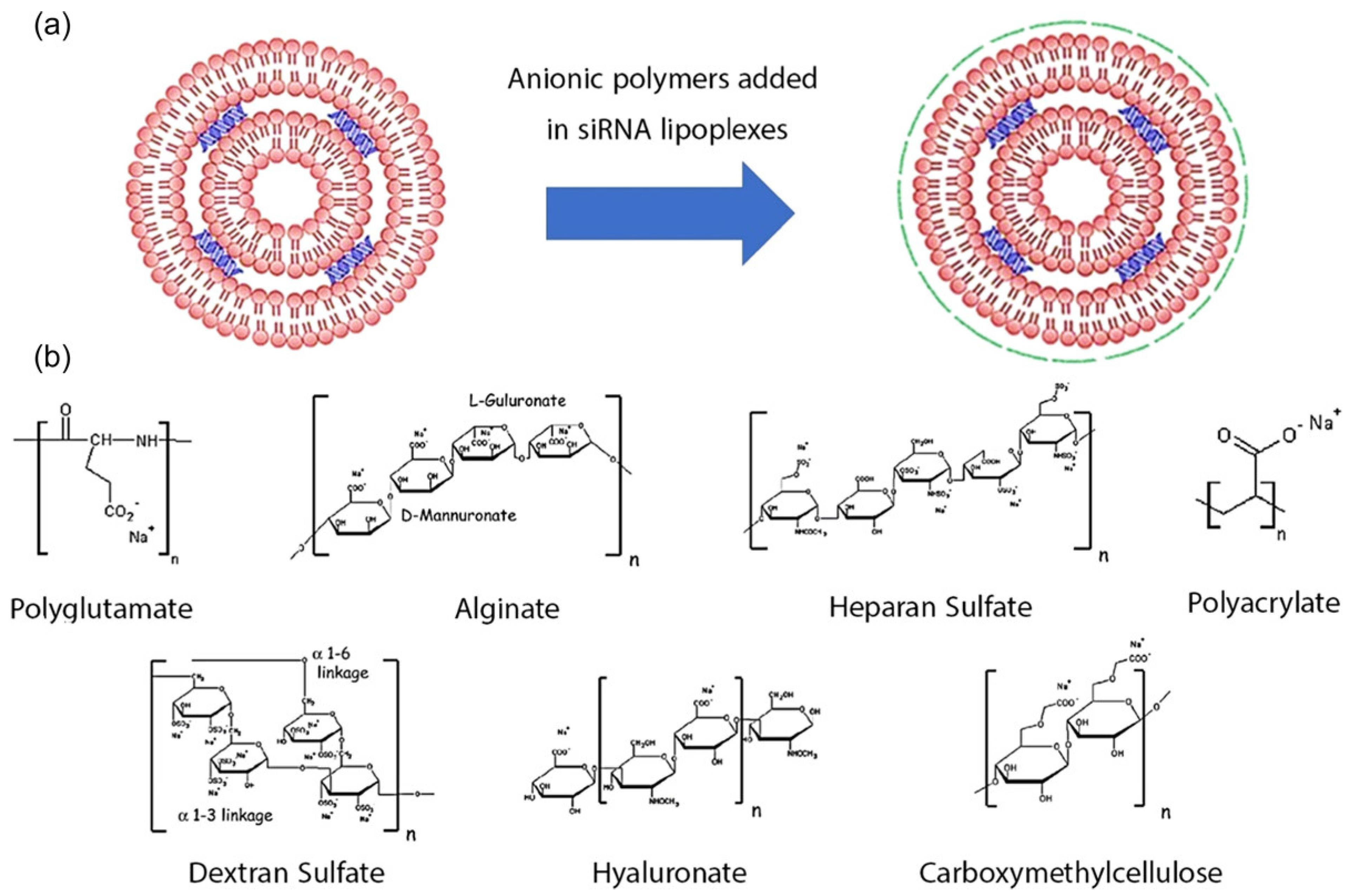
siRNA–lipid complexes are known as lipoplexes. Cationic liposomes combined with lipids have shown excellent outcomes in siRNA drug delivery for cancer therapy [21]. However, immune responses and undesired toxicities associated with lipoplexes should be addressed. Various approaches have been developed to diminish lipoplex toxicity. Although their entrapment efficiency might be low, neutral liposomes have reduced such toxicity. Further, cationic lipoplexes have been coated with anionic polymers, including sodium salts of hyaluronic acid, and alginic acid carboxymethyl cellulose. The coated cationic liposomes showed enhanced siRNA delivery to solid tumors (Figure 2) [21].
The benefit of polymeric nanocarriers is that they are not stimulators of the immune response. Electrostatic interactions between polymers and siRNA are readily achievable through linkages (thiol, maleimide, disulfide, etc.). The natural cationic polymers have led to efficient delivery approaches due to low toxicity, biodegradability, and biocompatibility [21,51,52,53].
The siRNA rigid structure incites weak interactions with polycation, and polyplexes are less effective in protecting siRNA against nucleases. Increasing the polycation amount could improve this pitfall while increasing toxicity, and hence an optimum polycation quantity should be utilized. Further, dendrimers have flexibility and prospects in siRNA loading [21,54].
Nanoparticles PEG-b-PLA or PEG-b-PLGA have received increased curiosity for siRNA drug delivery but are restricted by the reduced encapsulation efficacy of siRNA. To overcome this pitfall, a cationic lipid-induced NP platform known as CLAN with more siRNA encapsulation competence was fabricated [55].
Cell membrane coating has been established as an efficient approach to enhance the in vivo execution of NPs. Cell membrane decoration on NPs improves their colloidal stability and blood circulation and also benefits targeted drug delivery. Cancer cell membrane-decorated biomimetic NPs could deliver siRNA to homologous tumors in vivo [56,57]. Cell membrane-coated polyplexes are associated with problems in releasing siRNA into cytoplasm. To resolve this problem, charge-reversal polymers are utilized to facilitate siRNA release in cytoplasm [58,59]. PEI–siRNA polyplexes were coated with citraconic anhydride-grafted poly-L-lysine, which was then decorated with angiopep-2 peptide-coated red blood cell membrane (Ang-RBCm) [59]. The RBC membrane decoration offered long-term blood circulation, and targeting of angiopep-2 enhanced the accumulation of drugs into solid tumors. Such cell membrane-decorated charge reversal hybrid nanocomplexes proficiently inhibited tumor growth by knockdown of target genes via siRNA delivery.
Virus/polymer chimeric NPs permit simultaneous expression and silencing of numerous genes [60,61]. NPs comprising a Bcl-2-interacting mediator of cell death (BIM)-encoding adeno-associated virus (AAV) core and an acid-degradable polyketal (PK) shell were developed for enhanced siRNA delivery to tumors. After endocytosis of nanoparticles into the cell, the release of siRNA was facilitated by PK shell degradation. The BIM-encoding AAV was then transported into the cell nucleus for BIM expression. BIM enhanced the apoptosis of cancer cells. Thus, the polymer/virus chimeric NPs upregulated the BIM expression and simultaneously inhibited the cancer cells [43,60,61].
PLL produced by Streptomyces albus was utilized as a scaffold for the production of polyphenol-grafted bio-inspired polymers for siRNA delivery. Phenol, catechol, and pyrogallol were conjugated onto PLL through a simple reaction, and the cellular uptake of polyphenol-grafted polymer-siRNA complex by HeLa cells was examined [62]. A series of polyphenol-grafted low molecular weight polymer–siRNA complexes was effective in siRNA delivery inspired by natural polyphenol. The phenolic moieties enhanced the siRNA binding ability and stability of the polyplex. Among the polyphenols, polycatechols demonstrated the highest performance in siRNA binding, cellular uptake, and gene silencing [62]. The polycatechol–siRNA platform could be a highly promising bioinspired polymer for siRNA delivery in solid tumors.
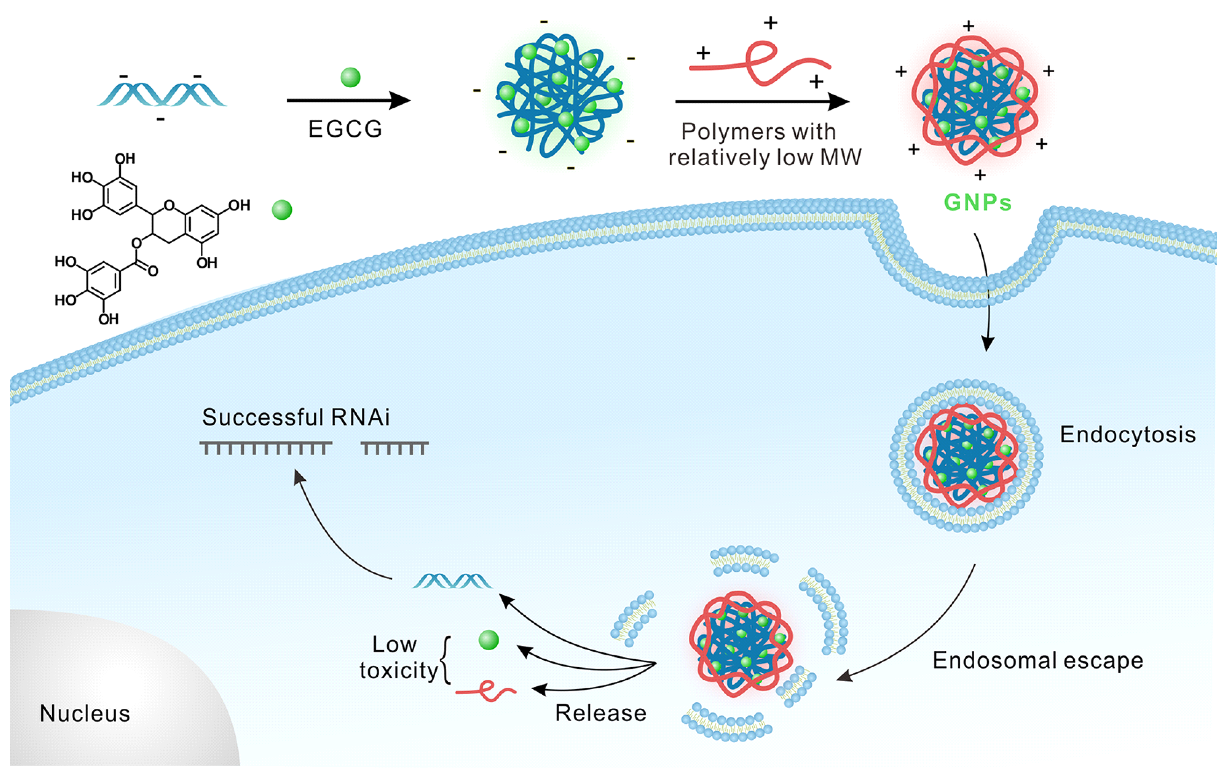
Green tea-based EGCG enables siRNA condensation by low molecular-weight polymers, yielding green NPs (GNPs), showing effective siRNA-induced gene silencing [63]. Biocompatible components of GNPs such as EGCG, siRNA, and PLL demonstrated reduced toxicity on the transfected cells. Similarly, antisense oligodeoxynucleotides, DNAzymes, and peptide nucleic acids may be utilized to design bioinspired polymer-based platforms for the fabrication of GNPs for a robust system that is promising for gene/siRNA delivery (Figure 3) [63].
Further, utilizing siRNA drugs for the treatment of hematological malignancies has been ineffective because blood cancer cells demonstrate significant resistance to standard transfection methods [64]. However, there are some studies concerning siRNA delivery to blood cancers. For example, CD20/CD44 dual targeted siRNA delivery using layer-by-layer NPs (LbL NPs, PLGA/PLA/siRNA/PLA; PLGA, poly-(D,L-lactide-co-glycolide); PLA, poly-l-arginine) with BCL-2 siRNA in B cell lymphoma and leukemia cells downregulated BCL-2, induced apoptosis, and reduced the blood cancer cells. Herein, the outermost layer of LbL NPs consists of hyaluronic acid (a CD44-ligand) covalently conjugated to CD20 antibodies. The CD20/CD44 dual-targeting outer layer offered precise binding to blood cancer cells, followed by receptor-mediated endocytosis of the LbL NPs [64].
In recent years, there have been many pre-clinical studies using biopolymer-induced siRNA therapeutics for the enhanced therapy of cancer. To date, several siRNA-mediated drugs have been in different phases of clinical trials or have received FDA approval for the treatment of a variety of diseases, including solid tumors, as demonstrated in Table 1 [65,66]. siRNA drugs including patisiran, givosiran, lumasiran, and inclisiran have received approval from the FDA [67]. As a result, siRNA drugs have received the enhanced attention of medical doctors and scientists. The design of apposite siRNA-assisted delivery systems is a vital issue for clinical applications in cancer therapy. These approaches could be potentially supportive in reducing cancer deaths in the near future.

4. Breast Cancer Therapy with Biopolymer-Based siRNA Nanoplatform

Breast cancer is the second leading cause of death in woman after lung cancer world-wide. However, cancer death rates including breast cancer have been decreasing due to the development of advanced screening technologies and significant improvement of immunotherapy, targeted therapy, and gene therapy [68]. Natural herbal compounds or biopolymers have significant roles in inhibiting drug resistance in breast cancer, impeding breast cancer stem cells (BCSCs). BCSCs utilize different cellular mechanisms, including membrane transporters, anti-apoptotic, pro-survival, and self-renewal-linked signaling pathways. Biopolymers may disrupt these mechanisms and hence impede breast tumors, inhibiting the BCSCs, and the induction of apoptosis [69]. Therefore, this review highlighted developments in biopolymer-mediated siRNA drug delivery systems for breast tumors. Biopolymer-induced delivery of chemotherapeutics and siRNA is promising and has demonstrated synergistic effects as a single delivery therapeutic [12]. Methotrexate (MTX) is a potential drug for breast cancer treatment, effectively inhibiting the dihydrofolate reductase (DHFR) receptor, which suppresses DNA synthesis. MTX can significantly induce apoptosis by inhibiting the STAT3 in the JAK/STAT pathway in tumor cells. Inhibition of the STAT3 protein by siRNA is an effective approach to hinder breast tumors [70]. A nanoparticle delivery system for the co-delivery of MTX and STAT3siRNA was developed and investigated against MCF-7 breast cancer cells in vitro. Mesoporous silica nanoparticles (MSNs) were functionalized with chitosan by covalent grafting facilitated by APTES using glutaraldehyde as a linker. APTES and grafted chitosan offered an amine rich surface coating on MSN NPs, permitting electrostatic loading of both MTX and siRNA on MSN [14]. The co-delivery of MTX and STAT3siRNA to breast cancer cells in vitro efficiently inhibited the DHFR receptor and inhibited breast tumors. Further, the co-delivery of doxorubicin (Dox) and p53 by deionized chitosan derivative functionalized MSNs tempted noteworthy apoptosis in breast cancer cells due to the synergistic effects of Dox and the p53 gene [18].
PEG-modified chitosan was synthesized for survivin-targeted siRNA delivery to breast cancer cells to inhibit breast tumors and to prevent metastasis [71]. The outcome of the study demonstrated that siRNA loaded in PEGylated chitosan (PEG-chitosan-siRNA) delivered to breast cancer cells showed enhanced cellular uptake and inhibited 4T1 tumor cell growth significantly. The flow cytometry study and confocal scanning microscopy study demonstrated that PEG-chitosan-siRNA particles were uptaken more easily than naked siRNA. The developed PEG-chitosan-siRNA nanoparticle significantly reduced tumor growth in vivo in a xenograft mouse model. The study exhibited that the PEG-chitosan-siRNA nanoplatform is safe, effective in tumor inhibition, and efficient in preventing metastasis [71].
Gold NPs modified with trimethyl chitosan (TMC) were developed as efficient delivery carriers to enhance the stability and cellular uptake of siRNA targeting EGFR in breast tumors. EGFRsiRNA was complexed with AuNPs-TMC through electrostatic interactions to develop the nanosystem AuNPs-TMC-EGFRsiRNA using a facile synthesis process. The synthesized AuNPs-TMC-EGFRsiRNA NPs effectively delivered siRNA to breast cancer cells. Therefore, AuNPs-TMC-EGFRsiRNA appeared as a prospective and potential nanodelivery system for breast cancer therapy [72].
A breast cancer-specific gene 1-siRNA (BCSG1-siRNA) plasmid was fabricated and then encapsulated by chitosan-silicon dioxide NPs. The slow release of BCSG1-siRNA to MCF-7 cells demonstrated enhanced cytotoxicity, reduced proliferation, and increased apoptosis. BCSG1-siRNA downregulated BCSG1 and inhibited tumor growth significantly. As a result, chitosan-silicon dioxide NP-BCSG1-siRNA is an effective siRNA delivery system to inhibit breast tumors [73].
A pH-responsive nanoplatform was fabricated introducing antiPOL siRNA and guanidine-CO2-functionalized chitosan at a neutral pH to knock down the POLR2 in TNBC tumors. The siRNA nanodrug was effectively internalized into TNBC cells harboring hemizygous TP53 deletion, overcoming intracellular trafficking at low pH. The downregulation of POLR2 caused TNBC cell death with little or no toxicity [74].
A triblock copolymer, a hollow-sphere polymersome PVPON-PDMS-PVPON was designed for siRNA delivery to breast cancer cell lines [75]. The polymersomes assembled in the presence of 1 μM PARPsiRNA-loaded siRNA inside the nanocarrier. When the developed nanovehicles were disrupted by ultrasound in vitro, the siRNA was released unaltered. siRNA-loaded polymersomes were tested against HER2+, trastuzumumab-resistant breast cancer cells in vitro, which reduced the PPARP1 protein level in the breast cancer cells and impeded MDR genes. Herein, a fluorescent dye was attached covalently to the outside of the nanocapsules. Thus, the targeting molecule was added to the nanocapsules to deliver with the polymersomes into the tumors. This nanosystem with PARP1siRNA was treated with TNBC cells in a tumor mouse model, and the delivered siRNA significantly knocked down the expression of PARP1 and increased by four-fold the survival of mice bearing TNBC tumors. PARPis are suitable for targeting TNBC tumors with defects in DNA repair and could alter the TME (tumor microenvironment) [76,77,78,79,80,81,82,83]. However, it has been challenging to combine many PAPRis with chemotherapy due to bone marrow suppression. Targeting PARP1i in the tumor may permit novel combination therapy approach. The polymersome-loaded PARP1siRNA nanosystem is a biodegradable, non-ionic polymeric nanovesicles proficient to encapsulate and deliver PARP1siRNA for knocking down PARP1 in vivo. This study provided an advanced nanopodium for the development of an effective drug delivery nanocarrier system for siRNA therapy of breast cancer, including TNBC [75].
A stable polyplex was developed using PEG-modified L-arginine oligo(-alkylaminosiloxane) grafted with PEI for siRNA delivery to breast cancer cells [84]. The polyplex was used to deliver the anti-luciferase siRNA to the luciferase-expressed TNBC cell line MDA-MB-231-Luc-D3H2LN, and anti-ABCB1 siRNA to doxorubicin-resistant MCF-7 ADR. Transfection of MDA-MB-231-Luc-D3H2LN cells with anti-luciferase siRNA demonstrated comprehensive knockdown of luciferase expression. When MCF-7 ADR cells were transfected with anti-ABCB1 siRNA, efficient downregulation of ABCB1 was observed. Therefore, this approach could be effective against breast cancer and TNBC therapy [84].
Lipid-based carriers are efficient in siRNA delivery [85]. Piperazine is frequently utilized in bioactive compounds and medicine [86,87]. A recent study reported that piperazine-containing ionizable lipid (Pi-Lipids) favorably deliver genes to immune cells in vivo without targeting the ligand [86]. Herein, the author demonstrated that Pi-A10 LNP (Pi-LNPs that contain PPZ-A10) effectively delivered mRNA/siRNA to solid tumors. LNP-10A was also formulated with siGFP and siLuc, and LNP10AsiGFP showed 25% knockdown of GFP, but no knockdown of luciferase was observed using LNP10AsiLuc. This study showed that high throughput in vivo investigations can recognize NPs with natural nonhepatocyte tropism, and the lipid with bioactive small molecule motifs can deliver mRNA/siRNA to immune cells in vivo. Further, LNP-10A was formulated with sipGP. This strategy could be applied to deliver siRNA to breast cancer cells [88].
CD44 expression is linked to the JAK2/STAT3 pathway in TNBC hostility. Incorporation of PEG–lipid complexes inside the lipid bilayers increased the sustained release of siRNA and evasion of the mononuclear phagocytic system. Cationic liposomes are widely used for siRNA delivery platforms for enhanced delivery, without enzymatic degradation, and effective cytoplasmic release [89].
Lipid-based polymeric nanocarriers are utilized for siRNA delivery to chemoresistant breast cancer. A nanoplatform formulated by hexadecylated PEI-siRNA encapsulated with lipid segments (DSPE-PEG, DSPE, cholesterol, tripalmitin) and fabricated with apolipoprotein was used for megalin-targeted siRNA delivery to chemo-treated breast cancer cells, which demonstrated enhanced delivery and a reduction of disease recurrence [89].
Calcium apatite NPs-siRNA downregulated BCL-2, EGFR, and ERBB-2 genes, resulting in the cell death-inhibiting PI3K/MAPK signaling pathway in a mouse model of breast tumors [90]. A calcium phosphate-PEG-polyanion polymer promoted the siRNA delivery targeting anti-apoptotic gene BCL-xL and BCL-2 in breast cancer cell line MCF-7. After 48 h of incubation the BCL-xL and BCL-2 genes were reduced by 49% and 23%, respectively [91].
Further, MCF-7 and MDA-MB-231 cell lines were studied to knock down SUR and BCL2 genes utilizing siSUR- and siBCL2-loaded bovine milk exosomes. siSUR was highly effective in inhibiting the breast cancer cells with ~80% knockdown of the SUR gene [33]. Bovine milk exosomes are natural, stable, safe, feasible, and promising nanocarriers for siRNA delivery to breast cancer cells.
NPs consisting of coupled PEI and folic acid to core/shell Fe3O4@SiO2 were fabricated to deliver Notch-shRNA to TNBC cell lines, MDA-MB-231 [92]. The study demonstrated that superparamagnetic Fe3O4@SiO2/PEI-FA/Notch-1shRNA NPs induced delivery of Notch-1shRNA to TNBC cells effectively with no toxicity. The nanodelivery cargo was capable of delivering Notch-1shRNA efficiently while protecting shRNA from degradation by exogenous DNaseI and nucleases. The preferential uptake of superparamagnetic Fe3O4@SiO2/PEI-FA/Notch-1shRNA NPs by TNBC cells MDA-MB-231 was revealed by MRI and fluorescence microscopy. Significantly decreased expression of Notch-1 was exhibited by MDA-MB-231 cells. Thus, the Fe3O4@SiO2/PEI-FA/Notch-1shRNA nanosystem is an effective theranostic system exploring both Notch-1 inhibition in TNBC cells and superparamagnetic property-enabled MRI and fluorescence imaging simultaneously [92].
A PEI nanocomplex was fabricated with EpCAM aptamer (EpApt) and EpCAM siRNA (siEP) to study its effects against breast cancer. The fabricated PEI-EpApt-siEp knocked down EpCAM and subdued the proliferation of breast cancer cells MCF-7. The study demonstrated that PEI-EpApt-siEp has higher binding ability to MCF-7 cells than EpApt or scrambled aptamer or PEI-ScrApt-SiEp [65,93].
A nanosystem was fabricated with lipid-NPs-siRNA coated with a Fab antibody to utilize against heparin-binding EGF-like growth factor (αHB-EGF LNP siRNA). A developed αHB-EGF LNP siRNA nanoplatform targeting PLK1 was examined for TNBC cell line MDA-MB-231 overexpressing HB-EGF on the cell surface. The result indicated the suppression of PLK1 protein, with the inhibition of the TNBC tumor revealing αHB-EGF LNP siRNA as a prospective target for TNBC tumors expressing HB-EGF [94].
A potential nanoplatform using a liposome–polycation–DNA complex (LPD) conjugated with tumor specific antibody (TLPD) was developed for siRNA delivery targeting EGFR in TNBC cells. TLPD was prepared by a self-assembled process followed by anti-EGFR Fab antibody conjugation (TLPD-FCC). The TLPD-FCC-siRNA demonstrated enhanced binding affinity and luciferase gene silencing (20%) in EGFR, overexpressing the TNBC cell line MDA-MB-231 in vitro. This study showed that TLPD-FCC has great prospects in delivering siRNA to EGFR overexpressing breast cancer cells [95].
A TNBC tumor targeting RGD-PEG-ECO/siDANCR nanosystem was developed for studying breast cancer cells. TNBC cell lines MDA-MB-231 and BT549 cells treated with the RGD-PEG-ECO/siDANCR NPs showed ~90% silencing of DANCR expression for up to 7 days, demonstrating an effective siRNA delivery approach for targeting and an enhanced therapeutic effect [96].
ICAM-1 targeted Lcn2 siRNA encapsulated liposome binds TNBC cells MDA-MB-231 significantly, and effective Lcn2 downregulation by ICAM-Lcn2-LPs led to a substantial lessening in VEGF from MDA-MB-231 cells, resulting in diminished angiogenesis, both in vitro and in vivo. The study demonstrated a promising approach for inhibiting angiogenesis and for tumor growth retardation in TNBC tumors [97].
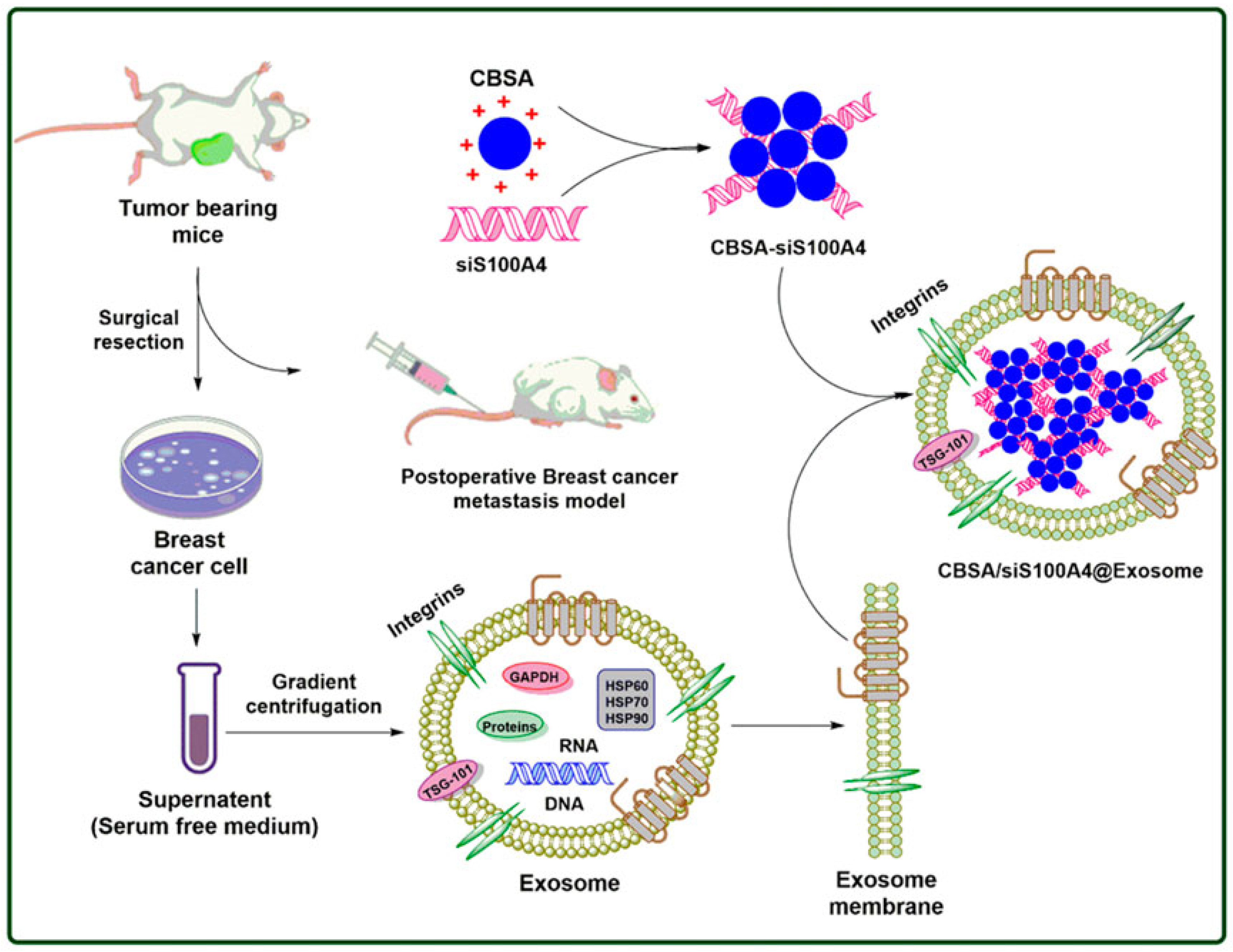
Cationic bovine serum albumin-conjugated siS100A4 was coated with exosome membrane to develop self-assembled nanosystem CBSA/siS100A4@Exosome for enhanced drug delivery to cancer cells (Figure 4). The in vivo study demonstrated that CBSA/siS100A4@Exosome has a higher affinity towards cancer cells compared to CBSA/siS100A4@liposomes, and it exhibited significant gene silencing as well as inhibiting the growth of malignant breast cancer cells. The CBSA/siS100A4@Exosome nanoplatform exhibits a promising strategy to impede postoperative metastasis in breast cancer [98,99].
Exosomes were collected from fibroblastic BJ cells (BJExo) to treat different cancer cells, including breast tumors [100,101,102,103,104]. PIK3CA was silenced by siPIK3CA to improve the breast tumor therapy outcome. PIK3CA silencing by siPIK3CA delivery reduced cell migration capacity, impeding the metastasis process. This study confirmed the PI3K/AKT/mTOR pathway and PIK3CA as an effective target for breast cancer therapy and introduced BJ-derived exosomes as a promising nanocarrier for siRNA delivery. The study utilized siPIK3CA to target MDA-MB-231 and MDA-MB-453 cell lines and led to the reduction in mRNA expression of PIK3CA, AKT, and mTOR [104]. The study outcome was supported by previous results [101,102,103].
Exosome-loaded siRNA therapeutics have been effective in downregulating BCL-2, PLK1, KRAS, and survivin anti-apoptotic proteins that induce tumor cell mitosis and cell growth factor. The exosome-mediated siRNA therapy was effective in inhibiting cell proliferation and migration. BCL-2 siRNA-loaded exosome derived from NK cells was utilized to treat ER+ breast cancer cells and led to increased apoptosis in breast cancer cells [105].
RNA-NP-modified exosome was fabricated to target three cancer cells, including breast cancer cells, simultaneously. In the structure of the RNA-NP, the pRNA of phage phi29 was extended into an arrow shape to connect with the RNA ligands, and fluorescent alexa647 was added for imaging [106]. The RNA ligand was utilized for specific targeting and exosome for effective membrane fusion. The resulting ligand-displaying exosome was capable of specific delivery of siRNA to cells and potentially inhibiting tumor growth. The exosome harboring EGFR aptamer-loaded siSurvivin NPs (EGFRapt/Exosome/siSurvivin) inhibited breast tumor in MDA-MB-468 cells orthotopic xenograft breast tumor bearing mice [106,107].
Ligands can be expressed, fusing with exosomal surface proteins by engineered exosome-producing cells, for targeting cancer cells [108]. Modified exosome-producing engineered HEK293T cells were utilized to target HER2+ breast cancer cells. The result demonstrated that HEK293T cells formed DARPin G3 on the surface of exosomes. These modified exosomes can bind to HER2/neu and deliver siRNA to SKBR3 breast cancer cells, downregulating gene expression up to 70%. This study exhibited a potential approach to facilitate gene and drug transfer to HER2+ cancer cells [108].
MCF-7 breast cancer cell-derived exosomes loaded with siRNA were used against CD44 in breast cancer cells. Exosome-siRNA targeting CD44 expression (Exos-siCD44) effectively silenced the expression. When cocultured on Exos-siCD44, breast cancer cells demonstrated diminished cell proliferation and improved sensitivity to doxorubicin. The same effect was detected in vivo in mice [109].
Nanoscale exosome-mimics (Ems) were generated from non-tumorigenic MCF-10A cells, and then siRNA was encapsulated into the EMs by the electroporation technique. The CDK4siRNA-loaded EMs significantly downregulated CDK4 mRNA protein expression in vitro and in vivo. Therefore, EMs from similar cell types may be effective as siRNA carriers for the development of innovative breast cancer therapeutics with no toxicity [110]. There have been numerous pre-clinical studies utilizing biopolymer-based siRNA delivery systems with promising results for breast cancer therapy (Table 2). Biopolymer-induced siRNA delivery is a prospective strategy to treat breast tumors to improve survival of breast cancer patients.

5. Conclusions and Future Perspective

Biopolymers have been proficiently applied in siRNA drug delivery to cancer cells, including breast cancer. The sources of the biopolymers are algae, fungi, plants, animals, microbes, etc. Further, exosomes are obtained from tissues, biological fluids, and cells. Biopolymers are effective for the progress of innovative anticancer drug delivery platforms. Biopolymer-based NP systems have been recognized as cost-effective environmentally friendly platforms for effective drug delivery with enhanced selectivity and reduced toxicity. Nanobiopolymers have promising stimuli-responsive properties and therefore can be used to enhance siRNA delivery platforms to undruggable MDR metastatic cancer cells, including breast cancer. The biopolymeric siRNA drug formulations can shield drugs from pH degradation, extracellular trafficking, nontargeted binding sites, and they are accordingly appropriate for drug delivery and internalization into tumors in a controlled release manner. Numerous natural polysaccharide polymers are utilized in siRNA delivery for cancer therapy. The key advantage of polysaccharides is the presence of various functional groups for modification and polymerization. The most frequently applied polysaccharide for siRNA drug delivery to cancer is chitosan, which is biodegradable, biocompatible, inexpensive, and has low toxicity. For increasing the solubility of chitosan and making it suitable for endo-cytoplasmic release, various modifications have been performed. CPP-based siRNA drugs are promising siRNA delivery vehicles, as they can easily cross the cell membrane. Exosome-loaded siRNA therapeutics have been effective in downregulating cancer genes as well as anti-apoptotic and other proteins that prompt tumor cell mitosis and cell growth factors. Exosome-mediated siRNA therapy is promising and prospective in inhibiting cell proliferation and migration in solid tumors, resulting in enhanced tumor inhibition in solid tumors, including breast tumors, improving the survival of patients. However, for producing biopolymer NPs, functionalization and modification should be performed perfectly and precisely for making them pertinent nanocarriers for siRNA-based drug delivery for overcoming systemic and extra-cellular hurdles. Further, before translation of biopolymer-based siRNA drugs into the clinic, their immune activation and toxicity profiles must be addressed effectively.

Author Contributions

M.A.S. conceived the idea of research, performed the study, prepare the manuscript, and revised it. V.P.T. contributed in manuscript revisions, and supervised the whole project. All authors have read and agreed to the published version of the manuscript.

Funding

This research received no external funding.

Institutional Review Board Statement

Not applicable.

Informed Consent Statement

Not applicable.

Data Availability Statement

Not applicable.

Acknowledgments

Authors acknowledged both SUST and Northeastern University.

Conflicts of Interest

The authors declare no conflict of interest.

References

  1. Byun, M.J.; Lim, J.; Kim, S.-N.; Park, D.-H.; Kim, T.-H.; Park, W.; Park, C.G. Advances in Nanoparticles for Effective Delivery of RNA Therapeutics. BioChip J. 2022, 16, 128–145. [Google Scholar] [CrossRef] [PubMed]
  2. Hou, X.; Zaks, T.; Langer, R.; Dong, Y. Lipid nanoparticles for mRNA delivery. Nat. Rev. Mater. 2021, 6, 1078–1094. [Google Scholar] [CrossRef] [PubMed]
  3. Xue, H.Y.; Liu, S.; Wong, H.L. Nanotoxicity: A key obstacle to clinical translation of siRNA-based nanomedicine. Nanomedicine 2014, 9, 295–312. [Google Scholar] [CrossRef] [PubMed] [Green Version]
  4. Ho, W.; Gao, M.; Li, F.; Li, Z.; Zhang, X.Q.; Xu, X. Next-generation vaccines: Nanoparticle-mediated DNA and mRNA delivery. Adv. Healthc. Mater. 2021, 10, e2001812. [Google Scholar] [CrossRef]
  5. Tenchov, R.; Bird, R.; Curtze, A.E.; Zhou, Q. Lipid nanoparticles- from liposomes to mRNA vaccine delivery, a landscape of research diversity and advancement. ACS Nano. 2021, 15, 16982–17015. [Google Scholar] [CrossRef]
  6. Smith, S.A.; Selby, L.I.; Johnston, A.P.R.; Such, G.K. The Endosomal Escape of Nanoparticles: Toward More Efficient Cellular Delivery. Bioconjugate Chem. 2019, 30, 263–272. [Google Scholar] [CrossRef]
  7. Şenel, B.; Büyükköroğlu, G. Applications of Lipidic and Polymeric Nanoparticles for siRNA Delivery. In Antisense Therapy; IntechOpen: London, UK, 2019. [Google Scholar] [CrossRef] [Green Version]
  8. Cavallaro, G.; Sardo, C.; Craparo, E.F.; Porsio, B.; Giammona, G. Polymeric nanoparticles for siRNA delivery: Production and applications. Int. J. Pharm. 2017, 525, 313–333. [Google Scholar] [CrossRef]
  9. Ragelle, H.; Vandermeulen, G.; Préat, V. Chitosan-based siRNA delivery systems. J. Control. Release 2013, 172, 207–218. [Google Scholar] [CrossRef]
  10. Mao, S.; Sun, W.; Kissel, T. Chitosan-based formulations for delivery of DNA and siRNA. Adv. Drug Deliv. Rev. 2010, 62, 12–27. [Google Scholar] [CrossRef]
  11. Peng, H.; Yang, H.; Song, L.; Zhou, Z.; Sun, J.; Du, Y.; Lu, K.; Li, T.; Yin, A.; Xu, J.; et al. Sustained delivery of siRNA/PEI complex from in situ forming hydrogels potently inhibits the proliferation of gastric cancer. J. Exp. Clin. Cancer Res. 2016, 35, 57. [Google Scholar] [CrossRef] [Green Version]
  12. Shakeran, Z.; Varshosaz, J.; Keyhanfar, M.; Mohammad-Beigi, H.; Rahimi, K.; Sutherland, D.S. Co-delivery of STAT3 siRNA and methotrexate in breast cancer cells. Artif. Cells Nanomed. Biotechnol. 2022, 50, 29–39. [Google Scholar] [CrossRef] [PubMed]
  13. Li, X.; Chen, Y.; Wang, M.; Ma, Y.; Xia, W.; Gu, H. A mesoporous silica nanoparticle—PEI—Fusogenic peptide system for siRNA delivery in cancer therapy. Biomaterials 2013, 34, 1391–1401. [Google Scholar] [CrossRef] [PubMed]
  14. Hao, N.; Jayawardana, K.W.; Chen, X.; Yan, M. One-Step Synthesis of Amine-Functionalized Hollow Mesoporous Silica Nanoparticles as Efficient Antibacterial and Anticancer Materials. ACS Appl. Mater. Interfaces 2015, 7, 1040–1045. [Google Scholar] [CrossRef] [PubMed] [Green Version]
  15. Tan, W.B.; Zhang, Y. Surface modification of gold and quantum dot nanoparticles with chitosan for bioapplications. J. Biomed. Mater. Res. Part A 2005, 75A, 56–62. [Google Scholar] [CrossRef]
  16. Lv, G.; Qiu, L.; Liu, G.; Wang, W.; Li, K.; Zhao, X.; Lin, J. pH sensitive chitosan-mesoporous silica nanoparticles for targeted delivery of a ruthenium complex with enhanced anticancer effects. Dalton Trans. 2016, 45, 18147–18155. [Google Scholar] [CrossRef]
  17. Nhavene, E.P.F.; Andrade, G.F.; Faria, J.A.Q.A.; Gomes, D.A.; de Sousa, E.M.B. Biodegradable Polymers Grafted onto Multifunctional Mesoporous Silica Nanoparticles for Gene Delivery. Chemengineering 2018, 2, 24. [Google Scholar] [CrossRef] [Green Version]
  18. Lin, J.-T.; Liu, Z.-K.; Zhu, Q.-L.; Rong, X.-H.; Liang, C.-L.; Wang, J.; Ma, D.; Sun, J.; Wang, G.-H. Redox-responsive nanocarriers for drug and gene co-delivery based on chitosan derivatives modified mesoporous silica nanoparticles. Colloids Surfaces B: Biointerfaces 2017, 155, 41–50. [Google Scholar] [CrossRef]
  19. Cao, Y.; Tan, Y.F.; Wong, Y.S.; Liew, M.W.J.; Venkatraman, S. Recent Advances in Chitosan-Based Carriers for Gene Delivery. Mar. Drugs 2019, 17, 381. [Google Scholar] [CrossRef] [Green Version]
  20. Soliman, O.Y.; Alameh, M.G.; De Cresenzo, G.; Buschmann, M.D.; Lavertu, M. Efficiency of Chitosan/Hyaluronan-Based mRNA Delivery Systems In Vitro: Influence of Composition and Structure. J. Pharm. Sci. 2020, 109, 1581–1593. [Google Scholar] [CrossRef]
  21. Sargazi, S.; Arshad, R.; Ghamari, R.; Rahdar, A.; Bakhshi, A.; Karkan, S.F.; Ajalli, N.; Bilal, M.; Díez-Pascual, A.M. siRNA-based nanotherapeutics as emerging modalities for immune-mediated diseases: A preliminary review. Cell Biol. Int. 2022, 46, 1320–1344. [Google Scholar] [CrossRef]
  22. Pilipenko, I.; Korzhikov-Vlakh, V.; Sharoyko, V.; Zhang, N.; Schafer-Korting, M.; Rühl, E.; Zoschke, C.; Tennikova, T. pH-sensitive chitosan-heparin nanoparticles for effective delivery of genetic drugs into epithelial cells. Pharmaceutics 2019, 11, 317. [Google Scholar] [CrossRef] [PubMed] [Green Version]
  23. Pathak, N.; Singh, P.; Singh, P.K.; Sharma, S.; Singh, R.P.; Gupta, A.; Mishra, R.; Mishra, V.K.; Tripathi, M. Biopolymeric nanoparticles based effective delivery of bioactive compounds toward the sustainable development of anticancerous therapeutics. Front. Nutr. 2022, 9, 963413. [Google Scholar] [CrossRef] [PubMed]
  24. Sarkar, S.; Ponce, N.T.; Banerjee, A.; Bandopadhyay, R.; Rajendran, S.; Lichtfouse, E. Green polymeric nanomaterials for the photocatalytic degradation of dyes: A review. Environ. Chem. Lett. 2020, 18, 1569–1580. [Google Scholar] [CrossRef]
  25. Farshchi, E.; Pirsa, S.; Roufegarinejad, L.; Alizadeh, M.; Rezazad, M. Photocatalytic/biodegradable film based on carboxymethyl cellulose, modified by gelatin and TiO2–Ag nanoparticles. Carbohydr Polym. 2019, 216, 189–196. [Google Scholar] [CrossRef] [PubMed]
  26. Kolangare, I.M.; Isloor, A.M.; Karim, Z.A.; Kulal, A.; Ismail, A.F.; Inamuddin; Asiri, A. M. Antibiofouling hollow-fiber membranes for dye rejection by embedding chitosan and silver-loaded chitosan nanoparticles. Environ. Chem. Lett. 2019, 17, 581–587. [Google Scholar] [CrossRef]
  27. Morin-Crini, N.; Lichtfouse, E.; Torri, G.; Crini, G. Applications of chitosan in food, pharmaceuticals, medicine, cosmetics, agriculture, textiles, pulp and paper, biotechnology, and environmental chemistry. Environ. Chem. Lett. 2019, 17, 1667–1692. [Google Scholar] [CrossRef] [Green Version]
  28. Bae, J.; Park, S.J.; Shin, D.S.; Lee, J.; Park, S.; Kim, H.J.; Kwon, O.S. A dual functional conductive hydrogel containing titania@polypyrrolecyclodextrin hybrid nanotubes for capture and degradation of toxic chemical. Biochip J. 2021, 15, 162–170. [Google Scholar] [CrossRef]
  29. Liang, Y.; Wang, Y.; Wang, L.; Liang, Z.; Li, D.; Xu, X.; Chen, Y.; Yang, X.; Zhang, H.; Niu, H. Self-crosslinkable chitosan-hyaluronic acid dialdehyde nanoparticles for CD44-targeted siRNA delivery to treat bladder cancer. Bioact. Mater. 2021, 6, 433–446. [Google Scholar] [CrossRef]
  30. Erdene-Ochir, T.; Ganbold, T.; Zandan, J.; Han, S.; Borjihan, G.; Baigude, H. Alkylation enhances biocompatibility and siRNA delivery efficiency of cationic curdlan nanoparticles. Int. J. Biol. Macromol. 2020, 143, 118–125. [Google Scholar] [CrossRef]
  31. O’Brien, K.; Breyne, K.; Ughetto, S.; Laurent, L.C.; Breakefield, X.O. RNA delivery by extracellular vesicles in mammalian cells and its applications. Nat. Rev. Mol. Cell Biol. 2020, 21, 585–606. [Google Scholar] [CrossRef]
  32. Luan, X.; Sansanaphongpricha, K.; Myers, I.; Chen, H.; Yuan, H.; Sun, D. Engineering exosomes as refined biological nanoplatforms for drug delivery. Acta Pharmacol. Sin. 2017, 38, 754–763. [Google Scholar] [CrossRef] [PubMed] [Green Version]
  33. Aqil, F.; Munagala, R.; Jeyabalan, J.; Agrawal, A.K.; Kyakulaga, A.-H.; Wilcher, S.A.; Gupta, R.C. Milk exosomes—Natural nanoparticles for siRNA delivery. Cancer Lett. 2019, 449, 186–195. [Google Scholar] [CrossRef] [PubMed]
  34. Yang, Z.; Shi, J.; Xie, J.; Wang, Y.; Sun, J.; Liu, T.; Zhao, Y.; Zhao, X.; Wang, X.; Ma, Y.; et al. Large-scale generation of functional mRNA-encapsulating exosomes via cellular nanoporation. Nat. Biomed. Eng. 2020, 4, 69–83. [Google Scholar] [CrossRef] [PubMed]
  35. Liang, S.-F.; Zuo, F.-F.; Yin, B.-C.; Ye, B.-C. Delivery of siRNA based on engineered exosomes for glioblastoma therapy by targeting STAT. Biomater. Sci. 2022, 10, 1582–1590. [Google Scholar] [CrossRef]
  36. Zhang, D.; Lee, H.; Wang, X.; Rai, A.; Groot, M.; Jin, Y. Exosome-Mediated Small RNA Delivery: A Novel Therapeutic Approach for Inflammatory Lung Responses. Mol. Ther. 2018, 26, 2119–2130. [Google Scholar] [CrossRef] [Green Version]
  37. Agrawal, A.K.; Aqil, F.; Jeyabalan, J.; Spencer, W.A.; Beck, J.; Gachuki, B.W.; Alhakeem, S.S.; Oben, K.; Munagala, R.; Bondada, S.; et al. Milk-derived exosomes for oral delivery of paclitaxel. Nanomedicine 2017, 13, 1627–1636. [Google Scholar] [CrossRef]
  38. Pi, F.; Binzel, D.W.; Lee, T.J.; Li, Z.; Sun, M.; Rychahou, P.; Li, H.; Haque, F.; Wang, S.; Croce, C.M.; et al. Nanoparticle orientation to control RNA loading and ligand display on extracellular vesicles for cancer regression. Nat. Nanotechnol. 2018, 13, 82–89. [Google Scholar] [CrossRef]
  39. Shoari, A.; Tooyserkani, R.; Tahmasebi, M.; Löwik, D.W.P.M. Delivery of Various Cargos into Cancer Cells and Tissues via Cell-Penetrating Peptides: A Review of the Last Decade. Pharmaceutics 2021, 13, 1391. [Google Scholar] [CrossRef]
  40. Jones, S.W.; Christison, R.; Bundell, K.; Voyce, C.J.; Brockbank, S.M.V.; Newham, P.; Lindsay, M.A. Characterisation of cell-penetrating peptide-mediated peptide delivery. Br. J. Pharmacol. 2005, 145, 1093–1102. [Google Scholar] [CrossRef]
  41. Dong, Y.; Chen, Y.; Zhu, D.; Shi, K.; Ma, C.; Zhang, W.; Rocchi, P.; Jiang, L.; Liu, X. Self-assembly of amphiphilic phospholipid peptide dendrimer-based nanovectors for effective delivery of siRNA therapeutics in prostate cancer therapy. J. Control. Release 2020, 322, 416–425. [Google Scholar] [CrossRef]
  42. Strand, M.S.; Krasnick, B.A.; Pan, H.; Zhang, X.; Bi, Y.; Brooks, C.; Wetzel, C.; Sankpal, N.; Fleming, T.; Goedegebuure, S.P.; et al. Precision delivery of RAS-inhibiting siRNA to KRAS driven cancer via peptide-based nanoparticles. Oncotarget 2019, 10, 4761–4775. [Google Scholar] [CrossRef] [PubMed] [Green Version]
  43. Wang, H.; Zhang, S.; Lv, J.; Cheng, Y. Design of polymers for siRNA delivery: Recent progress and challenges. VIEW 2021, 2, 20200026. [Google Scholar] [CrossRef]
  44. Peer, D.; Park, E.J.; Morishita, Y.; Carman, C.V.; Shimaoka, M. Systemic Leukocyte-Directed siRNA Delivery Revealing Cyclin D1 as an Anti-Inflammatory Target. Science 2008, 319, 627–630. [Google Scholar] [CrossRef] [PubMed] [Green Version]
  45. Lu, H.; Wang, D.; Kazane, S.; Javahishvili, T.; Tian, F.; Song, F.; Sellers, A.; Barnett, B.; Schultz, P.G. Synthesis of Bispecific Antibodies using Genetically Encoded Unnatural Amino Acids. J. Am. Chem. Soc. 2013, 135, 13885. [Google Scholar] [CrossRef] [Green Version]
  46. Kohata, A.; Hashim, P.K.; Okuro, K.; Aida, T. Transferrin-Appended Nanocaplet for Transcellular siRNA Delivery into Deep Tissues. J. Am. Chem. Soc. 2019, 141, 2862–2866. [Google Scholar] [CrossRef]
  47. Khan, O.F.; Kowalski, P.S.; Doloff, J.C.; Tsosie, J.K.; Bakthavatchalu, V.; Winn, C.B.; Haupt, J.; Jamiel, M.; Langer, R.; Anderson, D.G. Endothelial siRNA delivery in nonhuman primates using ionizable low–molecular weight polymeric nanoparticles. Sci. Adv. 2018, 4, eaar8409. [Google Scholar] [CrossRef] [Green Version]
  48. Xu, X.; Saw, P.E.; Tao, W.; Li, Y.; Ji, X.; Yu, M.; Mahmoudi, M.; Rasmussen, J.; Ayyash, D.; Zhou, Y.; et al. Tumor Microenvironment-Responsive Multistaged Nanoplatform for Systemic RNAi and Cancer Therapy. Nano Lett. 2017, 17, 4427–4435. [Google Scholar] [CrossRef]
  49. Yu, D.; Khan, O.F.; Suvà, M.L.; Dong, B.; Panek, W.K.; Xiao, T.; Wu, M.; Han, Y.; Ahmed, A.U.; Balyasnikova, I.V.; et al. Multiplexed RNAi therapy against brain tumor-initiating cells via lipopolymeric nanoparticle infusion delays glioblastoma progression. Proc. Natl. Acad. Sci. USA 2017, 114, E6147–E6156. [Google Scholar] [CrossRef] [Green Version]
  50. Dahlman, J.E.; Kauffman, K.J.; Xing, Y.; Shaw, T.E.; Mir, F.F.; Dlott, C.C.; Langer, R.; Anderson, D.G.; Wang, E.T. Barcoded nanoparticles for high throughput in vivo discovery of targeted therapeutics. Proc. Natl. Acad. Sci. USA 2017, 114, 2060–2065. [Google Scholar] [CrossRef]
  51. Srivastava, A.; Gowda, D.V.; Madhunapantula, S.V.; Shinde, C.G.; Iyer, M. Mucosal vaccines: A paradigm shift in the development of mucosal adjuvants and delivery vehicles. APMIS 2015, 123, 275–288. [Google Scholar] [CrossRef]
  52. Parmar, R.G.; Poslusney, M.; Busuek, M.; Williams, J.M.; Garbaccio, R.; Leander, K.; Walsh, E.; Howell, B.; Sepp-Lorenzino, L.; Riley, S.; et al. Novel Endosomolytic Poly(amido amine) Polymer Conjugates for Systemic Delivery of siRNA to Hepatocytes in Rodents and Nonhuman Primates. Bioconjugate Chem. 2014, 25, 896–906. [Google Scholar] [CrossRef] [PubMed]
  53. Farshbaf, M.; Davaran, S.; Zarebkohan, A.; Annabi, N.; Akbarzadeh, A.; Salehi, R. Significant role of cationic polymers in drug delivery systems. Artif. Cells Nanomed. Biotechnol. 2018, 46, 1872–1891. [Google Scholar] [CrossRef] [PubMed]
  54. Kim, B.; Park, J.-H.; Sailor, M.J. Rekindling RNAi Therapy: Materials Design Requirements for In Vivo siRNA Delivery. Adv. Mater. 2019, 31, e1903637. [Google Scholar] [CrossRef] [PubMed] [Green Version]
  55. Xu, C.F.; Iqbal, S.; Shen, S.; Luo, Y.L.; Yang, X.; Wang, J. Development of "CLAN" Nanomedicine for Nucleic Acid Therapeutics. Small 2019, 15, e1900055. [Google Scholar] [CrossRef] [PubMed]
  56. Xu, C.; Liu, W.; Hu, Y.; Li, W.; Di, W. Bioinspired tumor-homing nanoplatform for co-delivery of paclitaxel and siRNA-E7 to HPV-related cervical malignancies for synergistic therapy. Theranostics 2020, 10, 3325–3339. [Google Scholar] [CrossRef]
  57. Zhang, L.; Deng, S.; Zhang, Y.; Peng, Q.; Li, H.; Wang, P.; Fu, X.; Lei, X.; Qin, A.; Yu, X. Homotypic Targeting Delivery of siRNA with Artificial Cancer Cells. Adv. Health Mater. 2020, 9, 1900772. [Google Scholar] [CrossRef]
  58. Liu, Y.; Zou, Y.; Feng, C.; Lee, A.; Yin, J.; Chung, R.; Park, J.B.; Rizos, H.; Tao, W.; Zheng, M.; et al. Charge Conversional Biomimetic Nanocomplexes as a Multifunctional Platform for Boosting Orthotopic Glioblastoma RNAi Therapy. Nano Lett. 2020, 20, 1637. [Google Scholar] [CrossRef]
  59. Wang, Y.; Ji, X.; Ruan, M.; Liu, W.; Song, R.; Dai, J.; Xue, W. Worm-Like Biomimetic Nanoerythrocyte Carrying siRNA for Melanoma Gene Therapy. Small 2018, 14, e1803002. [Google Scholar] [CrossRef]
  60. Cho, S.K.; Kwon, Y.J. Simultaneous gene transduction and silencing using stimuli-responsive viral/nonviral chimeric nanoparticles. Biomaterials 2012, 33, 3316–3323. [Google Scholar] [CrossRef]
  61. Hong, C.A.; Cho, S.K.; Edson, J.A.; Kim, J.; Ingato, D.; Pham, B.; Chuang, A.; Fruman, D.A.; Kwon, Y.J. Viral/Nonviral Chimeric Nanoparticles To Synergistically Suppress Leukemia Proliferation via Simultaneous Gene Transduction and Silencing. ACS Nano 2016, 10, 8705–8714. [Google Scholar] [CrossRef] [Green Version]
  62. Shen, W.; Wang, R.; Fan, Q.; Gao, X.; Wang, H.; Shen, Y.; Li, Y.; Cheng, Y. Natural Polyphenol Inspired Polycatechols for Efficient siRNA Delivery. CCS Chem. 2020, 2, 146–157. [Google Scholar] [CrossRef]
  63. Shen, W.; Wang, Q.; Shen, Y.; Gao, X.; Li, L.; Yan, Y.; Wang, H.; Cheng; Y. Green Tea Catechin Dramatically Promotes RNAi Mediated by LowMolecular-Weight Polymers. ACS Cent. Sci. 2018, 4, 1326–1333. [Google Scholar] [CrossRef]
  64. Choi, K.Y.; Correa, S.; Min, J.; Li, J.; Roy, S.; Laccetti, K.H.; Dreaden, E.; Kong, S.; Heo, R.; Roh, Y.H.; et al. Binary Targeting of siRNA to Hematologic Cancer Cells In Vivo Using Layer-by-Layer Nanoparticles. Adv. Funct. Mater. 2019, 29, 1900018. [Google Scholar] [CrossRef]
  65. Hattab, D.; Bakhtiar, A. Bioengineered siRNA-Based Nanoplatforms Targeting Molecular Signaling Pathways for the Treatment of Triple Negative Breast Cancer: Preclinical and Clinical Advancements. Pharmaceutics 2020, 12, 929. [Google Scholar] [CrossRef] [PubMed]
  66. Gangopadhyay, S.; Gore, K.R. Advances in siRNA therapeutics and synergistic effect on siRNA activity using emerging dual ribose modifications. RNA Biol. 2022, 19, 452–467. [Google Scholar] [CrossRef] [PubMed]
  67. Cao, W.; Li, R.; Pei, X.; Chai, M.; Sun, L.; Huang, Y.; Wang, J.; Barth, S.; Yu, F.; He, H. Antibody–siRNA conjugates (ARC): Emerging siRNA drug formulation. Med. Drug Discov. 2022, 15, 100128. [Google Scholar] [CrossRef]
  68. Islam, F.; Ward, E.M.; Sung, H. Annual Report to the Nation on the Status of Cancer, Part 1: National Cancer Statistics. J. Natl. Cancer Inst. 2021, 113, 1648–1669. [Google Scholar] [CrossRef]
  69. Bozorgi, A.; Khazaei, S.; Khademi, A.; Khazaei, M. Natural and herbal compounds targeting breast cancer, a review based on cancer stem cells. Iran J. Basic Med. Sci. 2020, 23, 970–983. [Google Scholar] [CrossRef]
  70. Hanafi-Bojd, M.Y.; Ansari, L.; Malaekeh-Nikouei, B. Codelivery of anticancer drugs and siRNA by mesoporous silica nanoparticles. Ther. Deliv. 2016, 7, 649–655. [Google Scholar] [CrossRef]
  71. Sun, P.; Huang, W.; Jin, M.; Wang, Q.; Fan, B.; Kang, L.; Gao, Z. Chitosan-based nanoparticles for survivin targeted siRNA delivery in breast tumor therapy and preventing its metastasis. Int. J. Nanomed. 2016, 11, 4931–4945. [Google Scholar] [CrossRef] [PubMed] [Green Version]
  72. Baghani, L.; Noroozi Heris, N.; Khonsari, F.; Dinarvand, S.; Dinarvand, M.; Atyabi, F. Trimethyl-Chitosan Coated Gold Nanoparticles Enhance Delivery, Cellular Uptake and Gene Silencing Effect of EGFR-siRNA in Breast Cancer Cells. Front. Mol. Biosci. 2022, 9, 871541. [Google Scholar] [PubMed]
  73. Cui, L.; Zheng, R.; Lui, W.; Shen, P.; Tang, Y.; Luo, J.; Zhang, W.; Jia, G.; Wang, Y.; Zhao, S.; et al. Preparation of chitosan silicon dioxide/BCSG1 siRNA nanoparticles to enhance therapeutic efficacy in breast cancer cells. Mol. Med. Rep. 2018, 17, 436–441. [Google Scholar] [CrossRef] [PubMed] [Green Version]
  74. Xu, J.; Liu, Y.; Li, Y.; Wang, H.; Stewart, S.; Van Der Jeught, K.; Agarwal, P.; Zhang, Y.; Liu, S.; Zhao, G.; et al. Precise targeting of POLR2A as a therapeutic strategy for human triple negative breast cancer. Nat. Nanotechnol. 2019, 14, 388–397. [Google Scholar] [CrossRef] [PubMed]
  75. Yang, Y.; Kozlovskaya, V.; Zhang, Z.; Xing, C.; Zaharias, S.; Dolmat, M.; Qian, S.; Zhang, J.; Warram, J.M.; Yang, E.S.; et al. Poly(N-vinylpyrrolidone)-block-Poly(dimethylsiloxane)-block-Poly(N-vinylpyrrolidone) Triblock Copolymer Polymersomes for Delivery of PARP1 siRNA to Breast Cancers. ACS Appl. Bio Mater. 2022, 5, 1670–1682. [Google Scholar] [CrossRef] [PubMed]
  76. Ashworth, A. A Synthetic Lethal Therapeutic Approach: Poly(ADP) Ribose Polymerase Inhibitors for the Treatment of Cancers Deficient in DNA Double-Strand Break Repair. J. Clin. Oncol. 2008, 26, 3785–3790. [Google Scholar] [CrossRef]
  77. Telli, M.L.; Ford, J.M. Novel Treatment Approaches for Triple-Negative Breast Cancer. Clin. Breast Cancer 2010, 10, E16–E22. [Google Scholar] [CrossRef]
  78. Zimmer, A.S.; Gillard, M.; Lipkowitz, S.; Lee, J.-M. Update on PARP Inhibitors in Breast Cancer. Curr. Treat. Options Oncol. 2018, 19, 21. [Google Scholar] [CrossRef]
  79. Lee, A.; Djamgoz, M.B. Triple negative breast cancer: Emerging therapeutic modalities and novel combination therapies. Cancer Treat. Rev. 2018, 62, 110–122. [Google Scholar] [CrossRef]
  80. Loibl, S.; O'Shaughnessy, J.; Untch, M.; Sikov, W.M.; Rugo, H.S.; McKee, M.D.; Huober, J.; Golshan, M.; von Minckwitz, G.; Maag, D.; et al. Addition of the PARP inhibitor veliparib plus carboplatin or carboplatin alone to standard neoadjuvant chemotherapy in triple-negative breast cancer (BrighTNess): A randomised, phase 3 trial. Lancet Oncol. 2018, 19, 497–509. [Google Scholar] [CrossRef]
  81. Timms, K.M.; Abkevich, V.; Hughes, E.; Neff, C.; Reid, J.; Morris, B.; Kalva, S.; Potter, J.; Tran, T.V.; Chen, J.; et al. Association of BRCA1/2 Defects with Genomic Scores Predictive of DNA Damage Repair Deficiency Among Breast Cancer Subtypes. Breast Cancer Res. 2014, 16, 475. [Google Scholar] [CrossRef] [Green Version]
  82. Garufi, G.; Palazzo, A.; Tortora, G.; Bria, E.; Carbognin, L. Homologous Repair Deficiency Status and Response to Neoadjuvant Chemotherapy for Triple-Negative Breast Cancer: The Best Current Biomarker to Select the Most Appropriate Treatment? J. Cancer Sci. Clin. Ther. 2021, 5, 124–133. [Google Scholar] [CrossRef]
  83. Li, Y.; Zhan, Z.; Yin, X.; Fu, S.; Deng, X. Targeted Therapeutic Strategies for Triple-Negative Breast Cancer. Front. Oncol. 2021, 11, 4517. [Google Scholar] [CrossRef]
  84. Lu, S.; Morris, V.B.; Labhasetwar, V. Effectiveness of Small Interfering RNA Delivery via Arginine-Rich Polyethylenimine-Based Polyplex in Metastatic and Doxorubicin-Resistant Breast Cancer Cells. J. Pharmacol. Exp. Ther. 2019, 370, 902–910. [Google Scholar] [CrossRef] [PubMed]
  85. Rietwyk, S.; Peer, D. Next-Generation Lipids in RNA Interference Therapeutics. ACS Nano 2017, 11, 7572–7586. [Google Scholar] [CrossRef] [PubMed]
  86. Magriotis, P.A. Recent progress toward the asymmetric synthesis of carbon-substituted piperazine pharmacophores and oxidative related heterocycles. RSC Med. Chem. 2020, 11, 745–759. [Google Scholar] [CrossRef] [PubMed]
  87. Girase, P.S.; Dhawan, S.; Kumar, V.; Shinde, S.R.; Palkar, M.B.; Karpoormath, R. An appraisal of anti-mycobacterial activity with structure-activity relationship of piperazine and its analogues: A review. Eur. J. Med. Chem. 2021, 210, 112967. [Google Scholar] [CrossRef]
  88. Ni, H.; Hatit, M.Z.C.; Zhao, K.; Loughrey, D.; Lokugamage, M.P.; Peck, H.E.; Del Cid, A.; Muralidharan, A.; Kim, Y.; Santangelo, P.J.; et al. Piperazine-derived lipid nanoparticles deliver mRNA to immune cells in vivo. Nat. Commun. 2022, 13, 4766. [Google Scholar] [CrossRef]
  89. Ashique, S.; Almohaywi, B.; Haider, N.; Yasmin, S.; Hussain, A.; Mishra, N.; Garg, A. siRNA-based nanocarriers for targeted drug delivery to control breast cancer. Adv. Cancer Biol. Metastasis 2022, 4, 100047. [Google Scholar] [CrossRef]
  90. Hafiz, M.M.; Mazatulikhma, M.Z.; Faiz, F.M.; Saifulaman, M.M. Targeted RNAi of the mitogen-activated protein kinase pathway genes in acute myeloid leukemia cells. Sains Malays. 2013, 42, 1131–1137. [Google Scholar]
  91. Kamaruzman, N.I.; Tiash, S.; Ashaie, M.; Chowdhury, E.H. siRNAs Targeting Growth Factor Receptor and Anti-Apoptotic Genes Synergistically Kill Breast Cancer Cells through Inhibition of MAPK and PI-3 Kinase Pathways. Biomedicines 2018, 6, 73. [Google Scholar] [CrossRef] [PubMed]
  92. Yang, H.; Li, Y.; Li, T.; Xu, M.; Chen, Y.; Wu, C.; Dang, X.; Liu, Y. Multifunctional Core/Shell Nanoparticles Cross-linked Polyetherimide-folic Acid as Efficient Notch-1 siRNA Carrier for Targeted Killing of Breast Cancer. Sci. Rep. 2014, 4, 7072. [Google Scholar] [CrossRef] [Green Version]
  93. Subramanian, K.; Kanwar, J.R.; Athalya, P.K.; Janakiraman, N.; Khetan, V.; Kanwar, R.K.; Eluchuri, S.; Krishnakumar, S. EpCAM aptamer mediated cancer cell specific delivery of EpCAM siRNA using polymeric nanocomplex. J. Biomed. Sci. 2015, 22, 4. [Google Scholar] [CrossRef] [Green Version]
  94. Okamoto, A.; Asai, T.; Hirai, Y.; Shimizu, K.; Koide, H.; Minamino, T.; Oku, N. Systemic Administration of siRNA with Anti-HB-EGF Antibody-Modified Lipid Nanoparticles for the Treatment of Triple-Negative Breast Cancer. Mol. Pharm. 2018, 15, 1495–1504. [Google Scholar] [CrossRef] [PubMed]
  95. Gao, J.; Liu, W.; Xia, Y.; Sun, J.; Chen, H.; Li, B.; Zhang, D.; Qian, W.; Meng, Y.; Deng, L.; et al. The promotion of siRNA delivery to breast cancer overexpressing epidermal growth factor receptor through anti-EGFR antibody conjugation by immunoliposomes. Biomaterials 2011, 32, 3459–3470. [Google Scholar] [CrossRef] [PubMed]
  96. Vaidya, A.M.; Sun, Z.; Ayat, N.; Schilb, A.; Liu, X.; Jiang, H.; Sun, D.; Scheidt, J.; Qian, V.; He, S.; et al. Systemic Delivery of Tumor-Targeting siRNA Nanoparticles against an Oncogenic LncRNA Facilitates Effective Triple-Negative Breast Cancer Therapy. Bioconjugate Chem. 2019, 30, 907–919. [Google Scholar] [CrossRef] [PubMed]
  97. Guo, P.; Yang, J.; Di Jia, M.A.M.; Auguste, D.T. ICAM-1-targeted, Lcn2 siRNA-encapsulating liposomes are potent anti-angiogenic agents for triple negative breast cancer. Theranostics 2016, 6, 1–13. [Google Scholar] [CrossRef] [Green Version]
  98. Zhao, L.; Gu, C.; Gan, Y.; Shao, L.; Chen, H.; Zhu, H. Exosome-mediated siRNA delivery to suppress postoperative breast cancer metastasis. J. Control. Release 2020, 318, 1–15. [Google Scholar] [CrossRef] [PubMed]
  99. Goyal, R.; Chopra, H.; Singh, I.; Dua, K.; Gautam, R.K. Insights on prospects of nano-siRNA based approaches in treatment of Cancer. Front. Pharmacol. 2022, 13, 985670. [Google Scholar] [CrossRef]
  100. Kucharzewska, P.; Christianson, H.C.; Welch, J.E.; Svensson, K.J.; Fredlund, E.; Ringnér, M.; Mörgelin, M.; Bourseau-Guilmain, E.; Bengzon, J.; Belting, M. Exosomes reflect the hypoxic status of glioma cells and mediate hypoxia-dependent activation of vascular cells during tumor development. Proc. Natl. Acad. Sci. USA 2013, 110, 7312–7317. [Google Scholar] [CrossRef] [Green Version]
  101. Keam, B.; Kim, S.; Ahn, Y.-O.; Kim, T.M.; Lee, S.-H.; Kim, D.-W.; Heo, D.S. In vitro anticancer activity of PI3K alpha selective inhibitor BYL719 in head and neck cancer. Anticancer. Res. 2015, 35, 175–182. [Google Scholar]
  102. Kim, M.J.; Lee, S.J.; Ryu, J.H.; Kim, S.H.; Kwon, I.C.; Roberts, T.M. Combination of KRAS gene silencing and PI3K inhibition for ovarian cancer treatment. J. Control. Release 2020, 318, 98–108. [Google Scholar] [CrossRef] [PubMed]
  103. Yuan, Y.; Wen, W.; Yost, S.E.; Xing, Q.; Yan, J.; Han, E.S.; Mortimer, J.; Yim, J.H. Combination therapy with BYL719 and LEE011 is synergistic and causes a greater suppression of p-S6 in triple negative breast cancer. Sci. Rep. 2019, 9, 7509. [Google Scholar] [CrossRef] [PubMed] [Green Version]
  104. Silva, R.; Ferreira, D.; Rodrigues, L.R. Exosome-based delivery of RNAi leads to breast cancer inhibition. J. Drug Deliv. Sci. Technol. 2022, 78, 103931. [Google Scholar] [CrossRef]
  105. Kaban, K.; Hinterleitner, C.; Zhou, Y.; Salva, E.; Kantarci, A.; Salih, H.; Märklin, M. Therapeutic Silencing of BCL-2 Using NK Cell-Derived Exosomes as a Novel Therapeutic Approach in Breast Cancer. Cancers 2021, 13, 2397. [Google Scholar] [CrossRef] [PubMed]
  106. Inoue, S.; Patil, R.; Portilla-Arias, J.; Ding, H.; Konda, B.; Espinoza, A.; Mongayt, D.; Markman, J.L.; Elramsisy, A.; Phillips, H.W.; et al. Nanobiopolymer for Direct Targeting and Inhibition of EGFR Expression in Triple Negative Breast Cancer. PLoS ONE 2012, 7, e31070. [Google Scholar] [CrossRef] [PubMed] [Green Version]
  107. Zhang, Y.; Liu, Q.; Zhang, X.; Huang, H.; Tang, S.; Chai, Y.; Yang, C. Recent advances in exosome-mediated nucleic acid delivery for cancer therapy. J. Nanobiotechnol. 2022, 20, 279. [Google Scholar] [CrossRef] [PubMed]
  108. Limoni, S.K.; Moghadam, M.F.; Moazzeni, S.M.; Gomari, H.; Salimi, F. Engineered Exosomes for Targeted Transfer of siRNA to HER2 Positive Breast Cancer Cells. Appl. Biochem. Biotechnol. 2019, 187, 352–364. [Google Scholar] [CrossRef]
  109. Wang, X.; Cheng, K.; Zhang, G.; Jia, Z.; Yu, Y.; Guo, J.; Hua, Y.; Guo, F.; Li, X.; Zou, W.; et al. Enrichment of CD44 in Exosomes from Breast Cancer Cells Treated with Doxorubicin Promotes Chemoresistance. Front. Oncol. 2020, 10, 960. [Google Scholar] [CrossRef]
  110. Yang, Z.; Xie, J.; Zhu, J.; Kang, C.; Chiang, C.; Wang, X.; Wang, X.; Kuang, T.; Chen, F.; Chen, Z.; et al. Functional exosome-mimic for delivery of siRNA to cancer: In vitro and in vivo evaluation. J. Control. Release 2016, 243, 160–171. [Google Scholar] [CrossRef] [Green Version]
  111. Pillé, J.Y.; Li, H.; Blot, E.; Bertrand, J.-R.; Pritchard, L.-L.; Opolon, P.; Maksimenko, A.; Lu, H.; Vannier, J.-P.; Soria, J.; et al. Intravenous delivery of anti-RhoA small interfering RNA loaded in nanoparticles of chitosan in mice: Safety and e_cacy in xenografted aggressive breast cancer. Hum. Gene Ther. 2006, 17, 1019–1026. [Google Scholar] [CrossRef]
  112. Aliabadi, H.M.; Landry, B.; Mahdipoor, P.; Uludağ, H. Induction of Apoptosis by Survivin Silencing through siRNA Delivery in a Human Breast Cancer Cell Line. Mol. Pharm. 2011, 8, 1821–1830. [Google Scholar] [CrossRef] [PubMed]
  113. Da Silva, L.C.G.; Santos, A.O.; Bimbo, L.M.; Moura, V.; Ramalho, J.S.; De Lima, M.C.P.; Simões, S.; Moreira, J.N. Toward a siRNA-containing nanoparticle targeted to breast cancer cells and the tumor microenvironment. Int. J. Pharm. 2012, 434, 9–19. [Google Scholar] [CrossRef] [PubMed]
  114. Salva, E.; Kabasakal, L.; Eren, F.; Özkan, N.; Çakalağaoğlu, F.; Akbuğa, J. Local delivery of chitosan/VEGF siRNA nanoplexes reduces angiogenesis and growth of breast cancer in vivo. Nucleic Acid Ther. 2012, 22, 40–48. [Google Scholar] [CrossRef] [PubMed]
  115. Jing, H.; Cheng, W.; Li, S.; Wu, B.; Leng, X.; Xu, S.; Tian, J. Novel cell-penetrating peptide-loaded nanobubbles synergized with ultrasound irradiation enhance EGFR siRNA delivery for triple negative Breast cancer therapy. Colloids Surf. B Biointerfaces 2016, 146, 387–395. [Google Scholar] [CrossRef]
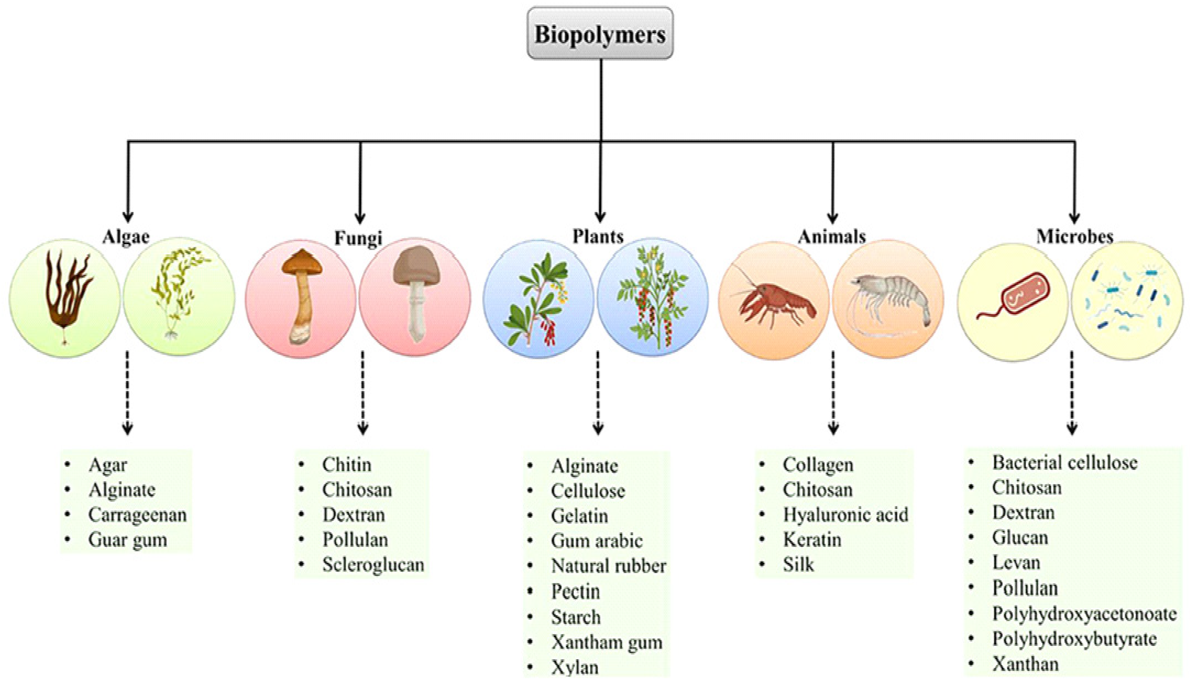
Figure 1. Biopolymers could be achieved for different sources. Adapted with permission from Ref. [23]. Copyright © 2023 Pathak, Singh, Singh, Sharma, Singh, Gupta, Mishra, Mishra and Tripathi.
Pharmaceutics 15 00153 g001
Figure 2. (a) Anionic polymers included in siRNA lipoplexes. (b) Different anionic polymers. Adapted with permission from Ref. [21]. Copyright © 2023–2022 John Wiley & Sons, Inc.
Pharmaceutics 15 00153 g002
Figure 3. GNPs prepared by the supramolecular approach exhibited a gene silencing mechanism with high efficiency and low toxicity. Adapted with permission from Ref. [63]. Copyright © 2023 American Chemical Society.
Pharmaceutics 15 00153 g003
Figure 4. Post-operation metastatic inhibition in breast tumors through CBSA/siS100A4@Exosome. Adapted with permission from Ref. [99]. Copyright @2022, Goyal, Chopra, singh, Dua and Gautam.
Pharmaceutics 15 00153 g004
Table 1. siRNA therapeutics in different stages of clinical trials.
siRNA DrugsDelivery SystemTarget
Gene
DiseaseCompany Status/
Phase
PatisiranLNP-siRNATTRTTR-mediated amyloidosisAlnylamApproved,
FDA 2018
GivosiranGalNAc-siRNAALAS1Acute hepatic porphyriaAlnylamApproved,
FDA 2019
LumasiranGalNAc-siRNAHAO1Primary heperoxaluria
type 1
AlnylamApproved,
FDA 2020
InclisiranGalNAc-siRNAPCSK9HypercholesterolemiaAlnylam
Novartis
Approved,
FDA 2021
Vutrisiran
(ALN-TTRSCO2)
GalNAc-siRNATTRTTR-mediated amyloidosisAlnylamIII
Nedosiran
(DCR-PHXC)
GalNAc-siRNALDHAPrimary heperoxaluriaAlnylamIII
Fitusiran
(ALN-AT3SC)
GalNAc-siRNAATHemophilia A and B and rare blood disorderAlnylam
Sanofi
Genzyme
III
Atu027Liposomal-siRNA, AtuPLEXPKN3Solid tumors, advanced or metastatic pancreatic cancerSilence TherapeuticsII
completed
TMK-PLK1LNP-siRNAPLK1Solid tumors, adrenal cortical carcinoma, hepatocellular carcinomaArbutus Biopharma II
completed
siG12D LODERsiG12D-LODER™
system and chemotherapy
G12D mutated KRASPancreatic cancer, pancreatic ductal adenocarcinomaSilenseed Ltd.II
ongoing
ALN-VSP02LNP-siRNAVEGF, KSPSolid tumorsAlnylamI
completed
Table 2. Preclinical studies of selected biopolymer-induced siRNA delivery platforms for breast cancer treatment.
Target NanoparticleCell LineReference
RhoAChitosan-coated polyisohexylcyanoacrylate
(PIHCA) NPs
MDA-MB-231[111]
SurvivinLipid substituted polymerMDA-MB-231[112]
HB-EGF Fab’s antibody-modified LNPMDA-MB-231[94]
EGFRFab conjugated liposomal NPs MDA-MB-231[95]
EGFRPMLA-based nanobioconjugateMDA-MB-468[106]
eGFPF3-targeted liposomal NPsMDA-MB-231
MDA-MB-435
[113]
VEGF-A, VEGFR-1, VEGFR-2, and neuropilin-1Chitosan nanocomplexesBreast tumor induced in rat using N-nitroso-N-methylurea [114]
Lipocalin-2ICAM-1 conjugated liposomesMDA-MB-231[97]
HREpCAM aptamer polyethylineimineMCF-7[93]
EGFRCPP-loaded nanobubbles MDA-MB-231[115]
CD44ExosomeMCF-7[109]
PIK3CABJExo MDA-MB-231, MDA-MB-453[104]
SUR, Bcl2Bovine milk exosomesMCF-7, MDA-MB-231[33]
ABCB1PEG-L-arginine oligo(-alkylaminosiloxane)-PEI polyplexMCF-7 ADR, MDA-MB-231[84]
Disclaimer/Publisher’s Note: The statements, opinions and data contained in all publications are solely those of the individual author(s) and contributor(s) and not of MDPI and/or the editor(s). MDPI and/or the editor(s) disclaim responsibility for any injury to people or property resulting from any ideas, methods, instructions or products referred to in the content.