
PEG-Free Polyion Complex Nanocarriers for Brain-Derived Neurotrophic Factor

 , ,
, ,  and
and 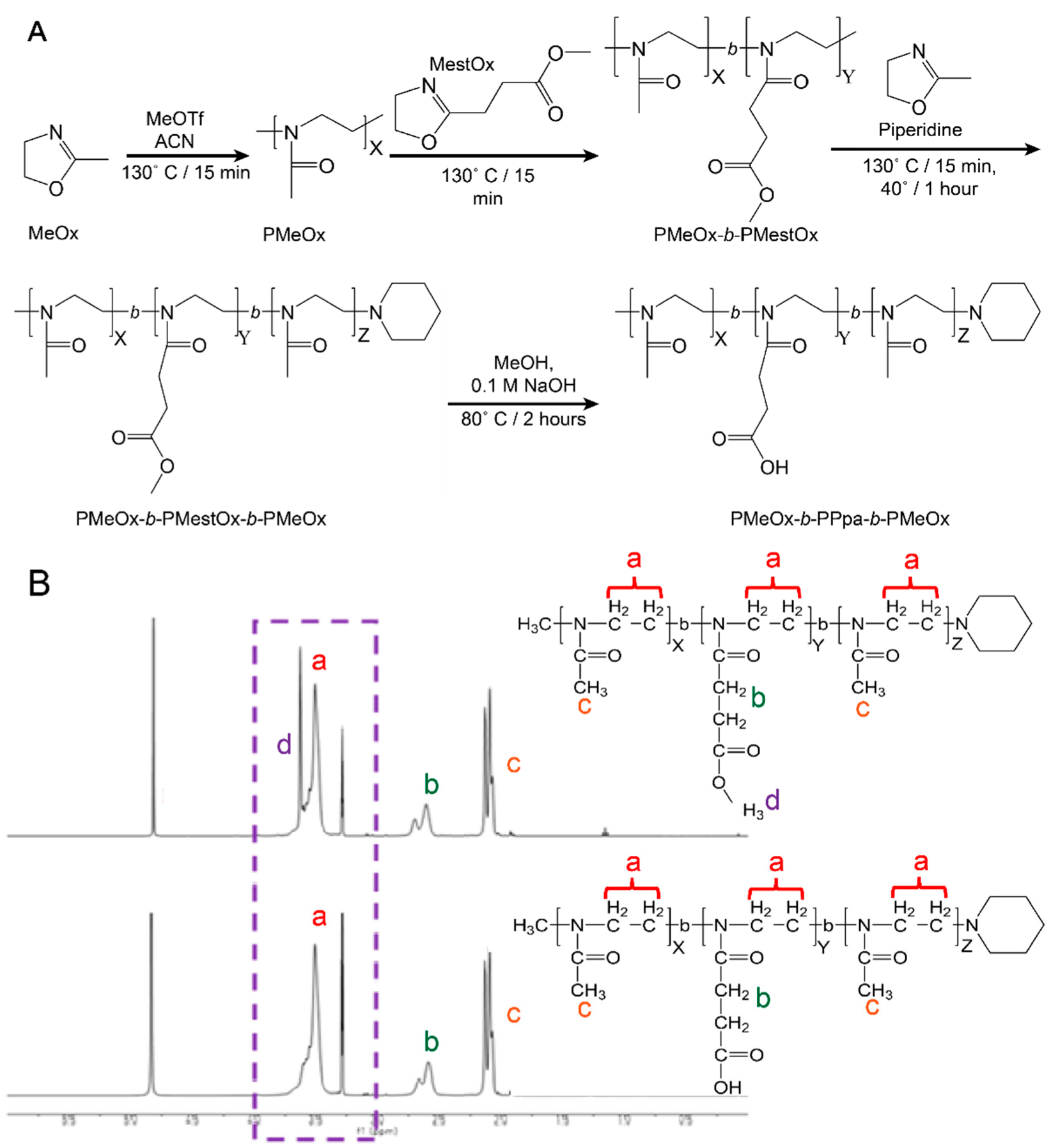
Abstract
:1. Introduction
2. Materials and Methods
2.1. Materials
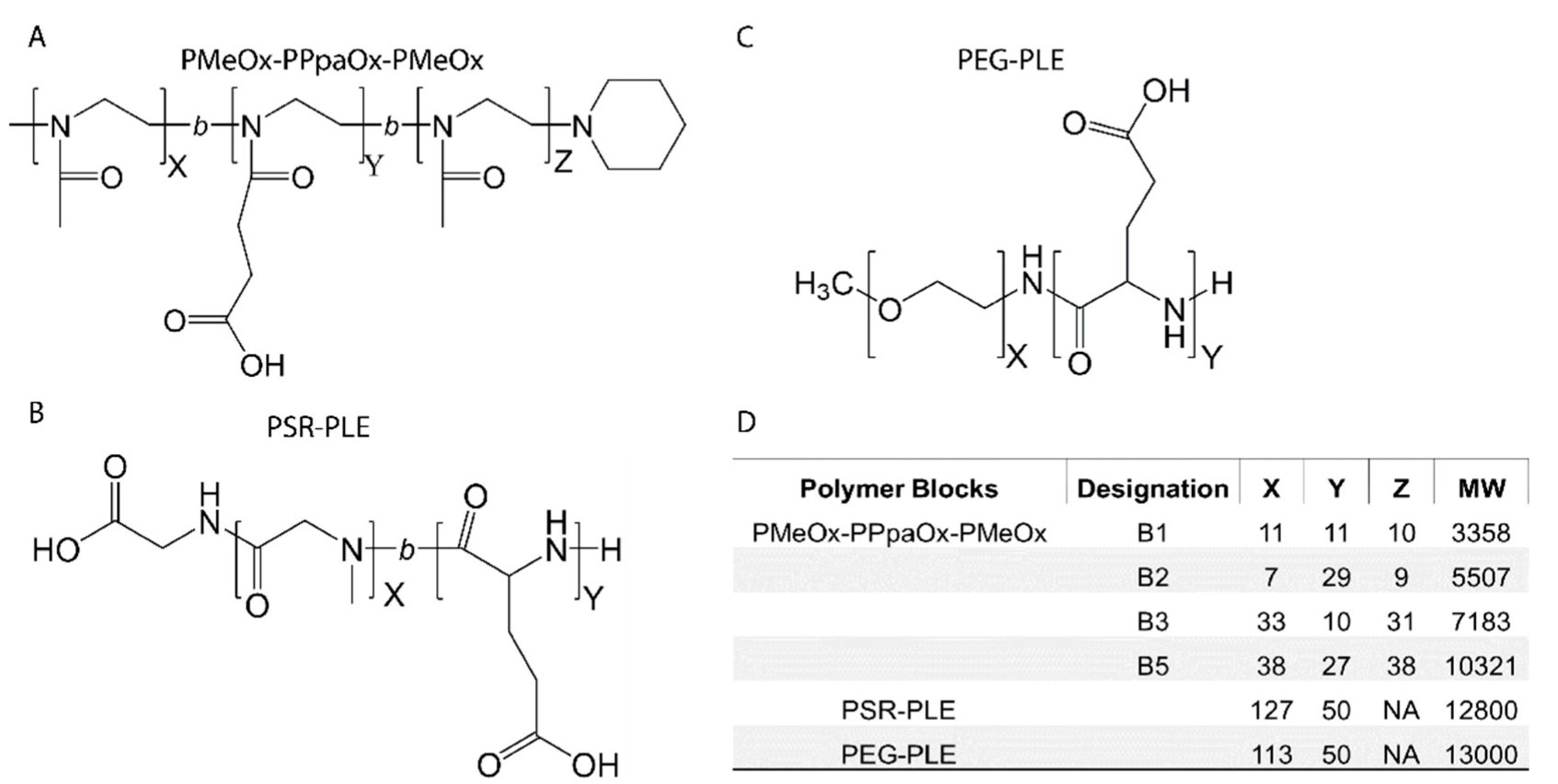
2.2. Polymer Synthesis
2.3. Polymer Characterization by 1H NMR
2.4. Preparation of PIC by Manual Mixing
2.5. Preparation of PIC Using a Microfluidic Mixer
2.6. Nanoparticle Size, Dispersity, and Zeta Potential
2.7. Transmission Electron Microscopy (TEM)
2.8. Horizontal Agarose Gel Electrophoresis (HAGE)
2.9. Isothermal Titration Calorimetry (ITC)
2.10. Potentiometric Titration of Polymers
2.11. Cell Culture
2.12. BDNF Stimulation
3. Results
3.1. Manufacture and Characterization of Poly(2-oxazoline) Block Copolymers
3.2. Nano-BDNF Candidate Innowing
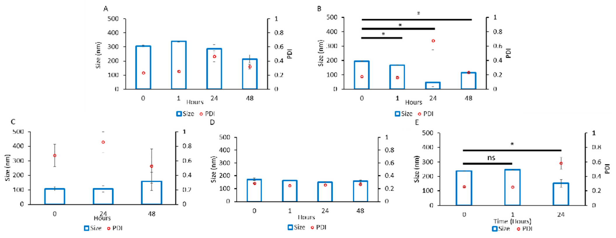
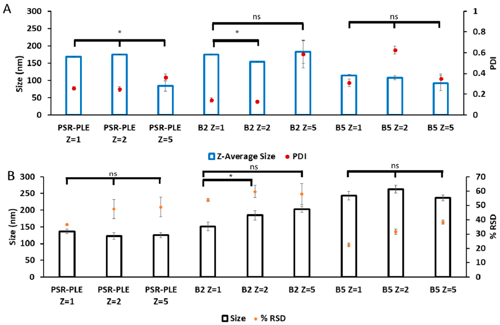
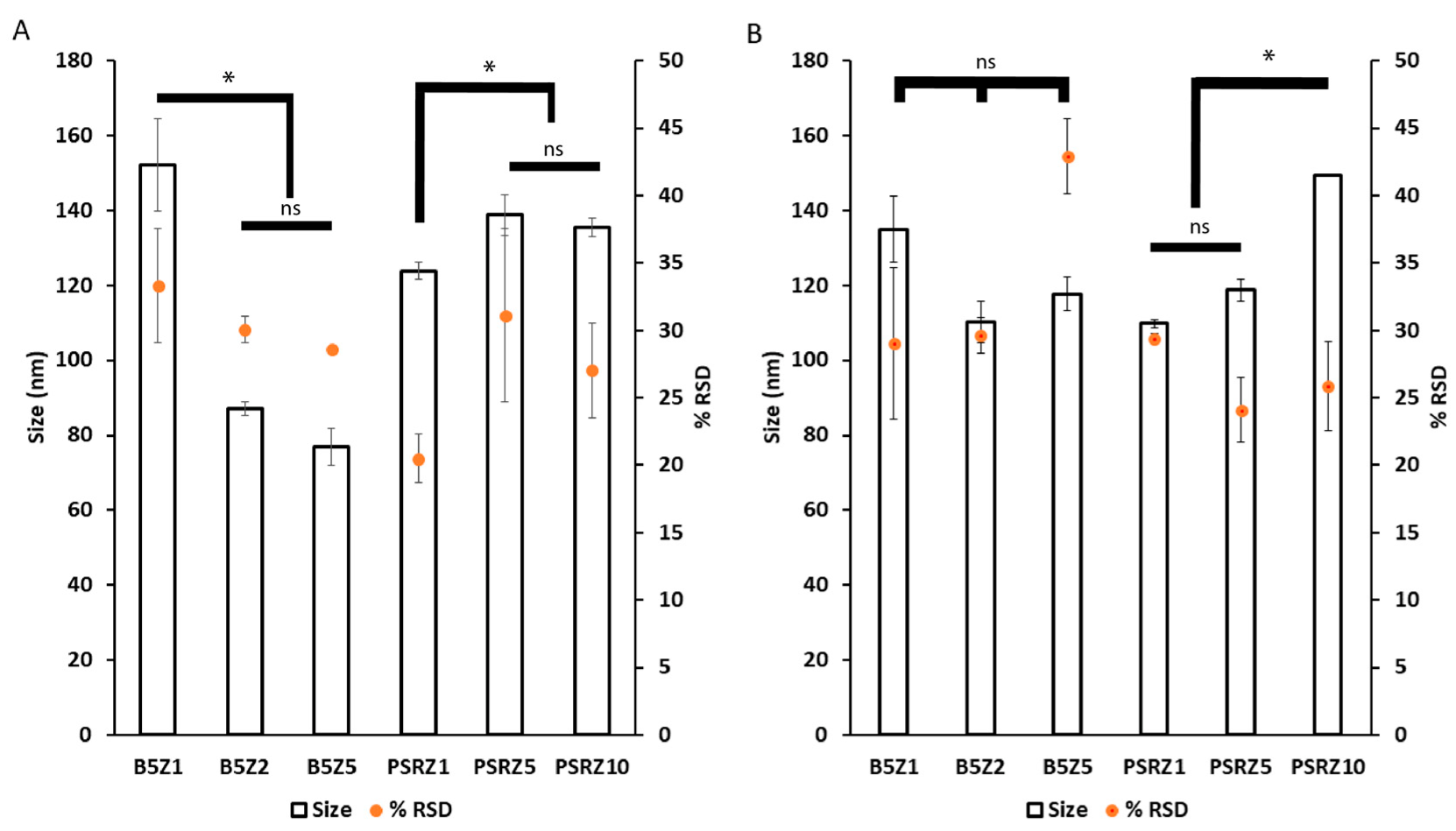
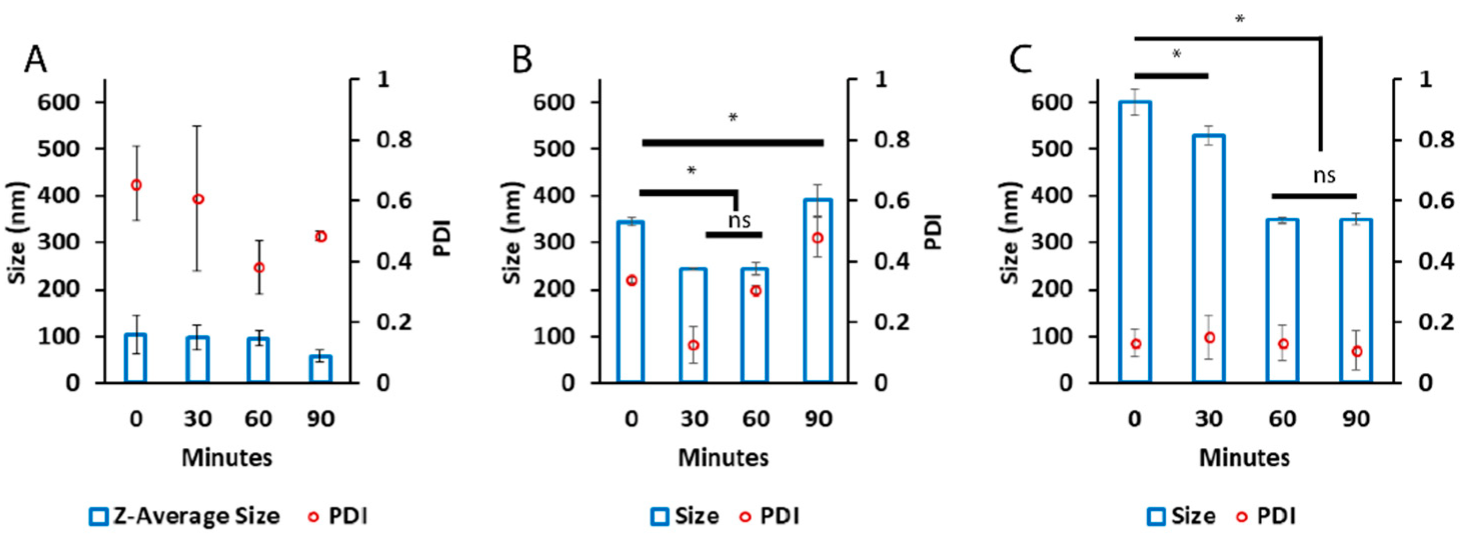
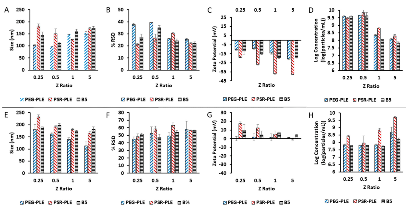
3.3. Preparation of Nano-BDNF Particles Suitable for Injection
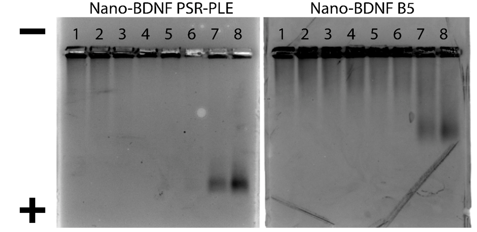
3.4. HAGE Visualization of Nano-BDNF
3.5. Zeta-Potential Characterization of Nano-BDNF
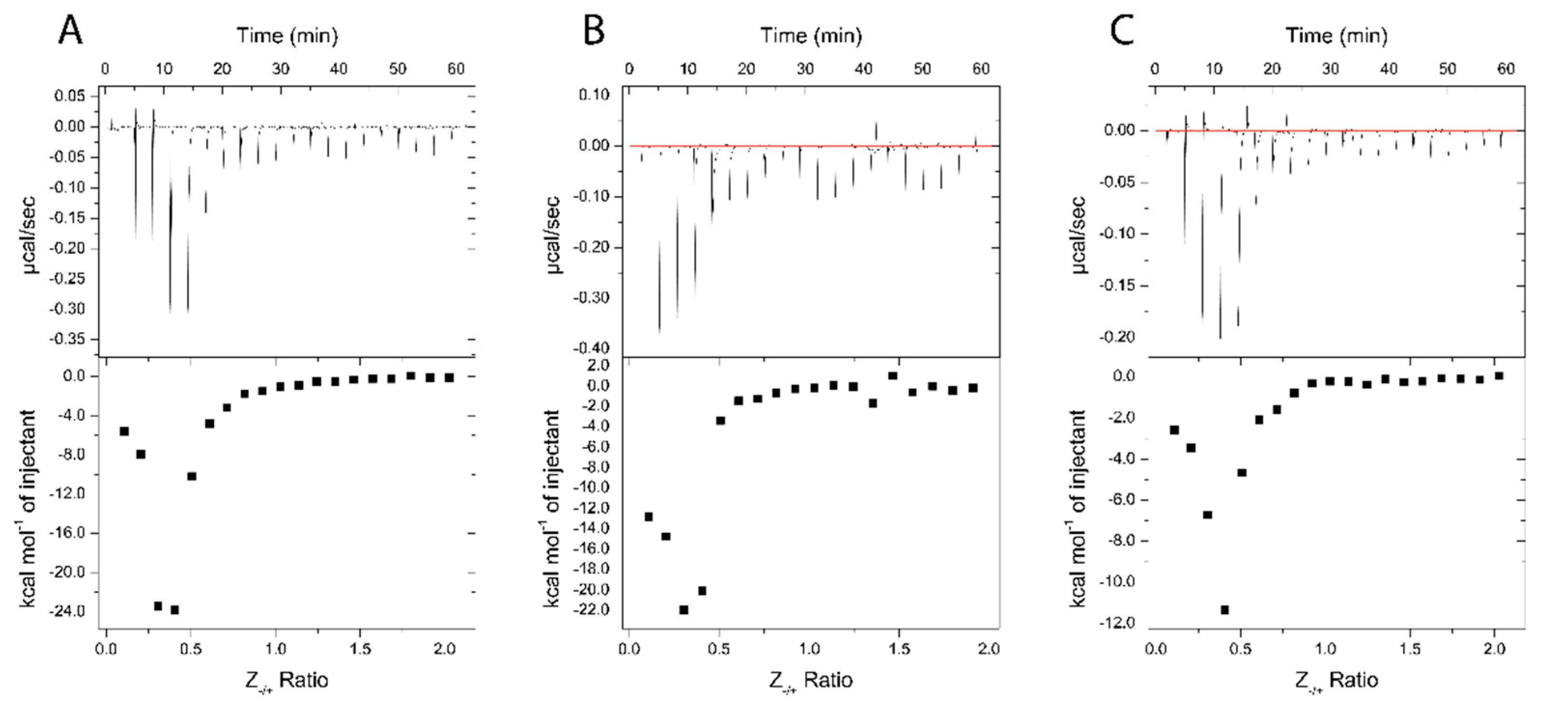
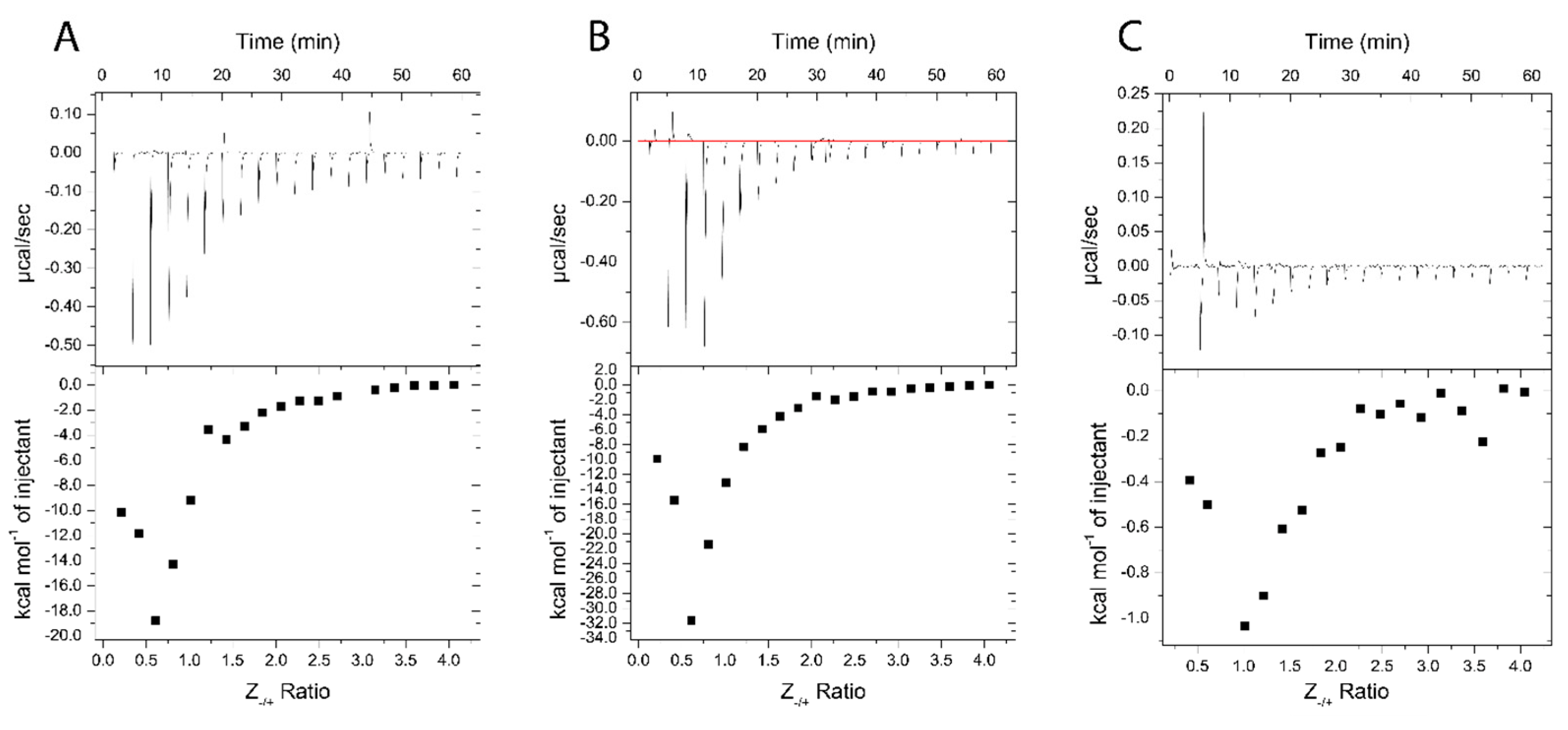
3.6. Thermodynamic Analysis of BDNF-Polyanion Association
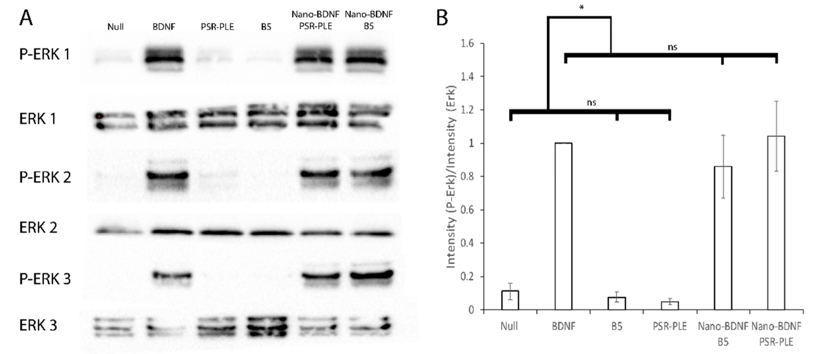
3.7. Stimulation of the TrkB Pathway
4. Discussion
5. Conclusions
Supplementary Materials
Author Contributions
Funding
Institutional Review Board Statement
Informed Consent Statement
Data Availability Statement
Acknowledgments
Conflicts of Interest
References
- Gref, R.; Minamitake, Y.; Peracchia, M.T.; Torchilin, V.; Langer, R.; Peracchia, T.; Trubetskoy, V.; Torchilin, V.; Langerllf, R. Biodegradable Long-Circulating Polymeric Nanospheres. Science 1994, 263, 1600–1603. [Google Scholar] [CrossRef] [PubMed] [Green Version]
- Armstrong, J.K. PEGylated Protein Drugs: Basic Science and Clinical Applications; Birkhauser: Basel, Switzerland, 2009; ISBN 9783764386788. [Google Scholar]
- Ishida, T.; Masuda, K.; Ichikawa, T.; Ichihara, M.; Irimura, K.; Kiwada, H. Accelerated Clearance of a Second Injection of PEGylated Liposomes in Mice. Int. J. Pharm. 2003, 255, 167–174. [Google Scholar] [CrossRef]
- Yang, Q.; Jacobs, T.M.; McCallen, J.D.; Moore, D.T.; Huckaby, J.T.; Edelstein, J.N.; Lai, S.K. Analysis of Pre-Existing IgG and IgM Antibodies against Polyethylene Glycol (PEG) in the General Population. Anal. Chem. 2016, 88, 11804–11812. [Google Scholar] [CrossRef] [PubMed]
- Risma, K.A.; Edwards, K.M.; Hummell, D.S.; Little, F.F. Potential Mechanisms of Anaphylaxis to COVID-19 MRNA Vaccines. J. Allergy Clin. Immunol. 2019, 147, 2075–2082. [Google Scholar] [CrossRef] [PubMed]
- Haddad, H.F.; Burke, J.A.; Scott, E.A.; Ameer, G.A. Clinical Relevance of Pre-Existing and Treatment-Induced Anti-Poly (Ethylene Glycol) Antibodies. Regen. Eng. Transl. Med. 2021, 8, 32–42. [Google Scholar] [CrossRef] [PubMed]
- Moghimi, S.M. Allergic Reactions and Anaphylaxis to LNP-Based COVID-19 Vaccines. Mol. Ther. 2021, 29, 898–900. [Google Scholar] [CrossRef]
- Warren, C.M.; Snow, T.T.; Lee, A.S.; Shah, M.M.; Heider, A.; Blomkalns, A.; Betts, B.; Buzzanco, A.S.; Gonzalez, J.; Chinthrajah, R.S.; et al. Assessment of Allergic and Anaphylactic Reactions to MRNA COVID-19 Vaccines with Confirmatory Testing in a US Regional Health System. JAMA Netw. Open 2021, 4, e212552. [Google Scholar] [CrossRef]
- Somiya, M.; Mine, S.; Yasukawa, K.; Ikeda, S. Sex Differences in the Incidence of Anaphylaxis to LNP-MRNA COVID-19 Vaccines. Vaccine 2021, 39, 3313–3314. [Google Scholar] [CrossRef]
- Harris, N.M.; Ritzel, R.; Mancini, N.; Jiang, Y.; Yi, X.; Manickam, D.S.; Banks, W.A.; Kabanov, A.V.; McCullough, L.D.; Verma, R. Nano-Particle Delivery of Brain Derived Neurotrophic Factor after Focal Cerebral Ischemia Reduces Tissue Injury and Enhances Behavioral Recovery. Pharmacol. Biochem. Behav. 2016, 150–151, 48–56. [Google Scholar] [CrossRef] [Green Version]
- Jiang, Y.; Fay, J.M.; Poon, C.D.; Vinod, N.; Zhao, Y.; Bullock, K.; Qin, S.; Manickam, D.S.; Yi, X.; Banks, W.A.; et al. Nanoformulation of Brain-Derived Neurotrophic Factor with Target Receptor-Triggered-Release in the Central Nervous System. Adv. Funct. Mater. 2018, 28, 1703982. [Google Scholar] [CrossRef]
- Nagahara, A.H.; Merrill, D.A.; Coppola, G.; Tsukada, S.; Schroeder, B.E.; Shaked, G.M.; Wang, L.; Blesch, A.; Kim, A.; Conner, J.M.; et al. Neuroprotective Effects of Brain-Derived Neurotrophic Factor in Rodent and Primate Models of Alzheimer’s Disease. Nat. Med. 2009, 15, 331–337. [Google Scholar] [CrossRef] [PubMed] [Green Version]
- Tsukahara, T.M.D.; Takeda, M.M.; Shimohama, S.M.; Ohara, O.P.; Hashimoto, N.M. Effects of Brain-Derived Neurotrophic Factor on 1-Methyl-4-Phenyl-1,2,3,6-Tetrahydropyridine-Induced Parkinsonism in Monkeys. Neurosurgery 1995, 37, 733–741. [Google Scholar] [CrossRef] [PubMed]
- Zhao, H.; Alam, A.; San, C.-Y.; Eguchi, S.; Chen, Q.; Lian, Q.; Ma, D. Molecular Mechanisms of Brain-Derived Neurotrophic Factor in Neuro-Protection: Recent Developments. Brain Res. 2017, 1665, 1–21. [Google Scholar] [CrossRef] [PubMed]
- Pan, W.; Banks, W.A.; Fasold, M.B.; Bluth, J.; Kastin, A.J. Transport of Brain-Derived Neurotrophic Factor across the Blood-Brain Barrier. Neuropharmacology 1998, 37, 1553–1561. [Google Scholar] [CrossRef]
- Veronese, F.M.; Mero, A.; Pasut, G. Protein PEGylation, Basic Science and Biological Applications. In PEGylated Protein Drugs: Basic Science and Clinical Applications; Veronese, F.M., Ed.; Birkhäuser: Basel, Switzerland, 2009; pp. 11–31. ISBN 978-3-7643-8679-5. [Google Scholar]
- Armstrong, J.K. The Occurrence, Induction, Specificity and Potential Effect of Antibodies against Poly(Ethylene Glycol). In PEGylated Protein Drugs: Basic Science and Clinical Applications; Veronese, F.M., Ed.; Birkhäuser: Basel, Switzerland, 2009; pp. 147–168. ISBN 978-3-7643-8679-5. [Google Scholar]
- Abuchowski, A.; Es, T.; Van Palczuk, N.C.; Davis, F.F. Alteration of Immunological Properties of Bovine Serum Albumin by Covalent Attachment of Polyethylene Glycol. J. Biol. Chem. 1977, 252, 3578–3581. [Google Scholar] [CrossRef]
- Konradi, R.; Acikgoz, C.; Textor, M. Polyoxazolines for Nonfouling Surface Coatings—A Direct Comparison to the Gold Standard PEG. Macromol. Rapid Commun. 2012, 33, 1663–1676. [Google Scholar] [CrossRef]
- Son, K.; Ueda, M.; Taguchi, K.; Maruyama, T.; Takeoka, S. Evasion of the Accelerated Blood Clearance Phenomenon by Polysarcosine Coating of Liposomes. J. Control. Release 2020, 322, 209–216. [Google Scholar] [CrossRef]
- Luxenhofer, R.; Han, Y.; Schulz, A.; Tong, J.; He, Z.; Kabanov, A.V.; Jordan, R. Poly(2-Oxazoline)s as Polymer Therapeutics. Macromol. Rapid Commun. 2012, 33, 1613–1631. [Google Scholar] [CrossRef] [Green Version]
- Li, Y.; Bronich, T.K.; Chelushkin, P.S.; Kabanov, A.V. Dynamic Properties of Block Ionomer Complexes with Polyion Complex Cores. Macromolecules 2008, 41, 5863–5868. [Google Scholar] [CrossRef]
- He, Z.; Miao, L.; Jordan, R.; S-Manickam, D.; Luxenhofer, R.; Kabanov, A.V. A Low Protein Binding Cationic Poly(2-Oxazoline) as Non-Viral Vector. Macromol. Biosci. 2015, 15, 1004–1020. [Google Scholar] [CrossRef] [Green Version]
- Tong, J.; Yi, X.; Luxenhofer, R.; Banks, W.A.; Jordan, R.; Zimmerman, M.C.; Kabanov, A.V. Conjugates of Superoxide Dismutase 1 with Amphiphilic Poly(2-Oxazoline) Block Copolymers for Enhanced Brain Delivery: Synthesis, Characterization and Evaluation in Vitro and in Vivo. Mol. Pharm. 2013, 10, 360–377. [Google Scholar] [CrossRef] [PubMed] [Green Version]
- Jiang, Y.; Brynskikh, A.M.; S-Manickam, D.; Kabanov, A.V. SOD1 Nanozyme Salvages Ischemic Brain by Locally Protecting Cerebral Vasculature. J. Control. Release 2015, 213, 36–44. [Google Scholar] [CrossRef] [PubMed] [Green Version]
- Barz, M.; Luxenhofer, R.; Zentel, R.; Vicent, M.J. Overcoming the PEG-Addiction: Well-Defined Alternatives to PEG, from Structure–Property Relationships to Better Defined Therapeutics. Polym. Chem. 2011, 2, 1900–1918. [Google Scholar] [CrossRef]
- Gao, S.; Holkar, A.; Srivastava, S. Protein—Polyelectrolyte Complexes and Micellar Assemblies. Polymers 2019, 11, 1097. [Google Scholar] [CrossRef] [PubMed]
- Miyata, K.; Nishiyama, N.; Kataoka, K. Rational Design of Smart Supramolecular Assemblies for Gene Delivery: Chemical Challenges in the Creation of Artificial Viruses. Chem. Soc. Rev. 2012, 41, 2562–2574. [Google Scholar] [CrossRef]
- He, Z.; Wan, X.; Schulz, A.; Bludau, H.; Dobrovolskaia, M.A.; Stern, S.T.; Montgomery, S.A.; Yuan, H.; Li, Z.; Alakhova, D.; et al. Biomaterials A High Capacity Polymeric Micelle of Paclitaxel: Implication of High Dose Drug Therapy to Safety and in vivo Anti-Cancer Activity. Biomaterials 2016, 101, 296–309. [Google Scholar] [CrossRef] [Green Version]
- Vinod, N.; Hwang, D.; Azam, S.H.; Van Swearingen, A.E.D.; Wayne, E.; Fussell, S.C.; Sokolsky-Papkov, M.; Pecot, C.V.; Kabanov, A.V. High-Capacity Poly(2-Oxazoline) Formulation of TLR 7/8 Agonist Extends Survival in a Chemo-Insensitive, Metastatic Model of Lung Adenocarcinoma. Sci. Adv. 2020, 6, eaba5542. [Google Scholar] [CrossRef]
- Seo, Y.; Schulz, A.; Han, Y.; He, Z.; Bludau, H.; Wan, X.; Tong, J.; Bronich, T.K.; Sokolsky, M.; Luxenhofer, R.; et al. Poly(2-Oxazoline) Block Copolymer Based Formulations of Taxanes: Effect of Copolymer and Drug Structure, Concentration, and Environmental Factors. Polym. Adv. Technol. 2015, 26, 837–850. [Google Scholar] [CrossRef]
- He, Z.; Schulz, A.; Wan, X.; Seitz, J.; Bludau, H.; Alakhova, D.Y.; Darr, D.B.; Perou, C.M.; Jordan, R.; Ojima, I.; et al. Poly (2-Oxazoline) Based Micelles with High Capacity for 3rd Generation Taxoids: Preparation, in Vitro and in Vivo Evaluation. J. Control. Release 2015, 208, 67–75. [Google Scholar] [CrossRef] [Green Version]
- Hwang, D.; Ramsey, J.D.; Makita, N.; Sachse, C.; Jordan, R.; Sokolsky-papkov, M.; Kabanov, A.V. Novel Poly (2-Oxazoline) Block Copolymer with Aromatic Heterocyclic Side Chains as a Drug Delivery Platform. J. Control. Release 2019, 307, 261–271. [Google Scholar] [CrossRef]
- Hwang, D.; Dismuke, T.; Tikunov, A.; Rosen, E.P.; Kagel, J.R.; Ramsey, J.D.; Lim, C.; Zamboni, W.; Kabanov, A.V.; Gershon, T.R.; et al. Poly (2-Oxazoline) Nanoparticle Delivery Enhances the Therapeutic Potential of Vismodegib for Medulloblastoma by Improving CNS Pharmacokinetics and Reducing Systemic Toxicity. Nanomed. Nanotechnol. Biol. Med. 2021, 32, 102345. [Google Scholar] [CrossRef]
- Zalipsky, S.; Hansen, C.B.; Oaks, J.M.; Allen, T.M. Evaluation of Blood Clearance Rates and Biodistribution of Poly(2-Oxazoline)-Grafted Liposomes. J. Pharm. Sci. 1996, 85, 133–137. [Google Scholar] [CrossRef]
- Wan, X.; Beaudoin, J.J.; Vinod, N.; Min, Y.; Makita, N.; Bludau, H.; Jordan, R.; Wang, A.; Sokolsky, M.; Kabanov, A.V. Biomaterials Co-Delivery of Paclitaxel and Cisplatin in Poly (2-Oxazoline) Polymeric Micelles: Implications for Drug Loading, Release, Pharmacokinetics and Outcome of Ovarian and Breast Cancer Treatments. Biomaterials 2019, 192, 1–14. [Google Scholar] [CrossRef] [PubMed]
- Hwang, D.; Ramsey, J.D.; Kabanov, A.V. Polymeric Micelles for the Delivery of Poorly Soluble Drugs: From Nanoformulation to Clinical Approval. Adv. Drug Deliv. Rev. 2020, 156, 80–118. [Google Scholar] [CrossRef] [PubMed]
- Lau, K.H.A. Peptoids for Biomaterials Science. Biomater. Sci. 2014, 2, 627. [Google Scholar] [CrossRef] [PubMed] [Green Version]
- Cheung, D.L.; Hang, K.; Lau, A. Atomistic Study of Zwitterionic Peptoid Antifouling Brushes. Langmuir 2018, 35, 1483–1494. [Google Scholar] [CrossRef]
- Huesmann, D.; Sevenich, A.; Weber, B.; Barz, M. A Head-to-Head Comparison of Poly(Sarcosine) and Poly(Ethylene Glycol) in Peptidic, Amphiphilic Block Copolymers. Polymer 2015, 67, 240–248. [Google Scholar] [CrossRef]
- Chan, B.A.; Xuan, S.; Li, A.; Simpson, J.M.; Sternhagen, G.L.; Yu, T.; Darvish, O.A. Polypeptoid Polymers: Synthesis, Characterization, and Properties. Biopolymers 2017, 109, e23070. [Google Scholar] [CrossRef]
- Hu, Y.; Hou, Y.; Wang, H.; Lu, H. Polysarcosine as an Alternative to PEG for Therapeutic Protein Conjugation. Bioconjug. Chem. 2018, 29, 2232–2238. [Google Scholar] [CrossRef]
- Soppet, D.; Escandon, E.; Maragos, J.; Middlemas, D.S.; Raid, S.W.; Blair, J.; Burton, L.E.; Stanton, B.R.; Kaplan, D.R.; Hunter, T. The Neurotrophic Factors Brain-Derived Neurotrophic Factor and Neurotrophin-3 Are Ligands for the TrkB Tyrosine Kinase Receptor. Cell 1991, 65, 895–903. [Google Scholar] [CrossRef]
- Luxenhofer, R.; Schulz, A.; Roques, C.; Li, S.; Bronich, T.K.; Batrakova, E.V.; Jordan, R.; Kabanov, A.V. Doubly-Amphiphilic Poly(2-Oxazoline)s as High Capacity Delivery Systems for Hydrophobic Drugs. Biomaterials 2011, 31, 4972–4979. [Google Scholar] [CrossRef] [PubMed] [Green Version]
- Kabanov, V.A. Polyelectrolyte Complexes in Solution and in Bulk. Russ. Chem. Rev. 2005, 74, 3–20. [Google Scholar] [CrossRef]
- Chelushkin, P.S.; Lysenko, E.A.; Bronich, T.K.; Eisenberg, A.; Kabanov, V.A.; Kabanov, A.V. Polyion Complex Nanomaterials from Block Polyelectrolyte Micelles and Linear Polyelectrolytes of Opposite Charge. 2. Dynamic Properties. J. Phys. Chem. B 2008, 112, 7732–7738. [Google Scholar] [CrossRef] [PubMed]
- Lu, M.; Ozcelik, A.; Grigsby, C.L.; Zhao, Y.; Guo, F.; Leong, K.W.; Huang, T.J. Microfluidic Hydrodynamic Focusing for Synthesis of Nanomaterials. Nano Today 2016, 11, 778–792. [Google Scholar] [CrossRef]
- Guimaraes Sa Correia, M.; Briuglia, M.L.; Niosi, F.; Lamprou, D.A. Microfluidic Manufacturing of Phospholipid Nanoparticles: Stability, Encapsulation Efficacy, and Drug Release. Int. J. Pharm. 2017, 516, 91–99. [Google Scholar] [CrossRef]
- Burns, M.L.; Malott, T.M.; Metcalf, K.J.; Hackel, B.J.; Chan, J.R.; Shusta, E.V. Directed Evolution of Brain-Derived Neurotrophic Factor for Improved Folding and Expression in Saccharomyces Cerevisiae. Appl. Environ. Microbiol. 2014, 80, 5732–5742. [Google Scholar] [CrossRef] [Green Version]
- ITC Data Analysis in Origin, 7th. ed.; MicroCal LLC: Northhampton, MA, USA, 2004.
- Ulbricht, J.; Jordan, R.; Luxenhofer, R. On the Biodegradability of Polyethylene Glycol, Polypeptoids and Poly(2-Oxazoline)S. Biomaterials 2014, 35, 4848–4861. [Google Scholar] [CrossRef]
- Koshkina, O.; Westmeier, D.; Lang, T.; Bantz, C.; Hahlbrock, A.; Würth, C.; Resch-Genger, U.; Braun, U.; Thiermann, R.; Weise, C.; et al. Tuning the Surface of Nanoparticles: Impact of Poly(2-Ethyl-2-Oxazoline) on Protein Adsorption in Serum and Cellular Uptake. Macromol. Biosci. 2016, 16, 1287–1300. [Google Scholar] [CrossRef]
- Kim, W.; Yamasaki, Y.; Jang, W.D.; Kataoka, K. Thermodynamics of DNA Condensation Induced by Poly(Ethylene Glycol)-Block-Polylysine through Polyion Complex Micelle Formation. Biomacromolecules 2010, 11, 1180–1186. [Google Scholar] [CrossRef]
- Kim, W.; Yamasaki, Y.; Kataoka, K. Development of a Fitting Model Suitable for the Isothermal Titration Calorimetric Curve of DNA with Cationic Ligands. J. Phys. Chem. B 2006, 110, 10919–10925. [Google Scholar] [CrossRef]
- Bronich, T.; Kabanov, A.V.; Marky, L.A. A Thermodynamic Characterization of the Interaction of a Cationic Copolymer with DNA. J. Phys. Chem. B 2001, 105, 6042–6050. [Google Scholar] [CrossRef]
- Friedman, N. Hydrogen Bonding and Heat of Solution. J. Chem. Educ. 1977, 54, 248. [Google Scholar] [CrossRef]
- Kabanov, A.V.; Bronich, T.K.; Kabanov, V.A.; Yu, K.; Eisenberg, A. Soluble Stoichiometric Complexes from Poly (N-Ethyl-4-Vinylpyridinium) Cations and Poly (Ethylene Oxide)-Block-Polymethacrylate Anions. Society 1996, 9297, 6797–6802. [Google Scholar] [CrossRef] [Green Version]
- Borukhov, I.; Andelman, D.; Borrega, R.; Cloitre, M.; Leibler, L.; Orland, H. Polyelectrolyte Titration: Theory and Experiment. J. Phys. Chem. B 2000, 104, 11027–11034. [Google Scholar] [CrossRef] [Green Version]
- Bodnarchuk, M.S.; Doncom, K.E.B.; Wright, D.B.; Heyes, D.M.; Dini, D.; O’Reilly, R.K. Polyelectrolyte PKa from Experiment and Molecular Dynamics Simulation. RSC Adv. 2017, 7, 20007–20014. [Google Scholar] [CrossRef] [Green Version]


Publisher’s Note: MDPI stays neutral with regard to jurisdictional claims in published maps and institutional affiliations. |
© 2022 by the authors. Licensee MDPI, Basel, Switzerland. This article is an open access article distributed under the terms and conditions of the Creative Commons Attribution (CC BY) license (https://creativecommons.org/licenses/by/4.0/).
Share and Cite
Fay, J.M.; Lim, C.; Finkelstein, A.; Batrakova, E.V.; Kabanov, A.V. PEG-Free Polyion Complex Nanocarriers for Brain-Derived Neurotrophic Factor. Pharmaceutics 2022, 14, 1391. https://doi.org/10.3390/pharmaceutics14071391
Fay JM, Lim C, Finkelstein A, Batrakova EV, Kabanov AV. PEG-Free Polyion Complex Nanocarriers for Brain-Derived Neurotrophic Factor. Pharmaceutics. 2022; 14(7):1391. https://doi.org/10.3390/pharmaceutics14071391
Chicago/Turabian StyleFay, James M., Chaemin Lim, Anna Finkelstein, Elena V. Batrakova, and Alexander V. Kabanov. 2022. "PEG-Free Polyion Complex Nanocarriers for Brain-Derived Neurotrophic Factor" Pharmaceutics 14, no. 7: 1391. https://doi.org/10.3390/pharmaceutics14071391
APA StyleFay, J. M., Lim, C., Finkelstein, A., Batrakova, E. V., & Kabanov, A. V. (2022). PEG-Free Polyion Complex Nanocarriers for Brain-Derived Neurotrophic Factor. Pharmaceutics, 14(7), 1391. https://doi.org/10.3390/pharmaceutics14071391