Article

Lambda gpP-DnaB Helicase Sequestration and gpP-RpoB Associated Effects: On Screens for Auxotrophs, Selection for RifR, Toxicity, Mutagenicity, Plasmid Curing

Department of Microbiology and Immunology, College of Medicine, University of Saskatchewan, Saskatoon, SK S7N 5E5, Canada
*
Author to whom correspondence should be addressed.
Viruses 2016, 8(6), 172; https://doi.org/10.3390/v8060172
Submission received: 18 March 2016 / Revised: 25 May 2016 / Accepted: 9 June 2016 / Published: 22 June 2016
(This article belongs to the Section Bacterial Viruses)

Abstract

:
The bacteriophage lambda replication initiation protein P exhibits a toxic effect on its Escherichia coli (E. coli) host, likely due to the formation of a dead-end P-DnaB complex, sequestering the replicative DnaB helicase from further activity. Intracellular expression of P triggers SOS-independent cellular filamentation and rapidly cures resident ColE1 plasmids. The toxicity of P is suppressed by alleles of P or dnaB. We asked whether P buildup within a cell can influence E. coli replication fidelity. The influence of P expression from a defective prophage, or when cloned and expressed from a plasmid was examined by screening for auxotrophic mutants, or by selection for rifampicin resistant (RifR) cells acquiring mutations within the rpoB gene encoding the β-subunit of RNA polymerase (RNAP), nine of which proved unique. Using fluctuation assays, we show that the intracellular expression of P evokes a mutator effect. Most of the RifR mutants remained PS and localized to the Rif binding pocket in RNAP, but a subset acquired a PR phenotype, lost sensitivity to ColE1 plasmid curing, and localized outside of the pocket. One PR mutation was identical to rpo*Q148P, which alleviates the UV-sensitivity of ruv strains defective in the migration and resolution of Holliday junctions and destabilizes stalled RNAP elongation complexes. The results suggest that P-DnaB sequestration is mutagenic and supports an earlier observation that P can interact with RNAP.

Graphical Abstract

1. Introduction

A lambda (λ) prophage is maintained within the chromosome of Escherichia coli (E. coli) by an action of the CI repressor encoded by gene cI. The inhibition of replication initiation from the prophage oriλ site positioned midway within gene O [1,2,3] (Figure 1A) is blocked by CI protein binding to operator sites overlapping promoters directing transcription leftward from pL, e.g., copying genes N-int, or rightward from pR for expressing genes cro-cII-O-P-Q. The product of gene P (gpP, P) participates in loading the E. coli DnaB helicase [4] onto DNA during formation of a DnaB:P:O:oriλ preprimosomal complex [5,6,7], each factor being required for replication initiation from oriλ. DnaB unwinds double-stranded (ds) DNA at the replication fork, by encircling and translocating along the 5′ lagging strand using energy provided by ATP hydrolysis [4]. The interaction between DnaG primase and the N-terminal end of DnaB [8] increases both the NTPase and helicase activities of DnaB and the synthesis of RNA primers by DnaG [4,8,9,10,11,12,13,14,15]. DnaB can promote the progression of Holliday junctions, believed important in the repair of DNA damage occurring near advancing replication forks [16,17]. DnaB functions as a hexamer, with about 20 hexamers per cell [18,19], with each binding up to six ATP [20,21]. It forms a complex with the host replication initiation protein DnaC to which the majority of DnaB in a cell is bound [22]. This interferes with the intrinsic single-stranded (ss) DNA binding activity of DnaB [20,22,23,24,25,26]. The DnaC-DnaB complex acquires cryptic ssDNA binding activity specific to the bacterial origin of replication, oriC, where it begins unwinding dsDNA after a number of complex changes [27,28,29,30,31,32,33,34].
The λ P protein has evolved to compete for and dissociate DnaC-DnaB complexes [5,38] and two to six P monomers can bind to every DnaB hexamer [5,39,40], forming an enzymatically inactive “dead-end” complex [22]. High levels of P were found deleterious to host cells [41,42] and it was suggested that P sequestered, i.e., bound-up, DnaB and thus interfered with the initiation of host DNA synthesis. ColE1 replication establishment or propagation [43] is extremely sensitive to P [35]. The effect is suppressed by alleles of dnaB or P, which suggests that P can impact a cellular replication event by acting outside of the DnaA and oriC-dependent replication initiation step. This same study showed that P-dependent cellular filamentation arose in cells defective for SOS induction, suggesting that P was influencing replication propagation or restart mechanisms. For example, the sequestration of DnaB by P could perturb or impede origin-independent stable DNA replication [44,45,46,47,48], where both cellular replication restart and ColE1 replication depend upon PriA helicase activity [49,50,51,52] at R-loops.
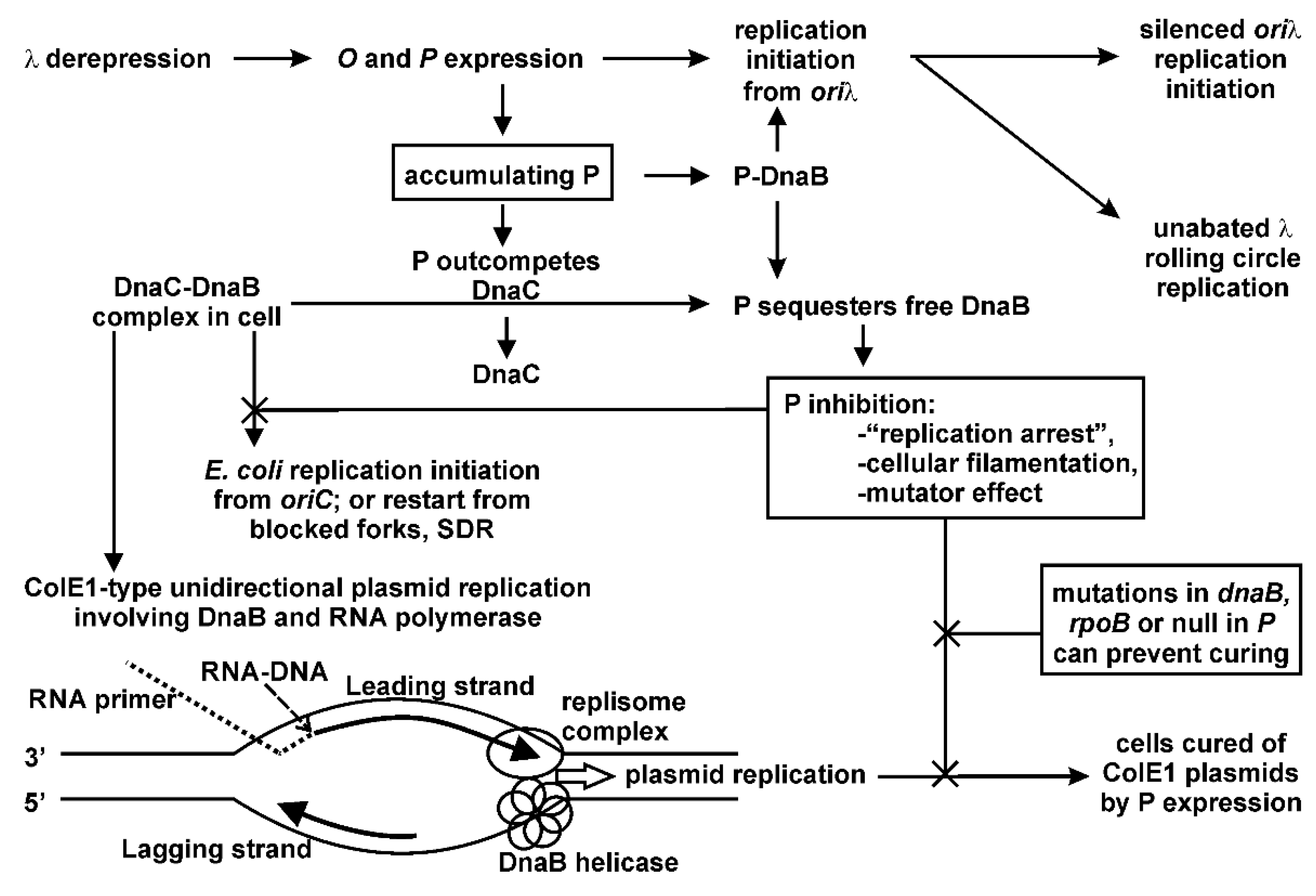
Multiple possibilities present for the P-lethality/inhibition observations, and are summarized in Figure 2. The 233-amino acid P protein is very stable, with a half-life of an hour [53,54]. In addition to binding Oλ [55], P interacts with the host proteins DnaA [56,57,58], DnaB [59,60,61], GrpE [62], DnaJ and DnaK [63], and possibly RNA polymerase (RNAP) [64]. As is understood from in vitro studies, the Hsp70 chaperone complex removes P from DnaB when the P-DnaB complex is bound to oriλ:O [40] in a two-step reaction involving DnaJ binding to complexed P-DnaB, which enhances P-DnaK binding, ATP hydrolysis, and the release of a P-DnaK-ADP complex. There is a requirement for a high concentration of DnaK unless GrpE is present, and a proposal that DnaK changes the conformation of P from a native to a folded state that is no longer able to bind DnaB [65]. An existing hypothesis for P-lethality/inhibition is that by binding up DnaB (or DnaA), P will inhibit chromosomal replication initiation from oriC. Greater understanding of the extent of intracellular binding between P and DnaB, which occurs beyond that involved in the formation of an oriλ preprimosomal complex is required to help explain DnaB sequestration. We showed that: (a) very low levels of P are necessary for the limited oriλ initiation required for λ replication [35]; and (b) that λ replication resulting in high phage burst from an induced λcI[Ts]857 Sam7 prophage blocked for cell lysis occurred in cells that were simultaneously fully derepressed for P expression from pcIpR-P-timm—conditions where DnaB would be sequestered.
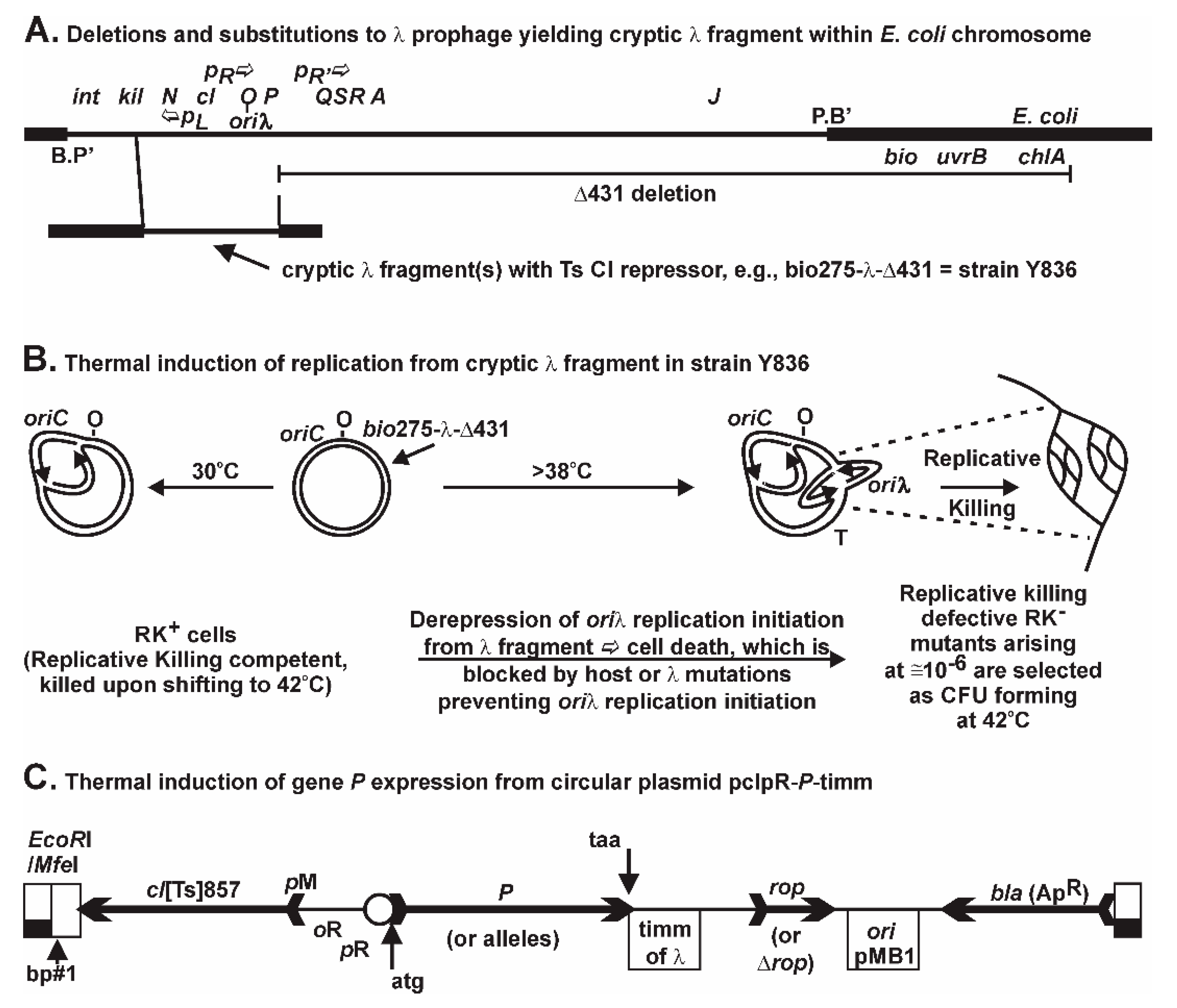
The initiation of oriλ replication from a chromosomally integrated, but non-excisable λ gene fragment (Figure 1A) is repressed by the cI [Ts] repressor. When CI[Ts] is inactivated upon shifting cells growing at 30 °C to 42 °C, O-P are transcribed, replication initiation arises at oriλ, and the Replicative Killing, RK+, phenotype is triggered, resulting in massive cell death [66]. Rare mutations nullifying the RK+ phenotype permit the selection for colony forming units (CFU) at 42 °C, each representing RK mutants in host or prophage genes (not merely in P) that block some aspect of λ replication initiation [66,67,68,69]. For example, we characterized within RK mutants small deletions in O that shift the reading frame and yield close-by stop codons polar for downstream gene expression. These RK mutants form CFU at 42 °C, likely surviving by limiting downstream P expression. Thus, it appears these induced mutants can tolerate some level of λ P expression (or that of any other toxic λ fragment protein, such as the unstable cII gene product). Since P expression had been shown to be toxic, confusion existed relative to its contribution to the RK+ phenotype. We suggested the terms cis Replicative Killing and trans P-lethality to distinguish mechanistically these two ideas. We found that the RK+ phenotype was uniquely dependent upon multiple, non-repairable λ replication forks arising from oriλ within a trapped prophage fragment, resulting in very rapid, nonreversible cell death, whereas there are likely multiple possibilities for P-lethality, which can be reversed even after hours of P expression [35]. In short, limited levels of P can be metabolized. The dnaBgrpD55 allele [70], which encodes missense mutations V256I and E426K [35] was shown to fully prevent oriλ replication initiation and to suppress cell killing by 106-fold from the lambda prophage fragment in strain Y836 (Figure 2C in reference [71]) when the cells were shifted from 30 °C to 42 °C, even though P expression remained constitutive from the induced defective prophage. This allele was shown Ts for λ replication but not for E. coli cell growth [70]. When removing the complication of the defective prophage and simply expressing P from a plasmid, we found that the same allele of dnaB suppressed the various P-lethality phenotypic manifestations [35]. This suggested that the P-lethality phenotype was dependent upon a P-DnaB interaction, possibly DnaB sequestration, and required further study.
In order to test whether P-DnaB interactions influence cellular replication events around the chromosome, rather than just at oriC, we explored a hypothesis that P buildup within a cell can perturb host replication fidelity. Since blocks to replication restart can arise around the chromosome, we first surveyed for an increase in the appearance of auxotrophic mutations of any type, and then for a targeted increase in rifampicin resistant (RifR) mutations arising in rpoB. The assumption made was that perturbing replication restart can be error prone. Herein, we observed a λ-dependent mutator effect that was linked to P. We observed that P expression creates the potential for sequestration of DnaB and results in a dramatic increase in cellular mutagenesis, which is nullified by inactivated or altered alleles of P and by an allele of dnaB.

2. Results

2.1. Examination for Auxotrophs within RK Population

To determine if the events related to replication initiation from oriλ influence cellular mutation, we asked if the selected RK clones acquire additional untargeted mutations within the chromosome. Since there are hundreds of host genes involved in cell metabolism [72], representing a very large genetic target of perhaps 1/6th of the chromosome, we examined whether any of the RK clones derived from a prototrophic RK+ host acquired an auxotrophic phenotype. If mutations conferring auxotrophy arise during the selection for RK mutants, then individual RK clones will appear at higher frequency when their selection is plated on RM compared to MM where they cannot form a colony. Accordingly, the ratio of RK CFU arising on RM/MM should be greater than unity. Indeed, this was observed (refer to rightmost columns in Table 1 and Table 2).
In order to undertake this experiment we first prepared a His+ transductant of Y836 his (moved from host 594) (Figure 1A), and then moved the λ cIII-ren fragment from Y836 into prototrophic host strains 594 and W3101 to create 594::(cIII-ren)λ and W3101::(cIII-ren)λ. We next demonstrated that these RK+ strains and their parents plated with equal efficiency on rich (RM) and minimal (MM) media (Table S1) at 30 °C. Then, RK CFU from Y836 his and the Y836 His+ transductant were isolated on RM, MM, or MM supplemented with histidine, biotin, or Casamino acids (not vitamin-free), and the RK frequency was compared (Table 1). The appearance of RK CFU was equivalent on RM or Casamino acids-supplemented MM (shown to be equivalent to RM), but was lower on un-supplemented MM, which did not support the growth of many RK mutants acquiring an auxotrophic mutation. This suggested that auxotrophic mutations were co-selected during the selection for RK clones. Thirty-seven RK isolates from Y836 his were screened for their auxotrophic defect [73] using the stabbing technique of Holliday [74], but including histidine in all of the supplemented MM plates. Sixteen categories of auxotrophs were identified. Some were further characterized by reversion analysis and yielded revertants at frequencies between 9.5 × 10⁻⁸ and 1.3 × 10⁻⁷, suggesting that the auxotrophs had acquired missense mutations rather than deletions, which agreed with a parallel observation that a high proportion of the RK clones had acquired a Ts auxotrophic phenotype when plated on MM. Since there was no obvious difference between the original Y836 his grown on MM + histidine and the Y836 His+ transductant grown on MM, we continued to use the original Y836 his strain to avoid any unaccounted transduced traits moved into Y836 His+, and hereafter refer to Y836 his as Y836.
Is an intrinsic, λ-independent, mutator effect in Y836 cells responsible for the selection of auxotrophic markers within the selected RK CFU? RK CFU were selected on RM and MM using prototrophic 594 and W3101 cells into which the (cIII-ren)λ fragment was moved by transduction from Y836. The frequency of RK CFU from both 594::(cIII-ren)λ and W3101::(cIII-ren)λ cells was reduced on MM compared to that found on RM. The similarity of the results to those with Y836 cells suggested the effect was linked to the (cIII-ren)λ fragment and not to a “λ-independent” activity activated at 42 °C (Table 1). Y836 was made lexA3[Ind] or ΔrecA to block SOS induction, or made dinB (DNA polymerase IV) or umuCumuD (DNA polymerase V) to prevent DNA damage tolerance [75]. Neither removing the capacity for SOS induction nor Pol IV or Pol V activities eliminated the appearance of auxotrophs within the selected RK CFU (Table 1). What is responsible for the RK frequency being higher on RM than on MM?

2.2. Hypothesis of P as Mutator, Evaluating Using Screen for Auxotrophy

Table 2 reveals that the RM/MM CFU ratio remains at unity for all strains where λ-fragment derepression was blocked by pCI+ but was elevated when oriλ replication initiation was blocked but transcription of the λ fragment was allowed at 42 °C. This suggests that the putative mutator effect accounting for the reduction in colony formation on MM plates at 42 °C is linked to the induction of gene expression from the (cIII-ren)λ fragment but that it does not require actual replication initiation from oriλ. The conclusions from Table 2 are limited to the suggestion that any of the inducible λ fragment gene products could be responsible for the increased recovery of auxotrophs on RM vs. MM. However, it is important to insert prior information that is relevant to this experiment: we previously found (Figure 2C in reference [71]) that the addition to strain Y836 of plasmid pCI+ expressing wild-type immλ cI repressor, or the addition by transduction of the dnaBgrpD55 allele, each prevented replication initiation and blocked Replicative Killing when cells with a λ prophage fragment were shifted from 30 °C to 42 °C. The plasmid pCI+ blocked both derepression of λ fragment transcription and replication initiation from oriλ. The dnaBgrpD55 allele fully suppressed Replicative Killing of Y836 cells shifted from 30 °C to 42 °C (Figure 2C in [71]), even better than with the pCI+ plasmid, but did not block the induced expression of the λ fragment genes. In addition, we previously compared cell viability (Table 6 in reference [35]) for 594 dnaBgrpD55[pcIpR-P-timm] cells grown up in culture at 25 °C by plating them at 25 °C (cell viability assumed 1.0), 37 °C, 39 °C (viabilities averaged 0.99), and 42 °C (viability 1.0). This showed that the dnaBgrpD55 allele did not impart cellular toxicity over this temperature range, and that it could completely suppress the toxicity resulting from P expression from this plasmid. In comparison, 594[pcIpR-P-timm] cells had a viability of 0.001 when plated at 42 °C. Therefore, it can obliquely be argued from Table 2 that the increase in the RM/MM CFU ratio for the dnaBgrpD55 cells is more complex than simply equating it to a manifestation of cell toxicity.
To examine the suggestion that λ gene expression could contribute to the mutator effect, a stab assay was evoked to screen for the proportion of acquired auxotrophic mutations within selected RK CFU, Table 3. This permitted a direct analysis of the linkage and potential co-selection between prophage induction and the appearance of auxotrophs within selected RK mutants. Using strain Y836 his as an example, the frequency of RK CFU selected at 42 °C on RM (for ten independent selections, all values × 10−6) was: 16.9, 11.2, 13.5, 16.5, 2, 3.5, 1.1, 5.6, 1.3, and 0.9. Individual CFU arising on RM plates incubated at 30 °C were stabbed to MM plates incubated at 30 °C to check whether any spontaneous auxotrophs arose within the starting cells. Similarly, all the selected CFU (large or tiny) arising per RM plate(s) that were incubated at 42 °C and yielded ~10 to ~100 CFU/selection plate were picked with sterile toothpicks onto two MM+Histidine plates. One stab plate was incubated at 30 °C and another at 42 °C to determine if the CFU possessed a Ts auxotrophic mutation, and a parallel set were stabbed to an RM plate incubated at 30 °C to ensure transfer of cells had occurred. All stab plates were incubated for 48 h. Potential auxotrophs were picked from the RM control plate and streaked onto MM for auxotrophy confirmation. Stabs for RK mutants from Y836 were also made to MM+His+biotin plates, which allowed us to demonstrate that the drop in RK frequency on MM relative to RM was not frequently linked to loss of the bio operon introduced by the bio275 addition (Figure 1A), which if deleted along with the λ fragment would confer a Bio phenotype. In summary, among CFU arising at 30 °C or 42 °C from strains without the (cIII-ren)λ fragment, or where the fragment was blocked for λ gene expression by pCI+, less than 0.01% were auxotrophs. In contrast, for strains with the (cIII-ren)λ fragment that were derepressed for λ gene expression, about 3.4% of the CFU forming at 42 °C were auxotrophs. No auxotrophs were observed among the stabbed CFU when the Y836 strain was engineered to contain a partial gene replacement substituting KanR within P, or if P was inactivated as in the RK mutant 566a with a spontaneous IS2 insertion in P. The observation that cell growth, with concomitant λ gene P expression, elevates the frequency of CFU acquiring an auxotrophic phenotype suggests the hypothesis that P expression confers a mutator phenotype in growing cells.

2.3. P Expression Stimulates Selection for Rifampicin-Resistant (RifR) CFU

To examine if the influence of P expression could be duplicated using a different scheme, we switched from screening for auxotrophs to a direct forward selection for rifampicin resistant (RifR) CFU. The influence of P expression was measured in cells derepressed for the (cIII-ren)λ fragment, or 594 cells transformed with plasmid pcIpR-P-timm (Figure 1C) in Table 4. When P is expressed in cells with a dnaB+ allele there is an enormous increase in the number of selected RifR CFU. Inactivation of P by insertion, deletion, or alteration by a point mutation (Pπ39991) nullified the mutator effect of P expression, as did replacing dnaB+ with the dnaBgrpD55 allele. Both the prophage and plasmid data suggest that a P-DnaB interaction is involved in the P-mutator phenotype.
When the RK+ phenotype is induced (i.e., combining cis-killing with trans-P-lethality) the frequency of RifR mutants was stimulated by >20-fold over situations with trans-P-lethality, but lacking cis-killing. For example, in Table 4: divide the RifR stimulation factors 88,727, 42,500, or 51,485 for the O+ P+ RK+ strains Y836 and 594::(cIII-cI857-O+-P+-ren)λ by the factor of 2167 for the P+ strain defective in O, i.e., Y836 O223a P+. Mutant O223a carries a deletion in O resulting in a frameshift, and a premature nonsense codon that may evoke polarity for the expression of downstream P. The 20-fold enhancement in rpoB mutagenesis when the RK+ phenotype is induced requires further explanation. Do higher levels of P yield progressively higher mutagenic consequences? Additionally, further properties of the DnaBgrpD55 protein, i.e., beyond its ability to block oriλ replication initiation [71] and to nullify P- lethality [35], remain to be explored.
Two fluctuation assays (FA) were employed to determine if the RifR mutations arising in rpoB preexisted the induction of P expression. In FA#1, we grew up 40 FA tubes of cells for 26–27 generations to saturation at 25 °C (without any P expression) [77]. From each of the 40 tubes, parallel aliquots were removed (each aliquot representing 1−2 × 10⁸ CFU) and spread on RM+100 µg/mL rifampicin plates (Table 5). One plate from the split aliquots was incubated at 25 °C and the other at 37 °C (which permitted partial expression of P from the plasmid). The 80 incubation plates yielded between 0 CFU (observed for 45 plates) to 11 RifR CFU per plate. Aliquots taken from 23 of the 40 tubes yielded RifR CFU at either 25 °C or 37 °C, or on both plates. Thirty individual RifR CFU were subcloned and the rpoB gene from each mutant was sequenced. Eighteen of these CFU were selected from the 25 °C plates and 12 CFU were from the 37 °C plates. Twenty distinct rpoB mutation sites were identified within the 30 RifR CFU. One mutation (1691, P564L) occurred four times, four arose two times, and the remainder were separate mutational events. Only one FA tube yielded the same rpoB mutation (P564L) for the parallel 25 °C and 37 °C selections. Because different mutations occurred in the cells incubated at 37 °C compared to their counterparts at 25 °C in almost all cases, and because very few RifR colonies were recovered on each plate, the mutations have most likely occurred long after the culture was split into two aliquots and spread, i.e., the RifR mutations mostly arose during growth on the spread plates, where one set of cells was exposed to some P (plates incubated at 37 °C) and the other set was not (plates incubated at 25 °C).
In FA#1 we also asked if there was a toxic effect of P expression at 37 °C that influenced the appearance of RifR mutants. Several of the 40 FA#1 culture tubes from the 48 h incubation at 25 °C were diluted in buffer and spread on RM agar plates (without rifampicin) that were incubated at 25 °C or 37 °C. The titers for cells from culture tubes that were incubated on RM plates at 25 °C were ~2 × 10⁹ CFU/mL. The titers for the same diluted cells that were incubated on RM plates at 37 °C were on average 82-fold less, showing the toxicity of low level P expression from the plasmid at 37 °C (i.e., where P was not fully induced), when compared to growth on plates at 25 °C where P expression from the plasmid was repressed. Clearly, the RifR CFU arising on the 37 °C plates in FA#1 were exposed to 82-fold cellular P-toxicity. The results suggest that P triggers a mutagenic effect resulting in an increase in RifR mutations, and that some of the RifR CFU arising on the 37 °C Rif100 plates can resist/survive P toxicity. In FA#2 and subsequent experiments, we examined the influence of P on the selection of RifR CFU in order to help address the possibilities that RifR mutants selected in the presence of P are resistant to P-toxicity and that P triggers mutagenesis.
In FA#2, it was possible to superimpose the influence of P expression toxicity on 40 populations of 594[pcIpR-P-timm] cells derived from cultures each grown to saturation at 25 °C and then spread on parallel plates that were incubated for 72 h at higher temperatures (Figure 3). We show that the RifR CFU were increased on the parallel Rif100 plates incubated at 35–36 °C (Figure 3B,C). A nine-fold increase in RifR CFU arising on the Rif100 plates incubated at 36 °C (Figure 3B) was seen even though the incubation reduced the viability of the cells spread from the 25 °C growth tubes by 39-fold. Full derepression of P from pcIpR-P-timm occurs between 39 °C and 42 °C, but at lower temperatures some CI[Ts] repressor activity remains to bind oR and limit P expression from pR [35]. The partially induced P expression at 36 °C from pcIpR-P-timm is both toxic and mutagenic. The result shows that many of the beneficial mutations conferring RifR arise after imposing the P-expression selection rather than being preexisting, which supports the formation of a mutagenic state resulting from P accumulation. The increased expression of P in cells incubated at 37 °C caused 83-fold cell lethality. This reduced the selected RifR CFU by six-fold compared to incubation at 36 °C (Figure 3B) suggesting that most of the acquired RifR rpoB mutations do not confer resistance to P-lethality.

2.4. Assessing if Selecting RifR CFU co-Selects for PR Cells

Cellular sensitivity to P expression from pcIpR-P-timm was demonstrated [35] by showing that sensitive cells are transformable at 25–30 °C, where the CI[Ts] repressor is active, but not at 37 °C where the CI[Ts] repressor activity is reduced, permitting some P expression. Two categories of RifR mutants were screened in Table 6, those from cells where P had never been introduced into the cell (Table S2), and survivors from FA#1 that were exposed to P, had lost pcIpR-P-timm during their growth on plates at 37 °C, and had survived a 82-fold toxic effect of P-exposure. Two of the 11 RifR CFU selected without cell exposure to P showed weak PR and the remainder were PS. Six of the 11 RifR CFU arising from the FA#1 incubation at 37 °C varied from being high to moderately PR, revealing that some RifR mutations confer PR, while most others are PS (Figure 4).
Table S3 shows 109 characterized mutations within rpoB conferring RifR. Figure 4 summarizes 53 RifR mutations we localized to 34 independent sites within rpoB, representing 25 mutations that were previously identified, and 9 unique mutations including base pair sites (designated by ##, Table S3) 433(I145F), 1351(R451S), 1532(L511R), 1595(A532E), 1600(G534S), 1609(G537C) and the deletions 1319-1324(ΔGEV440-442V), 1604-1612(ΔPGGL535-538P), and 1605-1613(ΔPGGL535-538P). One previous deletion in rpoB conferring RifR, ∆1589-97 base pairs, was described [78]. The 109 RifR mutations localize to five general regions of rpoB (expanded from Figure 1 of [79]): region I, codons 139-153(181); II, codons 395-451; III, codons 507-574 (major group with 79 of 109 mutations; previously subdivided into three groups [79]); IV, codons 631-687; and V, codons 1244-1260. Structural studies of Thermus aquaticus RNA polymerase with Rif revealed that Rif binds and is inhibited through the same biochemical mechanism as for E. coli RNAP [79]. These authors identified deep within the main DNA/RNA channel a Rif binding pocket in RpoB (Figure 5 in reference [79]), which for E. coli represents amino acids (clockwise circular orientation) 511, 510, 512, 513, 514, 515, 526, 516, 518, 522, 521, 686, 563, 564, 573, 529, 572, 531, 532, 533, and 534. Thirty-four of the 52 rpoB mutations we sequenced were missense mutations within codons for these amino acids (Figure 4). The two deletions ∆PGGL535-538P (Figure 4) fell just outside of this pocket. The seven PR mutations (Figure 4) Q148L, Q148P, ∆GEV440-442V, R451S, S509R, G537C, and L571Q (italicized showing strong PR) fell outside of the pocket and may define independent contact points between RpoB and P.

2.5. Sensitivity of ColE1 Plasmid Replication to P-inhibition

Culture cells with a grpD55 allele of dnaB have a PR phenotype and are refractory to curing of the ColE1-based pcIpR-P-timm plasmid when grown at 37 °C or 42 °C [35], whereas PS dnaB+ culture cells are cured of pcIpR-P-timm when incubated at or above 35 °C. The influence of RifR mutations on P-dependent ColE1 curing is shown in Figure 5. The RifR mutants were transformed with pcIpR-P-timm and examined for retention of the plasmid at 30 °C, 37 °C and 42 °C. The PS RifR mutants 3-37-C1 and T325C10 were cured of pcIpR-P-timm during cell growth at 37 °C or 42 °C as was the RifS 594 parent. The remaining PR RifR mutants 3-37-B1, -B8, -C4, -D2 and -D6 showed varying levels of plasmid retention at 37 °C and 42 °C. These mutants were sequenced through dnaA to determine if they possessed an rpl (resistance to P-lethality) mutation shown to map within dnaA [56]. Each PR RifR mutant was wild type for dnaA. The results show that some alleles of rpoB, i.e., the same ones conferring PR, can suppress ColE1 plasmid curing by P.
The original non-transformed RifR mutants (shown left column, Figure 5) were transduced to Rifs by P1 transduction of TetR RifS from donor cells. The TetR transductants were screened for replacement of the RifR rpoB allele with a RifS rpoB+ allele and loss of the RifR phenotype. Each of the TetR RifS transductants were sequenced for rpoB and shown to have lost the rpoB mutation originally conferring the RifR phenotype.
All of the sequenced TetR RifS transductants were then transformed with pcIpR-P-timm at 25 °C and then examined for plasmid loss after culture cell growth at 30, 37, or 42 °C. Each of the TetR RifS transductants retained the pcIpR-P-timm plasmid when grown at 30, but completely lost the plasmid during culture growth at 37 °C or 42 °C, Figure 5. These results show that replacement of the RifR allele with the rpoB+ allele restores the sensitivity of ColE1 to P.
The sequenced PR RifR isolates and their TetR RifS transductants (see Figure 5) were also transformed with pcIpR-P-timmΔrop to increase selective pressure from the higher copy plasmid (Table 7 and Figure S1). These results further support the results in Figure 5 showing that some RifR rpoB alleles, especially those that show a PR phenotype, interfere with the sensitivity of ColE1 to P.

3. Discussion

The initiation of replication from a trapped λ fragment (Figure 1B) results in backward, or wrong-orientation replication forks that can be problematic for cell viability [44,48,80,81]. The leftward fork from oriλ can undergo head-on collisions with the rightward replication fork arising from oriC, or with rightward-directed transcription arising, e.g., from five of the seven rrn operons: rrnC, A, B, E, and H, positioned between oriC and oriλ. Such head-on collisions are likely responsible for the powerful RK+ phenotype. We previously compared the kinetics of cell death resulting from P expression from pcIpR-P-timm or Replicative Killing by initiation from a trapped oriλ [35]. The cis-RK+ phenotype became irreversible by 20 min while trans-P lethality/inhibition was slower and reversible for several hours. The RK+ phenotype (Figure 1B) seems uniquely dependent upon unresolved collisions between replication arising from oriλ and the rightward fork from oriC or with cellular transcripts. Of course, when the trapped λ fragment is induced, both cis-RK+ and trans-P-lethality/inhibition phenotypes are jointly produced. Normal λ induction would likely escape the cis-RK+ phenotype because the prophage excises from the chromosome, which is not possible for the defective prophage. The combined lethal effects may amplify the selective pressure for survivors and be responsible for the 20-fold increase in mutants arising compared to situations where only P is expressed. This extreme selective pressure may account for the very high frequency of co-selected auxotrophs among the selected RK mutants, especially for the RK CFU acquiring a mutation conferring Ts auxotrophy. For example, any co-selected Ts auxotrophic mutation arising in an amino acid synthesis or utilization pathway would inhibit protein synthesis at the limiting temperature and reduce λ fragment gene expression, in turn lowering the buildup of P, and help such RK mutants be selected for colony formation during 42 °C selection period. As previously noted, RK mutants arising on RM at 42 °C are defective for oriλ replication and can form a CFU at 42 °C. Any host mutation that facilitates colony formation during cell growth at 42 °C could be co-selected. While never previously explored in the literature, co-selected Ts auxotrophs might be particularly supportive in situations where the RK mutation does not inactivate P, for example mutations nullifying O activity. However, none of the original characterized O-defective P+ RK starting strains employed herein had acquired an auxotrophic mutation when originally isolated (and all were then maintained at non-inducing temperatures to prevent further selective pressure). Nevertheless, a high proportion of auxotrophs appeared from these strains when re-isolated CFU were obtained from 42 °C plates when P+ was expressed. Given the subsequent results suggesting that P conferred a mutator effect, we explain the high proportion of auxotrophs (refer to Table 3 section “Induced defective λ lysogens”) as representing a combination of P mutagenesis combined with a powerful unrealized selection for cellular mutations that limited P expression toxicity during cell growth at 42 °C. It is also entirely possible that some of the RifR rpoB CFU selected at 37 °C (cells with plasmid) or 42 °C served to reduce cellular toxicity to 
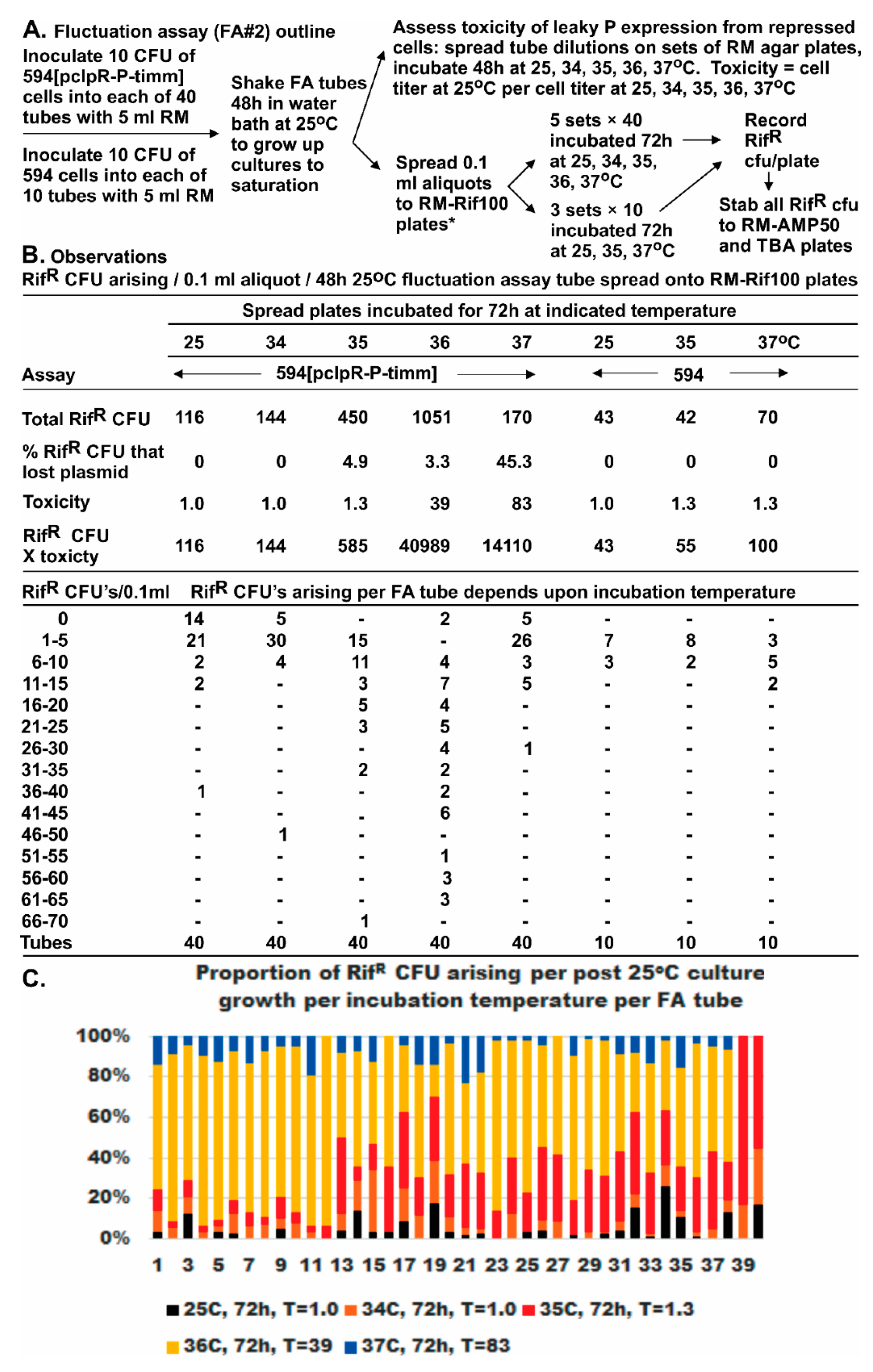
P expression. Further investigation is required to explore potential manifestations of P toxicity on alternative mutation selection; however, this may simply be an inherent limitation in all studies where a mutagenic agent is also toxic, and where toxicity can influence selection. One possibility is that the selected mutants are resistant to the toxic agent. We explored this difficulty in FA#2 by accounting for both toxicity and recovered RifR mutants for each selection temperature. We could demonstrate that many RifR mutants were sensitive to P-toxicity, and, for selections at 36 °C, which were less toxic than those at 37 °C, there was a significant increase in the appearance of RifR mutant CFU (see below). A separate manuscript is in preparation on the complementation and examined toxicity of cloned vs. defective prophage (single copy) expressed λ imm-rep gene products.
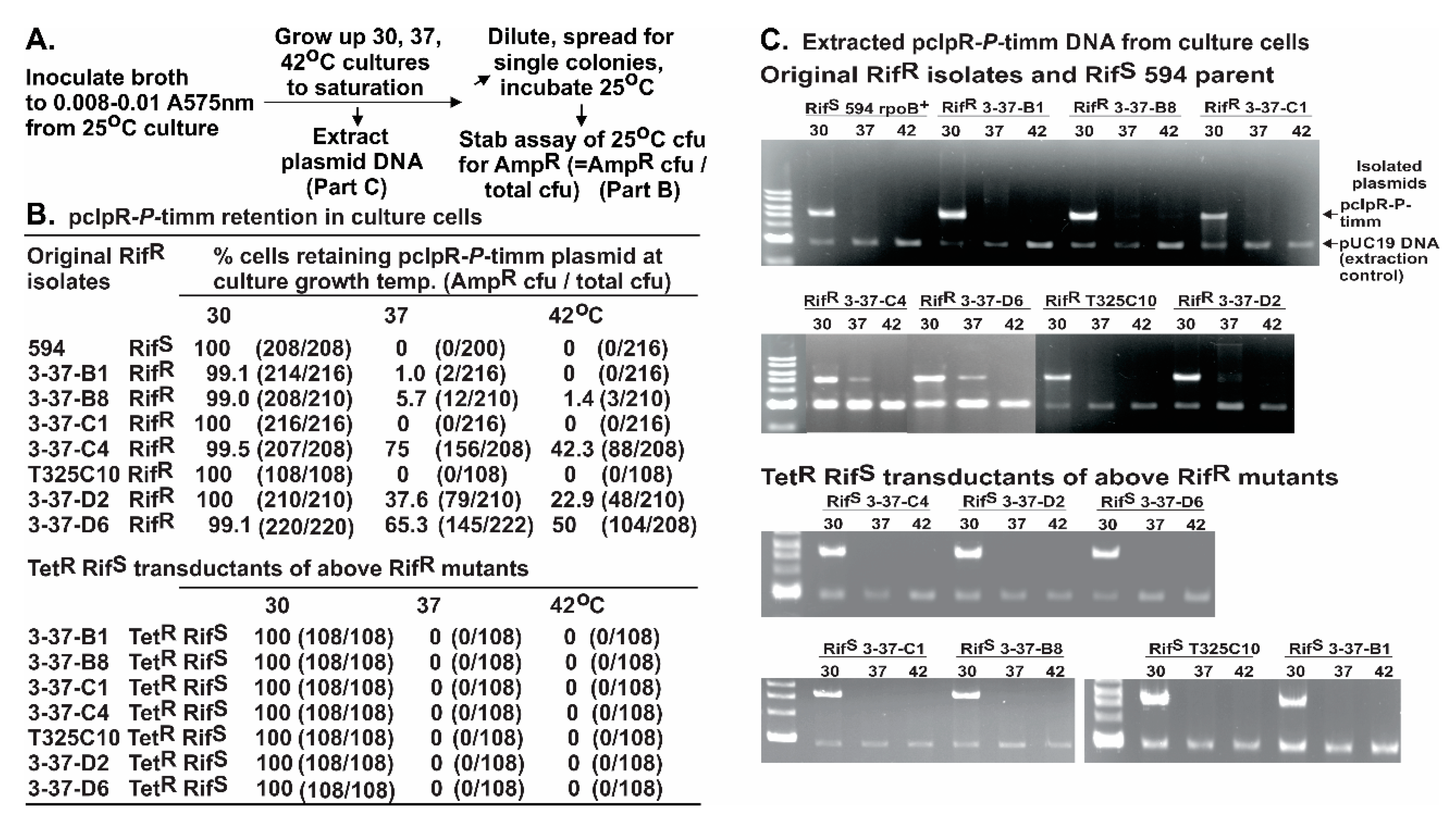
Fluctuation assays were used to determine if the RifR mutations arising in rpoB preexisted the expression of P. As noted, this was complicated by observations that 80-fold cellular toxicity occurred in cells with pcIpR-P-timm incubated at 37 °C, and that most of the RifR CFU were PS. On lowering the incubation temperature to 36 °C, P-lethality was reduced by half but the recovered RifR mutants increased by nine-fold compared to 30 °C. With incubation lowered yet another degree to 35 °C, the toxicity of P was minimal (1.3-fold), ColE1 curing of cells was slight (5%), yet the RifR mutants recovered were 3.9-fold higher than at 30 °C. These observations suggest that the increase in RifR mutants was dependent upon P expression, rather than selection conditions favoring preexisting PR RifR mutants.
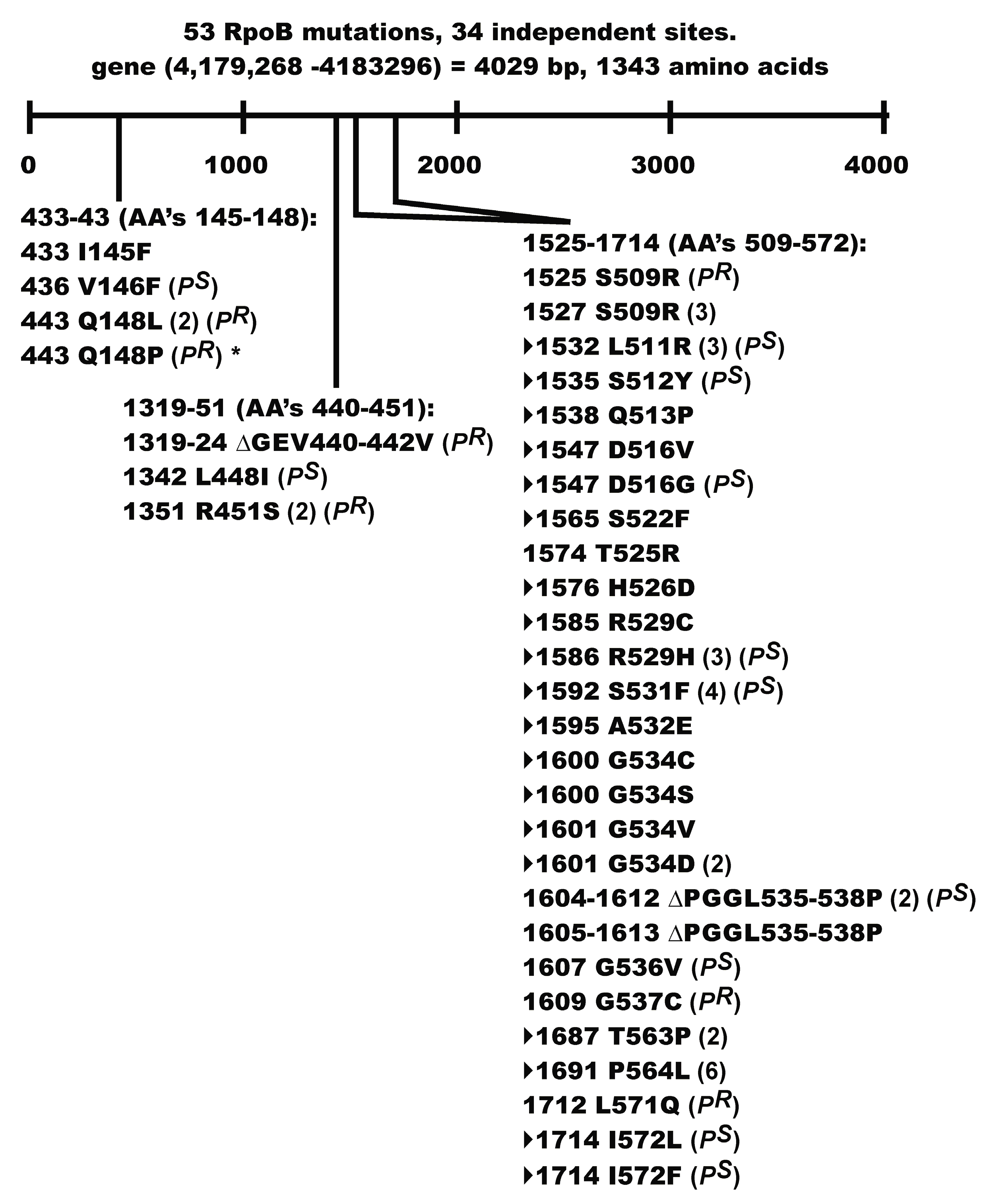
Seven of the RifR mutants acquired a PR phenotype. Two of these mutations, G537C and L571Q map close to the Rif binding pocket in RpoB [79], but the remaining mutations Q148L, Q148P, ∆GEV440-442V, R451S, and S509R fell outside of the pocket. These mutations may help define a contact point(s) between RpoB and P that has long been suggested [64]. Our PR mutation Q148P in rpoB is identical to rpo*148 [82]. A small collection of rpo* mutations, which possess some level of RifR [82], were isolated based on the discovery [83] that mutations in RNAP alleviate the UV-sensitive phenotype of ruv strains, e.g., rpo*148 suppresses the extreme sensitivity of UV treatment to a ∆relAspoT ruv strain. This property was linked to the rpo* mutation destabilizing RNAP open complexes and stalled elongation complexes, thereby reducing the occurrence of stalled RNAP(s) at lesions in the DNA template [82,83]. Co-directional collisions between the replisome and RNAP in an arrested (backtracked) elongation complex can lead to DNA double strand breaks [84]. Transcription pausing and stalling regulators include ppGpp which destabilizes open complexes, and the RNAP modulators DskA, GreA, GreB, and Mfd [81,84,85]. The failure of rpo* RNAP to pause and backtrack, and their ability to reduce the accumulation of RNAP arrays [81], helps explain why rpo* mutations can suppress the formation of DSB [84]. Assuming that our PR Q148P RifR mutation will reduce the accumulation of RNAP arrays, can this explain a PR phenotype and the suppression of P-dependent ColE1 plasmid curing? Future studies require determining if the other PR mutants share an rpo* phenotype.
Several unanswered problems arise in relation to the observed mutator effect caused by P expression. Based on the low λ requirement for P, coupled with P sequestration of the low cellular amount of DnaB [18,19], either P expression from λ requires a high degree of negative regulation to prevent P-lethality/inhibition (probably not fully appreciated), or there is an unknown mechanism for limiting the PS phenotype. The apparent complete escape of λ replication from P is contrasted with the exquisite sensitivity of ColE1 plasmid replication to P, and its mutagenic effect on E. coli. One possible explanation is that late λ rolling circle replication may be independent of replication restart. Whereas, the mechanism for initiation of ColE1 [43] is similar to restart (see [35]), and likely requires loading DnaB for each initiation event, which breaks down with P-DnaB sequestration. Just how PR RifR mutants suppress ColE1 curing by P, and seemingly overcome P-DnaB sequestration remain open questions.
The NH2-terminal portion of P was suggested to bind O and its C-terminal domain to interact with host proteins [86,87]. The NH2-terminal domain of DnaC is involved in binding to DnaB. Both P and DnaC share a stretch of amino acids with high homology at their NH2 termini [88]. The in-frame ∆76 deletion within codons 9–86 at the NH2-terminal end of P suppressed: (a) the curing of ColE1 plasmids; (b) P-lethality at 37–39 °C [35]; and (c) prevented a P-dependent increase in RifR mutations or mutagenic effect. These observations suggest that the NH2 end of P can compete for the DnaC binding site on DnaB, and that P binding at this site can account for PS phenotype: its cellular toxicity, ColE1 curing, and mutator activity.
The cell construct, Figure 1A, possessed a mutagenic activity that was linked to the expression of P from the λ fragment within the chromosome. Independently, cells that included a plasmid where P was the only inducible gene product exhibited a mutator phenotype when P was expressed. The mutator activity was assessed by screening for auxotrophic mutations arising during Replicative Killing selection for RK clones, or the selection of RifR mutations within rpoB. Since alleles of the host dnaB replicative helicase, or of P, can nullify the mutator phenotype, and since P interacts with DnaB to form an enzymatically inactive “dead-end” P-DnaB complex [22], i.e., the so-called sequestered DnaB, reducing or eliminating cellular DnaB activity is linked to the mutator phenotype.
Under conditions of DnaB sequestration, any replication fork collapse could be problematic for replication re-initiation and generate gaps in DNA synthesis needing repair. Studies reported with yeast suggested that the stalling of transcription at abasic (AP) sites is highly mutagenic [89] and mechanisms, such as mutations arising via translesion synthesis through AP sites were proposed. Most of the auxotrophs we characterized acquired temperature sensitive mutations, characteristic of a missense arising from a point mutation. We did not find that blocking SOS induction, deleting recA, or inactivating individual E. coli mutator polymerases Pol IV or Pol V eliminated P-dependent mutagenesis. Since three E. coli DNA polymerases, Pol II, Pol IV and Pol V, are involved in induced mutagenesis [90], this was not evaluated rigorously as the lack of any one could be substituted for by another. Further work is needed to determine if the sequestration of DnaB by P stimulates the appearance of gaps and AP sites around the chromosome, linking them to the mutagenic state associated with elevated levels of P. This would agree with the observation that P can stimulate SOS-independent filamentation [35]. We propose a model that P sequestration of DnaB has several cellular outcomes: (a) it prevents the loading of DnaB helicase needed for the initiation of ColE1 replication (Figure 2) resulting in rapid, complete plasmid curing; (b) it prevents replication restart requiring the reloading of DnaB by DnaC; and (c) when a faster-moving Pol III replisome complex inevitably collides with RNAP, the displaced replisome components along with RNAP from the leading strand are uncoupled from DNA polymerase copying the lagging strand, leading to ssDNA regions that become a target for DNA damage which increase the probability for spontaneous mutation. We speculate that P may exacerbate this situation by gaining access to DnaB remaining bound to the lagging strand, and so interfere with rebinding of the β-clamp and Pol III, resulting in replication stuttering and the formation of leading strand gap(s) as illustrated for the co-directional collisions drawn in Figure 5e of reference [91]. We imagine that mutations in RNAP that limit P-interaction, or limit arrested RNAP complex formation on DNA, hence reducing collisions between the replisome and arrested RNAP, will lessen the influence of P-lethality/inhibition.

4. Materials and Methods

4.1. Strains Employed

The bacteria and plasmids employed are listed in Table 8 or in reference [35].

4.2. Oligonucleotides Employed for PCR Fragment Amplification and DNA Sequencing

The DNA oligonucleotides employed are shown in Table S4.

4.3. Growth Medium, Buffers, PCR Amplification, DNA Sequencing, and Fluctuation Assays

Solid support growth medium designated RM (rich medium) includes 10 g Bacto agar, 10 g Bacto tryptone and 5 g of NaCl per liter. MM (minimal medium) includes 11 g Bacto agar, 100 mL of 10× M9 salts (70 g Na2HPO4, 30 g KH2PO4, 5 g NaCl, 10 g NH4Cl in 1 L deionized water), 16 mL 25% glucose, 0.1 mL 1 M CaCl2, 1 mL 1 M MgSO4·7H2O, and 0.2 mL 0.3% ferric citrate per liter [98]. The broth versions of RM and MM omit agar. MM agar plates were, when noted, supplemented with histidine (100 µg/mL), biotin (1 µg/mL), or 0.3% Bacto casamino acids (not vitamin-free), and adjusted to 0.01 M Tris-HCl, pH 7.6. Ampicillin (Amp), rifampicin (Rif), or tetracycline (Tet) were added to RM or MM agar preparations at 50, 100, or 15 µg/mL, respectively. MM agar plates were made up for auxotroph typing using the combinations of amino acids (each at 0.1 mg/mL), vitamins and purine/pyrimidine pools (concentration/mL) as per Holliday [74]: adenine (0.05 mg/mL), hypoxanthine (0.05 mg/mL), folic acid (0.5 µg/mL), ornithine (0.1 mg/mL), pantothenic acid (0.5 µg/mL), guanine (0.05 mg/mL), pyridoxine (0.5 µg/mL), sodium thiosulfate (0.05 mg/mL), thymine (0.01 µg/mL), p-aminobenzoic acid (0.5 µg/mL), uracil (0.05 mg/mL), riboflavin (2.5 µg/mL), nicotinic acid (0.5 µg/mL), choline (10 µg/mL), inositol (5 µg/mL), and biotin (0.01 µg/mL). Plates containing Rif were held in foil covers and discarded after a week without use. The growth medium employed for transformation and transduction was LB (10 g Bacto tryptone, 5 g Bacto yeast extract, 5 g NaCl per liter), and for electroporation was SOB {2% w/v Bacto tryptone, 0.5% yeast extract, 10 mM NaCl, plus Mg2+ (2 mL 1 M MgCl2·6H2O and 2 mL 1 M MgSO4·7H2O per 200 mL SOB)}, and for SOC was SOB + Mg2+ made 1.8 µg/mL with 25% glucose. The Φ80 buffer used for cell dilutions, and the buffers for DNA handling, plasmid purification, and electrophoresis, and the methods for PCR fragment amplification and for DNA sequencing were previously described [76]. The methodology employed for fluctuation assays #1 and #2 is described in Table 5 and Figure 2, respectively.

4.4. Transformation, Transduction, Electroporation, and Recombineering

Procedure for transduction: P1vir lysates were prepared on strains with a TetR marker (Table 8); this was repeated to prepare a secondary lysate grown up on the same host. All the lysates employed had titers of 5 × 10⁸ PFU/mL or higher. A single colony of a TetS RifR E. coli mutant was grown up overnight at 30 °C in LB broth; 5.0 mL of these cells were centrifuged at 6 K rpm for 6 min, decanted, and the cell pellet resuspended in an equal volume of sterile MC buffer (0.1 M MgSO4, 0.005 M CaCl2). The resuspended cells were aerated for 15 min in a shaking water bath at 30 °C. Cells-phage mixtures and mock infections were incubated at 37 °C for 20 min to permit phage adsorption, then an equal volume of sterile 0.1 M citrate buffer (0.1 M citric acid, pH 5.5) was added to prevent the re-adsorption of P1 phage. The mixtures were added to 7.8 mL of pre-warmed LB broth + 0.2 mL of 0.1 M citrate buffer, and then incubated at 37 °C in a shaking water bath for 60–90 min. The samples were pelleted at 6 K for 10 min, washed, and the cell pellet was resuspended in 400 µL of 0.1 M Citrate buffer. One hundred microliters aliquots were spread on agar plates with 15 µg/mL of tetracycline and incubated at 30 °C overnight. The TetR transduced single colonies arising were streaked onto fresh tetracycline and rifampicin plates to identify the transduced TetR RifS and TetR RifR CFU.
The procedure used for transformation, was from reference [35]. For electroporation and recombineering we followed the procedures outlined in reference [96].

4.5. Moving Chromosomal Fragments by PI and Complementation for P

Every P1vir lysate was serially passaged twice on the donor strain to avoid any carry-over of markers before being used to move gene fragments into recipient cells. Strain CAG12147 nadA57::Tn10 possesses a tetracycline resistance (TetR) marker at 16.85 min on the E. coli linkage map. TetR was moved into Y836 his+. A second step was to co-transduce the TetR marker plus the cryptic λ prophage genes (cIII-ren)λ into two recipient prototrophic strains 594 and W3101. TetR recipients were screened for the immλ fragment using the functional immunity (FI) recombination assay [71]. Strain CAG12164 has a malF::Tn10 TetR marker at ~91.5 min. The TetR marker was moved by transduction into DE407 lexA3[Ind] recipient and the TetR CFU were examined for increased sensitivity to UV to confirm that they retained the lexA3[Ind] allele. Both TetR and lexA3[Ind] alleles were transduced from DE407malF::Tn10 into recipient strain Y836, which represents bio275[substituting for int-kil]-cIII-ren-∆431 (Figure 1A). The TetR CFU were screened for acquisition of lexA3[Ind] by assaying for increased UV sensitivity. Note that the bio275 addition in Y836 confers UvrB+ and Bio+ phenotypes to cells with deletion ∆431. The presence of immλ phenotype was confirmed by the CFU being lysed by λvir and resistant to λcI72 at 30 °C. Strain SF2006 has a dinB::Tn5 at 5 min and can form CFU on plates with kanamycin. A similar approach was used to introduce umuC112:Tn5 into strain Y836. The ethylmethanesulfonate (EMS) spot assay was performed on the KanR transductants to verify movement of the dinB allele into Y836. KanR transductants were grown overnight, washed twice and 0.1 mL aliquots and spread onto MM+His plates in quadruplet. Two of the four plates were spotted with 5 µL concentrations of EMS, incubated 48 h at 30 °C, and compared. Cells defective for DinB mutator polymerase gave fewer auxotrophs in region of EMS spot (compared to DinB+ cells) as the concentration of EMS was increased. The rpoB+ gene was moved by P1vir transduction from TetR donor strain CAG18500 thiC:Tn10 into RifR isolates of 594. TetR CFU were isolated and TetR RifS co-transductants (occurring at about 80%) were distinguished by stabbing on agar plates containing 100 µg/mL of rifampicin.
The phage λimm434 Pam3 carries the amber mutation CAG(Gln)39786TAG and can only form plaques on host cells that can complement for P, or on a host with a nonsense suppressor mutation, as TC600 supE. For complementation assays the phage is diluted into Φ80 buffer and aliquots were mixed with 0.3 mL of assayed host cells plus top agar and poured onto RM plates incubated between 25 °C to 42 °C. The efficiency of plating (EOP) for assay conditions is compared to parallel plates where TC600 is the permissive host (assumed to give a %EPO of 100) and 594 the non-permissive host, upon which P+-revertant phages arise at a frequency of about 1 × 10⁻⁷. Complementation for P expression from plasmid pcIpR-P-timm was described in reference [35]. Some examples include (%EOP at 38–39 °C/30 °C): Y836 (65/0.05), Y836 ilr 534c (41/0.02), Y836 ilr O208b (67/0.001), Y836 ilrO223a (35/0.008), and Y836 ilr P::IS2 (0.0002/0.00008). “ilr” represents a characterized λ mutation conferring “initiation of λ replication” defective phenotype.

Supplementary Materials

The following are available online at https://www.mdpi.com/1999-4915/8/6/172/s1, Figure S1. pcIpR-P-timmΔrop transformation and retention in RifR isolates and transductants. Table S1: Plating efficiency of RK+ strains on RM and MM at 30 °C. Table S2: Spontaneous RifR mutations obtained without cell exposure to P. Table S3: 107 RifR mutations localized to rpoB. Table S4: Oligonucleotides employed for PCR fragment amplification and DNA sequencing.

Acknowledgments

This work was supported by NSERC Canada Discovery grant to S.H. We thank L. Song for assistance in sequencing some of the rpoB mutants.

Author Contributions

Performed the experiments and helped analyze the data: W.W., K.R., A.C., A.B., and C.H. Conceived, designed the experiments, and wrote the paper: S.H.

Conflicts of Interest

The authors declare no conflict of interest.

References

  1. Denniston-Thompson, K.; Moore, D.D.; Kruger, K.E.; Furth, M.E.; Blattner, F.R. Physical structure of the replication origin of bacteriophage lambda. Science 1977, 198, 1051–1056. [Google Scholar] [CrossRef] [PubMed]
  2. Furth, M.E.; Blattner, F.R.; McLeester, C.; Dove, W.F. Genetic structure of the replication origin of bacteriophage lambda. Science 1977, 198, 1046–1051. [Google Scholar] [CrossRef] [PubMed]
  3. Scherer, G. Nucleotide sequence of the O gene and of the origin of replication in bacteriophage lambda DNA. Nucleic Acids Res. 1978, 5, 3141–3156. [Google Scholar] [CrossRef] [PubMed]
  4. LeBowitz, J.H.; McMacken, R. The Escherichia coli DnaB replication protein is a DNA helicase. J. Biol. Chem. 1986, 261, 4738–4748. [Google Scholar] [PubMed]
  5. Mallory, J.B.; Alfano, C.; McMacken, R. Host virus interactions in the initiation of bacteriophage lambda DNA replication. Recruitment of Escherichia coli DnaB helicase by lambda P replication protein. J. Biol. Chem. 1990, 265, 13297–13307. [Google Scholar] [PubMed]
  6. Alfano, C.; McMacken, R. Ordered assembly of nucleoprotein structures at the bacteriophage lambda replication origin during the initiation of DNA replication. J. Biol. Chem. 1989, 264, 10699–10708. [Google Scholar] [PubMed]
  7. Zylicz, M.; Ang, D.; Liberek, K.; Georgopoulos, C. Initiation of lambda DNA replication with purified host- and bacteriophage-encoded proteins: the role of the dnaK, dnaJ and grpE heat shock proteins. EMBO J. 1989, 8, 1601–1608. [Google Scholar] [PubMed]
  8. Bailey, S.; Eliason, W.K.; Steitz, T.A. Structure of hexameric DnaB helicase and its complex with a domain of DnaG primase. Science 2007, 318, 459–463. [Google Scholar] [CrossRef] [PubMed]
  9. McMacken, R.; Ueda, K.; Kornberg, A. Migration of Escherichia coli dnaB protein on the template DNA strand as a mechanism in initiating DNA replication. Proc. Natl. Acad. Sci. USA 1977, 74, 4190–4194. [Google Scholar] [CrossRef] [PubMed]
  10. Zyskind, J.W.; Smith, D.W. NOVEL Escherichia coli dnaB mutant: Direct involvement of the dnaB252 gene product in the synthesis of an origin-ribonucleic acid species during initiaion of a round of deoxyribonucleic acid replication. J. Bacteriol. 1977, 129, 1476–1486. [Google Scholar] [PubMed]
  11. Lu, Y.B.; Ratnakar, P.; Mohanty, B.K.; Bastia, D. Direct physical interaction between DnaG primase and DnaB helicase of Escherichia coli is necessary for optimal synthesis of primer RNA. Proc. Natl. Acad. Sci. USA 1996, 93, 12902–12907. [Google Scholar] [CrossRef] [PubMed]
  12. Thirlway, J.; Turner, I.J.; Gibson, C.T.; Gardiner, L.; Brady, K.; Allen, S.; Roberts, C.J.; Soultanas, P. DnaG interacts with a linker region that joins the N- and C-domains of DnaB and induces the formation of 3-fold symmetric rings. Nucleic Acids Res. 2004, 32, 2977–2986. [Google Scholar] [CrossRef] [PubMed]
  13. Arai, K.; Kornberg, A. Mechanism of dnaB protein action. IV. General priming of DNA replication by dnaB protein and primase compared with RNA polymerase. J. Biol. Chem. 1981, 256, 5267–5272. [Google Scholar] [PubMed]
  14. Arai, K.; Kornberg, A. Mechanism of dnaB protein action. III. Allosteric role of ATP in the alteration of DNA structure by dnaB protein in priming replication. J. Biol. Chem. 1981, 256, 5260–5266. [Google Scholar] [PubMed]
  15. Corn, J.E.; Berger, J.M. Regulation of bacterial priming and daughter strand synthesis through helicase-primase interactions. Nucleic Acids Res. 2006, 34, 4082–4088. [Google Scholar] [CrossRef] [PubMed]
  16. Bujalowski, W. Expanding the physiological role of the hexameric DnaB helicase. Trends Biochem. Sci. 2003, 28, 116–118. [Google Scholar] [CrossRef]
  17. Kaplan, D.L.; O’Donnell, M. DnaB drives DNA branch migration and dislodges proteins while encircling two DNA strands. Mol. Cell 2002, 10, 647–657. [Google Scholar] [CrossRef]
  18. Nakayama, N.; Arai, N.; Bond, M.W.; Kaziro, Y.; Arai, K. Nucleotide sequence of dnaB and the primary structure of the dnaB protein from Escherichia coli. J. Biol. Chem. 1984, 259, 97–101. [Google Scholar] [PubMed]
  19. Ueda, K.; McMacken, R.; Kornberg, A. dnaB protein of Escherichia coli. Purification and role in the replication of phiX174 DNA. J. Biol. Chem. 1978, 253, 261–269. [Google Scholar] [PubMed]
  20. Arai, K.; Kornberg, A. Mechanism of dnaB protein action. II. ATP hydrolysis by dnaB protein dependent on single- or double-stranded DNA. J. Biol. Chem. 1981, 256, 5253–5259. [Google Scholar] [PubMed]
  21. Reha-Krantz, L.J.; Hurwitz, J. The dnaB gene product of Escherichia coli. I. Purification, homogeneity, and physical properties. J. Biol. Chem. 1978, 253, 4043–4050. [Google Scholar] [PubMed]
  22. Biswas, S.B.; Biswas, E.E. Regulation of dnaB function in DNA replication in Escherichia coli by dnaC and lambda P gene products. J. Biol. Chem. 1987, 262, 7831–7838. [Google Scholar] [PubMed]
  23. Kobori, J.A.; Kornberg, A. The Escherichia-Coli Dnac Gene-Product. 1. Overproduction of the Dnac Proteins of Escherichia-Coli and Salmonella-Typhimurium by Cloning into a High Copy Number Plasmid. J. Biol. Chem. 1982, 257, 13757–13762. [Google Scholar] [PubMed]
  24. Kobori, J.A.; Kornberg, A. The Escherichia coli dnaC gene product. II. Purification, physical properties, and role in replication. J. Biol. Chem. 1982, 257, 13763–13769. [Google Scholar] [PubMed]
  25. Kobori, J.A.; Kornberg, A. The Escherichia coli dnaC gene product. III. Properties of the dnaB-dnaC protein complex. J. Biol. Chem. 1982, 257, 13770–13775. [Google Scholar] [PubMed]
  26. Lanka, E.; Schuster, H. The dnaC protein of Escherichia coli. Purification, physical properties and interaction with dnaB protein. Nucleic Acids Res. 1983, 11, 987–997. [Google Scholar] [CrossRef] [PubMed]
  27. Davey, M.J.; Fang, L.; McInerney, P.; Georgescu, R.E.; O’Donnell, M. The DnaC helicase loader is a dual ATP/ADP switch protein. EMBO J. 2002, 21, 3148–3159. [Google Scholar] [CrossRef] [PubMed]
  28. Flowers, S.; Biswas, E.E.; Biswas, S.B. Conformational dynamics of DnaB helicase upon DNA and nucleotide binding: Analysis by intrinsic tryptophan fluorescence quenching. Biochemistry 2003, 42, 1910–1921. [Google Scholar] [CrossRef] [PubMed]
  29. Galletto, R.; Jezewska, M.J.; Bujalowski, W. Interactions of the Escherichia coli DnaB helicase hexamer with the replication factor the DnaC protein. Effect of nucleotide cofactors and the ssDNA on protein-protein interactions and the topology of the complex. J. Mol. Biol. 2003, 329, 441–465. [Google Scholar] [CrossRef]
  30. Galletto, R.; Jezewska, M.J.; Bujalowski, W. Unzipping mechanism of the double-stranded DNA unwinding by a hexameric helicase: quantitative analysis of the rate of the dsDNA unwinding, processivity and kinetic step-size of the Escherichia coli DnaB helicase using rapid quench-flow method. J. Mol. Biol. 2004, 343, 83–99. [Google Scholar] [CrossRef] [PubMed]
  31. Jezewska, M.J.; Bujalowski, W. Global conformational transitions in Escherichia coli primary replicative helicase DnaB protein induced by ATP, ADP, and single-stranded DNA binding. Multiple conformational states of the helicase hexamer. J. Biol. Chem. 1996, 271, 4261–4265. [Google Scholar] [PubMed]
  32. Jezewska, M.J.; Kim, U.S.; Bujalowski, W. Binding of Escherichia coli primary replicative helicase DnaB protein to single-stranded DNA. Long-range allosteric conformational changes within the protein hexamer. Biochemistry 1996, 35, 2129–2145. [Google Scholar] [CrossRef] [PubMed]
  33. San Martin, C.; Radermacher, M.; Wolpensinger, B.; Engel, A.; Miles, C.S.; Dixon, N.E.; Carazo, J.M. Three-dimensional reconstructions from cryoelectron microscopy images reveal an intimate complex between helicase DnaB and its loading partner DnaC. Structure 1998, 6, 501–509. [Google Scholar] [CrossRef]
  34. Yu, X.; Jezewska, M.J.; Bujalowski, W.; Egelman, E.H. The hexameric E. coli DnaB helicase can exist in different Quaternary states. J. Mol. Biol. 1996, 259, 7–14. [Google Scholar] [CrossRef] [PubMed]
  35. Hayes, S.; Erker, C.; Horbay, M.A.; Marciniuk, K.; Wang, W.; Hayes, C. Phage Lambda P Protein: Trans-Activation, Inhibition Phenotypes and their Suppression. Viruses 2013, 5, 619–653. [Google Scholar] [CrossRef] [PubMed]
  36. Casjens, S.R.; Hendrix, R.W. Bacteriophage lambda: Early pioneer and still relevant. Virology 2015, 479–480, 310–330. [Google Scholar] [CrossRef] [PubMed]
  37. Hayes, S.; Slavcev, R.A. Polarity within pM and pE promoted phage lambda cI-rexA-rexB transcription and its suppression. Can. J. Microbiol. 2005, 51, 37–49. [Google Scholar] [CrossRef] [PubMed]
  38. Konieczny, I.; Marszalek, J. The requirement for molecular chaperones in lambda DNA replication is reduced by the mutation pi in lambda P gene, which weakens the interaction between lambda P protein and DnaB helicase. J. Biol. Chem. 1995, 270, 9792–9799. [Google Scholar] [PubMed]
  39. Learn, B.A.; Um, S.J.; Huang, L.; McMacken, R. Cryptic single-stranded-DNA binding activities of the phage lambda P and Escherichia coli DnaC replication initiation proteins facilitate the transfer of E. coli DnaB helicase onto DNA. Proc. Natl. Acad. Sci. USA 1997, 94, 1154–1159. [Google Scholar] [CrossRef] [PubMed]
  40. Osipiuk, J.; Georgopoulos, C.; Zylicz, M. Initiation of lambda DNA replication. The Escherichia coli small heat shock proteins, DnaJ and GrpE, increase DnaK’s affinity for the lambda P protein. J. Biol. Chem. 1993, 268, 4821–4827. [Google Scholar] [PubMed]
  41. Klinkert, J.; Klein, A. Cloning of the replication gene P of bacteriophage lambda: Effects of increased P-protein synthesis on cellular and phage DNA replication. Mol. Gen. Genet. 1979, 171, 219–227. [Google Scholar] [CrossRef] [PubMed]
  42. Tsurimoto, T.; Matsubara, K. Replication of lambda dv plasmid in vitro promoted by purified lambda O and P proteins. Proc. Natl. Acad. Sci. USA 1982, 79, 7639–7643. [Google Scholar] [CrossRef] [PubMed]
  43. Lilly, J.; Camps, M. Mechanisms of Theta Plasmid Replication. Microbiol. Spectr. 2015, 3, PLAS-0029–2014. [Google Scholar] [PubMed]
  44. Dimude, J.U.; Stockum, A.; Midgley-Smith, S.L.; Upton, A.L.; Foster, H.A.; Khan, A.; Saunders, N.J.; Retkute, R.; Rudolph, C.J. The Consequences of Replicating in the Wrong Orientation: Bacterial Chromosome Duplication without an Active Replication Origin. MBio 2015, 6, e01294–e01215. [Google Scholar] [CrossRef] [PubMed]
  45. Kogoma, T. Stable DNA replication: Interplay between DNA replication, homologous recombination, and transcription. Microbiol. Mol. Biol. Rev. 1997, 61, 212–238. [Google Scholar] [PubMed]
  46. Fukuoh, A.; Iwasaki, H.; Ishioka, K.; Shinagawa, H. ATP-dependent resolution of R-loops at the ColE1 replication origin by Escherichia coli RecG protein, a Holliday junction-specific helicase. EMBO J. 1997, 16, 203–209. [Google Scholar] [CrossRef] [PubMed]
  47. Hong, X.; Cadwell, G.W.; Kogoma, T. Escherichia coli RecG and RecA proteins in R-loop formation. EMBO J. 1995, 14, 2385–2392. [Google Scholar] [PubMed]
  48. Rudolph, C.J.; Upton, A.L.; Harris, L.; Lloyd, R.G. Pathological replication in cells lacking RecG DNA translocase. Mol. Microbiol. 2009, 73, 352–366. [Google Scholar] [CrossRef] [PubMed]
  49. Rudolph, C.J.; Upton, A.L.; Stockum, A.; Nieduszynski, C.A.; Lloyd, R.G. Avoiding chromosome pathology when replication forks collide. Nature 2013, 500, 608–611. [Google Scholar] [CrossRef] [PubMed]
  50. Marians, K.J. PriA-directed replication fork restart in Escherichia coli. Trends Biochem. Sci. 2000, 25, 185–189. [Google Scholar] [CrossRef]
  51. Masai, H.; Arai, K. DnaA- and PriA-dependent primosomes: Two distinct replication complexes for replication of Escherichia coli chromosome. Front. Biosci. 1996, 1, d48–d58. [Google Scholar] [CrossRef] [PubMed]
  52. Masai, H.; Nomura, N.; Kubota, Y.; Arai, K. Roles of phi X174 type primosome- and G4 type primase-dependent primings in initiation of lagging and leading strand syntheses of DNA replication. J. Biol. Chem. 1990, 265, 15124–15133. [Google Scholar] [PubMed]
  53. Miwa, T.; Akaboshi, E.; Matsubara, K. Instability of bacteriophage lambda initiator O and P proteins in DNA replication. J. Biochem. 1983, 94, 331–338. [Google Scholar] [PubMed]
  54. Wyatt, W.M.; Inokuchi, H. Stability of lambda O and P replication functions. Virology 1974, 58, 313–315. [Google Scholar] [CrossRef]
  55. Tomizawa, J. Functional cooperation of genes O and P. In The Bacteriophage Lambda; Hershey, A.D., Ed.; Cold Spring Harbor Laboratory: Cold Spring Harbor, NY, USA, 1971; pp. 549–552. [Google Scholar]
  56. Datta, I.; Banik-Maiti, S.; Adhikari, L.; San, S.; Das, N.; Mandal, N.C. The mutation that makes Escherichia coli resistant to lambda P gene-mediated host lethality is located within the DNA initiator gene dnaA of the bacterium. J. Biochem. Mol. Biol. 2005, 38, 89–96. [Google Scholar] [CrossRef] [PubMed]
  57. Datta, I.; Sau, S.; Sil, A.K.; Mandal, N.C. The bacteriophage lambda DNA replication protein P inhibits the oriC DNA- and ATP-binding functions of the DNA replication initiator protein DnaA of Escherichia coli. J. Biochem. Mol. Biol. 2005, 38, 97–103. [Google Scholar] [CrossRef] [PubMed]
  58. Wegrzyn, G.; Wegrzyn, A.; Pankiewicz, A.; Taylor, K. Allele specificity of the Escherichia coli dnaA gene function in the replication of plasmids derived from phage lambda. Mol. Gen. Genet. 1996, 252, 580–586. [Google Scholar] [CrossRef] [PubMed]
  59. Georgopoulos, C.P.; Herskowitz, I. Escherichia coli mutants blocked in lambda DNA synthesis. In The Bacteriophage Lambda; Hershey, A.D., Ed.; Cold Spring Harbor Laboratory: Cold Spring Harbor, NY, USA, 1971; pp. 553–564. [Google Scholar]
  60. Klein, A.; Lanka, E.; Schuster, H. Isolation of a complex between the P protein of phage lambda and the dnaB protein of Escherichia coli. Eur. J. Biochem. 1980, 105, 1–6. [Google Scholar] [CrossRef] [PubMed]
  61. Saito, H.; Uchida, H. Organization and expression of the dnaJ and dnaK genes of Escherichia coli K12. Mol. Gen. Genet. 1978, 164, 1–8. [Google Scholar] [CrossRef] [PubMed]
  62. Zylicz, M.; Ang, D.; Georgopoulos, C. The grpE protein of Escherichia coli. Purification and properties. J. Biol. Chem. 1987, 262, 17437–17442. [Google Scholar] [PubMed]
  63. Yochem, J.; Uchida, H.; Sunshine, M.; Saito, H.; Georgopoulos, C.P.; Feiss, M. Genetic analysis of two genes, dnaJ and dnaK, necessary for Escherichia coli and bacteriophage lambda DNA replication. Mol. Gen. Genet. 1978, 164, 9–14. [Google Scholar] [CrossRef] [PubMed]
  64. McKinney, M.D.; Wechsler, J.A. RNA polymerase interaction with dnaB protein and lambda P protein during lambda replication. J. Virol. 1983, 48, 551–554. [Google Scholar] [PubMed]
  65. Hoffmann, H.J.; Lyman, S.K.; Lu, C.; Petit, M.A.; Echols, H. Activity of the Hsp70 chaperone complex—DnaK, DnaJ, and GrpE—In initiating phage lambda DNA replication by sequestering and releasing lambda P protein. Proc. Natl. Acad. Sci. USA 1992, 89, 12108–12111. [Google Scholar] [CrossRef] [PubMed]
  66. Hayes, S.; Hayes, C.; Bull, H.J.; Pelcher, L.A.; Slavcev, R.A. Acquired mutations in phage lambda genes O or P that enable constitutive expression of a cryptic lambdaN+cI[Ts]cro- prophage in E. coli cells shifted from 30 °C to 42 °C, accompanied by loss of immlambda and Rex+ phenotypes and emergence of a non-immune exclusion-state. Gene 1998, 223, 115–128. [Google Scholar] [PubMed]
  67. Hayes, S. Mutations suppressing loss of replication control. Genetic analysis of bacteriophage lambda-dependent replicative killing, replication initiation, and mechanisms of mutagenesis. In DNA Replication and Mutagenesis; Moses, R.E., Summers, W.C., Eds.; American Society for Microbiology: Washington, DC, USA, 1988; pp. 367–377. [Google Scholar]
  68. Hayes, S.; Duincan, D.; Hayes, C. Alcohol treatment of defective lambda lysogens is deletionogenic. Mol. Gen. Genet. 1990, 222, 17–24. [Google Scholar] [PubMed]
  69. Hayes, S.; Hayes, C. Spontaneous lambda OR mutations suppress inhibition of bacteriophage growth by nonimmune exclusion phenotype of defective lambda prophage. J. Virol. 1986, 58, 835–842. [Google Scholar]
  70. Bull, H.J.; Hayes, S. The grpD55 locus of Escherichia coli appears to be an allele of dnaB. Mol. Gen. Genet. 1996, 252, 755–760. [Google Scholar] [PubMed]
  71. Hayes, S.; Asai, K.; Chu, A.M.; Hayes, C. NinR- and red-mediated phage-prophage marker rescue recombination in Escherichia coli: Recovery of a nonhomologous immlambda DNA segment by infecting lambdaimm434 phages. Genetics 2005, 170, 1485–1499. [Google Scholar] [CrossRef] [PubMed]
  72. Hua, Q.; Yang, C.; Oshima, T.; Mori, H.; Shimizu, K. Analysis of gene expression in Escherichia coli in response to changes of growth-limiting nutrient in chemostat cultures. Appl. Environ. Microbiol. 2004, 70, 2354–2366. [Google Scholar] [CrossRef] [PubMed]
  73. Chu, A.M. Mutator Phenotype of Induced Cryptic Coliphage Lambda Prophage. Master’s Thesis, University of Saskatchewan, Saskatoon, SK, Canada, 2004. [Google Scholar]
  74. Holliday, R. A new method for the identification of biochemical mutants of micro-organisms. Nature 1956, 178, 987. [Google Scholar] [CrossRef]
  75. Schlacher, K.; Goodman, M.F. Lessons from 50 years of SOS DNA-damage-induced mutagenesis. Nat. Rev. Mol. Cell. Biol. 2007, 8, 587–594. [Google Scholar] [CrossRef] [PubMed]
  76. Hayes, S.; Horbay, M.A.; Hayes, C. A CI-Independent Form of Replicative Inhibition: Turn Off of Early Replication of Bacteriophage Lambda. PLoS ONE 2012, 7, e36498. [Google Scholar] [CrossRef] [PubMed]
  77. Banerjee, A. Influence of Bacteriophage Lambda Gene P Expression on Host Escherichia coli Cells. Master’s Thesis, University of Saskatchewan, Saskatoon, SK, Canada, 2011. [Google Scholar]
  78. Ovchinnikov Yu, A.; Monastyrskaya, G.S.; Gubanov, V.V.; Lipkin, V.M.; Sverdlov, E.D.; Kiver, I.F.; Bass, I.A.; Mindlin, S.Z.; Danilevskaya, O.N.; Khesin, R.B. Primary structure of Escherichia coli RNA polymerase nucleotide substitution in the beta subunit gene of the rifampicin resistant rpoB255 mutant. Mol. Gen. Genet. 1981, 184, 536–538. [Google Scholar] [CrossRef] [PubMed]
  79. Campbell, E.A.; Korzheva, N.; Mustaev, A.; Murakami, K.; Nair, S.; Goldfarb, A.; Darst, S.A. Structural mechanism for rifampicin inhibition of bacterial RNA polymerase. Cell 2001, 104, 901–912. [Google Scholar] [CrossRef]
  80. Lau, I.F.; Filipe, S.R.; Soballe, B.; Okstad, O.A.; Barre, F.X.; Sherratt, D.J. Spatial and temporal organization of replicating Escherichia coli chromosomes. Mol. Microbiol. 2003, 49, 731–743. [Google Scholar] [CrossRef] [PubMed]
  81. Rudolph, C.J.; Dhillon, P.; Moore, T.; Lloyd, R.G. Avoiding and resolving conflicts between DNA replication and transcription. DNA Repair 2007, 6, 981–993. [Google Scholar] [CrossRef] [PubMed]
  82. Trautinger, B.W.; Lloyd, R.G. Modulation of DNA repair by mutations flanking the DNA channel through RNA polymerase. EMBO J. 2002, 21, 6944–6953. [Google Scholar] [CrossRef] [PubMed]
  83. McGlynn, P.; Lloyd, R.G. Modulation of RNA polymerase by (p)ppGpp reveals a RecG-dependent mechanism for replication fork progression. Cell 2000, 101, 35–45. [Google Scholar] [CrossRef]
  84. Dutta, D.; Shatalin, K.; Epshtein, V.; Gottesman, M.E.; Nudler, E. Linking RNA polymerase backtracking to genome instability in E. coli. Cell 2011, 146, 533–543. [Google Scholar] [CrossRef] [PubMed]
  85. Trautinger, B.W.; Jaktaji, R.P.; Rusakova, E.; Lloyd, R.G. RNA polymerase modulators and DNA repair activities resolve conflicts between DNA replication and transcription. Mol. Cell. 2005, 19, 247–258. [Google Scholar] [CrossRef] [PubMed]
  86. Reiser, W.; Leibrecht, I.; Klein, A. Structure and function of mutants in the P gene of bacteriophage lambda leading to the pi phenotype. Mol. Gen. Genet. 1983, 192, 430–435. [Google Scholar] [CrossRef] [PubMed]
  87. Wickner, S.H. DNA replication proteins of Escherichia coli and phage lambda. Cold Spring Harb. Symp. Quant. Biol. 1979, 43, 303–310. [Google Scholar] [CrossRef] [PubMed]
  88. Ludlam, A.V.; McNatt, M.W.; Carr, K.M.; Kaguni, J.M. Essential amino acids of Escherichia coli DnaC protein in an N-terminal domain interact with DnaB helicase. J. Biol. Chem. 2001, 276, 27345–27353. [Google Scholar] [CrossRef] [PubMed]
  89. Yu, S.L.; Lee, S.K.; Johnson, R.E.; Prakash, L.; Prakash, S. The stalling of transcription at abasic sites is highly mutagenic. Mol. Cell. Biol. 2003, 23, 382–388. [Google Scholar] [CrossRef] [PubMed]
  90. Napolitano, R.; Janel-Bintz, R.; Wagner, J.; Fuchs, R.P. All three SOS-inducible DNA polymerases (Pol II, Pol IV and Pol V) are involved in induced mutagenesis. EMBO J. 2000, 19, 6259–6265. [Google Scholar] [CrossRef] [PubMed]
  91. Pomerantz, R.T.; O’Donnell, M. The replisome uses mRNA as a primer after colliding with RNA polymerase. Nature 2008, 456, 762–766. [Google Scholar] [CrossRef] [PubMed]
  92. Bachmann, B.J. Derivations and genotypes of some mutant derivatives of Escherichia coli K-12. In Escherichia coli and Salmonella typhimurium: Cellular and Molecular Biology; Neidhardt, F.C., Ingraham, J.I., Low, K.B., Magasanik, B., Schaechter, M., Umbargr, H.E., Eds.; American Society for Microbiology: Washington, DC, USA, 1987; Volume 2, pp. 1192–1219. [Google Scholar]
  93. Horbay, M.A. Inhibition phenotype specific for ori-lambda replication dependent phage growth, and a reappraisal of the influence of lambda P expression on Escherichia coli cell metabolism: P-interference phenotype. University of Saskatchewan: Saskatoon, SK, Canada, 2005. [Google Scholar]
  94. Nichols, B.P.; Shafiq, O.; Meiners, V. Sequence analysis of Tn10 insertion sites in a collection of Escherichia coli strains used for genetic mapping and strain construction. J. Bacteriol. 1998, 180, 6408–6411. [Google Scholar] [PubMed]
  95. Hayes, S.; Gamage, L.N.; Hayes, C. Dual expression system for assembling phage lambda display particle (LDP) vaccine to porcine Circovirus 2 (PCV2). Vaccine 2010, 28, 6789–6799. [Google Scholar] [CrossRef] [PubMed]
  96. Thomason, L.; Court, D.L.; Bubunenko, M.; Constantino, N.; Wilson, H.; Dattaa, S.; Oppenheim, A. Recombineering: Genetic engineering in bacteria using homologous recombination. In Current Protocols in Molecular Biology; John Wiley & Sons, Inc.: Hoboken, NJ, USA, 2005. [Google Scholar]
  97. Chan, W.; Costantino, N.; Li, R.; Lee, S.C.; Su, Q.; Melvin, D.; Court, D.L.; Liu, P. A recombineering based approach for high-throughput conditional knockout targeting vector construction. Nucleic Acids Res. 2007, 35, e64. [Google Scholar] [CrossRef] [PubMed]
  98. Arber, W.; Enquist, L.; Hohn, B.; Murray, N.; Murray, K. Experimental methods for use with lambda. In Lambda II; Hendrix, R.W., Roberts, J.W., Stahl, F.W., Weisberg, R.A., Eds.; Cold Spring Harbor Laboratory: Cold Spring Harbor, NY, USA, 1983; pp. 433–466. [Google Scholar]
Figure 1. Modes for expression of P: (A) cryptic prophage map (for more λ gene detail refer to Figure 1B in reference [35] or Figure 1 in reference [36]); (B) λ gene expression is induced by shifting cells from 30 °C to 42 °C, which inactivates the encoded Ts (temperature sensitive) cI[Ts]857 repressor, permitting rightward transcription from promoter pR; and (C) an exact copy of P (or alleles of P) was cloned into the synthetic expression plasmid pcIpR-timm [35] (shown by arrows at ATG and TAA). P expression from pcIpR-P-timm is controlled by the λ CI[Ts] repressor. The circle to the right of pR and left of ATG represents the ribosomal binding site for the deleted intervening gene cro. The transcription of P from the plasmid terminates at the transcriptional terminator timm, which in the wild type λ sequence prevents both the low maintenance and high level establishment modes of cI-rexA-rexB transcription from transcribing leftward from pM or pE into oL-pL [37].
Viruses 08 00172 g001
Figure 2. Summary of P lethality/inhibition observations, including observations provided herein. Arrows terminating in X show inhibitory activities. “SDR” is stable DNA replication [44,45,46,47,48]. P can bind DnaB helicase and replace pre-bound DnaC.
Viruses 08 00172 g002
Figure 3. Fluctuation assay to examine the influence of P expression on the screen for RifR CFU: (A) outline of methodology; (B) observations; and (C) relative proportion of RifR CFU arising on spread plates incubated in parallel at 25 °C to 37 °C, where T = the toxicity of the treatment, determined by CFU formed on RM plates without rifampicin at 25 °C per CFU formed on RM plates without rifampicin after incubation at 25, 34, 35, 36, or 37 °C.
Viruses 08 00172 g003
Figure 4. Location of selected RifR mutations in rpoB. Mutants designated PS are transformable at 30 °C but not 37 °C by pcIpR-P-timm, whereas the PR mutants were transformable at both temperatures. The rpoB mutant designated with * is identical to mutant rpo*148 (see Discussion). The missense mutations (designated by sideways triangle) each fall within codons for amino acids found in the Rif binding pocket of rpoB.
Viruses 08 00172 g004
Figure 5. pcIpR-P-timm retention in PS and PR RifR isolates and for RifS transductants. (A) Outline of experimental method is identical to that described for measuring P-induced plasmid loss in Figure 3, reference [35]: Cultures made from single CFU for each isolate grown up on Amp50 plates were inoculated and grown to stationary phase in RM plus 50 µg/ml ampicillin for 48 h at 25 °C. Cell aliquots from the cultures were diluted into fresh RM (no ampicillin) as shown in outline A and incubated for about 20 h in shaking baths between 30 to 42 °C. (B) The percentage of the TetS RifR cells that acquired the TetR RifS phenotypes after transduction (see text) was 81%, 85%, 75%, 94%, 69%, 88%, and 90%, for the RifR mutants B1, B8, C1, C4, C10, D2, and D6, respectively. (C) Extracted plasmids from identical culture cells (described in A, quantitated in B) grown between 30 to 42 °C.
Viruses 08 00172 g005
Table 1. RK mutation frequencies.
RK+ StrainsRK CFU Frequency on RM ×10−6 (SE × 10−6) aRK CFU Frequency on MM ×10−6 (SE × 10−6) bSupplement to MM bRK CFU at 42 °C Arising on RM/MM c
RK mutant frequencies determined from selection at 42 °C on RM and MM a.
Y836 his d5.88 (1.0)0.373 (0.057)histidine15.8
Y836 his5.00.33histidine + biotin15.1
Y836 his e4.05 (0.3)6.08 (0.83)casamino acids g0.7
Y836 f13.4 (0.67)1.56 (0.24)none8.6
Does Y836 have intrinsic mutator activity? Transduce Y836 ( cIII-ren)λ into 594 and W3101.
594::(cIII-ren2.21 (0.48)0.178 (0.085)none12.4
W3101::(cIII-ren11.9 (4.6)1.50 (0.11)none7.9
Assay requirement of SOS gene products for dual RK plus auxotrophic mutation(s).
Y836 his lexA3[Ind]0.260.011histidine23.6
Y836 his ΔrecA4.43 (0.43)0.405 (0.062)histidine10.9
Y836 his umuC:Tn521.3 (0.96)0.268 (0.032)histidine79.5
Y836 his dinB:Kan5.00 (0.85)0.244 (0.11)histidine20.5
594::(cIII-rendinB:Kan1.90.090none21.1
a Frequency of RK mutants forming colonies at 42 °C per colony forming units (CFU) at 30 °C on RM or MM × 10−6. (SE × 10−6) is the standard error, determined for ≥3 independent assays; b Frequency of RK mutants forming CFU at 42 °C per CFU at 30 °C on MM (minimal medium, or where noted, MM with the supplement nutrient added shown in adjacent column) × 10−6 (standard error × 10−6); c The frequency of RK mutants forming CFU on RM/frequency of RK mutants forming CFU on MM, or where noted on MM+ supplement; d Average of 10 independent assays and (standard error); e Average of 6 independent assays and (standard error); f Average of 5 independent assays and (standard error); g Medium includes Bacto Casamino Acids (not “Bacto Vitamin-Free Casamino Acids”).
Table 2. Blocking λ gene expression and oriλ replication initiation, or only oriλ initiation.
RK+ Strains aCFU at 42 °C/CFU at 30 °CRatio CFU on RM/MM
RMMM+Histidine
Inhibiting λ fragment gene expression at 42 °C by cI+ expressed on plasmid pCI
Y836 his [pCI+]0.95 (0.076)0.85 (0.033)1.1
W3101::(cIII-ren)λ [pCI+]1.21.21.0
594: (cIII-ren)λ [pCI+]0.920.901.0
Inhibiting replication initiation at 42 °C from oriλ on λ fragment by host mutation
Y836 his dnaBgrpD550.91 (0.084)0.15 (0.073)6.1
Y836 his dnaBgrpD55 [pCI+]0.770.820.9
a The identical λ fragments in each of the RK+ strains carry a cI857 mutation conferring to the CI857 repressor a temperature sensitive phenotype. The pCI+ plasmid encodes a WT cI+ gene and the CI+ repressor remains active (trans dominant), preventing transcription from promoters pL and pR at 30 and 42 °C. Values in parentheses represent standard error for multiple assays.
Table 3. Stab assay screen for auxotrophic colony forming units (CFU).
StrainsAssayed CFU aAuxotrophs/CFU a (Ts CFU) a
Nonlysogens or lysogens with noninduced prophage
CFU from strains spread on RM, 30 °C
5945510
594 dnaBgrpD55 b4130
Y83625200
Y836 dnaBgrpD55 b11790
594: (cIII-cI857-O+-P+-ren3470
Y836 RK Bib11t O+ P:Kan c3240
Y836 RK 566a O+ P:IS2 d2800
Y836 RK O208b P+ e3220
Y836 RK O223a P+ f2230
Y836 RK 534c O+ P+ g1670
CFU from strains spread on RM, 42 °C
5942600
594 dnaBgrpD55 b919 1 h
Y836 [pCI]880
Y836 dnaBgrpD55 [pCI]1000
594: (cIII-cI857-O+-P+-ren)λ [pCI]1100
Total78031
RK CFU isolated from Induced RK+ strains (each expressing P at 42 °C)
Y836 90494 (79)
Y836 dnaBgrpD5543314 (11)
Y836 ΔrecA25936 (36)
Y836 dinB:Kan1008 (8)
594: (cIII-cI857-O+-P+-ren)λ RK+42757 (57)
Characterized λ replication defective RK mutants (forming CFU at high viability at 42 °C)
Y836 RK Bib11t O+ P:Kan7160
Y836 RK 566a O+ P:IS26430
Y836 RK O208b P+16051 (1)
Y836 RK O223a P+65411 (11)
Y836 RK 534c O+ P+8751 (1)
Total6607222 (204)
a Isolated CFU appearing on RM agar plates, that were incubated at 30 or 42 °C, were picked to RM plates that were incubated at 30 °C, or to parallel MM+histidine plates that were incubated both at 30 °C and 42 °C to distinguish acquired auxotrophs with a temperature sensitive (Ts) phenotype. Values in “( )” represent number of auxotrophic CFU with a Ts phenotype forming CFU on both RM and MM+His plates incubated at 30 °C, but showing no growth on MM+histidine plates incubated at 42 °C; b The grpD55 mutation in host gene dnaB prevents λ replication initiation, but does not noticeably influence the growth of the E. coli host carrying the mutation; c SH insertion, clone Bib11t of kanamycin resistance (promoter-orf-terminator of gene aph from TN903 removed from plasmid p51kan [76]) gene into P by recombineering, substituting bp 39651-39838 of P. The Kan insertion includes a synthetic 11 bp all-translational stop sequence at each end of the insertion; d The RK 566a mutation P:IS2 represents an insertion in P right of 39690 with 6 bp repeat of bases 39685-39690 AGGTAG –IS2–AGGTAG. Blast of about 1000 bp sequence downstream of 39690 shows that the insertion is IS2; however, sequencing from both ends shows a 6 bp (not characteristic 5 bp) repeat sequence straddling each end of the insertion. The sequence of oop-O is wild type; e The RK mutation O208b is a 7 bp deletion in O (bases 39147-39153, fusing bases 39146-39154) creating −1 frameshift, with stop codon at 21 codons downstream from fused codon; f The RK mutation O223a is a 44bp deletion in O (bases 39044-39087, fusing ITN1 with ITN3) creating −1 frameshift, with stop codon 33 codons downstream from fused codon; g The RK mutant 534c was shown to have a wild type λ sequence from gene cI857 through cII-oop-O-P-ren through base 40713 in ninB. The strain likely survives replicative killing at 42 °C via an uncharacterized host mutation that prevents replication initiation from oriλ within the λ DNA fragment in strain Y836.
Table 4. Influence of P expression on the selection of rifampicin-resistant (RifR) CFU.
Strains with Fragment of λ ProphageFrequency RifR CFU at 42 °C/30 °C a [Average Frequency RifR CFU at 30 °C]
5941 [8.3 × 10⁻⁸]
Exp. A. b
Y836 O+ P+88,727 (42,027) [4.33 × 10⁻⁸]
Y836 O+ P+ dnaBgrpD553 (1.5) [1.38 × 10⁻⁷]
Y836 O+ P:Kan6 (1.9) [5.3 × 10⁻⁸]
Y836 O+ P:IS23 (0.24) [2.83 × 10⁻⁸]
Y836 O223a P+2167 (730) [8.5 × 10⁻⁸]
Exp. B. b
Y836 O+ P+42,500 (17,000) [5.4 × 10⁻⁸]
Y836 O+ P+ dnaBgrpD552 [1.3 × 10⁻⁸]
594::(cIII-cI857-O+-P+-ren51,485 [1.0 × 10⁻⁷]
Y836 O+ P+ [pcI]1 [2.4 × 10⁻⁸]
Y836 O+ P+ dnaBgrpD55 [pCI]1 [1.0 × 10⁻⁹]
594::(cIII-cI857-O+-P+-ren)λ [pCI]1 [5.0 × 10⁻⁹]
Strains with a PlasmidFrequency RifR CFU at 37 °C/30 °C [Average Frequency RifR CFU at 30 °C] e
594 [pcIpR-P-timm]122 (52) [3.0 × 10⁻⁸]
594 [pcIpR-Pπ39991-timm] c2 [3.3 × 10⁻⁸]
594 [pcIpR- P∆76-timm] d3 [4.3 × 10⁻⁸]
594 dnaBgrpD55 [pcIpR-P-timm]2 (1.1) [1.3 × 10⁻⁸]
594 dnaBgrpD55 [pcIpR-Pπ-timm]4 [3.5 × 10⁻⁸]
594 dnaBgrpD55 [pcIpR-ΔP-timm]0.1 (0.05) [2.0 × 10⁻⁹]
a Averaged results for three to seven experiments (standard error). Other results show average determinations; b Experiments A and B were undertaken by different workers; c The mutation Pπ39991 is G to A transition, R137Q in P; d The mutation P∆76 is an in frame 76 codon deletion fusing part of codon 9 with 86, and deleting λ bases 39609-39836 in NH2 terminal end of P; e Determination of the frequency of RifR CFU was measured for multiple assays at 25 °C, 30 °C and 37 °C (the results per temperature is an average). The reduced level of P expression was measured at 37 °C from the pcIpR-P-timm plasmid [35]. The frequencies of RifR CFU selected at 25 °C and 30 °C were close and only the results for 30 °C are shown.
Table 5. Isolation and characterization of RifR CFU from 40 fluctuation assay (FA #1) tubes a.
40 FA #1 TubesRifR CFU/Spread PlateSequence of One (or Multiple) d RifR CFU from
Inoculate 15 CFU/mL, Grow 48 h at 25 °CSpread 0.1 mL to Rif100 Plate, Incubate 72 h at 25 °C b,cSpread 0.1 mL to Rif100 Plate, Incubate 72 h at 37 °C b,cRif100 Plate Incubated at 25 °CRif100 Plate Incubated at 37 °C
A43 b01585:CtoT, R529Cnone
A61 b01595:CtoA, A532Enone
A75 b11527:CtoA, S509Rlost
A91 b41687:AtoC, T563Plost
A101 b6 c1592:CtoT, S531F1547:AtoG, D516G
B16 b2 c1605-13: ΔAGGCGGTCT, PGGL535-538P443:AtoC, Q148P
B802 cnone1712:TtoA, L571Q (2) d
B108 b01600:GtoT, G534Cnone
C102 cnone1691:CtoT, P564L (2)
C44 b1 c1687:AtoC, T563P1319-24:ΔGCGAAG, GEV440-442V (2)
C53 b4 c1691:CtoT, P564L1691:CtoT,P564L
C611 b1 c1592:CtoT, S531F1351:CtoA, R451S (2)
C719 blost1601:GtoA, G534D (3)
C101 b01586:GtoA, R529Hnone
D11 b1 c1574:CtoG, T525R1714:AtoC, I572L (3)
D22 b1 c1576:CtoG, H526D1351:CtoA, R451S (2)
D307 cnone436:GtoT, V146F (2)
D51 b01527:CtoA, S509Rnone
D66 b2 c1601:GtoT, G534V1609:GtoT, G537C
D702nonelost
D82 b01691:CtoT, P564Lnone
D91 b11565:CtoT, S522Flost
D101 b01604-12: ΔCAGGCGGTC, PGGL535-538Pnone
a Forty culture tubes with one mL RM broth, numbered A1–A10, B1–B10, C1–C10 and D1–D10, were inoculated with ~15 CFU (determined by parallel titration of the inoculum) of fresh 594[pcIpR-P-timm] culture, single colony 3. The tubes from which no RifR CFU were obtained are omitted in the left column, e.g., A1–A3, A5, etc. All the inoculated tubes were shaken in a water bath at 25 °C for 48 h. Thereupon, 0.1 mL aliquots, representing about ~2 × 10⁸ CFU were spread on two RM agar plates containing 100 µg/mL rifampicin. One plate was incubated at 25 °C and the other at 37 °C and the CFU arising are shown in columns 1 and 2, respectively; b Isolated, restreaked clone(s) retained the plasmid pcIpR-P-timm during growth in culture tube from 15 CFU/mL to ~2 × 10⁹ CFU/mL, as evidenced by efficient growth on Amp50; RM agar plates; c Isolated, restreaked clone(s) had lost plasmid pcIpR-P-timm during growth in culture tube from 15 CFU/mL to ~2 × 10⁹ CFU per mL; d Individual clones sequenced shown in parentheses.
Table 6. Transformation (×10−7) of pcIpR-P-timm/100 ng plasmid into RifR mutants of 594 a.
Strains/MutantsTransformants at 25 °CTransformants at 37 °C
594520<0.17
594 RifR mutants selected from cells never exposed to gene P b
1-25A27900<0.18
3-25E570<0.17
1-37A217,000<0.19
3-37D270<0.18
T-3-37-B10580<0.2
T-3-37-C51200<0.2
T-3-25-D10780<0.3
Td-3-25-A7,scg312800<0.23
Td-3-37-C7,sc1f451200<0.3
Td-3-25-C1038001.2
Td-3-25-D910002.4
594 RifR mutants selected at 37 °C from 594[pcIpR-P-timm] cultures that had lost the plasmid c
3-37-A10270<0.3
3-37-B14106.0
3-37-B81200430
3-37-C1, 3-37-C5 d180<0.12
3-37-C4860140
3-37-D1100<0.17
3-37-C6, 3-37-D2 e1100390
3-37-D3190<0.2
3-37-D663013
a RifR isolates were streaked for sc’s on fresh LB agar plates with 50 µg/mL rifampicin, A CFU was selected, inoculated into LB broth and grown overnight to saturation. These cells were used for transformation with pcIpR-P-timm as described in Materials and Methods. Values with “<” had no recovered AmpR-transformants and the frequency shown was obtained by dividing “1” by the cell titer on LB agar plates. The results for one experiment are shown, but are representative of numerous repeats; b Refer to Table S2 for isolates employed; c Refer to Table 5 for isolates employed; d Both mutants with RifR mutation at 1691:CtoT, P564L, were PS, data for 3-37-C1; e Both mutants with RifR mutation at 1351:CtoA, R451S, were PR, data for 3-37-D2.
Table 7. pcIpR-P-timm∆rop transformation (× 10−7) into TetS rpoB RifR mutants and their transductants made TetR rpoB + RifS.
Strains/Mutants/PhenotypeTransformants at 25 °CTransformants at 37 °C
594 (TetS RifS rpoB +)800<6.3 × 10⁻⁹
594 TetS rpoB RifR mutants
RifR 3-37-C41500110
RifR 3-37-D29080
RifR 3-37-D61100700
594 RifR mutants transduced to TetR rpoB + RifS
TetR RifS 3-37-C4300<1.3 × 10⁻⁹
TetR RifS 3-37-D21800<2.3 × 10⁻⁹
TetR RifS 3-37-D6600<2.8 × 10⁻⁹
a Cells were transformed with 1710 ng (in 2 µL) of pcIpR-P-timm∆rop plasmid.
Table 8. Bacteria and plasmids employed.
Bacterial StrainsCharacteristics or GenotypeSource/Ref.’; Hayes Lab # a
594F lac-3350 galK2 galT22 rpsL 179 IN(rrnD-rrnE)1; also called R594[71,92] SH lab; B10
594 dnaB-grpD55grpD55 allele malF3089:Tn10; TetR, λR at 42 °C, λrepP22S[71,93] NB295
594 lexA3[Ind] malB:Tn9LexA repressor induction defective[35] C. Erker (CE), NB293
594 Δ(srlR-recA)306:Tn10deletion of recA TetR UVS[35] CE, B318
594:nadA:Tn10 [~cIII-ren]λTn10 [zbh29 at 16.8 min] bio+ transductant = 594 bio275cIII-cI[Ts]857-O-P-ren) Δ431[73] = AC; this strain = NY1057
W3101: nadA:Tn10 [~cIII-ren]λTn10 [zbh29 at 16.8 min] bio+ transductant = 594 bio275cIII-cI[Ts]857-O-P-ren) Δ431AC, NY1057
CAG12147=nadA57:Tn10 at 16.85 minλ, nadA57:Tn10, rph-1C.A. Gross (CAG); NY1053
CAG12164malF:Tn10, 4,241,898 [94]Coli genetic stock center cgsc.biology.yale.edu
CAG12185argE:Tn10, 4,151,734 [94]cgsc.biology.yale.edu
CAG18500thiC:Tn10, 4,192,143 [94]cgsc.biology.yale.edu
Y836SA500(λbio275cI[Ts]857 Δ431) his[68,69] SH, NY1049
Y836 [pCI+]λ prophage fragment genes rendered noninducible by WT CI+[71]
Y836his+his+ transductant of Y836 his = Y836AC, NY1046
Y836 RK P:kan (Bib11t)SA500 (λbio275 cI[Ts]857 O+P:kan Δ431) his KanR[35] SH, NY1153
Y836 RK ilr566a O+ P:IS2CH, MY843 RK
Y836 dnaBgrpD55[pCI]λ prophage fragment genes rendered noninducible by WT CI+[71] AC, NY1054
Y836:Tn10nadA57:Tn10 at ~16.85 min CG12147AC, NY1047
594::(cIII-ren)λnadA57:Tn10 and bio275 = Bio+cIII-cI[Ts]857-ren) Δ431 transduced from Y836:Tn10AC, NY1057
594::(cIII-ren ilr566a)λnadA57:Tn10 and [bio275 λcIII-cI[Ts]857-ren) Δ431 transduced from Y836 RK ilr566a nadA57:Tn10AC, NY1065
594::(cIII-ren)λ [pCI]λ prophage fragment genes rendered noninducible by WT CI+AC, NY1055
W3101galT22 IN(rrnD-rrnE)B. Backmann, B25-b
W3101::(cIII-ren)λnadA57:Tn10AC, NY1051
W3101::(cIII-ren)λ[pCI+]λ prophage fragment genes rendered noninducible by WT CI+AC, NY1059
SF2006 and SF2139dinB:KanM. Goodman,
GW3200AB1157 umuD44D. Ennis, B396
GW2100AB1157 umuC112:Tn5 (Kan)G. Walker, B317
Y836 dinB:KandinB:Kan from SF2006AC, 1045
Y836 umuC112:Tn5umuC112:Tn5 (Kan)AC, 1052
Y836 ∆recA TetR UVS∆(srlR-recA)306::Tn10 [35]AC, Y916, NY1048
Y836 lexA3[Ind] malB:Tn9LexA repressor induction defectiveAC, from DE407 [35]
PlasmidsTransformed into strain 594Source/Ref.’; Hayes lab # a
pCI+used to place WT CI+ repressor expression in strains to prevent λ prophage fragment derepression upon culture shift from 30 to 42 °CM. Horbay; [71]
pcIpR-D-CAP-timmD-CAP expression CH, P459 [95]
pcIpR-P-timmBamHI-ClaI fragment from λcI857, replacing D-CAP in P459, with λ bp’s 39582-40280CH, P466 [35]
pcIpR-P-timmΔropP651 = 4371bp; Δ rop region, i.e., 613bp between PciI to EagI and includes 23937-24310bpλ) from pSJH-D[pR-MT-GFP-timm][EGFP]+λSO = P593, was inserted into P466 replacing its rop region between bp 938-2472CH, P593; KR P651
pSIM6inducible λ Red genesL. Thomason [96,97]
a The strain numbers are from Hayes laboratory collections. All gene inserts within the plasmids were sequenced to confirm genetic integrity of the inserted fragment.

