Article

Comparative Endosymbiont Community Structures of Nonviruliferous and Rice Stripe Virus-Viruliferous Laodelphax striatellus (Hemiptera: Delphacidae) in Korea

1
Department of Plant Medicals, Gyeongkuk National University, Andong 36729, Republic of Korea
2
Crop Protection Division, National Institute of Agriculture Sciences, Rural Development Administration, Wanju 55365, Republic of Korea
*
Author to whom correspondence should be addressed.
These authors contribute equally to this work.
Viruses 2025, 17(8), 1074; https://doi.org/10.3390/v17081074
Submission received: 6 December 2024 / Revised: 13 July 2025 / Accepted: 30 July 2025 / Published: 1 August 2025
(This article belongs to the Special Issue Plant Viruses and Their Vectors: Epidemiology and Control)

Abstract

Insects and their bacterial endosymbionts form intricate ecological relationships, yet their role in host–pathogen interactions are not fully elucidated. The small brown planthopper (Laodelphax striatellus), a polyphagous pest of cereal crops, acts as a key vector for rice stripe virus (RSV), a significant threat to rice production. This study aimed to compare the endosymbiont community structures of nonviruliferous and RSV-viruliferous L. striatellus populations using 16S rRNA gene sequencing with high-throughput sequencing technology. Wolbachia was highly dominant in both groups; however, the prevalence of other endosymbionts, specifically Rickettsia and Burkholderia, differed markedly depending on RSV infection. Comprehensive microbial diversity and composition analyses revealed distinct community structures between nonviruliferous and RSV-viruliferous populations, highlighting potential interactions and implications for vector competence and virus transmission dynamics. These findings contribute to understanding virus-insect-endosymbiont dynamics and could inform strategies to mitigate viral spread by targeting symbiotic bacteria.

1. Introduction

Rice stripe virus (RSV: Tenuivirus oryzaclavatae) was first reported in Japan in the 1930s and is now known to be widespread in many countries, including Korea, Japan, and China [1,2]. RSV belongs to the genus Tenuivirus and has been assigned to the family Phenuiviridae in the order Bunyavirales [3]. The negative/ambisense RNA genome of RSV consists of four single-stranded (ss) RNA segments [4], and it can infect various plants and crops, such as corn, wheat, oats, and other weeds in addition to rice [5,6]. RSV is persistently transmitted by small brown planthoppers (SBPH, Laodelphax striatellus) [5]. RSV is acquired by SBPH through sap-sucking, spreading internally to the midgut, salivary glands, and ovaries, resulting in persistent infection and transovarial transmission through eggs [7].
SBPH, which has a significant impact on the transmission of RSVs, is one of the pests affecting various crops. SBPH is known to be prevalent in Palearctic regions such as China, Japan, Germany, Italy, Russia, and Kazakhstan [8]. SBPH is known to damage rice, maize, oat, wheat and barley through sap-sucking, and it is known as a vector insect that spreads viruses such as RSV, rice black-streaked dwarf virus (RBSDV, Fijivirus alporyzae; genus Fijivirus), and rice dwarf virus (RDV, Phytoreovirus alphaoryzae; genus Phytoreovirus) [5].
Various living beings, including humans, insects, and plants, harbor a diverse assortment of microorganisms collectively termed the “microbiota”. The compilation of microbial genetic material within a host is termed the “microbiome” [9]. These communities of microorganisms engage in interactions with their hosts, augmenting the ability of organisms to adapt to fluctuations in environmental conditions [10,11,12,13,14].
Among these symbionts, bacteria play diverse roles and have been extensively studied [15]. Insect endosymbionts actively engage in various facets of host life cycles, exerting profound influences on the biological characteristics of their insect hosts [16,17,18]. They also play pivotal roles in nutritional provision and fortifying the gut against colonization by exogenous species, including pathogens that could affect the respective vectors [12]. Consequently, there has been a surge in research interest surrounding insect symbionts in recent years [19]. A plethora of studies has underscored the involvement of endosymbionts in facilitating pathogen transmission within insect host vectors [20,21,22,23]. Notably, certain endosymbionts aid in pathogen entry into insects. For instance, GroEL proteins from various endosymbionts play a pivotal role in virus transmission in aphids and whiteflies by binding to virus particles, thus shielding them from rapid proteolytic degradation in the insect hemolymph [23,24,25,26].
Sequencing of the 16S ribosomal RNA (rRNA) gene has been widely employed as the primary method for bacterial identification [27,28]. The approximately 1500 base pairs of the 16S rDNA sequence provide ample material for bioinformatics analyses [29], and it is universally present across bacterial species with a well-defined function [30]. However, this approach encounters challenges in polymicrobial samples, where the presence of multiple bacterial species results in ambiguous Sanger sequencing results [31]. Next-generation sequencing (NGS) technologies have revolutionized high-throughput functional genomic research [32]. Notably, Illumina technology generates millions of DNA sequence reads in a single run, significantly altering the landscape of genetic studies [33]. Currently, NGS technologies offer novel avenues for analyzing the structure and content of microbial genomes [34], with applications spanning the investigation of microbial communities associated with insects such as beetles [35], ants [36], thrips [37], and planthoppers [38,39].
Despite advances in these analytical methods, limited studies have investigated the interactions between endosymbionts of SBPH and RSV, despite their significant impact on rice yield. Therefore, in this study, we used Illumina 16S rRNA gene MiSeq sequencing to observe changes in the endosymbiont of virus-free and RSV-viruliferous SBPH in Korea. This study explores the impact of RSV on the composition of the endosymbiont of SBPH and aims to provide a broader perspective for the control of RSV.

2. Results

2.1. Differences in the Diversity of SBPH with or Without RSV Infection

We utilized Illumina MiSeq to investigate SBPH to analyze bacterial microbiota diversity between virus-free SBPH (VF) and RSV-viruliferous SBPH (RSV). After filtering out low-quality reads, 241,586–327,278 useful reads were obtained from the samples. Operational taxonomic unit (OTU) clustering was performed using the SILVA 16s database, and a total of 61 OTUs were identified in both groups (VF, RSV). At a similarity cutoff of 99% (1% dissimilarity), 54 OTUs were identified in the VF group, whereas 27 OTUs were identified in the RSV-viruliferous group. In the two groups, the Shannon entropy, phylogenetic diversity, and Simpson’s index were 0.62 and 0.63; 1.59 and 1.11; 0.15 and 0.18, respectively (Table 1). Shannon entropy measures the diversity and evenness of an ecosystem. Phylogenetic diversity evaluates biodiversity based on the evolutionary distance among species. Simpson’s index reflects the dominance of particular species, where higher values indicate lower evenness and a greater presence of dominant species. Shannon entropy and Simpson’s index. Although Shannon entropy and Simpson’s index values were similar between the two groups, the phylogenetic diversity notably differed. Specifically, the virus-free SBPH population exhibited higher phylogenetic diversity, suggesting the presence of a broader range of bacterial lineages compared to the RSV-viruliferous populations. The ASV analysis also used the SILVA 16s database as a reference database. In the ASV analysis, Shannon entropy and Simpson’s index values were consistent with the OTU analysis, again showing similar diversity and evenness between the two groups. Additionally, phylogenetic diversity followed the same pattern observed in OTU analysis, reinforcing the conclusion of reduced bacterial diversity in RSV-viruliferous SBPH. However, the number of microbial communities identified was different between the OTU and ASV.

2.2. Bacterial Community Composition

To evaluate the OTUs distribution among different SBPH populations, we identified 20 OTUs that were common to both populations. This indicates that more than half of the endosymbionts in the RSV group belonged to the shared portion, with most OTUs classified under the genus Wolbachia. Further, the number of unique OTUs was 34 and 7 in the VF and RSV populations, respectively, which were represented by a low proportion (Table S1). Comparing the two groups, at least four phyla were identified in each sample at the phylum level (Figure 1A). Proteobacteria comprised the majority of all samples, with 99.4% in the VF group and 99.9% in the RSV group, respectively. Further analysis was performed to show the relative bacterial abundances at the class, order, family and genus levels (Figure 1B–E). At the class level, Alphaproteobacteria dominated in both groups, accounting for 97% in the virus-free (non-viruliferous) group and 95.5% in the RSV-viruliferous group, with smaller proportions of Gammaproteobacteria (2.3% and 4.3%, respectively) (Figure 1B). At the family level, apart from Anaplasmataceae, which accounted for 96.5% in the Virus-free group, Enterobacteriaceae made up 1.7%. In the RSV-viruliferous group, besides Anaplasmataceae, which accounted for 95.5%, Burkholderiaceae represented 4.2% (Figure 1D). At the genus level, a total of 35 genera were classified across both groups, with 12 genera common to all SBPH groups. Additionally, 21 genera were unique to the VF group, and 2 unique genera were identified in the RSV group. Wolbachia was dominant in both the VF group (96.5%) and the RSV group (95.5%). Other genera showed lower percentage, with Enterobacter and Rickettsia comprising 1.4% and 0.5%, respectively, in the VF group, and Burkholderia accounting for 4.2% in the RSV group (Figure 1E and Figure S1). This fact demonstrate that the bacterial distribution is predominantly dominated by the genus Wolbachia, with some variations depending on RSV infection.
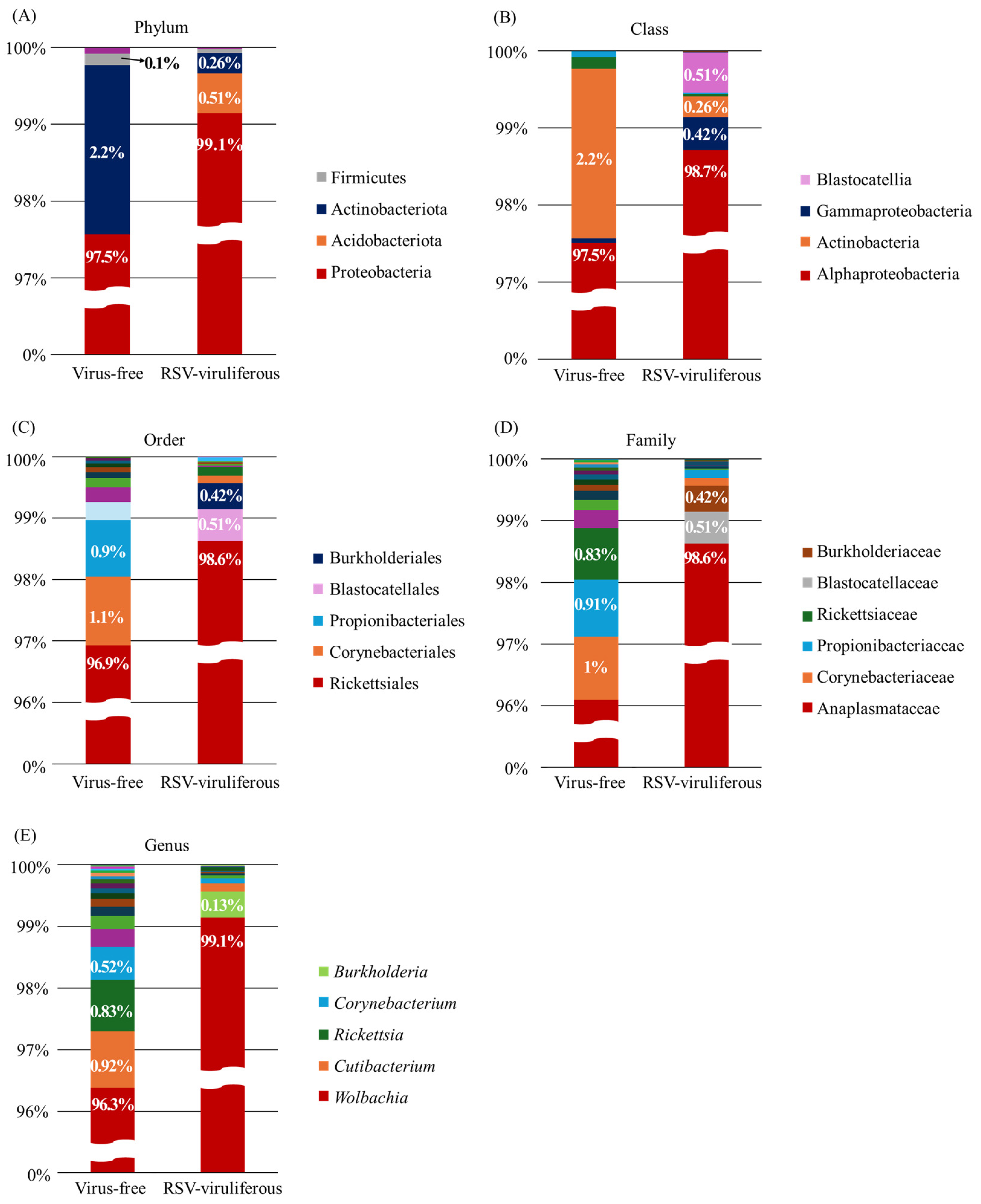
The ASV analysis identified seven ASVs common to both populations. Consistent with the OTU analysis, the majority of the ASVs were classified in the genus Wolbachia, with a low percentage of unique ASVs (17 and 10, respectively). Comparing the two groups, at least four phyla were identified at the phylum level (Figure 2A). In both groups, Proteobacteria comprised the majority of the samples at 97.5% and 99.1%, respectively. Further analysis organized the relative abundance of bacteria at the class, order, family, and genus levels (Figure 2B–E). At the class level, Alphaproteobacte-ria was dominant in both groups, with 97.5% in the virus-free (non-viruliferous) group and 98.7% in the RSV-viruliferous group, slightly higher in the RSV-viruliferous group, contrary to the OTU analysis (Figure 2B). At the family level, Corynebacteriaceae, Propionibacteriaceae, and Rickettsiaceae comprised the majority of the virus-free group, except for Anaplasmataceae, which accounted for 96% and 98.6%, respectively, while Blastocatellaceae and Burkholderiaceae comprised the remainder of the RSV-viruliferous group (Figure 2D). At the genus level, bacterial community composition trends were largely consistent between OTU and ASV analyses. However, notable differences included the absence of Enterobacter in the virus-free group in ASV analysis, which was present in OTU analysis, and a significant reduction in the abundance of Burkholderia from 4.2% (OTU) to 0.13% (ASV) in the RSV-viruliferous group. These discrepancies highlight the methodological sensitivity and underscore the importance of integrating multiple analytical approaches.

2.3. Validation of Sequencing Data

To validate our sequencing results, PCR was performed to specifically detect and confirm the presence of selected endosymbionts, particularly targeting genera (Rickettsia and Burkholderia) whose relative abundances varied significantly between groups, excluding the highly dominant genus Wolbachia (validation shown in Figure S1). The PCR validation results were fully consistent with our sequencing data. Specifically, amplification of Rickettsia DNA was successful only in virus-free SBPH samples, while Burkholderia DNA was exclusively amplified in RSV-viruliferous SBPH samples, clearly confirming the sequencing-based findings.

3. Discussion

In this study, 16S rRNA sequencing was used to identify changes in the endosymbiont of SBPH according to RSV infection. The most common clustering method in metagenomic studies is identity clustering, which uses a fixed sequence identity criterion (usually 97% for sequences from the same species) to generate operational taxonomic units (OTUs) [40]. In recent years, denoising has been introduced as an alternative clustering method that forms clusters through a process of predicting and correcting for noise. This method is called amplicon sequence variants (ASVs) [41,42,43] and forms clusters after correcting for true sequencing error (noise). Denoising approaches utilize well-established statistical models to identify low-frequency sequences and consider them as sequence variants. This produces fewer clusters, but with greater consistency and precision between clusters and thoroughly validated results [44,45,46]. Recently, ASVs methods have been increasingly used in microbiota research alongside traditional OTUs methods. Employing both OTU and ASV analyses allowed us not only to cross-validate our results but also to capture both coarse-grained (OTU) and fine-grained (ASV) insights into the endosymbiont community composition. Although general patterns were consistent, differences highlighted by ASV analysis provide additional resolution, enabling a deeper understanding of microbial dynamics.
In this study, we utilized both OTU and ASV analyses to characterize bacterial communities and identified Wolbachia as the overwhelmingly dominant endosymbiont, comprising 96.5% in the VF group and 95.5% in the RSV group. This result aligns well with previous reports highlighting Wolbachia’s prevalence in SBPH [47]. Wolbachia is a widely distributed endosymbiont belonging to the Alphaproteobacteria, known to infect numerous hosts including insects, isopods, and spiders [48,49,50,51]. Wolbachia are maternally transmitted and have evolved several strategies to facilitate their own proliferation and transmission. These include parthenogenesis, feminization, male killing, and cytoplasmic incompatibility [52,53,54,55,56,57], and these strategies regulate the reproduction of their hosts. Teixeira et al. [58] reported that Wolbachia infection increased D. melanogaster’s resistance to RNA viruses (e.g., Drosophila C virus, Nora virus and Flock house virus), reducing the viral load of infected flies. When Wolbachia was introduced into the mosquito Aedes aegypti, it induced resistance to Dengue virus and interfered with virus transmission [59]. Wolbachia strains from various insects transferred to A. aegypt have limited the replication of arboviruses such as dengue virus (DNEV), chikungunya virus (CHIKV), yellow fever virus (YFV), and zika virus (ZIKV) [60]. In addition, Wolbachia regulate the reproduction of insects by enhancing their gene transmission through the female germline. The most common variation is referred to as cytoplasmic incompatibility (CI).
In SBPH, Wolbachia infection is widely distributed in the head, thorax, abdomen, salivary gland, gut, ovary, and testis [61]. Wolbachia was able to induce strong CI in SBPH, which was one of the main features of the symbiosis between Wolbachia and SBPH [62,63]. CI occurs when eggs laid by Wolbachia-free (W-) females fail to develop after mating with Wolbachia-infected (W+) males. In contrast, the other three mating combinations (W+ female × W- male, W+ female × W+ male, and W- female × W- male) result in normal offspring development [48,64,65]. In addition, Zhang et al. [66] used 454-FLX pyrosequencing to show that the Wolbachia transcriptome in non-viral SBPH is four times larger than in RSV-viruliferous insects [48,64,65]. Zhang et al. [66] identified several Wolbachia genes in the SBPH transcriptome, and these genes may participate in various cellular processes such as molecular transport, balance maintenance, and degradation of harmful compounds, which may contribute to the adaptation of Wolbachia to the SBPH intracellular environment. Wolbachia may also provide metabolites needed by the host [67], but information on metabolite supply by Wolbachia in SBPH is still lacking.
Apart from the predominant Wolbachia, other genera were present at substantially lower proportions. Notably, Enterobacter (1.4%) and Rickettsia (0.5%) accounted for meaningful proportions of the endosymbiotic community in the VF group. In contrast, within the RSV-viruliferous group, Burkholderia emerged as the predominant genus (4.2%) among minor taxa, a finding clearly supported by our PCR validation experiments (Figure S1). Bacteria belonging to the genus Burkholderia are widely distributed in the natural environment and are also diverse in their lifestyle, environment, and ecological roles [68,69,70]. Some strains are pathogenic to plants (e.g., B. gladioli and B. glumae), humans, and other animals [69].; however, most Burkholderia species are commonly distributed in soil and roots and have non-pathogenic effects on other organisms [71]. Some of them have established beneficial symbiotic relationships with a variety of eukaryotes, including phytopathogenic and endophytic bacteria, various plants, soil amoebae, and insects [69,70]. To date, in insects, mutualistic symbiotic relationships with Burkholderia have been reported for phytophagous stinkbugs (Hemiptera, Pentatomomorpha), ants of the genus Tetraponera (Formicidae: Pseudomyrmecinae), Lagrinii beetles (Coleoptera, Tenebrionidae), Acronicta aceris (Noctudiae), and Gossyparia spuria (Eriococcidae) [72,73,74,75]. To date, the role of Burkholderia in SBPH remains unclear. Even if Burkholderia potentially influences SBPH-RSV interactions, its overall impact on viral transmission may be limited due to its considerably lower abundance relative to the dominant Wolbachia population.
In conclusion, the results of MiSeq sequencing showed that Wolbachia, the main endosymbiont, was similar between the two groups, and that Enterobacter, Rickettsia, and Burkholderia made up a small proportion of the remaining endosymbionts, but the expected important endosymbionts such as Buchnera and Hamiltonella were not found. These are known to be host-specific symbionts (e.g., aphids). These genera may either not naturally occur in SBPHs, or they might exist at concentrations too low in RSV-viruliferous SBPHs to be detected using MiSeq sequencing. In addition, as shown in this study, since Wolbachia is a highly dominant endosymbiotic in SBPH in both groups, it opens up the possibility of utilizing Wolbachia for suppressing SBPH’s ability to transmit RSV. As mentioned above, Wolbachia-induced CI can be used to suppress SBPH populations. However, there have been limited studies on the effects of other identified endosymbionts, including Wolbachia, on RSV transmission in SBPH. Although our study provided comprehensive insights into community composition changes in response to RSV infection, further functional validation through targeted experimental manipulation of these endosymbionts is needed to definitively elucidate their roles in SBPH biology and RSV transmission dynamics. Therefore, further studies are warranted. Collectively, our results underline the importance of integrating advanced sequencing techniques and complementary PCR validation to elucidate endosymbiotic dynamics, providing a foundation for developing novel strategies for SBPH control and RSV management.

4. Materials and Methods

4.1. Insect Rearing

SBPH populations (Non-viruliferous RSV and RSV-viruliferous) used in this study were provided from Kyungpook National University (Sangju, Republic of Korea). RSV-infected rice was also provided, and two-leafed rice was fed every two weeks to maintain SBPH. Confirmation of RSV infection was diagnosed using RSV-specific primers, and the RT-PCR cycles and primers used are described in Section 4.2 and Table S2. SBPH was reared in acrylic cages (40 × 40 × 40, W × D × H, cm) and grown at room temperature under 16:8 (L:D) photoperiod and humidity conditions of 60–70%. Additionally, every two weeks, rice seedlings that had been sprouted for two weeks were newly supplied. The RSV isolate used in this study is the same strain previously sequenced by our research team (NCBI GenBank accession numbers OL438909, OL998468–OL998470).

4.2. RNA Extraction and RSV Diagnosis

Twenty SBPHs were randomly selected. RNA was extracted NucleoSpin RNA Plant kit (Machery-Nagel, Düren, Germany) according to the manufacturer’s instructions. RNA concentration and purity were measured using a Nanodrop NP80 (Implen, Munich, Germany). RNA samples were stored at −80 °C before use.
To confirm the RSV, PCR was performed using total RNA and primers for the RNA-dependent RNA polymerase (RdRp) region of RSV. The SuPrimeScript RT-PCR Kit (2X) (Genetbio, Daegeon, Republic of Korea) was used with the following conditions: 50 °C for 30 min, 95 °C for 5 min, followed by 35 cycles of 95 °C for 30 s, 56 °C for 30 s, and 72 °C for 1 min, and a final extension at 72 °C for 5 min. The results were confirmed through electrophoresis. The primers used are described in Table S2.

4.3. DNA Extraction, Library Construction and Sequencing

To obtain sufficient high-quality DNA for sequencing with Illumina MiSeq Platform, DNA was extracted from two populations (virus-free and RSV). Each population contained 20 insects (5th-instar nymphs and adults). Genomic DNA was extracted from each SBPH population using the NucleoSpin DNA Insect kit (Machery-Nagel), according to the manufacturer’s instructions. DNA concentration and purity were quantified using a NanoPhotometer® NP80 spectrophotometer (Implen).
Before sequencing, sample quality was measured using the picogreen method with Victor 3 fluorometry. After measurement, the library for the V3-V4 hypervariable region of the bacterial 16s rRNA gene was prepared using the 16S Amplicon PCR Forward Primer = 5′TCGTCGGCAGCGTCAGATGTGTATAAGAGACAGCCTACGGGNGGCWGCAG and 16S Amplicon PCR Reverse Primer = 5′GTCTCGTGGGCTCGGAGATGTGTATAAGAGACAGGACTACHVGGGTATCTAATCC, along with Herculase II Fusion DNA Polymerase (Agilent Technologies, Santa Clara, CA, USA) and the Nextera XT Index V2 Kit (Illumina, San Diego, CA, USA), following the manufacturer’s instructions. The quality of the libraries was also checked using the picogreen method. NGS was performed on the MiSeq platform, producing paired-end 301 bp raw reads, and Library construction and sequencing were conducted by Macrogen (Seoul, Republic of Korea).

4.4. 16S Taxonomy Analysis

The raw reads were analyzed using CLC Genomics Workbench software (version 25.0; QIAGEN, Hilden, Germany). Adapters were removed and high-quality reads were separated using the ‘trim read’ tool (quality limit = 0.001, discard below length = 15).

4.4.1. OTU Analysis

The trimmed reads were then used for OTU clustering with the ‘OTU clustering’ tool, analyzed at 99% similarity using the SILVA (SSU 138.1) database. To assess bacterial diversity, indices (Shannon entropy, Phylogenetic diversity, Simpson’s index) were calculated using the ‘alpha diversity’ tool.

4.4.2. ASV Analysis

The trimmed reads were then used for Amplicon Sequence Variants analysis with the ‘Detect Amplicon Sequence Variants and Assign Taxonomies’ tool. The reference database was used the SILVA (SSU 138.1). To assess bacterial diversity, indices (Shannon entropy, Phylogenetic diversity, Simpson’s index) were calculated using the ‘alpha diversity’ tool.
The abundance graphs were created using Excel, and all data analyses were conducted with QIAGEN CLC Genomics Workbench software (version 24.0).

4.5. PCR for Endosymbiont Detection

To confirm the presence of endosymbionts in SBPH, genomic DNA extracted from 20 randomly selected individuals (as described in Section 4.3) was used for PCR. PCR amplifications were performed using the SuPrimeScript RT-PCR Kit (2X) (Genetbio), under the identical cycling conditions as described previously (Section 4.2), excluding only the initial reverse transcription step at 50 °C for 30 min. The primers specific for Rickettsia and Burkholderia were designed to amplify target genes, as listed in Table S2. Actin was used as an internal control.

Supplementary Materials

The following supporting information can be downloaded at: https://www.mdpi.com/article/10.3390/v17081074/s1, Figure S1: PCR validation of selected bacterial genera in SBPH populations. Table S1: Raw read counts of 35 bacterial genera from different SBPH populations. Table S2: List of primers used in this study. References [76,77,78] are cited in Supplementary Materials files.

Author Contributions

Conceptualization, E.-J.K. and B.C.L.; methodology, J.J. and M.K.; software, J.J.; validation, J.J. and M.K.; investigation, J.J.; resources, B.C.L.; data curation, E.-J.K.; writing—original draft preparation, J.J. and M.K.; writing—review and editing, E.-J.K. and B.C.L.; visualization, J.J. and M.K.; supervision, E.-J.K.; project administration, E.-J.K.; funding acquisition, E.-J.K. All authors have read and agreed to the published version of the manuscript.

Funding

This research was funded by “Cooperative Research Program for Agriculture Science & Technology Development (Project No. PJ01556601)” Rural Development Administration, Republic of Korea.

Institutional Review Board Statement

Not applicable.

Informed Consent Statement

Not applicable.

Data Availability Statement

The data supporting the findings of this study are available from the corresponding author upon reasonable request. The 16S rRNA sequencing data have been deposited in the NCBI Sequence Read Archive (SRA). No additional data were generated that require specific archiving.

Conflicts of Interest

The authors declare no conflicts of interest.

References

  1. Hibino, H. Biology and epidemiology of rice viruses. Annu. Rev. Phytopathol. 1996, 34, 249–274. [Google Scholar] [CrossRef]
  2. Wang, Y.; Xue, Y.; Li, J. Towards molecular breeding and improvement of rice in China. Trends Plant Sci. 2005, 10, 610–614. [Google Scholar] [CrossRef]
  3. Lefkowitz, E.J.; Dempsey, D.M.; Hendrickson, R.C.; Orton, R.J.; Siddell, S.G.; Smith, D.B. Virus taxonomy: The database of the International Committee on Taxonomy of Viruses (ICTV). Nucleic Acids Res. 2017, 46, D708–D717. [Google Scholar] [CrossRef]
  4. Ishikawa, K.; Omura, T.; Hibino, H. Morphological characteristics of rice stripe virus. J. Gen. Virol. 1989, 70, 3465–3468. [Google Scholar] [CrossRef]
  5. Falk, B.W.; Tsai, J.H. Biology and molecular biology of viruses in the genus Tenuivirus. Annu. Rev. Phytopathol. 1998, 36, 139–163. [Google Scholar] [CrossRef] [PubMed]
  6. Wei, T.-Y.; Yang, J.-G.; Liao, F.-L.; Gao, F.-L.; Lu, L.-M.; Zhang, X.-T.; Li, F.; Wu, Z.-J.; Lin, Q.-Y.; Xie, L.-H. Genetic diversity and population structure of rice stripe virus in China. J. Gen. Virol. 2009, 90, 1025–1034. [Google Scholar] [CrossRef]
  7. Jia, D.; Chen, Q.; Mao, Q.; Zhang, X.; Wu, W.; Chen, H.; Yu, X.; Wang, Z.; Wei, T. Vector mediated transmission of persistently transmitted plant viruses. Curr. Opin. Virol. 2018, 28, 127–132. [Google Scholar] [CrossRef]
  8. Zhu, J.; Jiang, F.; Wang, X.; Yang, P.; Bao, Y.; Zhao, W.; Wang, W.; Lu, H.; Wang, Q.; Cui, N. Genome sequence of the small brown planthopper, Laodelphax striatellus. Gigascience 2017, 6, gix109. [Google Scholar] [CrossRef]
  9. Lederberg, J.; McCray, A.T. Ome SweetOmics—A genealogical treasury of words. Scientist 2001, 15, 8. [Google Scholar]
  10. Coon, K.L.; Vogel, K.J.; Brown, M.R.; Strand, M.R. Mosquitoes rely on their gut microbiota for development. Mol. Ecol. 2014, 23, 2727–2739. [Google Scholar] [CrossRef]
  11. Minard, G.; Mavingui, P.; Moro, C.V. Diversity and function of bacterial microbiota in the mosquito holobiont. Parasites Vectors 2013, 6, 146. [Google Scholar] [CrossRef]
  12. Dillon, R.J.; Dillon, V. The gut bacteria of insects: Nonpathogenic interactions. Annu. Rev. Entomol. 2004, 49, 71–92. [Google Scholar] [CrossRef]
  13. Charroux, B.; Royet, J. Drosophila immune response: From systemic antimicrobial peptide production in fat body cells to local defense in the intestinal tract. Fly 2010, 4, 40–47. [Google Scholar] [CrossRef] [PubMed]
  14. Ryu, J.-H.; Kim, S.-H.; Lee, H.-Y.; Bai, J.Y.; Nam, Y.-D.; Bae, J.-W.; Lee, D.G.; Shin, S.C.; Ha, E.-M.; Lee, W.-J. Innate immune homeostasis by the homeobox gene caudal and commensal-gut mutualism in Drosophila. Science 2008, 319, 777–782. [Google Scholar] [CrossRef] [PubMed]
  15. Engel, P.; Moran, N.A. The gut microbiota of insects–diversity in structure and function. FEMS Microbiol. Rev. 2013, 37, 699–735. [Google Scholar] [CrossRef] [PubMed]
  16. Baumann, P. Biology of bacteriocyte-associated endosymbionts of plant sap-sucking insects. Annu. Rev. Microbiol. 2005, 59, 155–189. [Google Scholar] [CrossRef]
  17. Engelstädter, J.; Hurst, G.D. The ecology and evolution of microbes that manipulate host reproduction. Annu. Rev. Ecol. Evol. Syst. 2009, 40, 127–149. [Google Scholar] [CrossRef]
  18. Feldhaar, H. Bacterial symbionts as mediators of ecologically important traits of insect hosts. Ecol. Entomol. 2011, 36, 533–543. [Google Scholar] [CrossRef]
  19. Moran, N.A.; McCutcheon, J.P.; Nakabachi, A. Genomics and evolution of heritable bacterial symbionts. Annu. Rev. Genet. 2008, 42, 165–190. [Google Scholar] [CrossRef]
  20. Banerjee, S.; Hess, D.; Majumder, P.; Roy, D.; Das, S. The interactions of Allium sativum leaf agglutinin with a chaperonin group of unique receptor protein isolated from a bacterial endosymbiont of the mustard aphid. J. Biol. Chem. 2004, 279, 23782–23789. [Google Scholar] [CrossRef]
  21. Hancock, P.A.; Sinkins, S.P.; Godfray, H.C.J. Strategies for introducing Wolbachia to reduce transmission of mosquito-borne diseases. PLoS Neglected Trop. Dis. 2011, 5, e1024. [Google Scholar] [CrossRef]
  22. Kambris, Z.; Cook, P.E.; Phuc, H.K.; Sinkins, S.P. Immune activation by life-shortening Wolbachia and reduced filarial competence in mosquitoes. Science 2009, 326, 134–136. [Google Scholar] [CrossRef]
  23. Morin, S.; Ghanim, M.; Zeidan, M.; Czosnek, H.; Verbeek, M.; van den Heuvel, J.F. A GroEL homologue from endosymbiotic bacteria of the whiteflyBemisia tabaciis implicated in the circulative transmission of tomato yellow leaf curl virus. Virology 1999, 256, 75–84. [Google Scholar] [CrossRef] [PubMed]
  24. Gottlieb, Y.; Zchori-Fein, E.; Mozes-Daube, N.; Kontsedalov, S.; Skaljac, M.; Brumin, M.; Sobol, I.; Czosnek, H.; Vavre, F.; Fleury, F. The transmission efficiency of tomato yellow leaf curl virus by the whitefly Bemisia tabaci is correlated with the presence of a specific symbiotic bacterium species. J. Virol. 2010, 84, 9310–9317. [Google Scholar] [CrossRef] [PubMed]
  25. Gray, S.M.; Banerjee, N. Mechanisms of arthropod transmission of plant and animal viruses. Microbiol. Mol. Biol. Rev. 1999, 63, 128–148. [Google Scholar] [CrossRef]
  26. Van den Heuvel, J.; Bruyere, A.; Hogenhout, S.A.; Ziegler-Graff, V.; Brault, V.; Verbeek, M.; Van Der Wilk, F.; Richards, K. The N-terminal region of the luteovirus readthrough domain determines virus binding to Buchnera GroEL and is essential for virus persistence in the aphid. J. Virol. 1997, 71, 7258–7265. [Google Scholar] [CrossRef] [PubMed]
  27. Petti, C.; Polage, C.; Schreckenberger, P. The role of 16S rRNA gene sequencing in identification of microorganisms misidentified by conventional methods. J. Clin. Microbiol. 2005, 43, 6123–6125. [Google Scholar] [CrossRef]
  28. Ranjith, M.; Harish, E.; Girija, D.; Nazeem, P. Bacterial communities associated with the gut of tomato fruit borer, Helicoverpa armigera (Hübner)(Lepidoptera: Noctuidae) based on Illumina Next-Generation Sequencing. J. Asia-Pac. Entomol. 2016, 19, 333–340. [Google Scholar] [CrossRef]
  29. Patel, J.B. 16S rRNA gene sequencing for bacterial pathogen identification in the clinical laboratory. Mol. Diagn. 2001, 6, 313–321. [Google Scholar] [CrossRef]
  30. Janda, J.M.; Abbott, S.L. 16S rRNA gene sequencing for bacterial identification in the diagnostic laboratory: Pluses, perils, and pitfalls. J. Clin. Microbiol. 2007, 45, 2761–2764. [Google Scholar] [CrossRef]
  31. Drancourt, M.; Bollet, C.; Carlioz, A.; Martelin, R.; Gayral, J.-P.; Raoult, D. 16S ribosomal DNA sequence analysis of a large collection of environmental and clinical unidentifiable bacterial isolates. J. Clin. Microbiol. 2000, 38, 3623–3630. [Google Scholar] [CrossRef]
  32. Morozova, O.; Marra, M.A. Applications of next-generation sequencing technologies in functional genomics. Genomics 2008, 92, 255–264. [Google Scholar] [CrossRef]
  33. Mardis, E.R. The impact of next-generation sequencing technology on genetics. Trends Genet. 2008, 24, 133–141. [Google Scholar] [CrossRef]
  34. Forde, B.M.; O’Toole, P.W. Next-generation sequencing technologies and their impact on microbial genomics. Brief. Funct. Genom. 2013, 12, 440–453. [Google Scholar] [CrossRef] [PubMed]
  35. Miller, K.E.; Hopkins, K.; Inward, D.J.; Vogler, A.P. Metabarcoding of fungal communities associated with bark beetles. Ecol. Evol. 2016, 6, 1590–1600. [Google Scholar] [CrossRef] [PubMed]
  36. Chua, K.-O.; Song, S.-L.; Yong, H.-S.; See-Too, W.-S.; Yin, W.-F.; Chan, K.-G. Microbial community composition reveals spatial variation and distinctive core microbiome of the weaver ant Oecophylla smaragdina in Malaysia. Sci. Rep. 2018, 8, 10777. [Google Scholar] [CrossRef] [PubMed]
  37. Kaczmarczyk-Ziemba, A.; Wagner, G.K.; Grzywnowicz, K.; Kucharczyk, M.; Zielińska, S. The microbiome profiling of fungivorous black tinder fungus beetle Bolitophagus reticulatus reveals the insight into bacterial communities associated with larvae and adults. PeerJ 2019, 7, e6852. [Google Scholar] [CrossRef]
  38. Bing, X.L.; Zhao, D.S.; Peng, C.W.; Huang, H.J.; Hong, X.Y. Similarities and spatial variations of bacterial and fungal communities in field rice planthopper (Hemiptera: Delphacidae) populations. Insect Sci. 2020, 27, 947–963. [Google Scholar] [CrossRef]
  39. Shentu, X.; Xiao, Y.; Song, Y.; Cao, Z.; Fan, J.; Yu, X. Comparative analysis of the diversity of the microbial communities between non-fertilized and fertilized eggs of brown planthopper, Nilaparvata lugens Stål. Insects 2020, 11, 49. Insects 2020, 11, 49. [Google Scholar] [CrossRef]
  40. Nguyen, N.-P.; Warnow, T.; Pop, M.; White, B. A perspective on 16S rRNA operational taxonomic unit clustering using sequence similarity. NPJ Biofilms Microbiomes 2016, 2, 16004. [Google Scholar] [CrossRef]
  41. Edgar, R.C. UNOISE2: Improved error-correction for Illumina 16S and ITS amplicon sequencing. BioRxiv 2016, 081257. [Google Scholar] [CrossRef]
  42. Amir, A.; McDonald, D.; Navas-Molina, J.; Kopylova, E.; Morton, J.; Zech Xu, Z.; Kightley, E.; Thompson, L.; Hyde, E.; Gonzalez, A. Deblur Rapidly Resolves Single-Nucleotide Community Sequence Patterns. mSystems 2017, 2, R79. [Google Scholar] [CrossRef]
  43. Callahan, B.J.; McMurdie, P.J.; Rosen, M.J.; Han, A.W.; Johnson, A.J.A.; Holmes, S.P. DADA2: High-resolution sample inference from Illumina amplicon data. Nat. Methods 2016, 13, 581–583. [Google Scholar] [CrossRef]
  44. Callahan, B.J.; McMurdie, P.J.; Holmes, S.P. Exact sequence variants should replace operational taxonomic units in marker-gene data analysis. ISME J. 2017, 11, 2639–2643. [Google Scholar] [CrossRef] [PubMed]
  45. Nearing, J.T.; Douglas, G.M.; Comeau, A.M.; Langille, M.G. Denoising the Denoisers: An independent evaluation of microbiome sequence error-correction approaches. PeerJ 2018, 6, e5364. [Google Scholar] [CrossRef]
  46. Prodan, A.; Tremaroli, V.; Brolin, H.; Zwinderman, A.H.; Nieuwdorp, M.; Levin, E. Comparing bioinformatic pipelines for microbial 16S rRNA amplicon sequencing. PLoS ONE 2020, 15, e0227434. [Google Scholar] [CrossRef] [PubMed]
  47. Li, S.; Zhou, C.; Chen, G.; Zhou, Y. Bacterial microbiota in small brown planthopper populations with different rice viruses. J. Basic Microbiol. 2017, 57, 590–596. [Google Scholar] [CrossRef]
  48. Werren, J.H. Biology of wolbachia. Annu. Rev. Entomol. 1997, 42, 587–609. [Google Scholar] [CrossRef]
  49. Zug, R.; Hammerstein, P. Still a host of hosts for Wolbachia: Analysis of recent data suggests that 40% of terrestrial arthropod species are infected. PLoS ONE 2012, 7, e38544. [Google Scholar] [CrossRef]
  50. Zhang, K.J.; Han, X.; Hong, X.Y. Various infection status and molecular evidence for horizontal transmission and recombination of Wolbachia and Cardinium among rice planthoppers and related species. Insect Sci. 2013, 20, 329–344. [Google Scholar] [CrossRef]
  51. Turelli, M.; Cooper, B.S.; Richardson, K.M.; Ginsberg, P.S.; Peckenpaugh, B.; Antelope, C.X.; Kim, K.J.; May, M.R.; Abrieux, A.; Wilson, D.A. Rapid global spread of wRi-like Wolbachia across multiple Drosophila. Curr. Biol. 2018, 28, 963–971.e8. [Google Scholar] [CrossRef]
  52. Weeks, A.; Breeuwer, J. Wolbachia–induced parthenogenesis in a genus of phytophagous mites. Proc. R. Soc. Lond. Ser. B Biol. Sci. 2001, 268, 2245–2251. [Google Scholar] [CrossRef]
  53. Zheng, Y.; Ren, P.-P.; Wang, J.-L.; Wang, Y.-F. Wolbachia-induced cytoplasmic incompatibility is associated with decreased Hira expression in male Drosophila. PLoS ONE 2011, 6, e19512. [Google Scholar] [CrossRef]
  54. Beckmann, J.F.; Ronau, J.A.; Hochstrasser, M. A Wolbachia deubiquitylating enzyme induces cytoplasmic incompatibility. Nat. Microbiol. 2017, 2, 17007. [Google Scholar] [CrossRef] [PubMed]
  55. LePage, D.P.; Metcalf, J.A.; Bordenstein, S.R.; On, J.; Perlmutter, J.I.; Shropshire, J.D.; Layton, E.M.; Funkhouser-Jones, L.J.; Beckmann, J.F.; Bordenstein, S.R. Prophage WO genes recapitulate and enhance Wolbachia-induced cytoplasmic incompatibility. Nature 2017, 543, 243–247. [Google Scholar] [CrossRef] [PubMed]
  56. Miyata, M.; Konagaya, T.; Yukuhiro, K.; Nomura, M.; Kageyama, D. Wolbachia-induced meiotic drive and feminization is associated with an independent occurrence of selective mitochondrial sweep in a butterfly. Biol. Lett. 2017, 13, 20170153. [Google Scholar] [CrossRef] [PubMed]
  57. Harumoto, T.; Fukatsu, T.; Lemaitre, B. Common and unique strategies of male killing evolved in two distinct Drosophila symbionts. Proc. R. Soc. B Biol. Sci. 2018, 285, 20172167. [Google Scholar]
  58. Teixeira, L.; Ferreira, Á.; Ashburner, M. The bacterial symbiont Wolbachia induces resistance to RNA viral infections in Drosophila melanogaster. PLoS Biol. 2008, 6, e1000002. [Google Scholar] [CrossRef]
  59. Bian, G.; Xu, Y.; Lu, P.; Xie, Y.; Xi, Z. The endosymbiotic bacterium Wolbachia induces resistance to dengue virus in Aedes aegypti. PLoS Pathog. 2010, 6, e1000833. [Google Scholar] [CrossRef]
  60. Pimentel, A.C.; Cesar, C.S.; Martins, M.; Cogni, R. The antiviral effects of the symbiont bacteria Wolbachia in insects. Front. Immunol. 2021, 11, 626329. [Google Scholar] [CrossRef]
  61. Kang, L.; Ma, X.; Cai, L.; Liao, S.; Sun, L.; Zhu, H.; Chen, X.; Shen, D.; Zhao, S.; Li, C. Superinfection of Laodelphax striatellus with Wolbachia from Drosophila simulans. Heredity 2003, 90, 71–76. [Google Scholar] [CrossRef]
  62. Noda, H.; Koizumi, Y.; Zhang, Q.; Deng, K. Infection density of Wolbachia and incompatibility level in two planthopper species, Laodelphax striatellus and Sogatella furcifera. Insect Biochem. Mol. Biol. 2001, 31, 727–737. [Google Scholar] [CrossRef]
  63. Kawai, S.; Matsumoto, Y.; Gotoh, T.; Noda, H. Transinfection of Wolbachia in planthoppers: Nymphal injection of cultured Wolbachia and infection dynamics. Environ. Entomol. 2009, 38, 1626–1633. [Google Scholar] [CrossRef]
  64. Breeuwer, J.A. Wolbachia and cytoplasmic incompatibility in the spider mites Tetranychus urticae and T. turkestani. Heredity 1997, 79, 41–47. [Google Scholar] [CrossRef]
  65. Bourtzis, K.; Dobson, S.L.; Braig, H.R.; O’Neill, S.L. Rescuing Wolbachia have been overlooked. Nature 1998, 391, 852–853. [Google Scholar] [CrossRef]
  66. Zhang, F.; Guo, H.; Zheng, H.; Zhou, T.; Zhou, Y.; Wang, S.; Fang, R.; Qian, W.; Chen, X. Research article Massively parallel pyrosequencing-based transcriptome analyses of small brown planthopper (Laodelphax striatellus), a vector insect transmitting rice stripe virus (RSV). BMC Genom. 2010, 11, 303. [Google Scholar] [CrossRef]
  67. Gill, A.C.; Darby, A.C.; Makepeace, B.L. Iron necessity: The secret of Wolbachia’s success? PLoS Neglected Trop. Dis. 2014, 8, e3224. [Google Scholar] [CrossRef] [PubMed]
  68. Coenye, T.; Vandamme, P. Diversity and significance of Burkholderia species occupying diverse ecological niches. Environ. Microbiol. 2003, 5, 719–729. [Google Scholar] [CrossRef] [PubMed]
  69. Compant, S.; Nowak, J.; Coenye, T.; Clément, C.; Ait Barka, E. Diversity and occurrence of Burkholderia spp. in the natural environment. FEMS Microbiol. Rev. 2008, 32, 607–626. [Google Scholar] [CrossRef] [PubMed]
  70. Kaltenpoth, M.; Flórez, L.V. Versatile and dynamic symbioses between insects and Burkholderia bacteria. Annu. Rev. Entomol. 2020, 65, 145–170. [Google Scholar] [CrossRef]
  71. Viallard, V.; Poirier, I.; Cournoyer, B.; Haurat, J.; Wiebkin, S.; Ophel-Keller, K.; Balandreau, J. Burkholderia graminis sp. nov., a rhizospheric Burkholderia species, and reassessment of [Pseudomonas] phenazinium, [Pseudomonas] pyrrocinia and [Pseudomonas] glathei as Burkholderia. Int. J. Syst. Evol. Microbiol. 1998, 48, 549–563. [Google Scholar] [CrossRef]
  72. Michalik, K.; Szklarzewicz, T.; Kalandyk-Kołodziejczyk, M.; Jankowska, W.; Michalik, A. Bacteria belonging to the genus Burkholderia are obligatory symbionts of the eriococcids Acanthococcus aceris Signoret, 1875 and Gossyparia spuria (Modeer, 1778) (Insecta, Hemiptera, Coccoidea). Arthropod Struct. Dev. 2016, 45, 265–272. [Google Scholar] [CrossRef]
  73. Flórez, L.V.; Kaltenpoth, M. Symbiont dynamics and strain diversity in the defensive mutualism between Lagria beetles and Burkholderia. Environ. Microbiol. 2017, 19, 3674–3688. [Google Scholar] [CrossRef]
  74. Takeshita, K.; Kikuchi, Y. Riptortus pedestris and Burkholderia symbiont: An ideal model system for insect–microbe symbiotic associations. Res. Microbiol. 2017, 168, 175–187. [Google Scholar] [CrossRef] [PubMed]
  75. Wierz, J.C.; Gaube, P.; Klebsch, D.; Kaltenpoth, M.; Flórez, L.V. Transmission of bacterial symbionts with and without genome erosion between a beetle host and the plant environment. Front. Microbiol. 2021, 12, 715601. [Google Scholar] [CrossRef] [PubMed]
  76. Kato, C.Y.; Chung, I.H.; Robinson, L.K.; Austin, A.L.; Dasch, G.A.; Massung, R.F. Assessment of real-time PCR assay for detection of Rickettsia spp. and Rickettsia rickettsii in banked clinical samples. J. Clin. Microbiol. 2013, 51, 314–317. [Google Scholar] [CrossRef] [PubMed]
  77. Kim, B.K.; Cho, M.S.; Kim, M.H.; Choi, H.J.; Kang, M.J.; Shim, H.S.; Ahn, T.Y.; Kim, J.; Park, D.S. Rapid and specific detection of Burkholderia glumae in rice seed by real-time Bio-PCR using species-specific primers based on an rhs family gene. Plant Dis. 2012, 96, 577–580. [Google Scholar] [CrossRef]
  78. Lee, B.C.; Hong, Y.K.; Kwak, D.Y.; Oh, B.G.; Park, S.T.; Kim, S.C. Detection of rice stripe virus using RT-PCR. Res. Plant Dis. 2004, 10, 30–33. [Google Scholar] [CrossRef]
Figure 1. Bacterial community composition of virus-free and RSV-viruliferous SBPH populations based on OTU analysis. (A) Phylum-level: Proteobacteria was dominant in both groups. (B) Class-level: Alphaproteobacteria dominated virus-free (97%) and RSV-viruliferous (95.5%) groups; Gammaproteobacteria was present in lower abundances (2.3% virus-free; 4.3% RSV-viruliferous). (C) Order-level: Rickettsiales was predominant; smaller proportions of Burkholderiales and Enterobacterales were also detected. (D) Family-level: Anaplasmataceae dominated both groups, with minor representation from Burkholderiaceae and Enterobacteriaceae. (E) Genus-level: Wolbachia dominated in both groups, while Rickettsia and Enterobacter were present only in virus-free samples, and Burkholderia was notably observed in RSV-viruliferous samples. Percentages indicate relative abundance at each taxonomic level.
Viruses 17 01074 g001
Figure 2. Bacterial community composition of virus-free and RSV-viruliferous SBPH populations based on ASV analysis. (A) Phylum-level: Proteobacteria dominated both groups. (B) Class-level: Alphaproteobacteria were predominant (97.5% virus-free; 98.7% RSV-viruliferous), accompanied by small proportions of other classes. (C) Order-level: Rickettsiales was dominant, with minor occurrences of Corynebacteriales, Propionibacteriales, Blastocatellales, and Burkholderiales. (D) Family-level: Anaplasmataceae dominated in both groups, with minor contributions from Corynebacteriaceae, Propionibacteriaceae, Rickettsiaceae, Blastocatellaceae, and Burkholderiaceae. (E) Genus-level: Wolbachia dominated both groups; small proportions of Rickettsia, Cutibacterium, and Corynebacterium were identified in virus-free samples, whereas Burkholderia was minimally detected (0.13%) in RSV-viruliferous samples. Percentages indicate relative abundance at each taxonomic level.
Viruses 17 01074 g002
Table 1. Summary of trimmed read counts and alpha diversity indices for bacterial communities of virus-free (VF) and RSV-viruliferous (RSV) SBPH populations analyzed by OTU and ASV methods (99% sequence identity cutoff, equivalent to 1% dissimilarity).
PopulationSampleTotal Reads After TrimmingOTUs or ASVsTotal OTUs or ASVs of PopulationShannon EntropyPhylogenetic DiversitySimpson’s Index
NonviruliferousVF-1-OTU241,58654610.621.590.15
RSV-viruliferousRSV-1-OTU327,278270.631.110.18
NonviruliferousVF-1-ASV241,58624345.781.460.98
RSV-viruliferousRSV-1-ASV327,278175.721.080.98
Disclaimer/Publisher’s Note: The statements, opinions and data contained in all publications are solely those of the individual author(s) and contributor(s) and not of MDPI and/or the editor(s). MDPI and/or the editor(s) disclaim responsibility for any injury to people or property resulting from any ideas, methods, instructions or products referred to in the content.