Article

Detection of a Novel Gull-like Clade of Newcastle Disease Virus and H3N8 Avian Influenza Virus in the Arctic Region of Russia (Taimyr Peninsula)

1
Federal Research Center of Fundamental and Translational Medicine, 630060 Novosibirsk, Russia
2
Federal Research Center “Krasnoyarsk Science Center of the Siberian Branch of the Russian Academy of Sciences”, Research Institute of Agriculture and Ecology of the Arctic, 663300 Norilsk, Russia
3
Division of Transboundary Animal Disease, National Institute of Animal Health, Tsukuba 305-0856, Japan
4
Department of Biosciences and Bioengineering, Indian Institute of Technology Guwahati, Guwahati 781039, India
5
Department of Natural Sciences, Novosibirsk State University, 630090 Novosibirsk, Russia
*
Author to whom correspondence should be addressed.
These authors contributed equally to this work.
Viruses 2025, 17(7), 955; https://doi.org/10.3390/v17070955
Submission received: 23 May 2025 / Revised: 30 June 2025 / Accepted: 5 July 2025 / Published: 7 July 2025
(This article belongs to the Special Issue Evolution and Adaptation of Avian Viruses)

Abstract

Wild waterbirds are circulating important RNA viruses, such as avian coronaviruses, avian astroviruses, avian influenza viruses, and avian paramyxoviruses. Waterbird migration routes cover vast territories both within and between continents. The breeding grounds of many species are in the Arctic, but research into this region is rare. This study reports the first Newcastle disease virus (NDV) detection in Arctic Russia. As a result of a five-year study (from 2019 to 2023) of avian paramyxoviruses and avian influenza viruses in wild waterbirds of the Taimyr Peninsula, whole-genome sequences of NDV and H3N8 were obtained. The resulting influenza virus isolate was phylogenetically related to viruses that circulated between 2021 and 2023 in Eurasia, Siberia, and Asia. All NDV sequences were obtained from the Herring gull, and other gull sequences formed a separate gull-like clade in the sub-genotype I.1.2.1, Class II. This may indirectly indicate that different NDV variants adapt to more host species than is commonly believed. Further surveillance of other gull species may help to test the hypothesis of putative gull-specific NDV lineage and better understand their role in the evolution and global spread of NDV.

1. Introduction

Orthoavulavirus javaense, also recently known as avian paramyxovirus 1 (APMV-1) or Newcastle disease virus (NDV, used hereafter for this paper), is an enveloped negative-sense RNA virus in the Paramyxoviridae family. This family has been described as one of the families of RNA viruses with the potential to switch hosts rapidly [1].
The Orthoavulavirus javaense species is represented by genetically heterogeneous variants that are divided into two lineages (Class I and Class II) with multiple genotypes and sub-genotypes [2]. The diversity of genotypes results in a diversity of pathogenicity variants and host species. Highly pathogenic variants cause Newcastle disease in many wild and domestic bird species. Low-pathogenic variants cause subclinical infections or mild respiratory disease and are found in birds worldwide [3].
The diversity of susceptible species facilitates the global circulation of NDV [4]. No natural reservoirs have been identified for all NDV genotypes, but wild waterbirds are thought to play an important role. During routine surveillance, low pathogenic isolates are often found in Anseriformes and Charadriiformes in the same locations and periods as AIV (avian influenza virus) [4,5,6]. There are also known cases of detection of pathogenic NDV isolates in double-crested cormorants and herring gulls [7,8].
The influenza A virus (Alphainfluenzavirus influenzae, family Orthomyxoviridae) [9] is an enveloped virus with a segmented RNA genome. The influenza A virus infects humans, horses, pigs, dogs, cats, marine mammals, bats, and birds [10,11,12,13]. Natural reservoirs of influenza A virus are wild waterbirds. The global circulation of this virus, like NDV, also includes domestic birds [14,15]. Developed trade and the peculiarities of keeping poultry, along with vaccination, create special conditions for the spread and evolution of viruses. But since all subtypes of the influenza A virus infecting mammals and poultry have directly or indirectly originated from wild waterbirds [16], monitoring viruses in this natural reservoir is still an important part of epidemiological research. A study of the spread of H5N1 showed that the spread of this subtype around the world was associated with both the trade in wild and domestic birds and the seasonal migrations of wild waterbirds [15].
The migration routes of waterbirds are extensive and include the Arctic [17]. Many waterbird species migrate seasonally to the Arctic regions for breeding. Nesting sites are associated with high population density and immuno-naïve juveniles. These factors may create special conditions for the spread and evolution of viruses. Polar regions have a number of specific characteristics related to weather and seasonality, remoteness from other areas, and high levels of animal endemism [18]. Marine mammals and migratory birds are the major changing components of polar ecosystems. Polar regions could become new frontiers for the emergence and spread of significant viral pathogens. The worldwide epizootic of highly pathogenic H5N1, which began in Asia in 1996, reached Antarctica in 2023 [19,20,21]. The consequences of the mass death of marine mammals and birds and the continued circulation of highly pathogenic influenza in the Antarctic ecosystem have yet to be assessed.
The Arctic is poorly studied, with particularly few data on NDV and AIV. There is especially little information about the Arctic region of Russia [22]. Insufficient information on infectious agents in the Arctic limits understanding of the genetic diversity and evolution of viruses with a potential impact on agri-livestock farming and especially the poultry industry and on species biodiversity conservation in general.
Here, we report the results of a five-year study of NDV and AIV in waterbirds on the Taimyr Peninsula (Arctic region of Russia).

2. Materials and Methods
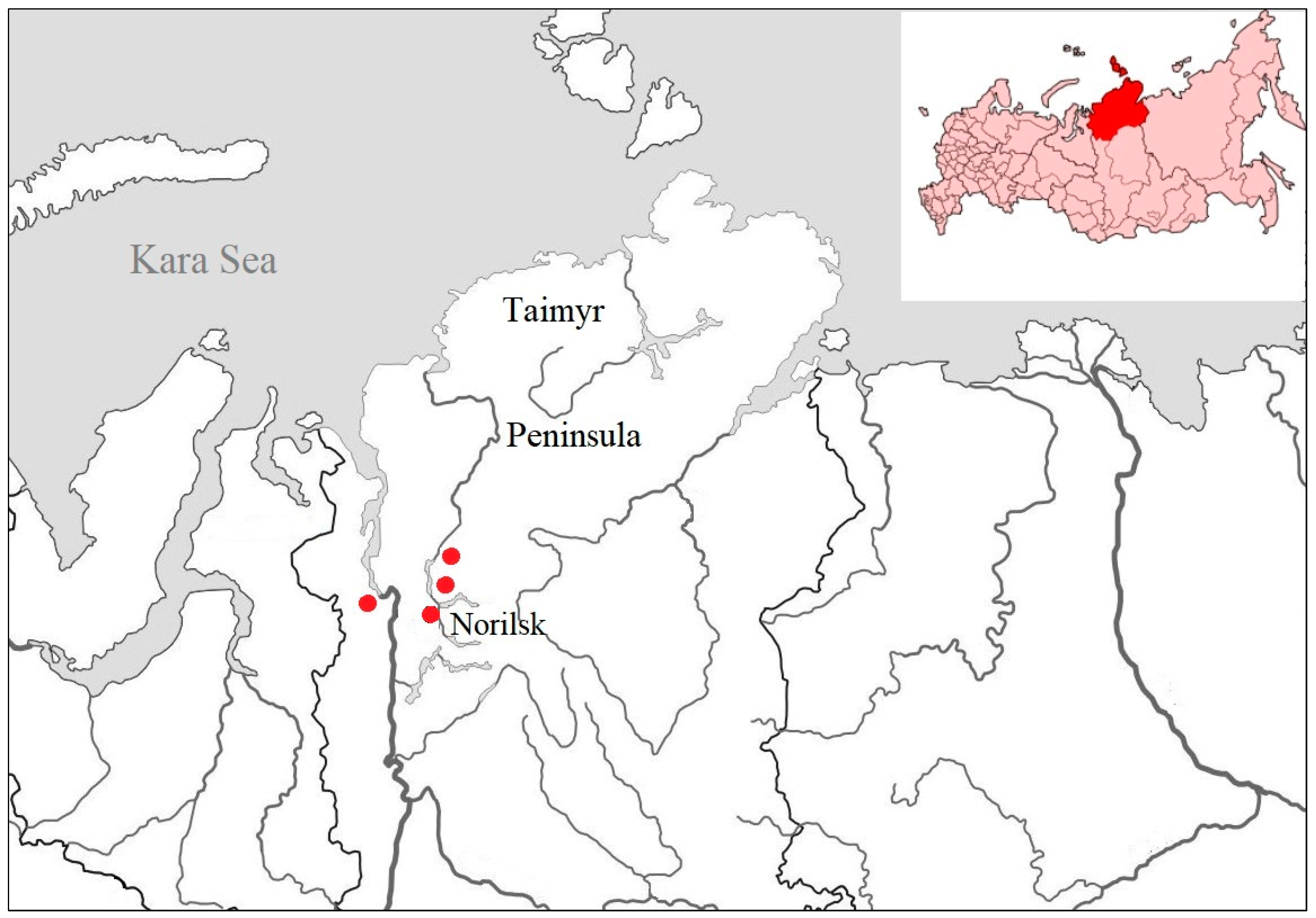
2.1. Sampling

The collection of biological material from birds was carried out as part of the annual monitoring of the influenza A virus on the Taimyr Peninsula (Krasnoyarsk region, Russia) (Figure 1). Samples were collected during the state hunting season under a license from the regional ministry of ecology and natural resources within the framework of the Program for the Study of Infectious Diseases of Wild Animals of the Federal Research Center of Fundamental and Translational Medicine (FRC FTM), Novosibirsk.
Cloacal swabs from wild birds were collected during the official hunting season in 2 mL tubes filled with 1 mL viral transport medium [23]. Samples were stored and transported in liquid nitrogen following the WHO guidelines [23].
The present study was conducted as per the approval and requirements of the Biomedical Ethics Committee of the FRC FTM, Novosibirsk (protocol Nos. 2013-23 and 2021-10). The study utilized the Biosafety Level-3 (BSL-3) facilities of the FRC FTM.

2.2. Virus Isolation

For the isolation of AIV and APMV (avian paramyxoviruses) in 10-day specific pathogen-free (SPF) embryonated chicken eggs, samples were prepared according to a standard protocol [24]. All virus isolation work was performed in the BSL-3 laboratory of the FRC FTM. After cultivation, 2 mL of allantoic fluid from each embryo infected with one sample was extracted and used for a hemagglutination test (HA) with 5% chicken red blood cells [25]. All HA-positive samples were aliquoted for AIV M and APMV RDRP gene PCR testing.

2.3. Avian Paramyxovirus and Avian Influenza Virus Detection

RNA was extracted from allantoic fluid samples using a kit for nucleic acid extraction (Medical-Biological Union LLC, Novosibirsk, Russia) following the manufacturer’s protocol. The presence of conservative M gene regions of the influenza A virus was determined by real-time PCR using the Influenza A Virus Real-Time RT-PCR Kit (Medical-Biological Union LLC, Novosibirsk, Russia), adapted to detect both human and avian influenza virus.
HA-positive allantoic fluid was also tested for the presence of APMV by PCR. For this purpose, cDNA was synthesized using the REVERTA-L Kit (AmpliSens, Moscow, Russia). To detect avian paramyxoviruses, family-wide oligonucleotides (PMX1 5′-GAR-GGI-YII-TGY-CAR-AAR-NTN-TGG-AC-3′ and PMX2 5′-TIA-YIG-CWA-TIR-IYT-GRT-TRT-CNC-C-3′) specific to domain III of the RNA-dependent RNA polymerase gene were used [26]. Oligonucleotides were diluted to a concentration of 50 pmol/μL. A reaction mixture was prepared using 25 μL of HS-Taq PCR-Color (2×) Master Mix (Biolabmix, Novosibirsk, Russia), 1 μL of forward and reverse oligonucleotides, and 5 μL of cDNA. Water was then added to achieve a final volume of 50 µL. The reaction mixture was incubated at 95 °C for 5 min, then for 40 cycles at 95 °C for 15 s, at 41 °C for 30 s, at 72 °C for 15 s, and then a final extension at 72 °C for 7 min.
The reaction products were visualized by electrophoresis in 1.5% agarose gel in the gel documentation system “E-Box CX5” (VILBER, Eberhardzell, Germany). A 100 bp DNA ladder DNA marker Step100 (Biolabmix, Novosibirsk, Russia) was used to estimate the amplicon size. Samples in which amplicons of the target size were detected were prepared for whole-genome sequencing.

2.4. Sequencing

Avian paramyxovirus sequences were obtained at the National Institute of Animal Health, Japan, using a MiSeq genome sequencer (Illumina, San Diego, CA, USA), as described previously [27]. Briefly, RNA was isolated from allantoic fluid using the RNeasy Mini Kit (Qiagen, Hilden, Germany). cDNA libraries were prepared using the NEBNext Ultra RNA Library Prep Kit (New England Biolabs, Ipswich, MA, USA). Sequencing was performed using the MiSeq Reagent Kit v.2 (Illumina, San Diego, CA, USA). Consensus sequences were constructed using Workbench software v.12.0.2 (Qiagen, Hilden, Germany).
The whole-genome sequence of the influenza A virus was obtained using the Illumina MiSeq platform (Illumina, San Diego, CA, USA) and the corresponding reagent kits according to the manufacturer’s methodology. Sequencing was performed at FRC FTM, Russia. RNA was extracted using the QIAamp Viral RNA Mini Kit (Qiagen, Hilden, Germany). Whole-genome amplification was performed using a modified protocol [28]. DNA libraries were prepared using the Nextera DNA Flex Library Prep Kit (Illumina, San Diego, CA, USA) and sequenced using the MiSeq Reagent Kit v3 (600 cycles) (Illumina, San Diego, CA, USA). Consensus sequences were generated using Bowtie software (version 1.3.1) [29].

2.5. Phylogenetic Analysis

2.5.1. Newcastle Disease Virus

For phylogenetic analysis, the complete CDS F gene (1662 nt) of the obtained isolates and the most closely related sequences found in BLAST, as well as from the dataset described by Dimitrov et al., were used [2]. For phylogenetic analysis of whole genomes, the most closely related sequences found in BLAST and sequences from Class I isolates as an outgroup were used. Sequence alignments were obtained using the MUSCLE alignment algorithm. The phylogenetic tree was constructed using the maximum likelihood method based on the General Time Reversible model with a discrete gamma distribution (+G) and allowing for invariant sites (+I) with statistical analysis based on 1000 bootstrap replicates, as implemented in MEGA X [30]. The tree was drawn to scale, with branch lengths measured in the number of substitutions per site.

2.5.2. Avian Influenza Virus

For each of the obtained genome segment sequences, a search for the most identical sequences was conducted using the BLAST algorithm [31] from the GISAID database [32].
The obtained sets of sequences were subjected to multiple sequence alignment using the MAFFT v7.520 multiple alignment algorithm (downloadable version) [33] with the following default parameters: gap opening penalty: 1.53, gap extension penalty: 0.0. Then, the files containing multiple alignments were opened in the Unipro UGENE software, version 50.0 [34], where they were manually checked, and sites containing deletions were removed from the final alignment.
Phylogenetic trees were constructed using the IQ-TREE version 1.6 ML (maximum likelihood) method [35]. Branch support was assessed using ultrafast bootstrap v2.4.0 [36] and the SH-aLRT branch test with 1000 iterations. The nucleotide substitution model for each gene segment was determined using ModelFinder through IQ-TREE version 1.6 [37].
Tree visualization and topology analysis were performed using iTOL v.6 [38].

3. Results

3.1. Sampling and Virus Isolation

As part of the annual influenza A virus monitoring program on the Taimyr Peninsula from 2019 to 2023, 323 samples were collected from wild birds. Samples were obtained from 18 species of four orders: Charadriiformes (n = 167), Anseriformes (n = 147), Gaviiformes (n = 7), and Passeriformes (n = 2). The most numerous species in the sample were: herring gull (Larus argentatus) (n = 158), northern pintail (Anas acuta) (n = 30), european wigeon (Mareca penelope) (n = 28), and velvet scoter (Melanitta fusca) (n = 28) (Table 1).
During five years of monitoring, only four APMV isolates and one AIV isolate were detected in the summer and autumn periods. This constituted 1.24% and 0.31% of the total number of samples collected, respectively. All APMV isolates belonged to the Orthoavulavirus javaense species. The AIV isolate belonged to the H3N8 subtype.

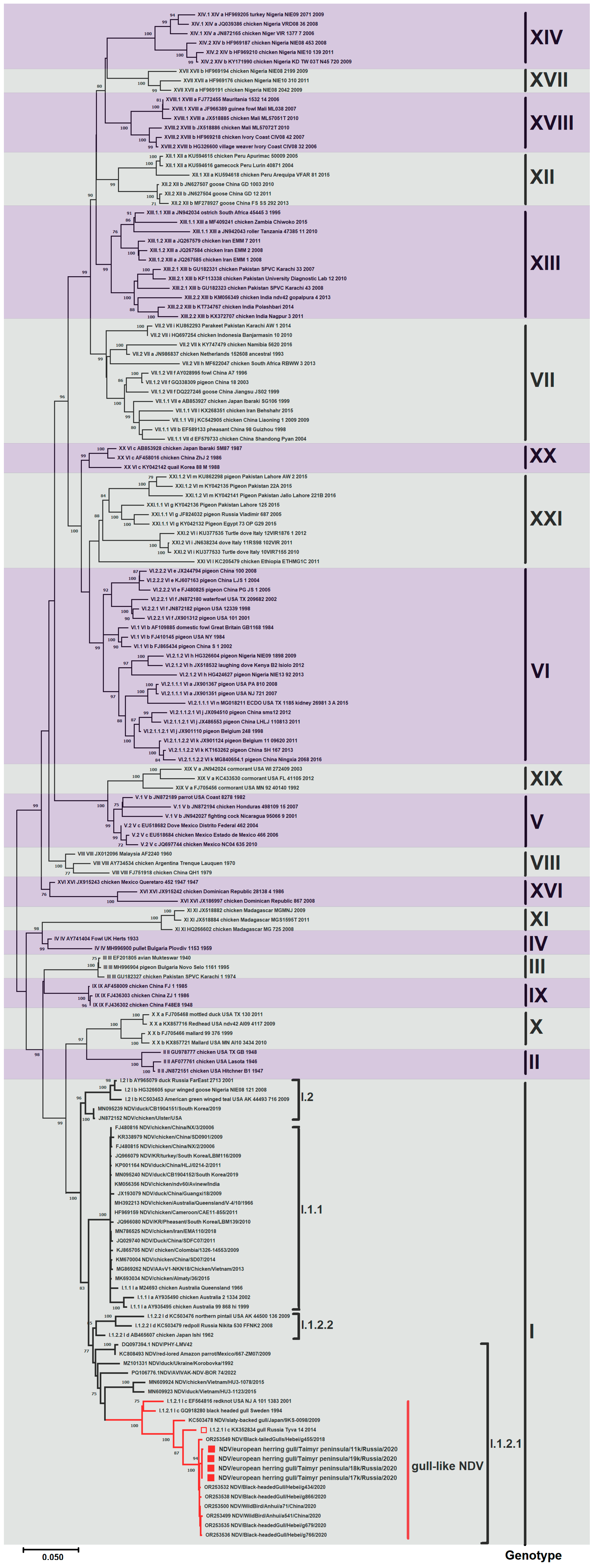
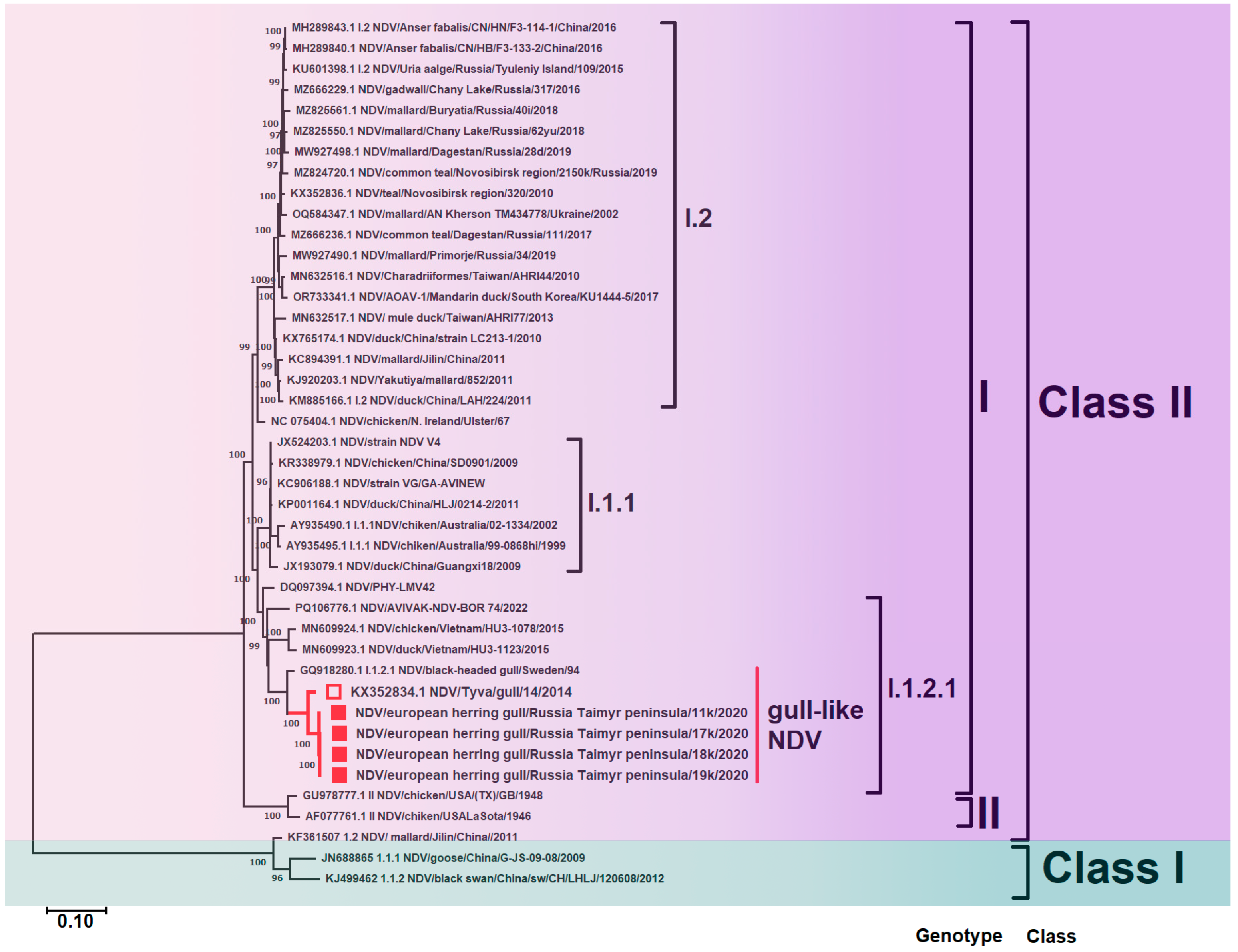
3.2. Phylogenetic Analysis of Newcastle Disease Virus

In the current study, four whole genome sequences of NDV were obtained: NDV/herring gull/Taimyr Peninsula/11k/Russia/2020 (NDV/11k), NDV/herring gull/Taimyr Peninsula/17k/Russia/2020 (NDV/17k), NDV/herring gull/Taimyr Peninsula/18k/Russia/2020 (NDV/18k), and NDV/herring gull/Taimyr Peninsula/19k/Russia/2020 (NDV/19k) (GenBank accession numbers: PV032627-PV032630).
BLAST analysis of the identity of the nucleotide sequences of the whole genome showed that the Taimyr isolates had the highest identity with the isolate we previously obtained from a gull in the south of Eastern Siberia in 2014. Since the GenBank database contains significantly more nucleotide sequences of the F gene of NDV than whole-genome sequences, CDS of the F gene was also used to determine the nucleotide identity. The highest identity (99.46–99.82%) was observed with isolates from China obtained from the black-headed gull, black-tailed gull, and unspecified wild birds in 2018 and 2020 (Table 2).

3.2.1. Gene F

Phylogenetic analysis of the complete CDS gene F (1662 nt) revealed that all Taimyr isolates belonged to genotype I within Class II. When combined with other isolates obtained from various regions of Eurasia, they formed a distinct gull-like clade in sub-genotype I.1.2.1 (see Figure 2). The clade was mainly formed by isolates from different species of gulls: black-tailed gull, black-headed gull, little gull, herring gull, and slaty-backed gull. These isolates were collected sporadically and with long intervals between occurrences from different locations across the continent. Also in the clade was an isolate obtained in the USA from the red knot shorebird, belonging to the same order as gulls, Charadriformes. NDV/BHG/Sweden/94 and APMV-1/red knot/US(NJ)/A101-1383/2001, the oldest in the clade, formed a separate branch.

3.2.2. Whole Genome

When comparing the whole genome sequences of NDV, it was seen that a gull-like clade also formed in sub-genotype I.1.2.1 (Figure 3). Currently, the NCBI database has significantly fewer available open-access whole-genome sequences of NDV than sequences of the F gene. Therefore, the gull-like clade on the phylogenetic tree of the whole genome is represented only by isolates from the Taimyr Peninsula, the south of Eastern Siberia, and Sweden.
Isolates used in this study are shown in red. Roman numerals indicate each isolate’s corresponding genotype and sub-genotype according to the classification proposed by Dimitrov et al. [2]. The percentage of trees in which associated taxa clustered together in the bootstrap test (1000 replicates) is shown next to the branches.

3.2.3. Amino Acid Substitutions

Analysis of amino acid substitutions of the F protein showed that the sequences obtained in this study contained the F protein cleavage site 112-GKQGR↓L-117, characteristic of lentogenic virus. Other viruses in sub-genotype I.1.2.1, including vaccine strains, contained the same cleavage site (Table 3). In sub-genotype I.1.2.2, the F protein cleavage site was the same as in isolates of sub-genotype I.1.2.1, but a variation at position 112 was noted in the isolate northern pintail/USA/AK/44500/136/2009.
Sub-genotype I.1.1 contained isolates characteristic of both highly pathogenic and low-pathogenic viruses, reflecting the historical composition of sub-genotype I.1.1 [14]. For sub-genotype I.2, cases of detection of only lentogenic isolates were described, which was confirmed by the corresponding cleavage site.

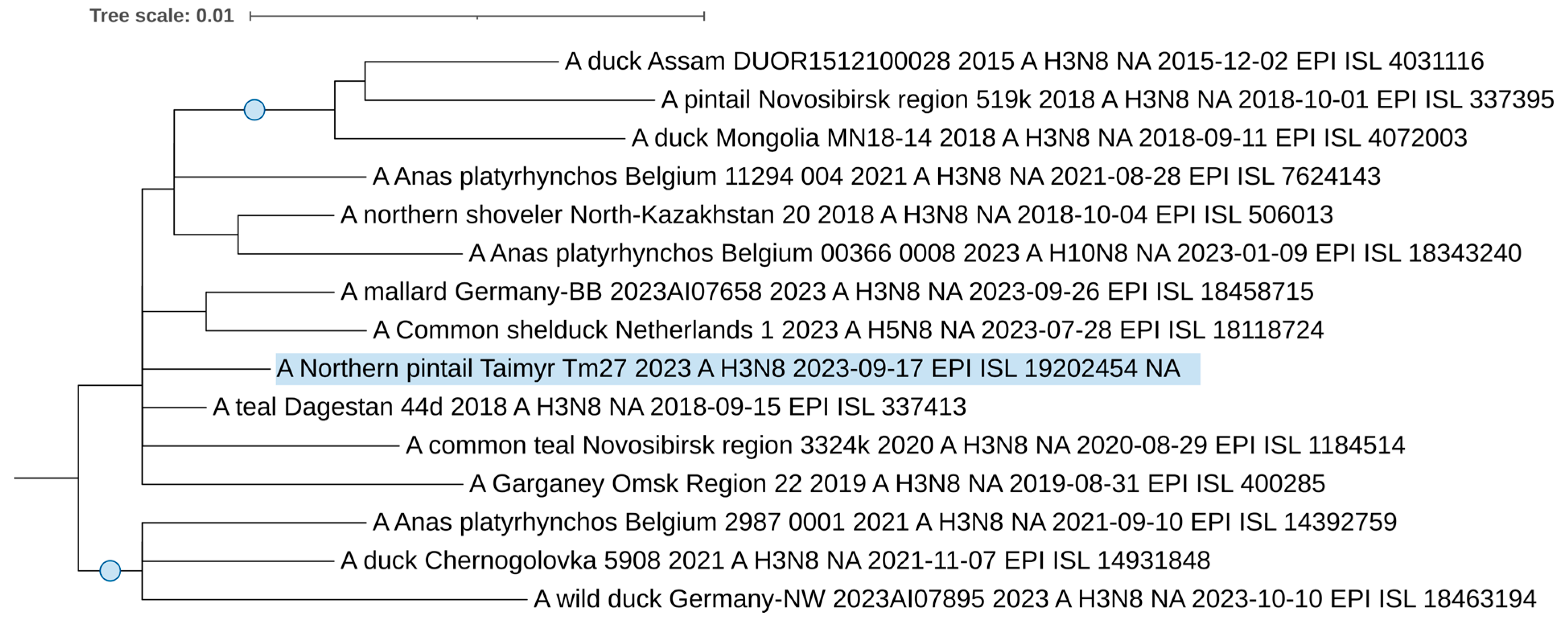
3.3. Phylogenetic Analysis of Avian Influenza Virus

From 2019 to 2023, one H3N8 avian influenza virus, A/northern pintail/Taimyr/Tm27/2023 (EPI_ISL_19202454), was isolated and sequenced. Phylogenetic dendrograms were constructed using genome segment sequences from the isolated influenza virus strain, and closely related sequences from around the world were identified through BLAST analysis.
The H3 segment sequence forms a distinct branch, sharing a common ancestor with European H3 viruses, including strains from Belgium and Germany, and H3N2 strains isolated from a wild duck in the Moscow Pond in 2023 (Figure 4) [39]. This hemagglutinin virus belongs to the classical, avian-like clade of Eurasian low-pathogenic H3Nx viruses [40].
Conversely, based on the neuraminidase segment, the N8 sequence (Figure 5) does not group within this specific European (Belgian–German) clade. Instead, it resides in a more diverse clade of H3N8 viruses. Notably, the clade also contains a unique H5N8 virus that was isolated from a duck in the Netherlands in 2023.
The viruses that share the specific clades of our studied H3 and N8 segments are all contemporary strains circulated between 2021 and 2023. This supports a phylogenetic relationship and suggests that these strains are circulating in northern regions, such as Taimyr, and are likely spread by wild, migrating birds.
Phylogenetic analysis of internal segments showed that all are of Eurasian lineage. Figures are presented in the Supplementary Material (Figures S1–S6). Interestingly, the polymerase complex segments exhibit distinct phylogenetic relationships: PB1 and PB2 group within European clades, whereas the PA segment clusters with East and Central Asian viruses isolated in Eastern Siberia, Japan, China, and Bangladesh. Additionally, the NS segment of the virus we isolated belongs to allele B, typical for avian influenza viruses, instead of allele A, which is also found in mammalian viruses [41].
Using the FluSurver tool [42], we analyzed genome segment sequences to identify mutations resulting in amino acid substitutions. The identified substitutions are detailed in Table S1. While some substitutions, such as I262V in neuraminidase, are known to confer increased resistance to antiviral drugs, potentially, all identified substitutions at critical positions in our analysis presented residues different from those previously associated with the described effect. Our findings indicate the presence of substitutions that, based on current knowledge, do not elevate the risk of adaptation to mammals. However, a key limitation is that the functional roles of these substitutions have primarily been characterized in H5N1 influenza viruses and have not yet been definitively shown for low-pathogenic avian influenza viruses (LPAIs).
The phylogenetic analysis revealed no specific pattern for A/northern pintail/Taimyr/Tm27/2023. All segments belong to the Eurasian clades of classic avian-like influenza viruses. The close phylogenetic relationships of the virus with Asian and Siberian viruses on the one hand, and with European viruses on the other, suggest broad connections and ways of virus transmission throughout Eurasia into Arctic regions, as we demonstrated previously for Eurasian regions [43,44,45].

4. Discussion

During a five-year study in the summer and autumn periods on the Taimyr Peninsula (Arctic region of Russia), only four NDV isolates and one AIV isolate were detected.
The H3N8 subtype is one of the most commonly detected avian influenza viruses in wild birds, particularly in Northern Asia [44]. While H3N8 viruses are widespread in birds, human infections are rare [46]. However, it is important to monitor these viruses due to their potential to evolve and cause human disease, as demonstrated by isolated cases and a recent fatal case in China. We did not find any substitutions increasing virulence to mammals or segments of highly pathogenic avian influenza lineages, confirming the low pathogenicity of the virus studied.
Polar regions may become new frontiers for the emergence and spread of significant viral pathogens. In Antarctica, mass deaths of penguins of unknown etiology were recorded back in the 1970s [18]. Currently, influenza A virus and Newcastle disease virus have been detected in the region. The first studies reported the detection of antibodies against these viruses in the Adélie penguin (Pygoscelis adeliae) and the south polar skua (Stercorarius maccormicki) [47,48]. Current studies have confirmed the presence of H5N1 HPAIV in birds and seals, and low-pathogenicity variants of NDV in penguins [21,49]. A study of the spread of H5N1 HPAIV from South America to Antarctica has confirmed the involvement of seals and several bird species: brown skua (Stercorarius antarcticus), kelp gull (Larus domincanus), and antarctic tern (Sterna vittata) [19,20].
A recent comprehensive review of influenza A virus ecology in the Arctic found that Alaska is the best-studied region of the Arctic [22]. There has been very little research on the influenza A virus in the Arctic region of Russia. The only known large-scale surveillance of avian influenza viruses on the Taimyr Peninsula, which involved testing over 1000 geese of various species during their spring migration, did not detect any avian influenza viruses [50]. The Arctic region of Russia includes subarctic, low Arctic, and high Arctic zones with a variety of wetlands and climate conditions that attract different species of waterbirds. The introduction of viruses into relatively isolated polar regions with low species diversity could have detrimental effects. More research in this region will contribute to our understanding of the genetic flow and distribution of avian influenza viruses.
We focused on analyzing unique NDV isolates obtained from herring gulls in autumn 2020. As a result of phylogenetic analysis, these isolates and other isolates from gulls formed a strongly supported clade in sub-genotype I.1.2.1, Class II.
Genotype I is one of the four “historical” genotypes, currently represented by four sub-genotypes: I.1.1, I.2, I.1.2.1, and I.1.2.2 [2]. Viruses of sub-genotypes I.2 and I.1.1 have been detected in wild and domestic birds worldwide. Isolates of sub-genotype I.2 are low-pathogenic, while sub-genotype I.1.1 also includes highly pathogenic isolates [14]. These sub-genotypes also include the vaccine strains Queensland/V-4/chicken/Australia/1966 and Ulster/chicken/Ireland/1967. The isolates that formed the I.1.2.2 sub-genotype were isolated from waterfowl, shorebirds, and redpoll from 2002 to 2009 and from poultry in Japan in 1962.
Sub-genotype I.1.2.1 is represented by low-pathogenic isolates from wild and domestic birds and vaccine strains NDV/AVIVAK-NDV-BOR/74/2022 (used in Russia) and NDV/PHY-LMV42/Hungary. Viruses from domestic birds were detected in Ukraine in 1992 and Vietnam in 2015. Viruses from wild birds (mostly from gulls) were detected in Sweden in 1994, the USA in 2001, Japan in 2009, and Russia in 2014. Recently, sequences from China, obtained from two gull species and unidentified wild birds in 2018 and 2020, were added to the NCBI database. Together with the sequences obtained during this study, they formed a separate gull-like clade that has not been previously identified.
The gull-like clade is represented by sequences obtained in different regions and with long intervals between occurrences. The oldest isolate in the clade was obtained from a black-headed gull in Sweden in 1994. The sample was collected in the autumn at the Ottenby Bird Observatory, on the Baltic coast [51]. The sample included 430 samples from 57 bird species, but no other data on APMV detection in this sample were described. Retrospective and ongoing analyses of samples from 1986 to 2006 in the United States yielded 268 NDV isolates from wild waterbirds and live bird market birds [6]. Among them, only one isolate, obtained from a red knot in 2001, was most closely related to the isolate from Sweden. During monitoring of AIV and APMV in migratory birds in Alaska, Russia, and Japan from 2007 to 2010, 73 NDV isolates were obtained. Only one isolate, obtained from a slaty-backed gull in Japan in 2009, was phylogenetically close to isolates from Sweden and the USA, which led to the identification of a new sub-genotype I.1.2.1 (formerly 1c) [52]. In 2014, our laboratory staff obtained a full-genome sequence of NDV from a little gull due to cultivation. Biological material from the bird was collected in April at Ubsu-Nur Lake on the border of Russia and Mongolia. From 2014 to 2021, we did not detect any other APMV isolates in this region. In 2018 and 2020, several NDV sequences were obtained from a black-headed gull, a black-tailed gull, and unspecified wild bird species in China. Seven of the ten isolates were isolated in March 2020, two in November 2020, and one in September 2018. All were phylogenetically related to the gull isolates described above. These sequences from unspecified species may belong to gulls or other species of the order Charadriiformes. However, there is no precise information. Identifying the host bird species or genus during sample collection is important for monitoring viruses with relevance for the poultry industry. Detection of the affiliation of a specific viral lineage to a particular host species helps to monitor the direction of virus evolution and adjust preventive measures.
In the current study, we found NDV in four herring gulls. This is a common bird species on the Taimyr Peninsula. These birds arrive in the spring and build nests on islands and along the shores of large lakes and rivers, forming colonies. The first spring migrations to the Taimyr Peninsula begin in late April. The bulk of nesting sites for this species are concentrated in the west of the peninsula (the Yenisei River floodplain) and the southeast (the foothills of the Putorana Plateau). Birds migrate to the eastern part of the peninsula from the south and southeast along the Kotuy, Popigai, and Anabar Rivers. Some flocks of herring gulls fly to the shores of Taimyr and Severnaya Zemlya from the Laptev Sea, from the New Siberian Islands. Autumn migration to wintering grounds begins in the last ten days of August and continues until the end of September. However, in the area of city dumps, which are located within the boundaries of the Norilsk industrial region, large aggregations of herring gulls linger for a longer period: until the end of the first ten days of October and even later. They winter in Japan, Korea, and China [53]. Within Russia, they winter on the eastern coast of the Baltic Sea, the northern coast of the Black and Azov Seas, the Caspian Sea, in the south of Primorsky Krai, and in the Kuril Islands [54]. Thus, the ecological characteristics of the species make it possible for viruses to spread not only within the Central Asian and East Asian Flyways, but also in areas of large bird concentrations at the intersection of several flyways (for example, on the western coast of the Caspian Sea).
The NDV isolates obtained in this study contained the F protein cleavage site characteristic of low-virulence viruses [55]. Samples were collected from asymptomatic gulls. Avirulent isolates cause subclinical infections or mild respiratory disease and are not important for agri-livestock farming, but their role in the evolution of NDV remains to be elucidated. In a recent study, avirulent NDV isolates were obtained during an outbreak of highly pathogenic avian influenza in the UK [56]. Avirulent NDV was isolated from both a chicken in one of the poultry houses and the carcasses of a magpie (Pica pica) found near the poultry houses. Co-infection of one organism with several virus species is associated with the emergence of complex interspecific relationships under selection pressure [57]. Thus, avirulent isolates may contribute more to the formation of the overall genetic landscape of NDV.
Based on the results of phylogenetic analysis, the gull isolates obtained in this study in 2020 are most closely related to the isolates obtained in China in 2018 and 2020 and in the south of Eastern Siberia (Ubsu-Nur Lake, Russia) in 2014. The nucleotide identity of the F gene was 97.35–99.82%. This may indicate the exchange of viruses within the Central Asian and East Asian Flyways. Given that gull NDV sequences, which were obtained sporadically and with long intervals between occurrences from different locations across the continent, formed a distinct clade in sub-genotype I.1.2.1, we suggest that within Class II there may be a larger number of genetic lineages leading to adaptation to a larger number of hosts.
The diversity of susceptible hosts expands the range of conditions for the emergence of different quasispecies during the virus life cycle. A study conducted in 2010 showed that virulent viral populations can arise during the replication of avirulent field isolates [58]. The presence of such virulent populations did not lead to a change in the phenotypic properties of the avirulent isolate. However, the selective pressure caused by passaging led to an increase in the percentage of virulent quasispecies. The authors believe that the selection pressure can lead to the dominance of virulent quasi-species and, as a consequently, to a change in phenotypic properties. The diversity of susceptible hosts with a rapid shift in generations and individual biological characteristics can also change the parameters of immune pressure, thereby changing the direction of selection in the evolution of the virus. The existence of a lineage of NDV specific to species from the order Charadriiformes can be accompanied by the accumulation of mutations arising under the influence of population immunity. If the biological features that influence population immunity differ significantly between the orders Anseriformes and Charadriiformes, then virus specialization to Charadriiformes species will increase the diversity of the overall gene pool that is subject to evolutionary events. Further surveillance of different gull and shorebird species may help better understand their role in the evolution and global spread of NDV.
NDV and H3N8 isolates were obtained from herring gulls and northern pintails. The northern pintail is recognized as a reservoir for avian influenza virus and can carry the virus asymptomatically, playing an important role in long-distance transmission along migratory routes [59,60]. Newcastle disease virus has also been detected in clinically healthy northern pintails [61]. Northern pintail migrations include transcontinental routes, so it is important to monitor for viral pathogens in these bird populations. Herring gulls are opportunistic scavengers and have been implicated in the spread of avian pathogens. Their ecological behavior may be key to the spread of the virus. For example, a similar species, the kelp gull (Larus dominicanus), has been documented scavenging on elephant seal (Mirounga leonina) carcasses, highlighting the potential for interspecies transmission and ecological persistence of viral pathogens [62].
Nevertheless, our five-year study has identified certain limitations that should be acknowledged. The main limitation is that the sample size was small (n = 323). In addition, the study included 48.92% of samples from the herring gull, which may introduce bias. However, we wanted to estimate the general viral diversity in a poorly studied area. Charadriiformes (51.7%) and Anseriformes (45.51%) are informative groups for an initial assessment of viral diversity.

5. Conclusions

In this study, we report the first detection of Newcastle disease virus (NDV) in wild birds in Arctic Russia. All NDV sequences were obtained from the herring gull and, together with other gull sequences, formed a separate gull-like clade in the sub-genotype I.1.2.1, Class II. The gull isolates obtained in this study in 2020 are closely related to those isolates obtained in China in 2018 and 2020 and in the south of Eastern Siberia (Russia) in 2014. The nucleotide identity of the F gene was 97.35–99.82%. This may indicate the exchange of viruses within the Central Asian and East Asian Flyways.
Our study also identified an avian influenza virus subtype H3N8 isolated from a northern pintail. This isolate was phylogenetically related to viruses circulating between 2021 and 2023 in Eurasia, Siberia, and Asia.
Further research in this region will further our understanding of the distribution and evolution of avian paramyxoviruses and avian influenza viruses.

Supplementary Materials

The following supporting information can be downloaded at: https://www.mdpi.com/article/10.3390/v17070955/s1, Table S1: Amino acid substitutions in key positions of influenza virus proteins according to the FluServer (https://flusurver.bii.a-star.edu.sg/); Figure S1: Maximum likelihood phylogenetic tree of the MP genome segment of avian influenza viruses isolated in the Taimyr Peninsula; Figure S2: Maximum likelihood phylogenetic tree of the NP genome segment of avian influenza viruses isolated in the Taimyr Peninsula. The blue circle symbol denotes branches with SH-aLRT values > 80% and UFboot values > 95%; Figure S3: Maximum likelihood phylogenetic tree of the NS genome segment of avian influenza viruses isolated in the Taimyr Peninsula; Figure S4: Maximum likelihood phylogenetic tree of the PA genome segment of avian influenza viruses isolated in the Taimyr Peninsula. The blue circle symbol denotes branches with SH-aLRT values > 80% and UFboot values > 95%; Figure S5: Maximum likelihood phylogenetic tree of the PB1 genome segment of avian influenza viruses isolated in the Taimyr Peninsula. The blue circle symbol denotes branches with SH-aLRT values > 80% and UFboot values > 95%; Figure S6: Maximum likelihood phylogenetic tree of the PB2 genome segment of avian influenza viruses isolated in the Taimyr Peninsula. The blue circle symbol denotes branches with SH-aLRT values > 80% and UFboot values > 95%.

Author Contributions

A.D. conceptualization, data curation, formal analysis, methodology, visualization, software, writing—original draft preparation, writing—review and editing; N.D. validation, formal analysis, methodology, visualization, software, writing—review and editing; A.P. resources, data curation; J.M. data curation, methodology, formal analysis, software; R.T. data curation, methodology, validation, software; Y.U. data curation, methodology, validation, software; T.S. data curation, methodology, validation, software; N.K. formal analysis, visualization, software; A.L. methodology; I.S. formal analysis, methodology; S.K. conceptualization, writing—review and editing; A.S. project administration; K.S. conceptualization, data curation, formal analysis, validation, project administration, writing—original draft, writing—review and editing. All authors have read and agreed to the published version of the manuscript.

Funding

The study was partially supported by the following sources: RSF project 23-64-00005, state-funded budget project 225020408196-1.

Institutional Review Board Statement

All aspects of the present study were approved by the Committee on Biomedical Ethics at the Federal Research Center of Fundamental and Translational Medicine (Novosibirsk, Russia) (protocol Nos. 2013-23, 2019-3 and 2021-10). The experiments were conducted in Biosafety Level-3 facilities.

Data Availability Statement

All sequences from the study are available in the GenBank database (accession numbers: PV032627-PV032630) and in the GISAID EpiFlu database (accession numbers: EPI_ISL_19202454).

Acknowledgments

Construction of the phylogenetic trees was carried out using sequences deposited in GenBank. We are grateful to the GenBank database (https://www.ncbi.nlm.nih.gov/nucleotide/ accessed on 4 July 2025) and to the authors who provided sequence information. We are grateful to the reviewers for their significant assistance in working on the manuscript. The study was partly supported by the Centers for Collective Use of scientific equipment “Proteomic analysis,” supported by funding from the Ministry of Science and Higher Education of the Russian Federation (agreement No. 075-15-2021-691), and “Modern Optical Systems” of the Research Center of Fundamental and Translational Medicine (Novosibirsk, Russia). We would like to thank the Technical Sequencing Center of the Institute of Virology and, in particular, Okhlopkova O.V., Moshkin A.D., Stolbunova K.A., and Stepanyuk M.A for sequencing A/northern pintail/Taimyr/Tm27/2023 (EPI_ISL_19202454).

Conflicts of Interest

The authors declare no conflict of interest.

References

  1. Kitchen, A.; Shackelton, L.A.; Holmes, E.C. Family Level Phylogenies Reveal Modes of Macroevolution in RNA Viruses. Proc. Natl. Acad. Sci. USA 2011, 108, 238–243. [Google Scholar] [CrossRef] [PubMed]
  2. Dimitrov, K.M.; Abolnik, C.; Afonso, C.L.; Albina, E.; Bahl, J.; Berg, M.; Briand, F.-X.; Brown, I.H.; Choi, K.-S.; Chvala, I.; et al. Updated Unified Phylogenetic Classification System and Revised Nomenclature for Newcastle Disease Virus. Infect. Genet. Evol. 2019, 74, 103917. [Google Scholar] [CrossRef]
  3. Alexander, D.J.; Aldous, E.W.; Fuller, C.M. The Long View: A Selective Review of 40 Years of Newcastle Disease Research. Avian Pathol. 2012, 41, 329–335. [Google Scholar] [CrossRef] [PubMed]
  4. Sharshov, K.; Dubovitskiy, N.; Derko, A.; Loginova, A.; Kolotygin, I.; Zhirov, D.; Sobolev, I.; Kurskaya, O.; Alekseev, A.; Druzyaka, A.; et al. Does Avian Coronavirus Co-Circulate with Avian Paramyxovirus and Avian Influenza Virus in Wild Ducks in Siberia? Viruses 2023, 15, 1121. [Google Scholar] [CrossRef] [PubMed]
  5. Coffee, L.L.; Hanson, B.A.; Luttrell, M.P.; Swayne, D.E.; Senne, D.A.; Goekjian, V.H.; Niles, L.J.; Stallknecht, D.E. Avian Paramyxoviruses in Shorebirds and Gulls. J. Wildl. Dis. 2010, 46, 481–487. [Google Scholar] [CrossRef]
  6. Kim, L.M.; King, D.J.; Curry, P.E.; Suarez, D.L.; Swayne, D.E.; Stallknecht, D.E.; Slemons, R.D.; Pedersen, J.C.; Senne, D.A.; Winker, K.; et al. Phylogenetic Diversity among Low-Virulence Newcastle Disease Viruses from Waterfowl and Shorebirds and Comparison of Genotype Distributions to Those of Poultry-Origin Isolates. J. Virol. 2007, 81, 12641–12653. [Google Scholar] [CrossRef]
  7. Diel, D.G.; Miller, P.J.; Wolf, P.C.; Mickley, R.M.; Musante, A.R.; Emanueli, D.C.; Shively, K.J.; Pedersen, K.; Afonso, C.L. Characterization of Newcastle Disease Viruses Isolated from Cormorant and Gull Species in the United States in 2010. Avian Dis. 2012, 56, 128–133. [Google Scholar] [CrossRef]
  8. Kuiken, T.; Leighton, F.A.; Wobeser, G.; Danesik, K.L.; Riva, J.; Heckert, R.A. An Epidemic of Newcastle Disease in Double-Crested Cormorants from Saskatchewan. J. Wildl. Dis. 1998, 34, 457–471. [Google Scholar] [CrossRef]
  9. Relich, R.F.; Loeffelholz, M.J. Taxonomic Changes for Human Viruses, 2020 to 2022. J. Clin. Microbiol. 2022, 61, e0033722. [Google Scholar] [CrossRef]
  10. Webster, R.G.; Bean, W.J.; Gorman, O.T.; Chambers, T.M.; Kawaoka, Y. Evolution and Ecology of Influenza A Viruses. Microbiol. Rev. 1992, 56, 152–179. [Google Scholar] [CrossRef]
  11. Sreenivasan, C.C.; Li, F.; Wang, D. Emerging Threats of Highly Pathogenic Avian Influenza A (H5N1) in US Dairy Cattle: Understanding Cross-Species Transmission Dynamics in Mammalian Hosts. Viruses 2024, 16, 1703. [Google Scholar] [CrossRef] [PubMed]
  12. Echeverri-De la Hoz, D.; Martínez-Bravo, C.; Gastelbondo-Pastrana, B.; Rivero, R.; López, Y.; Bertel, V.; Alemán-Santos, M.; Garay, E.; Hoyos, R.; Arrieta, G.; et al. Genomics of Novel Influenza A Virus (H18N12) in Bats, Caribe Colombia. Sci. Rep. 2025, 15, 6507. [Google Scholar] [CrossRef] [PubMed]
  13. Gadzhiev, A.; Petherbridge, G.; Sharshov, K.; Sobolev, I.; Alekseev, A.; Gulyaeva, M.; Litvinov, K.; Boltunov, I.; Teymurov, A.; Zhigalin, A.; et al. Pinnipeds and Avian Influenza: A Global Timeline and Review of Research on the Impact of Highly Pathogenic Avian Influenza on Pinniped Populations with Particular Reference to the Endangered Caspian Seal (Pusa caspica). Front. Cell. Infect. Microbiol. 2024, 14, 1325977. [Google Scholar] [CrossRef]
  14. Dimitrov, K.M.; Ramey, A.M.; Qiu, X.; Bahl, J.; Afonso, C.L. Temporal, Geographic, and Host Distribution of Avian Paramyxovirus 1 (Newcastle Disease Virus). Infect. Genet. Evol. 2016, 39, 22–34. [Google Scholar] [CrossRef] [PubMed]
  15. Kilpatrick, A.M.; Chmura, A.A.; Gibbons, D.W.; Fleischer, R.C.; Marra, P.P.; Daszak, P. Predicting the Global Spread of H5N1 Avian Influenza. Proc. Natl. Acad. Sci. USA 2006, 103, 19368–19373. [Google Scholar] [CrossRef]
  16. Yoon, S.-W.; Webby, R.J.; Webster, R.G. Evolution and Ecology of Influenza A Viruses. In Influenza Pathogenesis and Control—Volume I; Compans, R.W., Oldstone, M.B.A., Eds.; Springer International Publishing: Cham, Switzerland, 2014; pp. 359–375. ISBN 978-3-319-11155-1. [Google Scholar]
  17. Boere, E.G.C.; Galbraith, C.A.; Stroud, D.A.; Thompson, D.B.A.; Underhill, L.G. Waterbirds Around the World; Stationery Office: Edinburgh, UK, 2006. [Google Scholar]
  18. Smeele, Z.E.; Ainley, D.G.; Varsani, A. Viruses Associated with Antarctic Wildlife: From Serology Based Detection to Identification of Genomes Using High Throughput Sequencing. Virus Res. 2018, 243, 91–105. [Google Scholar] [CrossRef]
  19. Banyard, A.C.; Bennison, A.; Byrne, A.M.P.; Reid, S.M.; Lynton-Jenkins, J.G.; Mollett, B.; De Silva, D.; Peers-Dent, J.; Finlayson, K.; Hall, R.; et al. Detection and Spread of High Pathogenicity Avian Influenza Virus H5N1 in the Antarctic Region. Nat. Commun. 2024, 15, 7433. [Google Scholar] [CrossRef]
  20. Ogrzewalska, M.; Vanstreels, R.T.; Pereira, E.; Campinas, E.; Junior, L.C.; Melo, J.O.; Macedo, L.; Appolinario, L.; Arantes, I.; Brandao, M.L.; et al. Genomic Analysis of High Pathogenicity Avian Influenza Viruses from Antarctica Reveals Multiple Introductions from South America. 12 June 2025. Preprint (Version 1). Available online: https://doi.org/10.21203/rs.3.rs-6727501/v1 (accessed on 28 June 2025).
  21. Kuiken, T.; Vanstreels, R.E.T.; Banyard, A.; Begeman, L.; Breed, A.C.; Dewar, M.; Fijn, R.; Serafini, P.P.; Uhart, M.; Wille, M. Emergence, Spread, and Impact of High-pathogenicity Avian Influenza H5 in Wild Birds and Mammals of South America and Antarctica. Conserv. Biol. 2025, 39, e70052. [Google Scholar] [CrossRef]
  22. Gass, J.D.; Kellogg, H.K.; Hill, N.J.; Puryear, W.B.; Nutter, F.B.; Runstadler, J.A. Epidemiology and Ecology of Influenza A Viruses among Wildlife in the Arctic. Viruses 2022, 14, 1531. [Google Scholar] [CrossRef]
  23. World Health Organization. World Health Organization Collecting, Preserving and Shipping Specimens for the Diagnosis of Avian Influenza A(H5N1) Virus Infection: Guide for Field Operations; World Health Organization: Geneva, Switzerland, 2006. [Google Scholar]
  24. Avian Influenza—WOAH—World Organisation for Animal Health. Available online: https://www.woah.org/en/disease/avian-influenza/ (accessed on 28 November 2024).
  25. Killian, M.L. Hemagglutination Assay for Influenza Virus. Methods Mol. Biol. 2014, 1161, 3–9. [Google Scholar] [CrossRef]
  26. van Boheemen, S.; Bestebroer, T.M.; Verhagen, J.H.; Osterhaus, A.D.M.E.; Pas, S.D.; Herfst, S.; Fouchier, R.A.M. A Family-Wide RT-PCR Assay for Detection of Paramyxoviruses and Application to a Large-Scale Surveillance Study. PLoS ONE 2012, 7, e34961. [Google Scholar] [CrossRef] [PubMed]
  27. Mine, J.; Tsunekuni, R.; Tanikawa, T.; Uchida, Y.; Dubovitskiy, N.; Derko, A.; Sobolev, I.; Shestopalov, A.; Sharshov, K.; Saito, T. Genetics of Japanese H5N8 High Pathogenicity Avian Influenza Viruses Isolated in Winter 2020–2021 and Their Genetic Relationship with Avian Influenza Viruses in Siberia. Transbound. Emerg. Dis. 2022, 69, e2195–e2213. [Google Scholar] [CrossRef] [PubMed]
  28. Zhou, B.; Donnelly, M.E.; Scholes, D.T.; George, K.S.; Hatta, M.; Kawaoka, Y.; Wentworth, D.E. Single-Reaction Genomic Amplification Accelerates Sequencing and Vaccine Production for Classical and Swine Origin Human Influenza A Viruses. J. Virol. 2009, 83, 10309–10313. [Google Scholar] [CrossRef] [PubMed]
  29. Langmead, B.; Trapnell, C.; Pop, M.; Salzberg, S.L. Ultrafast and Memory-Efficient Alignment of Short DNA Sequences to the Human Genome. Genome Biol. 2009, 10, R25. [Google Scholar] [CrossRef]
  30. Kumar, S.; Stecher, G.; Li, M.; Knyaz, C.; Tamura, K. MEGA X: Molecular Evolutionary Genetics Analysis Across Computing Platforms. Mol. Biol. Evol. 2018, 35, 1547–1549. [Google Scholar] [CrossRef]
  31. Altschul, S.F.; Gish, W.; Miller, W.; Myers, E.W.; Lipman, D.J. Basic Local Alignment Search Tool. J. Mol. Biol. 1990, 215, 403–410. [Google Scholar] [CrossRef]
  32. Khare, S.; Gurry, C.; Freitas, L.; Schultz, M.B.; Bach, G.; Diallo, A.; Akite, N.; Ho, J.; Lee, R.T.; Yeo, W.; et al. GISAID’s Role in Pandemic Response. China CDC Wkly. 2021, 3, 1049–1051. [Google Scholar] [CrossRef]
  33. Katoh, K.; Rozewicki, J.; Yamada, K.D. MAFFT Online Service: Multiple Sequence Alignment, Interactive Sequence Choice and Visualization. Brief. Bioinform. 2019, 20, 1160–1166. [Google Scholar] [CrossRef]
  34. Okonechnikov, K.; Golosova, O.; Fursov, M.; The UGENE Team. Unipro UGENE: A Unified Bioinformatics Toolkit. Bioinformatics 2012, 28, 1166–1167. [Google Scholar] [CrossRef]
  35. Trifinopoulos, J.; Nguyen, L.-T.; von Haeseler, A.; Minh, B.Q. W-IQ-TREE: A Fast Online Phylogenetic Tool for Maximum Likelihood Analysis. Nucleic Acids Res. 2016, 44, W232–W235. [Google Scholar] [CrossRef]
  36. Hoang, D.T.; Chernomor, O.; Von Haeseler, A.; Minh, B.Q.; Vinh, L.S. UFBoot2: Improving the Ultrafast Bootstrap Approximation. Mol. Biol. Evol. 2018, 35, 518–522. [Google Scholar] [CrossRef] [PubMed]
  37. Kalyaanamoorthy, S.; Minh, B.Q.; Wong, T.K.F.; von Haeseler, A.; Jermiin, L.S. ModelFinder: Fast Model Selection for Accurate Phylogenetic Estimates. Nat. Methods 2017, 14, 587–589. [Google Scholar] [CrossRef] [PubMed]
  38. Letunic, I.; Bork, P. Interactive Tree Of Life (iTOL) v5: An Online Tool for Phylogenetic Tree Display and Annotation. Nucleic Acids Res. 2021, 49, W293–W296. [Google Scholar] [CrossRef]
  39. Postnikova, Y.; Treshchalina, A.; Gambaryan, A.; Belyakova, A.; Ishmukhametov, A.; Matrosovich, M.; Sadykova, G.; Prilipov, A.; Lomakina, N.; Boravleva, E. Evolutionary Dynamics of Avian Influenza Viruses Isolated from Wild Birds in Moscow. Int. J. Mol. Sci. 2023, 24, 3020. [Google Scholar] [CrossRef]
  40. Cui, H.; Shi, Y.; Ruan, T.; Li, X.; Teng, Q.; Chen, H.; Yang, J.; Liu, Q.; Li, Z. Phylogenetic Analysis and Pathogenicity of H3 Subtype Avian Influenza Viruses Isolated from Live Poultry Markets in China. Sci. Rep. 2016, 6, 27360. [Google Scholar] [CrossRef] [PubMed]
  41. Turnbull, M.L.; Wise, H.M.; Nicol, M.Q.; Smith, N.; Dunfee, R.L.; Beard, P.M.; Jagger, B.W.; Ligertwood, Y.; Hardisty, G.R.; Xiao, H.; et al. Role of the B Allele of Influenza A Virus Segment 8 in Setting Mammalian Host Range and Pathogenicity. J. Virol. 2016, 90, 9263–9284. [Google Scholar] [CrossRef]
  42. Flusurver—Prepared for the Next Wave. Available online: https://flusurver.bii.a-star.edu.sg/ (accessed on 25 February 2025).
  43. Murashkina, T.; Sharshov, K.; Gadzhiev, A.; Petherbridge, G.; Derko, A.; Sobolev, I.; Dubovitskiy, N.; Loginova, A.; Kurskaya, O.; Kasianov, N.; et al. Avian Influenza Virus and Avian Paramyxoviruses in Wild Waterfowl of the Western Coast of the Caspian Sea (2017–2020). Viruses 2024, 16, 598. [Google Scholar] [CrossRef]
  44. Marco, M.A.D.; Sharshov, K.; Gulyaeva, M.; Delogu, M.; Ciccarese, L.; Castrucci, M.R.; Shestopalov, A. Ecology of Avian Influenza Viruses in Siberia; Nova Publishers: New York, NY, USA, 2016; Chapter 4. [Google Scholar]
  45. Druzyaka, A.V.; Druzyaka, O.R.; Sharshov, K.A.; Kasianov, N.; Dubovitskiy, N.; Derko, A.A.; Frolov, I.G.; Torniainen, J.; Wang, W.; Minina, M.A.; et al. Stable Isotope Analysis Reveals Common Teal (Anas crecca) Molting Sites in Western Siberia: Implications for Avian Influenza Virus Spread. Microorganisms 2024, 12, 357. [Google Scholar] [CrossRef]
  46. Sun, H.; Li, H.; Tong, Q.; Han, Q.; Liu, J.; Yu, H.; Song, H.; Qi, J.; Li, J.; Yang, J.; et al. Airborne Transmission of Human-Isolated Avian H3N8 Influenza Virus Between Ferrets. Cell 2023, 186, 4074–4084.e11. [Google Scholar] [CrossRef]
  47. Austin, F.J.; Webster, R.G. Evidence of Ortho- and Paramyxoviruses in Fauna from Antarctica. J. Wildl. Dis. 1993, 29, 568–571. [Google Scholar] [CrossRef]
  48. Morgan, I.R.; Westbury, H.A. Studies of Viruses in Penguins in the Vestfold Hills. Hydrobiologia 1988, 165, 263–269. [Google Scholar] [CrossRef]
  49. Thomazelli, L.M.; Araujo, J.; Oliveira, D.B.; Sanfilippo, L.; Ferreira, C.S.; Brentano, L.; Pelizari, V.H.; Nakayama, C.; Duarte, R.; Hurtado, R.; et al. Newcastle Disease Virus in Penguins from King George Island on the Antarctic Region. Vet. Microbiol. 2010, 146, 155–160. [Google Scholar] [CrossRef]
  50. Sivay, M.V.; Silko, N.Y.; Sharshov, K.A.; Prokudin, A.V.; Li, L.; Yang, M.; Cao, S.; Shestopalov, A.M. The Role of Wild Goose (Anser) Populations of Russia and the Tibet Plateau in the Spread of the Avian Influenza Virus. Chin. Birds 2011, 2, 140–146. [Google Scholar] [CrossRef]
  51. Munir, M.; Linde, A.-M.; Zohari, S.; Ståhl, K.; Baule, C.; Holm, K.; Engström, B.; Berg, M. Complete Genome Analysis of an Avian Paramyxovirus Type 1 Strain Isolated in 1994 from an Asymptomatic Black-Headed Gull (Larus ridibundus) in Southern Sweden. Avian Dis. 2010, 54, 923–930. [Google Scholar] [CrossRef]
  52. Ramey, A.M.; Reeves, A.B.; Ogawa, H.; Ip, H.S.; Imai, K.; Bui, V.N.; Yamaguchi, E.; Silko, N.Y.; Afonso, C.L. Genetic Diversity and Mutation of Avian Paramyxovirus Serotype 1 (Newcastle Disease Virus) in Wild Birds and Evidence for Intercontinental Spread. Arch. Virol. 2013, 158, 2495–2503. [Google Scholar] [CrossRef] [PubMed]
  53. Larus [Vegae or Mongolicus] (Vega or Mongolian Gull)—Avibase. Available online: https://avibase.bsc-eoc.org/species.jsp?lang=EN&avibaseid=F01A756B334DF946 (accessed on 15 February 2025).
  54. Birds of Russia. Herring Gull/Larus Argentatus (Pontoppidan, 1763). Available online: https://www.egir.ru/bird/87.html (accessed on 15 February 2025).
  55. World Organisation for Animal Health (WOAH), Newcastle Disease (Infection with Newcastle Disease Virus). WOAH Terrestrial Manual 2021; World Organisation for Animal Health: Paris, France, 2021; Chapter 3.3.14. [Google Scholar]
  56. Reid, S.M.; Byrne, A.M.P.; Lean, F.Z.X.; Ross, C.S.; Pascu, A.; Hepple, R.; Dominguez, M.; Frost, S.; Coward, V.J.; Núñez, A.; et al. A Multi-Species, Multi-Pathogen Avian Viral Disease Outbreak Event: Investigating Potential for Virus Transmission at the Wild Bird—Poultry Interface. Emerg. Microbes Infect. 2024, 13, 2348521. [Google Scholar] [CrossRef] [PubMed]
  57. Wille, M.; Avril, A.; Tolf, C.; Schager, A.; Larsson, S.; Borg, O.; Olsen, B.; Waldenström, J. Temporal Dynamics, Diversity, and Interplay in Three Components of the Virodiversity of a Mallard Population: Influenza A Virus, Avian Paramyxovirus and Avian Coronavirus. Infect. Genet. Evol. 2015, 29, 129–137. [Google Scholar] [CrossRef]
  58. Kattenbelt, J.A.; Stevens, M.P.; Selleck, P.W.; Gould, A.R. Analysis of Newcastle Disease Virus Quasispecies and Factors Affecting the Emergence of Virulent Virus. Arch. Virol. 2010, 155, 1607–1615. [Google Scholar] [CrossRef]
  59. Yamaguchi, N.; Hupp, J.W.; Higuchi, H.; Flint, P.L.; Pearce, J.M. Satellite-tracking of Northern Pintail Anas Acuta during Outbreaks of the H5N1 Virus in Japan: Implications for Virus Spread. Ibis 2010, 152, 262–271. [Google Scholar] [CrossRef]
  60. Jahangir, A.; Watanabe, Y.; Chinen, O.; Yamazaki, S.; Sakai, K.; Okamura, M.; Nakamura, M.; Takehara, K. Surveillance of Avian Influenza Viruses in Northern Pintails (Anas acuta) in Tohoku District, Japan. Avian Dis. 2008, 52, 49–53. [Google Scholar] [CrossRef]
  61. Jahangir, A.; Ruenphet, S.; Ueda, S.; Ueno, Y.; Shoham, D.; Shindo, J.; Okamura, M.; Nakamura, M.; Takehara, K. Avian Influenza and Newcastle Disease Viruses from Northern Pintail in Japan: Isolation, Characterization and Inter-Annual Comparisons during 2006–2008. Virus Res. 2009, 143, 44–52. [Google Scholar] [CrossRef] [PubMed]
  62. Uhart, M.M.; Vanstreels, R.E.T.; Nelson, M.I.; Olivera, V.; Campagna, J.; Zavattieri, V.; Lemey, P.; Campagna, C.; Falabella, V.; Rimondi, A. Epidemiological Data of an Influenza A/H5N1 Outbreak in Elephant Seals in Argentina Indicates Mammal-to-Mammal Transmission. Nat. Commun. 2024, 15, 9516. [Google Scholar] [CrossRef] [PubMed]
Figure 1. Sampling sites in the Taimyr Peninsula (Krasnoyarsk region, Russia).
Viruses 17 00955 g001
Figure 2. Maximum likelihood phylogenetic tree of the F gene (1662 nt) of NDV Class II (—sequence from the study; —sequence we obtained in a previous study). The branches of the tree that form the gull-like clade are colored in red. Isolates used in this study are shown in red. Roman numerals indicate each isolate’s corresponding genotype and sub-genotype according to the classification proposed by Dimitrov et al. [2]. The percentage of trees in which the associated taxa clustered together in the bootstrap test (1000 replicates) is shown next to the branches.
Viruses 17 00955 g002
Figure 3. Maximum likelihood phylogenetic tree of the whole genome of NDV Class II (—sequence from the study; —sequence we obtained in a previous study).
Viruses 17 00955 g003
Figure 4. Maximum likelihood phylogenetic tree of the HA (H3) genome segment of avian influenza viruses isolated in the Taimyr Peninsula. The blue circle symbol denotes branches with values SH-aLRT > 80% and UFboot > 95%.
Viruses 17 00955 g004
Figure 5. Maximum likelihood phylogenetic tree of the NA (N8) genome segment of avian influenza viruses isolated in the Taimyr Peninsula. The blue circle symbol denotes branches with values SH-aLRT > 80% and UFboot > 95%.
Viruses 17 00955 g005
Table 1. Composition and results of virus detection in wild birds of the Taimyr Peninsula, 2019–2023.
OrderFamilySpeciesTotal NumberNumber of NDVNumber of AIV
AnseriformesAnatidaeGreater white-fronted goose (Anser albifrons)900
12 species
(n = 147)
AnatidaeCommon merganser (Mergus merganser)400
AnatidaeLong-tailed duck (Clangula hyemalis)1500
AnatidaeCommon goldeneye (Bucephala clangula)700
AnatidaeTaiga bean-goose (Anser fabalis)100
AnatidaeEurasian wigeon (Mareca penelope)2800
AnatidaeVelvet scoter (Melanitta fusca)2800
AnatidaeGadwall (Mareca strepera)100
AnatidaeGreater scaup (Aythya marila)1800
AnatidaeTufted duck (Aythya fuligula)200
AnatidaeGreen-winged teal (Anas crecca)400
AnatidaeNorthern pintail (Anas acuta)3001
CharadriiformesStercorariidaeLong-tailed jaeger (Stercorarius longicaudus)200
4 species
(n = 167)
CharadriidaeEuropean golden-plover (Pluvialis apricaria)400
ScolopacidaeRuff (Calidris pugnax)300
LaridaeHerring gull (Larus argentatus)15840
Gaviiformes
1 species
(n = 7)
GaviidaeArctic loon (Gavia arctica)700
Passeriformes
1 species
(n = 2)
CorvidaeHooded crow (Corvus cornix)200
Total71832341
Table 2. Nucleotide identity of Taimyr isolates.
StrainHostRelated StrainCountryHostWhole-Genome Identity, %Gene F Identity, %
NDV/11kLarus
argentatus
APMV-1/WildBird/Anhui/a71/2020ChinaWild bird- 199.76
APMV-1/Black-headedGull/Hebei/g679/2020ChinaChroicocephalus ridibundus-99.70
NDV/Tyva/gull/14/2014RussiaHydrocoloeus minutus97.3097.29
APMV-1/slaty-backed gull/Japan/9KS-0098/2009JapanLarus schistisagus-96.63
APMV-1/red knot/US(NJ)/A101-1383/2001USACalidris canutus-94.04
NDV/BHG/Sweden/94SwedenChroicocephalus ridibundus94.4493.98
PHY-LMV42HungaryVaccine strain91.4191.16
AVIVAK-NDV-BOR 74RussiaVaccine strain90.7291.82
NDV/chicken/Vietnam/HU3-1078/2015VietnamChicken90.3190.55
NDV/17kLarus
argentatus
APMV-1/WildBird/Anhui/a71/2020ChinaWild bird-99.82
APMV-1/Black-headedGull/Hebei/g679/2020ChinaChroicocephalus ridibundus-99.76
NDV/Tyva/gull/14/2014RussiaHydrocoloeus minutus97.2497.35
APMV-1/slaty-backed gull/Japan/9KS-0098/2009JapanLarus schistisagus-96.69
APMV-1/red knot/US(NJ)/A101-1383/2001USACalidris canutus-94.10
NDV/BHG/Sweden/94SwedenChroicocephalus ridibundus94.4094.04
PHY-LMV42HungaryVaccine strain91.3791.16
AVIVAK-NDV-BOR 74RussiaVaccine strain90.6891.88
NDV/chicken/Vietnam/HU3-1078/2015VietnamChicken90.3090.61
NDV/18kLarus
argentatus
APMV-1/WildBird/Anhui/a71/2020ChinaWild bird-99.82
APMV-1/Black-headedGull/Hebei/g679/2020ChinaChroicocephalus ridibundus-99.76
NDV/Tyva/gull/14/2014RussiaHydrocoloeus minutus97.2397.35
APMV-1/slaty-backed gull/Japan/9KS-0098/2009JapanLarus schistisagus-96.69
APMV-1/red knot/US(NJ)/A101-1383/2001USACalidris canutus-94.10
NDV/BHG/Sweden/94SwedenChroicocephalus ridibundus94.3994.04
PHY-LMV42HungaryVaccine strain91.3691.22
AVIVAK-NDV-BOR 74RussiaVaccine strain90.6891.88
NDV/chicken/Vietnam/HU3-1078/2015VietnamChicken90.2990.55
NDV/19kLarus
argentatus
APMV-1/WildBird/Anhui/a71/2020ChinaWild bird-99.82
APMV-1/Black-headedGull/Hebei/g679/2020ChinaChroicocephalus ridibundus-99.76
NDV/Tyva/gull/14/2014RussiaHydrocoloeus minutus97.2297.35
APMV-1/slaty-backed gull/Japan/9KS-0098/2009JapanLarus schistisagus-96.69
APMV-1/red knot/US(NJ)/A101-1383/2001USACalidris canutus-94.10
NDV/BHG/Sweden/94SwedenChroicocephalus ridibundus94.3994.04
PHY-LMV42HungaryVaccine strain91.3591.22
AVIVAK-NDV-BOR 74RussiaVaccine strain90.6791.88
NDV/chicken/Vietnam/HU3-1078/2015VietnamChicken90.2990.61
1 Complete genome sequences are not available.
Table 3. Amino acid substitutions of the F protein of NDV.
StrainCleavage Site Amino acid SequenceYear of
Isolation
Sub-GenotypeGenBank Accession No.
BHG/Sweden/94112GKQGR↓L1171994I.1.2.1GQ918280
Slaty-backed gull/Japan/9KS-0098/20092009I.1.2.1KC503478
Tyva/gull/14/20142014I.1.2.1KX352834
Black-headedGull/Hebei/g679/20202020I.1.2.1OR253535
WildBird/Anhui/a71/20202020I.1.2.1OR253500
Red knot/USA/NJ/A101/1383/20012001I.1.2.1EF564816
NDV/11k2020I.1.2.1PV032627
NDV/17k2020I.1.2.1PV032628
NDV/18k2020I.1.2.1PV032629
NDV/19k2020I.1.2.1PV032630
NDV/chicken/Vietnam/HU3-1078/20152015I.1.2.1MN609924
AVIVAK-NDV-BOR 742022I.1.2.1PQ106776
PHY-LMV42 I.1.2.1DQ097394
Northern pintail/USA/AK/44500/136/2009112EKQGR↓L1172009I.1.2.2KC503476
Chicken/Japan/Ishi/1962112GKQGR↓L1171962I.1.2.2AB465607
Chicken/Australia/Queensland/V-4/10/1966112GKQGR↓L1171966I.1.1MH392213
Chicken/Australia/2/1334/2002112RRQRRF1172002I.1.1AY935490
Chicken/Australia99/86 hi/1999112RRQGR↓L1171999I.1.1AY935495
Chicken/Ulster/USA112GKQGR↓L1171967I.2JN872152
Duck/Russia/FarEast/2713/20012001I.2AY965079
Disclaimer/Publisher’s Note: The statements, opinions and data contained in all publications are solely those of the individual author(s) and contributor(s) and not of MDPI and/or the editor(s). MDPI and/or the editor(s) disclaim responsibility for any injury to people or property resulting from any ideas, methods, instructions or products referred to in the content.

