Article

Syzygium aromaticum Phytoconstituents Target SARS-CoV-2: Integrating Molecular Docking, Dynamics, Pharmacokinetics, and miR-21 rs1292037 Genotyping

by
Mustafa Ahmed Muhmood
1,2,
Faiza Safi
3,
Mohammed Mukhles Ahmed
4,* and
Safaa Abed Latef Almeani
4
1
Department of Biology, Faculty of Science, University of Sfax, Sfax 3029, Tunisia
2
Department of Medical Laboratory Techniques, University of Al-Ma’aref, Anbar 31001, Iraq
3
Department of Pediatrics, Hedi Chaker Hospital, Faculty of Medicine, University of Sfax, Sfax 3029, Tunisia
4
Department of Biotechnology, Faculty of Science, University of Anbar, Anbar 31001, Iraq
*
Author to whom correspondence should be addressed.
Viruses 2025, 17(7), 951; https://doi.org/10.3390/v17070951 (registering DOI)
Submission received: 31 May 2025 / Revised: 2 July 2025 / Accepted: 3 July 2025 / Published: 5 July 2025
(This article belongs to the Special Issue Recent Advances in Antiviral Natural Products 2025)

Abstract

Background and aim: The COVID-19 pandemic, caused by SARS-CoV-2, remains a global health crisis despite vaccination efforts, necessitating novel therapeutic strategies. Natural compounds from Syzygium aromaticum (clove), such as eugenol and β-caryophyllene, exhibit antiviral and anti-inflammatory properties, while host genetic factors, including miR-21 rs1292037 polymorphism, may influence disease susceptibility and severity. This study investigates the dual approach of targeting SARS-CoV-2 via Syzygium aromaticum phytoconstituents while assessing the role of miR-21 rs1292037 in COVID-19 pathogenesis. Methods: Firstly, molecular docking and molecular dynamics simulations were employed to assess the binding affinities of eugenol and caryophyllene against seven key SARS-CoV-2 proteins—including Spike-RBD, 3CLpro, and RdRp—using SwissDock (AutoDock Vina) and the Desmond software package, respectively. Secondly, GC-MS was used to characterize the composition of clove extract. Thirdly, pharmacokinetic profiles were predicted using in silico models. Finally, miR-21 rs1292037 genotyping was performed in 100 COVID-19 patients and 100 controls, with cytokine and coagulation markers analyzed. Results: Docking revealed strong binding of eugenol to viral Envelope Protein (−5.267 kcal/mol) and caryophyllene to RdRp (−6.200 kcal/mol). ADMET profiling indicated favorable absorption and low toxicity. Molecular dynamics simulations confirmed stable binding of methyl eugenol and caryophyllene to SARS-CoV-2 proteins, with caryophyllene–7Z4S showing the highest structural stability, highlighting its strong antiviral potential. Genotyping identified the TC genotype as prevalent in patients (52%), correlating with elevated IL-6 and D-dimer levels (p ≤ 0.01), suggesting a hyperinflammatory phenotype. Males exhibited higher ferritin and D-dimer (p < 0.0001), underscoring sex-based disparities. Conclusion: The bioactive constituents of Syzygium aromaticum exhibit strong potential as multi-target antivirals, with molecular simulations highlighting caryophyllene’s particularly stable interaction with the 7Z4S protein. Methyl eugenol also maintained consistent binding across several SARS-CoV-2 targets. Additionally, the miR-21 rs1292037 polymorphism may influence COVID-19 severity through its role in inflammatory regulation. Together, these results support the combined application of phytochemicals and genetic insights in antiviral research, pending further clinical verification.

Graphical Abstract

1. Introduction

Although coronaviruses (CoVs) have been recognized since the 1940s, human infections associated with mild respiratory symptoms were not documented until the 1960s. In December 2019, a novel respiratory illness of viral origin emerged in Wuhan, China, and swiftly escalated into a global outbreak. The causative agent was identified as SARS-CoV-2, named for its close genetic resemblance—approximately 82% similarity in genomic RNA—to the original SARS-CoV responsible for the severe acute respiratory syndrome. The illness resulting from SARS-CoV-2 infection was subsequently designated as Coronavirus Disease 2019 (COVID-19) [1]. As of 8 June 2025, global health data report a total of 778,186,599 confirmed COVID-19 cases and 7,097,227 confirmed deaths. Despite the widespread impact of the pandemic, efforts to mitigate the virus’s spread have led to the administration of approximately 13,642,098,070 vaccine doses worldwide, with the most recent vaccination data updated on December 31, 2023. These figures underscore the extensive reach of the SARS-CoV-2 virus and the global response efforts aimed at curbing its transmission and associated mortality (https://www.who.int/emergencies/diseases/novel-coronavirus-2019, accessed on 25 June 2025) [2]. The dynamic viral evolution, characterized by emergent variants capable of immune evasion and breakthrough infections, continues to challenge containment and therapeutic strategies [3,4].
SARS-CoV-2 primarily initiates infection through the interaction of its spike (S) glycoprotein with the human angiotensin-converting enzyme 2 (ACE2) receptor, facilitating viral entry into respiratory epithelial cells [5]. Following cellular entry, viral replication is coordinated by several essential enzymes including the main protease (3CLpro), RNA-dependent RNA polymerase (RdRp), and helicase (NSP13), which collectively enable viral genome replication and protein processing [6]. Additionally, non-structural proteins such as NSP16 (2′-O-methyltransferase) play critical roles in immune evasion by mimicking host mRNA capping mechanisms, thus subverting host innate immune recognition [7].
The persistent threat posed by SARS-CoV-2, especially given its high mutation rate and immune-modulatory capabilities, has motivated the search for novel antiviral agents. Medicinal plants are rich in diverse secondary metabolites—such as alkaloids, terpenoids, flavonoids, and phenolic compounds—that underpin their pharmacological activities. Alkaloids (e.g., morphine, nicotine) exhibit notable anti-inflammatory and anticancer effects. Terpenoids, built from isoprene units, offer antimicrobial and anti-tumor benefits. Phenolic compounds, abundant in fruits and vegetables, serve as powerful antioxidants [8]. Natural products, particularly phytochemicals derived from medicinal plants, offer a rich source of structurally diverse bioactive compounds with demonstrated antiviral potential [9,10]. Syzygium aromaticum (clove), traditionally employed as both a culinary spice and a medicinal herb, contains phenolic constituents such as eugenol, β-caryophyllene, and gallic acid that exhibit antiviral, antioxidant, and anti-inflammatory properties [11].
Recent computational and experimental studies have suggested that clove phytochemicals can inhibit SARS-CoV-2 by targeting key viral proteins. Eugenol and β-caryophyllene, two major bioactive constituents of Syzygium aromaticum, have demonstrated significant binding affinities to critical viral targets including the spike receptor-binding domain (RBD), 3CLpro, RdRp, and helicase NSP13, suggesting a multi-target antiviral mechanism [12,13]. Pharmacokinetic modeling further supports the drug-likeness and favorable ADMET (absorption, distribution, metabolism, excretion, and toxicity) profiles of both compounds, particularly eugenol and β-caryophyllene, positioning them as promising candidates for antiviral drug development [14].
Concurrently, host genetic variability—particularly in regulatory non-coding RNAs such as microRNAs (miRNAs)—has garnered attention for its role in modulating individual susceptibility and disease severity in COVID-19. miRNAs regulate gene expression post-transcriptionally and influence key immune and inflammatory pathways [15]. Single-nucleotide polymorphisms (SNPs) within miRNA genes or their target sites can alter miRNA function, thereby impacting host antiviral defenses and inflammatory responses [16].
miR-21, one of the most extensively studied miRNAs, modulates inflammation, fibrosis, and immune regulation. Its dysregulation correlates with various pathologies, and recent evidence implicates it in the pathogenesis of COVID-19, notably through the modulation of cytokines such as interleukin-6 (IL-6) and transforming growth factor-beta (TGF-β) [17,18]. The rs1292037 polymorphism within the miR-21 gene has been associated with altered miRNA expression profiles and immune outcomes, yet its impact on susceptibility to and progression of COVID-19 remains inadequately characterized [19].
Given the integral role of miRNAs in immune regulation and the inflammatory milieu of severe COVID-19, exploring the relationship between miR-21 rs1292037 polymorphism and clinical outcomes could elucidate host–virus interaction dynamics. Biomarkers of inflammation such as IL-6, ferritin, and D-dimer have been consistently linked to COVID-19 severity, suggesting an interaction between genetic predisposition and immune dysregulation [20,21].
This study aimed to integrate molecular docking and pharmacokinetic evaluation of Syzygium aromaticum phytoconstituents, focusing on eugenol and β-caryophyllene, with genotypic analysis of miR-21 rs1292037 to provide a comprehensive pharmacogenomic perspective. By elucidating the molecular interactions with SARS-CoV-2 target proteins alongside host genetic influences on disease severity, we aim to advance the understanding of phytochemical therapeutics in COVID-19 management.

2. Materials and Methods

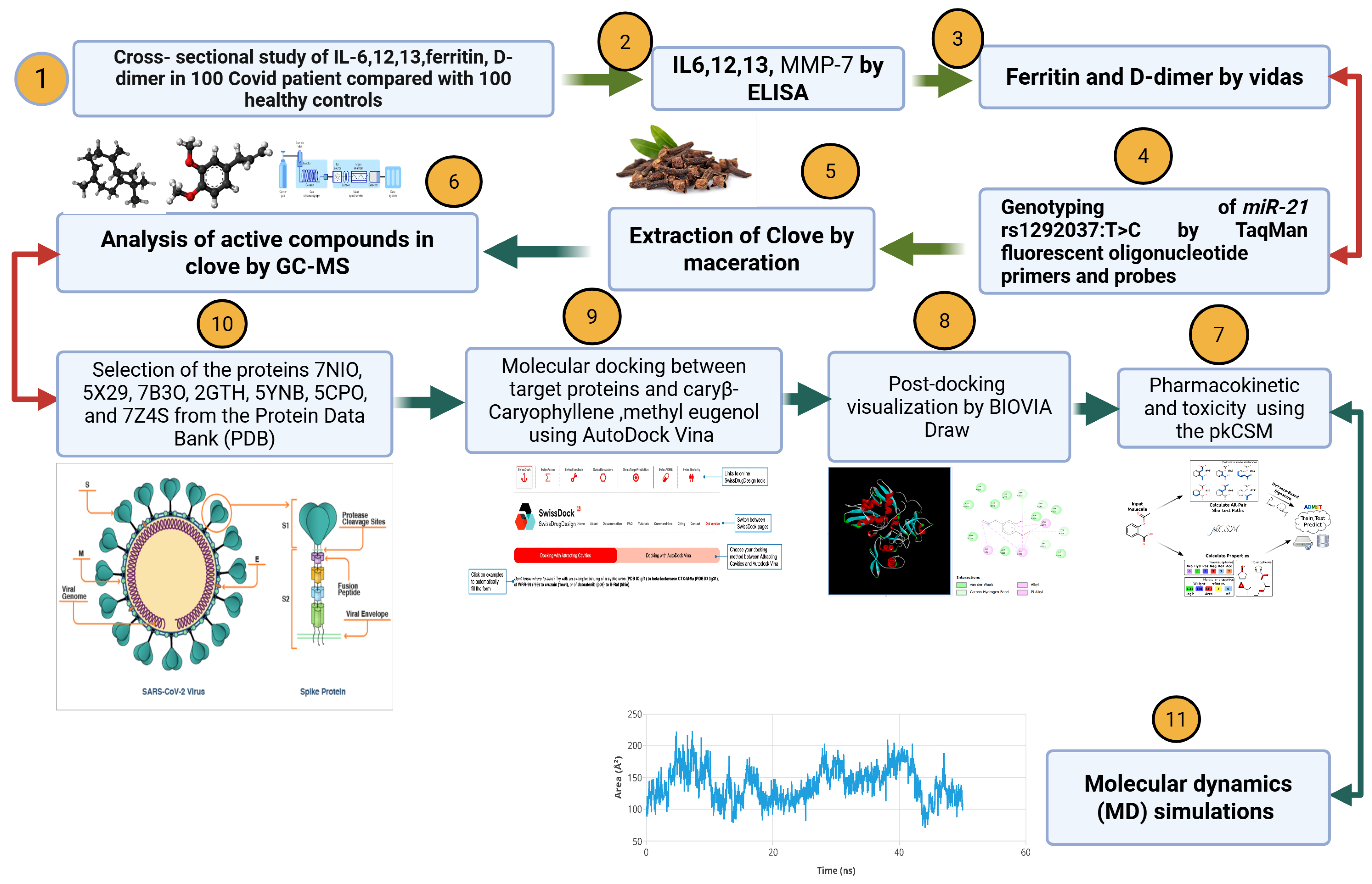
Figure 1 illustrates an integrative study combining clinical analysis, phytochemical investigation, and computational modeling to explore the potential of Syzygium aromaticum (clove) compounds against COVID-19. The research begins with a clinical comparison of 100 COVID-19 patients and 100 healthy individuals, focusing on inflammatory markers IL-6, IL-12, IL-13, ferritin, and D-dimer levels. Additionally, genetic analysis is conducted to examine the miR-21 rs1292037 T>C polymorphism using TaqMan fluorescent probes. Clove samples are extracted through maceration, and their active compounds are identified using gas chromatography–mass spectrometry (GC-MS). Two major constituents, β-caryophyllene and methyl eugenol, are further analyzed for their pharmacokinetic properties and toxicity using the pkCSM platform. These compounds are subjected to molecular docking studies against a panel of viral and microbial resistance proteins obtained from the Protein Data Bank, including SARS-CoV-2 targets. The docking interactions are visualized using BIOVIA Draw, and molecular dynamics simulations are performed to assess the stability of the ligand–protein complexes. The overall workflow provides a comprehensive approach that integrates experimental and computational methods to identify promising antiviral candidates from natural sources.

2.1. Study Design

This was a cross-sectional study aimed at investigating the relationship between IL-6, IL-12, and IL-13 levels and the presence of D-dimers and ferritin in COVID-19 patients compared with a control group. The study was conducted at Al-Ramadi hospitals in Anbar Province, Iraq, from March 2020 to September 2022.

2.1.1. Sampling and Markers

A purposive sampling method was used to select participants. The patient group included 100 individuals who presented with symptoms and sought evaluation and medical intervention for COVID-19. The patients were diagnosed through clinical evaluation by medical consultants, and the diagnosis was confirmed via real-time PCR (Jiangsu Bioperfectus Technologies Co., Taizhou, Jiangsu, China) and the BIOZEK COVID-19 rapid test (Biozek Medical, Apeldoorn, The Netherlands). The control group consisted of 100 healthy individuals selected from the general population and matched by age to the patient group, also ensuring that they had no history of COVID-19 infection or related symptoms.

2.1.2. Sample Size Calculation

A sample size of 200 individuals was determined on the basis of power calculations to detect statistically significant differences in the levels of IL-6, IL-12, IL-13, D-dimers, and ferritin between COVID-19 patients and controls. The calculation considered a confidence level of 95%, a power of 80%, and an effect size derived from preliminary data on inflammatory markers in COVID-19 patients. Additionally, the inclusion criteria ensured a diverse age range (18–72 years) to capture a representative sample of the population affected by COVID-19. The data for each individual were collected via a specialized questionnaire, as shown in the Supplementary Materials.

2.2. Blood Sample Collection

A 5 mL sample of venous blood was collected from each participant via a disposable syringe (Bio-Med Healthcare, Faridabad, India) to determine cytokine levels. The blood was transferred into 10 mL plain vacutainers with gel for interleukin measurement, allowed to coagulate at 25 °C for 30 min, and then centrifuged at 3000 RPM (Fisher Scientific, Pittsburgh, PA, USA) for 15 min to separate the sera. The sera were then collected into Eppendorf tubes (Bio-Med Healthcare, Faridabad, India).

2.3. Measured Parameters

2.3.1. Assay Principles of Human Interleukin ELISA Kits

The levels of interleukin 6 (IL-6), IL-12, and IL-13 were measured via human interleukin ELISA kits (SUNLONG Biotech Co., Ltd., Hangzhou, China). The experimental procedure adhered to the manufacturer’s instructions www.sunlongbiotech.com (accessed on 1 January 2022).

2.3.2. Determination of D-Dimer and Ferritin Levels

Ferritin levels in the serum and D-dimer levels in the plasma of both patients and controls were assessed via the VIDAS® instrument (bioMérieux, Marcy-l’Étoile, France).

2.4. Genotyping

2.4.1. Genomic DNA Extraction and Quantification

DNA was extracted from frozen blood samples via the Wizard® Genomic DNA Purification Kit, CAT. A1120 (Promega Corporation, Madison, WI, USA). To assess the integrity of the extracted DNA, the samples were subjected to electrophoresis on 1% agarose gel (Merck, Darmstadt, Germany). Additionally, the concentration of extracted DNA was measured using a Quantus Fluorometer (Promega Corporation, Madison, WI, USA) to evaluate the quality of the samples for use in subsequent applications.

2.4.2. Genotyping of miR-21 rs1292037:T>C

The miR-21 gene polymorphisms of interest were amplified via the use of TaqMan fluorescent oligonucleotide primers and probes (Macrogen Company, Seoul, Republic of Korea). The primers and probe sequences utilized in this study are provided in Table 1, and these primers were provided by Macrogen Company in lyophilized form. To prepare the primers for use, the lyophilized primers were dissolved in nuclease-free water, resulting in a final concentration of 100 pmol/μL as a stock solution. A working solution of these primers was created by combining 10 μL of the primer stock solution (which was stored at −20 °C in a freezer) with 90 μL of nuclease-free water (Bioneer, Daejeon, Republic of Korea), resulting in a working primer solution with a concentration of 10 pmol/μL.
For this assay, DNA samples from both COVID-19 patients and healthy controls were genotyped for the above-mentioned SNPs. The total volume of the RT–PCR mixture was 20 µL, composed of 10 µL of TaqMan master mix, 1 µL of each fluorescence probe, 1 µL of each forward and reverse primer (10 μM), 2 µL of template DNA, and 4 µL of nuclease-free water. RT–PCR amplification was performed via a programmed thermocycler with 1 hold cycle of 95 °C for 5 min, and 40 cycles of 95 °C for 30 s, 60 °C for 30 s, and 72 °C for 30 s.

2.5. Extraction of Clove Extract

Clove buds and leaves from Syzygium aromaticum were subjected to extraction via the maceration technique to isolate the ethanol-based fraction. Specifically, 100 g of the plant material was immersed in 500 mL of 70% ethanol, maintaining a sample-to-solvent ratio of 1:5. The mixture was incubated for 24 h under continuous stirring. Following incubation, the mixture was filtered and the resulting solution was concentrated using a rotary evaporator at 45 °C to yield the ethanol extract of clove [22].

2.5.1. GC-MS Analysis

Plant extracts were analyzed using a GC-MS system (Agilent 7820A Santa Clara, CA, USA) equipped with an HP-5ms Ultra Inert capillary column (30 m × 250 µm × 0.25 µm). A 1 µL sample was injected in split mode with helium (99.99%) as the carrier gas at 11.933 psi. The injector and inlet temperatures were set at 250 °C, while the auxiliary heater was maintained at 310 °C. The scan range was m/z 50–500. The oven temperature program started at 60 °C (held for 1 min), and was ramped up to 180 °C at 7 °C/min and then to 280 °C at the same rate, with a total run time of approximately 33 min.

2.5.2. Ligand Preparation

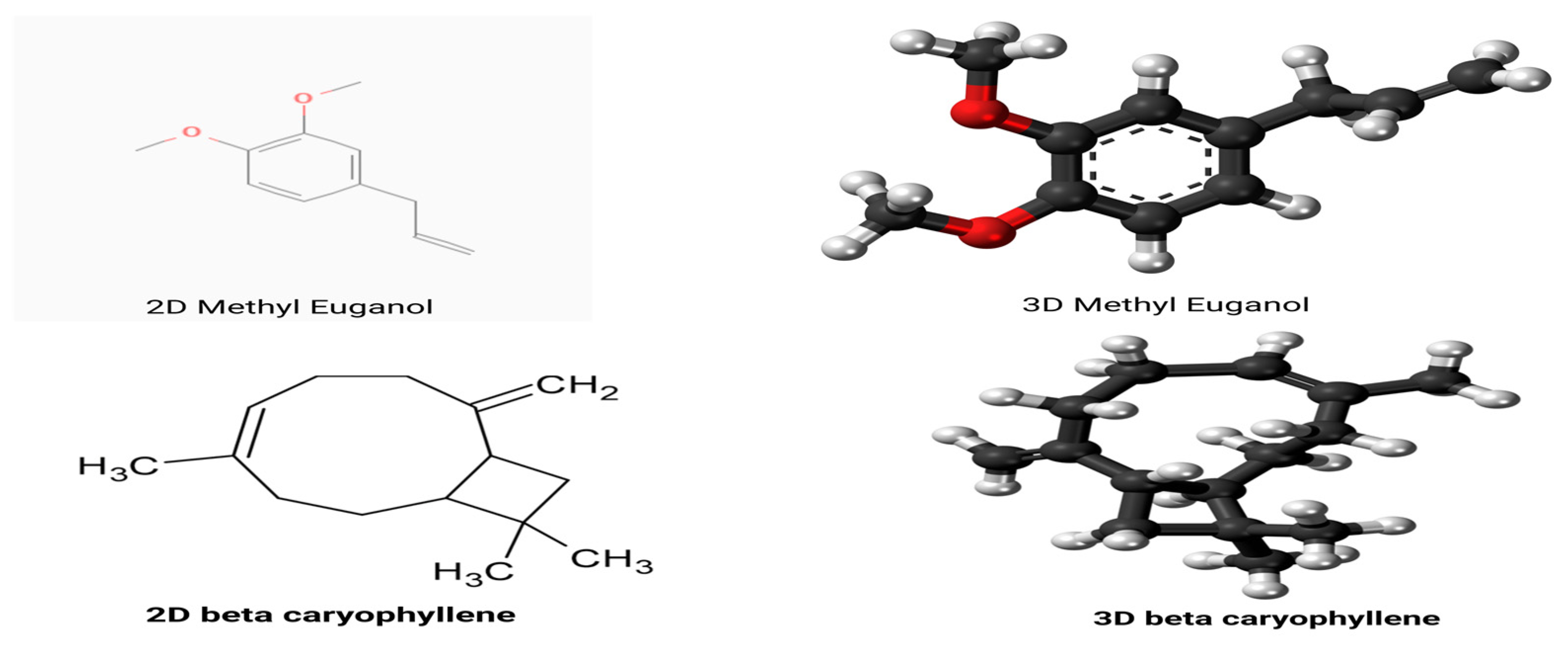
The ligand used for molecular docking was a terpenoid compound retrieved from the PubChem database under CID 20831623 (SMILES: C/C/1=C\CCC(=C)[C@H]2CC([C@@H]2CC1)(C)C). Additionally, eugenol (PubChem CID: 3314), a key phytochemical from Syzygium aromaticum, was selected based on prior research indicating its antiviral potential and known interaction with viral proteins, as shown in Figure 2. Ligand structures were initially obtained in SMILES format, converted to 3D conformations using Open Babel (https://openbabel.org) (accessed on 1 June 2025) [23], and optimized using the MMFF94 force field. Protonation was performed at physiological pH (7.4), and energy-minimized structures were saved in PDBQT format for compatibility with docking protocols.

2.6. Target Protein Selection and Preparation

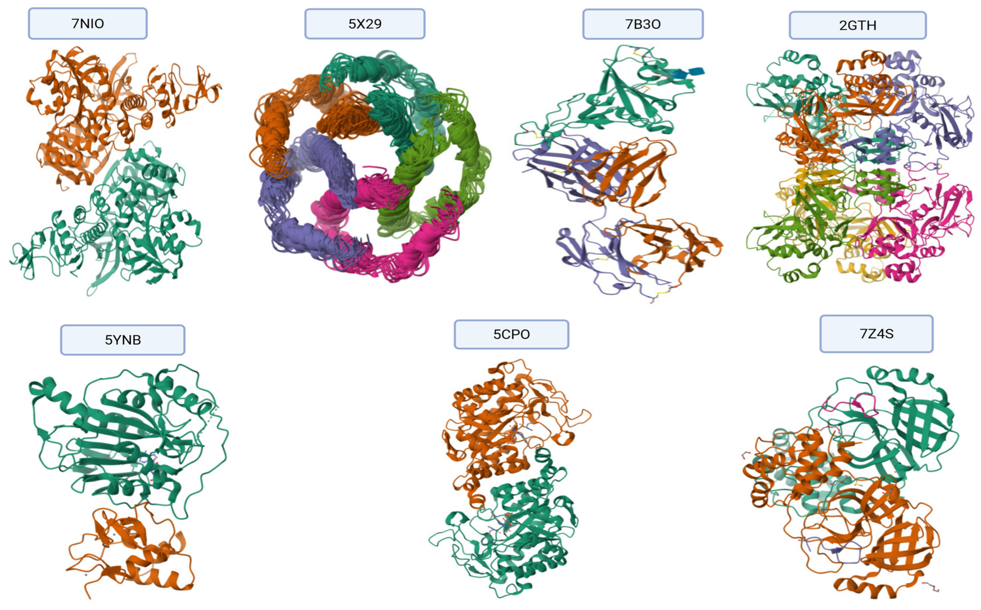
A total of seven SARS-CoV-2 proteins involved in key viral functions were selected as molecular targets based on their druggability and role in viral replication, immune evasion, and host interaction, as shown in Figure 3. Their crystallographic structures were obtained from the Protein Data Bank (PDB), https://www.rcsb.org accessed on 1 June 2025) [24], and the corresponding IDs are shown in Table 2.
Protein structures were pre-processed using AutoDock Tools (v1.5.7) by removing water molecules and native ligands, adding polar hydrogens, and assigning Gasteiger charges. The final files were saved in PDBQT format.

2.7. Molecular Docking Procedure

Molecular docking was conducted using AutoDock Vina 1.2.0 https://vina.scripps.edu (accessed on 1 June 2025) [25], (https://autodock.scripps.edu/) (accessed on 1 June 2025) [26], integrated via the SwissDock platform (2024 release), http://www.swissdock.ch [27]. Docking simulations were performed within a grid box centered around the active site of each protein using dimensions of 20 × 20 × 20 Å and an exhaustiveness level of 4 to ensure sufficient conformational sampling, as shown in Table 3.

2.8. Scoring and Binding Affinity Evaluation

Binding affinities (ΔG, kcal/mol) were predicted by AutoDock Vina for each protein–ligand complex. The best-ranked poses based on the most negative binding energy were selected for further analysis. The top 20 conformations were recorded for each docking simulation.

2.9. Visualization and Interaction Analysis

Post-docking visualization was performed using Discovery Studio Visualizer and BIOVIA Draw, https://discover.3ds.com/discovery-studio-visualizer-download (accessed on 1 June 2025), where interactions such as hydrogen bonding, π-π stacking, and hydrophobic contacts were analyzed [28].

2.10. Pharmacokinetic and Toxicity Prediction

Pharmacokinetic and toxicity properties of the selected compound were predicted using the pkCSM online tool, which utilizes graph-based signatures to estimate ADMET-related characteristics. The tool is freely available at https://biosig.lab.uq.edu.au/pkcsm/ (accessed on 1 June 2025) [29].

2.11. Molecular Dynamics (MD) Simulation

The molecular dynamics simulations were performed using the Desmond software package accessed through the Maestro graphical interface (Schrödinger Release 2023–4; Schrödinger, LLC, New York, NY, USA), https://www.schrodinger.com/products/desmond (accessed on 1 June 2025). Protein–ligand complexes obtained from docking studies were prepared and embedded within an explicit solvent environment modeled by TIP3P water molecules. An orthorhombic simulation box was defined with a 10 Å margin surrounding the solute to ensure adequate solvation.
To mimic physiological ionic conditions, the system was neutralized by the addition of counter ions and supplemented with 0.15 M sodium chloride. The OPLS4 force field was applied to parameterize all components of the system. Following energy minimization and a stepwise equilibration protocol supplied by Desmond, production simulations were conducted in the isothermal–isobaric (NPT) ensemble. Temperature and pressure were maintained at 300 K and 1 atm, respectively, using the Nose–Hoover thermostat and the Martyna–Tobias–Klein barostat.
The simulation was carried out over a duration of 50 nanoseconds with a 2-femtosecond integration time step. Trajectory frames were saved every 100 picoseconds for subsequent analysis. The resulting trajectories were analyzed to assess structural stability and dynamic behavior by computing parameters such as root mean square deviation (RMSD), root mean square fluctuation (RMSF), radius of gyration (Rg), and hydrogen bonding interactions throughout the simulation timeframe [30].

2.12. Protein Structure Preparation Using Swiss-PdbViewer

The three-dimensional structures of target proteins were retrieved in PDB format and processed using Swiss-PdbViewer (version v4.1. Upon loading, the software automatically scanned the protein files for missing atoms, side chains, or incomplete residues. Residues with missing atoms were reconstructed using built-in rotamer libraries, and incomplete side chains were corrected using the “Fix Sidechains” function. Hydrogen atoms were subsequently added to stabilize the structure and prepare it for downstream docking analysis. Local energy minimization was performed where necessary to resolve unfavorable geometries. The refined structures were then saved and used in subsequent molecular docking and dynamics simulations.

2.13. Re-Docking Protocol

To validate the accuracy of the docking procedure, a re-docking analysis was performed using the co-crystallized ligand extracted from the native protein–ligand complex. The ligand was separated from the protein structure and re-docked into the original binding site using the same docking parameters applied throughout the study. Root mean square deviation (RMSD) between the predicted and crystallographic poses was calculated to assess reproducibility. An RMSD value below 2.0 Å was considered acceptable, indicating that the docking protocol reliably reproduces the experimentally observed binding conformation. All re-docking calculations were conducted using AutoDock Vina 1.2.0 [25], with grid dimensions and scoring functions kept consistent with those used in the virtual screening phase.

2.14. Statistical Analysis

Statistical analysis of serum parameters was conducted using ANOVA in GraphPad Prism® 5.0, with data presented as means  ±  SDs or percentages. Group comparisons were made via Student’s t-test, with p ≤ 0.05 and 0.01 indicating significance for IL-6, IL-12, IL-13, D-dimers, and ferritin. Correlations between cytokines and biomarkers were assessed using Pearson or Spearman coefficients. Hardy–Weinberg equilibrium and allele/genotypic differences were analyzed using the chi-square test, with odds ratios calculated for risk and protective alleles, considering p < 0.05 statistically significant.

3. Results

3.1. Participant Characteristics

The study included 200 individuals, comprising 100 COVID-19 patients and 100 matched healthy controls. Their demographic and clinical features are outlined in Table 4. The patient group included 60% males and 40% females, with 61% aged ≥40 years. In comparison, the control group had 54% males and 46% females, with 53% aged ≥40 years. The BMI distribution showed a higher prevalence of obesity in females in the COVID-19 group.

3.2. Inflammatory and Coagulation Markers in COVID-19 Patients

Table 5 shows that COVID-19 patients had significantly elevated levels of IL-6, IL-12, IL-13, MMP-7, ferritin, and D-dimer (p ≤ 0.01), reflecting a strong inflammatory and pro-thrombotic response.

3.3. Sex-Based Differences in Biomarkers

Stratified analysis (Table 6) showed that males had significantly higher ferritin and D-dimer levels than females in both patients and controls. These differences were highly significant (p < 0.0001), suggesting greater susceptibility to hypercoagulability in males.

3.4. Correlation Between Biomarkers

The results of the correlation analysis present in Table 7 demonstrate significant relationships between cytokines and coagulation factors (Table 4). IL-12 and D-dimer were strongly correlated (r = 0.847), similarly to ferritin and D-dimer (r = 0.796).

3.5. Genotype Distribution of rs1292037:T>C

Genotyping of the miR-21 rs1292037 polymorphism revealed a significant association with COVID-19 susceptibility. The TC genotype was most frequent among patients (52%), while the CC genotype was more common among controls (30%). The C allele increased the odds of COVID-19 by 67% (OR = 1.67, p = 0.01).

3.6. Cytokine and Coagulation Levels by Genotype

Table 8 and Table 9: Cytokine and Coagulation Levels by Genotype.

3.7. Statistical Comparison Between Groups

In Table 10, the t-test results between patients and controls for each marker show statistically significant differences in IL-10 (p = 0.00479) and IL-20 (p = 0.00004), highlighting their discriminative potential.
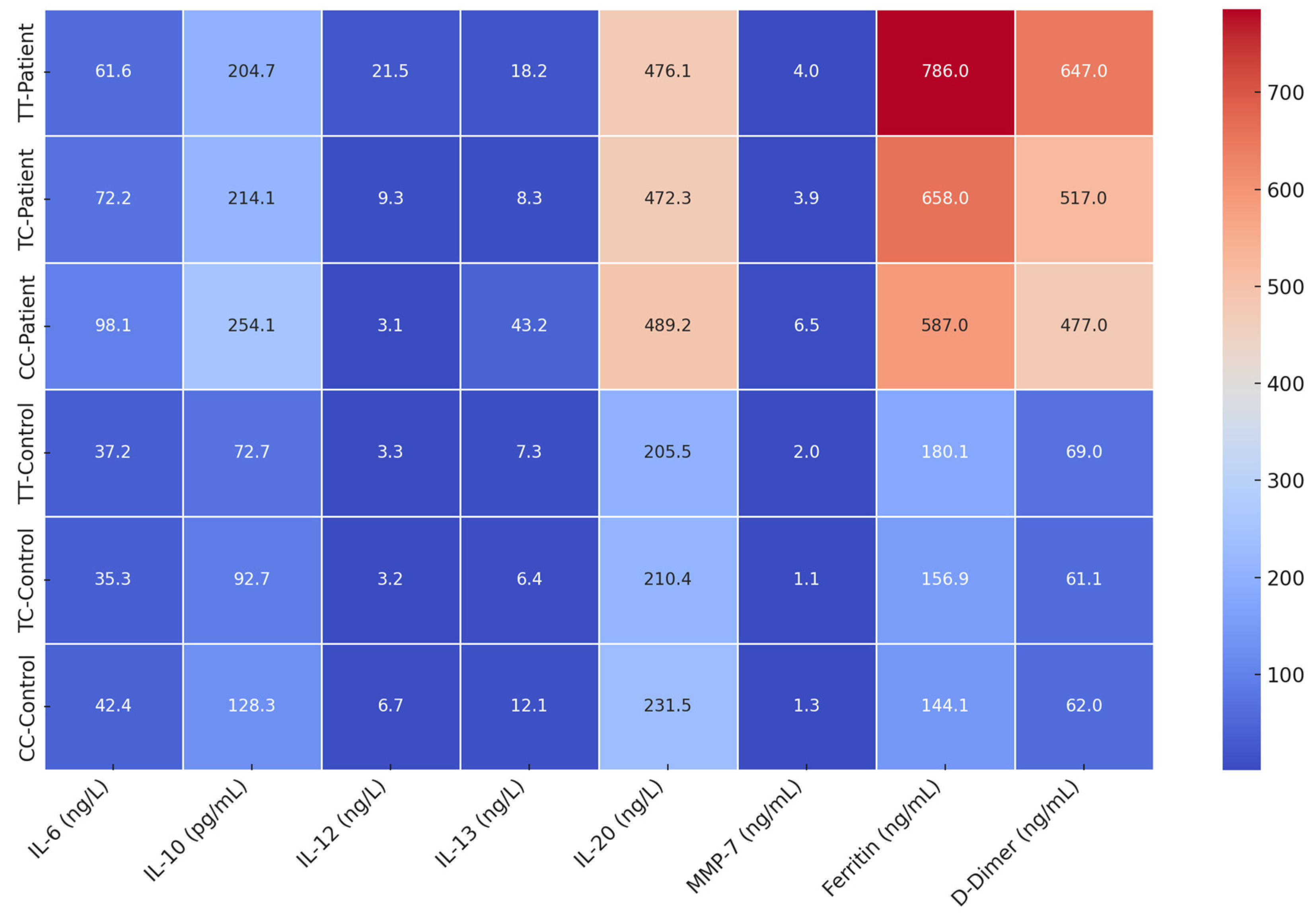
A heatmap was generated to visually summarize all results (Figure 4). It displays elevated marker levels across all COVID-19 patient genotypes, with especially high expression in CC patients. Controls had consistently lower levels.

3.8. GC-MS Profile

As shown in Figure 5 and Table 11, the GC-MS analysis revealed a diverse range of compounds with varying chemical structures and potential bioactivities. Caryophyllene, a sesquiterpene known for its anti-inflammatory and antimicrobial properties, was the most abundant compound (47.55%). Methyl eugenol (17.75%) and eugenyl acetate (14.85%) were also present in high amounts; both contribute to the characteristic clove aroma and exhibit biological activities. Other notable compounds included eugenol (4.21%) and humulene (7.20%), both recognized for their therapeutic potential. Minor constituents such as methyl salicylate, α-farnesene, caryophyllene oxide, and several esters and hydrocarbons added to the chemical complexity, possibly enhancing the overall pharmacological profile. The presence of various terpenes, esters, and aromatic derivatives suggests that the sample possesses a rich mixture of volatile compounds with potential applications in the medicinal, aromatic, and industrial fields.

3.9. Molecular Docking Results of Eugenol Derivative and Caryophyllene Against SARS-CoV-2 Proteins

Table 12 and Figure 6 present the results of the comparative molecular docking analysis of two phytocompounds—eugenol and caryophyllene—against seven critical SARS-CoV-2 proteins using AutoDock Vina. These proteins are involved in key stages of the viral life cycle, including RNA replication, protein maturation, and immune evasion.
The docking results indicate that eugenol generally exhibited stronger binding affinities than caryophyllene across most targets, particularly against the Envelope (5X29, −5.267 kcal/mol), Spike-RBD (7B3O, −4.979 kcal/mol), and NSP15 (2GTH, −5.317 kcal/mol) proteins. These values suggest potential for eugenol in interfering with viral entry and immune modulation.
In contrast, caryophyllene showed superior binding to NSP12 (RNA polymerase, 5CPO, −6.200 kcal/mol) and NSP16 (2′-O-methyltransferase, 5YNB, −6.000 kcal/mol), implying a possible inhibitory effect on viral genome replication and RNA capping processes. Caryophyllene also showed a moderately strong affinity for NSP5 (7Z4S, −5.500 kcal/mol), suggesting a role in inhibiting viral protease activity (Figure 7).
Overall, while eugenol demonstrated consistent moderate binding across several viral targets, caryophyllene appeared to selectively bind more tightly to proteins involved in replication and post-transcriptional modification. These findings underscore the complementary potential of these two natural compounds as part of a multi-targeted antiviral strategy against SARS-CoV-2.
  • Molecular Interactions Between (COC1=C(C=C(C=C1)CC=C)OC) and SARS-CoV-2 Protein Targets
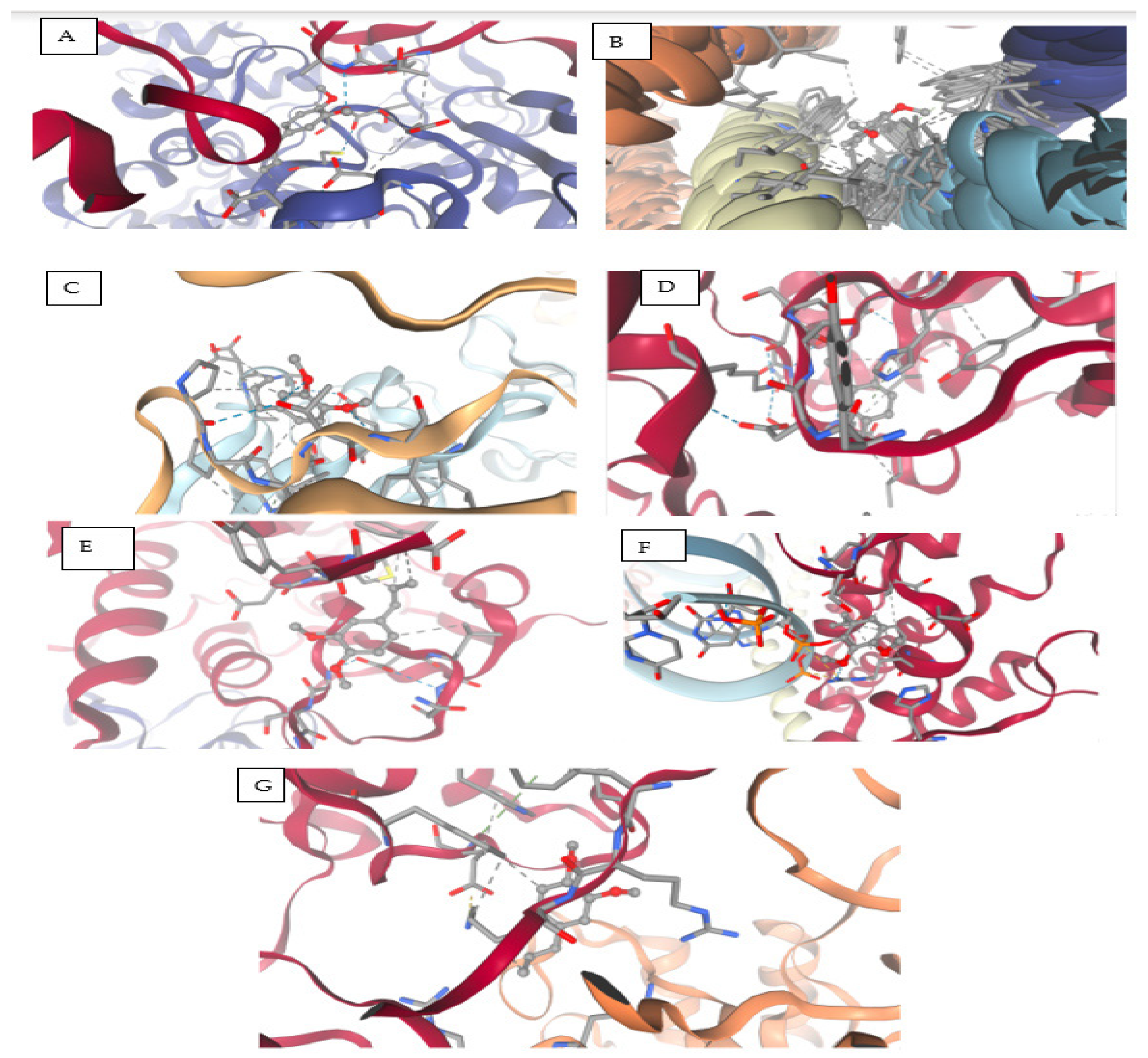
The compound (COC1=C(C=C(C=C1)CC=C)OC), a methoxy-substituted aromatic derivative, exhibits a diverse array of molecular interactions with critical amino acid residues across multiple SARS-CoV-2 protein structures, as revealed by molecular docking studies. The binding behavior includes hydrogen bonding, electrostatic contacts, π-π stacking, hydrophobic interactions, and van der Waals forces, contributing to ligand stabilization within active or allosteric sites of the viral targets, as shown in Figure 8 and Table 13.
  • Target PDB: 7NIO (nsp16/nsp10 methyltransferase complex)
This ligand engages in hydrogen bonding and hydrophobic interactions with Ala553, supporting mixed-polarity contact. Thr552 lies in close proximity, participating in potential polar contacts, while Glu551 facilitates electrostatic stabilization, likely via a salt bridge or hydrogen bond. Cys556 and Asp578/580 further enhance binding through van der Waals and ionic interactions. Notably, Leu581 creates a hydrophobic microenvironment, reinforcing non-polar affinity.
  • Target PDB: 5X29 (Main protease Mpro)
Aromatic interactions are predominant here, with Phe25–26 forming π-π stacking contacts, indicative of strong binding to aromatic regions of the protein. A hydrophobic patch formed by Ala, Leu, and Val (residues 19–29) stabilizes the ligand via van der Waals and alkyl interactions.
  • Target PDB: 7B3O (Helicase NSP13)
Key anchoring is established through hydrogen bonding and π–alkyl interactions with Phe99, Leu45, Ala60, and Pro95, suggesting stable aromatic and alkyl engagement. Additional contributions from Ser, Asp, Trp, Glu, and Thr residues form weak van der Waals contacts that may fine-tune the ligand’s spatial orientation within the binding site.
  • Target PDB: 2GTH (Papain-like protease)
Moderate C–H and π–alkyl interactions occur with His304 and Ser301, while multiple residues including Tyr, Asp, Leu, Lys, Ile, Arg, and Val form a network of van der Waals contacts, reinforcing the fit through hydrophobic and spatial complementarity. Ala89 and Leu279 further stabilize the complex through alkyl interactions.
  • Target PDB: 5YNB (RNA-dependent RNA polymerase complex)
Significant electrostatic and hydrogen bonding is observed with Asp99, 130, and 133 via π–anion interactions, C–H bonds, and van der Waals forces. A diverse set of residues (Asn101, Gly71/73, Met131, and Tyr132) contribute to polar and van der Waals contacts, indicating a broad interaction surface. Leu100 and Phe149 provide hydrophobic anchoring through π–alkyl interactions.
  • Target PDB: 9CPO (Exonuclease nsp14-nsp10 complex)
Strong stabilizing effects are noted with Arg844 and His448, mediated through hydrogen bonds and π–alkyl interactions. However, steric clashes may arise with U:P32–33 and A:P31, as indicated by unfavorable bumps. Several additional residues (Asp, Pro, Gln, Ile, and Ser) contribute to non-specific van der Waals stabilization.
  • Target PDB: 7Z4S (Spike receptor-binding domain, RBD)
Weak polar interactions such as carbon–hydrogen bonds are observed with Arg4 and Leu282, while Lys5, Trp207, and Phe291 establish π–alkyl interactions, securing the ligand in an aromatic-rich environment. Glu288 and Phe3 form additional van der Waals contacts, suggesting a supplementary role in complex stabilization.
  • Molecular Docking Analysis of Caryophyllene with Various Protein Targets (Figure 5 and Table 14)
A comprehensive molecular docking study was conducted to assess the binding interactions of caryophyllene, a non-polar sesquiterpene, with diverse protein targets implicated in viral replication, microbial resistance, and quorum sensing.
Figure 9A displays the docking results of caryophyllene with the main SARS-CoV-2 protease (PDB ID: 7NIO). The interaction diagram reveals hydrophobic Pi–alkyl contacts with LEU405, ARG560, HIS554, and PRO408, highlighted in pink. Additionally, van der Waals interactions are observed with ASP534, LEU412, and LEU417 (green shading). The absence of polar or hydrogen bonds reflects the non-polar nature of caryophyllene, indicating that hydrophobic and steric factors largely drive binding stabilization in this protease.
Figure 9B illustrates the interaction between caryophyllene and the quorum sensing receptor protein (PDB ID: 5X29). The ligand engages in Pi–alkyl interactions with PHE28 and LEU19, suggesting strong hydrophobic anchoring. A single van der Waals contact with SER16 contributes to ligand stabilization. The pocket’s predominantly hydrophobic environment supports caryophyllene’s fit and potential to interfere with receptor activity.
Figure 9C shows caryophyllene docked into the bacterial resistance-associated protein 7B3O. Here, the ligand forms an alkyl interaction with LEU455, alongside multiple van der Waals contacts with LYS417, TYR453, and ARG403. These findings support a non-polar binding mechanism, consistent with caryophyllene’s hydrophobic nature, and suggest possible interference with protein function.
Figure 9D presents the interaction of caryophyllene with the eukaryotic protein 2GTP. The ligand displays alkyl interactions with LEU227, VAL160, and ALA89, supported by van der Waals contacts involving ASP300, GLU68, GLY158, TYR88, ASP155, and ASP87. This combination of non-polar and polar residues highlights a moderate binding mode, largely governed by hydrophobic interactions.
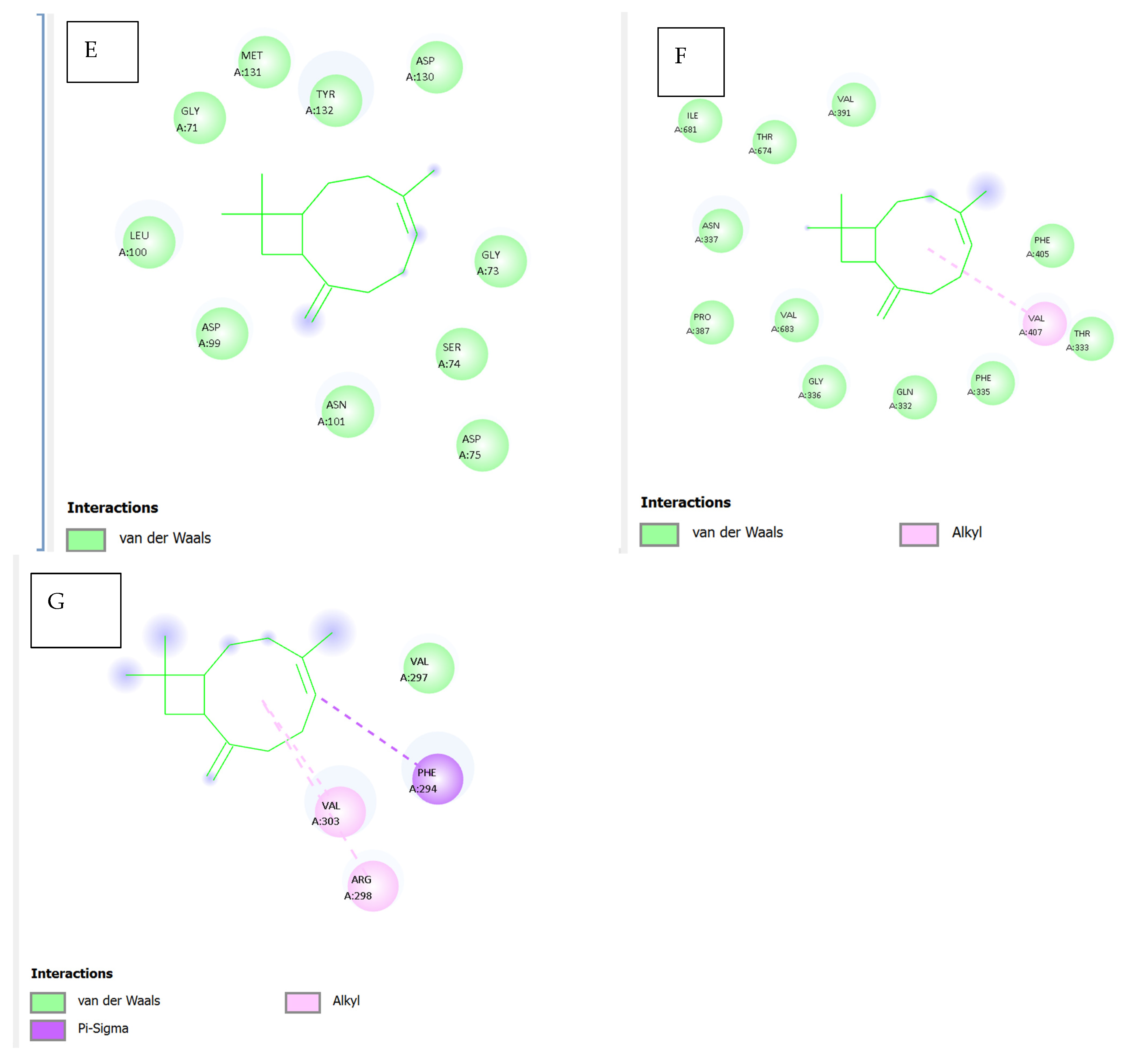
Figure 9E illustrates the docking pose of caryophyllene within the bacterial protein 5YNB. All observed contacts are van der Waals in nature, involving residues such as GLY71, GLY73, ASP75, ASN101, LEU100, and TYR132. The absence of stronger interactions (e.g., hydrogen bonds or π interactions) suggests a lower binding affinity and a relatively passive accommodation within the binding cavity.
Figure 9F shows the interaction with the 5CPO protein. Caryophyllene forms alkyl interactions with VAL407 and PHE405, alongside van der Waals contacts involving ILE681, VAL391, GLN332, ASN337, and PRO387, among others. The diversity of surrounding residues indicates a flexible and largely hydrophobic binding site, suitable for the bulky, cyclic nature of the ligand.
Figure 9G highlights caryophyllene’s interactions with the 7Z4S protein. Notable contacts include van der Waals and alkyl interactions with VAL297, VAL303, and PHE294, in addition to a potential pi–sigma interaction involving PHE294. These hydrophobic and aromatic interactions may support stable binding within the protein’s functional domain, although experimental studies are needed to confirm this.
Table 14. Summary of caryophyllene–protein interactions.
FigureProtein (PDB ID)Key InteractionsNotable ResiduesBinding Nature
9A7NIO (main protease)Pi–alkyl, van der WaalsLEU405, ARG560, ASP534Hydrophobic
9B5X29 (QS receptor)Pi–alkyl, van der WaalsPHE28, LEU19, SER16Hydrophobic
9C7B3O (resistance)Alkyl, van der WaalsLEU455, TYR453, ARG403Hydrophobic
9D2GTPAlkyl, van der WaalsLEU227, VAL160, ASP300Mixed hydrophobic
9E5YNBVan der Waals onlyGLY71, ASP130, TYR132Weak/non-specific
9F5CPOAlkyl, van der WaalsVAL407, PHE405, ILE681Hydrophobic
9G7Z4SAlkyl, van der Waals, π-σVAL297, PHE294, ARG298Aromatic–hydrophobic
This analysis underscores that caryophyllene preferentially binds to hydrophobic and semi-polar regions of diverse proteins through non-covalent interactions, predominantly alkyl and van der Waals forces. Its lack of hydrogen bonding is consistent with its lipophilic terpenoid structure, suggesting that its biological activity may derive from steric interference with active or regulatory sites in proteins.
The comparative ADMET profile presented in Table 15 offers a comprehensive evaluation of methyl eugenol and β-caryophyllene using predictive in silico models, emphasizing key pharmacokinetic and toxicological parameters essential for drug development and safety assessment.
In terms of absorption, eugenol displays a markedly higher aqueous solubility than caryophyllene (log S = −2.671 vs. −5.555), which favors its dissolution and potential oral bioavailability. Both compounds show comparable Caco-2 permeability and high human intestinal absorption, suggesting effective gastrointestinal uptake. Despite their favorable absorption profiles, both exhibit relatively low skin permeability, with eugenol being slightly less permeable dermally. Notably, neither molecule is a substrate or inhibitor of P-glycoproteins, indicating a reduced risk of efflux-related drug interactions and enhanced systemic retention.
Regarding distribution, caryophyllene shows a higher volume of distribution (log VDss = 0.652) compared to eugenol (0.265), suggesting more extensive tissue penetration. Both compounds exhibit moderate plasma protein binding, ensuring an adequate unbound drug fraction. Additionally, caryophyllene demonstrates a greater capacity to permeate the blood–brain barrier, though both show limited CNS permeability, which may restrict their central effects.
Under metabolic profiling, neither compound is a substrate or inhibitor of major cytochrome P450 enzymes except for CYP1A2, where eugenol acts as an inhibitor. This interaction implies a potential for metabolic interference when co-administered with substrates of this enzyme. Otherwise, their metabolic stability appears favorable, with low risk of enzymatic inhibition-based drug interactions.
In the context of excretion, caryophyllene exhibits higher predicted total clearance, suggesting faster systemic elimination. Neither compound is likely to be excreted via renal OCT2-mediated pathways.
Concerning toxicity, a notable distinction arises in AMES test predictions: eugenol may possess mutagenic potential, whereas caryophyllene is predicted to be non-mutagenic. Eugenol, however, is tolerated at a higher dose in humans and demonstrates lower acute and chronic toxicity in rodent models. Both compounds show potential for skin sensitization. Environmentally, eugenol exhibits higher minnow toxicity, while caryophyllene is more toxic to T. pyriformis, indicating species-specific ecological risks.
In summary, eugenol and caryophyllene share several favorable ADMET traits, such as high absorption and low hepatic toxicity, but differ in solubility, metabolic enzyme interaction, and certain toxicity endpoints. These differences could influence their suitability in pharmaceutical or nutraceutical applications, depending on the target profile and safety requirements.
  • Molecular Dynamics Simulation of Methyl Eugenol Bound to Key SARS-CoV-2 Proteins
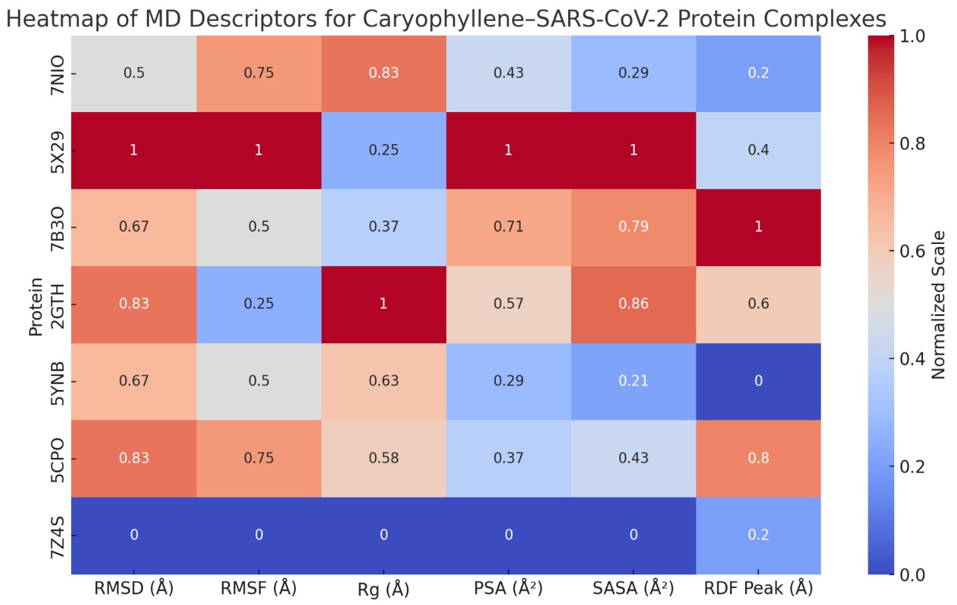
A suite of molecular dynamics (MD) simulations was conducted to evaluate the dynamic stability, structural adaptability, and interaction profiles of methyl eugenol in complex with seven critical SARS-CoV-2 target proteins: 7NIO (nucleocapsid phosphoprotein), 5X29 (Envelope Protein), 7B3O (RNA-dependent RNA polymerase), 2GTH (main protease), 5YNB (RNA-binding domain), 5CPO (papain-like protease), and 7Z4S (spike receptor-binding domain). The simulations were carried out over a 50 ns timescale under explicit solvent conditions, with systematic evaluation of key structural descriptors, including root mean square deviation (RMSD), root mean square fluctuation (RMSF), radius of gyration (Rg), molecular and polar surface area (MSA/PSA), solvent-accessible surface area (SASA), radial distribution function (RDF), and ligand–protein contact profiles as shown in Supplementary S1, Figure 10, and Table 16.
  • RMSD and Conformational Equilibrium
Across all protein complexes, RMSD values stabilized within the first 5–6 ns, with most systems maintaining deviations below 2.5 Å. This included the 7Z4S and 7NIO complexes, which exhibited particularly low RMSD plateaus around 1.8–2.1 Å, underscoring rapid convergence and minimal global rearrangement. These results reflect strong structural resilience upon ligand binding, with methyl eugenol being effectively accommodated in shallow grooves, hydrophobic pockets, or active site clefts without inducing deleterious conformational changes. The 7B3O and 5CPO systems similarly stabilized early, with average RMSD values suggesting tight ligand anchoring and thermodynamic compatibility.
  • RMSF and Residue-Level Mobility
The RMSF analyses revealed that loop regions and termini consistently displayed the highest fluctuations (>2.0 Å), while structured α-helices and β-strands remained notably rigid (≤1.2 Å). Binding-site residues in all complexes showed suppressed mobility, particularly in the 7Z4S RBD, where ACE2-interacting loops exhibited reduced flexibility, hinting at potential inhibitory effects through rigidification. In the catalytic proteins (5CPO, 2GTH), ligand-induced dampening of flexibility in enzymatically active residues supports the hypothesis of functional interference. The 5YNB complex, involving the RNA-binding domain, displayed ligand-induced stabilization within its β-sheet core, aligning with the notion of conformational anchoring.
  • Radius of Gyration (Rg) and Tertiary Structure Stability
All protein systems maintained compact tertiary structures throughout the simulations, with Rg fluctuations confined within ±0.3 Å of their initial values. No evidence of large-scale unfolding or domain collapse was observed, even in structurally sensitive systems like 2GTH and 5CPO. This compactness confirms the biophysical compatibility of methyl eugenol with diverse viral protein architectures and further supports its non-denaturing interaction profile.
  • MSA, PSA, and Surface Adaptation
The MSA and PSA trends were consistent across simulations, with moderate increases during early equilibration phases, reflecting solvent reorganization and mild residue rearrangements. PSA elevations (typically ~300 Å2) were most notable in proteins with solvent-facing polar grooves (e.g., 5X29, 7B3O), suggesting polar residue reorientation around the ligand. In the 7Z4S system, a slight reduction in MSA indicated hydrophobic cavity occupation by methyl eugenol, leading to reduced surface entropy and enthalpic stabilization.
  • RDF and Interaction Specificity
RDF profiles across all complexes exhibited sharp, high-density peaks between 2.2 and 4.8 Å, confirming persistent and spatially localized interactions. Notably, 5CPO and 7Z4S showed strong RDF peaks corresponding to active-site residues and receptor-binding motifs, respectively, indicating sustained contact zones critical for function modulation. The narrowness and symmetry of RDF peaks in all systems confirmed single, well-defined binding poses, with no evidence of alternative orientations or transient dissociation events.
  • SASA and Solvent Stability
The SASA trajectories across complexes revealed minor oscillations consistent with protein breathing motions. Importantly, no spikes indicative of partial unfolding were observed. Slight SASA reductions in the vicinity of the ligand (e.g., 5CPO and 7Z4S) reflected hydrophobic shielding and potential reductions in conformational entropy—features correlated with enhanced binding affinity and reduced solvation energy penalties.
  • Ligand–Protein Interaction
Methyl eugenol exhibited diverse, yet consistent interaction profiles, dominated by hydrophobic contacts, hydrogen bonds, and π-π or π–cation interactions. In catalytic proteins such as 5CPO and 2GTH, key residues like Cys111 and His272 engaged in stabilizing interactions with the ligand, suggesting active-site targeting. In the 7Z4S RBD, aromatic residues (Tyr453, Tyr505) formed π-π stacking interactions, while Gln493 and Asn487 provided hydrogen bonds—highlighting a pharmacophoric alignment potentially disruptive to ACE2 engagement. The interaction persistence across simulations supports the ligand’s high residence time and specificity.
Finally, methyl eugenol demonstrates a robust and adaptable binding profile across several critical SARS-CoV-2 proteins, maintaining structural stability and inducing local conformational reinforcement at functional domains. Its capacity to stably occupy enzymatic clefts, receptor interfaces, and RNA-binding grooves, while preserving protein fold and compactness, highlights its promise as a phytochemical scaffold for antiviral intervention. These molecular dynamics simulations offer compelling evidence for its further exploration through in vitro validation, SAR optimization, and structure-based drug design initiatives targeting viral replication and host-cell entry.
  • Comprehensive Molecular Dynamics Simulation of Caryophyllene Bound to Key SARS-CoV-2 Proteins
A series of molecular dynamics (MD) simulations were conducted to assess the dynamic behavior and structural stability of caryophyllene when bound to seven critical SARS-CoV-2 proteins: nucleocapsid phosphoprotein (7NIO), Envelope Protein (5X29), RNA-dependent RNA polymerase (7B3O), main protease (2GTH), RNA-binding domain (5YNB), papain-like protease (5CPO), and the spike receptor-binding domain (7Z4S). Each protein–ligand complex was simulated over a 50 ns timescale, and key structural descriptors were evaluated to understand the conformational response, interaction patterns, and overall compatibility of the non-polar sesquiterpene ligand, as shown in Supplementary S1, Figure 11, and Table 17.
  • Stability and Compactness: RMSD, Rg, and SASA Analyses
Across all complexes, RMSD values stabilized within the first 4–6 ns, with average deviations ranging from ~1.8 Å (7Z4S) to ~2.4 Å (5X29), indicating high structural fidelity upon ligand binding. No global unfolding or large-scale structural rearrangements were observed. The radius of gyration (Rg) profiles remained within a narrow ±0.3 Å band, further underscoring the preservation of protein compactness. Similarly, SASA trends demonstrated minimal deviation, suggesting that ligand binding did not provoke abnormal solvent exposure or collapse of tertiary structures.
  • Local Flexibility and Functional Regions: RMSF Insights
RMSF analyses showed generally low residue-level fluctuations (<1.5 Å) across the α-helical, β-sheet, and catalytic cores. Notably, loop regions in RNA-binding or receptor-interacting domains displayed slightly elevated mobility, yet this remained within expected dynamic limits. In many cases (e.g., 7B3O, 2GTH, 5YNB), ligand presence actually dampened flexibility near active or binding sites, suggesting a stabilizing influence on functionally relevant residues.
  • Surface and Solvation Behavior: MSA, PSA, and RDF Observations
Molecular surface area (MSA) increased modestly in most systems due to accommodation of the ligand’s hydrophobic volume. However, polar surface area (PSA) remained largely stable, indicating that critical polar interactions and solvation interfaces were conserved. Radial distribution function (RDF) analyses consistently revealed sharp peaks between 4.3 and 4.8 Å, reflecting organized hydration shells that supported ligand stability via water-mediated van der Waals and dispersive forces.
  • Interaction Mechanisms: Ligand–Protein Contact Analysis
Despite lacking polar functional groups, caryophyllene consistently engaged in van der Waals, alkyl–alkyl, and π–alkyl interactions across all protein targets. Key residues involved included Leu50 and Val73 in 7NIO, Leu28 and Phe56 in 5X29, Ile548 and Phe793 in 7B3O, Met49 and His163 in 2GTH, Phe55 and Ile89 in 5YNB, Leu162 and Val266 in 5CPO, and Tyr489 and Leu455 in 7Z4S. These hydrophobic contacts formed stable clusters that anchored the ligand within shallow grooves, clefts, or surface pockets, without disrupting polar interaction networks or essential catalytic motifs.
  • Functional Implications and Structural Impact
The simulation data suggest that caryophyllene maintains protein structural integrity while inducing localized rigidification in flexible or catalytically important regions. In spike RBD (7Z4S), reduced mobility in the receptor-binding loop implies potential interference with ACE2 docking. In proteases (2GTH, 5CPO), occupancy near the catalytic triad hints at possible non-competitive inhibition. For RNA-interacting proteins (7NIO, 5YNB), the ligand may modulate RNA accessibility without obstructing functional core motifs.
Finally, the molecular dynamics simulations across seven SARS-CoV-2 proteins demonstrated that caryophyllene binding is structurally non-disruptive and thermodynamically favorable. The ligand’s hydrophobic nature enabled stable anchoring through non-polar contacts, while the preservation of compactness, surface integrity, and functional domain architecture suggests strong compatibility. These findings advocate for the potential of caryophyllene as a scaffold for further antiviral drug development, particularly in targeting protein surfaces involved in viral assembly, RNA binding, or host interaction.
4. Discussion

Medicinal and natural plants have been utilized for centuries in combating infectious diseases, including viral infections. They are valued for their wide array of biologically active compounds that can act at multiple stages of the viral cycle. Compounds such as flavonoids, terpenes, alkaloids, and phenolics can disrupt viral attachment, inhibit replication enzymes, or interfere with viral protein synthesis. This multifaceted activity makes plant-based extracts promising tools for the development of antiviral therapies, especially against resistant or emerging viruses [31]. The present study provides comprehensive pharmacogenomic and molecular insight into the antiviral potential of Syzygium aromaticum bioactives—primarily eugenol and caryophyllene—against SARS-CoV-2, integrating GC-MS profiling, molecular docking, pharmacokinetic modeling, and miRNA-21 rs1292037 genotyping. These findings build on prior knowledge of phytotherapeutic agents by combining host genetics and viral inhibition in a unified therapeutic framework.
The GC-MS analysis revealed a rich composition of volatile compounds, with caryophyllene (47.55%), methyl eugenol (17.75%), and eugenyl acetate (14.85%) as the major constituents. Caryophyllene, a sesquiterpene hydrocarbon, has documented anti-inflammatory and antiviral activity, partly through modulation of the CB2 cannabinoid receptor and the NF-κB pathway [32,33]. Methyl eugenol and eugenyl acetate contribute not only to aroma but also to antioxidant and cytoprotective effects, which could support host immunity [34]. While eugenol comprised a relatively smaller fraction (4.21%), its broad-spectrum bioactivity, including antibacterial, antifungal, and antiviral properties, is well-established [35,36].
Molecular docking results indicated that both compounds interact with multiple SARS-CoV-2 proteins involved in replication, immune evasion, and viral maturation. Eugenol displayed stronger binding to the Envelope Protein (5X29), Spike RBD (7B3O), and NSP15 (2GTH), indicating its potential to interfere with viral entry and endoribonuclease activity. These findings align with studies by Aboubakr et al. (2021) and Islam et al. (2021), which demonstrated similar docking behavior for eugenol [37,38]. Caryophyllene, in contrast, showed greater binding affinity to the viral RNA polymerase (NSP12, 5CPO) and methyltransferase (NSP16, 5YNB), suggesting that it may inhibit replication and RNA capping post-entry. These complementary interactions support the rationale for a multi-target strategy against SARS-CoV-2.
Furthermore, docking interaction maps confirmed that eugenol forms hydrogen bonds, π-π stacking, and van der Waals contacts with key amino acids across different protein targets. These interactions may enhance ligand stability and specificity, as described in prior structural studies [8]. Caryophyllene, being hydrophobic, formed predominantly alkyl and van der Waals interactions, consistent with its non-polar nature. While this may lead to lower binding energies, it suggests a mechanism involving steric hindrance or membrane interaction rather than active-site inhibition [39].
The ADMET profiling supports the potential of both compounds as safe and orally bioavailable agents. Eugenol displayed superior solubility and a favorable intestinal absorption profile, though its potential mutagenicity in the AMES test warrants caution and dose optimization [40]. Caryophyllene, with its higher tissue distribution and non-mutagenic profile, may offer complementary pharmacokinetic properties. Neither compound showed significant inhibition of cytochrome P450 enzymes (except CYP1A2 for eugenol), reducing the likelihood of metabolic drug–drug interactions. These properties collectively highlight their suitability for repurposing as antiviral agents or for formulation into adjunctive therapies.
The molecular dynamics simulations conducted for methyl eugenol and caryophyllene across seven SARS-CoV-2 target proteins—7NIO, 5X29, 7B3O, 2GTH, 5YNB, 5CPO, and 7Z4S—reveal significant insights into their binding stability, dynamic behavior, and structural influence on protein–ligand complexes.
For methyl eugenol, most complexes demonstrated early RMSD fluctuations that settled into stable trajectories, particularly with the 7NIO, 5X29, and 7Z4S proteins, indicating strong conformational retention and equilibrium. The RMSF values, especially for 7NIO and 7B3O, exposed localized flexibility at loops and termini, while the protein cores remained rigid. Radius of gyration (Rg) measurements confirmed the preservation of protein compactness, with minimal expansion throughout the simulations. Both MSA and PSA exhibited minor oscillations, suggesting biological adaptability without destabilization. RDF peaks at short atomic distances in all cases signified consistent atomic-scale interactions, and the stability of SASA readings across simulations further confirmed the exposure and persistence of methyl eugenol at the binding interfaces.
Notably, surface area metrics (MSA and PSA) showed controlled fluctuation across all systems, reinforcing the notion that caryophyllene does not significantly distort protein topology. RDF plots further revealed distinct binding events at proximity levels typically associated with van der Waals and hydrophobic interactions. SASA remained consistent across all simulations, supporting the stable solvation of the ligand–protein interfaces.
When comparing both ligands, caryophyllene demonstrated a slight edge in complex stability, particularly with 7Z4S and 5CPO, where the lowest residue fluctuations and sustained spatial compactness were observed. On the other hand, methyl eugenol showed stronger atomic proximity in the RDF plots for proteins like 2GTH and 7NIO, possibly due to its smaller molecular size and better fit within tighter pockets. Nonetheless, both ligands showed broad-spectrum interaction potential, evidenced by stable binding across multiple viral targets.
Overall, the 7Z4S–caryophyllene complex emerged as the most dynamically stable among all combinations tested, marked by early structural convergence, minimal surface disruption, and preserved protein compactness. These findings highlight caryophyllene’s potential as a lead antiviral scaffold and justify further experimental exploration.
The miR-21 rs1292037 polymorphism analysis added a critical host genetic dimension. Our results demonstrated a significant association between genotype frequency and COVID-19 susceptibility. The TC genotype was more prevalent among infected individuals, while the CC genotype appeared protective. This is consistent with findings from Zhang et al. (2021) and Li et al. (2022), who reported altered miRNA expression profiles in severe COVID-19 cases [41,42]. As miR-21 regulates inflammatory pathways—including TGF-β, PTEN/PI3K-AKT, and NF-κB—its altered function due to SNP variation could significantly modulate cytokine expression.
Indeed, the elevated IL-6, D-dimer, and ferritin levels observed in the TT and TC genotypes suggest a link between the polymorphism and hyperinflammatory states. Prior research has correlated miR-21 upregulation with IL-6-driven cytokine storms and endothelial damage in COVID-19 [43,44]. Furthermore, the positive correlation of IL-6 with IL-13 and D-dimer in our study reflects the concurrent activation of Th2 cytokines and coagulation pathways, which have been previously implicated in disease progression and mortality [45,46].
Sex- and age-specific analyses provided further insight. Male patients exhibited higher inflammatory marker levels, consistent with known immunological differences and sex hormone influences on cytokine regulation [47]. Older patients showed increased disease severity, likely reflecting immune senescence, as reported by Akbar and Gilroy (2020) [48].
The integration of phytochemical profiling, molecular docking, and SNP genotyping provides a multifactorial approach to understanding antiviral resistance and host response. From a translational perspective, these data support the use of Syzygium aromaticum extracts—particularly eugenol and caryophyllene—in targeted therapeutics, especially in genetically predisposed individuals. However, further in vitro and clinical validation is essential before these agents can be recommended as standalone antivirals or immunomodulators.

5. Conclusions

This study presents an integrative analysis of the antiviral potential of Syzygium aromaticum bioactives—particularly eugenol and caryophyllene—against SARS-CoV-2, combining GC-MS profiling, molecular docking, pharmacokinetic modeling, and host genetic analysis via miR-21 rs1292037 genotyping. Both compounds demonstrated strong binding affinities toward critical viral proteins implicated in entry, replication, and RNA processing, supporting a multi-target antiviral mechanism. The methoxy-substituted eugenol derivative, in particular, exhibited favorable interactions with NSP15, NSP16, and the Envelope Protein, underscoring its potential as a promising therapeutic scaffold. ADMET evaluations further indicated that these phytochemicals possess suitable pharmacokinetic and safety profiles, although careful dose optimization remains necessary for eugenol. Caryophyllene and methyl eugenol both demonstrated stable binding with SARS-CoV-2 target proteins in molecular dynamics simulations. Among all complexes, the caryophyllene–7Z4S complex showed the highest structural stability, suggesting caryophyllene as a promising antiviral candidate. From a host genetic perspective, elevated levels of IL-6, IL-10, IL-13, MMP-7, ferritin, and D-dimer were observed in COVID-19 patients across all genotypes, with the CC genotype associated with the highest concentrations, suggesting a heightened inflammatory and pro-thrombotic state. These findings implicate the miR-21 rs1292037>C polymorphism as a critical modulator of immune and coagulation responses during SARS-CoV-2 infection. The association between the CC genotype and increased inflammatory burden positions this SNP as a potential biomarker for disease susceptibility and severity. Thus, genotype-informed risk stratification may enhance clinical decision-making and therapeutic targeting. Collectively, these insights highlight the dual importance of phytochemical intervention and host genetic profiling in the development of personalized antiviral strategies. Future in vitro and in vivo investigations are essential to validate these findings and translate them into clinical applications.
Availability of data and materials: Supplementary S1 and S2 illustrate the molecular dynamics simulations of methyl eugenol and caryophyllene in complex with SARS-CoV-2 target proteins.

Supplementary Materials

The following supporting information can be downloaded at: https://www.mdpi.com/article/10.3390/v17070951/s1, which details the molecular dynamics between β-caryophyllene and eugenol with SARS-CoV-2 target proteins. Supplementary S1 presents the molecular dynamics analysis of methyl eugenol with SARS-CoV-2 target proteins, while Supplementary S2 focuses on the interaction of β-caryophyllene with the same viral proteins.

Author Contributions

Methodology: M.A.M.; supervision: F.S.; review, formal analysis S.A.L.A., and editing: M.M.A. All authors have read and agreed to the published version of the manuscript.

Funding

Self-funded with no support from universities or external sources.

Institutional Review Board Statement

The Ethics Committee of the Affiliated University of Anbar, Iraq, approved the present study (Approval number: 7832). All experiments adhered to the principles outlined in the Helsinki Declaration and institutional guidelines. Written informed consent was obtained from all participants, and all methods were conducted in accordance with relevant guidelines, regulations, and good clinical practices.

Informed Consent Statement

Informed consent was obtained from all individuals involved in the study, in accordance with the Ethics Committee of the Affiliated University of Anbar guidelines and approved protocol.

Data Availability Statement

The original contributions presented in this study are included in the article/Supplementary Materials.

Conflicts of Interest

The authors declare that they have no conflicts of interest.

References

  1. Silva, R.C.; Freitas, H.F.; Campos, J.M.; Kimani, N.M.; Silva, C.H.T.P.; Borges, R.S.; Pita, S.S.R.; Santos, C.B.R. Natural products-based drug design against SARS-CoV-2 Mpro 3CLpro. Int. J. Mol. Sci. 2021, 22, 11739. [Google Scholar] [CrossRef] [PubMed]
  2. Sathianathan, P.; Walling, K.I.; Pulikkottil, A.V. Global COVID 19 distribution and its association with the selected demographic variables of the countries: A Cross-sectional Infodemiological approach at 10th month of Pandemic. Int. J. Nurs. Educ. Res. 2022, 10, 2. [Google Scholar]
  3. Chen, J.; Wang, R.; Gilby, N.B.; Wei, G.-W. Omicron variant (B. 1.1. 529): Infectivity, vaccine breakthrough, and antibody resistance. J. Chem. Inf. Model. 2022, 62, 412–422. [Google Scholar] [CrossRef] [PubMed]
  4. Al Meani, S.A.L.; Ahmed, M.M.; Abdulkareem, A.H.; Hamid, N.M.; Ibrahim, M.O. Effect of COVID-19 Virus on Biomass Index of Infected Patients. Syst. Rev. Pharm. 2020, 11, 1134–1136. [Google Scholar]
  5. Hoffmann, M.; Kleine-Weber, H.; Pöhlmann, S. A multibasic cleavage site in the spike protein of SARS-CoV-2 is essential for infection of human lung cells. Mol. Cell 2020, 78, 779–784. [Google Scholar] [CrossRef]
  6. Gao, Y.; Yan, L.; Huang, Y.; Liu, F.; Zhao, Y.; Cao, L.; Wang, T.; Sun, Q.; Ming, Z.; Zhang, L.; et al. Structure of the RNA-dependent RNA polymerase from COVID-19 virus. Science 2020, 368, 779–782. [Google Scholar] [CrossRef]
  7. Decroly, E.; Ferron, F.; Lescar, J.; Canard, B. Conventional and unconventional mechanisms for capping viral mRNA. Nat. Rev. Microbiol. 2012, 10, 51–65. [Google Scholar] [CrossRef]
  8. Nour, H.; Abchir, O.; Mounadi, N.; Samadi, A.; Salah, B.; Chtita, S. Exploration of natural products for the development of promising Cholinesterase inhibitors in Alzheimer’s disease treatment. Heliyon 2025, 11, e42479. [Google Scholar] [CrossRef]
  9. Newman, D.J.; Cragg, G.M. Natural products as sources of new drugs over the nearly four decades from 01/1981 to 09/2019. J. Nat. Prod. 2020, 83, 770–803. [Google Scholar] [CrossRef]
  10. Abdulkareem, A.H.; Alalwani, A.K.; Ahmed, M.M.; Al-Meani, S.A.L.; Al-Janaby, M.S.; Al-Qaysi, A.-M.K.; Edan, A.I.; Lahij, H.F. Impact of Solidago virgaurea Extract on Biofilm Formation for ESBL-Pseudomonas aeruginosa: An In Vitro Model Study. Pharmaceuticals 2023, 16, 1383. [Google Scholar] [CrossRef]
  11. Prashar, A.; Locke, I.C.; Evans, C.S. Cytotoxicity of clove (Syzygium aromaticum) oil and its major components to human skin cells. Cell Prolif. 2006, 39, 241–248. [Google Scholar] [CrossRef] [PubMed]
  12. Tutun, H.; Yipel, M. Phenolic acids as potent antioxidant agents. In Advancement of Phenolic Acids in Drug Discovery; Elsevier: Amsterdam, The Netherlands, 2024; pp. 145–175. [Google Scholar]
  13. Sherif, Y.E.; Gabr, S.A.; Hosny, N.M.; Alghadir, A.H.; Alansari, R. Research Article Phytochemicals of Rhus spp. as Potential Inhibitors of the SARS-CoV-2 Main Protease: Molecular Docking and Drug-Likeness Study. Plants 2021, 10, 2480. [Google Scholar] [CrossRef]
  14. Lipinski, C.A. Lead-and drug-like compounds: The rule-of-five revolution. Drug Discov. Today Technol. 2004, 1, 337–341. [Google Scholar] [CrossRef]
  15. O’Brien, J.; Hayder, H.; Zayed, Y.; Peng, C. Overview of microRNA biogenesis, mechanisms of actions, and circulation. Front. Endocrinol. 2018, 9, 402. [Google Scholar] [CrossRef]
  16. Vastrad, B.; Vastrad, C. Identifying differentially expressed genes, miRNAs and TFs in major depressive disorder by bioinformatics analysis of next generation sequencing data. Preprint 2025. [Google Scholar] [CrossRef]
  17. Liu, G.; Friggeri, A.; Yang, Y.; Milosevic, J.; Ding, Q.; Thannickal, V.J.; Kaminski, N.; Abraham, E. miR-21 mediates fibrogenic activation of pulmonary fibroblasts and lung fibrosis. J. Exp. Med. 2010, 207, 1589–1597. [Google Scholar] [CrossRef] [PubMed]
  18. Daseke, M. Role of Neutrophils in Wound Healing Following Myocardial Infarction; The University of Mississippi Medical Center: Jackson, MI, USA, 2021. [Google Scholar]
  19. Xu, J.; Geng, J.; Zhang, Q.; Fan, Y.; Qi, Z.; Xia, T. Association of three micro-RNA gene polymorphisms with the risk of cervical cancer: A meta-analysis and systematic review. World J. Surg. Oncol. 2021, 19, 346. [Google Scholar] [CrossRef]
  20. Del Valle, D.M.; Kim-Schulze, S.; Huang, H.-H.; Beckmann, N.D.; Nirenberg, S.; Wang, B.; Lavin, Y.; Swartz, T.H.; Madduri, D.; Stock, A.; et al. An inflammatory cytokine signature predicts COVID-19 severity and survival. Nat. Med. 2020, 26, 1636–1643. [Google Scholar] [CrossRef]
  21. Huang, C.; Wang, Y.; Li, X.; Ren, L.; Zhao, J.; Hu, M. Clinical features of patients infected with 2019 novel coronavirus in Wuhan, China. Lancet 2020, 395, 497–506. [Google Scholar] [CrossRef]
  22. Afrendi, E.; Prastya, M.E.; Astuti, R.I.; Wahyuni, W.T.; Batubara, I. Bioactivity of the ethanol extract of clove (Syzygium aromaticum) as antitoxin. Int. J. Food Sci. 2023, 2023, 3245210. [Google Scholar] [CrossRef]
  23. O’Boyle, N.M.; Banck, M.; James, C.A.; Morley, C.; Vandermeersch, T.; Hutchison, G.R. Open Babel: An open chemical toolbox. J. Cheminform. 2011, 3, 33. [Google Scholar] [CrossRef] [PubMed]
  24. Berman, H.M.; Westbrook, J.; Feng, Z.; Gilliand, G.; Bhat, T.N.; Weissig, H. The protein data bank. Nucleic Acids Res. 2000, 28, 235–242. [Google Scholar] [CrossRef] [PubMed]
  25. Trott, O.; Olson, A.J. AutoDock Vina: Improving the speed and accuracy of docking with a new scoring function, efficient optimization, and multithreading. J. Comput. Chem. 2010, 31, 455–461. [Google Scholar] [CrossRef]
  26. Morris, G.M.; Huey, R.; Lindstrom, W.; Sanner, M.F.; Belew, R.K.; Goodsell, D.S.; Olson, A.J. AutoDock4 and AutoDockTools4: Automated docking with selective receptor flexibility. J. Comput. Chem. 2009, 30, 2785–2791. [Google Scholar] [CrossRef] [PubMed]
  27. Bugnon, M.; Röhrig, U.F.; Goullieux, M.; Perez, M.A.S.; Daina, A.; Michielin, O.; Zoete, V. SwissDock 2024: Major enhancements for small-molecule docking with Attracting Cavities and AutoDock Vina. Nucleic Acids Res. 2024, 52, W324–W332. [Google Scholar] [CrossRef]
  28. Studio, B.D. Discovery Studio Visualizer; Biovia Discovery Studio: San Diego, CA, USA, 2017; Volume 936. [Google Scholar]
  29. Pires, D.E.V.; Blundell, T.L.; Ascher, D.B. pkCSM: Predicting small-molecule pharmacokinetic and toxicity properties using graph-based signatures. J. Med. Chem. 2015, 58, 4066–4072. [Google Scholar] [CrossRef]
  30. D. E. Shaw Research. Shaw Research. Desmond Molecular Dynamics System, Release 4; Schrödinger: New York, NY, USA, 2017. [Google Scholar]
  31. Razmjoue, D.; Pirhadi, M.; Bahmani, M. Medicinal Plants and Natural Antioxidants Effective Against Corona: A Systematic Review. Coronaviruses 2025, 6, E290224227549. [Google Scholar] [CrossRef]
  32. Gertsch, J.; Leonti, M.; Raduner, S.; Racz, I.; Chen, J.-Z.; Xie, X.-Q.; Altmann, K.-H.; Karsak, M.; Zimmer, A. Beta-caryophyllene is a dietary cannabinoid. Proc. Natl. Acad. Sci. USA 2008, 105, 9099–9104. [Google Scholar] [CrossRef]
  33. Legault, J.; Pichette, A. Potentiating effect of β-caryophyllene on anticancer activity of α-humulene, isocaryophyllene and paclitaxel. J. Pharm. Pharmacol. 2007, 59, 1643–1647. [Google Scholar] [CrossRef]
  34. da Silva, F.F.M.; Monte, F.J.Q.; de Lemos, T.L.G.; do Nascimento, P.G.G.; de Medeiros Costa, A.K.; de Paiva, L.M.M. Eugenol derivatives: Synthesis, characterization, and evaluation of antibacterial and antioxidant activities. Chem. Cent. J. 2018, 12, 34. [Google Scholar] [CrossRef]
  35. Khwaza, V.; Aderibigbe, B.A. Antibacterial Activity of Selected Essential Oil Components and Their Derivatives: A Review. Antibiotics 2025, 14, 68. [Google Scholar] [CrossRef] [PubMed]
  36. Aouad, M.R.; Khan, D.J.O.; Said, M.A.; Ai-Kaff, N.S.; Rezki, N. Novel 1, 2, 3-Triazole Derivatives as Potential Inhibitors against Covid-19 Main Protease: Synthesis, Characterization, Molecular Docking and DFT Studies. ChemistrySelect 2021, 6, 3468–3486. [Google Scholar] [CrossRef]
  37. Kumar, U.; Singhal, S.; Khandia, R.; Munjal, A. Current scenario OF the pandemic COVID-19: An overview. Dr. Kuldeep Dhama 2020, 158. [Google Scholar] [CrossRef]
  38. Zahran, E.M.; Elfoly, E.; Hemied, M.S.; Elsayed, T.A.; Hisham, M. Unveiling the Broad-Spectrum Efficacy of Volatile Terpenes to Fight Against SARS-COV-2-Associated Mucormycosis. Chem. Biodivers. 2025, 22, e202402847. [Google Scholar] [CrossRef]
  39. Diniz, L.R.L.; Perez-Castillo, Y.; Elshabrawy, H.A.; da Filho, S.M.B.; de Sousa, D.P. Bioactive terpenes and their derivatives as potential SARS-CoV-2 proteases inhibitors from molecular modeling studies. Biomolecules 2021, 11, 74. [Google Scholar] [CrossRef] [PubMed]
  40. Alok, A.; Kumar, J.; Shekhawat, M.S. Basics of CRISPR/Cas Mediated Plant Genome Editing; CRC Press: Boca Raton, FL, USA, 2024. [Google Scholar]
  41. Li, C.; Hu, X.; Li, L.; Li, J. Differential microRNA expression in the peripheral blood from human patients with COVID-19. J. Clin. Lab. Anal. 2020, 34, e23590. [Google Scholar] [CrossRef] [PubMed]
  42. Lai, X.; Yang, T.; Wei, C.; Zhu, S.; Li, J. Integrative Analysis of Metabolomic and Transcriptomic Data Reveals Metabolic Signatures and Major Metabolic Pathways in Primary Aldosteronism. Endocrine Metab. Immune Disord. Targets 2025. [Google Scholar] [CrossRef]
  43. Maranini, B.; Ciancio, G.; Ferracin, M.; Cultrera, R.; Negrini, M.; Sabbioni, S.; Govoni, M. microRNAs and inflammatory immune response in SARS-CoV-2 infection: A narrative review. Life 2022, 12, 288. [Google Scholar] [CrossRef]
  44. Hong, L.-Z.; Shou, Z.-X.; Zheng, D.-M.; Jin, X. The most important biomarker associated with coagulation and inflammation among COVID-19 patients. Mol. Cell. Biochem. 2021, 476, 2877–2885. [Google Scholar] [CrossRef]
  45. Chen, G.; Wu, D.; Guo, W.; Cao, Y.; Huang, D.; Wang, H.; Wang, T.; Zhang, X.; Chen, H.; Yu, H.; et al. Clinical and immunological features of severe and moderate coronavirus disease 2019. J. Clin. Investig. 2020, 130, 2620–2629. [Google Scholar] [CrossRef]
  46. Iba, T.; Levy, J.H.; Levi, M.; Thachil, J. Coagulopathy in COVID-19. J. Thromb. Haemost. 2020, 18, 2103–2109. [Google Scholar] [CrossRef] [PubMed]
  47. Scully, E.P.; Haverfield, J.; Ursin, R.L.; Tannenbaum, C.; Klein, S.L. Considering how biological sex impacts immune responses and COVID-19 outcomes. Nat. Rev. Immunol. 2020, 20, 442–447. [Google Scholar] [CrossRef] [PubMed]
  48. Akbar, A.N.; Gilroy, D.W. Aging immunity may exacerbate COVID-19. Science 2020, 369, 256–257. [Google Scholar] [CrossRef] [PubMed]
Figure 1. Comprehensive clinical and computational framework investigating clove phytochemicals as potential therapeutics against COVID-19. This figure presents an integrative approach combining clinical biomarker analysis, phytochemical profiling, and computational modeling to explore the antiviral potential of Syzygium aromaticum compounds. Key steps include cytokine and genetic profiling in COVID-19 patients, GC-MS analysis of clove extracts, pharmacokinetic evaluation, molecular docking with viral and resistance-related proteins, and validation through molecular dynamics simulations. This figure was designed by the researcher Mohammed Mukhles Ahmed using the BioRender platform under a paid subscription, https://app.biorender.com/illustrations/685e788053a15de0a8c031cb (accessed on 1 July 2025).
Viruses 17 00951 g001
Figure 2. Two-dimensional and three-dimensional structure of methyl eugenol β-Caryophyllene.
Viruses 17 00951 g002
Figure 3. Selected proteins of COVID-19 were obtained from the Protein Data Bank (PDB) https://www.rcsb.org [24]. Each color represents an individual polypeptide chain or structural domain for visual clarity.
Viruses 17 00951 g003
Figure 4. Heatmap showing cytokine and coagulation levels across genotypes in patients and controls. Heatmap showing the concentration levels of inflammatory and biochemical markers (IL-6, IL-10, IL-12, IL-13, IL-20, MMP-7, Ferritin, and D-Dimer) across different study groups: CC-Control, TC-Control, Ce-Control, TC-Patient, and TP-Patient. The color gradient reflects the magnitude of expression, with higher concentrations indicated by warmer colors.
Viruses 17 00951 g004
Figure 5. GC-MS of active compounds for clove extract.
Viruses 17 00951 g005
Figure 6. Interaction of ligand COC1=C(C=C(C=C1)CC=C)OC with multiple SARS-CoV-2 targets. (A): 7nio_modified.pdb; (B): Envelope (E) Protein (5X29); (C): Spike (S) Protein RBD (7b3o); (D): NSP15 (2gth); (E): 5ynb_modified.pdb; (F): 9cpo; (G): 7z4s.
Viruses 17 00951 g006
Figure 7. Interaction of ligand C/C/1=C\CCC(=C)[C@H]2CC([C@@H]2CC1)(C)C) with multiple SARS-CoV-2 targets. (A): 7nio_modified.pdb; (B): Envelope (E) Protein (5X29); (C): Spike (S) Protein RBD (7b3o); (D): NSP15 (2gth); (E): 5ynb_modified.pdb; (F): 9cpo; (G): 7z4s.
Viruses 17 00951 g007
Figure 8. Interaction of ligand COC1=C(C=C(C=C1)CC=C)OC with multiple SARS-CoV-2 targets (2D). (A): 7nio_modified.pdb; (B): Envelope (E) Protein (5X29); (C): Spike (S) Protein RBD (7b3o); (D): NSP15 (2gth); (E): 5ynb_modified.pdb; (F): 9cpo; (G): 7z4s.
Viruses 17 00951 g008
Figure 9. Interaction of ligand C/C/1=C\CCC(=C)[C@H]2CC([C@@H]2CC1)(C)C with multiple SARS-CoV-2 targets (2D). (A): 7nio_modified.pdb; (B): Envelope (E) Protein (5X29); (C): Spike (S) Protein RBD (7b3o); (D): NSP15 (2gth); (E): 5ynb_modified.pdb; (F): 9cpo; (G): 7z4s.
Viruses 17 00951 g009aViruses 17 00951 g009b
Figure 10. Heat map of molecular dynamics parameters for SARS-CoV-2 protein complexes with methyl eugenol.
Viruses 17 00951 g010
Figure 11. Heat map of molecular dynamics parameters for SARS-CoV-2 protein complexes with β-caryophyllene.
Viruses 17 00951 g011
Table 1. Primer and probe sequences for the studied SNPs.
SNPsPrimersProbes
rs1292037:T>CForward:
ATGGAGGGAGGATTTTATGGAGAA
rs1292037P/C:
FamAAGCTGCACTGTGGGT
Reverse:
GAAGGTCAAGTAACAGTCATACAGC
rs1292037-P/T:
Hex-ACTAAGCTGCATTGTGGGT
Annealing temp.: 60 °C. The mutation data are already available on the NCBI https://pubmed.ncbi.nlm.nih.gov/34791386/ (accessed on 1 January 2022)
Table 2. Target proteins of COVID-19.
No.Protein NameFull NameFunctionJustification for TargetingPDB ID
1NSP5 (Mpro)Main protease (3CLpro)Cleaves viral polyproteinInhibition may block viral protein maturation7Z4S
2NSP12RNA-dependent RNA polymerase (RdRp)Viral RNA synthesisTarget for replication inhibition9CPO
3NSP162′-O-methyltransferaseViral mRNA cappingPrevents immune evasion5YNB
4NSP15Endoribonuclease (NendoU)RNA processingDisrupts immune escape2GTH
5Spike Protein (RBD)Receptor-binding domainHost cell entryBlocking ACE2 interaction7B3O
6NSP13RNA helicaseUnwinds viral RNAInterrupts genome replication7NIO
7Envelope (E) ProteinStructural Envelope ProteinViral assembly and releaseReduces virion formation5X29
Table 3. Characterization of docking for selected target proteins.
No.Target ProteinPDB IDGrid Center Coordinates (x, y, z)
1NSP15 (Endoribonuclease)2GTH(17.108, −63.576, −78.744)
2NSP13 (RNA Helicase)7NIO−17.958, 19.866, −27.020)
3Envelope Protein (E)5X29(−11.008, −6.004, 7.319)
4Spike Protein (RBD)7B3O(−58.000, −9.000, 27.000)
5NSP16 (2′-O-Methyltransferase)5YNB(63.000, 86.000, 147.000)
6NSP12 (RNA-dependent RNA Polymerase)5CPO(146.000, 122.000, 122.000)
7NSP5 (Main Protease, Mpro)7Z4S(13.000, 9.000, −4.000)
Table 4. Demographic characteristics of the study population.
VariableCOVID-19 Patients (n = 100)Controls (n = 100)
Sex
Male60 (60%)54 (54%)
Female40 (40%)46 (46%)
Age Group
<40 years39 (39%)47 (47%)
≥40 years61 (61%)53 (53%)
BMI Classification
Underweight0 (0%)0 (0%)
Normal Weight42 (35 M, 7 F)34 (15 M, 19 F)
Overweight34 (24 M, 10 F)40 (23 M, 17 F)
Obese24 (4 M, 20 F)26 (15 M, 11 F)
Table 5. Cytokine and coagulation marker levels.
MarkerPatients (Mean ± SD)Controls (Mean ± SD)p-Value
IL-6 (ng/L)77.3 ± 7.338.3 ± 5.5≤0.01
IL-12 (ng/L)11.3 ± 1.14.4 ± 0.4≤0.01
IL-13 (ng/L)23.2 ± 1.58.6 ± 0.7≤0.01
MMP-7 (ng/mL)4.8 ± 0.51.5 ± 0.1≤0.01
Ferritin (ng/mL)677 ± 107160.4 ± 20.7≤0.01
D-Dimer (ng/mL)547 ± 67.964.0 ± 13.3≤0.01
Table 6. Sex-based differences in ferritin and D-dimer.
MarkerGenderPatients (Mean ± SD)Controls (Mean ± SD)p-Value
Ferritin (ng/mL)Male594.0 ± 34.956.2 ± 9.60.00003
Female476.9 ± 37.974.2 ± 10.4<0.0001
D-Dimer (ng/mL)Male742.2 ± 69.4166.4 ± 21.7<0.0001
Female581.3 ± 79.0152.5 ± 17.00.0001
Table 7. Correlation coefficients (Pearson’s r) among markers.
Biomarker PairCorrelation (r)Strength
IL-6 vs. IL-120.719Strong
IL-6 vs. IL-130.787Strong
IL-12 vs. D-Dimer0.847Strong
Ferritin vs. D-Dimer0.796Strong
IL-13 vs. Ferritin0.386Weak
Table 8. Cytokine and coagulation profiles by genotype in COVID-19 patients.
GenotypeIL-6IL-10IL-12IL-13IL-20MMP-7FerritinD-Dimer
TT61.6204.721.518.2476.14.0786647
TC72.2214.19.38.3472.33.9658517
CC98.1254.13.143.2489.26.5587477
Table 9. Cytokine and coagulation profiles by genotype in healthy controls.
GenotypeIL-6IL-10IL-12IL-13IL-20MMP-7FerritinD-Dimer
TT37.272.73.37.3205.51.98180.169.0
TC35.392.73.26.4210.41.14156.961.1
CC42.4128.36.712.1231.51.26144.162.0
Table 10. t-test results for biomarkers (patients vs. controls).
BiomarkerT-Statisticp-ValueSignificance
IL-63.5310.06436Not Significant
IL-105.6880.00479Significant
IL-121.2490.32912Not Significant
IL-131.3890.29291Not Significant
IL-2027.7920.00004Significant
Table 11. Compounds identified in clove extract via GC-MS.
PeakRetention Time (min)Compound Name(s)Area %CAS Number(s)
15.699β-Pinene, Bicyclo[3.1.1]heptane, 6,6-dimethyl-2-methylene0.30000127-91-3/018172-67-3
26.642Acetic acid, dec-2-yl ester; 5-methylhex-2-yl ester0.461000367-89-6/others
38.0441,5-Heptadiene, 3,3-dimethyl-, (E); 3,6-dimethyl-; esters0.37067682-47-7/others
49.697Methyl salicylate1.21000119-36-8
512.251α-Cubebene, α-Copaene0.30017699-14-8/others
612.545Methyl Eugenol17.75000097-53-0
713.592Eugenol, Phenol, 2-methoxy-3-(2-propenyl)-4.21000097-53-0/001941-12-4
813.887β-Caryophyllene, Bicyclo[5.2.0]nonane derivative47.55000087-44-5/242794-76-9
914.242trans-Isoeugenol, Eugenol, 3-Allyl-6-methoxyphenol0.41005932-68-3/others
1014.3971,4,7-Cycloundecatriene, Humulene7.20006753-98-6/1000062-61-9
1114.562γ-Muurolene, other terpenes0.70030021-74-0/others
1214.873α-Farnesene1.18000502-61-4
1315.142Eugenyl acetate, Eugenol14.85000093-28-7/000097-53-0
1415.263trans-Calamenene, other hydrocarbons0.361000374-17-3/others
1515.410Naphthalene derivative, α-Cubebene0.57016728-99-7/017699-14-8
1615.531Benzenamine, 2-bromo-6-chloro-4-nitro-, quinazoline derivative0.61000099-29-6/others
1716.198Caryophyllene oxide1.03001139-30-6
1817.5652′,3′,4′-Trimethoxyacetophenone0.31013909-73-4
1920.811Hexadecanoic acid, methyl ester0.31000112-39-0
2023.1489-Octadecenoic acid, methyl ester (E)0.34001937-62-8
Table 12. Comparative binding affinities of eugenol and caryophyllene with SARS-CoV-2 target proteins using AutoDock Vina.
No.Protein (Function)PDB IDRole in Viral Life CycleEugenol Binding Affinity (kcal/mol)Caryophyllene Binding Affinity (kcal/mol)
1NSP13 (Helicase)7NIOUnwinding of viral RNA−4.599−4.082
2Envelope (E) Protein5X29Virus assembly, budding, and pathogenesis−5.267−3.573
3Spike (S) Protein—RBD7B3OACE2 receptor binding; viral entry−4.979−1.152
4NSP15 (Endoribonuclease)2GTHViral RNA processing; immune evasion−5.317−4.588
5NSP16 (2′-O-Methyltransferase)5YNBRNA cap methylation; immune evasion−5.170−6.000
6NSP12 (RNA-dependent RNA Polymerase)5CPOViral RNA replication−4.415−6.200
7NSP5 (Main Protease—3CLpro)7Z4SCleavage of polyproteins; viral maturation−4.638−5.500
Table 13. Interaction of (COC1=C(C=C(C=C1)CC=C)OC) with amino acid residues among SARS-CoV-2 targets.
Target PDB IDResidue (Chain:Position)Interaction TypeBond/Feature DescriptionRemarks
7nioALA (A:553/E:553)Hydrogen bond/hydrophobicGreen dashed/pink dashedPolar + non-polar interaction
THR (A:552/E:552)Polar contactNearby, not bondedWithin interactive range
GLU (A:551/E:551)Electrostatic/polarSalt bridge or hydrogen bondCharge-based stabilization
CYS (E:556)Van der Waals/polarGreen dashedBinding stabilization
ASP (E:578/E:580)Electrostatic/polarGreen dashed/nearbyIonic/secondary interactions
LEU (E:581)HydrophobicPink dashedHydrophobic pocket formation
5X29PHE (A/B:25–26), A:23Pi–Pi stacking/hydrophobicRed dashed/π-interactionStrong aromatic contact
ALA, LEU, VAL (A/B/C:19–29)Hydrophobic/van der WaalsPink dashed/nearbyNon-polar stabilizing contacts
7b3oPHE (L:99), LEU (H:45), ALA (H:60), PRO (L:95)H-bond/C–H bond/Pi–alkylDirectional H-bond/Pi-electron cloudKey anchoring interactions
SER, ASP, TRP, GLU, THR (Various)Van der WaalsWeak non-specificMinor stabilization
2gthHIS:304, SER:301C–H/Pi–alkylLight gray/light purpleModerate interaction
TYR, ASP, LEU, SER, LYS, ILE, ARG, VAL (Various)Van der WaalsGreenHydrophobic + spatial fitting
ALA:89, LEU:279AlkylPurpleStabilizing hydrophobic bonds
5ynbASP:A:99,130,133Pi–anion/C–H bond/van der WaalsOrange/light green/solid greenElectrostatic + hydrogen bonding
ASN:A:101, GLY:A:71,73, MET:A:131, TYR:A:132Mixed polar/VdWVariousComplex surface binding
LEU:A:100, PHE:A:149Pi-AlkylPink dashedAromatic and hydrophobic pocket
9cpoARG:A:844, HIS:A:448Hydrogen bond/Pi–alkylGreen/pink dashedStrong and stabilizing contacts
U:P:32–33, A:P:31Van der Waals/unfavorable bumpSolid green/red dashedPossible steric hindrance
ASP, PRO, GLN, ILE, SER (Various)Van der WaalsSolid greenNon-specific interactions
7z4sARG:A/B:4, LEU:A:282Carbon–hydrogen bondLight green dashedWeak polar interaction
LYS:A/B:5, TRP:A:207, PHE:A:291Pi–alkylPink dashedAromatic hydrophobic anchoring
GLU:A:288, PHE:A:3Van der WaalsSolid green circleAdditional stabilizers
Table 15. Comparative ADMET profiles of eugenol and caryophyllene predicted using in silico models.
CategoryModel ParameterEugenolCaryophylleneUnit/TypeInterpretation
AbsorptionWater Solubility−2.671−5.555log mol/LEugenol exhibits greater aqueous solubility than caryophyllene, enhancing its bioavailability potential.
Caco-2 Permeability1.5281.423log Papp (10−6 cm/s)Both compounds demonstrate moderate permeability across intestinal epithelial cells.
Intestinal Absorption (Human)94.53294.845% AbsorbedBoth compounds show high predicted human intestinal absorption.
Skin Permeability−1.916−1.58log KpEugenol shows lower skin permeability than caryophyllene, indicating weaker dermal penetration.
P-glycoprotein SubstrateNoNoYes/NoNeither compound is a P-gp substrate, reducing the risk of efflux-mediated bioavailability loss.
P-glycoprotein I InhibitorNoNoYes/NoNo inhibitory interaction with P-gp I, suggesting low transporter-based drug interaction risk.
P-glycoprotein II InhibitorNoNoYes/NoSimilarly, both compounds do not inhibit P-gp II.
DistributionVolume of Distribution (VDss, Human)0.2650.652log L/kgCaryophyllene shows wider distribution throughout body tissues than eugenol.
Fraction Unbound (Human)0.2080.263FuBoth exhibit moderate plasma protein binding, allowing a reasonable free fraction in circulation.
Blood–Brain Barrier (BBB) Permeability0.4220.733log BBCaryophyllene has a higher potential to cross the BBB compared to eugenol.
CNS Permeability−1.922−2.172log PSBoth compounds demonstrate poor permeability into CNS tissue.
MetabolismCYP2D6 SubstrateNoNoYes/NoNeither compound is a substrate for CYP2D6, reducing metabolic variability.
CYP3A4 SubstrateNoNoYes/NoNeither is a substrate for CYP3A4, suggesting limited metabolism via this major enzyme.
CYP1A2 InhibitorYesNoYes/NoEugenol may inhibit CYP1A2, potentially affecting the metabolism of co-administered drugs.
CYP2C19 InhibitorNoNoYes/NoBoth show no inhibition of CYP2C19.
CYP2C9 InhibitorNoNoYes/NoNo interaction with CYP2C9 predicted for either compound.
CYP2D6 InhibitorNoNoYes/NoNeither compound is a CYP2D6 inhibitor.
CYP3A4 InhibitorNoNoYes/NoNeither compound interferes with CYP3A4-mediated drug metabolism.
ExcretionTotal Clearance0.3381.088log mL/min/kgCaryophyllene demonstrates a higher clearance rate, possibly due to enhanced metabolic or renal elimination.
Renal OCT2 SubstrateNoNoYes/NoNeither compound is eliminated via OCT2-mediated renal secretion.
ToxicityAMES ToxicityYesNoYes/NoEugenol may exhibit mutagenic potential; caryophyllene does not.
Max. Tolerated Dose (Human)0.9590.351log mg/kg/dayEugenol has a higher predicted safe daily dose in humans.
hERG I InhibitorNoNoYes/NoNeither compound shows cardiotoxic risk via hERG I inhibition.
hERG II InhibitorNoNoYes/NoBoth are non-inhibitors of hERG II channels.
Oral Rat Acute Toxicity (LD50)1.9731.617mol/kgEugenol is predicted to be less acutely toxic than caryophyllene in rats.
Oral Rat Chronic Toxicity (LOAEL)2.1191.416log mg/kg_bw/dayEugenol shows a higher threshold for chronic toxicity than caryophyllene.
HepatotoxicityNoNoYes/NoNeither compound is predicted to cause liver damage.
Skin SensitizationYesYesYes/NoBoth compounds may provoke allergic skin responses upon dermal exposure.
T. pyriformis Toxicity0.7421.401log µg/LCaryophyllene exhibits greater toxicity toward T. pyriformis.
Minnow Toxicity1.4490.504log mMEugenol shows higher aquatic toxicity than caryophyllene.
Table 16. Comparative molecular dynamics analysis of methyl eugenol binding to key SARS-CoV-2 proteins.
Protein (PDB ID)Avg. RMSD (Å)RMSF InsightRg StabilitySASA BehaviorKey InteractionsFunctional Implication
7NIO~2.1Suppressed flexibility in RNA-binding loops±0.3 ÅStable, no unfoldingHydrophobic and van der Waals contactsStabilizes RNA recognition domain
5X29~2.4Flexible polar loops, stable transmembrane region±0.3 ÅMinor fluctuations from surface reorganizationPolar groove reorganizationPreserves Envelope Protein topology
7B3O~2.2Reduced mobility in catalytic core±0.3 ÅStable with slight oscillationsπ–π stacking with aromatic residuesRigidifies polymerase catalytic cleft
2GTH~2.2Dampened motion in active site residues±0.3 ÅStable, hydrophobic shielding near ligandCys145, His41 (active-site)May inhibit protease activity
5YNB~2.0Stabilized β-sheet core±0.3 ÅMaintained solvent exposureAnchoring near RNA-binding surfaceAnchors RNA-binding domain
5CPO~2.2Suppressed catalytic triad mobility±0.3 ÅHydrophobic surface shieldingCys111, His272 (active-site)Suggests allosteric inhibition
7Z4S~1.8Rigidified ACE2-interacting loops±0.3 ÅReduced SASA near RBDTyr453, Gln493, π–π and H-bondsDisrupts ACE2-binding via loop rigidity
Table 17. Comparative molecular dynamics analysis of caryophyllene binding to Key SARS-CoV-2 proteins.
Protein (PDB ID)Avg. RMSD (Å)RMSF InsightRg StabilitySASA BehaviorKey InteractionsFunctional Implication
7NIO~2.1Stable core, flexible RNA-binding loops±0.3 ÅStable, no unfoldingLeu50, Val73 (hydrophobic)Stabilizes RNA-binding groove
5X29~2.4Stable transmembrane region, flexible polar loops±0.3 ÅMinor loop exposureLeu28, Phe56 (hydrophobic)Preserves envelope topology
7B3O~2.3Stable catalytic motifs, dampened binding site±0.2 ÅConsistent solvent accessibilityIle548, Phe793 (hydrophobic)Rigidifies polymerase active site
2GTH~2.2Catalytic dyad stabilized, flexible distal loops±0.3 ÅNo significant changeMet49, His163 (hydrophobic)Potential protease inhibition
5YNB~2.0Rigid β-sheet core, reduced loop mobility±0.2 ÅMaintained exposure patternPhe55, Ile89 (hydrophobic)May modulate RNA access
5CPO~2.2Catalytic triad stable, flexible peripheral loops±0.3 ÅSteady, with loop breathingLeu162, Val266 (hydrophobic)Allosteric interference possible
7Z4S~1.8Rigid core, reduced mobility in receptor loop±0.2 ÅSlight reduction due to compactionTyr489, Leu455 (hydrophobic)Blocks ACE2-binding loop flexibility
Disclaimer/Publisher’s Note: The statements, opinions and data contained in all publications are solely those of the individual author(s) and contributor(s) and not of MDPI and/or the editor(s). MDPI and/or the editor(s) disclaim responsibility for any injury to people or property resulting from any ideas, methods, instructions or products referred to in the content.