Article

Screening of Active Compounds Against Porcine Epidemic Diarrhea Virus in Hypericum japonicum Thunb. ex Murray Extracts

Guangdong Provincial Key Laboratory of Plant Stress Biology, State Key Laboratory of Biocontrol, Guangdong Engineering and Technology Research Center for Quality and Efficacy Re-Evaluation of Post Marketed TCM, School of Life Sciences, Sun Yat-sen University, Guangzhou 510275, China
*
Author to whom correspondence should be addressed.
These authors contributed equally to this work.
Viruses 2025, 17(7), 900; https://doi.org/10.3390/v17070900
Submission received: 14 May 2025 / Revised: 20 June 2025 / Accepted: 23 June 2025 / Published: 26 June 2025
(This article belongs to the Special Issue Recent Advances in Antiviral Natural Products 2025)

Abstract

Porcine epidemic diarrhea (PED) remains a persistent threat to global swine production, necessitating urgent development of targeted interventions. Our previous research established that Hypericum japonicum Thunb. ex Murray (HJT) extract exhibited significant anti-porcine epidemic diarrhea virus (PEDV) activity both in vivo and in vitro. Nevertheless, the principal bioactive constituents mediating this antiviral activity remained uncharacterized. In this study, it was demonstrated that ethanol eluates with 20% (v/v) and 60% (v/v) ethanol exhibited activity against PEDV. Phytochemical characterization revealed 66 distinct compounds, including 36 flavonoids and 13 organic acids identified as possible antiviral constituents. Among these, taxifolin-7-O-rhamnoside and quercetin-7-rhamnoside were identified as the most potent anti-PEDV components. Notably, neither compound exhibited significant antiviral efficacy as monotherapy. However, co-administration produced a reduction in PEDV-G2 titers. This study mechanistically links taxifolin-7-O-rhamnoside and quercetin-7-rhamnoside as core anti-PEDV phytochemicals in HJT extract. These findings support the further development of HJT as a potential therapeutic for PED.

Graphical Abstract

1. Introduction

Porcine epidemic diarrhea virus (PEDV), a member of the genus Alphacoronavirus (family Coronaviridae), imposes substantial economic burdens on global swine production systems. The viral particle comprises four structural proteins; the spike (S), envelope (E), and membrane (M) proteins are asymmetrically distributed on the virion surface, whereas the nucleocapsid (N) protein encapsulates the viral RNA genome in a helical conformation [1]. Notably, the N protein demonstrates the highest expression abundance during PEDV infection, functioning as both a potent immunogen eliciting host immune responses and a critical facilitator of viral replication through RNA binding activity [2]. This multifunctional role establishes the N protein as a key biomarker for evaluating antiviral therapeutics. Clinically, PEDV infection manifests as porcine epidemic diarrhea (PED), characterized by acute onset of watery diarrhea, projectile vomiting, and severe dehydration [3,4]. PEDV infects porcine populations across all breeds and age groups, with neonatal piglets showing particularly elevated morbidity and mortality rates (80–100%), establishing this virus as the predominant etiological agent of diarrheal disease in piglets [5]. The virus demonstrates efficient multimodal transmission via fecal–oral, aerosolized, and vertical pathways, enabling rapid intra- and inter-farm dissemination [6].
Current prevention of PEDV is mainly based on the preparation of vaccination against currently prevalent strains, though antigenic variation and genetic divergence among strains compromise vaccine efficacy [7]. Global surveillance data (Swine Disease Reporting System, August 2024) indicate PEDV detection rates of 5.1% in weaned pigs versus 4.3% in sows and adult swine [8]. Without implemented biosecurity protocols, a U.S. swine production network reported PEDV outbreaks affecting 36.5% of holdings within 21 weeks (2019–2020) [9]. Epidemiological surveys estimate a 44% PEDV prevalence in Chinese swine populations [10]. These epidemiological patterns demonstrate PEDV’s persistent socioeconomic impact on global swine production, emphasizing the imperative for improved intervention strategies.
Hypericum japonicum Thunb. ex Murray (HJT), a pharmacologically significant annual herb in traditional medicine systems, contains diverse bioactive constituents including flavonoids, phloroglucinols, xanthones, volatile oil, and metallic elements. These components mediate hepatoprotective, antitumor, antimicrobial, cardioprotective, and immunoregulatory activities [11]. Notably, HJT demonstrates broad-spectrum antiviral potential. Flavonoid-enriched fractions exhibit inhibition against hepatitis A virus in avian models (37.0% suppression) and hepatitis B surface antigen production (67.7% suppression) [12,13]. Moreover, seven meroterpenoids based on sericinic acid isolated from H. japonicum showed anti-Epstein–Barr virus activity [14]. In addition, multiple pairs of enantiomers extracted from H. japonicum showed good inhibitory activity against Kaposi’s sarcoma-associated herpesvirus [15].
In previous studies, we have demonstrated that HJT exhibits significant anti-PEDV activity both in vivo and in vitro, showing inhibitory effects during the viral replication phase [16]. Additionally, therapeutic administration ameliorated clinical manifestations and restored gut microbiota diversity. These findings position HJT aqueous extract as a promising phytotherapeutic candidate for PEDV intervention. However, the pharmacological substance basis of its efficacy remains unclear, making it difficult to establish corresponding quality standards, which hinders its development and promotion for PED treatment. This study was based on the column separation technique and the IPI-FX viral infection model to search for HJT extraction fractions with strong anti-PEDV effects and then combined with network pharmacology to screen the key compounds from them and carried out in vitro validation, thereby elucidating the pharmacological substance basis of the anti-PEDV activity of HJT extract, providing a foundation for its development and quality research.

2. Materials and Methods

2.1. Reagents

The HJT used in the experiments was purchased from Wuzhou, Guangxi Province, China, and the plant identification was conducted by Dr. Liao Wenbo (Sun Yat-sen University, China). Dulbecco’s modified eagle medium (DMEM), 0.25% Trypsin solution, phosphate buffer solution (PBS), Penicillin–Streptomycin Solution, and Penicillin–Streptomycin (10,000 U/mL) were purchased from Gibco Life Technologies Corporation (New York, NY, USA). The fetal bovine serum albumin (BSA) was purchased from Guangzhou Cellcook Biotech Co., Ltd. (Guangzhou, China). The fetal bovine serum (FBS) was purchased from Gibco Life Technologies Corporation (New York, NY, USA). The Cell Counting Kit-8 (CCK-8) was purchased from Dojindo China Co., Ltd. (Shanghai, China). The ribavirin was purchased from Shanghai Yuanye Bio-Technology Co., Ltd. (Shanghai, China). The RNA Easy kit was purchased from ZScience Biotechnology Co., Ltd. (Roseville, CA, USA). The RT-PCR kit was purchased from Nanjing Wolase Biotechnology Co., Ltd. (Nanjing, China). The GoTaq® qPCR Master Mix was purchased from Promega Co., Ltd. (Madison, WI, USA). The radio immunoprecipitation assay (RIPA) lysis buffer and 5 × SDS loading buffer were purchased from Shanghai Beyotime Biotechnology Co., Ltd. (Shanghai, China). The protease inhibitors were purchased from Yataihengxin Biotechnology Co., Ltd. (Beijing, China). The sodium dodecyl sulfate-polyacrylamide gel electrophoresis (SDS-PAGE), polyvinylidene fluoride (PVDF) membranes, and enhanced chemiluminescence (ECL) reagents were purchased from Bio-Rad Laboratories Co., Ltd. (Hercules, CA, USA). The tris-buffered saline containing Tween-20 (TBST) was purchased from GenStar Biotechnology Co., Ltd. (Beijing, China). The skim milk was purchased from Becton, Dickinson and Company (Franklin Lakes, NJ, USA). The mouse monoclonal antibody against GAPDH was purchased from Abcam Plc (Boston, MA, USA). The mouse monoclonal antibody against PEDV N protein was purchased from Beijing Biolead Biology Sci. & Tech. Co., Ltd. (Beijing, China). The 4% paraformaldehyde and Triton X-100 were purchased from Dalian Meilun Biotech Co., Ltd. (Dalian, China). The macroporous adsorption resin D101 was purchased from Reagent Expert Yunzuoke Co., Ltd. (Guangzhou, China).

2.2. Cell Line and Virus

The Immortal Pig Intestinal (IPI-FX) cells were supplied by Dr. Cao Yongchang (Sun Yat-sen University, China). The cells were cultured in DMEM supplemented with 10% inactivated FBS and 1% Penicillin-Streptomycin Solution at 5% CO2 and 37 °C. The PEDV-G2 strain was isolated and provided by Dr. Gong Lang (South China Agricultural University, China). The maintenance medium for PEDV propagation was DMEM supplemented with 7.5 μg/mL trypsin. Working stocks for PEDV strains were prepared as in previous studies [16].

2.3. Extraction of Herbal Extract and Chemical Compound

After removing the dirt and other impurities, the dry whole plant of HJT was taken, washed to remove the weeds and soil of the grass, and sliced into pieces of about 1–2 cm. Approximately 500 g of HJT was extracted three times using 6 L of deionized water for 0.5 h per extraction. After a total of three times of extractions, the extracts were combined and concentrated. Subsequently, the resin column was eluted sequentially with deionized water, a 20% (v/v) ethanol aqueous solution, and a 60% (v/v) ethanol aqueous solution at a flow rate of 3 bed volume/h, in order of decreasing polarity. Elution with the 60% ethanol aqueous solution was continued until the eluate became colorless, effectively eluting all flavonoid components. Each eluent was passed through the column for 3 column volumes, and the eluents were collected and concentrated. This process yielded 126.97 g of water eluate (T0), 176.39 g of 20% (v/v) ethanol eluate (T20), and 132.05 g of 60% (v/v) ethanol eluate (T60). All eluents were stored at 4 °C and diluted with methanol or DMEM before use.

2.4. Cytotoxicity Assay

Viability of IPI-FX was determined using commercial CCK-8 according to the manufacturer’s instructions. Briefly, cells were spread on a 96-well plate and cultured to 80% confluence and then incubated with T0, T20, and T60 for 48 h, respectively. Among them, the mock group was incubated with DMEM alone, and the solvent group was incubated with DMEM containing 0.1% DMSO as a ribavirin-treated solvent control group. After the cells were washed twice with 1 × PBS (pH 7.4), 10 μL of CCK 8 reagent and 90 μL of DMEM were mixed and added to each well, then incubated at 37 °C for 1 h. The CCK-8 signal was measured at an absorbance of OD450. The relative viability of the cells was calculated as the percentage of the optical density relative to that of the control sample.

2.5. Inhibition of Virus Infection

IPI-FX cells were inoculated into 12-well plates or 24-well plates and pretreated for 1 h with DMEM, ribavirin, or drugs (T0, T20, or T60), all of which were prepared in DMEM at the indicated concentrations. Cells were then infected with PEDV (MOI = 0.1) for 2 h. After infection, the inoculum was removed and replaced with DMEM of the same compounds and concentrations as used in the pretreatment. Each well received only one treatment condition, which was maintained throughout the experiment before and after virus infection [17]. The ribavirin-treated group was designated as the positive control. Ribavirin was dissolved in DMSO at a concentration of 100 mg/mL and subsequently diluted with DMEM medium to yield a ribavirin medium solution containing 100 μg/mL ribavirin. The inhibiting effect of drugs on PEDV replication was analyzed as described previously with some modifications [16]. Briefly, cells were collected at 24 h to determine whether any change occurred in the PEDV nucleocapsid (N) protein by Western blot assay and whether any change occurred in the PEDV N gene by qPCR assay. In addition, the inhibitory effect of drugs on viral proliferation was determined by TCID50 analysis [18].

2.6. qRT-PCR Analysis

Total RNA was extracted from IPI-FX cells using the RNA Easy kit. The RNA concentration was determined using a NanoPhotometer-N60 (Implen, Munich, Germany), and then total RNA (approximately 200 ng) was reverse-transcribed for cDNA synthesis using the RT-PCR kit. Real-time quantitative PCR (qPCR) was performed using GoTaq® qPCR Master Mix according to the manufacturer’s protocol. Experiments were performed on a LightCycler 480 system (Roche, Mannheim, Germany) under the following conditions: initial denaturation at 95 °C for 10 min, followed by 45 cycles of 95 °C for 10 s, 60 °C for 20 s, and 72 °C for 20 s; the last cycle consisted of 95 °C for 5 s and 65 °C for 1 min [16].
The specific primers for the experiments were synthesized by Shanghai Sangong Biotechnology Co. (Shanghai, China), and the specific primers are listed in Table 1. Fold change in gene expression was calculated using the 2−ΔΔct method, and all PCR reactions were performed in triplicate.

2.7. Western Blot Analysis

IPI-FX cells were first washed three times with pre-cooled PBS, and then lysed with RIPA Lysis Buffer supplemented with 1% protease inhibitor and centrifuged. Next, the supernatant was mixed with 5× SDS Sampling Buffer and boiled to obtain protein samples. An equal volume of the sample was separated by 10% SDS-PAGE and transferred to a PVDF membrane. The membrane was then incubated overnight at 4 °C with the following primary antibodies: mouse monoclonal antibody against PEDV N protein and mouse monoclonal antibody against GAPDH (1:10,000). The blot was then washed and incubated with HRP-conjugated goat anti-mouse IgG secondary antibody (1:10,000) for 1 h at room temperature. Protein bands were visualized using ECL reagent and imaged using the Tanon5200 system. Bands were densitometrically analyzed using ImageJ 1.54 software, and all experiments were performed in triplicate to ensure reproducibility.

2.8. TCID50 Analysis

Cells were seeded into a 96-well plate and cultured for 80% confluence, then rinsed with PBS. A 10-fold serial dilution of the virus sample was then added. Each set of 8 replicate wells was incubated at 37 °C for 4 days. An immunofluorescence assay was performed to visualize and document PEDV-infected cells as follows. Cells were fixed with 4% paraformaldehyde for 15 min, then permeabilized with 0.2% Triton X-100 for 10 min at room temperature. The cells were blocked with 1% BSA for 1 h. The cells were incubated with anti-PEDV N polyclonal antibody (1% BSA preparation) at 37 °C for 1 h (or stained overnight at 4 °C) and, finally, incubated with FITC-labeled goat anti-mouse antibody (1% BSA preparation) at 37 °C for 1 h. Each of the above operations was followed by three washes with PBST. The 96-well plate was placed under a fluorescence microscope (Leica DMi8, Leica Microsystems, Wetzlar, Germany), and then the TCID50 of the virus was calculated according to the Reed–Muench method.

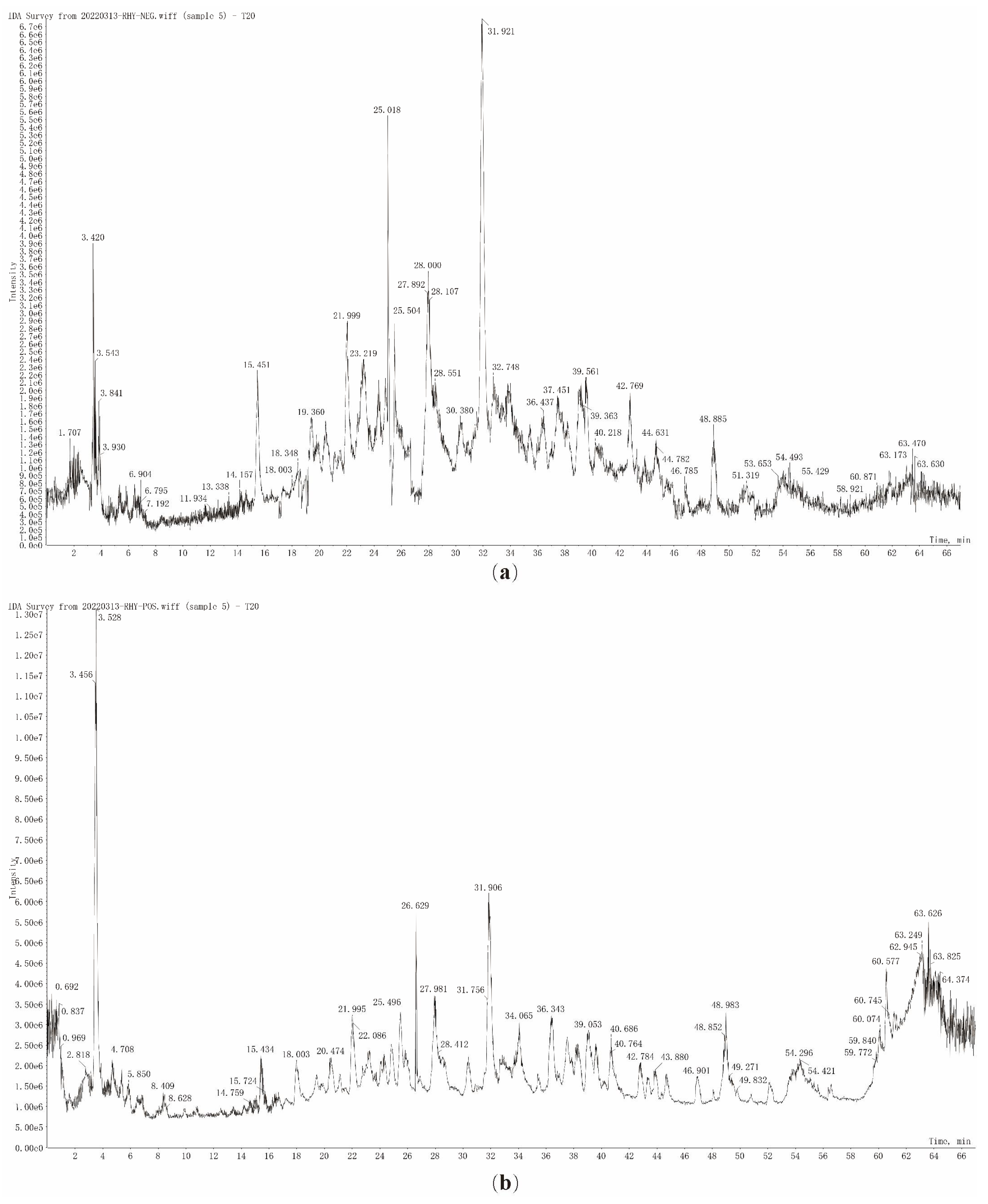
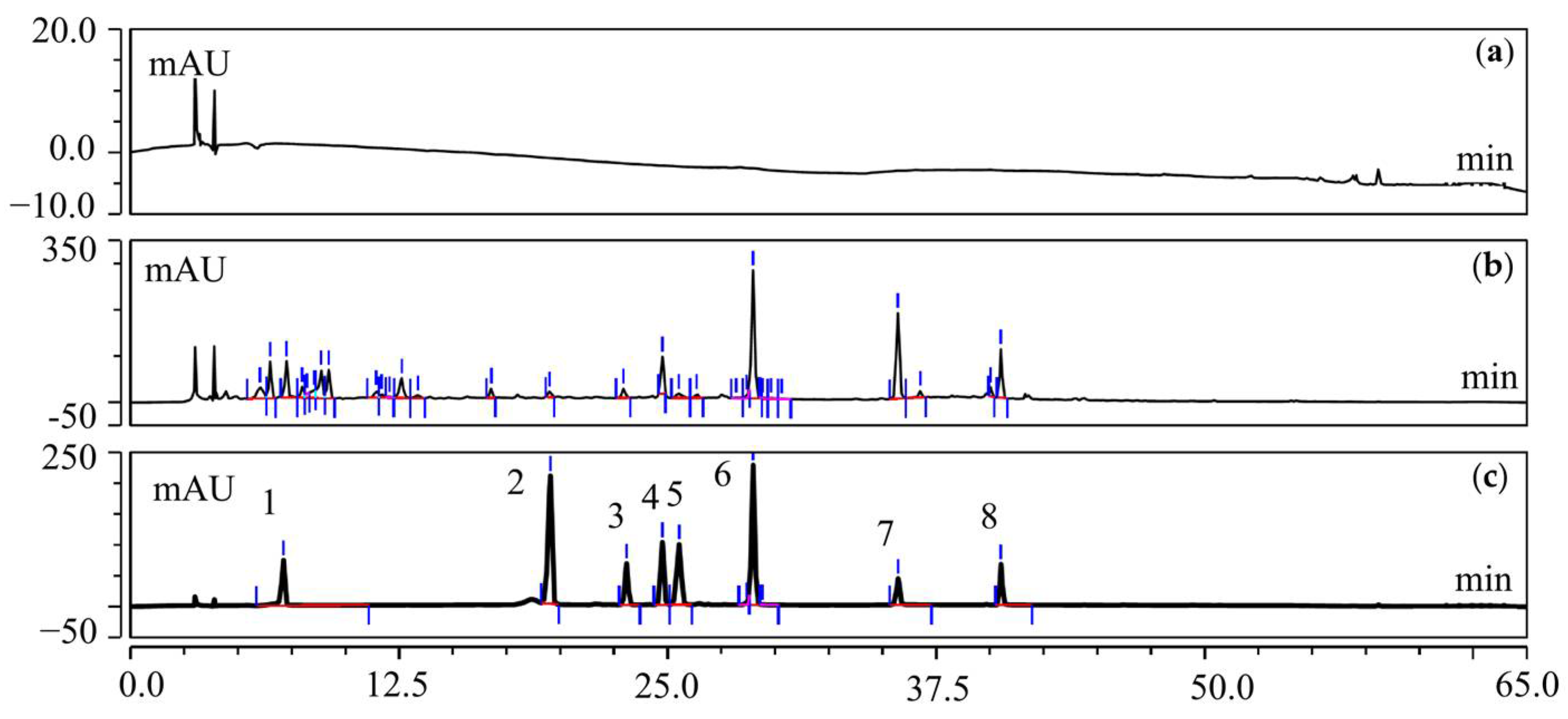
2.9. UFLC-Q-TOF-MS/MS Analysis of T20 and T60

A total of 3.0 g each of T20 and T60 were accurately weighed and sonicated in 10 mL of methanol for 30 min. The supernatant was filtered with 0.22 μm and then injected into an ultra-fast liquid chromatography/quadrupole-time-of-flight tandem mass spectrometry system for analysis (UFLC-Q-TOF-MS/MS). The column was an Acclaim™ Polar Advantage II C18 (4.6 × 250 mm, 5 μm, Thermo Scientific, Waltham, MA, USA), which was maintained at 30 °C. The mobile phases were composed of water with 0.1% formic acid (A) and methanol (B) using a gradient elution of 95–40% A at 0–55 min, 40–10% A at 55–60 min, and 10% A at 60–63 min. The flow rate into the mass spectrum adjusted by the diverter valve was about 0.3 mL/min. Other MS parameters were adopted to be the same as published work [19]. The sample volume injected was set at 10 μL. All the acquisition and analysis of data were controlled by the PeakView Software TM V. 1.1 (AB SCIEX, Foster City, CA, USA).

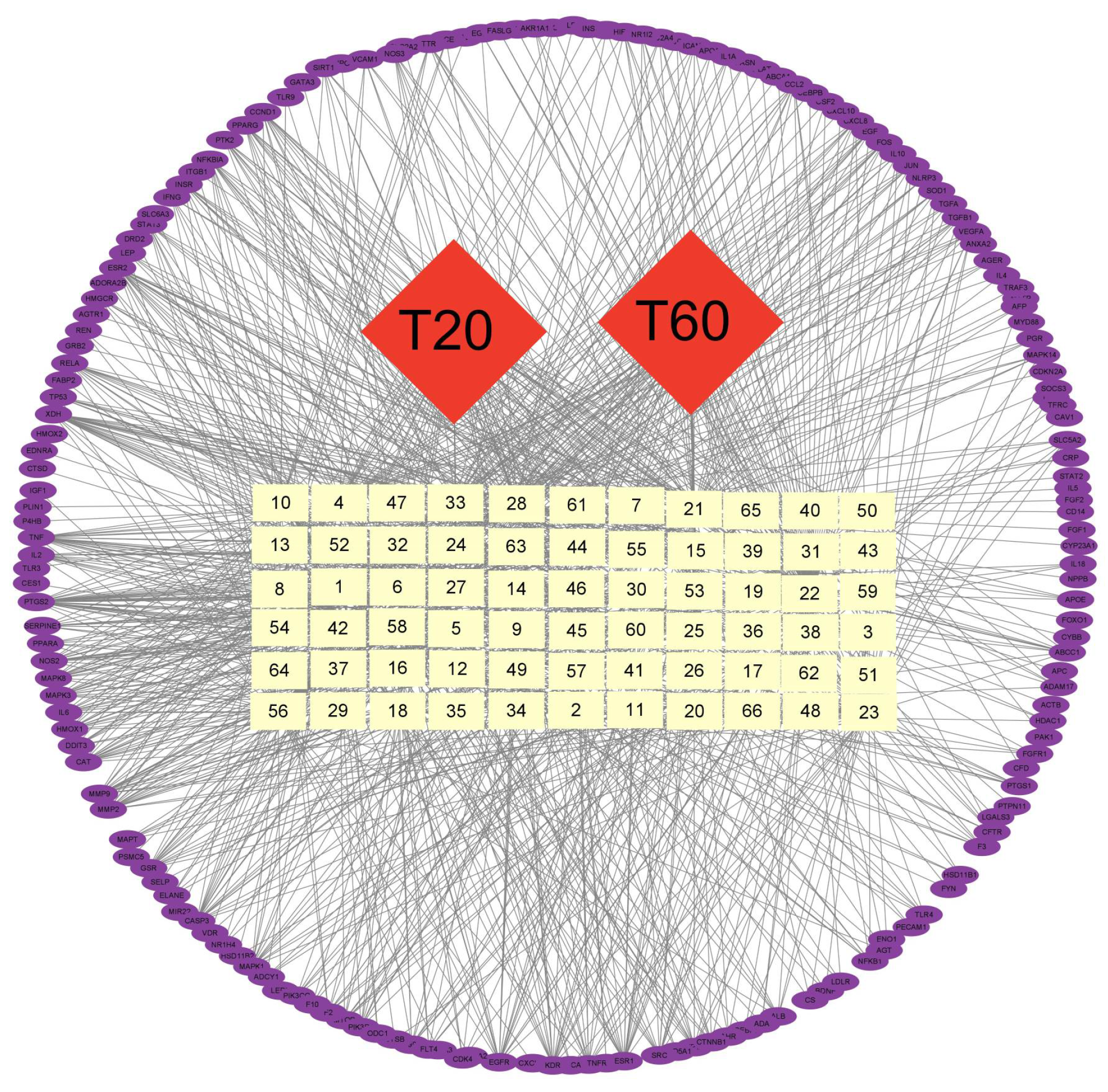
2.10. Network Pharmacological Analysis

The identified 66 components of T20 and T60 were searched and integrated using the SwissTargetPrediction http://www.swisstargetprediction.ch/ (accessed on 5 June 2023) and CTD https://ctdbase.org/ (accessed on 5 June 2023). In the GeneCards database https://www.genecards.org/ (accessed on 7 June 2023), “porcine epidemic diarrhea virus” was used as a keyword to search, download, and integrate all related genes. The intersection between the T20 and T60 targets and PEDV was used to obtain the therapeutic targets of HJT using the Venn diagram. A network diagram of the active components in T20 and T60 was constructed using Cytoscape 3.7.2 software, and then the network analysis tool in Cytoscape was used to analyze the topological parameters of each network and to evaluate the significance of the nodes according to centrality and node degree. The possible active ingredients in T20 and T60 that play an anti-PEDV role were sorted by degree value.

2.11. Determination of the Active Compounds and Contents of HJT Using HPLC

All of the reference standard compounds were purchased from Shanghai Yuanye Bio-Technology Co., Ltd., (Shanghai, China). Protocatechuic acid (10.0 mg), rutin (10.0 mg), isoquercetin (10.0 mg), quercetin (10.0 mg), taxifolin 7-rhamnoside (10.0 mg), taxifolin (10.0 mg), quercitrin (10.0 mg), and quercetin 7-rhamnoside (1.0 mg) were accurately weighed and dissolved in methanol to prepare reference standard solutions, each with a final volume of 10 mL. An HPLC analysis was carried out on an Ultimate 3000 DGLC system (Dionex company, Sunnyvale, CA, USA) equipped with Chromeleon 7.2 data processing software, DGP-3600SD dual-ternary Pump, SRD-3600 degasser, WPS-3000SL Automatic Sampler, TCC-3000RS column oven, and DAD detector. The HPLC system was used under the following chromatographic conditions: mobile phase A: acetonitrile, mobile phase B: 0.1% glacial acetic acid solution with pH 4.0 adjusted with triethylamine, column: Acclaim™ Polar Advantage II C18 (4.6 mm × 250 mm, 5 μm), gradient elution: 0–10 min, 10–15% A; 10–25 min, 15–23% A; 25–30 min, 23% A; 30–50 min, 23–60% A; and 50–53 min, 60–90% A. The detection wavelengths were 257 nm and 289 nm, while the column temperature was 30 °C.
The eight reference standard solutions were mixed and diluted to prepare a mixed reference solution containing protocatechuic acid (60 μg/mL), taxifolin 7-rhamnoside (150 μg/mL), rutin (100 μg/mL), isoquercetin (130 μg/mL), taxifolin (50 μg/mL), quercitrin (250 μg/mL), quercetin 7-rhamnoside (50 μg/mL), and quercetin (50 μg/mL). Then, the solution (10 μL) was injected into the liquid chromatograph, and peak areas were recorded. A calibration curve was constructed with five concentration levels of standard samples covering the range of 10–1000 μg/mL. The prepared test solution was continuously injected six times to record the chromatogram and evaluate precision.
A total of six samples were prepared for each herb solution, and reproducibility was confirmed using chromatography. The test solutions were analyzed at 0, 4, 12, 24, 36, 48, and 72 h after preparation to assess their stability. Recovery of analyses was assessed at medium concentration levels with 6 replicates. The content was determined by the corresponding peak area of each compound.

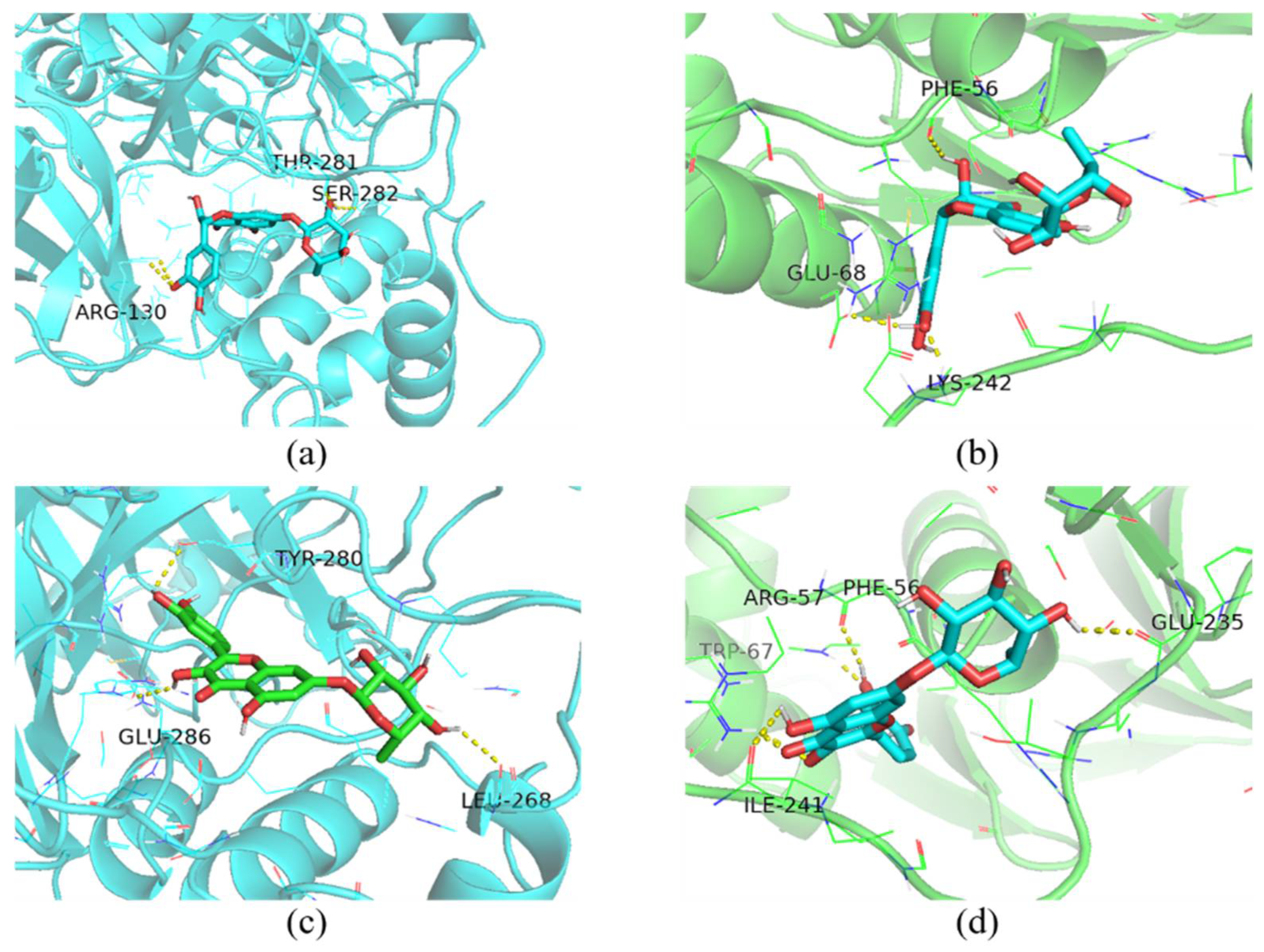
2.12. Molecular Docking

A molecular docking study was conducted to investigate the binding affinities of taxifolin-7-O-rhamnoside and quercetin-7-rhamnoside, as well as papain-like protease 2 (PLP-2) and 3C-like protease (3CLpro) of PEDV. The 2D structures of taxifolin-7-O-rhamnoside and quercetin-7-rhamnoside were obtained from the PubChem database, converted into 3D structures, and optimized for energy minimization using ChemBioOffice Ultra 13.0.2-Chem3D software. The protein structures of PLP-2 and 3CLpro were retrieved from the Protein Data Bank (PDB) database maintained by the RCSB. These structures were processed using the PyMOL 2.5.2 software to remove water and residual molecules. The docking grid points and dimensions were set in AutoDockTools-1.5.7 software, and semi-flexible molecular docking was carried out using Vina v1.1.2 software, where the higher absolute value of the binding energy represents the higher affinity of the receptor and ligand.

2.13. Statistical Analysis

Statistical comparisons were performed using GraphPad Prism 9 software. Accordingly, the significance of the differences between the treatment groups and mock group (cell viability, PFU, N mRNA, etc.) was determined by the ANOVA.

3. Results

3.1. Evaluation of Cytotoxicity of HJT Extract

CCK-8 was used to detect the effects of T0, T20, and T60 from HJT aqueous extract on the activity of IPI-FX cells for 48 h. The viability of the IPI-FX cells was unaffected by T0, T20, and T60 at concentrations of 0.21–6.7 mg (raw material)/mL, 0.27–8.7 mg (raw material)/mL, and 0.20–6.5 mg (raw material)/mL, respectively (Figure A1). Meanwhile, to exclude the effect of T0, T20, and T60 on pH, a pH meter was used to detect the pH change of DMEM medium after treatment. The results showed that the pH of the drug-containing medium below the maximum safe concentration was not significantly different from that of the negative control. Therefore, the above concentration ranges were selected for the cellular administration treatments without toxic effects on the cells.

3.2. Inhibition of Viral Infections In Vitro by HJT Extracts

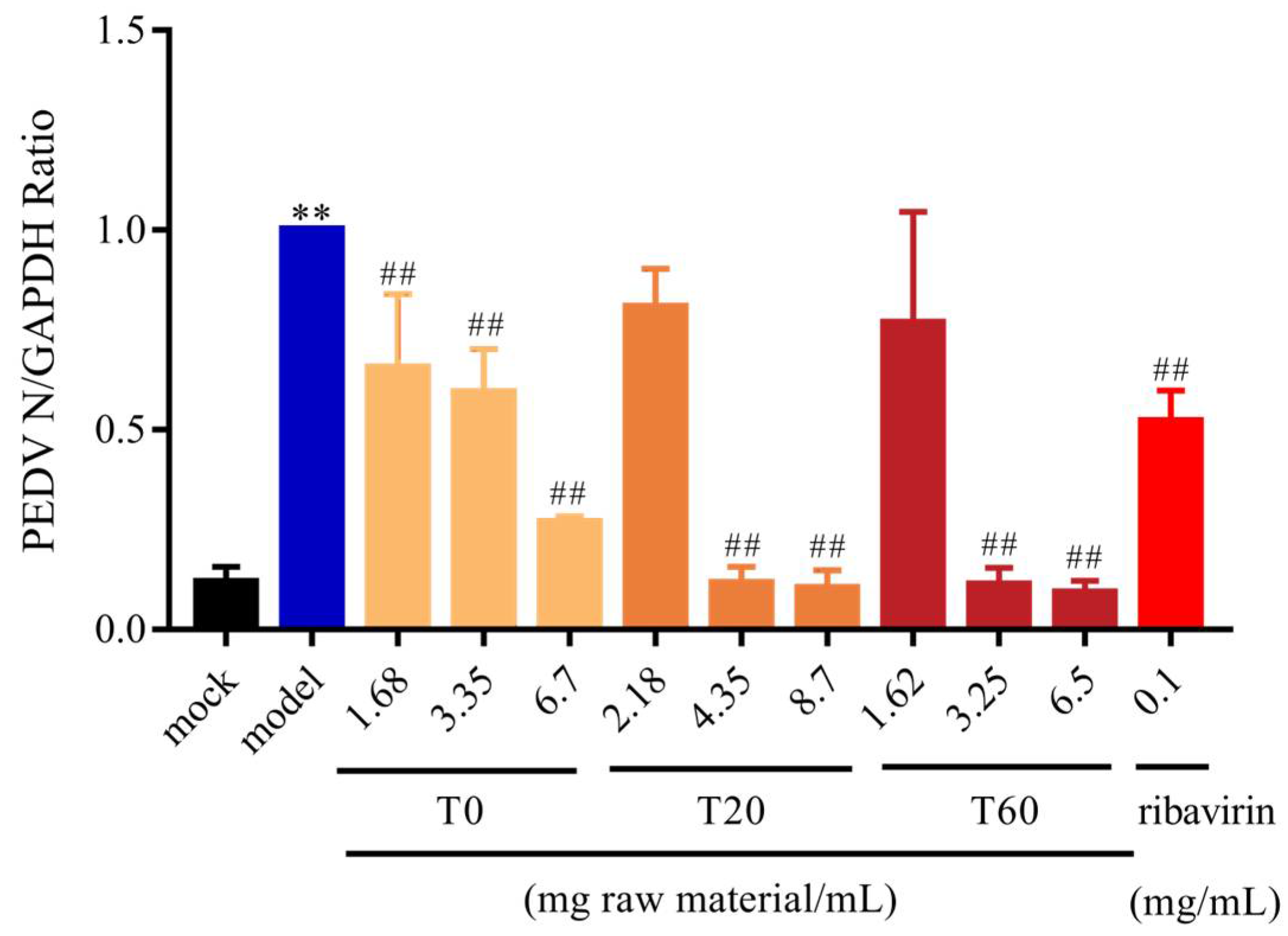
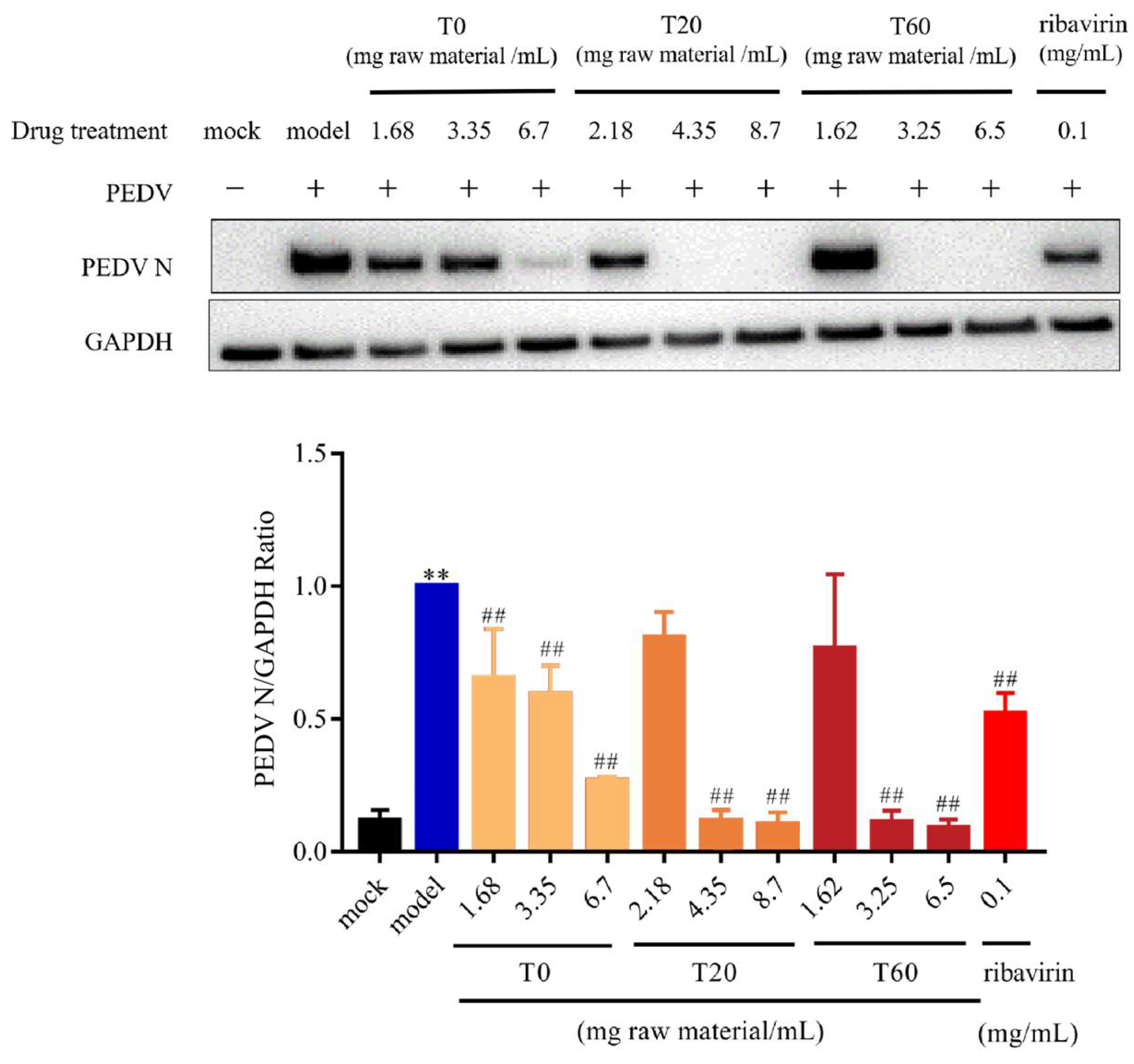
3.2.1. T0, T20, and T60 Inhibit PEDV N Protein Expression In Vitro

qPCR and WB analysis were used to detect the expression of the N protein in IPI-FX after PEDV infection. The results demonstrated that, in comparison with the mock group, the expression level of the N gene in the model group was significantly elevated. In comparison with the model group, N gene expression was found to be significantly reduced in the positive control group after ribavirin treatment. When the concentrations of T0, T20, and T60 reached 3.35 mg (raw material)/mL, 4.35 mg (raw material)/mL, and 1.62 mg (raw material)/mL, respectively, the expression of PEDV N gene was significantly reduced (Figure 1). Further analysis using Western blot showed that the expression of the PEDV N protein was also significantly reduced compared to the model group when the concentrations of T0, T20, and T60 were 1.68 mg (raw material)/mL, 4.35 mg (raw material)/mL, and 3.25 mg (raw material)/mL, respectively (Figure 2, Figure S1). These findings demonstrate that T0, T20, and T60 can significantly decrease the expression of the PEDV-G2 N protein in IPI-FX cells, indicating their in vitro anti-PEDV-G2 activity.

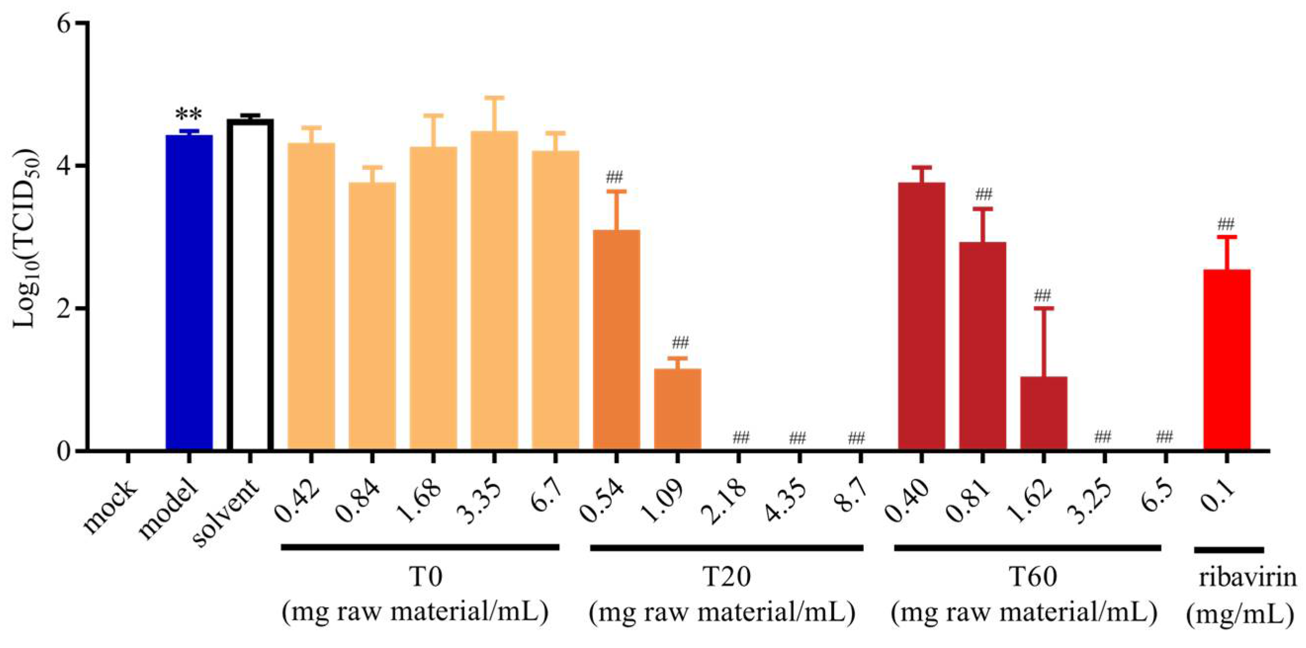
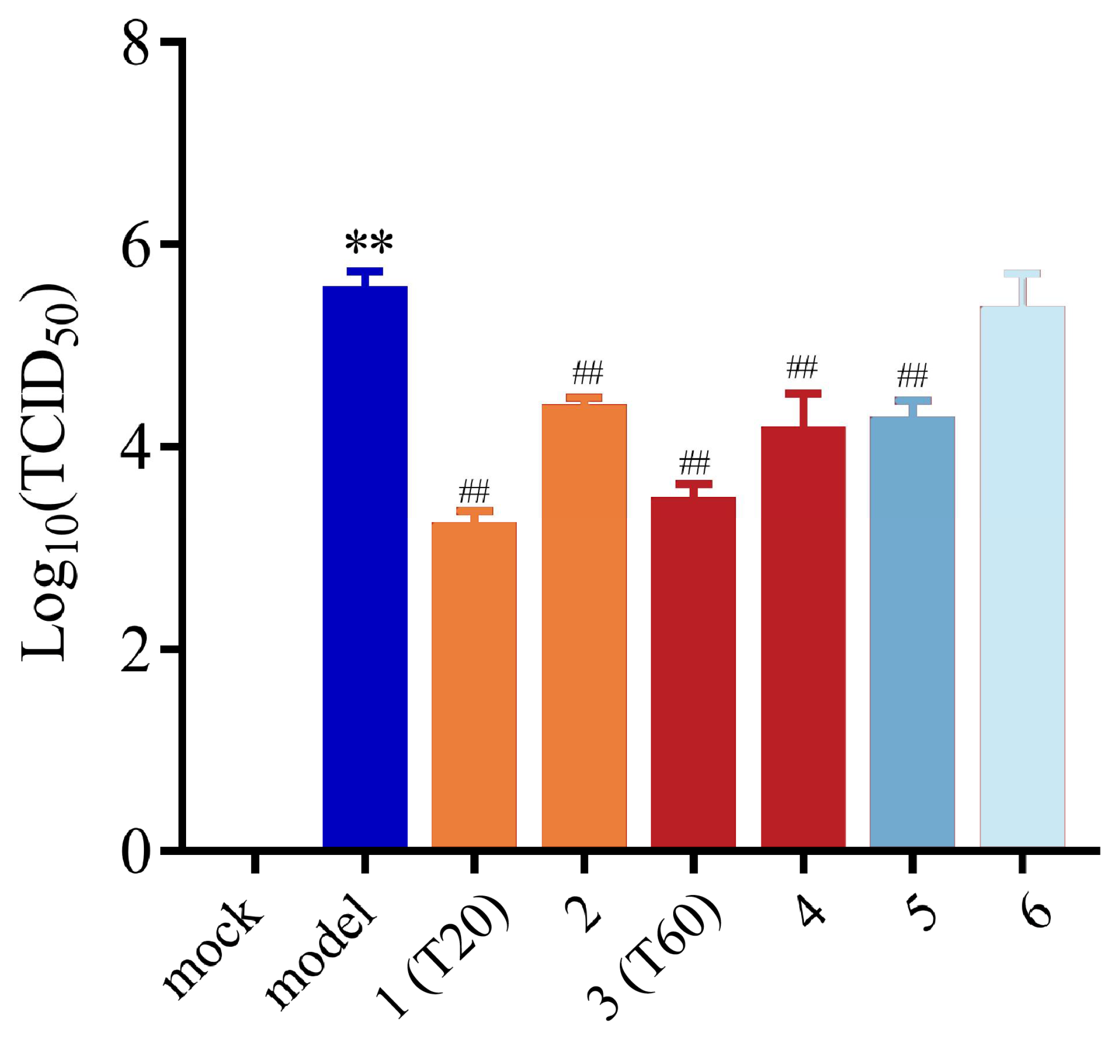
3.2.2. Effect of T0, T20, and T60 on PEDV Titers

For the further evaluation of the anti-PEDV effects of T0, T20, and T60, the changes in PEDV-G2 titers after T0, T20, and T60 treatments were determined using the endpoint dilution method. The results showed that the titers of PEDV-G2 decreased after T20 and T60 treatments, whereas T0 had no significant effect on the titers of PEDV-G2. Meanwhile, ribavirin treatment also significantly decreased the viral titers. In addition, when the concentrations of T20 and T60 reached 2.18 mg (raw material)/mL and 3.25 mg (raw material)/mL, respectively, the viral titers were reduced to 0 (Figure 3). This indicated that both T20 and T60 could make PEDV-G2 lose the ability to reinfect the cells and effectively prevent the transmission of PEDV, whereas T0 was unable to reduce the titers of PEDV. Therefore, it was hypothesized that T20 and T60 were the active fractions in the HJT extracts with anti-PEDV activity.

3.3. Anti-PEDV Compounds in HJT Extracts

3.3.1. Characterization of the Chemical Compound Profiles of T20 and T60

UFLC-Q-TOF-MS/MS analysis was used to detect the chemical compound profile of T20, and its total ion flow diagram was obtained (Figure 4). The mass spectrometry information, such as cleavage fragment ions and retention time of T20, was then compared with the database. In conclusion, 42 compounds were identified, including 26 flavonoids, 5 xanthones, 6 organic acids, and 5 other compounds. The cleavage fragments and peak attribution of each compound in positive and negative ion modes are shown in Table A1. The chemical composition of T60 was analyzed by the same method, and the total ion flow diagrams of T60 were obtained (Figure 5). A total of 46 compounds were identified in T60, including 26 flavonoids, 9 xanthones, 8 organic acids, and 3 other compounds. The cleavage fragments and peak assignments of each compound in positive and negative ion modes are shown in Table A2.

3.3.2. Network Pharmacological Prediction of Anti-PEDV Compounds

We integrated the component analysis results of T20 and T60, identifying a total of 66 compounds. To explore the active ingredients with potential anti-PEDV effects in T20 and T60, the SwissTargetPrediction and CTD databases were used to predict the targets of action of a total of 66 compounds in T20 and T60 (Table A3), and 1228 targets were obtained. After integrating with the 657 PEDV-related targets obtained from the GeneCards database, we obtained 185 potential targets of T20 and T60 against PEDV. The “extraction fraction-component-target” network diagram was constructed and analyzed, and it was found that the target-related compounds were mainly flavonoids and organic acids (Figure 6). Therefore, the anti-PEDV effects of T20 and T60 might be related to the 36 flavonoids and 13 organic acids in the extracts.

3.3.3. Determination of Active Compounds

The contents of the above 36 flavonoids and 13 organic acids were studied by HPLC determination. After mapping the conditions of content determination and methodological investigation, there were eight compounds, including protocatechuic acid, taxifolin 7-rhamnoside, rutin, isoquercitrin, taxifolin, quercitrin, quercetin 7-rhamnoside, and quercetin (Figure 7), that displayed the highest amount in T20 and T60. The content analysis results are shown in Table 2.

3.4. Analysis of Anti-PEDV Effects of HJT Active Compounds

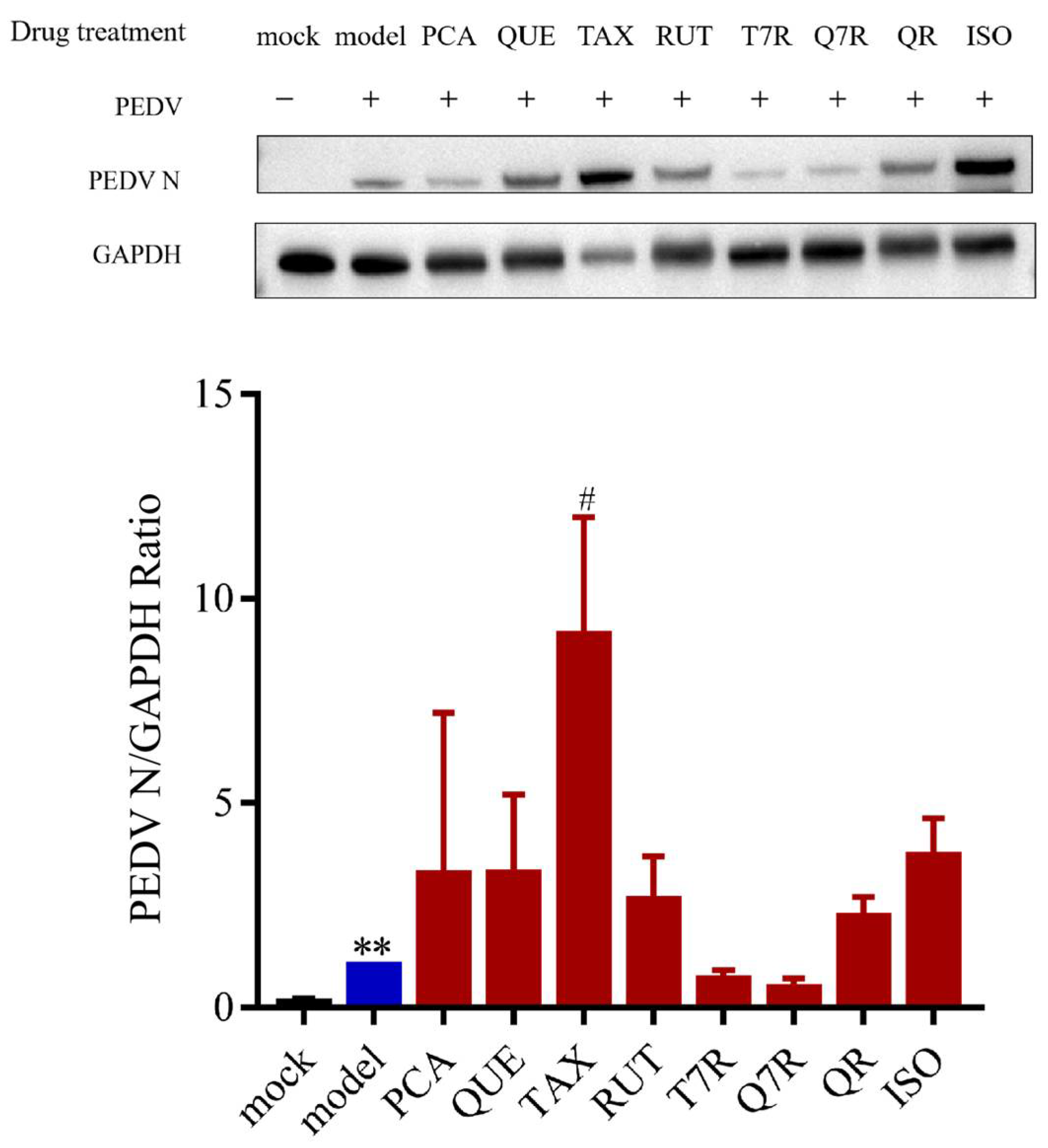
3.4.1. Anti-PEDV Effects of Individual Compounds

To investigate whether the anti-PEDV effect of HJT water extract is related to the eight major components mentioned above, this study first examined the effect of each compound on PEDV.
Western blot analysis was used to measure the expression levels of PEDV N protein in IPI-FX cells after treatment with the eight compounds. The results showed that, compared to the model group, none of the eight compounds significantly reduced the expression of PEDV N protein (Figure 8, Figure S2). Among them, only the groups treated with taxifolin-7-O-rhamnoside and quercetin-7-rhamnoside showed a trend toward downregulating PEDV N protein expression but without statistical significance. Therefore, it is speculated that the anti-PEDV effect of HJT water extract may be the result of the combined action of multiple compounds, possibly involving taxifolin-7-O-rhamnoside and quercetin-7-rhamnoside.
Furthermore, molecular docking analysis was performed on taxifolin-7-O-rhamnoside and quercetin-7-rhamnoside with key enzymes of PEDV replication (3CLpro and PLP-2) by utilizing the AutoDock-Vina 1.5.7 software (Figure 9). The results demonstrated that the docking binding energies of taxifolin-7-O-rhamnoside with 3CLpro and PLP-2 were −9.5 kcal/mol and −8.0 kcal/mol, respectively, and those of quercetin-7-rhamnoside with 3CLpro and PLP-2 were −10.4 kcal/mol and −8.2 kcal/mol. The specific interacting amino acid residues are shown in Table 3. It has been demonstrated that the two compounds exhibit a strong binding affinity for 3CLpro and PLP-2. The hypothesis is proposed that these compounds may possess an anti-PEDV effect through their interaction with PEDV replication-related proteins, thereby impeding the virus’s replication within host cells.

3.4.2. Anti-PEDV Effects of Mixed Controls

Next, we investigated whether it was the mixture reference standards of taxifolin-7-O-rhamnoside and quercetin-7-rhamnoside in the extracts of HJT that exerted the anti-PEDV effect. We prepared mixed controls by referring to the content determination results in 3.2.3 and used Western blot to determine T20 extract (1), a mixture of eight main compounds in T20 (2), T60 extract (3), a mixture of eight main compounds in T20 (4), a mixture of taxifolin-7-O-rhamnoside and quercetin-7-rhamnoside (5), and a mixture of six other compounds (6) and determined their inhibitory effects on PEDV-G2 in vitro. The effects of the six samples on PEDV titers were assessed using the endpoint dilution method. The results indicated that, compared to the model group, T20, T60, the mixed reference standards of T20 and T60, as well as the mixed reference standards of taxifolin-7-O-rhamnoside and quercetin-7-rhamnoside all significantly reduced the PEDV-G2 titers (Figure 10). This suggests that the mixed reference standards composed of taxifolin-7-O-rhamnoside and quercetin-7-rhamnoside have anti-PEDV effects, and it is speculated that these compounds are the key active ingredients responsible for the anti-PEDV activity in the HJT water extract.

4. Discussion

PEDV imposes severe socioeconomic impacts on global swine production systems through its high transmission efficiency and devastating mortality profiles, threatening the sustainability of intensive livestock operations. In recent years, plant natural ingredients have begun to be used in anti-PEDV therapy. For example, the aqueous leaf extract of Moringa oleifera can inhibit PEDV infection in vitro by inhibiting oxidative stress and apoptosis during the replication phase of PEDV [20]. Thai medicinal plant mulberry’s (Morus alba Linn.) leaf ethanolic aqueous crude extracts also showed promising anti-PEDV efficacy [21]. Nevertheless, phytochemical complexity and undefined bioactive markers pose challenges to quality standardization, process reproducibility, and clinical-grade manufacturing of botanical antivirals. Consequently, systematic characterization of bioactive constituents and mechanistic deconvolution of their antiviral pharmacodynamics are critical prerequisites for translating phytomedicines into clinical practice. N protein is widely used in the molecular biological diagnosis of PEDV and is a common pharmacodynamic indicator for evaluating the efficacy of anti-PEDV in vitro. This study measures the expression levels of the PEDV N gene and titers in IPI-FX cells after treatment with T0, T20, T60, and ribavirin. Ribavirin is a broad-spectrum antiviral drug that has been shown to have good antiviral effects against PEDV [22] and was used as a positive control in this experiment. In this study, we tested the anti-PEDV effect and analyzed the composition of different elution sites of HJT extracts separately and determined that T20 and T60 had good anti-PEDV efficacy. Although the N protein content was significantly reduced after treatment with high concentrations of T0, its viral titer did not change significantly. A decrease in N protein indicates that viral proliferation may be inhibited after treatment with a high concentration of T0, while no decrease in viral titer may indicate that the virus is not significantly deprived of its ability to infect. This also reflects the fact that antiviral activity needs to be tested in conjunction with both tests to reach a conclusion. Further, we preliminarily characterized 66 compounds in T20 and T60, which contained flavonoids, xanthones, organic acids, and so on. The anti-PEDV efficacy of T20 and T60 may be related to their rich compound composition.
Extensive research has demonstrated that plant-derived secondary metabolites exhibit significant pharmacological potential. These natural compounds have emerged as a promising focus for antiviral drug discovery, owing to their diverse biological activities, broad availability, favorable safety profiles, and demonstrated capacity to directly interact with viral particles (such as PEDV, dengue virus, transmissible gastroenteritis virus, etc.) [23,24]. Regarding prophylactic mechanisms, matrine has been shown to effectively block PEDV infection through specific interactions with viral surface spike proteins, thereby inhibiting both viral adsorption and cellular entry processes [25]. The PEDV 3CLpro, a crucial enzyme in viral replication, plays a pivotal role in both infection and transmission processes. Notably, phytochemicals including wogonin, tomatidine, chrysin, and naringenin exert dual-phase antiviral effects by targeting replicase proteins such as 3CLpro, which ultimately disrupts viral particle assembly during both preventive and post-entry stages [26,27,28]. Andrographolide demonstrates distinct therapeutic potential through its apoptosis-inducing mechanism mediated by JAK2-STAT3 pathway inhibition. This pharmacological action not only suppresses PEDV replication but also significantly ameliorates clinical symptoms and reduces mortality rates in infected piglets [29]. In addition, as supplements, ellagic acid and buddlejasaponin IVb can inhibit the inflammatory response induced by PEDV and the former also has the function of antioxidant damage and modulation of the interferon pathway, which improves intestinal homeostasis in PEDV-infected piglets [30,31]. These findings collectively indicate that phytochemical constituents may exert multi-target antiviral effects throughout various stages of viral pathogenesis. Therefore, it is important to investigate the interaction of key components with disease targets for accurate clinical application of natural compounds. In the present investigation, we employed network pharmacology approaches to predict potential antiviral components in HJT extracts, with particular focus on flavonoid and organic acid derivatives. Through content determination and analysis of the anti-PEDV effects of control compounds, taxifolin-7-O-rhamnoside and quercetin-7-rhamnoside were finally hypothesized to be the key anti-PEDV compounds in the extracts of HJT. Notably, antiviral effects were observed exclusively in the combined application of both compounds, demonstrating a statistically significant reduction in PEDV-G2 titers. Meanwhile, 3CLpro and PLP-2 are indispensable key enzymes in the replication process of coronaviruses such as PEDV. These enzymes can promote viral transmission by resisting host cellular immunity, making them important targets for antiviral drug development [32,33,34]. In this study, we found that both taxifolin-7-rhamnoside and quercetin-7-rhamnoside have a strong affinity for these two proteins through molecular docking results. It is hypothesized that they may inhibit viral replication by targeting the viral proteins, which is also supported by our previous findings.
In this study, we analyzed key compounds against PEDV and identified eight important components in HJT, including protocatechuic acid, taxifolin 7-rhamnoside, rutin, isoquercitrin, taxifolin, quercitrin, quercetin-7-rhamnoside, and quercetin. Among these components, taxifolin-7-O-rhamnoside and quercetin-7-rhamnoside were further validated to play a significant role in combating PEDV. Consistently, existing evidence indicates that quercetin-7-rhamnoside demonstrates notable anti-PEDV activity [35], coupled with specific antioxidant properties and cytopathic effect suppression [36]. Recent studies have revealed that plant extracts exhibit enhanced therapeutic efficacy through multi-component, multi-target mechanisms, highlighting the importance of not overlooking the potential roles of the other six compounds. Prior research has confirmed that inter-component interactions among phytochemicals can amplify antiviral (such as dengue virus serotype 2 and SARS-CoV-2) activity through synergistic or additive mechanisms [37,38]. There is an increasing preference for these phytoconstituents in modulating immune responses and enhancing barrier functions [39]. Importantly, gut microbiota-mediated bioconversion can metabolize ancillary components into bioactive derivatives with therapeutic potential [40]. For instance, protocatechuic acid can regulate gut microbiota, decrease levels of inflammatory factors, and modulate the redistribution of tight junction proteins [41]. Dietary supplementation with rutin in weaned piglets enhances intestinal barrier integrity, improves diarrheal resistance, and exerts combined anti-inflammatory and antioxidant effects [42]. Rutin [43], isoquercitrin [44], and quercitrin [45] undergo gut microbiota-mediated bioconversion to bioactive quercetin metabolites that upregulate tight junction protein expression and enhance intestinal epithelial integrity [46]. Taxifolin, on the other hand, activates the Wnt/β-catenin signaling pathway to stimulate tight junction protein synthesis and intestinal epithelial cell proliferation [47].
In summary, our findings demonstrate that taxifolin-7-O-rhamnoside and quercetin-7-rhamnoside represent promising candidate agents for PEDV intervention, with their quantitative levels serving as quality control biomarkers for standardizing HJT-based antiviral formulations. Nevertheless, the precise molecular mechanisms mediating their anti-PEDV activity require further elucidation. Systematic investigation of their antiviral mechanisms is therefore essential for optimizing the therapeutic development and clinical translation of HJT preparations. Notably, the other six major components in HJT extracts may exert ancillary protective roles, despite lacking direct antiviral efficacy under current experimental conditions.

5. Conclusions

In conclusion, we identified taxifolin-7-O-rhamnoside and quercetin-7-rhamnoside in the extracts of HJT with in vitro antiviral activity, which significantly reduced the expression of PEDV N protein and viral titers in IPI-FX cells. This study provides candidate compounds for the development of anti-PEDV drugs.

Supplementary Materials

The following supporting information can be downloaded at: https://www.mdpi.com/article/10.3390/v17070900/s1. Figure S1: WB-original image of Figure 2; Figure S2: WB-original image of Figure 8.

Author Contributions

Conceptualization, H.R. and P.L.; Data curation, W.W. (Wenlong Wang); Formal analysis, H.R.; Methodology, H.R.; Project administration, H.W.; Resources, W.S.; Software, S.L.; Supervision, H.W.; Validation, H.R. and S.L.; Visualization, W.W. (Weiyue Wang); Writing—original draft, S.L.; Writing—review and editing, P.L. All authors have read and agreed to the published version of the manuscript.

Funding

This research received no external funding.

Institutional Review Board Statement

Not applicable.

Informed Consent Statement

Not applicable.

Data Availability Statement

The data can be made available by addressing a request to the corresponding author.

Conflicts of Interest

The authors declare no conflicts of interest. The funders had no role in the design of the study; in the collection, analyses, or interpretation of data; in the writing of the manuscript; or in the decision to publish the results.

Abbreviations

The following abbreviations are used in this manuscript:
HJTHypericum japonicum Thunb. ex Murray
PEDVPorcine epidemic diarrhea virus
PEDPorcine epidemic diarrhea
DMEMDulbecco’s modified Eagle medium
PBSphosphate buffer solution
BSAbovine serum albumin
CCK-8Cell counting Kit-8
RIPARadio immunoprecipitation assay
SDS-PAGESodium dodecyl sulfate-polyacrylamide gel electrophoresis
PVDFpolyvinylidene fluoride
ECLenhanced chemiluminescence
TBSTtris-buffered saline containing tween-20
IPI-FXimmortal pig intestinal
UFLC-Q-TOF-MS/MSultra-fast liquid chromatography/quadrupole-time-of-flight tandem mass spectrometry system for analysis.
PLP-2papain-like protease 2
3CLpro3C-like protease
ARGArginine
SERSerine
THRThreonine
PHEPhenylalanine
GLUGlutamic acid
LYSLysine
LEULeucine
TYRTyrosine
TRPTryptophan
ILEIsoleucine

Appendix A

Table A1. Identification of the chemical profiles of T20 based on UPLC-Triple-TOF-MS/MS.
No.tR (min)Molecular FunctionCompounds[M + H]+[M − H]
16.65C7H12O6Quinic acid193.0705191.0562
222.03C13H20O3Vomifoliol225.1483\
323.17C16H18O9Chlorogenic acid355.1023353.0870
423.18C9H6O37-hydroxycoumarine163.0388\
523.26C33H44O82,5-cyclohexadien-1-one, 2-[[3-[(2e)-3,7-dimethyl-2,6-octadien-1-yl]-2,4,6-trihydroxy-5-(2-methyl-1-oxopropyl)phenyl]methyl]-3,5-dihydroxy-4,4-dimethyl-6-(2-methyl-1-oxopropyl)569.3128\
623.34C21H22O117-o-rhamnoside of flagellin451.1228449.1068
724.05C11H12O5Sinapic acid225.0755\
824.32C15H12O7Taxifolin305.0655303.0505
924.42C28H36O83,5-dihydroxy-4,4-dimethyl-2-(1-oxoisobutyl)-6-[[5-(1-oxoisobutyl)-3-(3-methyl-2-butenyl)-2,4,6-trihydroxyphenyl]methyl]-2,5-cyclohexadiene-1-one\499.2356
1024.44C15H14O6Catechin291.0862289.0713
1125.5C26H34O8Saroaspidin c475.2291473.2139
1226.67C15H14O6Epicatechin\289.0709
1326.73C14H10O45-hydroxy-1-methoxy-9h-xanthen-9-one243.0648\
1426.74C15H12O65,5′-methylenedisalicylic acid289.0704\
1527.36C27H30O15Kaempferol 7-o-rutinoside595.1644593.1484
1627.93C27H30O16Quercetin-7-o-rutinoside611.1595609.1445
1727.98C18H22O98-glucosyl-5,7-dihydroxy-2-isopropylchromone383.1330381.1178
1828.01C16H18O83-p-coumaroylquinic acid\337.0923
1930.25C27H32O15Ellipticoside597.1800595.1643
2030.57C21H22O11Astilbin\449.1062
2131.5C19H24O98-glucosyl-5,7-dihydroxy-2-(1-methylpropyl) chromone397.1488395.1330
2233.06C16H18O84-o-coumaroylquinic acid\337.0923
2333.38C27H30O16Rutin611.1558609.1442
2434.87C10H10O4Isoferulic acid\193.0505
2534.89C10H8O37-methoxycoumarin177.0544\
2635.43C31H34O84-[[3-benzoyl-2,6-dihydroxy-4-(3-methylbut-2-enoxy)phenyl]methyl]-3,5-dihydroxy-6,6-dimethyl-2-(2-methylpropanoyl)cyclohexa-2,4-dien-1-one\533.2121
2736.5C9H10O4Ethyl 3,4-dihydroxybenzoate183.0650181.0505
2836.93C27H30O15Quercetin 3,7-di-o-rhamnopyranoside\593.1486
2937.33C21H20O12Hyperoside465.1025463.0858
3038.85C24H20O8Kielcorin\435.1069
3138.95C15H12O7Taxifolin\303.0503
3239.27C27H30O16Quercetin 3-o-rutinoside611.1594609.1440
3342.41C21H20O10Afzelin433.1119431.0962
3442.76C21H20O12Isoquercitrin465.1020463.0859
3543.71C12H16O4(1s,3s)-3,4-dihydro-8-methoxy-3,5-dimethyl-1h-2-benzopyran-1,6-diol225.1099223.0956
3646.87C21H20O11Quercitrin449.1067447.0912
3748.89C21H20O11Vincetoxicoside b449.1070447.0913
3853.85C21H18O13Quercetin-3-o-glucuronide479.0815\
3956.88C15H10O7Quercetin303.0499301.0351
4057.52C13H8O61,3,5,6-tetrahydroxyxanthone\259.0246
4160.25C13H8O51,3,5-trihydroxyxanthone\243.0296
4263.41C28H42O5Hyperjaponol h\457.2943
Table A2. Identification of the chemical profiles of T60 based on UPLC-Triple-TOF-MS/MS.
NO.tR (min)Molecular FormulaCompounds[M + H]+[M − H]
16.61C7H12O6Quinic acid193.0703191.0563
216.62C7H6O5Gallic acid171,0646169.0144
323.14C7H6O4Protocatechuic acid155.0335153.0195
423.2C16H18O9Chlorogenic acid355.1025353.0876
523.2C7H6O34-hydroxybenzoic acid139.0386137.0244
623.32C21H22O11Taxifolin 7-rhamnoside451.1233449.1082
726.65C15H14O6L-epicatechin291.0864289.0720
827.51C45H38O18Procyanidin c1867.2130865.1984
927.96C18H22O95,7-dihydroxy-2-isopropylchromone-8-beta-d-glucoside383.1335381.1190
1028.23C16H18O83-p-coumaroylquinic acid\337.0928
1130.2C27H32O15Ellipticoside597.1809595.1664
1230.48C21H22O11Neoisoastilbin451.1233449.1084
1331.47C19H24O98-glucosyl-5,7-dihydroxy-2-(1-methylpropyl) chromone397.1495395.1345
1433.36C27H30O16Rutin611.1601609.1455
1534.86C10H10O4Isoferulic acid\193.0508
1635.31C21H22O10Aromadendrin 7-o-rhamnoside435.1283433.1136
1736.11C21H20O12Hyperoside465.1024463.0874
1836.46C9H10O4Ethyl 3,4-dihydroxybenzoate183.0645181.0513
1936.88C27H30O15Quercetin 3,7-di-o-rhamnopyranoside595.1648593.1501
2037.11C14H10O61,3,7-trihydroxy-2-methoxyxanthone275.0548273.0404
2137.33C14H10O51,7-dihydroxy-4-methoxyxanthone259.0600257.0457
2237.38C9H8O3Trans-4-hydroxycinnamic acid\163.0400
2338.49C24H20O9Cadesin a\451.1031
2438.86C30H24O14Dehydropolyester catechins\607.1087
2538.88C15H12O7Taxifolin305.0655303.0511
2639.04C32H36O8Hyperjaponicol a549.2421547.2280
2739.21C27H30O16Quercetin 3-o-rutinoside611.1602609.1458
2842.37C12H18O3Jasmonic acid\209.1182
2942.45C19H18O10Lancerin407.0971405.0822
3042.7C21H20O12Isoquercitrin465.1025463.0877
3143.2C19H28O92-cyclohexen-1-one, 4-[(1e)-4-(β-d-glucopyranosyloxy)-3-oxo-1-buten-1-yl]-4-hydroxy-3,5,5-trimethyl-, (4s)-401.1223399.1656
3244.41C15H12O66,8-dihydroxy-1,2-dimethoxy-9h-xanthen-9-one289.0708287.0562
3346.79C21H20O11Quercitrin449.1076447.0918
3448.88C21H20O117-(α-l-rhamnopyranosyloxy)-2-(3,4-dihydroxyphenyl)-3,5-dihydroxy-4h-1-benzopyran-4-one449.1074447.0917
3551.53C18H18O8Sampsone c363.1074361.0916
3653.53C13H8O6Norathyriol261.0393259.0248
3753.84C15H12O5Naringenin273.0756271.0611
3854.31C21H18O13Quercetin-3-o-glucuronide479.0816477.0661
3954.74C21H20O10Kaempferol-7-o-α-l-rhamnoside433.1129431.0959
4055.16C13H8O51,2,5-trihydroxyxanthone245.0445243.0299
4156.57C16H12O7Isorhamnetin317.0657315.0507
4256.77C15H10O7Quercetin303.0502301.0351
4357.52C13H8O61,3,5,6-tetrahydroxyxanthone261.0395259.0246
4460.06C12H16O43′,3′-dimethyl-6′-oxo-phlorisobutyrophenone225.1121223.0977
4561.62C15H10O6Kaempferol287.0552285.0403
4662.84C30H18O103,8′-biapigenin539.0967537.0822
Table A3. The chemical profiles of T20 and T60.
NO.Molecular
Formula
CompoundsCasCategorization
1C7H12O6Quinic acid77-95-2organic acid
2C7H6O34-hydroxybenzoic acid99-96-7organic acid
3C7H6O4Protocatechuic acid99-50-3organic acid
4C7H6O5Gallic acid149-91-7organic acid
5C7H6O4Ethyl 3,4-dihydroxybenzoate3943-89-3organic acid
6C9H6O37-hydroxycoumarin93-35-6flavonoid
7C9H8O3P-coumaric acid501-98-4organic acid
8C10H10O43-hydroxy-4-methoxycinnamic acid537-73-5organic acid
9C10H8O37-methoxycoumarin531-59-9else
10C11H12O54-hydroxy-3,5-dimethoxycinnamic acid530-59-6organic acid
11C12H16O4(1s,3s)-3,4-dihydro-8-methoxy-3,5-dimethyl-1h-2-benzopyran-1,6-diol\else
12C12H16O43′,3′-dimethyl-6′-oxo-phlorisobutyrophenone\xanthone
13C12H18O3Jasmonic acid6894-38-8organic acid
14C13H20O3Vomifoliol23526-45-6flavonoid
15C13H8O51,3,5-trihydroxyxanthone6732-85-0xanthone
16C13H8O51,2,5-trihydroxyxanthone156640-23-2xanthone
17C13H8O61,3,5,6-tetrahydroxyxanthone5084-31-1xanthone
18C13H8O6Norathyriol3542-72-1xanthone
19C14H10O43-methoxy-5-hydroxyxanthenone\xanthone
20C14H10O51,7-dihydroxy-4-methoxyxanthone87339-76-2xanthone
21C14H10O61,3,7-trihydroxy-2-methoxyxanthone211948-69-5xanthone
22C15H10O6Kaempferol520-18-3flavonoid
23C15H10O7Quercetin117-39-5flavonoid
24C15H12O5Naringenin480-41-1flavonoid
25C15H12O65,5′-methylenedisalicylic acid122-25-8organic acid
26C15H12O66,8-dihydroxy-1,2-dimethoxy-9h-xanthen-9-one25991-81-5xanthone
27C15H12O7Epitaxanthin153666-25-2flavonoid
28C15H12O7Taxifolin480-18-2flavonoid
29C15H14O6Cianidanol154-23-4flavonoid
30C15H14O6L-epicatechin490-46-0flavonoid
31C16H12O7Isorhamnetin480-19-3flavonoid
32C16H18O83-p-coumaroylquinic acid1899-30-5organic acid
33C16H18O84-p-coumaroylquinic acid53539-37-0organic acid
34C16H18O9Chlorogenic acid327-97-9organic acid
35C18H18O8Sampsone c\flavonoid
36C18H22O98-glucosyl-5,7-dihydroxy-2-isopropylchromone188785-44-6flavonoid
37C19H18O104-β-d-glucosyl-1,3,7-trihydroxyxanthenone81991-99-3xanthone
38C19H24O98-glucosyl-5,7-dihydroxy-2-(1-methylpropyl) chromone188818-27-1xanthone
39C19H28O92-cyclohexen-1-one,4-[(1e)-4-(β-d-glucopyranosyloxy)-3-oxo-1-buten-1-yl]-4-hydroxy-3,5,5-trimethyl-,(4s)-189344-54-5flavonoid
40C21H18O13Quercetin-3-o-glucuronide22688-79-5flavonoid
41C21H20O10Afzelin482-39-3flavonoid
42C21H20O10Kaempferol 7-o-rhamnoside20196-89-8flavonoid
43C21H20O11Quercitrin522-12-3flavonoid
44C21H20O117-(α-l-rhamnopyranosyloxy)-2-(3,4-dihydroxyphenyl)-3,5-dihydroxy-4h-1-benzopyran-4-one22007-72-3flavonoid
45C21H20O12Hyperoside482-36-0flavonoid
46C21H20O12Isoquercitrin482-35-9flavonoid
47C21H22O10Aromadendrin 7-o-rhamnoside69135-41-7flavonoid
48C21H22O11Taxifolin 7-rhamnoside137592-12-2flavonoid
49C21H22O112-(3,4-dihydroxyphenyl)-5,7-dihydroxy-3-(3,4,5-trihydroxy-6-methyl-oxa n-2-yl)oxy-chroman-4-one54141-72-9flavonoid
50C24H20O8Kielcorin64280-48-4xanthone
51C24H20O9Cadesin a64280-46-2else
52C26H34O8Saroaspidin c112663-70-4else
53C27H30O154h-1-benzopyran-4-one, 7-[[6-o-(6-deoxy-α-l-mannopyranosyl)-β-d-glucopyranosyl]oxy]-3,5-dihydroxy-2-(4-hydroxyphenyl)-103102-81-4flavonoid
54C27H30O15Quercetin 3,7-di-o-rhamnoside28638-13-3flavonoid
55C27H30O16Quercetin-7-o-rutinoside147714-62-3flavonoid
56C27H30O16Rutin153-18-4flavonoid
57C27H30O16Quercetin-3-o-rutinose949926-49-2flavonoid
58C27H32O15Ellipticoside128717-89-5flavonoid
59C28H36O83,5-dihydroxy-4,4-dimethyl-2-(1-oxoisobutyl)-6-[[5-(1-oxoisobutyl)-3-(3-methyl-2-butenyl)-2,4,6-trihydroxyphenyl]methyl]-2,5-cyclohexadiene-1-one19809-78-0flavonoid
60C28H42O5Hyperjaponol h\else
61C30H18O10Amentoflavone101140-06-1flavonoid
62C30H24O14Dehydropolyester catechins\flavonoid
63C31H34O84-[[3-benzoyl-2,6-dihydroxy-4-(3-methylbut-2-enoxy)phenyl]methyl]-3,5-dihydroxy-6,6-dimethyl-2-(2-methylpropanoyl)cyclohexa-2,4-dien-1-one96624-40-7flavonoid
64C32H36O8Hyperjaponicol a\flavonoid
65C33H44O82,5-cyclohexadien-1-one, 2-[[3-[(2e)-3,7-dimethyl-2,6-octadien-1-yl]-2,4,6-trihydroxy-5-(2-methyl-1-oxopropyl)phenyl]methyl]-3,5-dihydroxy-4,4-dimethyl-6-(2-methyl-1-oxopropyl)-105214-57-1flavonoid
66C45H38O18Procyanidin c137064-30-5flavonoid
Figure A1. Effects of different elution of HJT extracts on the viability of IPI-FX cells. (a) T0 treatment for 48 h, (b) T20 treatment for 48 h, (c) T60 treatment for 48 h. Data are expressed as mean ± SD, n = 3.
Viruses 17 00900 g0a1
Figure A2. Microscopy of IPI-FX.
Viruses 17 00900 g0a2

References

  1. Shen, Z.; Ye, G.; Deng, F.; Wang, G.; Cui, M.; Fang, L.; Xiao, S.; Fu, Z.F.; Peng, G. Structural Basis for the Inhibition of Host Gene Expression by Porcine Epidemic Diarrhea Virus Nsp1. J. Virol. 2018, 92, e01896-17. [Google Scholar] [CrossRef] [PubMed]
  2. Zhai, X.; Kong, N.; Zhang, Y.; Song, Y.; Qin, W.; Yang, X.; Ye, C.; Ye, M.; Tong, W.; Liu, C.; et al. N Protein of PEDV Plays Chess Game with Host Proteins by Selective Autophagy. Autophagy 2023, 19, 2338–2352. [Google Scholar] [CrossRef]
  3. Jung, K.; Saif, L.J.; Wang, Q. Porcine Epidemic Diarrhea Virus (PEDV): An Update on Etiology, Transmission, Pathogenesis, and Prevention and Control. Virus Res. 2020, 286, 198045. [Google Scholar] [CrossRef] [PubMed]
  4. Zhang, Y.; Chen, Y.; Zhou, J.; Wang, X.; Ma, L.; Li, J.; Yang, L.; Yuan, H.; Pang, D.; Ouyang, H. Porcine Epidemic Diarrhea Virus: An Updated Overview of Virus Epidemiology, Virulence Variation Patterns and Virus–Host Interactions. Viruses 2022, 14, 2434. [Google Scholar] [CrossRef]
  5. Lei, J.; Miao, Y.; Bi, W.; Xiang, C.; Li, W.; Zhang, R.; Li, Q.; Yang, Z. Porcine Epidemic Diarrhea Virus: Etiology, Epidemiology, Antigenicity, and Control Strategies in China. Animals 2024, 14, 294. [Google Scholar] [CrossRef]
  6. Li, M.; Pan, Y.; Xi, Y.; Wang, M.; Zeng, Q. Insights and Progress on Epidemic Characteristics, Genotyping, and Preventive Measures of PEDV in China: A Review. Microb. Pathog. 2023, 181, 106185. [Google Scholar] [CrossRef]
  7. Lee, C. Porcine Epidemic Diarrhea Virus: An Emerging and Re-Emerging Epizootic Swine Virus. Virol. J. 2015, 12, 193. [Google Scholar] [CrossRef] [PubMed]
  8. August-GSDMR-Report-8-6-2024.Pdf. Available online: https://www.swinehealth.org/global-disease-surveillance-reports/ (accessed on 16 December 2024).
  9. Kikuti, M.; Drebes, D.; Robbins, R.; Dufresne, L.; Sanhueza, J.M.; Corzo, C.A. Growing Pig Incidence Rate, Control and Prevention of Porcine Epidemic Diarrhea Virus in a Large Pig Production System in the United States. Porc. Health Manag. 2022, 8, 23. [Google Scholar] [CrossRef]
  10. Chen, X.; Zhang, X.-X.; Li, C.; Wang, H.; Wang, H.; Meng, X.-Z.; Ma, J.; Ni, H.-B.; Zhang, X.; Qi, Y.; et al. Epidemiology of Porcine Epidemic Diarrhea Virus among Chinese Pig Populations: A Meta-Analysis. Microb. Pathog. 2019, 129, 43–49. [Google Scholar] [CrossRef]
  11. Liu, L.-S.; Liu, M.-H.; He, J.-Y. Hypericum Japonicum Thunb. Ex Murray: Phytochemistry, Pharmacology, Quality Control and Pharmacokinetics of an Important Herbal Medicine. Molecules 2014, 19, 10733–10754. [Google Scholar] [CrossRef]
  12. Du, H.; Zhang, S.; Song, M.; Wang, Y.; Zeng, L.; Chen, Y.; Xiong, W.; Yang, J.; Yao, F.; Wu, Y.; et al. Assessment of a Flavone-Polysaccharide Based Prescription for Treating Duck Virus Hepatitis. PLoS ONE 2016, 11, e0146046. [Google Scholar] [CrossRef] [PubMed]
  13. Chen, G.; Liu, M.; Yu, M.; Zheng, C.; Wei, S. Study on the Chemical Constituents and Anti-HBV Activity of Hypericum Japonica. MATEC Web Conf. 2016, 60, 03002. [Google Scholar] [CrossRef]
  14. Hu, L.; Zhang, Y.; Zhu, H.; Liu, J.; Li, H.; Li, X.-N.; Sun, W.; Zeng, J.; Xue, Y.; Zhang, Y. Filicinic Acid Based Meroterpenoids with Anti-Epstein–Barr Virus Activities from Hypericum Japonicum. Org. Lett. 2016, 18, 2272–2275. [Google Scholar] [CrossRef]
  15. Hu, L.; Xue, Y.; Zhang, J.; Zhu, H.; Chen, C.; Li, X.-N.; Liu, J.; Wang, Z.; Zhang, Y.; Zhang, Y. (±)-Japonicols A–D, Acylphloroglucinol-Based Meroterpenoid Enantiomers with Anti-KSHV Activities from Hypericum Japonicum. J. Nat. Prod. 2016, 79, 1322–1328. [Google Scholar] [CrossRef] [PubMed]
  16. Rao, H.; Su, W.; Zhang, X.; Wang, Y.; Li, T.; Li, J.; Zeng, X.; Li, P. Hypericum Japonicum Extract Inhibited Porcine Epidemic Diarrhea Virus in Vitro and in Vivo. Front. Pharmacol. 2023, 14, 1112610. [Google Scholar] [CrossRef] [PubMed]
  17. Chen, X.; Chen, X.; Qu, Q.; Lin, Y.; Chen, R.; Zhu, Y.; Lv, W.; Guo, S. Lizhong Decoction Inhibits Porcine Epidemic Diarrhea Virus in Vitro and in Vivo. J. Ethnopharmacol. 2024, 333, 118428. [Google Scholar] [CrossRef]
  18. Reed, L.J.; Muench, H. A Simple Method of Estimating Fifty per Cent Endpoints12. Am. J. Epidemiol. 1938, 27, 493–497. [Google Scholar] [CrossRef]
  19. Zeng, X.; Su, W.; Zheng, Y.; Liu, H.; Li, P.; Zhang, W.; Liang, Y.; Bai, Y.; Peng, W.; Yao, H. UFLC-Q-TOF-MS/MS-Based Screening and Identification of Flavonoids and Derived Metabolites in Human Urine after Oral Administration of Exocarpium Citri Grandis Extract. Molecules 2018, 23, 895. [Google Scholar] [CrossRef]
  20. Cao, Y.; Zhang, S.; Huang, Y.; Zhang, S.; Wang, H.; Bao, W. The Aqueous Leaf Extract of M. Oleifera Inhibits PEDV Replication through Suppressing Oxidative Stress-Mediated Apoptosis. Animals 2022, 12, 458. [Google Scholar] [CrossRef]
  21. Maikhunthod, B.; Chaipayang, S.; Jittmittraphap, A.; Thippornchai, N.; Boonchuen, P.; Tittabutr, P.; Eumkeb, G.; Sabuakham, S.; Rungrotmongkol, T.; Mahalapbutr, P.; et al. Exploring the Therapeutic Potential of Thai Medicinal Plants: In Vitro Screening and in Silico Docking of Phytoconstituents for Novel Anti-SARS-CoV-2 Agents. BMC Complement. Med. Ther. 2024, 24, 274. [Google Scholar] [CrossRef]
  22. Graci, J.D.; Cameron, C.E. Mechanisms of Action of Ribavirin against Distinct Viruses. Rev. Med. Virol. 2006, 16, 37–48. [Google Scholar] [CrossRef] [PubMed]
  23. Menis Candela, F.; Soria, E.A.; Moliva, M.V.; Suárez Perrone, A.; Reinoso, E.B.; Giordano, W.; Sabini, M.C. Anti-DENV-2 Activity of Ethanolic Extracts from Arachis Hypogaea L.: Peanut Skin as a Relevant Resource of Bioactive Compounds against Dengue Virus. Plants 2024, 13, 2881. [Google Scholar] [CrossRef]
  24. Huang, X.; Chen, X.; Xian, Y.; Jiang, F. Anti-Virus Activity and Mechanisms of Natural Polysaccharides from Medicinal Herbs. Carbohydr. Res. 2024, 542, 109205. [Google Scholar] [CrossRef] [PubMed]
  25. Qiao, W.-T.; Yao, X.; Lu, W.-H.; Zhang, Y.-Q.; Malhi, K.K.; Li, H.-X.; Li, J.-L. Matrine Exhibits Antiviral Activities against PEDV by Directly Targeting Spike Protein of the Virus and Inducing Apoptosis via the MAPK Signaling Pathway. Int. J. Biol. Macromol. 2024, 270, 132408. [Google Scholar] [CrossRef]
  26. Wang, J.; Zeng, X.; Yin, D.; Yin, L.; Shen, X.; Xu, F.; Dai, Y.; Pan, X. In Silico and in Vitro Evaluation of Antiviral Activity of Wogonin against Main Protease of Porcine Epidemic Diarrhea Virus. Front. Cell. Infect. Microbiol. 2023, 13, 1123650. [Google Scholar] [CrossRef] [PubMed]
  27. Wang, P.; Bai, J.; Liu, X.; Wang, M.; Wang, X.; Jiang, P. Tomatidine Inhibits Porcine Epidemic Diarrhea Virus Replication by Targeting 3CL Protease. Vet. Res. 2020, 51, 136. [Google Scholar] [CrossRef]
  28. Gong, M.; Xia, X.; Chen, D.; Ren, Y.; Liu, Y.; Xiang, H.; Li, X.; Zhi, Y.; Mo, Y. Antiviral Activity of Chrysin and Naringenin against Porcine Epidemic Diarrhea Virus Infection. Front. Vet. Sci. 2023, 10, 1278997. [Google Scholar] [CrossRef]
  29. He, C.; Zhang, R.; Yang, L.; Xiang, B. Andrographolide Inhibits Porcine Epidemic Diarrhea Virus by Inhibiting the JAK2-STAT3 Pathway and Promoting Apoptosis. Vet. Microbiol. 2024, 298, 110235. [Google Scholar] [CrossRef]
  30. Sun, P.; Wang, M.; Li, J.; Qiu, Y.; Li, H.; Lv, M.; Bo, Z.; Shen, H.; Li, L. Inhibitory Effect of Buddlejasaponin IVb on Porcine Epidemic Diarrhea Virus in Vivo and in Vitro. Vet. Microbiol. 2022, 272, 109516. [Google Scholar] [CrossRef]
  31. Song, Z.; Deng, C.; Chen, Q.; Zhao, S.; Li, P.; Wu, T.; Hou, Y.; Yi, D. Protective Effects and Mechanisms of Ellagic Acid on Intestinal Injury in Piglets Infected with Porcine Epidemic Diarrhea Virus. Front. Immunol. 2024, 15, 1323866. [Google Scholar] [CrossRef]
  32. Stefanelli, I.; Corona, A.; Cerchia, C.; Cassese, E.; Improta, S.; Costanzi, E.; Pelliccia, S.; Morasso, S.; Esposito, F.; Paulis, A.; et al. Broad-Spectrum Coronavirus 3C-like Protease Peptidomimetic Inhibitors Effectively Block SARS-CoV-2 Replication in Cells: Design, Synthesis, Biological Evaluation, and X-Ray Structure Determination. Eur. J. Med. Chem. 2023, 253, 115311. [Google Scholar] [CrossRef] [PubMed]
  33. Fu, X.; Xu, W.; Yang, Y.; Li, D.; Shi, W.; Li, X.; Chen, N.; Lv, Q.; Shi, Y.; Xu, J.; et al. Diverse Strategies Utilized by Coronaviruses to Evade Antiviral Responses and Suppress Pyroptosis. Int. J. Biol. Macromol. 2025, 296, 139743. [Google Scholar] [CrossRef] [PubMed]
  34. Pathak, R.K.; Kim, W.-I.; Kim, J.-M. Targeting the PEDV 3CL Protease for Identification of Small Molecule Inhibitors: An Insight from Virtual Screening, ADMET Prediction, Molecular Dynamics, Free Energy Landscape, and Binding Energy Calculations. J. Biol. Eng. 2023, 17, 29. [Google Scholar] [CrossRef]
  35. Choi, H.-J.; Kim, J.-H.; Lee, C.-H.; Ahn, Y.-J.; Song, J.-H.; Baek, S.-H.; Kwon, D.-H. Antiviral Activity of Quercetin 7-Rhamnoside against Porcine Epidemic Diarrhea Virus. Antiviral Res. 2008, 81, 77. [Google Scholar] [CrossRef]
  36. Song, J.H.; Shim, J.K.; Choi, H.J. Quercetin 7-Rhamnoside Reduces Porcine Epidemic Diarrhea Virus Replication via Independent Pathway of Viral Induced Reactive Oxygen Species. Virol. J. 2011, 8, 460. [Google Scholar] [CrossRef] [PubMed]
  37. Flores-Ocelotl, M.R.; Rosas-Murrieta, N.H.; Moreno, D.A.; Vallejo-Ruiz, V.; Reyes-Leyva, J.; Domínguez, F.; Santos-López, G. Taraxacum Officinale and Urtica Dioica Extracts Inhibit Dengue Virus Serotype 2 Replication in Vitro. BMC Complement. Altern. Med. 2018, 18, 95. [Google Scholar] [CrossRef]
  38. Schadich, E.; Kaczorová, D.; Béres, T.; Džubák, P.; Hajdúch, M.; Tarkowski, P.; Ćavar Zeljković, S. Secondary Metabolite Profiles and anti-SARS-CoV-2 Activity of Ethanolic Extracts from Nine Genotypes of Cannabis sativa L. Arch. Pharm. 2025, 358, e2400607. [Google Scholar] [CrossRef]
  39. Jantan, I.; Ahmad, W.; Bukhari, S.N.A. Plant-Derived Immunomodulators: An Insight on Their Preclinical Evaluation and Clinical Trials. Front. Plant Sci. 2015, 6, 655. [Google Scholar] [CrossRef]
  40. Lin, T.-L.; Lu, C.-C.; Lai, W.-F.; Wu, T.-S.; Lu, J.-J.; Chen, Y.-M.; Tzeng, C.-M.; Liu, H.-T.; Wei, H.; Lai, H.-C. Role of Gut Microbiota in Identification of Novel TCM-Derived Active Metabolites. Protein Cell 2020, 12, 394. [Google Scholar] [CrossRef]
  41. Yang, X.; Sun, X.; Zhou, F.; Xiao, S.; Zhong, L.; Hu, S.; Zhou, Z.; Li, L.; Tan, Y. Protocatechuic Acid Alleviates Dextran-Sulfate-Sodium-Induced Ulcerative Colitis in Mice via the Regulation of Intestinal Flora and Ferroptosis. Molecules 2023, 28, 3775. [Google Scholar] [CrossRef]
  42. So, B.R.; Kim, S.; Jang, S.H.; Kim, M.J.; Lee, J.J.; Kim, S.R.; Jung, S.K. Dietary Protocatechuic Acid Redistributes Tight Junction Proteins by Targeting Rho-Associated Protein Kinase to Improve Intestinal Barrier Function. Food Funct. 2023, 14, 4777–4791. [Google Scholar] [CrossRef] [PubMed]
  43. Ulluwishewa, D.; Montoya, C.A.; Mace, L.; Rettedal, E.A.; Fraser, K.; McNabb, W.C.; Moughan, P.J.; Roy, N.C. Biotransformation of Rutin in In Vitro Porcine Ileal and Colonic Fermentation Models. J. Agric. Food Chem. 2023, 71, 12487–12496. [Google Scholar] [CrossRef] [PubMed]
  44. Yuan, M.; Shi, D.-Z.; Wang, T.-Y.; Zheng, S.-Q.; Liu, L.-J.; Sun, Z.-X.; Wang, R.-F.; Ding, Y. Transformation of Trollioside and Isoquercetin by Human Intestinal Flora in Vitro. Chin. J. Nat. Med. 2016, 14, 220–226. [Google Scholar] [CrossRef] [PubMed]
  45. Chen, J.; Li, G.; Sun, C.; Peng, F.; Yu, L.; Chen, Y.; Tan, Y.; Cao, X.; Tang, Y.; Xie, X.; et al. Chemistry, Pharmacokinetics, Pharmacological Activities, and Toxicity of Quercitrin. Phytother. Res. 2022, 36, 1545–1575. [Google Scholar] [CrossRef]
  46. Wang, X.; Xie, X.; Li, Y.; Xie, X.; Huang, S.; Pan, S.; Zou, Y.; Pan, Z.; Wang, Q.; Chen, J.; et al. Quercetin Ameliorates Ulcerative Colitis by Activating Aryl Hydrocarbon Receptor to Improve Intestinal Barrier Integrity. Phytother. Res. 2024, 38, 253–264. [Google Scholar] [CrossRef]
  47. Liu, G.; Fang, Y.; Zhang, Y.; Zhu, M. Dihydroquercetin Improves the Proliferation of Porcine Intestinal Epithelial Cells via the Wnt/β-Catenin Pathway. Biochem. Biophys. Res. Commun. 2024, 734, 150460. [Google Scholar] [CrossRef]
Figure 1. Effect of different elution of HJT extracts on PEDV-G2 N gene. Data are expressed as mean ± SD, n = 3; ** p < 0.01 compared with the mock group; ## p < 0.01 compared with the model group. T0, T20 and T60 treatment groups are indicated by different colours respectively.
Viruses 17 00900 g001
Figure 2. Effect of different elution of HJT extracts on PEDV-G2 N protein. Data are expressed as mean ± SD, n = 3; ** p < 0.01 compared with the mock group; ## p < 0.01 compared with the model group. T0, T20 and T60 treatment groups are indicated by different colours respectively.
Viruses 17 00900 g002
Figure 3. Effect of different elution of HJT extracts on the titers of PEDV-G2. Data are expressed as mean ± SD, n = 3; ** p < 0.01 compared with the mock group; ## p < 0.01 compared with the model group. T0, T20 and T60 treatment groups are indicated by different colours respectively.
Viruses 17 00900 g003
Figure 4. TIC of T20 in the negative ion mode (a) and positive ion mode (b).
Viruses 17 00900 g004
Figure 5. TIC of T60 in the negative ion mode (a) and positive ion mode (b).
Viruses 17 00900 g005
Figure 6. The network of extraction-component-targets. Red quadrilateral, yellow square, and purple circle represent the extraction, the active constituent, and the target, respectively.
Viruses 17 00900 g006
Figure 7. Specificity test results. (a) Solvent, (b) sample solution: HJT extract, (c) mixed substance; peak 1: protocatechuic acid, peak 2: taxifolin 7-rhamnoside, peak 3: rutin, peak 4: isoquercetin, peak 5: taxifolin, peak 6: quercitrin, peak 7: quercetin 7-rhamnoside, peak 8: quercetin.
Viruses 17 00900 g007
Figure 8. Effect of 8 compounds on PEDV-G2 N protein. PCA: protocatechuic acid; QUE: quercetin; TAX: taxifolin; RUT: rutin; T7R: taxifolin-7-O-rhamnoside; Q7R: quercetin-7-rhamnoside; QR: quercitrin; ISO: isoquercetin. The expression level of N protein was calculated about the expression level of GAPDH. Data are expressed as mean ± SD, n = 3; ** p < 0.01 compared with the mock group; # p < 0.05, compared with the model group.
Viruses 17 00900 g008
Figure 9. Docking of taxifolin-7-O-rhamnoside and quercetin-7-rhamnoside with PEDV 3CLpro and PLP-2. (a) 3CLpro with taxifolin-7-O-rhamnoside, (b) PLP-2 with taxifolin-7-O-rhamnoside, (c) 3CLpro with quercetin-7-rhamnoside, (d) PLP-2 with quercetin-7-rhamnoside. Residues and the ligand are shown as sticks, and hydrogen bonds are represented by black dashed lines.
Viruses 17 00900 g009
Figure 10. Effect of mixed reference standards on the titers of PEDV-G2. 1: T20 extract; 2: mixture of reference standards of T20 containing 0.11 μg/mL protocatechuic acid, 3.35 μg/mL rutin, 8.45 μg/mL taxifolin-7-O-rhamnoside, 7.17 μg/mL isoquercetin, 1.47 μg/mL taxifolin, 6.81 μg/mL quercitrin, 5.12 μg/mL quercetin-7-rhamnoside, and 0.56 μg/mL quercetin; 3: T60 extract; 4: mixture of reference standards of T60 containing 0.05 μg/mL protocatechuic acid, 0.50 μg/mL rutin, 3.12 μg/mL taxifolin-7-O-rhamnoside, 7.45 μg/mL isoquercetin, 0.86 μg/mL taxifolin, 12.91 μg/mL quercitrin, 4.39 μg/mL quercetin-7-rhamnoside, and 1.62 μg/mL quercetin; 5: mixture of 8.45 μg/mL taxifolin-7-O-rhamnoside and 5.12 μg/mL quercetin-7-rhamnoside; 6: mixture of 0.11 μg/mL protocatechuic acid, 3.35 μg/mL rutin, 7.45 μg/mL isoquercetin, 1.47 μg/mL taxifolin, 12.91 μg/mL quercitrin, and 1.62 μg/mL quercetin. The expression level of N protein was calculated about the expression level of GAPDH. Data are expressed as mean ± SD, n = 3; ** p < 0.01 compared with the mock group; ## p < 0.01 compared with the model group.
Viruses 17 00900 g010
Table 1. Primer sequences used for the qRT-PCR.
GeneForward Primer (5′→3′)Reverse Primer (5′→3′)
PEDV NGAAAATCCTGACAGGCATAAGCATTGCCGCTGTTGTCAGACTT
GAPDHCCTTCCGTGTCCCTACTGC CAACGACGCCTGCTTCAC CACCTTCT
Table 2. The concentration of 8 compounds in T20 and T60.
Extraction FractionContent (mg/g Raw Material)
Protocatechuic AcidRutinTaxifolin 7-RhamnosideIsoquercitrinTaxifolinQuercitrinQuercetin 7-RhamnosideQuercetin
T200.0090.2750.6940.5890.1210.5600.4200.046
T600.0080.0760.4781.1400.1321.9780.6730.247
Table 3. Binding energy and interacting amino acid residues of ligands and proteins of PEDV.
LigandProteinBinding Energy
(Kcal/mol)
Amino Acid Residues Contributing to Interactions
taxifolin-7-O-rhamnoside3CLpro−9.5ARG-130, THR-281, SER-282
PLP-2−8.0PHE-56, GLU-68, LYS-242
quercetin-7-rhamnoside3CLpro−10.4LEU-268, TYR-280, GLU-286
PLP-2−8.2PHE-56, ARG-57, TRP-67, GLU-235, ILE-241
Disclaimer/Publisher’s Note: The statements, opinions and data contained in all publications are solely those of the individual author(s) and contributor(s) and not of MDPI and/or the editor(s). MDPI and/or the editor(s) disclaim responsibility for any injury to people or property resulting from any ideas, methods, instructions or products referred to in the content.

