Next Article in Journal
The Evolving Role of Zika Virus Envelope Protein in Viral Entry and Pathogenesis
Previous Article in Journal
Navigating Virology’s Frontiers in Africa: Global Virus Network 2024 Durban Meeting
 
 
Due to scheduled maintenance work on our database systems, there may be short service disruptions on this website between 10:00 and 11:00 CEST on June 14th.
Article

DENV-2 Circulation and Host Preference Among Highly Anthropophilic, Outdoor-Biting Aedes aegypti in Dar es Salaam, Tanzania

1
Vector Control Product Testing Unit, Environmental Health and Ecological Sciences Thematic Group, Ifakara Health Institute, Bagamoyo P.O. Box 74, Tanzania
2
Swiss Tropical and Public Health Institute, Kreuzstrasse 2, Allschwil, 4123 Basel, Switzerland
3
University of Basel, Petersplatz 1, 4001 Basel, Switzerland
4
School of Life Sciences and Bioengineering, The Nelson Mandela African Institution of Science and Technology (NM-AIST), Arusha P.O. Box 447, Tanzania
*
Author to whom correspondence should be addressed.
Viruses 2025, 17(6), 818; https://doi.org/10.3390/v17060818
Submission received: 23 April 2025 / Revised: 28 May 2025 / Accepted: 30 May 2025 / Published: 5 June 2025
(This article belongs to the Section Human Virology and Viral Diseases)

Abstract

:
In Tanzania, dengue outbreaks have occurred almost annually over the past decade, with each new outbreak becoming more severe. This study investigated the prevalence of dengue virus (DENV) serotypes in the wild Aedes aegypti and their blood sources to determine human exposure risk in Dar es Salaam, Tanzania. A two-year longitudinal survey was conducted in the Ilala, Kinondoni, and Temeke districts of Dar es Salaam to sample Ae. aegypti mosquitoes using Biogents Sentinel trap (BGS), Prokopack aspiration, and Gravid Aedes trap (GAT). Collected mosquitoes were pooled in groups of 10 and tested for DENV1–4 serotypes using reverse transcription polymerase chain reaction (RT-qPCR). Blood meal sources were identified using an enzyme-linked immunosorbent assay (ELISA). Of 854 tested pools, only DENV-2 was detected and was found in all three districts: Temeke (3/371 pools), Ilala (1/206 pools), and Kinondoni (1/277 pools). Blood meal analysis showed a strong preference for humans (81%) as well as for mixed blood meals that contained human blood and other hosts (17%). Out of 354 collected hosts seeking Ae. aegypti, 78.5% were captured outdoors and 21.5% indoors. This study confirms the circulation of DENV-2 in Ae. aegypti populations, indicating a potential dengue outbreak risk in Tanzania. This study also demonstrates that xenomonitoring may be feasible in this setting. The mosquitoes’ strong preference for human hosts and predominance in outdoor settings pose challenges for dengue control efforts.

Graphical Abstract

1. Introduction

Dengue fever is a significant global public health concern across tropical and sub-tropical regions [1]. Approximately 3.9 billion people, nearly half of the world’s population, are estimated to be at risk of infection [2]. The global incidence of dengue has risen dramatically in recent decades, with a record high occurring in 2023 [3,4] and 2024 [5]. Nearly 100 to 400 million new dengue cases occur each year [6], of which 90 million are manifested clinically, ranging from mild to severe symptoms, the latter being a life-threatening form of disease [6,7]. However, the vast majority of the cases are asymptomatic [8], leading to a likely underestimation of the true scale of the infection.
Dengue is now endemic in over 129 countries [9], with most cases occurring in Asia, which accounts for nearly two-thirds of the global burden, followed by the Americas and a small proportion of cases in the African region [10]. In Africa, dengue cases are likely to be underreported [6] due to misdiagnosis as malaria or urinary tract infection (UTI). Even when correctly clinically diagnosed, many health systems lack sufficient diagnostic capacity to detect dengue virus (DENV) [11]. Human activities contributing to climate change, globalisation, and unplanned urbanisation fuelled by rural-to-urban migration, accelerate the spread of dengue [12]. Additionally, it is predicted that with the rapid expansion of intra- and intercontinental trade, the disease is expected to spread further, potentially tripling in the next 50 years [13].
Dengue fever is caused by four antigenically distinctive virus serotypes (DENV 1–4) [14,15], which share around 65–70% genome similarity [16,17]. The virus is an enveloped, single-stranded ribonucleic acid (ssRNA) virus belonging to the Flaviviridae family and Flavivirus genus [18,19]. It is transmitted from a viraemic individual to another individual(s) through mosquito bites. Each serotype exhibits independent virological characteristics, where the infection by one serotype does not confer a permanent cross-immunity against the others [20]. Secondary infections with another serotype or mixed infection may potentially lead to severe forms of dengue [21,22,23]. The severity of the secondary infection is explained by the antibody-dependent enhancement (ADE) theory [24]: antibodies from a previous infection provide long-lasting immunity against the same serotype but only temporary cross-protection against others. Thus, during a subsequent infection with a different serotype, this short-lasting immunity fails to neutralise the new serotype and forms an immune complex that facilitates viral entry into host cells, enhancing virus replication and increasing disease severity. Although all four dengue virus serotypes (DENV 1–4) circulate in Africa [25,26], DENV-2 is the most prevalent [27,28,29], likely due to its greater transmissibility [30,31] and greater susceptibility among local vectors [32]. These factors have important epidemiological implications.
Dengue fever is primarily transmitted through mosquito bites, with Aedes aegypti and Aedes albopictus serving as the primary and secondary mosquito vectors, respectively [33]. Both species bite during the day, making vector control challenging, as people are active during this time, and most existing interventions offer limited protection against daytime exposure [34]. In the absence of antiviral drugs and an effective universal vaccine [35], dengue prevention and control remain dependent on vector control. Understanding Ae. aegypti host feeding preference and location is a critical aspect of monitoring transmission and identifying potential virus reservoirs [36]. Studies show that Ae. aegypti primarily feed on humans [36,37], but in the presence of alternative hosts, they may also feed on other hosts [38,39,40]. This suggests opportunistic feeding behaviour, dependent on host availability. Dengue vectors may bite indoors or outdoors [41,42,43,44], with an increased tendency to rest indoors [45] and adaptation to artificial lighting contributing to indoor biting behaviour [46].
In Tanzania, the first dengue case was reported in 1823 [25], and subsequent studies have confirmed its circulation [27,47,48,49,50,51,52]. Like other East African countries, Tanzania has recorded all four dengue serotypes [50,53,54,55], which have likely driven the frequent dengue outbreaks in the country. Reports have indicated the co-circulation of multiple virus serotypes [55,56], as seen in the 2018/2019 outbreak with DENV-1 and 3 serotypes [55]. This introduction of and co-circulation of dengue virus serotypes likely contributes to increasing disease severity. Over the past decade, dengue outbreaks in Tanzania have become increasingly frequent and severe. The most severe dengue outbreak occurred in 2019, when about 7000 cases and 13 deaths linked to DENV-1 were reported [57,58]. In 2014, over 1000 dengue cases and four deaths were recorded [59], with DENV-2 identified as the circulating serotype.
Despite frequent outbreaks recorded in recent years, dengue surveillance in Tanzania remains limited. In humans, dengue seroprevalence is mainly reported only during outbreaks [48,60,61], resulting in limited data on year-round transmission. Additionally, little information exists on the prevalence of dengue viruses in the mosquito population [62], indicating the possibility that the virus may be silently circulating in the area. Data for targeted control efforts, including Ae. aegypti host preference and the location (indoors or outdoors) where these mosquitoes are most likely to feed, are lacking.
Therefore, this study aimed to determine DENV prevalence in mosquitoes to assess the risk of possible dengue outbreaks and the role of xenomonitoring for low-cost and non-invasive surveillance. It also investigated the host preference and feeding location of wild Ae. aegypti to better understand the dengue transmission chain.

2. Materials and Methods
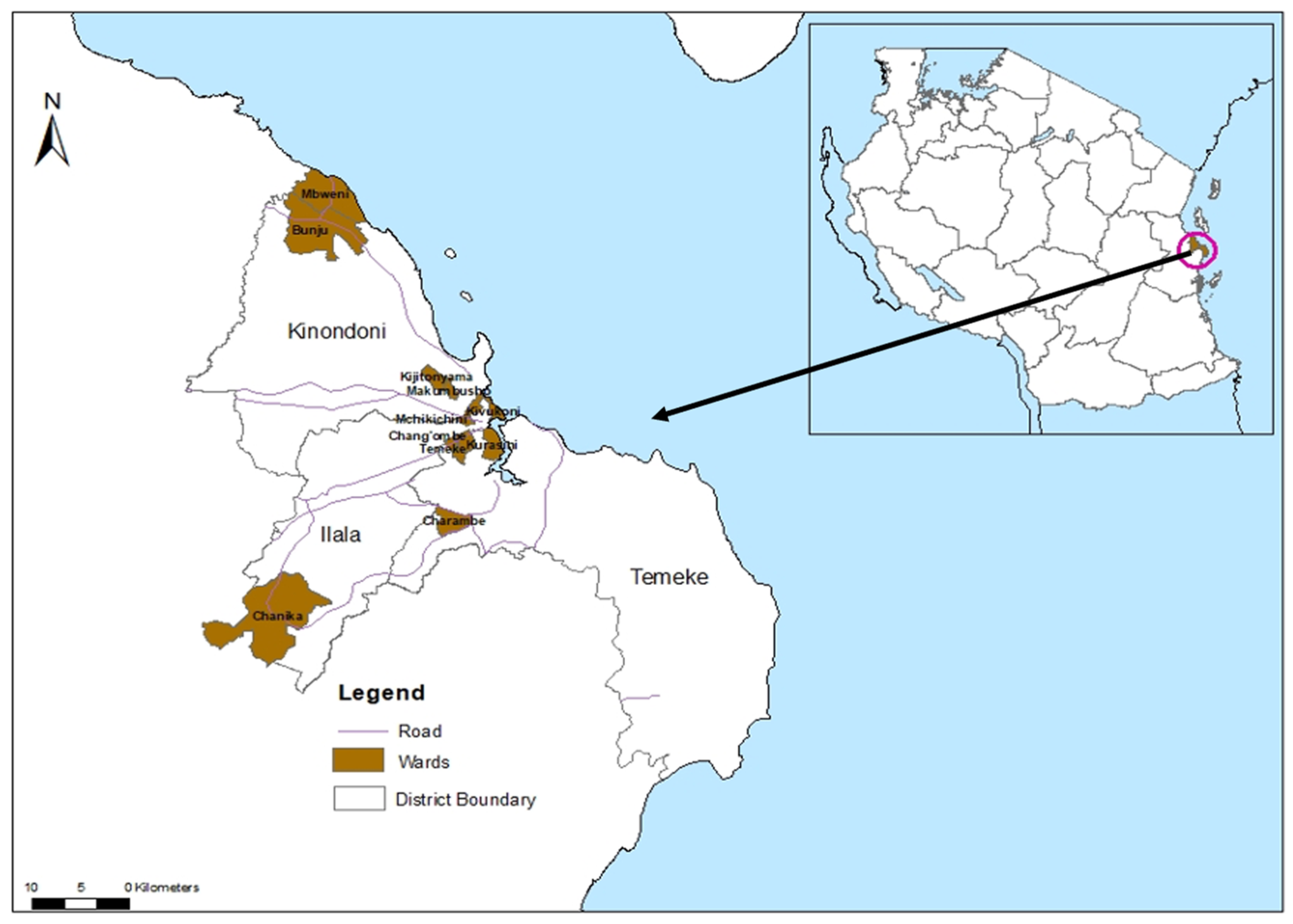
2.1. Study Area

The study was conducted in Dar es Salaam, Tanzania’s largest economic hub (Figure 1). The city is located at 6.48′ S and 39.17′ E on the Indian Ocean coast, with a population of approximately 7 million [63]. Administratively, it consists of five districts: Ilala, Kigamboni, Kinondoni, Temeke, and Ubungo. Based on previous dengue outbreaks [53,64], Ilala (1,649,912 people), Kinondoni (982,328), and Temeke (1,346,674) [63] were selected for this study.
Dar es Salaam has a tropical climate with high temperatures throughout the year and the hottest period occurring between October to February. The city experiences one dry season between June and October and receives an average annual rainfall of 1100 mm. The short rainy season occurs from November to December, and the long rainy season extends from March to May [65].

2.2. Mosquito Collection

Wild adult mosquitoes were collected from June 2022 to May 2024 using Biogents Sentinel (BGS) traps (Biogents AG, Regensburg, Germany) designed for catching host-seeking Aedes mosquitoes, locally made Gravid Aedes traps (BG-GAT) [66] for sampling gravid females, and Prokopack aspirators (John W Hock Company, Gainesville, FL, USA) for collecting resting adults. Four wards were selected from each district. In each ward, 20 houses were identified, and the traps (one BGS and one GAT) were deployed in one house per day for 24 h; then, the trapped mosquitoes were collected, followed by the collection of resting mosquitoes around the premises using a prokopack aspirator. This procedure was repeated monthly per each house for 24 months. The collected mosquitoes were morphologically identified to the species level following the Wilkerson et al., 2021 identification key [67]. Female Ae. aegypti were pooled in groups of 10 individuals and stored in 1.5 mL Eppendorf tubes containing RNA preservation solution locally made at Swiss Tropical and Public Health (TPH) institute. The RNA preservation solution was prepared by mixing 60 mL of 0.5 M EDTA with 37.5 mL of 1 M sodium citrate in 1400 mL MilliQ water. Afterwards, 1050 g ammonium sulfate was added, and the solution was filtered through a 0.2 µm filter. Mosquitoes were analysed for blood meal and the presence of DENV.
Additionally, an experiment was conducted in six houses per month per district for three months to determine the abundance of host-seeking Ae. aegypti mosquitoes indoors and outdoors. A pair of BGS were deployed indoors and outdoors for 24 h, after which female mosquitoes were collected, and recorded based on their capturing location.

2.3. RNA Extraction and Dengue Virus Detection

2.3.1. RNA Extraction

The extraction of RNA from mosquito pools was carried out using RNAzol® RT (Molecular Research Center, Cincinnati, OH, USA) according to the manufacturer’s instructions. Briefly, each pool of 10 individual mosquitoes was suspended in 200 µL of RNAzol in a 1.5 mL microcentrifuge tube and manually ground using a sterile plastic pestle designated for grinding mosquitoes. The mixture was then centrifuged at 12,000× g for 15 min, after which the supernatant was transferred to a new 1.5 mL microcentrifuge tube. An equal volume (200 µL) of 100% isopropanol was added to precipitate the RNA, followed by incubation for 15 min and centrifugation at 12,000× g for 10 min. The supernatant was removed and discarded. The RNA pellet was washed twice with 200 µL of 75% ethanol, centrifuged at 4000× g for 3 min, and the ethanol was carefully removed. Finally, the RNA pellets were eluted with 50 µL of RNAse-free water and stored at −80 °C for molecular analysis using reverse-transcription polymerase chain reaction (RT-qPCR) [68].

2.3.2. Dengue Virus Detection

A one-step multiplex RT-qPCR [69] was performed using the CFX96 Bio-Rad PCR machine (Bio-Rad Laboratories Inc., Hercules, CA, USA). The primers and probes used in the assay were adapted with modifications from Balingit et al. [70] (Table 1). The reaction was performed in 25 µL reaction volumes using the Luna® Universal Probe One-Step RT-qPCR Kit (New England Biolabs, Ipswich, MA, USA) consisting of 5 µL RNA template, 10 µL of Luna Universal Probe One-Step Reaction Mix (2×), 1 µL of Luna WarmStart RT Enzyme Mix (20×), 0.8 µL each of forward and reverse primers (10 µM), and 0.4 µL of probes (10 µM). Each sample was analysed in duplicates. The RT-qPCR cycling conditions were as follows: reverse transcription at 50 °C for 30 min, initialization at 95 °C for 2 min, followed by 45 cycles of denaturation at 95 °C for 15 s, and annealing/extension at 60 °C for 1 min. RNAse-free water was used as a template for the negative control. Samples with an average cycle threshold (Ct) higher than 37 were considered negative for either DENV serotype.

2.4. Blood Meal Source

Blood-fed Aedes mosquito samples collected over two sampling years and preserved in 1.5 mL Eppendorf microcentrifuge tubes containing locally made RNA preservation solution were selected and tested for polyclonal anti-IgG antibodies targeting vertebrates commonly found in the study area, including humans, dogs, chickens, and bovines, using an enzyme-linked immunosorbent assay (ELISA) as described by Beier et al. [71]. Briefly, the abdomen of each mosquito was separated from the rest of the body parts and triturated in 1x phosphate-buffered saline (PBS) using a handheld motorised micro-pestle (DWK Life Sciences, Faust Laborbedrf AG, Schaffhausen, Switzerland). A 96-well ELISA plate (Greiner Bio-One MicrolonTM, Monroe, NC, USA) was coated with 50 µL of Mab solution containing anti-human, anti-dog, anti-chicken, and anti-cow immunoglobulin G (IgG) antibodies produced in goat (Kirkegaard & Perry Laboratories, Gaithersburg, MD, USA) at 4 µg/mL and incubated for 30 min. After incubation, the contents were aspirated, and the excess liquid was removed by tapping the plate on a tissue paper. The wells were then filled with 250 µL of blocking buffer (BB) and incubated for 1 h. Following this, the buffer was drained, and 45 µL of BB was dispensed into each well. Next, 5 µL of each sample was loaded into the wells containing the 45 µL of BB and incubated for 2 h at room temperature. The same procedures were followed for positive and negative controls. After incubation, the plate contents were aspirated, and the wells were washed three times with 250 µL of washing buffer (PBS + Tween 20). A 50 µL aliquot of the appropriate conjugate solution (peroxidase-labelled antibody against human, dog, chicken, and cow IgG, manufactured by SeraCare Life Sciences, Milford, MA, USA) was then added to each well and incubated for 30 min at room temperature. The conjugate was removed by washing the wells four times with 250 µL of washing buffer (PBS + Tween 20). Finally, 100 µL of substrate solution (2,2′-azino-bis (3-ethylbenzothiazoline-6-sulfonic acid, ABTS)) was added to each well, followed by a 30 min incubation at room temperature. After incubation, the plates were read by observing the colour change using ELISA reader.

2.5. Data Analysis

All data obtained were analysed using STATA package version 16 (Stata Corp., College Station, TX, USA).

2.6. DENV Infection Rate in Mosquitoes

The infection rate per 1000 mosquitoes was calculated by determining the proportion of DENV-positive mosquitoes among those tested by qRT-PCR. Usually, the minimum infection rate (MIR) and maximum infection rate (MaxIR) are computed as follows:
M I R = x k × 1000
MaxIR = x × m k × 1000
However, since MIR tends to underestimate and MaxIR overestimates infection rates, both are imprecise. Therefore, the maximum likelihood estimate (MLE) with 95% confidence interval was used to provide a more accurate estimate.
MLE = 1 m l n 1 x n × 1000
where
k = total number of mosquitoes tested;
x = Number of positive pools;
m = Number of mosquitoes per pool (assuming equal pool size);
n = total number of pools tested.

2.7. Bloodmeal Preference

A descriptive analysis was performed to compare the percentage of blood-fed mosquitoes across different hosts. The anthropophagy percentage was defined as the proportion of mosquitoes with human blood meals across all districts.

2.8. Host Seeking Preference

A descriptive analysis was performed to compare the proportion of host-seeking mosquitoes collected indoors and outdoors. A negative binomial regression model was employed to determine if there was a statistically significant difference in host-seeking mosquitoes collected indoors versus those collected outdoors. The fixed terms in the model were location (indoors vs. outdoors), district, and ward, while day and household were included as random effects. The models estimated the mean incidence rate ratios (IRR) and 95% confidence intervals around the means.

3. Results

3.1. DENV Serotypes 1–4 Prevalence

A total of 854 pools, with 10 adult female mosquitoes per pool, were tested for DENV. Of these pools, 371 were from Temeke, 206 from Ilala and 277 from Kinondoni district (Table 2). DENV serotype 2 (DENV-2) was detected in all three districts, with Temeke having the highest maximum likelihood estimate of 0.81 per 1000 mosquitoes (Table 2). The viruses were detected in both years of mosquito sampling (Table 2), indicating endemic circulation.

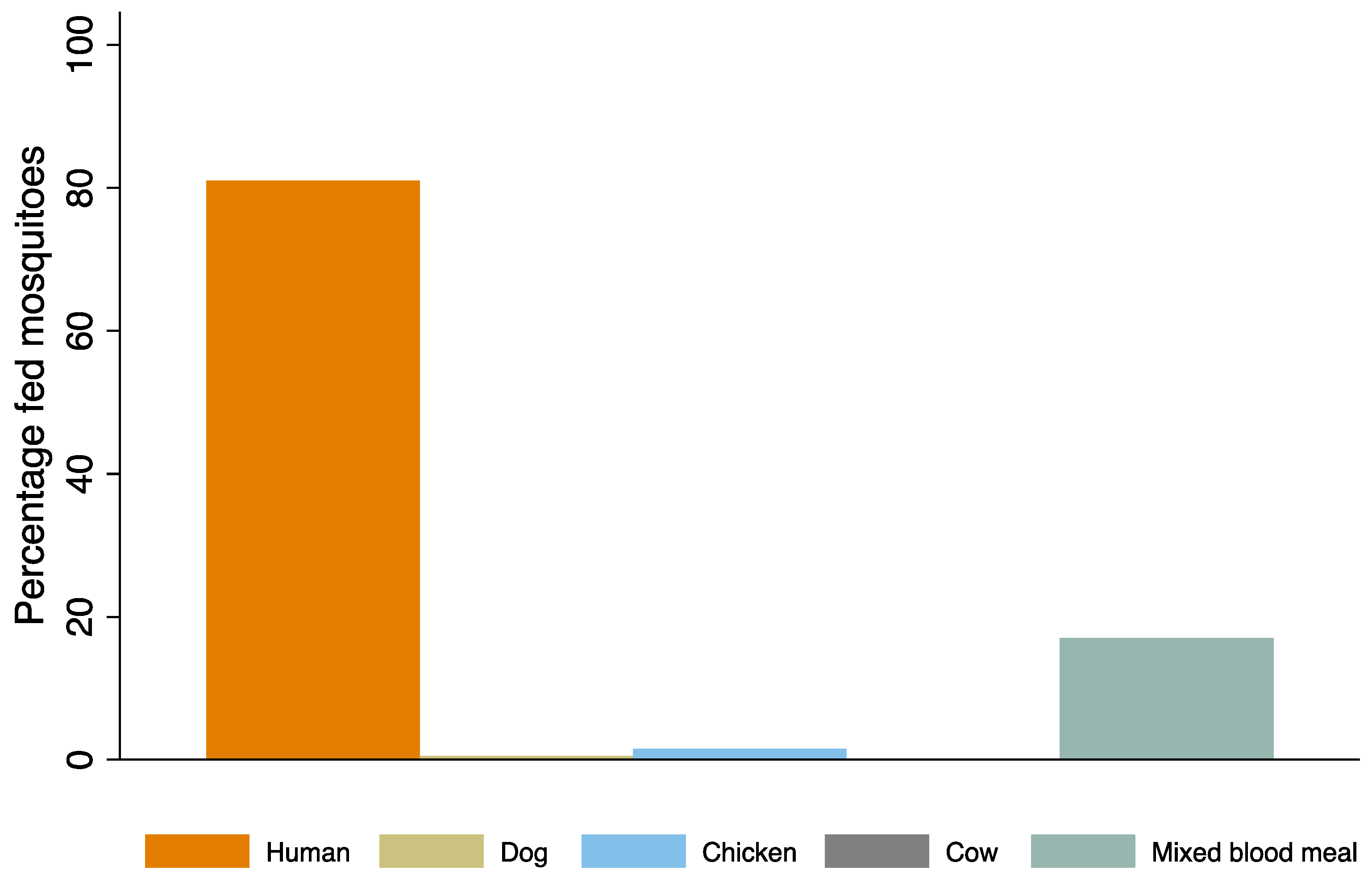
3.2. Host Preference

A total of 298 mosquito samples were tested for the origin of their blood meal from humans, dogs, chickens, and cows. Of these, 68.8% tested positive for either one or more blood meal sources (hosts) in the ELISA test, while 31.2% of samples showed no reaction.
Aedes aegypti showed a strong preference for human blood, with approximately 166 mosquitoes (81.0%) feeding on humans, followed by chicken (1.5%) and dog (0.5%) (Figure 2). About 17% of the mosquitoes had taken mixed blood meals from humans and other hosts, while none had fed on cow (Figure 2). Therefore, 98% of the mosquitoes had at least fed on humans. The majority of blood-fed mosquitoes (69.3%) were collected using the Prockopack aspirator, followed by BGS 27.8% and GAT 2.9% (Table 3).

3.3. Host Seeking Aedes aegypti Mosquitoes

Using BGS, a total of 354 female Ae. aegypti mosquitoes were collected from both indoor and outdoor locations. More than three quarters, 78.5% (n = 278), were caught outdoors, while 21.5% were collected indoors. The number of mosquitoes caught outdoors was 4.33 times higher than the indoor count (95% CI: [2.38–7.89], p-value < 0.001) (Table 4). Among the three districts, Temeke recorded a significantly higher mosquito count than Ilala (Table 4).

4. Discussion

Understanding pathogen circulation in vectors is crucial for disease control. This study reports the presence of DENV-2 circulating in mosquitoes from Dar es Salaam city throughout the two-year survey period, suggesting ongoing endemic virus circulation rather than a new introduction. The virus serotype reported in this study is the same as the one detected in the 2014 outbreak [53], highlighting the possibility that the virus has been persistent in the ecosystem since then. However, whole genome sequencing would be required to elucidate whether this is the case or if a new introduction of a different DENV-2 genotype occurred.
All four DENV serotypes (DENV 1–4) have circulated in Tanzania [50], with different serotypes predominating in each outbreak. This shifting pattern may explain the increasing number of dengue cases and deaths during subsequent outbreaks [58], a trend also observed in West Africa [72] and other endemic regions [4]. In 2019, WHO reported a dramatic increase in dengue cases across several countries in Africa, particularly in the sub-Saharan region [73]. This rise reflects a broader global increase in dengue in all WHO regions [6]. Despite the growing evidence, African countries, including Tanzania, lack comprehensive data on the exact magnitude of dengue virus distribution due to limited epidemiological, entomological, and virological surveillance since African vector control efforts focus mainly on malaria and dengue remains a neglected tropical disease.
Genotyping studies have shown a relatedness of dengue virus genotypes detected in East Africa, particularly Tanzania [53,59] and Kenya [74,75], to virus genotypes from Asian countries such as India and Singapore [55] as well as China [59]. This indicates that the viruses are being imported from the East via international travel and trade [59]. Africa’s rapid population growth and urbanisation will likely further accelerate the virus spread. By 2050, nearly 60% of the continent’s population is expected to live in cities [76]. Increased human mobility and urbanisation will be inevitable; therefore, deliberate dengue monitoring efforts are needed. In this context, routine screening at national and international entry points could be implemented to reduce introductions of new virus genotypes, although this may be cost prohibitive.
This study identified Temeke, Ilala, and Kinondoni districts as areas with dengue-infected mosquitoes, suggesting that these are priority areas for dengue xenomonitoring. Detection of arbovirus circulation in larval Aedes vectors has been conducted in 40 published studies [77] across Latin America and Southeast Asia, although this is the first study from sub-Saharan Africa to explore the potential utility of xenomonitoring for epidemic prediction. The current study evaluated DENV in adult mosquitoes using BGS, although low-cost monitoring using gravid traps is being explored [66]. The infected mosquitoes indicate a potential risk of dengue outbreaks in these areas, necessitating proactive Aedes mosquito surveillance. Additionally, it emphasises the need for government authorities to implement dengue control measures, including larval source reduction, targeted insecticide spraying, and public education on both mosquito control and the importance of seeking health care when experiencing dengue-like symptoms. Furthermore, it underscores the importance of implementing the International Health Regulations 2005 (IHR) to reduce the risk of virus transmission to other areas [78]. The IHR is a legal framework developed by WHO to guide the management of public health events and emergencies with the potential to cross borders. It calls on governments to undertake measures such as mapping and conducting susceptivity tests for Aedes vectors in urban areas, as well as implementing health surveillance at ports, airports, and borders for incoming travellers.
In Tanzania, dengue prevalence often exceeds 10% in human samples [48,56,59,60]. This study reports an average 0.04% dengue mosquito infection rate (0.41/1000), which is lower than that reported by Mboera et al. [53] during the 2014 outbreak. However, the earlier study was based on virus detection in larvae, whereas this study tested adult mosquitoes. Larval sampling may have overestimated infection rates, given they do not represent the host-seeking population, and the samples could have been biased if siblings from the same transovarially infected egg batch were sampled. Additionally, rates are likely higher due to the fact that an outbreak was ongoing. The findings from the present study align with those of Chilongola et al. [79] and Joseph et al. [80] in East Africa, Mojica et al. [81] in Nicaragua and Ecuador in Latin America, and Maneerattanasak et al. [82] in Southeast and South Asia who reported 0.3%, 2.7%, 0.7%, and 0.05–2.3% dengue infection rates, respectively, among adult mosquitoes. Similarly, this is consistent with reports on other female arthropod-transmitted diseases, such as malaria [83,84], where Plasmodium infection rates in Anopheles mosquitoes are typically very low, even in high endemic areas.
This study has demonstrated that, Ae. aegypti from Dar es Salaam are highly anthropophagic, more than 80% feeding only on humans and 98% overall with human blood. This behaviour significantly increases dengue transmission risk, as human–mosquito contact is a key driver of virus spread [85]. The findings of this study are consistent with research from West Africa (Senegal and Burkina Faso) [41,86,87] as well as India [39] South East Asia (Thailand) [36], Australia [88], Latin America (Brazil, Ecuador, and Peru) [89], and North America (USA) [42], which all indicated high anthropophagy among Ae. aegypti.
Aedes aegypti aegypti (Aaa) and Aedes aegypti formosus (Aaf) are the Ae. aegypti subspecies commonly found in Africa [90]. Aaa is considered an urban mosquito primarily responsible for urban dengue and yellow fever transmission [91]. Aaf inhabits peri-urban environments and serves as an agent for sylvatic dengue as well as yellow fever transmission [91,92] and is less competent for dengue [93] and less anthropophilic [94]. In this study, we used morphological identification only and were unable to distinguish between the two subspecies. Given the lack of comprehensive research on their coexistence in the country, our findings emphasise the need for genomic studies to accurately characterise them.
As reported in other studies [40,41,86], human–animal mixed blood meals were frequently observed in this study, highlighting the feeding flexibility of Ae. aegypti. This mosquito species is known for transmitting dengue virus and other viruses, including yellow fever virus (YFV), Chikungunya virus (CHIKV), and Zika virus (ZIKV), all of which pose a substantial public health burden. While the ability of Ae. aegypti mosquitoes to feed on multiple animal species has implications for virus transmission, such as cross-species transmission and vector competence; the role of animals as reservoirs for DENV, CHIKV, YFV, and ZIKV in urban settings remains unlikely. Studies have described systems demonstrating the potential co-infection of DENV serotypes in an individual [95,96]. With all four DENV serotypes circulating in the country [50], it is likely that the mosquitoes’ ability to feed on multiple human hosts could essentially lead to individuals being infected with more than one virus serotype; this will potentially increase the chance of individual developing severe dengue if he/she was previously infected with another virus serotype.
Moreover, the locations where mosquitoes seek blood meals from hosts and rest after feeding have important implications for vector control strategies. This study demonstrated that more than three quarters of Ae. aegypti preferentially seek blood meals outdoors, presenting a challenge for control. Most mosquito control interventions used in Tanzania are designed for malaria control and are applied indoors, targeting indoor host-seeking and resting mosquitoes. This finding is similar to studies of Ae. aegypti in Burkina Faso [87,97,98] and Ghana [99] that also indicate outdoor feeding. Consequently, larval source reduction [100] might be the most likely intervention to be successful at the community level. Further work is ongoing to measure the resting behaviour and susceptibility of the Dar es Salaam population of Ae. aegypti to insecticides used for mosquito control. In addition, the use of Wolbachia, an endosymbiotic alpha-proteobacterium naturally occurring in arthropods, has shown excellent efficacy in preventing dengue when deployed at a city scale [101]. Wolbachia blocks dengue virus replication, thereby reducing the ability of mosquitoes to transmit the disease. Its introduction also induces cytoplasmic incompatibility (CPI), whereby uninfected females that mate with Wolbachia-infected males cannot produce viable offspring. Because Wolbachia is maternally inherited, the trait spreads through the mosquito population over time, rendering it refractory to DENV. This intervention is self-sustaining and may be particularly valuable in rapidly growing urban areas like Dar es Salaam.

Study Limitations

To fully understand the indoor and outdoor ecology of Ae. aegypti, traps targeting host-seeking, resting, and oviposition behaviours need to be deployed concurrently both indoors and outdoors. However, we were unable to collect indoor mosquitoes using GATs due to the unpleasant smell of the infusion and with prokopack aspirators because of restricted access to homes, usually because residents were absent. Consequently, indoor collections were limited to BGS, a trap type designated for collecting host-seeking mosquitoes. Therefore, we recommend that for future studies, the indoor and outdoor mosquito collection should also include resting collections because the use of traps with a lure may bias the collections towards human-fed mosquitoes [89]. Additionally, blood meal analysis for host preference was performed on only four hosts (humans, dogs, chickens, and cows). However, we found that 93 samples did not react suggesting that the mosquitoes may have contained blood meals from hosts not included in the analysis. In Kenya, Ae. aegypti has been found to feed on goats, rats, and cats [101], which were also present in the present study site but not tested for. Therefore, future studies should include a broader range of potential hosts.

5. Conclusions

This study confirms the circulation of DENV-2 in the mosquito population in Dar es Salaam, highlighting the risk of a potential dengue outbreak in Tanzania. Dar es Salaam is one of Africa’s major metropolitan cities, with a population of nearly seven million. It serves as the economic hub of Tanzania, so it experiences a significant influx of local and international travellers. The presence of DENV-2 in mosquitoes year-round, as well as the strong human feeding preference of Ae. aegypti indicates the potential risk of DENV transmission to humans. These findings emphasise the need for enhanced surveillance and targeted proactive vector control measures, including the removal of breeding sites to mitigate dengue outbreaks.

Author Contributions

Conceptualisation: F.S.C.T. and S.J.M.; data curation: F.S.C.T., O.D., H.I.M. and H.M.; formal analysis: F.S.C.T.; investigation: F.S.C.T., O.D., H.I.M., H.M., S.H., L.D.B., J.M., J.J.M., N.S.L. and T.G.M.; methodology: F.S.C.T., P.M. and S.J.M.; supervision; S.J.M.; writing—original draft: F.S.C.T.; visualisation: S.J.M., P.M., S.H. and L.M.H.; writing—review and editing: S.J.M., P.M., S.H. and L.M.H.; funding acquisition: S.J.M. All authors have read and agreed to the published version of the manuscript.

Funding

This study was funded by Rudolf Geigy–Stiftung (RGS): 200902 and ArmaSuisse through Swiss TPH. The findings and conclusions contained within are those of the authors, herein stated is Ifakara Health Institute and Swiss TPH, and do not necessarily reflect positions or policies of the Rudolf Geigy Foundation and Armasuisse.

Institutional Review Board Statement

This study received ethical approval from Ifakara Health Institute Review Board (IHI-IRB) IHI/IRB/No: 7-2021 and National Institute for Medical Research Review Board (NIMR-RB) No. NIMR/HQ/R.8a/Vol. IX/3641.

Informed Consent Statement

Not applicable.

Data Availability Statement

The datasets generated during the study are available from Ifakara Health Institute and the corresponding author upon reasonable request.

Acknowledgments

The authors would like to thank Ifakara Health Institute staff for their support. Also, we would like to extend our heartfelt thanks to the RGS for funding this study and the excellent support from Lukas Meier at RGS.

Conflicts of Interest

The authors declare that they have no competing interests.

Abbreviations

BGSBiogent sentinel trap
GATGravid Aedes trap
DENVDengue virus
ITNInsecticide-treated net
IRSIndoor residual spray
DFDengue fever
YFYellow fever
CHIKChikungunya
ZIKZika
IHRInternational health regulations
WHOWorld health organisation
IRRIncidence rate ratio
OROdds ratio
ELISAEnzyme-linked immunosorbent assay.
qRT-PCRQuantitative reverse transcriptase polymerase chain reaction
MIRMosquito infection rate

References

  1. Guzman, A.; Istúriz, R.E. Update on the global spread of dengue. Int. J. Antimicrob. Agents 2010, 36, S40–S42. [Google Scholar] [CrossRef] [PubMed]
  2. Brady, O.J.; Gething, P.W.; Bhatt, S.; Messina, J.P.; Brownstein, J.S.; Hoen, A.G.; Moyes, C.L.; Farlow, A.W.; Scott, T.W.; Hay, S.I. Refining the global spatial limits of dengue virus transmission by evidence-based consensus. PLoS Negl. Trop. Dis. 2012, 6, e1760. [Google Scholar] [CrossRef]
  3. European Centre for Disease Prevention and Control. Dengue Cases January–December 2023. 2023. Available online: https://www.ecdc.europa.eu/en/publications-data/dengue-cases-january-december-2023 (accessed on 29 January 2024).
  4. World Health Organization. Dengue-Global Situation. 2024. Available online: https://www.who.int/emergencies/disease-outbreak-news/item/2024-DON518 (accessed on 30 May 2024).
  5. European Centre for Disease Prevention and Control. Dengue Worldwide Overview. 2024. Available online: https://www.ecdc.europa.eu/en/publications-data/twelve-month-dengue-virus-disease-case-notification-rate-100-000-population?utm_source=chatgpt.com (accessed on 3 February 2025).
  6. World Health Organization. Dengue and Severe Dengue. WHO. 2022. Available online: http://www.who.int/mediacentre/factsheets/fs117/en/ (accessed on 23 April 2023).
  7. Bhatt, S.; Gething, P.; Brady, O.; Messina, J.; Farlow, A.; Moyes, C. A distribuição global e o ônus da dengue. Nature 2013, 496, 504. [Google Scholar] [CrossRef] [PubMed]
  8. Waggoner, J.J.; Gresh, L.; Vargas, M.J.; Ballesteros, G.; Tellez, Y.; Soda, K.J.; Sahoo, M.K.; Nuñez, A.; Balmaseda, A.; Harris, E. Viremia and clinical presentation in Nicaraguan patients infected with Zika virus, chikungunya virus, and dengue virus. Clin. Infect. Dis. 2016, 63, 1584–1590. [Google Scholar] [CrossRef] [PubMed]
  9. World Health Organization. Dengue—Global Situation. 2023. Available online: https://www.who.int/emergencies/disease-outbreak-news/item/2023-DON498 (accessed on 23 April 2023).
  10. Bhatt, S.; Gething, P.W.; Brady, O.J.; Messina, J.P.; Farlow, A.W.; Moyes, C.L.; Drake, J.M.; Brownstein, J.S.; Hoen, A.G.; Sankoh, O. The global distribution and burden of dengue. Nature 2013, 496, 504–507. [Google Scholar] [CrossRef]
  11. Boillat-Blanco, N.; Klaassen, B.; Mbarack, Z.; Samaka, J.; Mlaganile, T.; Masimba, J.; Franco Narvaez, L.; Mamin, A.; Genton, B.; Kaiser, L. Dengue fever in Dar es Salaam, Tanzania: Clinical features and outcome in populations of black and non-black racial category. BMC Infect. Dis. 2018, 18, 644. [Google Scholar] [CrossRef]
  12. Guzman, M.G.; Harris, E. Dengue. Lancet 2015, 385, 453–465. [Google Scholar] [CrossRef]
  13. Messina, J.P.; Brady, O.J.; Golding, N.; Kraemer, M.U.; Wint, G.; Ray, S.E.; Pigott, D.M.; Shearer, F.M.; Johnson, K.; Earl, L. The current and future global distribution and population at risk of dengue. Nat. Microbiol. 2019, 4, 1508–1515. [Google Scholar] [CrossRef]
  14. Bennett, S.N. 17 Taxonomy and Evolutionary Relationships of Flaviviruses. In Dengue and Dengue Hemorrhagic Fever; CABI: Wallingford, UK, 2014; pp. 322–333. [Google Scholar]
  15. Guzman, M.G.; Halstead, S.B.; Artsob, H.; Buchy, P.; Farrar, J.; Gubler, D.J.; Hunsperger, E.; Kroeger, A.; Margolis, H.S.; Martínez, E. Dengue: A continuing global threat. Nat. Rev. Microbiol. 2010, 8, S7–S16. [Google Scholar] [CrossRef]
  16. Harapan, H.; Michie, A.; Sasmono, R.T.; Imrie, A. Dengue: A minireview. Viruses 2020, 12, 829. [Google Scholar] [CrossRef]
  17. Najri, N.; Mazlan, Z.; Jaimin, J.; Mohammad, R.; Yusuf, N.M.; Kumar, V.S.; Hoque, M. Genotypes of the dengue virus in patients with dengue infection from Sabah, Malaysia. Proc. J. Phys. Conf. Ser. 2019, 1358, 012019. [Google Scholar] [CrossRef]
  18. Holmes, E.C.; Twiddy, S.S. The origin, emergence and evolutionary genetics of dengue virus. Infect. Genet. Evol. 2003, 3, 19–28. [Google Scholar] [CrossRef] [PubMed]
  19. Sasmono, R.T.; Wahid, I.; Trimarsanto, H.; Yohan, B.; Wahyuni, S.; Hertanto, M.; Yusuf, I.; Mubin, H.; Ganda, I.J.; Latief, R. Genomic analysis and growth characteristic of dengue viruses from Makassar, Indonesia. Infect. Genet. Evol. 2015, 32, 165–177. [Google Scholar] [CrossRef] [PubMed]
  20. Rodenhuis-Zybert, I.A.; Wilschut, J.; Smit, J.M. Dengue virus life cycle: Viral and host factors modulating infectivity. Cell. Mol. Life Sci. 2010, 67, 2773–2786. [Google Scholar] [CrossRef]
  21. Guzman, M.G.; Alvarez, M.; Halstead, S.B. Secondary infection as a risk factor for dengue hemorrhagic fever/dengue shock syndrome: An historical perspective and role of antibody-dependent enhancement of infection. Arch. Virol. 2013, 158, 1445–1459. [Google Scholar] [CrossRef]
  22. Guzman, M.G.; Gubler, D.J.; Izquierdo, A.; Martinez, E.; Halstead, S.B. Dengue infection. Nat. Rev. Dis. Primers 2016, 2, 16055. [Google Scholar] [CrossRef]
  23. Vaughn, D.W.; Green, S.; Kalayanarooj, S.; Innis, B.L.; Nimmannitya, S.; Suntayakorn, S.; Endy, T.P.; Raengsakulrach, B.; Rothman, A.L.; Ennis, F.A. Dengue viremia titer, antibody response pattern, and virus serotype correlate with disease severity. J. Infect. Dis. 2000, 181, 2–9. [Google Scholar] [CrossRef]
  24. Moi, M.L.; Takasaki, T.; Omatsu, T.; Nakamura, S.; Katakai, Y.; Ami, Y.; Suzaki, Y.; Saijo, M.; Akari, H.; Kurane, I. Demonstration of marmosets (Callithrix jacchus) as a non-human primate model for secondary dengue virus infection: High levels of viraemia and serotype cross-reactive antibody responses consistent with secondary infection of humans. J. Gen. Virol. 2014, 95, 591–600. [Google Scholar] [CrossRef]
  25. Amarasinghe, A.; Kuritsky, J.N.; Letson, G.W.; Margolis, H.S. Dengue virus infection in Africa. Emerg. Infect. Dis. 2011, 17, 1349. [Google Scholar] [CrossRef]
  26. Sang, R.C. Dengue in Africa. In Report of the Scientific Working Group Meeting on Dengue; WHO Special Programme for Research and Training in Tropical Diseases: Geneva, Switzerland, 2007; pp. 50–52. [Google Scholar]
  27. Chipwaza, B.; Mugasa, J.P.; Selemani, M.; Amuri, M.; Mosha, F.; Ngatunga, S.D.; Gwakisa, P.S. Dengue and Chikungunya fever among viral diseases in outpatient febrile children in Kilosa district hospital, Tanzania. PLoS Negl. Trop. Dis. 2014, 8, e3335. [Google Scholar] [CrossRef]
  28. Parreira, R.; Centeno-Lima, S.; Lopes, A.; Portugal-Calisto, D.; Constantino, A.; Nina, J. Dengue virus serotype 4 and chikungunya virus coinfection in a traveller returning from Luanda, Angola, January 2014. Eurosurveillance 2014, 19, 20730. [Google Scholar] [CrossRef] [PubMed]
  29. Gautret, P.; Simon, F.; Askling, H.H.; Bouchaud, O.; Leparc-Goffart, I.; Ninove, L.; Parola, P. Dengue type 3 virus infections in European travellers returning from the Comoros and Zanzibar, February–April 2010. Eurosurveillance 2010, 15, 19541. [Google Scholar] [CrossRef] [PubMed]
  30. Gubler, D.J.; Nalim, S.; Tan, R.; Saipan, H. Variation in susceptibility to oral infection with dengue viruses among geographic strains of Aedes aegypti. Am. J. Trop. Med. Hyg. 1979, 28, 1045–1052. [Google Scholar] [CrossRef] [PubMed]
  31. Rosen, L.; Roseboom, L.E.; Gubler, D.J.; Lien, J.C.; Chaniotis, B.N. Comparative susceptibility of mosquito species and strains to oral and parenteral infection with dengue and Japanese encephalitis viruses. Am. J. Trop. Med. Hyg. 1985, 34, 603–615. [Google Scholar] [CrossRef]
  32. Armstrong, P.M.; Rico-Hesse, R. Efficiency of dengue serotype 2 virus strains to infect and disseminate in Aedes aegypti. Am. J. Trop. Med. Hyg. 2003, 68, 539. [Google Scholar] [CrossRef]
  33. Leta, S.; Beyene, T.J.; De Clercq, E.M.; Amenu, K.; Kraemer, M.U.; Revie, C.W. Global risk mapping for major diseases transmitted by Aedes aegypti and Aedes albopictus. Int. J. Infect. Dis. 2018, 67, 25–35. [Google Scholar] [CrossRef]
  34. Brady, O.J.; Hay, S.I. The Global Expansion of Dengue: How Aedes aegypti Mosquitoes Enabled the First Pandemic Arbovirus. Annu. Rev. Entomol. 2020, 65, 191–208. [Google Scholar] [CrossRef]
  35. Sridhar, S.; Luedtke, A.; Langevin, E.; Zhu, M.; Bonaparte, M.; Machabert, T.; Savarino, S.; Zambrano, B.; Moureau, A.; Khromava, A. Effect of dengue serostatus on dengue vaccine safety and efficacy. N. Engl. J. Med. 2018, 379, 327–340. [Google Scholar] [CrossRef]
  36. Ponlawat, A.; Harrington, L.C. Blood feeding patterns of Aedes aegypti and Aedes albopictus in Thailand. J. Med. Entomol. 2005, 42, 844–849. [Google Scholar] [CrossRef]
  37. Mann, J.G.; Washington, M.; Guynup, T.; Tarrand, C.; Dewey, E.M.; Fredregill, C.; Duguma, D.; Pitts, R.J. Feeding habits of vector mosquitoes in Harris County, TX, 2018. J. Med. Entomol. 2020, 57, 1920–1929. [Google Scholar] [CrossRef]
  38. Diallo, D.; Chen, R.; Diagne, C.T.; Ba, Y.; Dia, I.; Sall, A.A.; Weaver, S.C.; Diallo, M. Bloodfeeding patterns of sylvatic arbovirus vectors in southeastern Senegal. Trans. R. Soc. Trop. Med. Hyg. 2013, 107, 200–203. [Google Scholar] [CrossRef] [PubMed]
  39. Sivan, A.; Shriram, A.; Sunish, I.; Vidhya, P. Host-feeding pattern of Aedes aegypti and Aedes albopictus (Diptera: Culicidae) in heterogeneous landscapes of South Andaman, Andaman and Nicobar Islands, India. Parasitol. Res. 2015, 114, 3539–3546. [Google Scholar] [CrossRef] [PubMed]
  40. Olson, M.F.; Ndeffo-Mbah, M.L.; Juarez, J.G.; Garcia-Luna, S.; Martin, E.; Borucki, M.K.; Frank, M.; Estrada-Franco, J.G.; Rodríguez-Pérez, M.A.; Fernández-Santos, N.A. High rate of non-human feeding by Aedes aegypti reduces Zika virus transmission in South Texas. Viruses 2020, 12, 453. [Google Scholar] [CrossRef] [PubMed]
  41. Sene, N.M.; Diouf, B.; Gaye, A.; Gueye, A.; Seck, F.; Diagne, C.T.; Dia, I.; Diallo, D.; Diallo, M. Blood feeding patterns of Aedes aegypti populations in Senegal. Am. J. Trop. Med. Hyg. 2022, 106, 1402. [Google Scholar] [CrossRef]
  42. Pruszynski, C.A.; Stenn, T.; Acevedo, C.; Leal, A.L.; Burkett-Cadena, N.D. Human blood feeding by Aedes aegypti (Diptera: Culicidae) in the Florida Keys and a review of the literature. J. Med. Entomol. 2020, 57, 1640–1647. [Google Scholar] [CrossRef]
  43. Zahid, M.H.; Van Wyk, H.; Morrison, A.C.; Coloma, J.; Lee, G.O.; Cevallos, V.; Ponce, P.; Eisenberg, J.N. The biting rate of Aedes aegypti and its variability: A systematic review (1970–2022). PLoS Negl. Trop. Dis. 2023, 17, e0010831. [Google Scholar] [CrossRef]
  44. Martin, E.; Medeiros, M.C.; Carbajal, E.; Valdez, E.; Juarez, J.G.; Garcia-Luna, S.; Salazar, A.; Qualls, W.A.; Hinojosa, S.; Borucki, M.K. Surveillance of Aedes aegypti indoors and outdoors using Autocidal Gravid Ovitraps in South Texas during local transmission of Zika virus, 2016 to 2018. Acta Trop. 2019, 192, 129–137. [Google Scholar] [CrossRef]
  45. Dalpadado, R.; Amarasinghe, D.; Gunathilaka, N.; Ariyarathna, N. Bionomic aspects of dengue vectors Aedes aegypti and Aedes albopictus at domestic settings in urban, suburban and rural areas in Gampaha District, Western Province of Sri Lanka. Parasites Vectors 2022, 15, 148. [Google Scholar] [CrossRef]
  46. Chadee, D.D.; Martinez, R. Landing periodicity of Aedes aegypti with implications for dengue transmission in Trinidad, West Indies. J. Vector Ecol. J. Soc. Vector Ecol. 2000, 25, 158–163. [Google Scholar]
  47. Vairo, F.; Nicastri, E.; Meschi, S.; Schepisi, M.S.; Paglia, M.G.; Bevilacqua, N.; Mangi, S.; Sciarrone, M.R.; Chiappini, R.; Mohamed, J. Seroprevalence of dengue infection: A cross-sectional survey in mainland Tanzania and on Pemba Island, Zanzibar. Int. J. Infect. Dis. 2012, 16, e44–e46. [Google Scholar] [CrossRef]
  48. Vairo, F.; Nicastri, E.; Yussuf, S.M.; Cannas, A.; Meschi, S.; Mahmoud, M.A.; Mohamed, A.H.; Maiko, P.M.; De Nardo, P.; Bevilacqua, N. IgG against dengue virus in healthy blood donors, Zanzibar, Tanzania. Emerg. Infect. Dis. 2014, 20, 465. [Google Scholar] [CrossRef] [PubMed]
  49. Moi, M.L.; Takasaki, T.; Kotaki, A.; Tajima, S.; Lim, C.-K.; Sakamoto, M.; Iwagoe, H.; Kobayashi, K.; Kurane, I. Importation of dengue virus type 3 to Japan from Tanzania and Côte d’Ivoire. Emerg. Infect. Dis. 2010, 16, 1770. [Google Scholar] [CrossRef] [PubMed]
  50. Chipwaza, B.; Sumaye, R.D.; Weisser, M.; Gingo, W.; Yeo, N.K.-W.; Amrun, S.N.; Okumu, F.O.; Ng, L.F. Occurrence of 4 dengue virus serotypes and chikungunya virus in Kilombero Valley, Tanzania, during the dengue outbreak in 2018. In Open Forum Infectious Diseases; Oxford University Press: Cary, NC, USA, 2021; p. ofaa626. [Google Scholar]
  51. Hertz, J.T.; Munishi, O.M.; Ooi, E.E.; Howe, S.; Lim, W.Y.; Chow, A.; Morrissey, A.B.; Bartlett, J.A.; Onyango, J.J.; Maro, V.P. Chikungunya and dengue fever among hospitalized febrile patients in northern Tanzania. Am. J. Trop. Med. Hyg. 2012, 86, 171. [Google Scholar] [CrossRef] [PubMed]
  52. World Health Organization. Engue Outbreak in the United Republic of Tanzania; World Health Organization: Geneva, Switzerland, 2014. [Google Scholar]
  53. Mboera, L.E.; Mweya, C.N.; Rumisha, S.F.; Tungu, P.K.; Stanley, G.; Makange, M.R.; Misinzo, G.; De Nardo, P.; Vairo, F.; Oriyo, N.M. The risk of dengue virus transmission in Dar es Salaam, Tanzania during an epidemic period of 2014. PLoS Negl. Trop. Dis. 2016, 10, e0004313. [Google Scholar] [CrossRef]
  54. Kelly, M.E.; Msafiri, F.; Affara, M.; Gehre, F.; Moremi, N.; Mghamba, J.; Misinzo, G.; Thye, T.; Gatei, W.; Whistler, T. Molecular Characterization and Phylogenetic Analysis of Dengue Fever Viruses in Three Outbreaks in Tanzania Between 2017 and 2019. PLoS Negl. Trop. Dis. 2023, 17, e0011289. [Google Scholar] [CrossRef]
  55. Okada, K.; Morita, R.; Egawa, K.; Hirai, Y.; Kaida, A.; Shirano, M.; Kubo, H.; Goto, T.; Yamamoto, S.P. Dengue virus type 1 infection in traveler returning from Tanzania to Japan, 2019. Emerg. Infect. Dis. 2019, 25, 1782. [Google Scholar] [CrossRef]
  56. Mwanyika, G.; Mboera, L.E.; Rugarabamu, S.; Lutwama, J.; Sindato, C.; Paweska, J.T.; Misinzo, G. Co-circulation of Dengue Virus Serotypes 1 and 3 during the 2019 epidemic in Dar es Salaam, Tanzania. Pathog. Glob. Health 2021, 115, 467–475. [Google Scholar] [CrossRef]
  57. Mwanyika, G.O.; Sindato, C.; Rugarabamu, S.; Rumisha, S.F.; Karimuribo, E.D.; Misinzo, G.; Rweyemamu, M.M.; Hamid, M.M.A.; Haider, N.; Vairo, F. Seroprevalence and associated risk factors of chikungunya, dengue, and Zika in eight districts in Tanzania. Int. J. Infect. Dis. 2021, 111, 271–280. [Google Scholar] [CrossRef]
  58. SACIDS. Dengue Outbreaks in Tanzania: Recent Trends and Importance of Research Data in Disease Surveillance; Southern African Centre for Infectious Disease Surveillance: Morogoro, Tanzania, 2019. [Google Scholar]
  59. Vairo, F.; Mboera, L.E.; De Nardo, P.; Oriyo, N.M.; Meschi, S.; Rumisha, S.F.; Colavita, F.; Mhina, A.; Carletti, F.; Mwakapeje, E. Clinical, virologic, and epidemiologic characteristics of dengue outbreak, Dar es Salaam, Tanzania, 2014. Emerg. Infect. Dis. 2016, 22, 895. [Google Scholar] [CrossRef]
  60. Shauri, H.S.; Ngadaya, E.; Senkoro, M.; Buza, J.J.; Mfinanga, S. Seroprevalence of Dengue and Chikungunya antibodies among blood donors in Dar es Salaam and Zanzibar, Tanzania: A cross-sectional study. BMC Infect. Dis. 2021, 21, 911. [Google Scholar] [CrossRef]
  61. Kajeguka, D.C.; Kaaya, R.D.; Mwakalinga, S.; Ndossi, R.; Ndaro, A.; Chilongola, J.O.; Mosha, F.W.; Schiøler, K.L.; Kavishe, R.A.; Alifrangis, M. Prevalence of dengue and chikungunya virus infections in north-eastern Tanzania: A cross sectional study among participants presenting with malaria-like symptoms. BMC Infect. Dis. 2016, 16, 183. [Google Scholar] [CrossRef] [PubMed]
  62. Mustafa, U.-k.; Kreppel, K.S.; Sauli, E. Dengue virus transmission during non-outbreak period in Dar Es Salaam, Tanzania: A cross-sectional survey. BMC Infect. Dis. 2024, 24, 1219. [Google Scholar] [CrossRef] [PubMed]
  63. National Bureau of Statistics Ministry of Finance and Office of Chief Government Statistician President’s Office, Economy and Development Planning. Population and Housing Census. Administrative units Population Distribution and Age and Sex Distribution Report Tanzania–Volume1a. 2022. Available online: https://sensa.nbs.go.tz/publication/volume1a.pdf (accessed on 1 December 2022).
  64. Msellemu, D.; Gavana, T.; Ngonyani, H.; Mlacha, Y.P.; Chaki, P.; Moore, S.J. Knowledge, attitudes and bite prevention practices and estimation of productivity of vector breeding sites using a Habitat Suitability Score (HSS) among households with confirmed dengue in the 2014 outbreak in Dar es Salaam, Tanzania. PLoS Negl. Trop. Dis. 2020, 14, e0007278. [Google Scholar] [CrossRef] [PubMed]
  65. Tanzania Metrology Agency (TMA). Available online: http://www.meteo.go.tz (accessed on 1 December 2022).
  66. Machange, J.J.; Maasayi, M.S.; Mundi, J.; Moore, J.; Muganga, J.B.; Odufuwa, O.G.; Moore, S.J.; Tenywa, F.C. Comparison of the Trapping Efficacy of Locally Modified Gravid Aedes Trap and Autocidal Gravid Ovitrap for the Monitoring and Surveillance of Aedes aegypti Mosquitoes in Tanzania. Insects 2024, 15, 401. [Google Scholar] [CrossRef]
  67. Wilkerson, R.C.; Linton, Y.-M.; Strickman, D. Mosquitoes of the World; Johns Hopkins University Press: Baltimore, MD, USA, 2021. [Google Scholar]
  68. Bustin, S.A.; Benes, V.; Garson, J.A.; Hellemans, J.; Huggett, J.; Kubista, M.; Mueller, R.; Nolan, T.; Pfaffl, M.W.; Shipley, G.L. The MIQE Guidelines: Minimum Information for Publication of Quantitative Real-Time PCR Experiments; Oxford University Press: Oxford, UK, 2009. [Google Scholar]
  69. Pérez-Castro, R.; Castellanos, J.E.; Olano, V.A.; Matiz, M.I.; Jaramillo, J.F.; Vargas, S.L.; Sarmiento, D.M.; Stenström, T.A.; Overgaard, H.J. Detection of all four dengue serotypes in Aedes aegypti female mosquitoes collected in a rural area in Colombia. Mem. Do Inst. Oswaldo Cruz 2016, 111, 233–240. [Google Scholar] [CrossRef]
  70. Balingit, J.C.; Carvajal, T.M.; Saito-Obata, M.; Gamboa, M.; Nicolasora, A.D.; Sy, A.K.; Oshitani, H.; Watanabe, K. Surveillance of dengue virus in individual Aedes aegypti mosquitoes collected concurrently with suspected human cases in Tarlac City, Philippines. Parasites Vectors 2020, 13, 594. [Google Scholar] [CrossRef]
  71. Beier, J.C.; Perkins, P.V.; Wirtz, R.A.; Koros, J.; Diggs, D.; Gargan, T.P.; Koech, D.K. Bloodmeal identification by direct enzyme-linked immunosorbent assay (ELISA), tested on Anopheles (Diptera: Culicidae) in Kenya. J. Med. Entomol. 1988, 25, 9–16. [Google Scholar] [CrossRef]
  72. Gyasi, P.; Bright Yakass, M.; Quaye, O. Analysis of dengue fever disease in West Africa. Exp. Biol. Med. 2023, 248, 1850–1863. [Google Scholar] [CrossRef]
  73. World Health Organization. Dengue and Severe Dengue Fact Sheet; World Health Organization: Geneva, Switzerland, 2019. [Google Scholar]
  74. Masika, M.M.; Korhonen, E.M.; Smura, T.; Uusitalo, R.; Vapalahti, K.; Mwaengo, D.; Jääskeläinen, A.J.; Anzala, O.; Vapalahti, O.; Huhtamo, E. Detection of dengue virus type 2 of Indian origin in acute febrile patients in rural Kenya. PLoS Negl. Trop. Dis. 2020, 14, e0008099. [Google Scholar] [CrossRef]
  75. Langat, S.K.; Eyase, F.L.; Berry, I.M.; Nyunja, A.; Bulimo, W.; Owaka, S.; Ofula, V.; Limbaso, S.; Lutomiah, J.; Jarman, R. Origin and evolution of dengue virus type 2 causing outbreaks in Kenya: Evidence of circulation of two cosmopolitan genotype lineages. Virus Evol. 2020, 6, veaa026. [Google Scholar] [CrossRef]
  76. African Development Bank. Urbanization in Africa. 2012. Available online: https://www.afdb.org/fileadmin/uploads/afdb/Documents/Publications/African_Development_Report_2012.pdf (accessed on 1 December 2022).
  77. Rachmawati, Y.; Ekawardhani, S.; Fauziah, N.; Faridah, L.; Watanabe, K. Potential Way to Develop Dengue Virus Detection in Aedes Larvae as an Alternative for Dengue Active Surveillance: A Literature Review. Trop. Med. Infect. Dis. 2024, 9, 60. [Google Scholar] [CrossRef] [PubMed]
  78. World Health Organization. Urban Yellow Fever Risk Management: Preparedness and Response: Handbook for National Operational Planning; World Health Organization: Geneva, Switzerland, 2023. [Google Scholar]
  79. Chilongola, J.O.; Mwakapuja, R.S.; Horumpende, P.G.; Vianney, J.-M.; Shabhay, A.; Mkumbaye, S.I.; Semvua, H.S.; Mmbaga, B.T. Concurrent Infection with Dengue and Chikungunya Viruses in Humans and Mosquitoes: A Field Survey in Lower Moshi, Tanzania. E. Afr. Sci. 2022, 4, 78–86. [Google Scholar] [CrossRef]
  80. Joseph, N.K.; Mumo, E.; Morlighem, C.; Macharia, P.M.; Snow, R.W.; Linard, C. Mosquito-borne diseases in urban East African Community region: A scoping review of urban typology research and mosquito genera overlap, 2000–2024. Front. Trop. Dis. 2024, 5, 1499520. [Google Scholar] [CrossRef]
  81. Mojica, J.; Arévalo, V.; Juarez, J.G.; Galarza, X.; Gonzalez, K.; Carrazco, A.; Suazo, H.; Harris, E.; Coloma, J.; Ponce, P. A numbers game: Mosquito-based arbovirus surveillance in two distinct geographic regions of Latin America. J. Med. Entomol. 2025, 62, 220–224. [Google Scholar] [CrossRef] [PubMed]
  82. Maneerattanasak, S.; Ngamprasertchai, T.; Tun, Y.M.; Ruenroengbun, N.; Auewarakul, P.; Boonnak, K. Prevalence of dengue, Zika, and chikungunya virus infections among mosquitoes in Asia: A systematic review and meta-analysis. Int. J. Infect. Dis. 2024, 148, 107226. [Google Scholar] [CrossRef]
  83. Mapua, S.A.; Hape, E.E.; Kihonda, J.; Bwanary, H.; Kifungo, K.; Kilalangongono, M.; Kaindoa, E.W.; Ngowo, H.S.; Okumu, F.O. Persistently high proportions of plasmodium-infected Anopheles funestus mosquitoes in two villages in the Kilombero valley, South-Eastern Tanzania. Parasite Epidemiol. Control 2022, 18, e00264. [Google Scholar] [CrossRef]
  84. Lwetoijera, D.W.; Harris, C.; Kiware, S.S.; Dongus, S.; Devine, G.J.; McCall, P.J.; Majambere, S. Increasing role of Anopheles funestus and Anopheles arabiensis in malaria transmission in the Kilombero Valley, Tanzania. Malar. J. 2014, 13, 331. [Google Scholar] [CrossRef]
  85. Macdonald, G. Epidemiologic models in studies of vetor-borne diseases: The re dyer lecture. Public Health Rep. 1961, 76, 753. [Google Scholar] [CrossRef]
  86. Diouf, B.; Sene, N.M.; Ndiaye, E.H.; Gaye, A.; Ngom, E.H.M.; Gueye, A.; Seck, F.; Diagne, C.T.; Dia, I.; Diallo, M. Resting behavior of blood-fed females and host feeding preferences of Aedes aegypti (Diptera: Culicidae) morphological forms in Senegal. J. Med. Entomol. 2021, 58, 2467–2473. [Google Scholar] [CrossRef]
  87. Badolo, A.; Sombié, A.; Yaméogo, F.; Wangrawa, D.W.; Sanon, A.; Pignatelli, P.M.; Sanon, A.; Viana, M.; Kanuka, H.; Weetman, D. First comprehensive analysis of Aedes aegypti bionomics during an arbovirus outbreak in west Africa: Dengue in Ouagadougou, Burkina Faso, 2016–2017. PLoS Negl. Trop. Dis. 2022, 16, e0010059. [Google Scholar] [CrossRef]
  88. Jansen, C.C.; Webb, C.E.; Graham, G.C.; Craig, S.B.; Zborowski, P.; Ritchie, S.A.; Russell, R.C.; van den Hurk, A.F. Blood sources of mosquitoes collected from urban and peri-urban environments in eastern Australia with species-specific molecular analysis of avian blood meals. Am. J. Trop. Med. Hyg. 2009, 81, 849–857. [Google Scholar] [CrossRef] [PubMed]
  89. Melgarejo-Colmenares, K.; Cardo, M.V.; Vezzani, D. Blood feeding habits of mosquitoes: Hardly a bite in South America. Parasitol. Res. 2022, 121, 1829–1852. [Google Scholar] [CrossRef] [PubMed]
  90. Powell, J.R.; Tabachnick, W.J. History of domestication and spread of Aedes aegypti-a review. Memórias Do Inst. Oswaldo Cruz 2013, 108, 11–17. [Google Scholar] [CrossRef] [PubMed]
  91. Diallo, M.; Sall, A.A.; Moncayo, A.C.; Ba, Y.; Fernandez, Z.; Ortiz, D.; Coffey, L.L.; Mathiot, C.; Tesh, R.B.; Weaver, S.C. Potential role of sylvatic and domestic African mosquito species in dengue emergence. Am. J. Trop. Med. Hyg. 2005, 73, 445–449. [Google Scholar] [CrossRef]
  92. Valentine, M.J.; Murdock, C.C.; Kelly, P.J. Sylvatic cycles of arboviruses in non-human primates. Parasites Vectors 2019, 12, 463. [Google Scholar] [CrossRef]
  93. Chepkorir, E.; Lutomiah, J.; Mutisya, J.; Mulwa, F.; Limbaso, K.; Orindi, B.; Ng’ang’a, Z.; Sang, R. Vector competence of Aedes aegypti populations from Kilifi and Nairobi for dengue 2 virus and the influence of temperature. Parasit. Vectors 2014, 7, 435. [Google Scholar] [CrossRef]
  94. Gouck, H.K. Host preferences of various strains of Aedes aegypti and A. simpsoni as determined by an olfactometer. Bull. World Health Organ. 1972, 47, 680–683. [Google Scholar]
  95. Esteva, L.; Vargas, C. Coexistence of different serotypes of dengue virus. J. Math. Biol. 2003, 46, 31–47. [Google Scholar] [CrossRef]
  96. Feng, Z.; Velasco-Hernández, J.X. Competitive exclusion in a vector-host model for the dengue fever. J. Math. Biol. 1997, 35, 523–544. [Google Scholar] [CrossRef]
  97. Ouédraogo, W.M.; Zanré, N.; Sombié, A.; Yameogo, F.; Gnémé, A.; Sanon, A.; Costantini, C.; Kanuka, H.; Viana, M.; Weetman, D.; et al. Blood-Feeding Patterns and Resting Behavior of Aedes aegypti from Three Health Districts of Ouagadougou City, Burkina Faso. Am. J. Trop. Med. Hyg. 2024, 111, 1295–1301. [Google Scholar] [CrossRef]
  98. Captain-Esoah, M.; Kweku Baidoo, P.; Frempong, K.K.; Adabie-Gomez, D.; Chabi, J.; Obuobi, D.; Kwame Amlalo, G.; Balungnaa Veriegh, F.; Donkor, M.; Asoala, V.; et al. Biting Behavior and Molecular Identification of Aedes aegypti (Diptera: Culicidae) Subspecies in Some Selected Recent Yellow Fever Outbreak Communities in Northern Ghana. J. Med. Entomol. 2020, 57, 1239–1245. [Google Scholar] [CrossRef] [PubMed]
  99. Montenegro-Quiñonez, C.A.; Louis, V.R.; Horstick, O.; Velayudhan, R.; Dambach, P.; Runge-Ranzinger, S. Interventions against Aedes/dengue at the household level: A systematic review and meta-analysis. eBioMedicine 2023, 93, 104660. [Google Scholar] [CrossRef] [PubMed]
  100. Utarini, A.; Indriani, C.; Ahmad, R.A.; Tantowijoyo, W.; Arguni, E.; Ansari, M.R.; Supriyati, E.; Wardana, D.S.; Meitika, Y.; Ernesia, I.; et al. Efficacy of Wolbachia-Infected Mosquito Deployments for the Control of Dengue. N. Engl. J. Med. 2021, 384, 2177–2186. [Google Scholar] [CrossRef] [PubMed]
  101. Agha, S.B.; Tchouassi, D.P.; Turell, M.J.; Bastos, A.D.S.; Sang, R. Entomological assessment of dengue virus transmission risk in three urban areas of Kenya. PLoS Negl. Trop. Dis. 2019, 13, e0007686. [Google Scholar] [CrossRef] [PubMed]
Figure 1. Study area where mosquito collection was conducted. Mosquito trapping was performed in three districts, with four wards selected from each district.
Viruses 17 00818 g001
Figure 2. Host-feeding preference of Aedes aegypti. Mixed blood meal refers to mosquitoes that fed on all hosts. (Mixed blood meal = human + any of dog/chicken/cow blood meals).
Viruses 17 00818 g002
Table 1. Primers and Probes used for DENV serotyping from Aedes aegypti mosquito samples. The primers and probes were adapted as per Balingit et al. [70] modification.
DENV Serotype DetectedPrimer and ProbesNucleotide Sequence (5′ → 3′)Fluorophore and 3′ Quencher
DENV-1 DEN-1 forwardCAAAAGGAAGTCGTGCAATAFAM
DEN-1 reverseCTGAGTGAATTCTCTCTACTGAACC
DEN-1 probeCATGTGGTTGGGAGCACGC
DENV-2DEN-2 forwardCAGGCTATGGCACTGTCACHEX
DEN-2 reverseCCATTTGCAGCAACACCATC
DEN-2 probeCTCTCCGAGAACGGGCCTCGACTTCAA
DENV-3DEN-3 forwardGGACTGGACACACGCACTCACY5
DEN-3 reverseCATGTCTCTACCTTCTCGACTTGTCT
DEN-3 probeACCTGGATGTCGGCTGAAGGAGCTTG
DENV-4DEN-4 forwardTTGTCCTAATGATGCTGGTCGCY5.5

CY5/BHQ3
DEN-4 reverseTCCACCTGAGACTCCTTCCA
DEN-4 probeTTCCTACTCCTACGCATCGCATTCCG
Table 2. DENV serotype detected from pooled Aedes aegypti mosquitoes in Dar es Salaam.
DistrictsMosquito Samples
Pools TestedPositive DENV-SerotypeDetection Year Infection Rate per 1000 Mosquitoes
Temeke3713DENV-22023 and 20240.81 (0.18, 2.39)
Ilala2061DENV-220240.49 (0.012, 2.80)
Kinondoni2771DENV-220230.39 (0.009, 2.20)
Total8545DENV-22023 and 20240.41 (0.013, 1.10)
Table 3. Blood-fed Aedes aegypti mosquitoes collected by trap type.
TrapsBGSProkopack AspiratorGATTotal
Blood fed Aedes aegypti 571426205
Percentage blood fed27.869.32.9100
Table 4. Percentage and incidence rate ratio (IRR) of host-seeking Aedes aegypti collected indoors and outdoors.
Nn (%)IRR (95% CI)p-Value
Collection location
Indoors5476 (21.5)1-
Outdoors54278 (78.5)4.33 (2.38–7.89)<0.001
Districts
Ilala1842 (11.9)1-
Kinondoni18135 (38.1)4.24 (1.98–9.06)<0.001
Temeke18177 (50.0)5.03 (2.39–10.58)<0.001
Legend: N = collection days, n = number of mosquitoes, IRR = incidence rate ratio, 95% CI = 95% confidence interval.
Disclaimer/Publisher’s Note: The statements, opinions and data contained in all publications are solely those of the individual author(s) and contributor(s) and not of MDPI and/or the editor(s). MDPI and/or the editor(s) disclaim responsibility for any injury to people or property resulting from any ideas, methods, instructions or products referred to in the content.

