Article

Characterizations of Newly Isolated Erwinia amylovora Loessnervirus-like Bacteriophages from Hungary

1
Enviroinvest Corp., Kertvaros St. 2, H-7632 Pecs, Hungary
2
Department of Medical Microbiology and Immunology, Medical School, University of Pécs, Szigeti Str. 12, H-7624 Pecs, Hungary
3
Biopesticide Ltd., Kertvaros St. 2, H-7632 Pecs, Hungary
*
Author to whom correspondence should be addressed.
Viruses 2025, 17(5), 677; https://doi.org/10.3390/v17050677
Submission received: 7 October 2024 / Revised: 2 May 2025 / Accepted: 3 May 2025 / Published: 6 May 2025
(This article belongs to the Special Issue Recent Advances in Phage-Plant Interactions)

Abstract

:
This study explores alternative methods to combat bacterial infections like fire blight caused by Erwinia amylovora (Ea) using bacteriophages as potential antimicrobial agents. Two lytic phages, Ea PF 7 and Ea PF 9, were isolated from apple samples and classified as Loessnervirus-like based on their genomes. Both phages showed strong efficacy, lysing 95% of the tested 37 Ea strains. They inhibited bacterial growth for up to 10 h, even at low infection rates. The phages had a short latent period of 10 min and produced high burst sizes of 108 and 125 phage particles per infected cell. Stability tests revealed that both phages were stable at moderate temperatures (37–45 °C) and within a pH range of 4–10. However, their viability decreased at higher temperatures and extreme pH levels. Both phages exhibited notable desiccation tolerance and moderate resistance to UV-B radiation during UV testing. The phages were exposed to carefully controlled irradiation, considering factors like lamp type, radiation intensity, exposure time, and object distance. This method introduces a complex approach to research, ensuring repeatable and comparable results. These findings suggest that Ea PF 7 and Ea PF 9 hold promise as antimicrobial agents for therapeutic and biotechnological applications, potentially helping to combat antibiotic resistance in the future.

1. Introduction

Erwinia amylovora (Ea), is a Gram-negative bacterium causing a devastative disease, so-called fire blight, in various plants, such as apples, pears, quince, and other fruits from the Rosaceae family. These plant diseases caused by bacterial pathogens have arisen as significant challenges in agriculture, threatening global food security and economic stability [1,2,3,4]. In Hungary, Ea was first detected in apple orchards in 1996 [5]; since then, Ea has been detected in different plants and various areas [6,7,8].
Ea infection symptoms include wilting, spots, necrosis, and oozing bacterial exudates, leading to vast losses in fruit production and tree health [9,10]. Conventional control methods involving antibiotics (e.g., streptomycin and gentamicin), chemicals, and cultural practices have limitations, such as developing resistance in bacteria and their harmful effects on beneficial microorganisms [11,12]. There are different mechanisms for how bacteria can develop resistance toward phages [13,14]. The primary mechanisms for resistance in Ea are mutating or masking the targeted receptor [15] or the production of competitive inhibitor molecules, preventing phage–receptor binding [16]. Also, bacteria can protect themselves by hindering DNA injection [17] or the digestion of phage DNA in bacteria by R-M systems [18]. The search for alternative techniques has directed researchers to investigate the potential of bacteriophages as natural enemies of Ea.
Bacteriophages, the viruses that infect and replicate in bacteria, have gained considerable interest as potential biocontrol agents against bacterial pathogens [14,19]. These naturally occurring predators have evolved alongside bacteria and can specifically target and kill pathogenic strains, leaving beneficial microorganisms unharmed [20,21]. Their unique host-specificity and self-replicating properties make bacteriophages attractive candidates for developing targeted and sustainable approaches against Ea infection. Phages are considered environmentally safe options for managing bacterial infections in plants. They offer targeted action against host bacteria, pose no toxicity risks to plants or beneficial microbes, and effectively eliminate antibiotic-resistant bacteria. So, developing strategies using bacteriophages reduces chemical pesticide use while effectively combating bacterial diseases in agriculture [22,23].
Several recent studies have focused on isolating, characterizing, and applying bacteriophages to control Ea infections. These phages exhibit high specificity towards the pathogen, efficiently reducing bacterial populations in laboratory and field conditions [24,25,26]. In Hungary, researchers have successfully isolated bacteriophages that are effective against the bacterium Ea [27,28,29]. Bacteriophage cocktails containing several phages are developed to counteract the emergence of bacterial resistance [30,31]. Cocktail formulations provide a broader spectrum of activity, targeting different strains of Ea, thereby enhancing the efficacy and reliability of phage-based control strategies.
Integrating bacteriophages as sustainable tools to defeat Ea infections could be a more environmentally friendly and economically viable approach. This study focuses on Ea phages, which we isolated in Hungary. We characterized the comprehensive features of phages, such as their stability to biophysical factors, host range, one-step growth curve, genome, and morphology, to consider the application of bacteriophages as a new weapon for biological control of fire blight.

2. Materials and Methods

2.1. Bacterial Strains

For phage isolation, purification, and characterization, Ea strains were obtained from the National Collection of Agricultural and Industrial Microorganisms (Hungary) and are listed in Table 1. Bacterial strains were grown in an LB broth [32] and on LB 1.5% agar plates.

2.2. Isolation and Purification of Bacteriophages

Samples for bacteriophage isolation were collected in Hungary, Pecs, from an apple garden, where fire blight symptoms were seen. A total of 10 samples of apples and soil were taken. The Ea strains CFBP 1430 and Ea 4/82 were used to isolate the phages. Isolation was performed in parallel with these two strains in an LB broth, according to the method provided by Gill et al. [34]. Phage detection was conducted using a spot test and the double-agar overlay method [35,36,37]. Single plaques were taken and resuspended in an SM buffer (50 mM of Tris, 10 mM of NaCl, 8 mM of MgSO4, and pH 7.4) then centrifuged and filtered via a 0.22 mm membrane filter. This procedure was repeated 3 times. In order to choose likely new phages, to check the purity of phage suspensions, and to exclude cross-contamination, after every stage of purification, chosen plaques were tested by qPCR [38] with already-existing phage primers, such as Y2, M7, L1, S6, S2, Bue-1, EaH1, Ea H2, and Ea 35–70 [27,28,39,40].

2.3. Transmission Electron Microscopy

High-titer phage stocks (1011 PFU/mL) were obtained by the double-agar overlay method, scaping the top agar layer, as described by Makalatia Kh et al. [41]. Concentrated phages were analyzed under a JEM-1400 Flash transmission electron microscope (JEOL; Tokyo, Japan), according to the procedure by Koderi Valappil et al. [42].

2.4. Plaque Morphology and Quantitative and Qualitative Evaluation of Bacteriophages

To evaluate the phages’ infectivity and morphology, we performed quantitative, double-agar overlay investigations [37] and qualitative spot tests [36]. We characterized the plaque morphology of the phages based on the double-agar method. Infectivity is represented as the efficiency of plating (EOP) [37,43]. EOP was calculated as follows:
EOP = T T 0
where
T—titer of phage with tested bacteria;
T 0 —titer of phage with host bacteria.
If EOP = 1, the tested bacteria have the same output of phages as the host strain. If EOP > 1, the tested bacteria should be considered a better host strain. If E < 1, the tested bacteria have less phage output than the host strain.
Spot test results were analyzed as follows, according to Kutter E. [36]: 4+—confluent lysis (CL); 3+—semi-confluent lysis (SCL); 2+—confluent lysis with opacity due to secondary growth (OL); 1+—on the bacterial growth, individual lysis sites are small dots (IPOs); and no lysis.

2.5. In Vitro Infection Assay

A multi-plate reader assay was used to determine the efficiency of the phages in a liquid medium. Bacterial overnight (ON) cultures were adjusted to an optical density of OD600 = 0.5 then were diluted 10X in 1 mL of an LB broth. A total of 160 µL of LB, 20 µL of bacterial suspension, and 20 µL of phages from sufficient dilution were added to 96-well-plate wells to obtain an MOI (Multiplicity of Infection) of 1 to 0.000001. Incubation was performed at 26 °C for 16 h, with medium shaking, and the OD was recorded at 600 nm with a 30 min frequency. Each dilution was prepared in three replicates, and the experiment was repeated three times.

2.6. Stability of Bacteriophages

The stability of the phages was tested at different temperatures (50 °C, 60 °C, and 70 °C) for 30 min, 60 min, and 120 min, respectively [44]. A total of 100 µL from each suspension was aliquoted and diluted with serial dilutions. Titer was determined by the double-agar overlay method.
The stability of the phages was tested at different pHs of 3.0, 4.0, 6.0, 7.0, 8.0, and 9.0 for 24 h [44]. Phages were incubated in an LB broth. The exposure time could vary depending on the specific goals of the experiment, so sampling was performed after 30 min, 60 min, and 24 h. A total of 100 µL from each pH test was aliquoted and diluted with serial dilutions. Titer was determined by the double-agar overlay method.
The desiccation tolerance of the phages was examined by incubating 10 µL of phage suspensions on glass slides in three technical parallels. Sampling was performed for the following periods: T = 0, T = 30 min, and T = 24 h. After drying, for sample T0, 30 μL of an SM buffer was added and mixed well with a pipette tip, and then, 20 μL of it was diluted in a 180 μL SM buffer, and then, additional 10x dilutions were prepared from 10−2 to 10−9. The same method was used to test slide samples after 30 min and 24 h of incubation.
We selected UV-B radiation to assess the tolerance of phages to UV light. We determined the optimal exposure doses by measuring the irradiation of lamps at various distances. Based on these results, we determined the optimal distance and exposure time to achieve 1, 2, and 4 Joule/centimeter2 (J/cm2) exposure doses. Phage titer was evaluated by the double-agar overlay method. The exposure dose was calculated as follows:
E x p o s e   D o s e = I r r a d i a t i o n W a t t c m 2 E x p o s e   t i m e s e c o n d s
The following equation calculated the effect of environmental factors on phage viability:
P h a g e   V i a b i l i t y = Lg N 0 N t
where N0 is the initial titer of the phage, and Nt is phage titer after t time under the effect of physical factors.

2.7. One-Step Growth Curve

A bacterial ON culture was diluted in 10 mL of LB, and a sufficient amount of the phages was added to obtain MOI = 0.01, incubated for 10 min, and then centrifuged at 12,000× g for 2 min to remove unattached phages. The remaining sediment was re-diluted with 10 mL of an LB broth [45]. The sampling was conducted every 5 min for 20 min and then every 10 min for 120 min. Titer was determined by the double-agar overlay method.
The latent period refers to the interval between the initial infection of a bacterial cell by phage and the subsequent release of mature phage particles. The burst size measures the number of phage particles released from an infected bacterial cell. It is calculated as the ratio of phage particles released at the plateau level (when the bacterial culture reaches its maximum phage concentration) to the initial number of infected bacterial cells [46].

2.8. DNA Isolation and Genome Sequencing

According to the manufacturer’s instructions, DNA was isolated from purified phage suspension using a High Pure Viral Nucleic Acid Kit (Roche Diagnostics GmbH, Mannheim, Germany). For DNA sequencing, the DNA library was prepared with a Nextera XT DNA Library Prep Kit (Illumina Inc., San Diego, CA, USA), according to the manufacturer’s instructions. Phage DNA were sequenced by an Illumina (Illumina Inc., San Antonio, TX, USA) with the MiSeq System (v2 Reagent Tray 300 cycles-PE; 2 × 150 bp paired-ends mode). The assembly of sequence data was performed using SPAdes assembler V.3.9.0. ORFs were predicted and genomes annotated by the RAST-Rapid Annotation system using the Subsystem Technology server (https://rast.nmpdr.org (accessed on 20 December 2023)), and genes were predicted using https://proksee.ca/ (accessed on 20 December 2023) by Bakta v1.8.2 (DB: v5.0-Light). Genomes were blasted by Nucleotide Blast-BLASTn (https://blast.ncbi.nlm.nih.gov/Blast.cgi (accessed on 20 December 2023)). Pairwise and multiple sequence alignments were performed using Geneious Prime version 2025.1.2 (Biomatters Ltd., Auckland, New Zealand), employing the MAFFT algorithm (v7.490). Phylogenetic analyses were conducted in Geneious Prime with the Geneious Tree Builder tool, applying the Tamura–Nei genetic distance model and the Neighbor-Joining tree construction method. The following sequences were used for tree construction (GenBank accession numbers): MW349138; PQ051110; OQ818694; OK129343; OQ818696; OQ818698; OQ818701; NC_019504; NC_048875; OQ818704; OQ818695; OQ818699; OQ818700; OQ818697; and OQ818702, PQ431419, and PQ431420.

3. Results and Discussion

For the phage sample purification process, isolated plaques with different plaque morphologies were chosen. After every stage of the purification process, chosen plaques were tested with qPCR with already-existing phage primers, such as Y2, M7, L1, S6, S2, Bue-1, EaH1, Ea H2, and Ea 35–70. Based on the qPCR results from the final stage, 12 phages were positive with L1; 4 with EaH1; and 2 phages, EaPF7 and Ea PF9, provided negative results with all primers. Thus, these two phages were identified as probably novel and selected for further characterization, which was conducted with Ea 4/82.

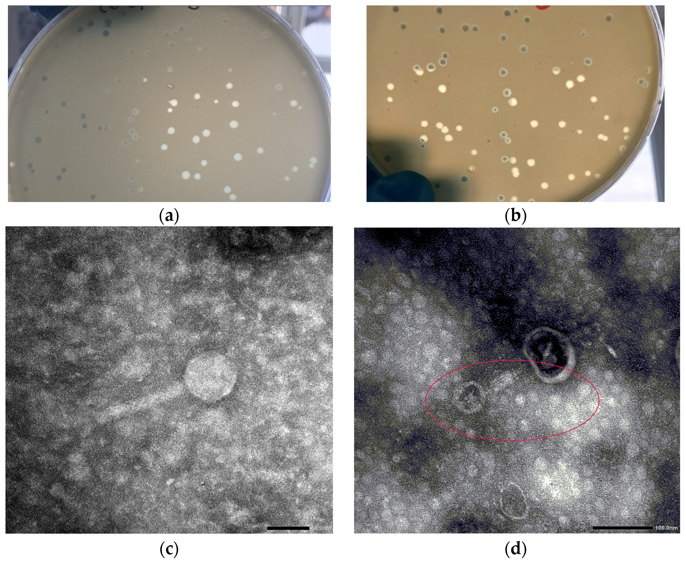
3.1. Viral and Plaque Morphology

The phages’ plaque morphology was evaluated on LB agar plates with the host bacteria Ea 4/82. For Ea PF 7, completely transparent plaques with a 2–4 mm diameter were obtained; however, Ea PF 9 showed more uniform plaques with a diameter of 4 mm (Figure 1).
A transmission electron microscopy analysis determined the morphology of the phages. These phages exhibited an icosahedral head with a size of 60 ± 2 nm, and they featured a long, contractile tail measuring 108 ± 2 nm (Figure 1).

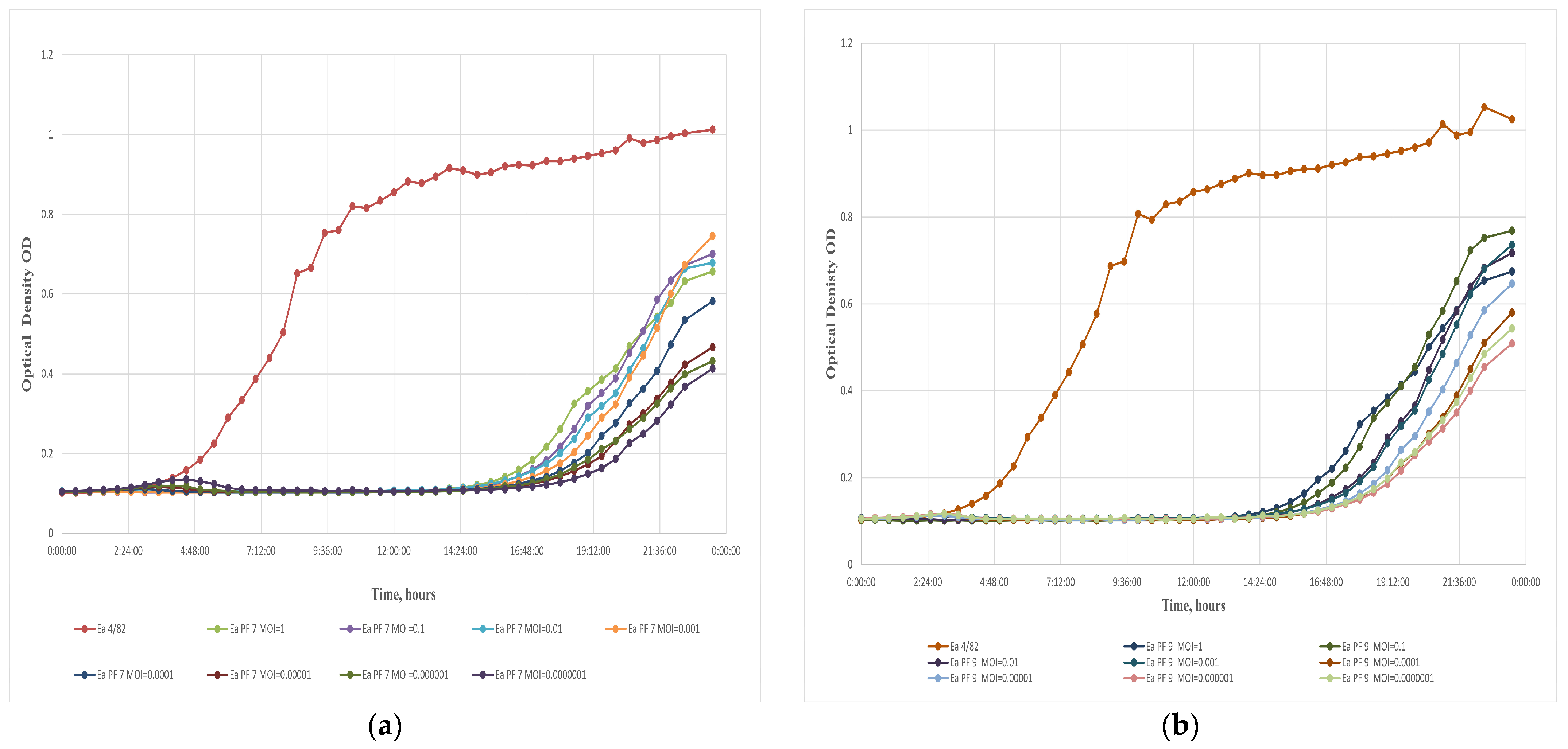
3.2. Quantitative and Qualitative Evaluation of Bacteriophages

Ea PF 7 and Ea PF 9 phage susceptibility was evaluated against 37 Ea strains from the National Collection of Agricultural and Industrial Microorganisms (Hungary). The lytic activity of the phages was determined with a spot test, while the double-agar method was used for quantitative evaluation (Table 2). The efficiency of each phage was evaluated at different MOIs in a liquid culture, using a 96-well-plate method with an uninterrupted time regime.
The spot test revealed that both the Ea PF 7 and Ea PF 9 phages could lyse 95% of the bacteria tested. However, Ea PF 7 showed a marginally higher efficacy than Ea PF 9. The quality of lysis induced by Ea PF 7 was as follows: a total of 43% of the bacteria exhibited confluent lysis, 40% exhibited semi-confluent lysis, and 12% exhibited opaque lysis. In comparison, Ea PF 9 induced confluent lysis in 40% of the bacteria, semi-confluent lysis in 38%, and opaque lysis in 17%, respectively. These results suggest that Ea PF 7 has slightly higher efficacy in lysing bacterial strains, highlighting the potential of both phages as helpful biocontrol agents.
Furthermore, the efficiency of plating (EOP) was calculated to be more than 0.7 in 86.5% of Ea strains for Ea PF 7 and 81% for Ea PF 9 compared to a host strain, which indicates that the phages can replicate and produce progeny phages efficiently.
The lytic activity of a bacteriophage, which refers to its ability to infect and destroy bacterial cells, is a critical factor in determining its prospect for various applications. In this study, the lytic activity of each phage was investigated at extremely low MOIs (MOI of 0.0000001), and in both cases, a significant inhibition of bacterial growth for nearly 15 h was observed (Figure 2a,b). The lytic activity of these phages was remarkable, even at deficient concentrations. This ultralow effective MOI could significantly reduce the costs associated with the practical application of the phages [47]. The development of phage-resistant mutants was observed in Ea bacteria, which is notable in the context of phage–bacteria interactions, as reported in previous studies [31,32]. Bacterial growth was detected after 15 h of phage exposure, suggesting the potential emergence of resistant mutants. To attribute the results obtained from the multi-well plate reader to the emergence of phage-resistant bacterial mutants, double-agar plate assays were performed. After 48 h of incubation, mutant colonies were observed and isolated. Their resistance was confirmed by spot testing [48].
The latent period of the phages Ea PF 7 and Ea PF 9 was short, lasting only 10 min (Figure 2c,d). The burst size, which refers to the number of phage particles released from a single infected bacterial cell, for Ea PF 7 and Ea PF 9 was 108 and 125 phage particles per infected cell. The combination of a short latent period and a high burst size, characteristic of highly effective lytic phages [49], likely explains the exceptional results observed at extremely low MOIs. These characteristics enable efficient phage propagation even when the initial phage concentration is minimal, ensuring rapid infection cycles and high phage yields relative to the available host population. These features make Ea PF 7 and Ea PF 9 particularly suitable for further phage cocktail formulations to combat bacterial infections, especially in environments where such traits are critical for efficiently controlling bacterial diseases.

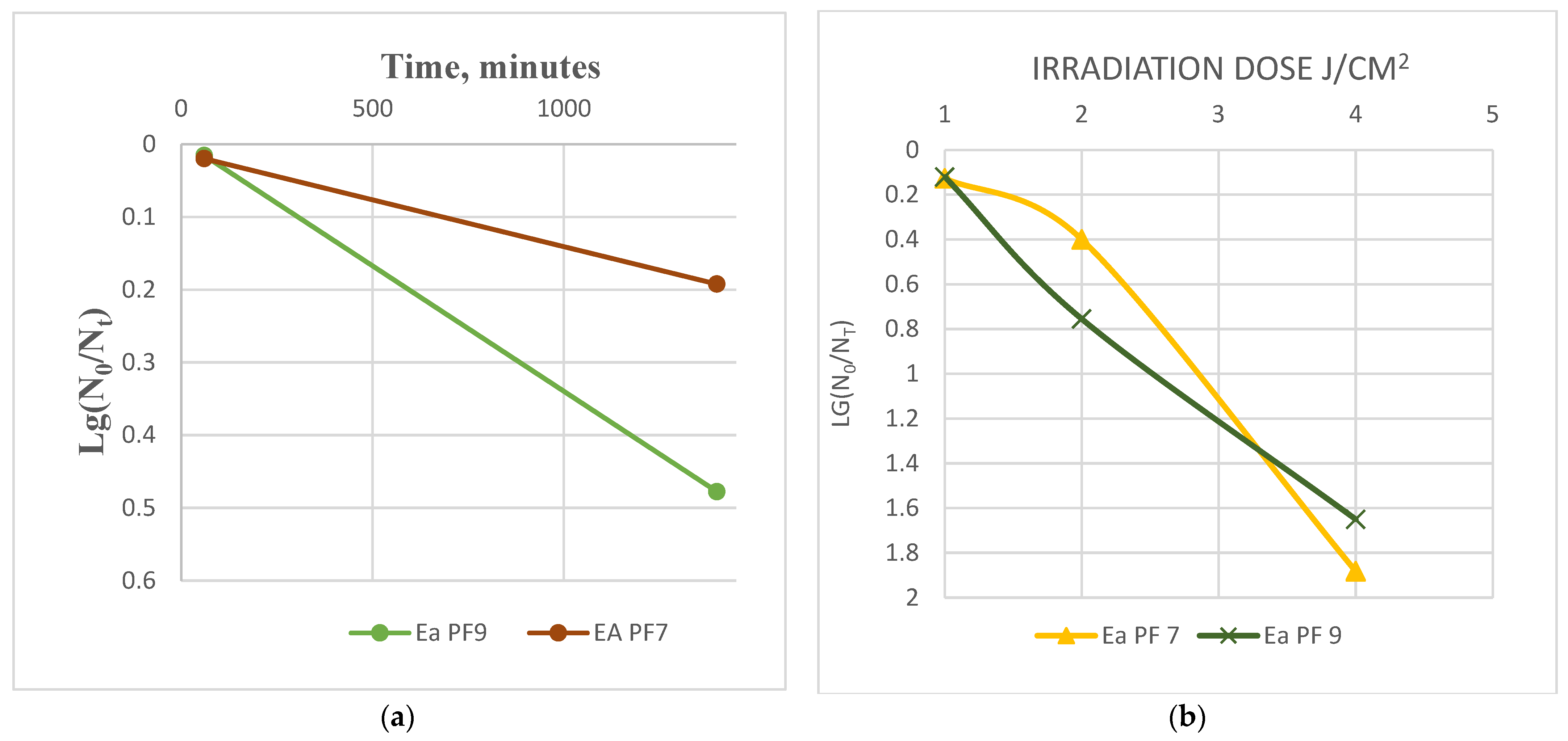
3.3. Stability to Biophysical Factors

Ea PF7 and Ea PF9 exhibited remarkable stability at moderate temperatures (37 °C and 45 °C), with no significant changes observed in their titers after a 120 min incubation. However, exposure to higher temperatures (50 °C to 55 °C) rapidly reduced their titer. Ea PF7 and Ea PF9 could not survive at 60 °C (Figure 3). In the case of Ea PF9, the data reveal that after 90 min of incubation, the phage titer dropped to 0, indicating complete inactivation. This outcome aligns with the rapid loss of viability observed at higher temperatures, particularly at 55 °C, as shown in Figure 3b.
The pH stability test revealed that both Ea PF7 and Ea PF9 can tolerate a wide range of pH conditions, from pH 4 to pH 10, after a 24 h incubation period. At the pH values pH 4 and pH 10, a notable reduction in titer by 2–3 logs was observed, indicating a loss of infectivity (Figure 4).
Our results also demonstrate that both phages show considerable desiccation tolerance, as evidenced by minimal changes in the titer after a 24 h drying period, Figure 5a. This is an important finding, as desiccation tolerance is crucial for the potential use of phages in various applications, including as therapeutics or in biotechnology processes that require long-term survival.
The irradiation dose is determined by several factors, including wavelength, lamp power (as specified by the manufacturer), distance from the lamp, exposure duration, and the age of the lamp, which impacts its power output over time. Experimental studies often omit specific parameters, resulting in incomplete datasets and outcomes that lack comparability and reproducibility. For instance, some studies report only the wavelength and exposure duration, omitting the lamp power and distance [50,51,52], while others provide the lamp power and duration but exclude details on the wavelength and distance [53]. Even when all parameters are given [54,55], the gradual decline in lamp power due to aging, introduces variability in the irradiation dose, even with the same lamp.
To standardize experimental conditions, this study recommends measuring the lamp intensity (W/cm2) at a defined distance at the time of use and calculating the precise irradiation dose (J/cm2) based on the exposure duration. This method allows for the accurate assessment of irradiation effects on phage viability and ensures the reproducibility of experimental results.
At UV doses of 1 and 2 J/cm2, the phages demonstrated a relatively high degree of stability, with a decrease in viability of only 0.4 to 0.9 logs. This suggests that these phages can tolerate UV radiation at these lower doses. However, at a higher UV dose of 4 J/cm2, the viability of the phages decreased significantly, with a reduction in viability of 1.6 to 1.9 logs (Figure 5b). The phages exhibited notable desiccation, temperature, and pH tolerance, as evidenced by minimal changes in relevant parameters close to natural environmental conditions. Phages are more sensitive to UV radiation at this higher dose, consistent with previous studies showing that UV radiation can damage the genetic material of phages, leading to a loss of infectivity [56].

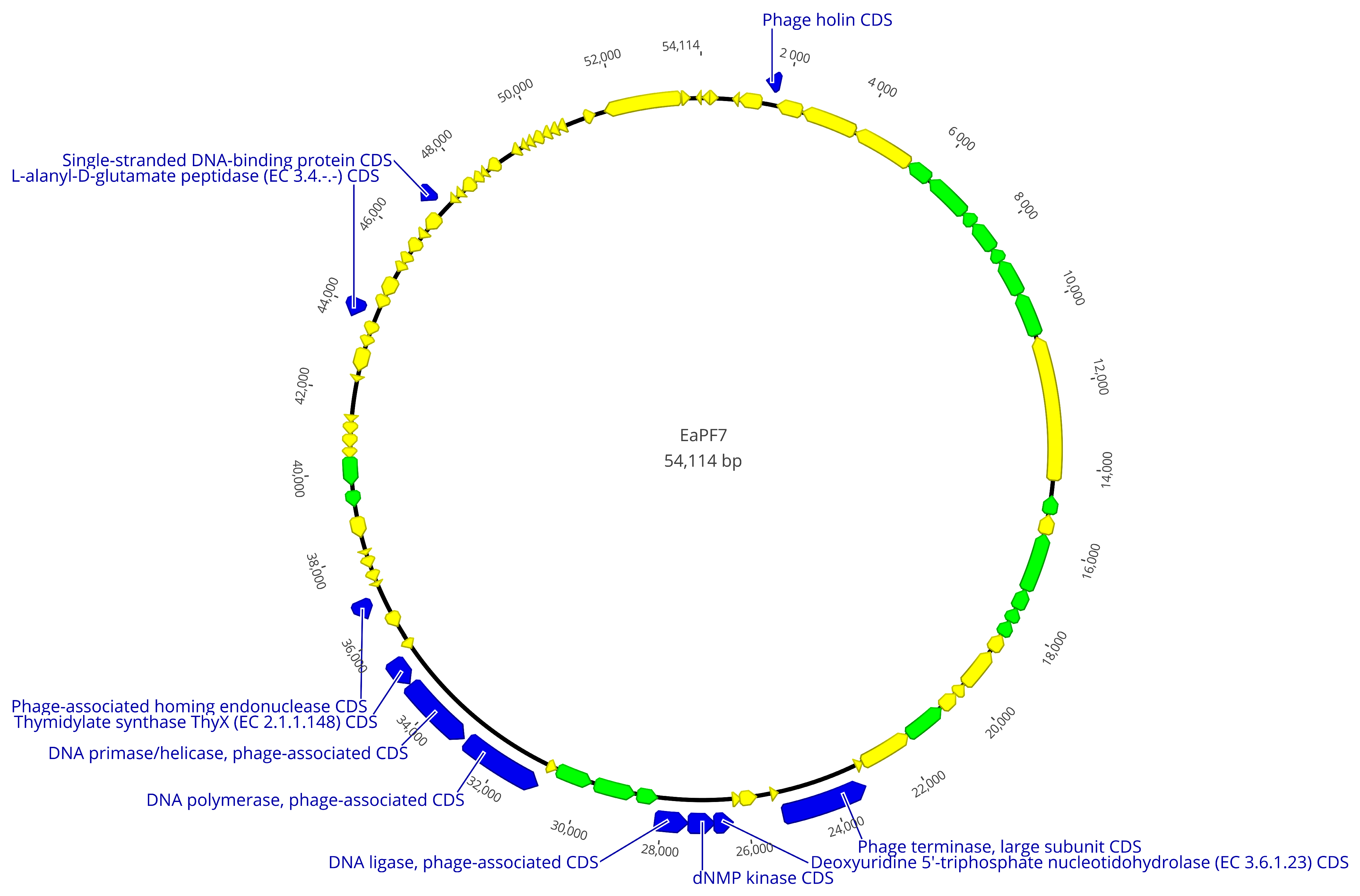
3.4. Genomic Analysis

Ea PF7 and Ea PF9 had 54,114 bps and 52,943 long genomes with a 43.9% and 44.0% G + C content, respectively. The DNA sequencing coverage achieved was 66X for Ea PF7 and 216X for Ea PF9. In the genome of Ea PF7, 87, and in Ea PF9, 84, open reading frames (ORF) were detected. The function of most ORFs could not be annotated, so they were defined as hypothetical proteins, but there are also 33 functional ones in both phage genomes. Based on annotation, structural proteins, proteins related to packaging, cell lysis proteins, and nucleic acid metabolism proteins were defined. However, no proteins associated with lysogeny, such as C1 repressor-like proteins [57], were identified, nor were any antibiotic resistance genes detected. This absence enhances its suitability as a biocontrol agent.
The phage genomes were analyzed using pairwise alignment, revealing a high degree of similarity, approximately 95.6%, in their genomic sequences. As the difference in genomes is 4.4%, only the whole genomic map of Ea PF 7 is presented in Figure 6. Despite this, they exhibited notable differences in other phenotypic characteristics during additional testing. Genomic differences were primarily located in non-coding regions or regions encoding hypothetical proteins, with the exception of the homing endonuclease genes, which exhibited 48.0% identity in pairwise alignment. Homing endionucleases represent an evolutionary strategy that phages use to ensure that their genetic elements persist and spread, while potentially providing benefits in phage–host competition and adaptability [58]. Phages with closely related genomes but distinct host ranges often have subtle genetic differences in genes coding for receptor-binding proteins or tail fibers [59]. While the annotation did not specifically identify these proteins, it is plausible that they are encoded by hypothetical proteins, which may conceal their functional roles.
According to the International Committee on Taxonomy of Viruses (ICTV), the genome analysis determined that both phages are Loessnervirus-like viruses. Genome blast results demonstrate that Phi Fifi044, Phi Fifi451 [50], and SNAUBM-27 [31] from Korea were the most related bacteriophages to Ea PF 7 and Ea PF 9.
Seventeen complete genomic sequences of Loessnervirus-like phages were used to construct phylogenetic trees, including the genomes of Ea PF 7 and Ea PF 9. Two distinct trees were generated: the first was based on concatenated sequences of the portal protein and terminase large subunit genes (Figure 7a), while the second utilized whole-genome sequences (Figure 7b). In the concatenated gene-based phylogeny, Ea PF 7 (GenBank accession no. PQ431420) and Ea PF 9 (PQ431419) clustered with OQ818694 and several other phages encoding putative portal proteins (MW349138, PQ051110, and OK129343) (Figure 7a). In contrast, the whole-genome-based phylogeny placed EaPf7 and EaPf9 in a distinct clade, separate from OQ818694 (Figure 7b). This topological incongruence may reflect a recombination or horizontal gene transfer event involving structural module genes, aligning with the modular evolution model of bacteriophages.
Admitting that the same phages have also been isolated in Hungary contributes to the broader knowledge of their geographical distribution and relevance. This not only contributes to understanding their distribution but also raises questions about the genetic variability and adaptability of these phages in different environmental and geographical contexts.

4. Conclusions

This study demonstrated that the phages Ea PF 7 and Ea PF 9 exhibited strong efficacy and stability under environmental conditions, characterized by a short latent period and large burst size. Genome sequencing revealed no genes associated with lysogeny or antibiotic resistance. While minor differences were observed between the two genomes, their biological features remained distinct. These findings position Ea PF 7 and Ea PF 9 as promising candidates for further cocktail formulations, which will be the focus of our future research. Developing bacteriophage-based biocontrol agents holds great potential for combating plant infectious diseases.

Author Contributions

Conceptualization, E.L. and T.K.; methodology, E.L. and T.K.; investigation, E.L.; resources, D.B. and R.P.-T.; writing—original draft preparation, E.L.; writing—review and editing, E.L. and T.K.; supervision, T.K., S.P. and G.S.; project administration, T.K. and S.P.; funding acquisition, T.K. All authors have read and agreed to the published version of the manuscript.

Funding

This research was funded by the European Union’s Horizon 2020 research and innovation program (H2020-EIC-FTI-2018-2020), the Phage Fire (no. 958645) and European Regional Development Fund, and by Hungarian Government (GINOP_PLUSZ-2.1.1-21-2022-00031—SuperAgroPhage). The APC was funded by European Regional Development Fund and by the Hungarian Government (GINOP_PLUSZ-2.1.1-21-2022-00031—SuperAgroPhage).

Institutional Review Board Statement

Not applicable.

Informed Consent Statement

Not applicable.

Data Availability Statement

We submitted assembled and annotated genomes to the GenBank, and the following accession numbers were used: PQ431420 for phage Ea PF 7 and PQ431419 for phage Ea PF 9.

Acknowledgments

We extend our gratitude to Fieseler Lars and Sandrine Kammerecker for their valuable suggestions and contributions to our investigations into UV tolerance.

Conflicts of Interest

The authors declare no conflicts of interest. E. Lomadze, S. Papp, and T. Kovács are employees of Enviroinvest Corp., and D. Bali, R. Princz-Tóth, and T. Kovács are employees of Biopesticide Ltd. This does not alter our adherence to Viruses MDPI policies on sharing data and material.

References

  1. Bonn, W.G.; van der Zwet, T. Distribution and economic importance of fire blight. In Fire Blight the Disease and Its Causative Agent, Erwinia amylovora; Vanneste, J.L., Ed.; CABI Publishing: Wallingford, UK, 2000; pp. 37–53. [Google Scholar]
  2. Momol, M.; Aldwinckle, H.S. Genetic Diversity and Host Range of Erwinia amylovora; CABI Publishing: Wallingford, UK, 2000; pp. 55–72. [Google Scholar]
  3. Thomson, S.V. Epidemiology of fire blight. In Fire Blight: The Disease and Its Causative Agent Erwinia Amylovora; Vanneste, J.L., Ed.; CAB International: Wallingford, UK, 2000; pp. 9–36. [Google Scholar]
  4. Mansfield, J.; Genin, S.; Magori, S.; Citovsky, V.; Sriariyanum, M.; Ronald, P.; Dow, M.; Verdier, V.; Beer, S.V.; Machado, M.A.; et al. Top 10 plant pathogenic bacteria in molecular plant pathology. Mol. Plant Pathol. 2012, 13, 614–629. [Google Scholar] [CrossRef] [PubMed]
  5. Hevesi, M. Az Erwinia amylovora (Burill) Winslow et al. hazai megjelenése almán. Növényvédelem 1996, 32, 225–228. [Google Scholar]
  6. Végh, A.; Hevesi, M.; Pájtli, É.; Palkovics, L. Characterization of Erwinia amylovora strains from Hungary. Eur. J. Plant Pathol. 2017, 147, 455–461. [Google Scholar] [CrossRef]
  7. Végh, A.; Némethy, Z.; Hajagos, L.; Palkovics, L. First report of Erwinia amylovora causing fire blight on plum (Prunus domestica L.) in Hungary. Plant Dis. 2012, 96, 759. [Google Scholar] [CrossRef]
  8. Végh, A.; Palkovics, L. First occurence of fire blight on apricot (Prunus armeniaca) in Hungary. Not. Bot. Horti Agrobot. Cluj-Napoca 2013, 41, 440–443. [Google Scholar] [CrossRef]
  9. Vanneste, J.L.; Eden-Green, S. Migration of Erwinia amylovora in host plant tissues. In Fire Blight: The Disease and Its Causative Agent, Erwinia amylovora; Vanneste, J.L., Ed.; CAB International: Wallingford, UK, 2000; pp. 73–83. [Google Scholar]
  10. Skoneczny, H.; Kubiak, K.; Spiralski, M.; Kotlarz, J.; Mikiciński, A.; Puławska, J. Fire Blight Disease Detection for Apple Trees: Hyperspectral Analysis of Healthy, Infected and Dry Leaves. Remote Sens. 2020, 12, 2101. [Google Scholar] [CrossRef]
  11. Zurn, J.D.; Norelli, J.L.; Montanari, S.; Bell, R.; Bassil, N.V. Dissecting Genetic Resistance to Fire Blight in Three Pear Populations. Phytopathology 2020, 110, 1305–1311. [Google Scholar] [CrossRef] [PubMed]
  12. Escursell, M.M.; Roschi, A.; Smits, T.H.M.; Rezzonico, F. Characterization and direct molecular discrimination of rpsL mutations leading to high streptomycin resistance in Erwinia amylovora. J. Plant Pathol. 2021, 103, 99–108. [Google Scholar] [CrossRef]
  13. Evrenosoğlu, Y.; Mertoglu, K.; Bilgin, N.; Misirli, A.; Altay, Y. An Analysis on Some Reciprocal Pear Hybridization Combinations in Terms of Transferring Resistance to Fire BlightUntersuchung einiger reziproker Birnen-Hybridisierungskombinationen im Hinblick auf die Übertragung von Feuerbrandresistenz. Erwerbs-Obstbau 2020, 62, 189–194. [Google Scholar] [CrossRef]
  14. Seed, K.D. Battling Phages: How Bacteria Defend against Viral Attack. PLoS Pathog. 2015, 11, e1004847. [Google Scholar] [CrossRef]
  15. Harvey, H.; Bondy-Denomy, J.; Marquis, H.; Sztanko, K.M.; Davidson, A.R.; Burrows, L.L. Pseudomonas aeruginosa defends against phages through type IV pilus glycosylation. Nat. Microbiol. 2018, 3, 47–52. [Google Scholar] [CrossRef] [PubMed]
  16. Destoumieux-Garzon, D.; Duquesne, S.; Peduzzi, J.; Goulard, C.; Desmadril, M.; Letellier, L.; Rebuffat, S.; Boulanger, P. The iron-siderophore transporter FhuA is the receptor for the antimicrobial peptide microcin J25: Role of the microcin Val11-Pro16 beta-hairpin region in the recognition mechanism. Biochem. J. 2005, 389 Pt 3, 869–876. [Google Scholar] [CrossRef]
  17. Bebeacua, C.; Lorenzo Fajardo, J.C.; Blangy, S.; Spinelli, S.; Bollmann, S.; Neve, H.; Cambillau, C.; Heller, K.J. X-ray structure of a superinfection exclusion lipoprotein from phage TP-J34 and identification of the tape measure protein as its target. Mol. Microbiol. 2013, 89, 152–165. [Google Scholar] [CrossRef] [PubMed]
  18. Dupuis, M.E.; Villion, M.; Magadan, A.H.; Moineau, S. CRISPR-Cas and restriction-modification systems are compatible and increase phage resistance. Nat. Commun. 2013, 4, 2087. [Google Scholar] [CrossRef]
  19. Aziz, R.K.; Bartels, D.; Best, A.A.; DeJongh, M.; Disz, T.; Edwards, R.A.; Formsma, K.; Gerdes, S.; Glass, E.M.; Kubal, M.; et al. The RAST Server: Rapid annotations using subsystems technology. BMC Genom. 2008, 9, 75. [Google Scholar] [CrossRef]
  20. Goodridge, L.D.; Abedon, S.T. Bacteriophage biocontrol: The technology matures. Microbiol. Aust. 2008, 29, 48–49. [Google Scholar] [CrossRef]
  21. Polaska, M.; Sokolowska, B. Bacteriophages—A new hope or a huge problem in the food industry. AIMS Microbiol. 2019, 5, 324–346. [Google Scholar] [CrossRef] [PubMed]
  22. Luo, J.; Dai, D.; Lv, L.; Ahmed, T.; Chen, L.; Wang, Y.; An, Q.; Sun, G.; Li, B. Advancements in the Use of Bacteriophages to Combat the Kiwifruit Canker Phytopathogen Pseudomonas syringae pv. actinidiae. Viruses 2022, 14, 2704. [Google Scholar] [CrossRef]
  23. Yin, Y.; Pei’en, N.; Bohan, D.; Shiping, W.; Wenping, X.; Wang, D. Isolation and characterisation of phages against Pseudomonas syringae pv. actinidiae. Acta Agric. Scand. Sect. B—Soil Plant Sci. 2019, 69, 199–208. [Google Scholar]
  24. Sieiro, C.; Areal-Hermida, L.; Pichardo-Gallardo, Á.; Almuiña-González, R.; de Miguel, T.; Sánchez, S.; Sánchez-Pérez, Á.; Villa, T.G. A Hundred Years of Bacteriophages: Can Phages Replace Antibiotics in Agriculture and Aquaculture? Antibiotics 2020, 9, 493. [Google Scholar] [CrossRef]
  25. Svircev, A.; Roach, D.; Castle, A. Framing the Future with Bacteriophages in Agriculture. Viruses 2018, 10, 218. [Google Scholar] [CrossRef]
  26. Jamal, M.; Bukhari, S.; Andleeb, S.; Ali, M.; Raza, S.; Nawaz, M.A.; Hussain, T.; Rahman, S.U.; Shah, S.S.A. Bacteriophages: An overview of the control strategies against multiple bacterial infections in different fields. J. Basic Microbiol. 2019, 59, 123–133. [Google Scholar] [CrossRef] [PubMed]
  27. Domotor, D.; Becsagh, P.; Rakhely, G.; Schneider, G.; Kovacs, T. Complete genomic sequence of Erwinia amylovora phage PhiEaH2. J. Virol. 2012, 86, 10899. [Google Scholar] [CrossRef] [PubMed]
  28. Meczker, K.; Domotor, D.; Vass, J.; Rakhely, G.; Schneider, G.; Kovacs, T. The genome of the Erwinia amylovora phage PhiEaH1 reveals greater diversity and broadens the applicability of phages for the treatment of fire blight. FEMS Microbiol. Lett. 2014, 350, 25–27. [Google Scholar] [CrossRef]
  29. Schwarczinger, I.; Kolozsváriné Nagy, J.; Künstler, A.; Szabó, L.; Geider, K.; Király, L.; Pogány, M. Characterization of Myoviridae and Podoviridae family bacteriophages of Erwinia amylovora from Hungary—Potential of application in biological control of fire blight. Eur. J. Plant Pathol. 2017, 149, 639–652. [Google Scholar] [CrossRef]
  30. Kim, H.J.; Jun, J.W.; Giri, S.S.; Kim, S.G.; Kim, S.W.; Kwon, J.; Lee, S.B.; Chi, C.; Park, S.C. Bacteriophage Cocktail for the Prevention of Multiple-Antibiotic-Resistant and Mono-Phage-Resistant Vibrio coralliilyticus Infection in Pacific Oyster (Crassostrea gigas) Larvae. Pathogens 2020, 9, 831. [Google Scholar] [CrossRef]
  31. Kim, S.G.; Lee, S.B.; Jo, S.J.; Cho, K.; Park, J.K.; Kwon, J.; Giri, S.S.; Kim, S.W.; Kang, J.W.; Jung, W.J.; et al. Phage Cocktail in Combination with Kasugamycin as a Potential Treatment for Fire Blight Caused by Erwinia amylovora. Antibiotics 2022, 11, 1566. [Google Scholar] [CrossRef]
  32. Knecht, L.E.; Born, Y.; Pelludat, C.; Pothier, J.F.; Smits, T.H.M.; Loessner, M.J.; Fieseler, L. Spontaneous Resistance of Erwinia amylovora Against Bacteriophage Y2 Affects Infectivity of Multiple Phages. Front. Microbiol. 2022, 13, 908346. [Google Scholar] [CrossRef]
  33. Falkenstein, H.; Bellemann, P.; Walter, S.; Zeller, W.; Geider, K. Identification of Erwinia amylovora, the Fireblight Pathogen, by Colony Hybridization with DNA from Plasmid pEA29. Appl. Environ. Microbiol. 1988, 54, 2798–2802. [Google Scholar] [CrossRef]
  34. Gill, J.J.; Svircev, A.M.; Smith, R.; Castle, A.J. Bacteriophages of Erwinia amylovora. Appl. Environ. Microbiol. 2003, 69, 2133–2138. [Google Scholar] [CrossRef]
  35. Adams, M.H. Bacteriophages; Interscience Publishers: New York, NY, USA, 1959. [Google Scholar]
  36. Kutter, E. Phage host range and efficiency of plating. Methods Mol. Biol. 2009, 501, 141–149. [Google Scholar] [PubMed]
  37. Kropinski, A.M.; Mazzocco, A.; Waddell, T.E.; Lingohr, E.; Johnson, R.P. Enumeration of bacteriophages by double agar overlay plaque assay. Methods Mol. Biol. 2009, 501, 69–76. [Google Scholar]
  38. Gayder, S.; Parcey, M.; Castle, A.J.; Svircev, A.M. Host Range of Bacteriophages Against a World-Wide Collection of Erwinia amylovora Determined Using a Quantitative PCR Assay. Viruses 2019, 11, 910. [Google Scholar] [CrossRef] [PubMed]
  39. Gayder, S.; Parcey, M.; Nesbitt, D.; Castle, A.J.; Svircev, A.M. Population Dynamics between Erwinia amylovora, Pantoea agglomerans and Bacteriophages: Exploiting Synergy and Competition to Improve Phage Cocktail Efficacy. Microorganisms 2020, 8, 1449. [Google Scholar] [CrossRef]
  40. Born, Y.; Fieseler, L.; Marazzi, J.; Lurz, R.; Duffy, B.; Loessner, M.J. Novel virulent and broad-host-range Erwinia amylovora bacteriophages reveal a high degree of mosaicism and a relationship to Enterobacteriaceae phages. Appl. Environ. Microbiol. 2011, 77, 5945–5954. [Google Scholar] [CrossRef]
  41. Makalatia, K.; Kakabadze, E.; Wagemans, J.; Grdzelishvili, N.; Bakuradze, N.; Natroshvili, G.; Macharashvili, N.; Sedrakyan, A.; Arakelova, K.; Ktsoyan, Z.; et al. Characterization of Salmonella Isolates from Various Geographical Regions of the Caucasus and Their Susceptibility to Bacteriophages. Viruses 2020, 12, 1418. [Google Scholar] [CrossRef]
  42. Koderi Valappil, S.; Shetty, P.; Deim, Z.; Terhes, G.; Urban, E.; Vaczi, S.; Patai, R.; Polgar, T.; Pertics, B.Z.; Schneider, G.; et al. Survival Comes at a Cost: A Coevolution of Phage and Its Host Leads to Phage Resistance and Antibiotic Sensitivity of Pseudomonas aeruginosa Multidrug Resistant Strains. Front. Microbiol. 2021, 12, 783722. [Google Scholar] [CrossRef]
  43. Duarte, J.; Pereira, C.; Costa, P.; Almeida, A. Bacteriophages with Potential to Inactivate Aeromonas hydrophila in Cockles: In Vitro and In Vivo Preliminary Studies. Antibiotics 2021, 10, 710. [Google Scholar] [CrossRef]
  44. Kim, S.G.; Jun, J.W.; Giri, S.S.; Yun, S.; Kim, H.J.; Kim, S.W.; Kang, J.W.; Han, S.J.; Jeong, D.; Park, S.C. Isolation and characterisation of pVa-21, a giant bacteriophage with anti-biofilm potential against Vibrio alginolyticus. Sci. Rep. 2019, 9, 6284. [Google Scholar] [CrossRef]
  45. Jo, S.J.; Kim, S.G.; Lee, Y.M.; Giri, S.S.; Kang, J.W.; Lee, S.B.; Jung, W.J.; Hwang, M.H.; Park, J.; Cheng, C.; et al. Evaluation of the Antimicrobial Potential and Characterization of Novel T7-Like Erwinia Bacteriophages. Biology 2023, 12, 180. [Google Scholar] [CrossRef]
  46. Erol, H.B.; Kaskatepe, B.; Ozturk, S.; Safi Oz, Z. The comparison of lytic activity of isolated phage and commercial Intesti bacteriophage on ESBL producer E. coli and determination of Ec_P6 phage efficacy with in vivo Galleria mellonella larvae model. Microb. Pathog. 2022, 167, 105563. [Google Scholar] [CrossRef]
  47. Peng, Q.; Ma, Z.; Han, Q.; Xiang, F.; Wang, L.; Zhang, Y.; Zhao, Y.; Li, J.; Xian, Y.; Yuan, Y. Characterization of bacteriophage vB_KleM_KB2 possessing high control ability to pathogenic Klebsiella pneumoniae. Sci. Rep. 2023, 13, 9815. [Google Scholar] [CrossRef]
  48. Knecht, L.E.; Heinrich, N.; Born, Y.; Felder, K.; Pelludat, C.; Loessner, M.J.; Fieseler, L. Bacteriophage S6 requires bacterial cellulose for Erwinia amylovora infection. Environ. Microbiol. 2022, 24, 3436–3450. [Google Scholar] [CrossRef] [PubMed]
  49. Merabishvili, M.; Vandenheuvel, D.; Kropinski, A.M.; Mast, J.; De Vos, D.; Verbeken, G.; Noben, J.P.; Lavigne, R.; Vaneechoutte, M.; Pirnay, J.P. Characterization of newly isolated lytic bacteriophages active against Acinetobacter baumannii. PLoS ONE 2014, 9, e104853. [Google Scholar] [CrossRef] [PubMed]
  50. Park, J.; Kim, B.; Song, S.; Lee, Y.W.; Roh, E. Isolation of Nine Bacteriophages Shown Effective against Erwinia amylovora in Korea. Plant Pathol. J. 2022, 38, 248–253. [Google Scholar] [CrossRef] [PubMed]
  51. Park, J.; Lee, G.M.; Kim, D.; Park, D.H.; Oh, C.S. Characterization of the Lytic Bacteriophage phiEaP-8 Effective against Both Erwinia amylovora and Erwinia pyrifoliae Causing Severe Diseases in Apple and Pear. Plant Pathol. J. 2018, 34, 445–450. [Google Scholar] [CrossRef]
  52. Luo, J.; Xie, L.; Yang, M.; Liu, M.; Li, Q.; Wang, P.; Fan, J.; Jin, J.; Luo, C. Synergistic Antibacterial Effect of Phage pB3074 in Combination with Antibiotics Targeting Cell Wall against Multidrug-Resistant Acinetobacter baumannii In Vitro and Ex Vivo. Microbiol. Spectr. 2023, 11, e0034123. [Google Scholar] [CrossRef]
  53. Jo, S.J.; Kim, S.G.; Park, J.; Lee, Y.M.; Giri, S.S.; Lee, S.B.; Jung, W.J.; Hwang, M.H.; Park, J.H.; Roh, E.; et al. Optimizing the formulation of Erwinia bacteriophages for improved UV stability and adsorption on apple leaves. Heliyon 2023, 9, e22034. [Google Scholar] [CrossRef]
  54. Born, Y.; Bosshard, L.; Duffy, B.; Loessner, M.J.; Fieseler, L. Protection of Erwinia amylovora bacteriophage Y2 from UV-induced damage by natural compounds. Bacteriophage 2015, 5, e1074330. [Google Scholar] [CrossRef]
  55. Fu, J.; Li, Y.; Zhao, L.; Wu, C.; He, Z. Characterization of vB_ValM_PVA8, a broad-host-range bacteriophage infecting Vibrio alginolyticus and Vibrio parahaemolyticus. Front. Microbiol. 2023, 14, 1105924. [Google Scholar] [CrossRef]
  56. Tom, E.F.; Molineux, I.J.; Paff, M.L.; Bull, J.J. Experimental evolution of UV resistance in a phage. PeerJ 2018, 6, e5190. [Google Scholar] [CrossRef] [PubMed]
  57. Sabri, M.; El Handi, K.; Valentini, F.; De Stradis, A.; Achbani, E.H.; Benkirane, R.; Resch, G.; Elbeaino, T. Identification and Characterization of Erwinia Phage IT22: A New Bacteriophage-Based Biocontrol against Erwinia amylovora. Viruses 2022, 14, 2455. [Google Scholar] [CrossRef] [PubMed]
  58. Brok-Volchanskaya, V.S.; Kadyrov, F.A.; Sivogrivov, D.E.; Kolosov, P.M.; Sokolov, A.S.; Shlyapnikov, M.G.; Kryukov, V.M.; Granovsky, I.E. Phage T4 SegB protein is a homing endonuclease required for the preferred inheritance of T4 tRNA gene region occurring in co-infection with a related phage. Nucleic Acids Res. 2008, 36, 2094–2105. [Google Scholar] [CrossRef] [PubMed]
  59. Chung, K.M.; Liau, X.L.; Tang, S.S. Bacteriophages and Their Host Range in Multidrug-Resistant Bacterial Disease Treatment. Pharmaceuticals 2023, 16, 1467. [Google Scholar] [CrossRef]
Figure 1. Photos of the plaques of the phages Ea PF 7 (a) and Ea PF9 (b). Transmission electron micrographs of phage Ea PF 7; scale bar = 50 nm (c). Transmission electron micrographs of phage Ea PF 7 contracted position; scale bar = 100 nm. Red circle highlight theEaPf7 bacteriophage with a contracted tail. (d).
Viruses 17 00677 g001
Figure 2. Effect of the bacteriophages on host bacteria growth. Evaluation of phage activity at different MOIs for phage Ea PF 7 (a) and for phage Ea PF 9 (b). One-step growth curve of Ea PF 7 (c) and of Ea PF 9 (d).
Viruses 17 00677 g002aViruses 17 00677 g002b
Figure 3. Temperature stability of phage Ea PF 7 (a) and Ea PF 9 (b). Comparative characterization of phages’ stability during 90 min incubation at different temperatures (c).
Viruses 17 00677 g003
Figure 4. pH stability of phage Ea PF 7 (a) and Ea PF 9 (b). Comparative characterization of phages’ stability during 30 min incubation at different pHs (c).
Viruses 17 00677 g004aViruses 17 00677 g004b
Figure 5. Desiccation tolerance of the phages (a). UV-B tolerance of the phages (b).
Viruses 17 00677 g005
Figure 6. Genomic maps of phage Ea PF 7. Blue label—functional proteins; green label—structural proteins; yellow label—hypothetical proteins.
Viruses 17 00677 g006
Figure 7. Phylogenetic trees of Ea PF 7 (GenBank accession no. PQ431420) and Ea PF 9 (PQ431419). (a) Tree constructed using concatenated amino acid sequences of the portal protein and terminase large subunit genes. (b) Tree constructed using complete whole-genome sequences.
Viruses 17 00677 g007
Table 1. List of the Ea strains used in this study.
EaStrain Collection and OriginIsolation Source
Ea B 01272National Collection of Agricultural and Industrial Microorganisms (Hungary)Apple
Ea B 01616Unknown (plant)
Ea B 01728Apple
Ea B 01729Apple
Ea B 01731Apple
Ea B 01733Apple
Ea B 01734Pear
Ea B 01735Quince
Ea B 01738Medlar
Ea B 01756Apple
Ea B 01757Apple
Ea B 01840Crataegus sp.
Ea B 01843Apple
Ea B 01844Pear
Ea B 01853Cotoneaster sp.
Ea B 01855Pyracantha sp.
Ea B 01896Unknown (plant)
Ea B 01898Apple
Ea B 01901Apple
Ea B 01903Apple
Ea B 01905Apple
Ea B 01906Apple
Ea B 01960Apple
Ea B 01961Apple
Ea B 01962Apple
Ea B 01963Apple
Ea B 01964Apple
Ea B 01971Apple
Ea B 01972Pear
Ea B 01973Pear
Ea B 01975Pear
Ea B 01978Pear
Ea B 01980Apple
Ea B 01983Quince
Ea G254Government Office of Baranya Country, HungaryApple
Ea G255Government Office of Baranya Country, HungaryApple
Ea B 0118TNational Collection of Agricultural and Industrial Microorganisms (Hungary)Pear
CFBP 1430CIRM-CFBP French Collection for Plant-Associated BacteriaCrataegus sp.
Ea 4/82Egypt, 1982 [33]Pear
Table 2. Results of qualitative and quantitate evaluation of phages. Lysis activity was analyzed as follows: 4—confluent lysis (CL); 3—semi-confluent lysis (SCL); 2—confluent lysis with opacity due to secondary growth (OL); 1—on the bacterial growth, individual lysis sites are as small dots (IPOs); and 0—no lysis. EOP was analyzed as follows: if EOP = 1, the tested bacteria have the same output of phages as the host strain; if EOP > 1, the tested bacteria should be considered a better host strain; and if EOP < 1, the tested bacteria have less phage output than the host strain.
Ea StrainsLysis ActivityEOP
Ea PF 7Ea PF 9Ea PF 7Ea PF 9
Ea B 01272300.50
Ea B 01616230.70.8
Ea B 01728430.70.7
Ea B 01729410.70.3
Ea B 017312110.1
Ea B 017334210.4
Ea B 01734440.90.9
Ea B 01735440.90.8
Ea B 01738430.90.9
Ea B 017564411
Ea B 017574411.1
Ea B 018404410.9
Ea B 01843330.60.2
Ea B 01844330.60.5
Ea B 01853340.70.8
Ea B 01855330.70.8
Ea B 01896230.70.9
Ea B 01898330.70.4
Ea B 01901431.11
Ea B 01903330.90.9
Ea B 01905330.91
Ea B 01906421.20.7
Ea B 01960340.81.1
Ea B 01961220.70.7
Ea B 019624410.9
Ea B 019634411
Ea B 01964440.91.1
Ea B 01971340.70.9
Ea B 01972330.80.8
Ea B 01973440.91.1
Ea B 01975330.70.9
Ea B 01978430.80.7
Ea B 01980140.21.1
Ea B 019833310.9
Ea G254340.81
Ea G255340.81
Ea B 0118T0201.1
CFBP 1430330.80.7
Ea 4/824411
Disclaimer/Publisher’s Note: The statements, opinions and data contained in all publications are solely those of the individual author(s) and contributor(s) and not of MDPI and/or the editor(s). MDPI and/or the editor(s) disclaim responsibility for any injury to people or property resulting from any ideas, methods, instructions or products referred to in the content.

