Article

A Novel Nobecovirus in an Epomophorus wahlbergi Bat from Nairobi, Kenya

by
Meredith C. VanAcker
1,2,*,†,
Koray Ergunay
3,4,5,6,†,
Paul W. Webala
7,
Maureen Kamau
8,
Janerose Mutura
8,
Rashid Lebunge
8,
Griphin Ochieng Ochola
8,
Brian P. Bourke
3,4,5,
Emily G. McDermott
9,
Nicole L. Achee
10,
Le Jiang
11,
John P. Grieco
10,
Erick Keter
12,
Audrey Musanga
13,
Suzan Murray
2,
Jared A. Stabach
14,
Meggan E. Craft
15,
Eric M. Fèvre
16,17,
Yvonne-Marie Linton
3,4,5 and
James Hassell
2,17,18
1
Department of Evolution, Ecology, and Organismal Biology, University of California, Riverside, CA 92521, USA
2
Global Health Program, Smithsonian Institution, National Zoo and Conservation Biology Institute, Washington, DC 20008, USA
3
Walter Reed Biosystematics Unit (WRBU), Smithsonian Institution, Museum Support Center, Suitland, MD 20746, USA
4
One Health Branch, Walter Reed Army Institute of Research (WRAIR), Silver Spring, MD 20910, USA
5
Department of Entomology, Smithsonian Institution, National Museum of Natural History (NMNH), Washington, DC 20560, USA
6
Virology Unit, Department of Medical Microbiology, Faculty of Medicine, Hacettepe University, Ankara 06100, Turkey
7
Department of Forestry and Wildlife Management, Maasai Mara University, Narok 20500, Kenya
8
Mpala Research Centre (MRC), Nanyuki 10400, Kenya
9
Department of Entomology and Plant Pathology, University of Arkansas, Fayetteville, AR 72701, USA
10
Department of Biological Sciences, Eck Institute for Global Health, University of Notre Dame, Notre Dame, IN 46556, USA
11
Viral and Rickettsial Diseases Department, Infectious Diseases Directorate, Naval Medical Research Center (NMRC), 503 Robert Grant Avenue, Silver Spring, MD 20910, USA
12
Department of Wildlife Management, University of Eldoret, Eldoret 30100, Kenya
13
College of Agriculture and Veterinary Sciences, University of Nairobi, Nairobi 00100, Kenya
14
Conservation Ecology Center, Smithsonian National Zoo and Conservation Biology Institute, Front Royal, VA 22630, USA
15
Department of Ecology, Evolution and Behavior, College of Biological Sciences, University of Minnesota, St. Paul, MN 55108, USA
16
Institute of Infection, Veterinary and Ecological Sciences, University of Liverpool, Liverpool L69 3BX, UK
17
International Livestock Research Institute (ILRI), Nairobi 00100, Kenya
18
Department of Epidemiology of Microbial Diseases, Yale School of Public Health, New Haven, CT 06520, USA
*
Author to whom correspondence should be addressed.
Shared first authorship.
Viruses 2025, 17(4), 557; https://doi.org/10.3390/v17040557
Submission received: 3 February 2025 / Revised: 25 March 2025 / Accepted: 8 April 2025 / Published: 12 April 2025
(This article belongs to the Special Issue Bat- and Rodent-Borne Zoonotic Viruses)

Abstract

:
Most human emerging infectious diseases are zoonotic, originating in animal hosts prior to spillover to humans. Prioritizing the surveillance of wildlife that overlaps with humans and human activities can increase the likelihood of detecting viruses with a high potential for human infection. Here, we obtained fecal swabs from two fruit bat species—Eidolon helvum (n = 6) and Epomophorus wahlbergi (n = 43) (family Pteropodidae)—in peridomestic habitats in Nairobi, Kenya, and used metagenome sequencing to detect microorganisms. A near-complete genome of a novel virus assigned taxonomically to the Coronaviridae family Betacoronavirus genus and Nobecovirus subclade was characterized from E. wahlbergi. Phylogenetic analysis indicates this unique Nobecovirus clade shares a common ancestor with Eidolon/Rousettus Nobecovirus subclades isolated from Madagascar, Kenya, and Cameroon. Recombination was detected across open reading frames, except the spike protein, in all BOOTSCAN analyses, indicating intra-host coinfection and genetic exchange between genome regions. Although Nobecoviruses are currently bat-specific and are not known to be zoonotic, the propensity of coronaviruses to undergo frequent recombination events and the location of the virus alongside high human and livestock densities in one of East Africa’s most rapidly developing cities justifies continued surveillance of animal viruses in high-risk urban landscapes.

1. Introduction

Infectious diseases are a major threat to human, wildlife, and livestock health and are a significant burden on global public health and economic systems. Efforts to elucidate the environmental, epidemiological, and ecological factors that drive zoonotic viral emergence specifically have increased our understanding of the detrimental and complex role of land-use change in spillover [1,2] while exposing the expansive knowledge gap on host–virus associations driven by unknown viral diversity [3]. A recent estimate posits that 10,000 viruses with zoonotic potential circulate in non-human mammals [3], the continued discovery of which greatly improves our understanding of global viral emergence patterns.
Several studies have examined whether taxonomic animal groups have predictable patterns in the number of zoonoses they host [4,5,6]. If certain host groups contribute disproportionately to the zoonotic potential of virus species [4], efforts toward zoonotic discovery and outbreak preparedness and response could be strategically prioritized toward those groups. Bats (Mammalia: Chiroptera) specifically have been hypothesized to be a “special” reservoir host based on immunological mechanisms that allow bats to tolerate viral infection [7,8,9] and due to the links between bat-derived coronaviruses and human epidemics, including severe acute respiratory syndrome (SARS-CoV), Middle East respiratory syndrome-related coronavirus (MERS-CoV) [10], and the ongoing SARS-CoV-2 pandemic—whose ancestral genome relates to bat Sarbecoviruses [11,12]. Alternative hypotheses have been proposed to predict which host species will likely harbor the next human-infecting virus, including an animal order’s evolutionary divergence from humans [4]; the life history strategies of taxonomic groups, such as rodents, that facilitate sympatry with humans [6]; and more recently, that the abundance of human-infecting viruses are linked to the number of viruses maintained by each reservoir group, predicted by a group’s species richness [5,13]. Although these hypotheses are still being debated, they serve as a guide for key factors to consider when conducting viral surveillance of wildlife reservoirs.
Urbanization is a key driver of zoonotic disease emergence, as anthropogenic disturbances from land conversion alter contact between humans, animals, and pathogens and can result in pathogen spillover across novel interfaces. It is critical to monitor the viral diversity circulating in urban reservoir hosts considering the role of urban development in driving zoonotic emergence and because subsequent pathogen spread through human populations in an urban landscape may occur at a higher frequency than in natural and rural landscapes. Bats and rodents frequently share habitats with humans and human-dominated landscapes, such as in cities, and represent the richest mammalian orders globally [14]. Further, the Afrotropical region (Africa south of the Sahara with Madagascar) hosts 31% of mammal diversity across the tropics globally [15], with approximately 20% of all recognized bat species occurring in continental Africa [16]. Over 100 species of bats are found in Kenya alone [17,18]. With diverse bat species occupying habitats that overlap with rapid, often unplanned urbanization, Nairobi, Kenya, is an ideal location for the surveillance of urban bats. Further, novel interactions and microbial exchanges between wildlife reservoirs, livestock, and humans were previously documented in Nairobi, flagging it as a potentially high-risk landscape for pathogen spillover [19]. Prior work has identified a range of viruses in Kenyan bat species, including adenoviruses, astroviruses, caliciviruses, coronaviruses, rhabdoviruses, rotaviruses, paramyxoviruses, and filoviruses [20,21,22,23,24,25,26].
Members of the family Coronaviridae are enveloped, positive-sense RNA viruses that infect four of the seven classes of vertebrates: mammals and birds (Orthocoronaviruses), amphibians (Letoviruses), and bony fish (Pitoviruses) [27]. In the subfamily Orthocoronavirinae, four genera are recognized, where Alphacoronavirus and Betacoronavirus are primarily associated with bats as hosts. The Betacoronavirus genus can be further broken down into subgenera: Sarbecovirus (hosted by bats in the family Rhinolophidae), Merbecovirus (hosted by bats in the family Vespertilionidae), Nobecovirus (hosted by bats in the family Pteropodidae), and Hibecovirus (hosted by bats in family Hipposideridae) [28,29,30,31]. Another subgenus, Embecovirus, is primarily associated with rodents and bovids, though a few bat hosts have been described. Here, we utilized metagenome sequencing to ensure unbiased pathogen screening of bat-derived samples and to characterize the genome of the novel Betacoronavirus we detected in an urban fruit bat. This work provides integral information on the complex epidemiology of coronaviruses in wild animal populations, especially those adapted to anthropogenically dominated landscapes with high densities of humans and livestock.

2. Materials and Methods

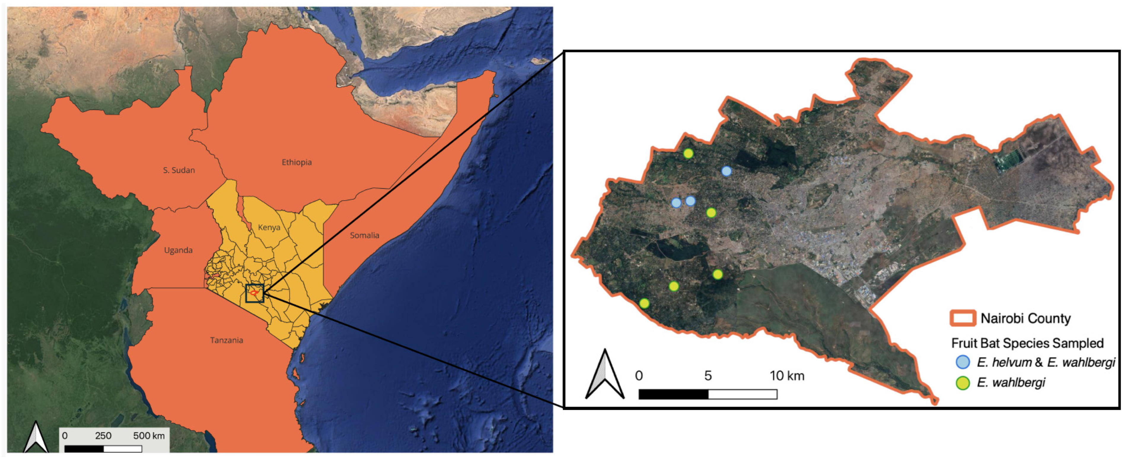
2.1. Study Area and Bat Sampling

Between June and August 2023, bat samples were obtained from E. wahlbergi and E. helvum at eight households in Nairobi, Kenya (Figure 1). Individual households were selected based on suitable habitats present for fruit bat foraging (e.g., fruiting trees), the presence of flyways (e.g., in riverine vegetation), and recent eco-epidemiological data that indicated the presence of E. wahlbergi and E. helvum in the neighborhood [32].
Bats were captured using two to three mist nets that were set up before sunset at flyways within the residential property and checked every 15 min. Only E. wahlbergi and E. helvum were retained for processing, while all other species were released at capture points. One trap night was conducted at each site and took place between the hours of 17:00 and 24:00 or until 9 targeted bats were trapped and sampled. Bats were placed in separate clean cloth bags to prevent cross-contamination and processed in order of capture. During bat handling, individuals were identified with species using published keys [17]. We recorded the body weight and forearm length of each bat and collected oropharyngeal and rectal swabs, whole blood, and fecal and urine samples when available (IACUC #SI-22051 and research permit WRTI-0247-11-22). All samples were directly stored in DNA/RNA Shield (Zymo Research, Irvine, CA, USA) for sample preservation and transferred to a −80 °C freezer within 24–48 h from field collection. Bats were released following sample collection from the processing area near the mist nets where individuals were trapped.

2.2. Sample Processing and Library Preparation

Swabs were individually processed for total nucleic acids, as described in detail in the protocol in [33]. Briefly, the samples were lysed in Proteinase K and ATL Lysis Buffer with Reagent DX (Qiagen, Valencia, CA, USA) with 0.1 mm zirconium oxide beads using a Bullet Blender (Next Advance, Troy, NY, USA). The lysate was centrifuged, and the supernatant was extracted using the IndiMag Pathogen Kit (Indical Bioscience, Leipzig, Germany) with the KingFisher™ Flex Purification System (ThermoFisher Scientific, Waltham, MA, USA). Nucleic acid concentrations were measured using a Qubit 4 Fluorometer following manufacturer instructions.
Oxford Nanopore Sequencing (ONS) cDNA libraries were prepared using NEBNext Ultra II RNA First Strand and Non-Directional RNA Second Strand Synthesis modules, utilizing random primer mix (New England Biolabs, Ipswich, MA, USA), following DNase treatment, as previously described [34]. Subsequently, double-stranded cDNA libraries were generated using the NEBNext Ultra II End repair/dA-tailing Module and Quick Ligation Modules (New England Biolabs), as well as a Ligation Sequencing Kit SQK-LSK109 (Oxford Nanopore Technologies, Oxford, UK). Cleanup and quantification were carried out using Agencourt AMPure XP reagent (Beckman Coulter Biosciences, Indianapolis, IN, USA) and a Qubit dsDNA HS Assay Kit (ThermoFisher Scientific). Samples were individually barcoded with the Native Barcoding Expansion 96—EXP-NBD 196 (Oxford Nanopore Technologies) and combined for sequencing. Sequencing libraries containing 24 barcoded pools were loaded on a single ONT R9.4.1 flow cell and run on a MinION Mk1C device (Oxford Nanopore Technologies) for 48 h.

2.3. Data Processing and Phylogenetic Analysis

Base calling and demultiplexing were accomplished on the device with MinKNOW operating software v21.11.7 (Oxford Nanopore Technologies) and Guppy v5.1.13 [35]. Raw reads were trimmed with Porechop to remove adapter sequences and then filtered with NanoFilt to remove reads with q-scores ≤9 and read lengths ≤100 bp [35,36]. Bat genomes were further removed using Minimap2 v2.24 and Samtools v1.9 [37,38]. The processed data were aligned to the National Center for Biotechnology Information (NCBI) non-redundant (NR) database using DIAMOND v2.0.14 and visualized using MEGAN6 (v6.25.9) [39,40]. The Flye assembler, developed for long single-molecule sequencing reads, was used for de novo assemblies [41]. Sequences were handled using Geneious Prime (v.2024.0.4) (Biomatters Ltd., Auckland, New Zealand). The BLASTn (2.13.0) and BLASTp (2.13.0) algorithms were used for similarity searches in the NCBI database [42]. CLUSTALW (2.0.11) was used for sequence alignment and pairwise comparisons [43]. Protein domain and motif searches were performed using the NCBI conserved domain search tool and MOTIF Search in the PFAM database [44,45].
Phylogenetic analysis was performed on sequences using IQ-TREE 2 and MEGA [46,47]. In IQ-TREE, the optimal evolutionary models and partitioning schemes were determined for amino acid sequence alignment using the automatic model selection tools (-mMFP+MERGE). Amino acid models were restricted to those designed for viral sequences (-msub viral). A 70% majority-rule consensus tree was constructed by maximum likelihood using 1000 replicates from the ultrafast bootstrap approximation approach (UFBoot) [48]. The UFBoot support values are more unbiased than normal bootstrap support, and significant clade support is considered at ≥95% [48,49]. Standard bootstrap analysis was carried out using MEGA v11.0.13 [47] for 500 replicates. The optimal analysis models were selected using the built-in “Find Best DNA/protein-substitution model” tools. Maximum likelihood trees based on nucleotide sequences were constructed using the General Time Reversible (GTR) model with a discrete Gamma distribution (+G) (ORF1a) and the General Time Reversible (GTR) model with a discrete Gamma distribution and invariant sites (+G+I) (ORF1b). Potential genetic exchange and recombination events were assessed using the RDP, GENECONV, BOOTSCAN, MAXCHI, CHIMAERA, SISCAN, and 3SEQ tools in automated and manual analyses with default settings [50]. BOOTSCAN plots were generated using SimPlot (version 3.5.1) [51].

3. Results

A total of 49 rectal swabs were processed from two fruit bat species, Wahlberg’s epauletted fruit bats (E. wahlbergi, 43/49, 87.7%) and straw-colored fruit bats (E. helvum, 6/49, 12.2%). Viral contigs were generated by de novo assembly from a single E. wahlbergi sample (1/49, 2.0%). Through the remapping of reads to assembled contigs and producing alignments, a single virus contig was generated that revealed the identities of several Nobecoviruses from Africa in BLAST queries.

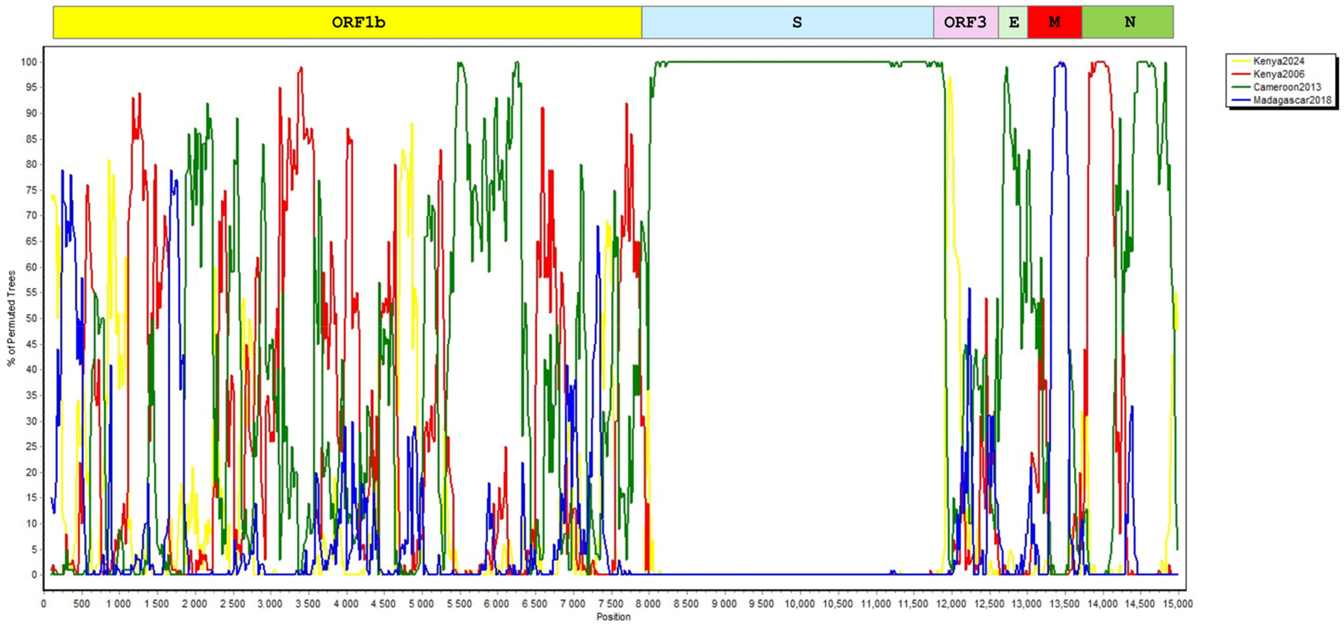
3.1. Genome Annotation and Phylogenetic and Recombination Analyses

The virus contig (referred to as NRB24) comprised 21,973 nucleotides, covering 75–76% of bat Nobecovirus genomes. Seven open reading frames (ORFs) were identified, including partial ORF1a (1−7099) and complete ORF1b (7099–15,114), spike (S) glycoprotein (15,086−18,910), ORF3 (18,911–19,609), envelope (E) protein (19,609−19,836), membrane (M) glycoprotein (19,841−20,509), and nucleocapsid (N) protein (20,564−21,973, Figure S1). As observed in coronaviruses, ORF1a and ORF1b partially overlap in NRB24, where ORF1b is in the −1 reading frame relative to the ORF1a stop codon (7097−7099), enabling the expression of ORF1b-encoded protein expression by cis-acting RNA elements that direct a fraction of elongating ribosomes to slip (programmed ribosomal frameshifting) [52].
Pairwise comparisons of individual ORFs with Nobecoviruses from Africa revealed identities of up to 77.6% and 91.9% in nucleotide and putative amino acid sequences, respectively (Table S1). Maximum likelihood analysis based on complete putative ORF1a and ORF1b nucleotide and amino acid sequences showed that NRB24 formed a separate subclade, distinct from previously described Nobecovirus clades, with strong bootstrap support (Figure 2 and Figure S2) [53]. Similar tree topologies were observed in maximum likelihood trees constructed using S, ORF3, and N putative amino acid alignments as well (Figure S3). In each tree, NRB24 remained distinct, sharing a common ancestor with the African Nobecovirus clade, which includes all geographically related virus genomes. Moreover, a comparable tree topology was observed during the analysis of a shorter nucleotide fragment encoding for virus RNA-dependent RNA polymerase (RdRp) (Figure 3), which was documented to delineate major Nobecovirus clades [53]. However, the separate grouping of NRB24 was not apparent in the maximum likelihood trees based on putative E and M amino acid alignments, presumably due to their relatively limited sizes (Figure S3).
We carried out further in silico analyses of complete ORF sequences to detect potential recombinations in Nobecoviruses from Africa, including NRB24. Probable recombinations involving multiple spots were detected using all tools. In the BOOTSCAN plot, recombination signals were observed to involve all ORFs but the S protein (Figure 4), suggesting coinfections and intra-host genetic exchange driving genome diversification.

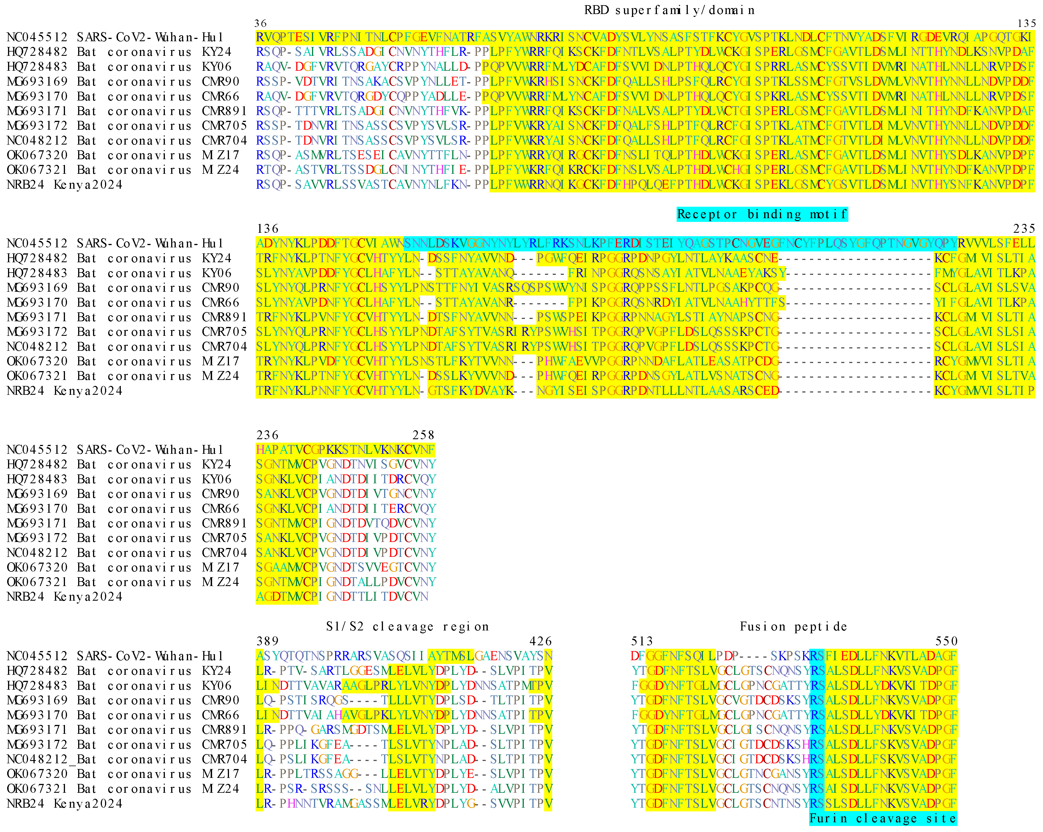
3.2. Analysis of Putative Viral Proteins

We examined the putative amino acid sequences of ORF1a (2365), ORF1b (2579), S glycoprotein (1274), ORF3 (232), E protein (75), M glycoprotein (222), and N protein (469). The ORF1a protein was partially identified with the virus non-structural protein (Nsp), and 3–10 conserved domains were identified (Table S2). Similarly, the complete ORF1b revealed conserved motifs of the RdRp catalytic core, as well as Nsp13–16, with various functions required for virus replication. The domains of the main coronavirus structural proteins, S, E, M, and N were further observed in the NRB24 contig (Table 1).
In coronaviruses, the S protein is critical for host range and virulence, as it mediates target cell attachment and the membrane fusion required for virus entry [27]. We identified the S1-S2 subunits, the S1/S2 and furin cleavage sites, the fusion peptide region, and associated motifs in NRB24 (Figure 5). Like other Nobecoviruses, NRB24 lacked the SARS-CoV-2 receptor binding motif and shared S1/S2 and furin cleavage sites and fusion peptide markers with Nobecoviruses.

4. Discussion

Here, we report a partial Betacoronavirus genome representing a distinct Nobecovirus subclade related to African Nobecoviruses, detected in a rectal swab from a Wahlberg’s epauletted fruit bat (E. wahlbergi) from Nairobi, Kenya. Tentatively named NRB24, the partial viral contig—covering >75% of Nobecovirus genomes—was generated by untargeted metagenome screening. The contig encompassed viral structural proteins (S, E, M, and N), as well as regions encoding for the core replication enzymes and main co-factors encoded by ORF1a and ORF1b. In phylogenetic analyses, it emerged as a novel Nobecovirus subclade, sharing a common ancestor with the Eidolon/Rousettus Nobecovirus subclade from Africa. We detected several functional domains on the NRB24 putative structural and non-structural proteins. Further analysis of the S protein revealed shared markers and cleavage sites with Nobecoviruses. We did not detect RNA derived from NRB24 in oral swabs from the same individual bat, suggesting probable tropism for gastrointestinal tissue.
Zoonotic capacity in particular subgenera, such as Sarbecoviruses (including SARS-CoV and SARS-CoV-2) and Merbecoviruses (including MERS-CoV) has been documented and caused spillover events with a significant health impact [28,29]. So far, no zoonotic potential has been recognized in Nobecoviruses, which are described exclusively in fruit bats of Pteropodidae [27,31]. Moreover, scarce information is available on Nobecovirus cell receptors and target cells. Compared to other bat-associated Sarbecovirus and Merbecoviruses, Nobecoviruses have been observed to infect fewer bat host species, which might indicate host specificity toward Rousettus and Eonycteris fruit bats (family Pteropodidae) [31,53]. Importantly, our findings describe another Pteropodidae species that can harbor Nobecoviruses.
All Pteropodidae species are distributed in tropical and subtropical areas of the Old World and have been documented to harbor zoonotic viruses, including the Ebola, Marburg, Hendra, and Nipah viruses [31,54]. Despite a lack of direct evidence of Nobecoviruses, examples of virus spillover to humans and recombination events have been documented [55,56], highlighting the potential of viruses hosted by Pteropodidae species. In this study, we documented potential recombination events among various Nobecoviruses of African origin, including the newly described NRB24, despite lacking the S protein, which is crucial for the host range. As exemplified by SARS-CoV and MERS-CoV, recombinations facilitate direct bat-to-human spillover and cross-species emergence via intermediary bridge hosts [28,29,57]. The potential of Nobecovirus to gain zoonotic potential through recombinations should not be underestimated, particularly when fruit bat hosts share habitats with potential intermediate hosts like livestock, as with our sites. It is imperative to continue active surveillance of fruit bat species in the Afrotropics and tropical and subtropical regions of Southeast Asia for emerging corona- and other viruses and to better understand their diverse virome. Despite challenges in identifying adequate bat habitats on private properties in a city environment, limiting our sample size, this study provided novel viral sequences obtained in a non-invasive manner from urban bats, contributing to our knowledge of Nobecoviruses and their bat host associations.

Supplementary Materials

The following supporting information can be downloaded at https://www.mdpi.com/article/10.3390/v17040557/s1: Figure S1: NRB24 genome annotation of seven open reading frames and genome coverage. Figure S2: Maximum likelihood tree of coronaviruses ORF1a (7472 nucleotides) and ORF1b (8033 nucleotides). Figure S3: Maximum likelihood consensus tree of coronavirus S (3949 amino acids), E (75 amino acids), ORF3 (266 amino acids), M (215 amino acids), and N (477 amino acids) sequences. Table S1: Pairwise comparison of nucleotide and deduced amino acid sequences of NRB24 and related Nobecovirus ORFs. Table S2: Conserved domains identified in NRB24 partial ORF1a putative amino acid sequence (n = 2365).

Author Contributions

Wildlife specimen collection and processing: M.C.V., P.W.W., E.K. and A.M.; Laboratory analysis: M.K., J.M., R.L. and G.O.O.; Data processing and interpretation: K.E. and B.P.B.; Manuscript drafting: K.E. and M.C.V.; Study design, M.C.V., J.H., E.M.F., J.A.S., M.E.C., Y.-M.L. and S.M.; Administrative processing: Y.-M.L., E.G.M., N.L.A., L.J. and J.P.G. All authors have read and agreed to the published version of the manuscript.

Funding

Sample collection was supported by the Smithsonian Institution’s George E. Burch Fellowship funding. This work was supported by the US Army Medical Research and Development Command under Contract Nos. W81XWH-21-C-0001, W81XWH-22-C-0093, and HT9425-23-C-0059. The views, opinions, and/or findings contained in this report are those of the author(s) and should not be construed as an official Department of the Army or Navy position, policy, or decision unless so designated by other documentation. The opinions or assertions contained herein are the private views of the authors and are not to be construed as official or as reflecting the true views of the Departments of the Army, Navy, or Defense. Material contained within this publication has been reviewed by the Walter Reed Army Institute of Research. There is no objection to its presentation and/or publication. This manuscript was prepared while K.E. and B.P.B. held National Research Council (NRC) Research Associateship Awards at the Walter Reed Biosystematics Unit through the Walter Reed Army Institute of Research, Silver Spring, MD, USA. Field sites for this project were established through the Urban Zoo project, supported by the UK Medical Research Council, the Biotechnology and Biological Science Research Council (UK), the Economic and Social Research Council (UK), and the Natural Environment Research Council (UK) through the Environmental & Social Ecology of Human Infectious Diseases Initiative (ESEI), Grant Reference: G1100783/1. This work also received support from the CGIAR One Health initiative “Protecting Human Health Through a One Health Approach”, which was supported by contributors to the CGIAR Trust Fund (https://www.cgiar.org/funders/, accessed on 1 February 2025).

Institutional Review Board Statement

The study protocol was approved by the National Zoological Park Institutional Animal Care and Use Committee (NZP-IACUC #SI-22051) and by the Kenyan Wildlife Research and Training Institute (WRTI-0247-11-22). All collection, sampling, and subsequent experiments were performed in accordance with the approved protocols.

Informed Consent Statement

Informed consent was obtained from all subjects involved in the study.

Data Availability Statement

Raw sequence data generated in this study are available in the NCBI Sequence Read Archive (SRA) under project PRJNA1237873 and the assembled virus genome sequence is available in GenBank under accession number PQ179293.

Acknowledgments

We want to deeply thank the household members who provided access to their properties for bat sampling. This work would not have been possible without their generosity.

Conflicts of Interest

The authors declare no conflicts of interest.

References

  1. Plowright, R.K. Land Use-Induced Spillover: A Call to Action to Safeguard Environmental. Anim. Hum. Health 2021, 5, e237–e245. [Google Scholar]
  2. Plowright, R.K.; Parrish, C.R.; McCallum, H.; Hudson, P.J.; Ko, A.I.; Graham, A.L.; Lloyd-Smith, J.O. Pathways to Zoonotic Spillover. Nat. Rev. Microbiol. 2017, 15, 502–510. [Google Scholar] [CrossRef]
  3. Carlson, C.J.; Zipfel, C.M.; Garnier, R.; Bansal, S. Global Estimates of Mammalian Viral Diversity Accounting for Host Sharing. Nat. Ecol. Evol. 2019, 3, 1070–1075. [Google Scholar] [CrossRef]
  4. Olival, K.J.; Hosseini, P.R.; Zambrana-Torrelio, C.; Ross, N.; Bogich, T.L.; Daszak, P. Host and Viral Traits Predict Zoonotic Spillover from Mammals. Nature 2017, 546, 646–650. [Google Scholar] [CrossRef]
  5. Mollentze, N.; Streicker, D.G. Viral Zoonotic Risk Is Homogenous among Taxonomic Orders of Mammalian and Avian Reservoir Hosts. Proc. Natl. Acad. Sci. USA 2020, 117, 9423–9430. [Google Scholar] [CrossRef]
  6. Han, B.A.; Kramer, A.M.; Drake, J.M. Global Patterns of Zoonotic Disease in Mammals. Trends Parasitol. 2016, 32, 565–577. [Google Scholar] [CrossRef] [PubMed]
  7. Brook, C.E.; Dobson, A.P. Bats as ‘Special’ Reservoirs for Emerging Zoonotic Pathogens. Trends Microbiol. 2015, 23, 172–180. [Google Scholar] [CrossRef] [PubMed]
  8. Ahn, M.; Anderson, D.E.; Zhang, Q.; Tan, C.W.; Lim, B.L.; Luko, K.; Wen, M.; Chia, W.N.; Mani, S.; Wang, L.C.; et al. Dampened NLRP3-Mediated Inflammation in Bats and Implications for a Special Viral Reservoir Host. Nat. Microbiol. 2019, 4, 789–799. [Google Scholar] [CrossRef]
  9. Brook, C.E.; Boots, M.; Chandran, K.; Dobson, A.P.; Drosten, C.; Graham, A.L.; Grenfell, B.T.; Müller, M.A.; Ng, M.; Wang, L.-F.; et al. Accelerated Viral Dynamics in Bat Cell Lines, with Implications for Zoonotic Emergence. eLife 2020, 9, e48401. [Google Scholar] [CrossRef]
  10. Li, W.; Shi, Z.; Yu, M.; Ren, W.; Smith, C.; Epstein, J.H.; Wang, H.; Crameri, G.; Hu, Z.; Zhang, H.; et al. Bats Are Natural Reservoirs of SARS-Like Coronaviruses. Science 2005, 310, 676–679. [Google Scholar] [CrossRef]
  11. Lytras, S.; Hughes, J.; Martin, D.; Swanepoel, P.; De Klerk, A.; Lourens, R.; Kosakovsky Pond, S.L.; Xia, W.; Jiang, X.; Robertson, D.L. Exploring the Natural Origins of SARS-CoV-2 in the Light of Recombination. Genome Biol. Evol. 2022, 14, evac018. [Google Scholar] [CrossRef]
  12. Lytras, S.; Xia, W.; Hughes, J.; Jiang, X.; Robertson, D.L. The Animal Origin of SARS-CoV-2. Science 2021, 373, 968–970. [Google Scholar] [CrossRef] [PubMed]
  13. Johnson, C.K.; Hitchens, P.L.; Pandit, P.S.; Rushmore, J.; Evans, T.S.; Young, C.C.W.; Doyle, M.M. Global Shifts in Mammalian Population Trends Reveal Key Predictors of Virus Spillover Risk. Proc. R. Soc. B Biol. Sci. 2020, 287, 20192736. [Google Scholar] [CrossRef]
  14. Mammal Diversity Database, Mammal Diversity Database (Version 2.0). Zenodo 2025. [CrossRef]
  15. Raven, P.H.; Gereau, R.E.; Phillipson, P.B.; Chatelain, C.; Jenkins, C.N.; Ulloa, C.U. The Distribution of Biodiversity Richness in the Tropics. Sci. Adv. 2020, 6, eabc6228. [Google Scholar] [CrossRef] [PubMed]
  16. Herkt, K.M.B.; Barnikel, G.; Skidmore, A.K.; Fahr, J. A High-Resolution Model of Bat Diversity and Endemism for Continental Africa. Ecol. Model. 2016, 320, 9–28. [Google Scholar] [CrossRef]
  17. Patterson, B.D.; Webala, P. Keys to the Bats (Mammalia: Chiroptera) of East Africa. Fieldiana Life Earth Sci. 2012, 2012, 1–60. [Google Scholar] [CrossRef]
  18. Musila, S.; Monadjem, A.; Webala, P.W.; Patterson, B.D.; Hutterer, H.; De Jong, Y.A.; Butynski, T.M.; Mwangi, G.; Chen, Z.Z. An Annotated Checklist of Mammals of Kenya. Zool. Res. 2018, 40, 3–52. [Google Scholar] [CrossRef]
  19. Hassell, J.M.; Muloi, D.M.; VanderWaal, K.L.; Ward, M.J.; Bettridge, J.; Gitahi, N.; Ouko, T.; Imboma, T.; Akoko, J.; Karani, M.; et al. Epidemiological Connectivity between Humans and Animals across an Urban Landscape. Proc. Natl. Acad. Sci. USA 2023, 120, e2218860120. [Google Scholar] [CrossRef]
  20. Conrardy, C.; Tao, Y.; Kuzmin, I.V.; Niezgoda, M.; Agwanda, B.; Breiman, R.F.; Anderson, L.J.; Rupprecht, C.E.; Tong, S. Molecular Detection of Adenoviruses, Rhabdoviruses, and Paramyxoviruses in Bats from Kenya. Am. J. Trop. Med. Hyg. 2014, 91, 258–266. [Google Scholar] [CrossRef]
  21. Kareinen, L.; Ogola, J.; Kivistö, I.; Smura, T.; Aaltonen, K.; Jääskeläinen, A.J.; Kibiwot, S.; Masika, M.M.; Nyaga, P.; Mwaengo, D.; et al. Range Expansion of Bombali Virus in Mops Condylurus Bats, Kenya, 2019. Emerg. Infect. Dis. 2020, 26, 3007–3010. [Google Scholar] [CrossRef] [PubMed]
  22. Kuzmin, I.V.; Niezgoda, M.; Franka, R.; Agwanda, B.; Markotter, W.; Breiman, R.F.; Shieh, W.J.; Zaki, S.R. Marburg Virus in Fruit Bat, Kenya. Emerg. Infect. Dis. 2010, 16, 352–354. [Google Scholar] [CrossRef]
  23. Tao, Y.; Tang, K.; Shi, M.; Conrardy, C.; Li, K.S.M.; Lau, S.K.P.; Anderson, L.J.; Tong, S. Genomic Characterization of Seven Distinct Bat Coronaviruses in Kenya. Virus Res. 2012, 167, 67–73. [Google Scholar] [CrossRef]
  24. Tong, S.; Conrardy, C.; Ruone, S.; Kuzmin, I.V.; Guo, X.; Tao, Y.; Niezgoda, M.; Haynes, L.; Agwanda, B.; Breiman, R.F.; et al. Detection of Novel SARS-like and Other Coronaviruses in Bats from Kenya. Emerg. Infect. Dis. 2009, 15, 482–485. [Google Scholar] [CrossRef]
  25. Waruhiu, C.; Ommeh, S.; Obanda, V.; Agwanda, B.; Gakuya, F.; Ge, X.-Y.; Yang, X.-L.; Wu, L.-J.; Zohaib, A.; Hu, B.; et al. Molecular Detection of Viruses in Kenyan Bats and Discovery of Novel Astroviruses, Caliciviruses and Rotaviruses. Virol. Sin. 2017, 32, 101–114. [Google Scholar] [CrossRef] [PubMed]
  26. Kamau, J.; Ergunay, K.; Webala, P.W.; Justi, S.A.; Bourke, B.P.; Kamau, M.W.; Hassell, J.; Chege, M.N.; Mwaura, D.K.; Simiyu, C.; et al. A Novel Coronavirus and a Broad Range of Viruses in Kenyan Cave Bats. Viruses 2022, 14, 2820. [Google Scholar] [CrossRef] [PubMed]
  27. Woo, P.; de Groot, R.; Haagmans, B.; Lau, S.; Neuman, B.; Perlman, S.; Sola, I.; van der Hoek, L.; Wong, A.; Yeh, S.-H. ICTV Virus Taxonomy Profile: Coronaviridae 2023. J. Gen. Virol. 2023, 104, 001843. [Google Scholar] [CrossRef]
  28. Hu, B.; Zeng, L.-P.; Yang, X.-L.; Ge, X.-Y.; Zhang, W.; Li, B.; Xie, J.-Z.; Shen, X.-R.; Zhang, Y.-Z.; Wang, N.; et al. Discovery of a Rich Gene Pool of Bat SARS-Related Coronaviruses Provides New Insights into the Origin of SARS Coronavirus. PLoS Pathog. 2017, 13, e1006698. [Google Scholar] [CrossRef]
  29. Anthony, S.J.; Gilardi, K.; Menachery, V.D.; Goldstein, T.; Ssebide, B.; Mbabazi, R.; Navarrete-Macias, I.; Liang, E.; Wells, H.; Hicks, A.; et al. Further Evidence for Bats as the Evolutionary Source of Middle East Respiratory Syndrome Coronavirus. mBio 2017, 8, e00373-17. [Google Scholar] [CrossRef]
  30. Frutos, R.; Serra-Cobo, J.; Pinault, L.; López-Roig, M.; Devaux, C. Emergence of Bat-Related Betacoronaviruses: Hazard and Risks. Front. Microbiol. 2021, 12, 591535. [Google Scholar] [CrossRef]
  31. Wong, A.C.P.; Li, X.; Lau, S.K.P.; Woo, P.C.Y. Global Epidemiology of Bat Coronaviruses. Viruses 2019, 11, 174. [Google Scholar] [CrossRef] [PubMed]
  32. Muloi, D.M.; Wee, B.A.; McClean, D.M.H.; Ward, M.J.; Pankhurst, L.; Phan, H.; Ivens, A.C.; Kivali, V.; Kiyong’a, A.; Ndinda, C.; et al. Population Genomics of Escherichia Coli in Livestock-Keeping Households across a Rapidly Developing Urban Landscape. Nat. Microbiol. 2022, 7, 581–589. [Google Scholar] [CrossRef] [PubMed]
  33. REDI-NET Consortium REDI-NET: SW-2 SWAB PROCESSING. protocols.io 2024. [CrossRef]
  34. REDI-NET Consortium REDI-NET: SW-4 SWAB TESTING. protocols.io 2024. [CrossRef]
  35. Wick, R.R.; Judd, L.M.; Holt, K.E. Performance of Neural Network Basecalling Tools for Oxford Nanopore Sequencing. Genome Biol. 2019, 20, 129. [Google Scholar] [CrossRef]
  36. De Coster, W.; D’Hert, S.; Schultz, D.T.; Cruts, M.; Van Broeckhoven, C. NanoPack: Visualizing and Processing Long-Read Sequencing Data. Bioinformatics 2018, 34, 2666–2669. [Google Scholar] [CrossRef]
  37. Danecek, P.; Bonfield, J.K.; Liddle, J.; Marshall, J.; Ohan, V.; Pollard, M.O.; Whitwham, A.; Keane, T.; McCarthy, S.A.; Davies, R.M.; et al. Twelve Years of SAMtools and BCFtools. Gigascience 2021, 10, giab008. [Google Scholar] [CrossRef]
  38. Li, H. Minimap2: Pairwise Alignment for Nucleotide Sequences. Bioinformatics 2018, 34, 3094–3100. [Google Scholar] [CrossRef]
  39. Buchfink, B.; Reuter, K.; Drost, H. Sensitive Protein Alignments at Tree-of-Life Scale Using DIAMOND. Nat. Methods 2021, 18, 366–368. [Google Scholar] [CrossRef]
  40. Huson, D.; Beier, S.; Flade, I.; Górska, A.; El-Hadidi, M.; Mitra, S.; Ruscheweyh, H.-J.; Tappu, R. MEGAN Community Edition—Interactive Exploration and Analysis of Large-Scale Microbiome Sequencing Data. PLoS Comput. Biol. 2016, 12, e1004957. [Google Scholar] [CrossRef]
  41. Kolmogorov, M.; Yuan, J.; Lin, Y.; Pevzner, P.A. Assembly of Long, Error-Prone Reads Using Repeat Graphs. Nat. Biotechnol. 2019, 37, 540–546. [Google Scholar] [CrossRef] [PubMed]
  42. Altschul, S.F.; Gish, W.; Miller, W.; Myers, E.W.; Lipman, D.J. Basic Local Alignment Search Tool. J. Mol. Biol. 1990, 215, 403–410. [Google Scholar] [CrossRef] [PubMed]
  43. Thompson, J.D.; Higgins, D.G.; Gibson, T.J. CLUSTAL W: Improving the Sensitivity of Progressive Multiple Sequence Alignment through Sequence Weighting, Position-Specific Gap Penalties and Weight Matrix Choice. Nucleic Acids Res. 1994, 22, 4673–4680. [Google Scholar] [CrossRef] [PubMed]
  44. Bateman, A.; Birney, E.; Cerruti, L.; Ettwiller, L.; Eddy, S.; Griffiths-Jones, S.; Howe, K.; Marshall, M.; Sonnhammer, E. The Pfam Protein Families Database. Nucleic Acids Res. 2002, 30, 276–280. [Google Scholar] [CrossRef]
  45. Marchler-Bauer, A.; Derbyshire, M.; Gonzales, N.; Lu, S.; Chitsaz, F.; Geer, L.; Geer, R.; He, J.; Gwadz, M.; Hurwitz, D.; et al. CDD: NCBI’s Conserved Domain Database. Nucleic Acids Res. 2014, 43, D222–D226. [Google Scholar] [CrossRef]
  46. Minh, B.Q.; Schmidt, H.A.; Chernomor, O.; Schrempf, D.; Woodhams, M.D.; Von Haeseler, A.; Lanfear, R. IQ-TREE 2: New Models and Efficient Methods for Phylogenetic Inference in the Genomic Era. Mol. Biol. Evol. 2020, 37, 1530–1534. [Google Scholar] [CrossRef]
  47. Tamura, K.; Stecher, G.; Kumar, S. MEGA11: Molecular Evolutionary Genetics Analysis Version 11. Mol. Biol. Evol. 2021, 38, 3022–3027. [Google Scholar] [CrossRef]
  48. Minh, B.Q.; Nguyen, M.A.T.; Von Haeseler, A. Ultrafast Approximation for Phylogenetic Bootstrap. Mol. Biol. Evol. 2013, 30, 1188–1195. [Google Scholar] [CrossRef]
  49. Hoang, D.T.; Chernomor, O.; von Haeseler, A.; Minh, B.Q.; Vinh, L.S. UFBoot2: Improving the Ultrafast Bootstrap Approximation. Mol. Biol. Evol. 2018, 35, 518–522. [Google Scholar] [CrossRef]
  50. Martin, D.P.; Murrell, B.; Golden, M.; Khoosal, A.; Muhire, B. RDP4: Detection and Analysis of Recombination Patterns in Virus Genomes. Virus Evol. 2015, 1, vev003. [Google Scholar] [CrossRef]
  51. Lole, K.S.; Bollinger, R.C.; Paranjape, R.S.; Gadkari, D.; Kulkarni, S.S.; Novak, N.G.; Ingersoll, R.; Sheppard, H.W.; Ray, S.C. Full-Length Human Immunodeficiency Virus Type 1 Genomes from Subtype C-Infected Seroconverters in India, with Evidence of Intersubtype Recombination. J. Virol. 1999, 73, 152–160. [Google Scholar] [CrossRef] [PubMed]
  52. Yan, S.; Zhu, Q.; Hohl, J.; Dong, A.; Schlick, T. Evolution of Coronavirus Frameshifting Elements: Competing Stem Networks Explain Conservation and Variability. Proc. Natl. Acad. Sci. USA 2023, 120, e2221324120. [Google Scholar] [CrossRef] [PubMed]
  53. Kettenburg, G.; Kistler, A.; Ranaivoson, H.C.; Ahyong, V.; Andrianiaina, A.; Andry, S.; DeRisi, J.L.; Gentles, A.; Raharinosy, V.; Randriambolamanantsoa, T.H.; et al. Full Genome Nobecovirus Sequences from Malagasy Fruit Bats Define a Unique Evolutionary History for this Coronavirus Clade. Front. Public Health 2022, 10, 786060. [Google Scholar] [CrossRef] [PubMed]
  54. Geldenhuys, M.; Mortlock, M.; Epstein, J.H.; Paweska, J.; Weyer, J.; Markotter, W. Overview of Bat and Wildlife Coronavirus Surveillance in Africa: A Framework for Global Investigations. Viruses 2021, 13, 936. [Google Scholar] [CrossRef]
  55. Chua, K.B.; Crameri, G.; Hyatt, A.; Yu, M.; Tompang, M.R.; Rosli, J.; McEachern, J.; Crameri, S.; Kumarasamy, V.; Eaton, B.T.; et al. A Previously Unknown Reovirus of Bat Origin is Associated with an Acute Respiratory Disease in Humans. Proc. Natl. Acad. Sci. USA 2007, 104, 11424–11429. [Google Scholar] [CrossRef]
  56. Huang, C.; Liu, W.J.; Xu, W.; Jin, T.; Zhao, Y.; Song, J.; Shi, Y.; Ji, W.; Jia, H.; Zhou, Y.; et al. A Bat-Derived Putative Cross-Family Recombinant Coronavirus with a Reovirus Gene. PLOS Pathog. 2016, 12, e1005883. [Google Scholar] [CrossRef]
  57. Wong, A.C.P.; Lau, S.K.P.; Woo, P.C.Y. Interspecies Jumping of Bat Coronaviruses. Viruses 2021, 13, 2188. [Google Scholar] [CrossRef]
Figure 1. Regional map of East Africa with Nairobi County highlighted. Within Nairobi County (right), we trapped foraging fruit bats in west and central Nairobi where suitable peridomestic habitats were found. Blue points indicate households where E. helvum and E. wahlbergi were trapped and sampled, and green points represent households where only E. wahlbergi was trapped and sampled.
Viruses 17 00557 g001
Figure 2. The maximum likelihood consensus tree of coronavirus ORF1a ((A): 2419 amino acids) and ORF1b ((B): 2528 amino acids) sequences. The trees were constructed using 1000 replicates. Branches achieving ≥95% bootstrap support are annotated with red dots. Viruses are indicated by GenBank accession, name, and isolate identifier. NRB24 and related virus genomes are marked. SARS-CoV-2 Wuhan-Hu-1 (Sarbecovirus) and Hipposideros coronaviruses CD36/PDF0663 (Hibecovirus) were included as outgroups as needed.
Viruses 17 00557 g002
Figure 3. The maximum likelihood consensus tree of the coronavirus RNA-dependent RNA polymerase (RdRp) sequences (256 nucleotides). The trees were constructed using 1000 replicates. Branches achieving ≥95% bootstrap support are annotated with red dots. Viruses are indicated by GenBank accession, name, and isolate identifier. Previously described Nobecovirus clades [53] are marked. SARS-CoV-2 Wuhan-Hu-1 (Sarbecovirus) and bat coronavirus Zhejiang2013 (Hibecovirus) were included as outgroups.
Viruses 17 00557 g003
Figure 4. BOOTSCAN plot of African bat Nobecovirus genomes. The alignment encompasses 15096 positions with the corresponding ORFs plotted on top. The plot was prepared within a sliding window of 200 bp, wide-centered on the position plotted, with a 20 bp step size, for 1000 replications (GapStrip: on, neighbor-joining, T/t: 2.0), with the bat coronavirus CMR704-P12 genome (NC048212, Eidolon helvum, Cameroon) as the query. Refer to Table 1 for a description of genomes in groups labeled Kenya2024 (NRB24), Kenya2006, Madagascar2018, and Cameroon2013.
Viruses 17 00557 g004
Figure 5. Alignment of domains involved in receptor binding of coronavirus S (spike) protein. Amino acid positions are provided according to the SARS-CoV2 Wuhan-Hu-1 strain. Conserved residues are highlighted (RBD domain, S1/S2 cleavage site, and fusion peptide: yellow; receptor binding motif and furin cleavage site: cyan).
Viruses 17 00557 g005
Table 1. Comparative topology and conserved domains identified in complete regions in NRB24 and closely related virus genomes.
HostEpomophorus wahlbergiEidolon
helvum
Rousettus
aegyptiacus
Eidolon helvumRousettus madagascariensis
Location, YearKenya, 2024Kenya 2006Kenya 2006Cameroon, 2013Madagascar, 2018
IsolateNRB24KY24KY06CMR900CMR66CMR891-892CMR705-P13CMR704-P12MIZ178MIZ240
GenBank AccessionPQ179293HQ728482.1HQ728483.1MG693169.1MG693170.1MG693171.1MG693172.1NC_048212.1OK067320.1OK067321.1
ORF1bSize2579257925882579258825792579257925792579
Catalytic core—RNA
polymerase (477363)
1–8231–8231–8231–8231–8231–8231–8231–8231–8231–823
Nsp13—zinc-binding
domain (cl41714)
824–918824–918824–918824–918824–918824–918824–918824–918824–918824–918
Nsp13—stalk (cl41715)922–969922–969922–969922–969922–969922–969922–969922–969922–969922–969
Nsp13—1B domain (cl41715)973–1051973–1051973–1051973–1051973–1051973–1051973–1051973–1051973–1051973–1051
Nsp13—helicase domain (cl41748)1074–14131074–14131074–14131074–14131074–14131074–14131074–14131074–14131074–14131074–1413
Nsp14 (cl40464)1428–19501428–19511428–19521428–19511428–19521428–19511428–19511428–19511428–19511428–1951
Nsp15—N terminal
domain (cl40469)
1954–20141954–20141955–20151954–20141955–20151954–20141954–20141954–20141954–20141954–2014
Nsp15—M domain (cl41717)2018–21462018–21462019–21432018–21462019–21432018–21462018–21462018–21462018–21462018–2146
Nsp15—endoribonuclease domain (cl41718)2144–22922144–22922141–22892144–22922141–22892144–22922144–22922144–22922144–22922144–2292
Nsp16—methyltransferase (461919, cl41719)2297–25782323–25372297–25782323–25372297–25782323–25372323–25372323–25372297–25782323–2537
SSize1274126412781273127812711269126912561265
S1—N terminal domain (cd21627)30–32333–31836–32733–32636–32733–32531–32231–32234–31242–321
Receptor binding domain (cl09656)367–521362–516371–521370–528371–521369–523366–524366–524356–511365–519
S1/S2 cleavage + S2 fusion domain (cd22381)538–1268533–1258538–1273545–1267538–1272540–1265541–1263541–1263528–1250536–1259
ORF3Size232238220238220238239239238238
ESize75757575757575757575
Envelope protein (cl40474)------4–634–634–634–63
MSize222221222221223221221221221221
Matrix protein (cl40475)11–2225–2227–2228–2217–2235–22110–22110–2218–2218–221
NSize469467468468468467470470467467
Nucleocapsid protein (cl47612)53–39653–37454–37554–37554–39554–37554–37554–37553–37453–374
Locations are provided according to the individual viral proteins. NCBI accessions are provided in parentheses.
Disclaimer/Publisher’s Note: The statements, opinions and data contained in all publications are solely those of the individual author(s) and contributor(s) and not of MDPI and/or the editor(s). MDPI and/or the editor(s) disclaim responsibility for any injury to people or property resulting from any ideas, methods, instructions or products referred to in the content.

