Article

DENV-1 Titer Impacts Viral Blocking in wMel Aedes aegypti with Brazilian Genetic Background

by
Jessica Corrêa-Antônio
1,
Mariana R. David
1,
Dinair Couto-Lima
1,
Gabriela Azambuja Garcia
1,
Milan S. G. Keirsebelik
1,
Rafael Maciel-de-Freitas
1,2 and
Márcio Galvão Pavan
1,*
1
Laboratório de Mosquitos Transmissores de Hematozoários, Instituto Oswaldo Cruz, Fiocruz, Rio de Janeiro 21040-900, Brazil
2
Department of Arbovirology, Bernhard Nocht Institute of Tropical Medicine, 20359 Hamburg, Germany
*
Author to whom correspondence should be addressed.
Viruses 2024, 16(2), 214; https://doi.org/10.3390/v16020214
Submission received: 27 December 2023 / Revised: 23 January 2024 / Accepted: 28 January 2024 / Published: 31 January 2024
(This article belongs to the Special Issue Molecular Epidemiology, Evolution, and Dispersion of Flaviviruses)

Abstract

:
Several countries have been using Wolbachia deployments to replace highly competent native Aedes aegypti populations with Wolbachia-carrying mosquitoes with lower susceptibility to arboviruses such as dengue, Zika, and chikungunya. In Rio de Janeiro, Wolbachia deployments started in 2015 and still present a moderate introgression with a modest reduction in dengue cases in humans (38%). Here, we evaluated the vector competence of wild-type and wMel-infected Ae. aegypti with a Brazilian genetic background to investigate whether virus leakage could contribute to the observed outcomes in Brazil. We collected the specimens in three areas of Rio de Janeiro with distinct frequencies of mosquitoes with wMel strain and two areas with wild Ae. aegypti. The mosquitoes were orally exposed to two titers of DENV-1 and the saliva of DENV-1-infected Ae. aegypti was microinjected into wMel-free mosquitoes to check their infectivity. When infected with the high DENV-1 titer, the presence of wMel did not avoid viral infection in mosquitoes’ bodies and saliva but DENV-1-infected wMel mosquitoes produced lower viral loads than wMel-free mosquitoes. On the other hand, wMel mosquitoes infected with the low DENV-1 titer were less susceptible to virus infection than wMel-free mosquitoes, although once infected, wMel and wMel-free mosquitoes exhibited similar viral loads in the body and the saliva. Our results showed viral leakage in 60% of the saliva of wMel mosquitoes with Brazilian background; thus, sustained surveillance is imperative to monitor the presence of other circulating DENV-1 strains capable of overcoming the Wolbachia blocking phenotype, enabling timely implementation of action plans.

1. Introduction

Arthropod-borne viruses (arboviruses) such as dengue (DENV), Zika (ZIKV), and chikungunya (CHIKV) have spread in urbanized areas around the world, representing a significant public health burden, with 3.9 billion people at risk in 129 countries [1]. Dengue is responsible for more than 90% of the arboviral cases reported in the Americas, reaching 2.8 million cases in 2022 [2,3]. Aedes aegypti (Diptera: Culicidae) is the primary vector, and it is highly associated with anthropogenic environments, where female mosquitoes lay eggs mainly in artificial containers inside or near human habitations and feed preferentially on humans [4,5].
Due to a lack of a vaccine broadly available for many arboviruses, the most recommended way to mitigate transmission is through intensifying vector control. Traditional control methods, such as the search for and elimination of breeding sites and the use of insecticides, present limited effectiveness in avoiding outbreaks [6]. Given this challenging scenario, designing alternative effective interventions is critical to reducing the disease burden. One of these strategies is the release of Ae. aegypti infected with the intracellular symbiont Wolbachia pipientis (Rickettsiales, Anaplasmataceae). This bacterium is naturally present in nearly 40% of arthropods but not in Ae. aegypti [7]. Its use is based on at least three phenotypes to support introgression into native mosquito populations: reduction in arbovirus replication and transmission in mosquitoes [8,9,10], maternal transmission (MT), and capacity to induce cytoplasmic incompatibility (CI) [11]. CI results in embryonic mortality in crosses between Wolbachia-free females and Wolbachia-infected males but not between females with Wolbachia and males with or without the bacteria [12,13].
Among the Wolbachia strains currently introgressed into Ae. aegypti, wMel (from Drosophila melanogaster) does not seem to influence mosquito fecundity and survival, but it reduces the oviposition success and the viability of eggs stored for many weeks under laboratory conditions [14,15,16]. Regarding the refractoriness to arboviruses, the wMel strain inhibits the transmission of CHIKV [8], Yellow Fever [17], and Mayaro viruses [18], but its protective effect is not complete against DENV-1 [19,20,21] and ZIKV [22]. Although the complete mechanism of Wolbachia-mediated blocking of arboviruses is not fully understood, there is evidence of mosquito immune priming, competition for lipids, and production of reactive oxygen species, among others (reviewed in [23,24]). The inhibition of arbovirus replication seems to also be bacterial density dependent, i.e., higher blocking is expected in individuals with higher Wolbachia density in mosquito tissues [25].
Wolbachia deployments in Ae. aegypti populations have been achieving promising outcomes around the globe. A randomized control trial in Yogyakarta, Indonesia, showed >70% wMel invasion after 4–7 months of release and a 77% reduction in dengue cases [26]. In North Queensland, Australia, dengue incidence reduced nearly 65% during wMel releases for 28 months between 2014 and 2017 and >95% 24 months after releases [27]. Another recent study in Colombia has shown that the Wolbachia deployments occurring since 2015 achieved an impressive dengue incidence decrease of 94–97%, with two out of the three cities that received the program being considered fully treated (60% of the local mosquitoes with Wolbachia [28,29]).
The first release of the wMel strain in Rio de Janeiro was conducted in 2015 at Tubiacanga, an isolated locality with around 750 premises. The release consisted of approximately 10,000 females/week for a period of 24 weeks. The wMel frequency in the field reached 80% in the 18th week of releases, and it remained between 85 and 95% one year after ceasing the releases [14]. From 2017 to 2019, the release areas were expanded, covering a total area of 86.8 km2 with around 890,000 inhabitants [30]. Niterói (13 km away from Rio de Janeiro) was the first city in Brazil to adopt citywide deployment of Wolbachia [31]. After a 2-year release program, the frequency of wMel-infected Ae. aegypti across the city varied from 33 to 90%, and an overall reduction of 69% in dengue cases was reported [32]. A more modest outcome was obtained in the city of Rio de Janeiro. After releasing 67 million wMel Ae. aegypti mosquitoes between 2017 and 2019, a 32% wMel introgression into the wild population was achieved, and a reduction of 38% and 10% of dengue and chikungunya notifications were reported, respectively [30]. The results of Rio de Janeiro are of interest, but a more in-depth investigation is warranted to assess the obstacles that limited wMel introgression and the reduction in arbovirus cases.
Several laboratory studies have been evaluating the effect of Wolbachia on the vector competence of Ae. aegypti with different genetic backgrounds to DENV strains, but most of them challenged mosquitoes with high doses of the virus (i.e., unrealistic viral loads) to ensure mosquito infection [19,20,21,33,34,35,36]. For instance, wMel Ae. aegypti mosquitoes with a Brazilian genetic background showed refractoriness to high doses of DENV-1 (106 and 107) and ZIKV (106 and 108) viruses [32]. In the field, Ae. aegypti, however, can also transmit arboviruses when fed the blood of infected vertebrates with low viremia [37]. Moreover, different genetic backgrounds of arboviruses and mosquitoes circulate in the field, which ultimately can result in contrasting infection and transmission scenarios. Herein, we provide an independent dataset involving vector competence assays to investigate the capacity of wMel, wMel-free, and wild Ae. aegypti collected in five locations of Rio de Janeiro with different invasion patterns to become infected and transmit DENV-1 through experimental oral infections using low and high viral titers.

2. Materials and Methods

2.1. Study Areas

Rio de Janeiro’s capital comprises 1200 km2, and it is spatially heterogeneous, with 163 neighborhoods where approximately 6.2 million people live [38]. Wolbachia releases first started in 2015 and ceased in 2021, covering 29 neighborhoods that represent 17.8% of the city [39]. Niterói is a city connected to Rio de Janeiro by a bridge across Guanabara Bay and has an area of 133 km2, with 482 thousand inhabitants and 52 neighborhoods [40]. wMel releases first started in Niterói in late 2015, and in 2021 75% of the territory has already received Wolbachia [41].
Ae. aegypti mosquitoes were sampled in five areas of Rio de Janeiro from September to early November 2019 (Figure 1), three of which with a previous history of Wolbachia releases: (A) Tubiacanga, Rio de Janeiro (22°47′06″ S; 43°13′32″ W), received wMel in 2015 and since then the wMel frequency has been near 100% (hereafter “FI” (Full Invasion)) [42]; (B) Bonsucesso, Rio de Janeiro (22°51′44″ S; 43°15′14″ W), received wMel in early 2019, ~6 months before mosquito samplings and in late 2019 the wMel frequency was ~40% (“PI1” (Partial Invasion)) [8], and (C) Fonseca, Niterói (22°52′37″ S; 43°4′32″ W), where wMel deployments occurred in late 2019, the same period as mosquito samplings (“PI2”), when wMel frequency was ~60% [31]. We also obtained mosquitoes from Urca (22°57′15″ S; 43°10′3″ W; D), a neighborhood of Rio de Janeiro distant from the wMel release areas, and from where mosquitoes were collected to be backcrossed with wMel Ae. aegypti to produce mosquitoes with local genetic backgrounds; therefore, wild Ae. aegypti (“NR1” (No wMel Releases)) and Vila Valqueire, Rio de Janeiro (22°52′40″ S; 43°21′47″ W; E), where no wMel strain releases occurred (i.e., wild Ae. aegypti) were not used in the backcrosses (hereafter “NR2”).
Two different sampling strategies were conducted simultaneously between September and November 2019. Using backpack aspirators, we captured 25 adult female Ae. aegypti in each of the study areas to check the prevalence and relative density of wMel in field mosquitoes. At the same time, 60 ovitraps were evenly distributed to obtain a minimum of 5000 eggs per area that best represents the genetic diversity of Ae. aegypti in the field. Five hundred eggs were hatched in plastic containers (45.5 cm × 28 cm × 7.7 cm) according to their area with 3 L of water and yeast, and larvae were fed daily with 4.5 mg of TetraMin® fish food (Tetra, Melle, Germany) until the pupae stage. Adult mosquitoes were identified using taxonomic keys [43]. Those identified as Ae. aegypti formed site-specific lab colonies that were kept under insectary conditions (80 ± 5% humidity and 25 ± 3 °C) with sugar solution (10%) ad libitum until 24 h before DENV infection.

2.2. Wolbachia DNA Detection and Quantification in Ae. aegypti

The DNA of Wolbachia was extracted as previously described [10] from both field-caught adults and lab-reared Ae. aegypti mosquitoes. wMel infection status was determined through a duplex PCR assay, which amplifies the Ae. aegypti ribosomal protein S17 (RpS17) as an internal mosquito control, and also the Wolbachia WD0513 gene to detect the wMel strain [19]. Relative quantification was performed with TaqManTM Fast 1-Step Master Mix (Applied Biosystems, Waltham, MA, USA), using the QuantStudioTM 6 Flex Real-Time PCR System (Applied Biosystems, Waltham, MA, USA) as previously published [44]. Five individuals from NR1 and NR2 were used as negative controls.

2.3. Viral Strain and Oral Infections

Dengue-1 MV17 strain, isolated from a human case in 2015 at Minas Gerais (DENV1/Homo sapiens/Brasil/Contagem/MG/MV17/2015) [45], was obtained after four passages in C6/36 cell culture. The virus was inoculated in C6/36 cell culture 5 days before the experimental infections. F1 adult Ae. aegypti were kept in cages with access to sugar solution (10%) until they were 6–7 days old. They were deprived of sugar solution 24 h before blood feeding, which consisted of 1 mL of erythrocytes + 1 mL of fresh virus suspended in L15 medium for the infected group and 1 mL of erythrocytes + 1 mL of L15 medium for the uninfected groups. Female mosquitoes from the five populations were orally fed using an artificial feeder (Hemotek, Great Hardwood, UK) with human blood (approved by Fiocruz Ethics Committee—CAAE 53419815.9.0000.5248) at 37 °C for approximately 30 min. Only visually fully engorged females were selected for the analyses. Two viral titers of the same virus culture were offered: 6 × 10⁸ FFU/mL (focus-forming units/mL; hereafter called ‘high titer’) or 3 × 10⁴ FFU/mL (hereafter called ‘low titer’). The two viral titers were chosen as representative of natural viral loads from naturally infected human hosts [37]. While the females were feeding, a virus aliquot was serially diluted and inoculated in C6/36 cells. Viral envelope protein E was detected by immunofluorescence in C6/36 cultures using DENV-specific monoclonal antibodies (purified from ascitic fluid anti-dengue virus 1; in-house lab LATAM production; product batch: 041118FDEN1P; technical expert: Tiago Pereira). The focus-forming units (FFUs) were counted in an EVOS® FL Auto Imaging System (Life Technologies, Carlsbad, CA, USA).

2.4. Mosquito Saliva Collection and Intrathoracic Microinjection

Mosquito females were anesthetized on ice, and their wings and legs were removed to collect their saliva at 14 dpi. Their proboscises were individually inserted into sterile filtered 10 µL pipette tips containing 10 µL of sterile Fetal Bovine Serum (Gibco, Thermo Fisher Scientific Inc., Waltham, MA, USA) mixed with blue food coloring and allowed to salivate for 30 min. Only the saliva of mosquitoes with blueish abdomens was collected. The F1 generation of Ae. aegypti females from NR1 were intrathoracically injected with 69 nL of saliva collected from DENV-1-infected females of FI, PI1, PI2, NR1, and NR2. Uninfected saliva from NR1 females was also inoculated as negative controls. Injections were carried out with a Nanoject II (Drummond Scientific Company, Broomall, PA, USA), as previously described [8].
Each saliva sample was inoculated in 15 female mosquitoes. Nineteen saliva samples of wild mosquitoes (NR1), 26 from areas with recent releases of wMel (PI1 = 10, PI2 = 16), and 13 from an area with early wMel releases (FI) were injected into 870 NR1 7-day-old female mosquitoes susceptible to DENV-1 [45]. We inoculated saliva only of those females that had their bodies positive for DENV-1 through RT-qPCR (excepting the negative controls). The injected female mosquitoes were killed seven days after saliva inoculations, and DENV-1 was screened through RT-qPCR (see next section for details). Saliva samples that produced at least one subsequent infection in the injected mosquitoes were classified as infective. In the case of females subjected to saliva microinjection, we assumed that susceptible mosquitoes would exhibit elevated viral loads in their bodies when exposed to saliva containing higher concentrations of infective virus. In essence, we postulated a positive correlation between the viral load in saliva-microinjected female bodies and the infective DENV load in the saliva.

2.5. DENV-1 RNA Detection and Quantification in Mosquitoes

The RNA of each sample was extracted individually from whole mosquitoes with a QlAamp Viral RNA Mini kit (Qiagen, Hilden, Germany) 14 dpi and seven days after intrathoracic injections. Detection and quantification of viral RNA was performed with TaqManTM Fast Virus 1-Step Master Mix (Applied Biosystems, Waltham, USA), using the QuantStudioTM 6 Flex Real-Time PCR System (Applied Biosystems, Waltham, USA). Each reaction was made with previously published primers and probes [46]. Amplification conditions consisted of 12 nmoles of forward and reverse primers, 9 nmoles of probe, 5 µL of TaqManTM Fast Virus 1-Step Master Mix (Applied Biosystems), and 5 µL of RNA. Cycling conditions were as follows: 45 °C for 15 min, 95 °C for 20 s, followed by 40 amplification cycles of 95 °C for 15 s, 58 °C for 5 s, and 60 °C for 30 s. Viral copy numbers were calculated by interpolation onto an internal standard curve made up of a six-point dilution series (101–106 FFU/mL) of DENV-1.

2.6. Statistical Analysis

DENV-1 loads in whole mosquitoes (14 dpi) and in susceptible mosquitoes intrathoracically injected with mosquito saliva (7 dpi) were compared according to their collected area, Wolbachia presence and density, and to the two DENV-1 titers (3 × 10⁴ FFU/mL and 6 × 10⁸ FFU/mL). The distribution of DENV-1 load was not normally distributed (Shapiro–Wilk test = 0.85, p < 0.001) and, therefore, was compared through Wilcoxon–Mann–Whitney tests in the R environment [47]. Moreover, logistic and linear regressions were used to estimate the effects of wMel and the viral titer on the infection status of mosquito bodies and saliva infectivity. The infection rate of mosquitoes was calculated as the number of mosquitoes infected with DENV-1 divided by the total number of mosquitoes tested. Linear regressions were also performed to infer the association between Wolbachia density and DENV-1 load. These analyses were performed with the ‘glm’ function using the binomial or Gaussian distributions in the R environment.

3. Results

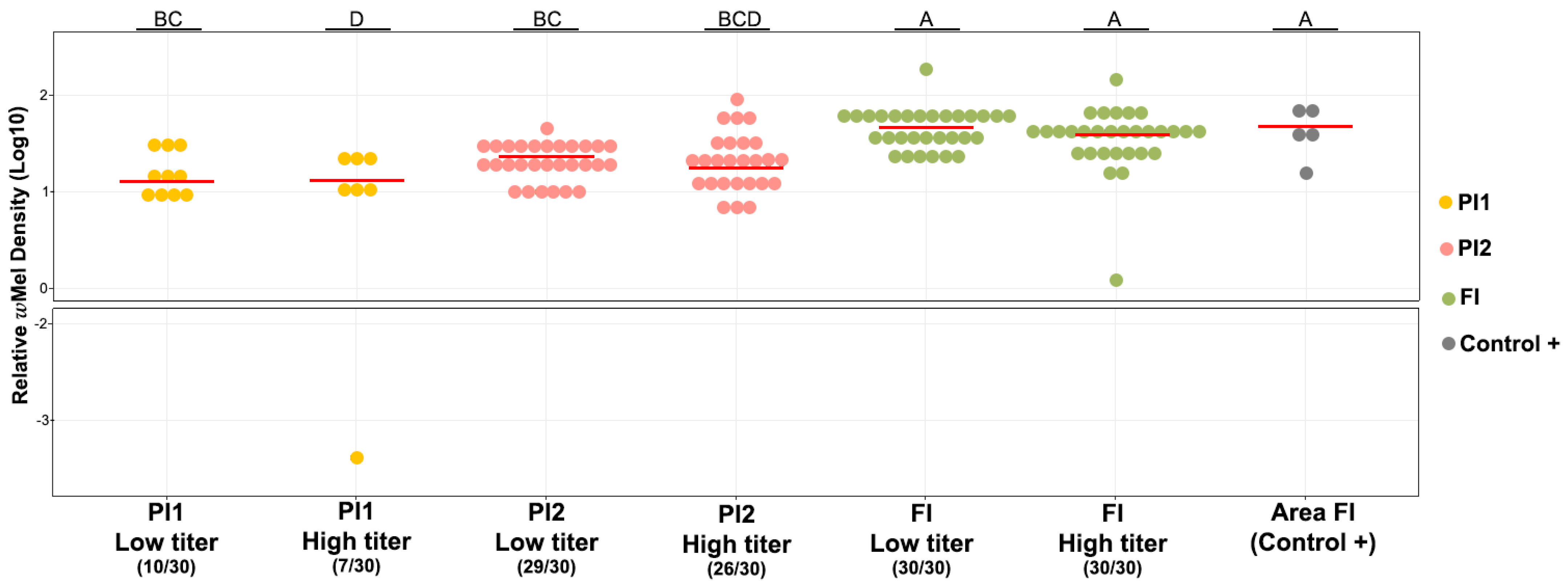
3.1. Prevalence and Density of wMel in Field and Lab Ae. aegypti Mosquitoes

We accessed the prevalence of Wolbachia in the five areas, three where wMel deployments took place before conducting the vector competence analyses (PI1, PI2, and FI; cf. Methods section for further details). The wMel strain was detected in 41 out of 75 mosquitoes (an overall frequency of 54.7%) of the field populations where wMel deployments occurred (Figure S1). All mosquitoes from FI (25/25) had the bacteria, while PI1 and PI2 had 20% (5/25) and 44% (11/25) of mosquitoes positive for wMel, respectively. Pairwise comparisons did not show significant differences between wMel densities in field-caught mosquitoes from the three deployed areas (W = 15; W = 107; W = 77 p > 0.05; Table S1). As expected, NR1 and NR2 mosquitoes were all wMel-free.
Considering the lab-reared F1 Ae. aegypti mosquitoes used in DENV-1 experimental infections, wMel was detected in 137/185 samples (74%; Figure S1): 65/65 from FI (100%), 17/60 (28.3%) from PI1 and 55/60 (91.7%) from PI2. NR1 and NR2 mosquitoes were negative for wMel. In general, lab-reared mosquitoes showed a higher relative density of wMel when compared with field mosquitoes (W = 64; p < 0.001; Figure S1, Table S1).

3.2. wMel Detection in Ae. aegypti Mosquitoes Orally Exposed to Two DENV-1 Titers

Considering the F1 mosquitoes used for oral infections with DENV-1, all Ae. aegypti from FI (where deployments started in 2015) were positive for wMel (30/30; both viral titers; Figure 2). Likewise, specimens from PI2, where releases started in late 2019, had Wolbachia in 86.7% (26/30) and 96.7% (29/30) of mosquitoes when subjected to oral feeding with high and low DENV-1 titers. Despite the Wolbachia deployments taking place in PI1 before PI2 (early and late 2019, respectively), Ae. aegypti from PI1 subjected to DENV-1 high and low titer infections exhibited wMel in only 23.3% (7/30) and 33.3% (10/30) of the total mosquitoes analyzed, respectively.
Mosquitoes collected in FI presented higher relative wMel density than mosquitoes from PI1 and PI2, where releases started five years later (W = 17–173.5, p < 0.001; Figure 2, Table S1). DENV-1-infected and non-infected mosquitoes had similar relative wMel densities. Regarding only DENV-1-infected specimens, linear regression analyses evidenced no correlation between wMel density and DENV-1 load (F = 2.354; p > 0.05 e R2 adjusted: 0.016, df = 80; Figure S2A).

3.3. Vector Competence Assays

A total of 216 Ae. aegypti F1 from FI, PI1, and PI2 and 144 mosquitoes from NR1 and NR2 were exposed to DENV-1 oral infections with low and high titers, and their whole bodies were tested for infection via RT-qPCR. Ten wMel and wild mosquitoes (from FI and NR1, respectively) non-exposed to DENV-1 were used as negative controls for arbovirus detection.
A total of 30 individuals from each locality were analyzed for each virus titer. In general, whole-body wMel mosquitoes had a lower DENV-1 infection rate than wMel-free mosquitoes (GLM: Wolbachia (Yes) estimate = −1.2, p = 0.0005; Figure 3A, Table S2). Regarding the low titer, PI1 had 12 positives for DENV-1 (40%), of which 4/10 were wMel Ae. aegypti and 8/20 were wMel-free mosquitoes. PI2 had 14 mosquitoes infected with DENV-1 (45.2%), of which 13/29 were wMel Ae. aegypti and 1/1 was a wMel-free mosquito. The FI had only 3/30 wMel mosquitoes infected with DENV-1 (10%). NR1 had 23/30 (76.7%) wild Ae. aegypti DENV-1 infected, while NR2 had 15/30 (50%) wild Ae. aegypti positive for DENV-1. Considering the wMel Ae. aegypti mosquitoes from the two PI areas, 40–45% were infected with DENV-1, contrasting with the 10%-infected mosquitoes of the FI area. Interestingly, DENV-1 load in wMel and wMel-free Ae. aegypti was similar in mosquito bodies, except for the comparison between NR1 and PI1 (W = 198; p = 0.04; Figure 3A).
Regarding the exposure to the high viral titer through oral infections, all 60 mosquitoes from PI1 and PI2 were positive for DENV-1, of which 7 were wMel Ae. aegypti and 23 were wMel-free mosquitoes, and 26 were wMel Ae. aegypti and 4 were wMel-free mosquitoes, respectively. A similar outcome was observed for mosquitoes from FI, of which 20/30 were wMel Ae. aegypti were infected with DENV-1 (96.7%). As expected, 59/60 wild mosquitoes were also infected for DENV-1 (NR1 = 30/30, NR2 = 29/30). Wild mosquitoes showed similar DENV-1 load (W = 473; p > 0.05; Figure 3B; Table S1). The number of DENV-1 copies in whole mosquitoes from FI was barely lower when compared with the wild mosquitoes from NR2 (W = 273; p = 0.02; Figure 3B, Table S1). Mosquitoes from PI1 and PI2 showed significantly different DENV-1 loads when compared to each other (W = 253; p < 0.05; Figure 3B) and to the controls (NR1 and NR2; W = 680; p < 0.05; W = 233; p < 0.05; W = 789; p < 0.05 e W = 123; p < 0.05; Figure 3B). Therefore, wMel Ae. aegypti showed a similar infection rate (GLM: Wolbachia (Yes) estimate = −0.32, p = 0.818) but lower DENV-1 viral load when compared to wild mosquitoes ((GLM: Wolbachia (Yes) estimate = −1.3, p-value < 0.001; Figure 3B, Table S2). We were unable to compare the DENV-1 load of wMel and wMel-free mosquitoes collected in sympatry due to the small sample size (PI1 = 23 wMel-free and 7 wMel mosquitoes; PI2 = 4 wMel-free and 26 wMel mosquitoes).

3.4. Saliva Infectivity for DENV-1

The saliva collected from mosquitoes with DENV-1 infectivity confirmed through RT-qPCR was injected into susceptible mosquitoes from NR1, and their whole bodies were analyzed seven days after injections. The DENV-1 infection rate of the injected saliva from the studied areas is shown in Table 1.
Surprisingly, when infected with DENV-1 low titers, the viral load in wild Ae. aegypti who received infective saliva of wMel Ae. aegypti was similar to the viral load observed in the control group (W = 86.5, p > 0.05; W = 48, p > 0.05; W = 52, p = 0.02; Figure 4, Table S1). On the other hand, when infected with the higher DENV-1 titer, there was a decrease in the viral load in wMel Ae. aegypti saliva when compared with wild Ae. aegypti mosquitoes (W = 108, p < 0.05; W = 83, p < 0.05; W = 220.5, p < 0.0001; Figure 4, Table S1). The linear regression analyses did not detect a statistically significant correlation between DENV-1 copies in saliva-microinjected mosquitoes and the relative wMel density of the mosquitoes (F = 2.605, p > 0.05, R2 = 0.063; Figures S2B and S3).

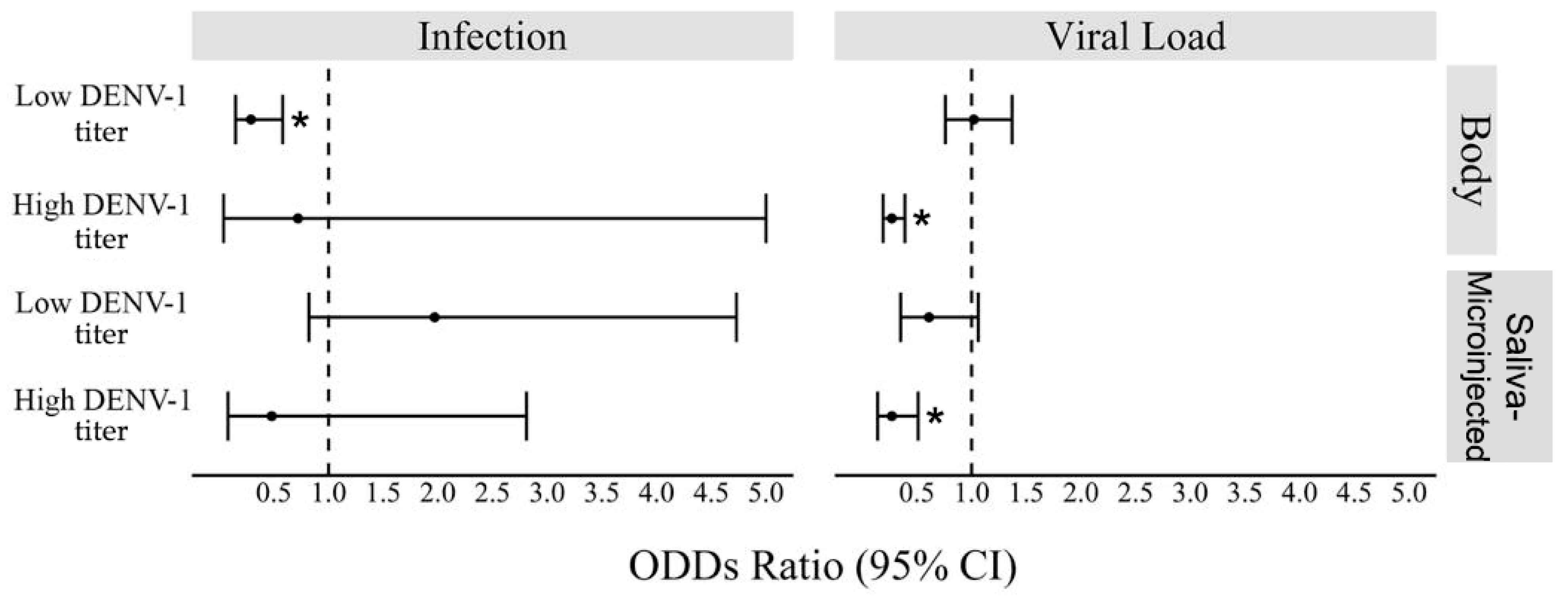
3.5. Interactions of wMel in DENV-1 Exposed Ae. aegypti

Generalized linear models revealed that when infected with low DENV-1 titer, wMel Ae. aegypti presented a lower infection rate in their whole bodies when compared to controls (wild Ae. aegypti; GLM: Wolbachia (Yes) estimate = −1.2, p = 0.0005; Figure S4, Table S2). Wild and wMel mosquitoes, however, had similar viral loads in their bodies (GLM: Wolbachia (Yes) estimate = 0.02, p = 0.885; Figure S4). When we analyzed the microinjected mosquitoes, both the rate of infection and the viral loads obtained seven days post injection of wMel mosquito saliva were similar to the controls (saliva of wild mosquitoes; GLM: Wolbachia (Yes) estimates = 0.6 and −0.4, p > 0.1, respectively; Table S2).
When infected with high DENV-1 titer, wMel Ae. aegypti mosquitoes showed similar infection rates as wild Ae. aegypti (GLM: Wolbachia (Yes) estimate = −0.32, p-value = 0.818; Figure S4; Table S2), but lower viral loads were observed in their whole bodies (GLM: Wolbachia (Yes) estimate = −1.3, p-value < 0.001; Figure S4) and in saliva-microinjected mosquitoes (GLM: Wolbachia (Yes) estimate = −1.3, p-value = 0.001; Figure 5, Table S2).

4. Discussion

In this study, the vector competence of Ae. aegypti was inferred in laboratory conditions by comparing the susceptibility and saliva infectivity of wMel and wMel-free mosquitoes collected in five localities of Rio de Janeiro, the first Brazilian city to receive Wolbachia deployment. We also tested if the relative density of wMel in mosquito bodies influences DENV-1 infection. We subjected wMel and wMel-free Ae. aegypti to oral infections containing two DENV-1 titers (6 × 10⁸ FFU/mL or 3 × 10⁴ FFU/mL) and microinjected mosquito saliva into wMel-free mosquitoes. Our results highlighted that when mosquitoes were infected with the higher DENV-1 titer, wMel did not avoid viral infection in mosquitoes’ bodies and saliva, but wMel mosquitoes produced lower DENV-1 loads than wMel-free mosquitoes (Figure 5). On the other hand, wMel mosquitoes subjected to a lower DENV-1 titer were less susceptible than wMel-free mosquitoes, but once infected, wMel and wMel-free mosquitoes exhibited similar viral loads in the body and to saliva-microinjected mosquitoes.
Several studies have been investigating the susceptibility of wMel and wMel-free Ae. aegypti to DENV [10,23,48,49,50,51], but there are few regarding the DENV-1 serotype [19,20,21]. In this study, we observed the superior DENV-1 infection rate (~69%) in wMel mosquitoes when infected with a higher viral titer, contrasting with those infected with a lower viral titer (~22%). Taking into consideration the saliva’s infection rate, we observed that ~80% of wild Ae. aegypti were infective, whereas ~60% of wMel Ae. aegypti were positive for DENV-1. Ferguson and colleagues [19] observed similar results for wMel and wMel-free Ae. aegypti from Cairns, Australia, exposed to viremic blood from acute dengue cases in Vietnam. The infection rate of mosquito body was 100%, irrespective of whether the mosquito carried wMel or not for DENV-1 titers of ~108 at 14 dpi, but wMel mosquitoes had only ~40% of their saliva infected (lower than we observed in this study), whereas ~85% of infective saliva was observed for wMel-free Ae. aegypti. The authors, however, did not observe infected bodies of wMel and wMel-free mosquitoes exposed to DENV-1 titer of ~105 at 10 dpi.
Another study in Vietnam involving four DENV serotypes has shown that 71.5% of wMel-free Ae. aegypti and 58.7% of wMel Ae. aegypti had their abdomens infected with DENV. Furthermore, the same held true for infectious viruses in the saliva, in which 38.5% and 22.8% for wMel-free and wMel Ae. aegypti, respectively, were infected with DENV [20]. In this study, the authors observed more mosquitoes with infective saliva when infected with DENV-1 than with DENV-4.
Flores and colleagues [21] also analyzed infection rates in wMel-Cairns Ae. aegypti (genetic background from Cairns, Australia), wMel-HCM Ae. aegypti (genetic background from Ho Chi Minh City, Vietnam) and wMel-free mosquitoes, respectively. They observed similar infection rates of abdomen/head–thorax/saliva to Carrington and colleagues [20] and did not find any difference in the prevalence of DENV in mosquitoes with different genetic backgrounds. The authors also described for DENV-1 intrathoracic microinjection a high infection rate (94%) and a ~103 viral load for wMel Ae. aegypti, while a 100% infection rate and ~105 TCID50/mL (for 1.2 × 10⁵ TCID50/mL titer) viral load were observed for Cairns wMel-free Ae. aegypti. Concerning the Brazilian Ae. aegypti genetic background, Souto-Maior et al. [52] observed through DENV-1 microinjections of five viral titers (104–108 TCID50/mL) that Wolbachia confers a slight protection against the virus.
Divergent results on studies regarding the vector competence to DENV of wMel Ae. aegypti might be related to the different virus genotypes [8,10,19,20,21,48,49,50,51]. In this context, the emergence of DENV variants able to replicate in wMel Ae. aegypti and surpass the viral blockage are of general concern. For example, an amino acid substitution (E203K) in the DENV-1 envelope protein has increased in frequency in virus populations following 20 passages in wMel Ae. aegypti compared to wMel-free Ae. aegypti cell cultures. Therefore, it is highly probable that wMel would exert selective pressure on dengue populations. It is still unknown, however, if these variants could efficiently replicate in the field where wMel mosquitoes have been released. Future studies regarding the genome sequence of DENV variants in the field may answer this question [53].
Wolbachia density has been positively linked to the strength of viral blocking to RNA viruses [9,54,55,56]. For instance, in Drosophila simulans, it was already observed that Wolbachia density is important for antiviral protection [57]. Studies involving cell lines have shown almost complete DENV inhibition only if the cells have high Wolbachia density [55,56]. Ae. albopictus is naturally infected with another Wolbachia strain, wAlbB, but is unable to block DENV, which has been linked to the lower bacterium density in this species. When wAlbB is transinfected into Ae. aegypti, it is present in higher densities in mosquito tissues, and a DENV-blocking phenotype is observed [56]. In our study, wMel Ae. aegypti from the field exhibit a lower density of the bacterium than the F1 lab-reared mosquitoes, and it may be explained by several factors such as temperature [58,59], fitness cost [13,60,61,62], and nutrition [63]. High temperatures can impact CI and Wolbachia density, leading to a reduction in wMel frequencies and densities in Ae. aegypti [58,59]. Although the fitness cost of having wMel is equal to both field and laboratory-reared mosquitoes [64,65], the latter are reared in optimal conditions of temperature, humidity, and nutrition, while field Ae. aegypti mosquitoes are subjected to adversities that may contribute to the lower wMel density. Regarding the lab-reared wMel Ae. aegypti exposed to DENV-1, there was no correlation between relative wMel density and virus infection of the mosquito’s body or saliva (Figure 5), as already reported [66,67]. Even though wMel density does not appear to influence DENV-1 infection, tissue-specific (ovaries, salivary glands, and saliva’s infectivity) analysis could shed a brighter light on this issue.
We observed contrasting scenarios in Rio regarding the wMel geographical invasion and relative density in mosquitoes and the viral blocking phenotype of Ae. aegypti. FI is an isolated neighborhood with low human density where wMel deployment was completed in 2015 and had the highest wMel frequency and density. Moreover, its wMel mosquitoes had the lowest DENV-1 loads (Figure 1, Figure 2 and Figure 5). PI1, a highly urbanized and human-populated area surrounded by places with few or no Wolbachia deployments, where Wolbachia deployments occurred in early 2019, wMel is still in low prevalence, and mosquitoes had high viral loads in their bodies and saliva. In PI2, where wMel releases occurred in late 2019 and the bacterium is highly prevalent, the mosquitoes also had high viral loads in the saliva. Therefore, even though there is not a positive linear relationship between Wolbachia density and DENV-1 infection, the results observed in FI suggest there may exist a Wolbachia density threshold, which allows better protection for the arboviruses. Another hypothesis would be that the positive relationship between Wolbachia density and protection may occur in some strategic tissues for Wolbachia but not in others, and this outcome is masked in this study once we analyze the whole mosquito bodies.
The release of Ae. aegypti with Wolbachia has been a promising strategy to mitigate the transmission of arboviruses such as DENV, ZIKV, and CHIKV in urban settings. Nonetheless, the success of Wolbachia as a strategy to reduce arboviruses varies given the complexity of each city’s environment and factors, such as the insecticide resistance of Ae. aegypti carrying Wolbachia, native field vector population sizes, and the maintenance of MT and CI in field conditions [30,36,68,69,70,71]. It is relevant to bear in mind that wMel deployment has a significant public health impact and likely achieves disease elimination in low/moderate arboviral transmission scenarios, but it seems to be less efficient to control the transmission in highly epidemic regions, particularly considering DENV-1 [19]. Studies in Rio de Janeiro, Indonesia, and Australia have shown a reduction in dengue cases using the wMel strain. Australia had a great outcome with a reduction of >95% of DENV cases. In Indonesia, a better outcome was observed when compared to Rio’s study, showing a reduction of 77% of the DENV cases against 38% [26,30]. These differences may be associated not just with the moderate wMel introgression into wild Ae. aegypti population (average 40%) in Rio de Janeiro after 4–5 years of Wolbachia deployments across the city [30,42], but also to the potential DENV leakage in the saliva of Wolbachia-infected individuals that could reduce the epidemiological effectiveness of this strategy. It is also important to highlight that there are differences in the incidence rate (IR) of dengue cases in these countries. In 2022, Australia had an IR of 1.55 cases of dengue (per 100,000 habitants), while Indonesia showed an IR of 45.7 cases, and Brazil had an impressive IR of 1097 cases [72,73]. Therefore, these distinct epidemiological scenarios may explain the different success rates in the reduction of dengue cases in these countries. Continuous monitoring of Wolbachia in the field and dengue cases in humans remains of utmost importance to identify any changes in the epidemiological scenario so action plans can be implemented in time.

5. Conclusions

This study provides insights into the tripartite interaction involving the wMel strain of Wolbachia, Ae. aegypti mosquitoes and DENV-1 in the highly epidemic scenario of Rio de Janeiro, Brazil. The presence of wMel did not avoid viral infection in mosquitoes’ bodies and saliva when subjected to a higher DENV-1 titer, but wMel mosquitoes exhibited lower viral loads than wild mosquitoes. Regarding the lower DENV-1 titer, wMel mosquitoes were less susceptible than wMel-free mosquitoes, but once infected, wMel and wild mosquitoes exhibited similar viral loads in the body and the saliva-microinjected saliva. Furthermore, DENV-1 infection and viral titer in the body or in saliva-microinjected mosquitoes seem to not be related to wMel density. Future studies concerning the vector competence of wMel and wild Ae. aegypti with varied genetic backgrounds are necessary to check the impact on wMel’s strategy to replace wild Ae. aegypti populations and diminish arbovirus transmission in the long term.

Supplementary Materials

The following supporting information can be downloaded at: https://www.mdpi.com/article/10.3390/v16020214/s1, Figure S1: Relative density of wMel Ae. aegypti mosquitoes separated into “FIELD” and “LAB” groups (A) and in field localities and laboratory F1 generation (B). Horizontal red bars represent the medians. Green, yellow, and pink circles represent samples infected with wMel from FI, PI1, and PI2, respectively. Figure S2: Regression analyses comparing relative wMel density in whole Ae. aegypti mosquitoes with (A) DENV-1 load in whole mosquitoes at 14 dpi, and (B) DENV-1 load in susceptible mosquitoes seven days post intrathoracic injection of mosquito saliva. Both regression analyses were not statistically significant (F = 2.354, p > 0.1289 and F = 2.605, p > 0.1202, respectively). Figure S3: Relative density of wMel in Ae. aegypti mosquitoes infected with DENV-1 at low (3 × 10⁴ FFU/mL) and high titer (6 × 10⁸ FFU/mL). The numbers inside parentheses (below the neighborhood name) indicate the number of wMel-positive mosquitoes out of the total mosquitoes tested. Different letters (above the graph) indicate statistically significant differences in wMel density (Wilcoxon–Mann–Whitney test; p < 0.05). Horizontal red bars represent the medians. Purple circles represent samples infected with wMel and untested for infective DENV-1 in the saliva. Yellow and green circles represent the samples with and without infective DENV-1 particles in the saliva, respectively. Figure S4: Number of DENV-1 copies in the whole body observed in wMel, wMel-free, and wild Ae. aegypti mosquitoes 14 days post infection with low titer (3 × 10⁴ FFU/mL) (A) and high titer (6 × 10⁸ FFU/mL) (B). The numbers inside parentheses (below the neighborhood name) indicate the number of DENV-1-positive mosquitoes out of the total mosquitoes tested. Different letters (above the graph) indicate statistically significant differences (Wilcoxon–Mann–Whitney test; p < 0.05). Horizontal red bars represent the medians. Purple and gray circles represent mosquitoes infected with wMel and wild, respectively, but untested for infective DENV-1 in the saliva. Yellow and pink circles represent mosquitoes with and without wMel, respectively, with infective DENV-1 in the saliva. Mosquitoes with and without wMel with DENV-1 non-infective saliva are represented by green and blue circles, respectively. All negative controls for DENV-1 infection were negative. Table S1: Results of the Wilcoxon–Mann–Whitney pairwise comparisons related to DENV-1 infection in the body and saliva and density of Wolbachia of Ae. aegypti mosquitoes. Table S2: Results of the generalized linear models (GLMs) related to DENV-1 infection in the body and saliva and density of Wolbachia of Ae. aegypti mosquitoes.

Author Contributions

Conceptualization, M.G.P. and R.M.-d.-F.; methodology, D.C.-L., G.A.G., J.C.-A., M.G.P., M.R.D. and M.S.G.K.; validation, D.C.-L., J.C.-A. and M.G.P.; formal analysis, J.C.-A., M.G.P. and M.R.D.; writing—original draft preparation, J.C.-A. and M.G.P.; writing—review and editing, D.C.-L., G.A.G., J.C.-A., M.G.P., M.R.D., M.S.G.K. and R.M.-d.-F.; supervision, M.G.P., M.R.D. and R.M.-d.-F.; funding acquisition, M.G.P. and R.M.-d.-F. All authors have read and agreed to the published version of the manuscript.

Funding

This research was funded by Fundação Carlos Chagas Filho de Amparo à Pesquisa do Estado do Rio de Janeiro—FAPERJ, grant numbers E26/2001.844/2017, E26/210.335/2022, E26/201.370/2022 and E26/211.159/2019, and Coordenação de Aperfeiçoamento de Pessoal de Nível Superior—CAPES (finance code 001).

Institutional Review Board Statement

The study was conducted in accordance with the Declaration of Helsinki and approved by the Ethics Committee of Fundação Oswaldo Cruz (CAAE 94070418.7.0000.5248 and date of approval as 28 April 2019). This protocol establishes the use of blood bags from anonymous donators of the Hematology Department of the State University of Rio de Janeiro to blood feed mosquitoes. The blood bags were supposed to be discarded due to low volume.

Informed Consent Statement

Not applicable.

Data Availability Statement

Data are contained within the article and supplementary materials.

Acknowledgments

We thank Marcelo Celestino dos Santos, Mauro Menezes Muniz, and Renato Carvalho de Andrade (in memoriam) for their tireless support in field collections.

Conflicts of Interest

The authors declare no conflicts of interest. The funders had no role in the design of the study; in the collection, analyses, or interpretation of data; in the writing of the manuscript; or in the decision to publish the results.

References

  1. Brady, O.J.; Gething, P.W.; Bhatt, S.; Messina, J.P.; Brownstein, J.S.; Hoen, A.G.; Moyes, C.L.; Farlow, A.W.; Scott, T.W.; Hay, S.I. Refining the Global Spatial Limits of Dengue Virus Transmission by Evidence-Based Consensus. PLoS Negl. Trop. Dis. 2012, 6, e1760. [Google Scholar] [CrossRef]
  2. WHO Dengue and Severe Dengue. Available online: https://www.who.int/news-room/fact-sheets/detail/dengue-and-severe-dengue#:~:text=Dengue%20is%20a%20viral%20infection,million%20infections%20occurring%20each%20year (accessed on 7 August 2023).
  3. PAHO Epidemiological Update for Dengue, Chikungunya and Zika in 2022. Available online: https://www3.paho.org/data/index.php/en/mnu-topics/indicadores-dengue-en/annual-arbovirus-bulletin-2022.html (accessed on 7 August 2023).
  4. Maciel-de-Freitas, R.; Marques, W.A.; Peres, R.C.; Cunha, S.P.; Lourenço-de-Oliveira, R. Variation in Aedes aegypti (Diptera: Culicidae) Container Productivity in a Slum and a Suburban District of Rio de Janeiro during Dry and Wet Seasons. Mem. Inst. Oswaldo Cruz 2007, 102, 489–496. [Google Scholar] [CrossRef]
  5. David, M.R.; Dantas, E.S.; Maciel-de-Freitas, R.; Codeço, C.T.; Prast, A.E.; Lourenço-de-Oliveira, R. Influence of Larval Habitat Environmental Characteristics on Culicidae Immature Abundance and Body Size of Adult Aedes aegypti. Front. Ecol. Evol. 2021, 9, 626757. [Google Scholar] [CrossRef]
  6. Girard, M.; Nelson, C.B.; Picot, V.; Gubler, D.J. Arboviruses: A Global Public Health Threat. Vaccine 2020, 38, 3989–3994. [Google Scholar] [CrossRef]
  7. Zug, R.; Hammerstein, P. Still a Host of Hosts for Wolbachia: Analysis of Recent Data Suggests That 40% of Terrestrial Arthropod Species Are Infected. PLoS ONE 2012, 7, e38544. [Google Scholar] [CrossRef] [PubMed]
  8. Moreira, L.A.; Iturbe-Ormaetxe, I.; Jeffery, J.A.; Lu, G.; Pyke, A.T.; Hedges, L.M.; Rocha, B.C.; Hall-Mendelin, S.; Day, A.; Riegler, M.; et al. A Wolbachia Symbiont in Aedes aegypti Limits Infection with Dengue, Chikungunya, and Plasmodium. Cell 2009, 139, 1268–1278. [Google Scholar] [CrossRef] [PubMed]
  9. Bian, G.; Xu, Y.; Lu, P.; Xie, Y.; Xi, Z. The Endosymbiotic Bacterium Wolbachia Induces Resistance to Dengue Virus in Aedes aegypti. PLoS Pathog. 2010, 6, e1000833. [Google Scholar] [CrossRef] [PubMed]
  10. Walker, T.; Johnson, P.H.; Moreira, L.A.; Iturbe-Ormaetxe, I.; Frentiu, F.D.; McMeniman, C.J.; Leong, Y.S.; Dong, Y.; Axford, J.; Kriesner, P.; et al. The wMel Wolbachia Strain Blocks Dengue and Invades Caged Aedes aegypti Populations. Nature 2011, 476, 450–453. [Google Scholar] [CrossRef] [PubMed]
  11. Werren, J.H.; Baldo, L.; Clark, M.E. Wolbachia: Master Manipulators of Invertebrate Biology. Nat. Rev. Microbiol. 2008, 6, 741–751. [Google Scholar] [CrossRef]
  12. Turelli, M. Evolution of incompatibility-inducing microbes and their hosts. Evolution 1994, 48, 1500–1513. [Google Scholar] [CrossRef] [PubMed]
  13. Ross, P.A.; Turelli, M.; Hoffmann, A.A. Evolutionary Ecology of Wolbachia Releases for Disease Control. Annu. Rev. Genet. 2019, 53, 93–116. [Google Scholar] [CrossRef]
  14. Garcia, G.d.A.; Sylvestre, G.; Aguiar, R.; da Costa, G.B.; Martins, A.J.; Lima, J.B.P.; Petersen, M.T.; Lourenço-de-Oliveira, R.; Shadbolt, M.F.; Rašić, G.; et al. Matching the Genetics of Released and Local Aedes aegypti Populations Is Critical to Assure Wolbachia Invasion. PLoS Negl. Trop. Dis. 2019, 13, e0007023. [Google Scholar] [CrossRef]
  15. Lau, M.-J.; Ross, P.A.; Hoffmann, A.A. Infertility and Fecundity Loss of Wolbachia-Infected Aedes aegypti Hatched from Quiescent Eggs Is Expected to Alter Invasion Dynamics. PLoS Negl. Trop. Dis. 2021, 15, e0009179. [Google Scholar] [CrossRef]
  16. Petersen, M.T.; Couto-Lima, D.; Garcia, G.A.; Pavan, M.G.; David, M.R.; Maciel-de-Freitas, R. Dengue Exposure and Wolbachia wMel Strain Affects the Fertility of Quiescent Eggs of Aedes aegypti. Viruses 2023, 15, 952. [Google Scholar] [CrossRef]
  17. Rocha, M.N.; Duarte, M.M.; Mansur, S.B.; e Silva, B.D.M.; Pereira, T.N.; Adelino, T.É.R.; Giovanetti, M.; Alcantara, L.C.J.; Santos, F.M.; Costa, V.R.d.M.; et al. Pluripotency of Wolbachia against Arboviruses: The Case of Yellow Fever. Gates Open Res. 2019, 3, 161. [Google Scholar] [CrossRef] [PubMed]
  18. Pereira, T.N.; Rocha, M.N.; Sucupira, P.H.F.; Carvalho, F.D.; Moreira, L.A. Wolbachia Significantly Impacts the Vector Competence of Aedes aegypti for Mayaro Virus. Sci. Rep. 2018, 8, 6889. [Google Scholar] [CrossRef]
  19. Ferguson, N.M.; Kien, D.T.H.; Clapham, H.; Aguas, R.; Trung, V.T.; Chau, T.N.B.; Popovici, J.; Ryan, P.A.; O’Neill, S.L.; McGraw, E.A.; et al. Modeling the Impact on Virus Transmission of Wolbachia-Mediated Blocking of Dengue Virus Infection of Aedes aegypti. Sci. Transl. Med. 2015, 7, 279ra37. [Google Scholar] [CrossRef]
  20. Carrington, L.B.; Tran, B.C.N.; Le, N.T.H.; Luong, T.T.H.; Nguyen, T.T.; Nguyen, P.T.; Nguyen, C.V.V.; Nguyen, H.T.C.; Vu, T.T.; Vo, L.T.; et al. Field- and Clinically Derived Estimates of Wolbachia-Mediated Blocking of Dengue Virus Transmission Potential in Aedes aegypti Mosquitoes. Proc. Natl. Acad. Sci. USA 2018, 115, 361–366. [Google Scholar] [CrossRef]
  21. Flores, H.A.; Taneja de Bruyne, J.; O’Donnell, T.B.; Tuyet Nhu, V.; Thi Giang, N.; Thi Xuan Trang, H.; Thi Thuy Van, H.; Thi Long, V.; Thi Dui, L.; Le Anh Huy, H.; et al. Multiple Wolbachia Strains Provide Comparative Levels of Protection against Dengue Virus Infection in Aedes aegypti. PLoS Pathog. 2020, 16, e1008433. [Google Scholar] [CrossRef]
  22. Caragata, E.P.; Rocha, M.N.; Pereira, T.N.; Mansur, S.B.; Dutra, H.L.C.; Moreira, L.A. Pathogen Blocking in Wolbachia-Infected Aedes aegypti Is Not Affected by Zika and Dengue Virus Co-Infection. PLoS Negl. Trop. Dis. 2019, 13, e0007443. [Google Scholar] [CrossRef]
  23. Ant, T.H.; Mancini, M.V.; McNamara, C.J.; Rainey, S.M.; Sinkins, S.P. Wolbachia-Virus Interactions and Arbovirus Control through Population Replacement in Mosquitoes. Pathog. Glob. Health 2023, 117, 245–258. [Google Scholar] [CrossRef] [PubMed]
  24. Boehm, E.C.; Jaeger, A.S.; Ries, H.J.; Castañeda, D.; Weiler, A.M.; Valencia, C.C.; Weger-Lucarelli, J.; Ebel, G.D.; O’Connor, S.L.; Friedrich, T.C.; et al. Wolbachia-Mediated Resistance to Zika Virus Infection in Aedes aegypti Is Dominated by Diverse Transcriptional Regulation and Weak Evolutionary Pressures. PLoS Negl. Trop. Dis. 2023, 17, e0011674. [Google Scholar] [CrossRef] [PubMed]
  25. Martinez, J.; Longdon, B.; Bauer, S.; Chan, Y.-S.; Miller, W.J.; Bourtzis, K.; Teixeira, L.; Jiggins, F.M. Symbionts Commonly Provide Broad Spectrum Resistance to Viruses in Insects: A Comparative Analysis of Wolbachia Strains. PLoS Pathog. 2014, 10, e1004369. [Google Scholar] [CrossRef] [PubMed]
  26. Utarini, A.; Indriani, C.; Ahmad, R.A.; Tantowijoyo, W.; Arguni, E.; Ansari, M.R.; Supriyati, E.; Wardana, D.S.; Meitika, Y.; Ernesia, I.; et al. Efficacy of Wolbachia-Infected Mosquito Deployments for the Control of Dengue. N. Engl. J. Med. 2021, 384, 2177–2186. [Google Scholar] [CrossRef] [PubMed]
  27. Ogunlade, S.T.; Adekunle, A.I.; Meehan, M.T.; McBryde, E.S. Quantifying the Impact of Wolbachia Releases on Dengue Infection in Townsville, Australia. Sci. Rep. 2023, 13, 14932. [Google Scholar] [CrossRef]
  28. Lenharo, M. Dengue Rates Drop after Release of Modified Mosquitoes in Colombia. Nature 2023, 623, 235–236. [Google Scholar] [CrossRef]
  29. Velez, I.D.; Tanamas, S.K.; Arbelaez, M.P.; Kutcher, S.C.; Duque, S.L.; Uribe, A.; Zuluaga, L.; Martínez, L.; Patiño, A.C.; Barajas, J.; et al. Reduced Dengue Incidence Following City-Wide WMel Wolbachia Mosquito Releases throughout Three Colombian Cities: Interrupted Time Series Analysis and a Prospective Case-Control Study. PLoS Negl. Trop. Dis. 2023, 17, e0011713. [Google Scholar] [CrossRef]
  30. Ribeiro Dos Santos, G.; Durovni, B.; Saraceni, V.; Souza Riback, T.I.; Pinto, S.B.; Anders, K.L.; Moreira, L.A.; Salje, H. Estimating the Effect of the WMel Release Programme on the Incidence of Dengue and Chikungunya in Rio de Janeiro, Brazil: A Spatiotemporal Modelling Study. Lancet Infect. Dis. 2022, 22, 1587–1595. [Google Scholar] [CrossRef]
  31. Pinto, S.B.; Riback, T.I.S.; Sylvestre, G.; Costa, G.; Peixoto, J.; Dias, F.B.S.; Tanamas, S.K.; Simmons, C.P.; Dufault, S.M.; Ryan, P.A.; et al. Effectiveness of Wolbachia-Infected Mosquito Deployments in Reducing the Incidence of Dengue and Other Aedes-Borne Diseases in Niterói, Brazil: A Quasi-Experimental Study. PLoS Negl. Trop. Dis. 2021, 15, e0009556. [Google Scholar] [CrossRef]
  32. Gesto, J.S.M.; Ribeiro, G.S.; Rocha, M.N.; Dias, F.B.S.; Peixoto, J.; Carvalho, F.D.; Pereira, T.N.; Moreira, L.A. Reduced Competence to Arboviruses Following the Sustainable Invasion of Wolbachia into Native Aedes aegypti from Southeastern Brazil. Sci. Rep. 2021, 11, 10039. [Google Scholar] [CrossRef]
  33. Calvez, E.; Guillaumot, L.; Girault, D.; Richard, V.; O’Connor, O.; Paoaafaite, T.; Teurlai, M.; Pocquet, N.; Cao-Lormeau, V.-M.; Dupont-Rouzeyrol, M. Dengue-1 Virus and Vector Competence of Aedes aegypti (Diptera: Culicidae) Populations from New Caledonia. Parasit. Vectors 2017, 10, 381. [Google Scholar] [CrossRef] [PubMed]
  34. Gutiérrez-Bugallo, G.; Boullis, A.; Martinez, Y.; Hery, L.; Rodríguez, M.; Bisset, J.A.; Vega-Rúa, A. Vector Competence of Aedes aegypti from Havana, Cuba, for Dengue Virus Type 1, Chikungunya, and Zika Viruses. PLoS Negl. Trop. Dis. 2020, 14, e0008941. [Google Scholar] [CrossRef] [PubMed]
  35. O’Connor, O.; Calvez, E.; Inizan, C.; Pocquet, N.; Richard, V.; Dupont-Rouzeyrol, M. Vector Competence of Aedes aegypti from New Caledonia for the Four Recent Circulating Dengue Virus Serotypes. PLoS Negl. Trop. Dis. 2020, 14, e0008303. [Google Scholar] [CrossRef]
  36. Amoa-Bosompem, M.; Kobayashi, D.; Itokawa, K.; Murota, K.; Faizah, A.N.; Azerigyik, F.A.; Hayashi, T.; Ohashi, M.; Bonney, J.H.K.; Dadzie, S.; et al. Determining Vector Competence of Aedes aegypti from Ghana in Transmitting Dengue Virus Serotypes 1 and 2. Parasit. Vectors 2021, 14, 228. [Google Scholar] [CrossRef]
  37. Duong, V.; Lambrechts, L.; Paul, R.E.; Ly, S.; Lay, R.S.; Long, K.C.; Huy, R.; Tarantola, A.; Scott, T.W.; Sakuntabhai, A.; et al. Asymptomatic Humans Transmit Dengue Virus to Mosquitoes. Proc. Natl. Acad. Sci. USA 2015, 112, 14688–14693. [Google Scholar] [CrossRef]
  38. IBGE-Brazilian Institute of Geography and Statistics. Cities and States—Rio de Janeiro. Available online: https://www.ibge.gov.br/cidades-e-estados/rj/rio-de-janeiro.html (accessed on 8 November 2023).
  39. WMP—World Mosquito Program. Global Progress—Brazil—Rio de Janeiro. Available online: www.worldmosquitoprogram.org/en/global-progress/brazil/niteroi (accessed on 8 November 2023).
  40. IBGE—Brazilian Institute of Geography and Statistics. Cities and States—Niterói. Available online: https://www.ibge.gov.br/cidades-e-estados/rj/niteroi.html (accessed on 8 November 2023).
  41. WMP—World Mosquito Program. Global Progress—Brazil—Rio de Janeiro. Available online: https://www.worldmosquitoprogram.org/en/global-progress/brazil/rio-de-janeiro (accessed on 8 November 2023).
  42. Gesto, J.S.M.; Pinto, S.B.; Dias, F.B.S.; Peixoto, J.; Costa, G.; Kutcher, S.; Montgomery, J.; Green, B.R.; Anders, K.L.; Ryan, P.A.; et al. Large-Scale Deployment and Establishment of Wolbachia Into the Aedes aegypti Population in Rio de Janeiro, Brazil. Front. Microbiol. 2021, 12, 711107. [Google Scholar] [CrossRef]
  43. Consoli, R.A.G.B.; Lourenço-de-Oliveira, R. Principais Mosquitos de Importância Sanitária No Brasil; Editora FIOCRUZ: Rio de Janeiro, Brazil, 1994. [Google Scholar]
  44. Dutra, H.L.C.; Rocha, M.N.; Dias, F.B.S.; Mansur, S.B.; Caragata, E.P.; Moreira, L.A. Wolbachia Blocks Currently Circulating Zika Virus Isolates in Brazilian Aedes aegypti Mosquitoes. Cell Host Microbe 2016, 19, 771–774. [Google Scholar] [CrossRef]
  45. Garcia, G.A.; Lord, A.R.; Santos, L.M.B.; Kariyawasam, T.N.; David, M.R.; Couto-Lima, D.; Tátila-Ferreira, A.; Pavan, M.G.; Sikulu-Lord, M.T.; Maciel-de-Freitas, R. Rapid and Non-Invasive Detection of Aedes aegypti Co-Infected with Zika and Dengue Viruses Using Near Infrared Spectroscopy. Viruses 2022, 15, 11. [Google Scholar] [CrossRef]
  46. Johnson, B.W.; Russell, B.J.; Lanciotti, R.S. Serotype-Specific Detection of Dengue Viruses in a Fourplex Real-Time Reverse Transcriptase PCR Assay. J. Clin. Microbiol. 2005, 43, 4977–4983. [Google Scholar] [CrossRef]
  47. R Core Team. R: A Language and Environment for Statistical Computing; R Core Team: Vienna, Austria, 2021. [Google Scholar]
  48. Frentiu, F.D.; Zakir, T.; Walker, T.; Popovici, J.; Pyke, A.T.; van den Hurk, A.; McGraw, E.A.; O’Neill, S.L. Limited Dengue Virus Replication in Field-Collected Aedes aegypti Mosquitoes Infected with Wolbachia. PLoS Negl. Trop. Dis. 2014, 8, e2688. [Google Scholar] [CrossRef]
  49. Ye, Y.H.; Carrasco, A.M.; Frentiu, F.D.; Chenoweth, S.F.; Beebe, N.W.; van den Hurk, A.F.; Simmons, C.P.; O’Neill, S.L.; McGraw, E.A. Wolbachia Reduces the Transmission Potential of Dengue-Infected Aedes aegypti. PLoS Negl. Trop. Dis. 2015, 9, e0003894. [Google Scholar] [CrossRef] [PubMed]
  50. Terradas, G.; Allen, S.L.; Chenoweth, S.F.; McGraw, E.A. Family Level Variation in Wolbachia-Mediated Dengue Virus Blocking in Aedes aegypti. Parasit. Vectors 2017, 10, 622. [Google Scholar] [CrossRef] [PubMed]
  51. Novelo, M.; Audsley, M.D.; McGraw, E.A. The Effects of DENV Serotype Competition and Co-Infection on Viral Kinetics in Wolbachia-Infected and Uninfected Aedes aegypti Mosquitoes. Parasit. Vectors 2021, 14, 314. [Google Scholar] [CrossRef] [PubMed]
  52. Souto-Maior, C.; Sylvestre, G.; Braga Stehling Dias, F.; Gomes, M.G.M.; Maciel-de-Freitas, R. Model-Based Inference from Multiple Dose, Time Course Data Reveals Wolbachia Effects on Infection Profiles of Type 1 Dengue Virus in Aedes aegypti. PLoS Negl. Trop. Dis. 2018, 12, e0006339. [Google Scholar] [CrossRef]
  53. Thi Hue Kien, D.; Edenborough, K.; da Silva Goncalves, D.; Thuy Vi, T.; Casagrande, E.; Thi Le Duyen, H.; Thi Long, V.; Thi Dui, L.; Thi Tuyet Nhu, V.; Thi Giang, N.; et al. Genome Evolution of Dengue Virus Serotype 1 under Selection by Wolbachia Pipientis in Aedes aegypti Mosquitoes. Virus Evol. 2023, 9, vead016. [Google Scholar] [CrossRef]
  54. Ye, Y.H.; Woolfit, M.; Rancès, E.; O’Neill, S.L.; McGraw, E.A. Wolbachia-Associated Bacterial Protection in the Mosquito Aedes aegypti. PLoS Negl. Trop. Dis. 2013, 7, e2362. [Google Scholar] [CrossRef]
  55. Frentiu, F.D.; Robinson, J.; Young, P.R.; McGraw, E.A.; O’Neill, S.L. Wolbachia-Mediated Resistance to Dengue Virus Infection and Death at the Cellular Level. PLoS ONE 2010, 5, e13398. [Google Scholar] [CrossRef] [PubMed]
  56. Lu, P.; Bian, G.; Pan, X.; Xi, Z. Wolbachia Induces Density-Dependent Inhibition to Dengue Virus in Mosquito Cells. PLoS Negl. Trop. Dis. 2012, 6, e1754. [Google Scholar] [CrossRef]
  57. Osborne, S.E.; Iturbe-Ormaetxe, I.; Brownlie, J.C.; O’Neill, S.L.; Johnson, K.N. Antiviral Protection and the Importance of Wolbachia Density and Tissue Tropism in Drosophila simulans. Appl. Environ. Microbiol. 2012, 78, 6922–6929. [Google Scholar] [CrossRef]
  58. Ulrich, J.N.; Beier, J.C.; Devine, G.J.; Hugo, L.E. Heat Sensitivity of WMel Wolbachia during Aedes aegypti Development. PLoS Negl. Trop. Dis. 2016, 10, e0004873. [Google Scholar] [CrossRef] [PubMed]
  59. Ross, P.A.; Axford, J.K.; Yang, Q.; Staunton, K.M.; Ritchie, S.A.; Richardson, K.M.; Hoffmann, A.A. Heatwaves Cause Fluctuations in WMel Wolbachia Densities and Frequencies in Aedes aegypti. PLoS Negl. Trop. Dis. 2020, 14, e0007958. [Google Scholar] [CrossRef] [PubMed]
  60. Axford, J.K.; Ross, P.A.; Yeap, H.L.; Callahan, A.G.; Hoffmann, A.A. Fitness of WAlbB Wolbachia Infection in Aedes aegypti: Parameter Estimates in an Outcrossed Background and Potential for Population Invasion. Am. J. Trop. Med. Hyg. 2016, 94, 507–516. [Google Scholar] [CrossRef] [PubMed]
  61. Fraser, J.E.; De Bruyne, J.T.; Iturbe-Ormaetxe, I.; Stepnell, J.; Burns, R.L.; Flores, H.A.; O’Neill, S.L. Novel Wolbachia-Transinfected Aedes aegypti Mosquitoes Possess Diverse Fitness and Vector Competence Phenotypes. PLoS Pathog. 2017, 13, e1006751. [Google Scholar] [CrossRef] [PubMed]
  62. Ant, T.H.; Herd, C.S.; Geoghegan, V.; Hoffmann, A.A.; Sinkins, S.P. The Wolbachia Strain WAu Provides Highly Efficient Virus Transmission Blocking in Aedes aegypti. PLoS Pathog. 2018, 14, e1006815. [Google Scholar] [CrossRef] [PubMed]
  63. Caragata, E.P.; Rancès, E.; O’Neill, S.L.; McGraw, E.A. Competition for Amino Acids between Wolbachia and the Mosquito Host, Aedes aegypti. Microb. Ecol. 2014, 67, 205–218. [Google Scholar] [CrossRef]
  64. Hoffmann, A.A.; Iturbe-Ormaetxe, I.; Callahan, A.G.; Phillips, B.L.; Billington, K.; Axford, J.K.; Montgomery, B.; Turley, A.P.; O’Neill, S.L. Stability of the WMel Wolbachia Infection Following Invasion into Aedes aegypti Populations. PLoS Negl. Trop. Dis. 2014, 8, e3115. [Google Scholar] [CrossRef]
  65. Ross, P.A.; Robinson, K.L.; Yang, Q.; Callahan, A.G.; Schmidt, T.L.; Axford, J.K.; Coquilleau, M.P.; Staunton, K.M.; Townsend, M.; Ritchie, S.A.; et al. A Decade of Stability for WMel Wolbachia in Natural Aedes aegypti Populations. PLoS Pathog. 2022, 18, e1010256. [Google Scholar] [CrossRef]
  66. Dutra, H.L.C.; Ford, S.A.; Allen, S.L.; Bordenstein, S.R.; Chenoweth, S.F.; Bordenstein, S.R.; McGraw, E.A. The Impact of Artificial Selection for Wolbachia-Mediated Dengue Virus Blocking on Phage WO. PLoS Negl. Trop. Dis. 2021, 15, e0009637. [Google Scholar] [CrossRef]
  67. Amuzu, H.E.; McGraw, E.A. Wolbachia-Based Dengue Virus Inhibition Is Not Tissue-Specific in Aedes aegypti. PLoS Negl. Trop. Dis. 2016, 10, e0005145. [Google Scholar] [CrossRef] [PubMed]
  68. Ryan, P.A.; Turley, A.P.; Wilson, G.; Hurst, T.P.; Retzki, K.; Brown-Kenyon, J.; Hodgson, L.; Kenny, N.; Cook, H.; Montgomery, B.L.; et al. Establishment of WMel Wolbachia in Aedes aegypti Mosquitoes and Reduction of Local Dengue Transmission in Cairns and Surrounding Locations in Northern Queensland, Australia. Gates Open Res. 2019, 3, 1547. [Google Scholar] [CrossRef] [PubMed]
  69. Nguyen, T.H.; Nguyen, H.L.; Nguyen, T.Y.; Vu, S.N.; Tran, N.D.; Le, T.N.; Vien, Q.M.; Bui, T.C.; Le, H.T.; Kutcher, S.; et al. Field Evaluation of the Establishment Potential of WMelPop Wolbachia in Australia and Vietnam for Dengue Control. Parasit. Vectors 2015, 8, 563. [Google Scholar] [CrossRef] [PubMed]
  70. Garcia, G.A.; Hoffmann, A.A.; Maciel-de-Freitas, R.; Villela, D.A.M. Aedes aegypti Insecticide Resistance Underlies the Success (and Failure) of Wolbachia Population Replacement. Sci. Rep. 2020, 10, 63. [Google Scholar] [CrossRef]
  71. Pavan, M.G.; Garcia, G.A.; David, M.R.; Maciel-de-Freitas, R. The Double-Edged Sword Effect of Expanding Wolbachia Deployment in Dengue Endemic Settings. Lancet Reg. Health Am. 2023, 27, 100610. [Google Scholar] [CrossRef]
  72. ECDC Dengue Worldwide Overview. Available online: https://www.ecdc.europa.eu/en/dengue-monthly (accessed on 28 September 2023).
  73. United Nations. Department of Economic and Social Affairs Population Division. World Population Prospects: The 2022 Revision. Available online: https://population.un.org/wpp/ (accessed on 28 September 2023).
Figure 1. Study areas in Rio de Janeiro. A—FI (Tubiacanga); B—PI1 (Bonsucesso); C—PI2 (Fonseca); D—NR1 (Urca); E—NR2 (Vila Valqueire). In green, wMel current released areas, and in orange, wMel-free areas.
Viruses 16 00214 g001
Figure 2. Relative density of wMel in Ae. aegypti mosquitoes infected with DENV-1 at low (3 × 10⁴ FFU/mL) and high titers (6 × 10⁸ FFU/mL). The numbers inside parentheses indicate the number of wMel-positive mosquitoes out of the total mosquitoes tested. Different letters (above the graph) indicate statistically significant differences in wMel density (Wilcoxon–Mann–Whitney test; p < 0.05). Horizontal red bars represent the medians. Yellow circles represent samples infected with wMel from PI1, pink circles from PI2, green circles from FI, and gray circles from wMel-positive control (FI).
Viruses 16 00214 g002
Figure 3. Number of DENV-1 copies in the whole body of wMel, wMel-free, and wild Ae. aegypti mosquitoes 14 days after the infection with the low titer (3 × 10⁴ FFU/mL) (A) and the high titer (6 × 10⁸ FFU/mL) (B). The numbers inside parentheses indicate the number of DENV-1-positive mosquitoes out of the total mosquitoes tested per area. Different letters (above the graph) indicate statistically significant differences (Wilcoxon–Mann–Whitney test; p < 0.05). Horizontal red bars represent the medians. Blue circles represent wMel-free mosquitoes, red circles represent mosquitoes with wMel, and brown circles represent wild mosquitoes. All negative controls for DENV-1 infection were negative.
Viruses 16 00214 g003
Figure 4. DENV-1 load in susceptible wild Ae. aegypti mosquitoes seven days after injecting the saliva of mosquitoes with confirmed infection of DENV-1 in the whole body. Each saliva was intrathoracically injected into 10 susceptible mosquitoes. The injected saliva was obtained from mosquitoes with and without wMel and subjected to DENV-1 oral infections with low (3 × 10⁴ FFU/mL) and high titer (6 × 10⁸ FFU/mL). The number of DENV-1 infective saliva out of the total number of mosquitoes tested is in parenthesis (below the neighborhood name). Horizontal red bars represent the medians. Different letters (above the graph) indicate statistically significant differences (Wilcoxon–Mann–Whitney test; p < 0.05). All mosquitoes that received saliva from DENV-1-negative mosquitoes were negative.
Viruses 16 00214 g004
Figure 5. Forest plot showing the odds ratio (OR) and 95% confidence intervals for interactions of wMel in DENV-1-exposed Ae. aegypti mosquitoes. “Infection” results are based on the number of infected and non-infected mosquitoes, and “Viral load” results are the absolute quantity of DENV-1 virus particles. Mosquito bodies were analyzed 14 days post infection (“Body”), and susceptible mosquitoes were analyzed 7 days after receiving an intrathoracic injection of saliva from a mosquito exposed to DENV-1 (“Saliva-microinjected”). Mosquitoes were fed on blood infected with DENV-1 at 3 × 10⁴ FFU/mL (“Low DENV-1 titer”) or 6 × 10⁸ FFU/mL (“High DENV-1 titer”). The asterisks denote statistically significant negative association (i.e., OR < 1.0) between having wMel and body infection or viral loads.
Viruses 16 00214 g005
Table 1. DENV-1 infection rate per area observed in wild Ae. aegypti mosquitoes 7 days post saliva microinjection with low titer (3 × 10⁴ FFU/mL) and high titer (6 × 10⁸ FFU/mL). The numbers inside parentheses indicate the number of DENV-1-positive mosquitoes out of the total mosquitoes tested per area.
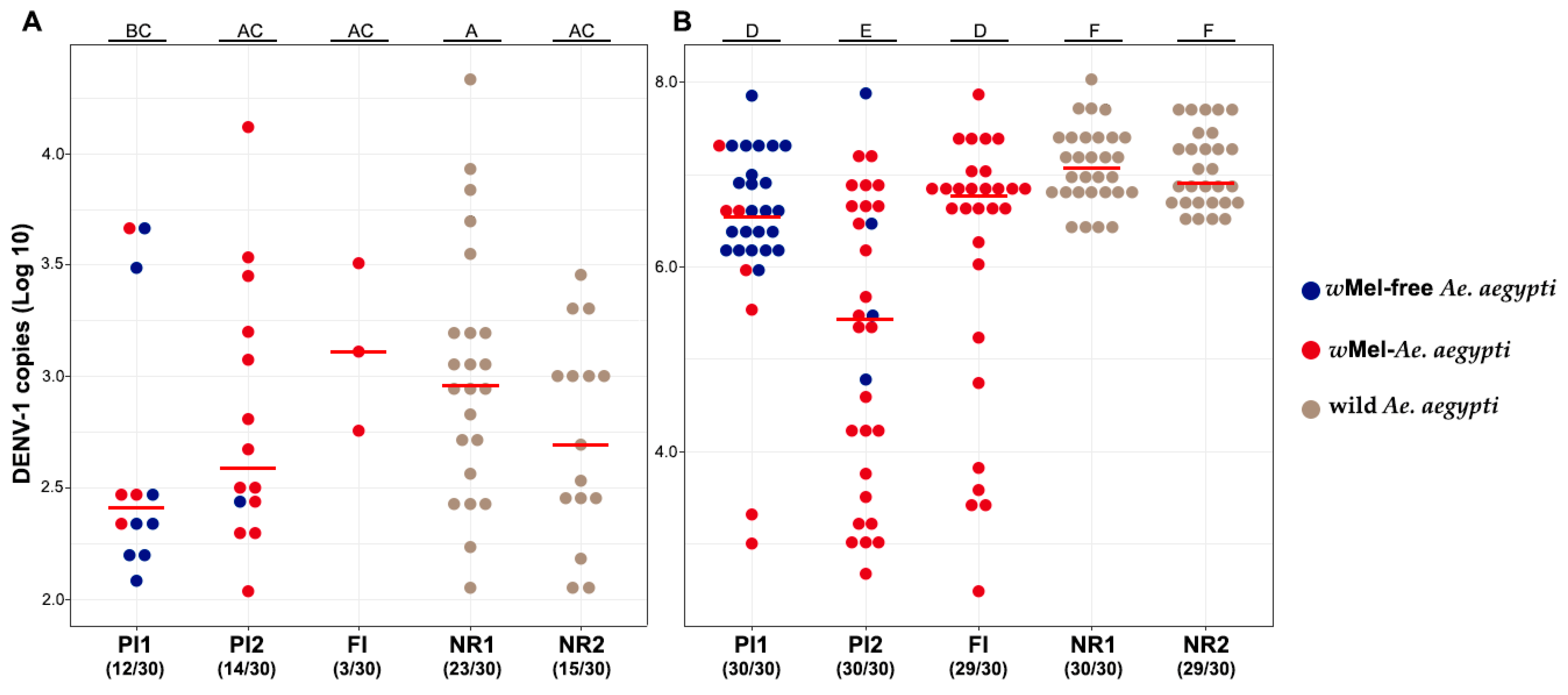
DENV-1 Infection Rate
Areas Low Titer High Titer
NR180% (8/10)77.77% (7/9)
PI150% (2/4)50% (3/6)
PI275% (6/8)50% (4/8)
FI66.66% (2/3)80% (8/10)
Disclaimer/Publisher’s Note: The statements, opinions and data contained in all publications are solely those of the individual author(s) and contributor(s) and not of MDPI and/or the editor(s). MDPI and/or the editor(s) disclaim responsibility for any injury to people or property resulting from any ideas, methods, instructions or products referred to in the content.

