Article

Enhanced Transcription of Human Endogenous Retroviruses and TRIM28 Downregulation in Patients with Inflammatory Bowel Disease

1
Department of Public Health and Pediatric Sciences, University of Turin, Piazza Polonia 94, 10126 Turin, Italy
2
Department of Medical Sciences, Division of Gastroenterology, University of Turin, 10123 Turin, Italy
3
Pediatric Laboratory, Department of Public Health and Pediatric Sciences, University of Turin, Regina Margherita Children’s Hospital, Piazza Polonia 94, 10126 Turin, Italy
*
Author to whom correspondence should be addressed.
Viruses 2024, 16(10), 1570; https://doi.org/10.3390/v16101570
Submission received: 20 August 2024 / Revised: 2 October 2024 / Accepted: 3 October 2024 / Published: 5 October 2024
(This article belongs to the Section Human Virology and Viral Diseases)

Abstract

:
Inflammatory bowel disease (IBD) includes patients affected by Crohn’s disease or ulcerative colitis. IBD is thought to be a chronic immune-mediated disease, but its origin remains elusive, and this limits new therapeutic approaches. Human endogenous retroviruses (HERVs) originate from ancestral infections and represent 8% of the human genome. HERVs are no longer infectious, but some retroviral sequences can be activated, and their aberrant expressions have been implicated in inflammatory and autoimmune disorders. HERV transcription is regulated by TRIM28 and SETDB1, which are also directly involved in epigenetic processes and modulation of the immune response. Using a PCR real-time Taqman amplification assay, we assessed, for the first time, the transcription levels of pol genes of HERV-H, -K, and -W families of env genes of syncytin 1 (SYN1), SYN2, and HERV-W, as well as of TRIM28 and SETDB1 in the whole blood of 48 patients with Crohn’s disease (CD), 20 with ulcerative colitis (UC), and in healthy controls (HC) of comparable age. The transcriptional levels of HERV-H-pol (p = 0.0003) and HERV-K-pol (p = 0.001) were significantly higher in IBD patients compared with HC, with no differences between patients with CD and UC. No significant differences were found for the remaining HERVs between IBD patients and HC. The transcript levels of TRIM28 were significantly downregulated in IBD patients (p < 0.001), without differences between CD and UC, while the SETDB1 levels were preserved. The enhanced transcription of HERV-H-pol and HERV-K-pol, as well as the impaired activation of TRIM28, were not influenced by clinical disease activity and type of treatment. The overexpression of HERVs and impaired transcription of TRIM28 in patients affected by CD or UC suggest that they might be the main actors in the pathophysiology of IBD, opening the way to innovative targeted interventions.

1. Introduction

Inflammatory bowel disease (IBD) is a major cause of chronic disability. It is considered an immune-mediated disease, but its pathogenesis remains poorly understood and this limits the identification of novel therapeutic measures [1,2]. The contribution of hereditable genetic factors is increasingly recognized [3,4,5]. Epigenetics is an emerging element through which environmental factors can modulate gene expressions without changing their fundamental structure. An accumulating body of literature highlights that epigenetic factors are highly involved in the development Crohn’s disease (CD) and ulcerative colitis (UC) [3,4,6,7,8]. Recent studies have also shown that the gut microbiota plays a pivotal role in maintaining gut homeostasis, and regulating local and systemic immune responses, while its imbalance may give rise to microbial metabolites able to trigger epigenetic variations in IBD patients [9,10]. Toll-like receptors (TLRs) participate in the immune response to microbial components. They contribute to preserving the intestinal epithelial barrier that can be compromised by dysregulated TLR signaling, allowing microbes to penetrate and trigger inflammatory responses in IBD [11].
Human endogenous retroviruses (HERVs) originate from the ancient infections of germinal cells of primates millions of years ago. Due to evolutionary mutations, they have lost the ability to produce infectious particles. However, HERVs still retain their retroviral structure, which consists of three principal genes: group-associated antigens (gag), polymerase (pol), and envelope (env), flanked by two regulatory long terminal repeats (LTRs) [12]. Most HERVs are inactive, but some elements are transcribed and a few encode proteins. Increasing evidence documents the essential role of HERVs during the intrauterine life. For instance, two envelope proteins, referred to as syncytin 1 (SYN1) [13] and syncytin 2 (SYN2) [14], contribute to the placental syncytiotrophoblast formation and the materno-foetal tolerance through their vigorous immune-suppressive effects [15]. Postnatally, the physiologic actions of HERVs are unclear: while activated retroviruses can act as the promoters or enhancers of cellular genes [16,17], their RNAs can be retrotranscribed and reintegrated into the DNA or, being recognized as non-self by TLRs, induce innate and specific immune responses [17,18,19]. In fact, HERVs have been studied and proposed in the pathophysiology of many inflammatory immune-mediated diseases [20,21,22,23,24]. HERVs also have a mutual influence on microbiota, and their enhanced expression promotes microbiota-mediated gut inflammation [25].
HERV transcription is influenced by environmental factors through epigenetic mechanisms, such as DNA methylation and histone modification, leading to heterochromatin silencing. The tripartite motif-containing 28 (TRIM28, also known as KAP1 or TIF1-b), functions as a nuclear corepressor for Krüppel-associated box domain zink finger proteins (KRAB-ZFPs), which are the largest family of transcriptional regulators in the human genome [26]. SET domain bifurcated histone lysine methyltransferase 1 (SETDB1) is a methyltransferase highly specific for the lysine 9 residue of histone H3 [27]. It has multifaced biological properties, such as T cell development, intestinal epithelial cell differentiation, and the prevention of gut inflammation [28,29,30]. Both TRIM28 and SETBD1 are specific tags for the epigenetic transcriptional repression of HERVs [31,32,33]. Furthermore, they regulate the transactivation of thousands of cellular genes [34,35] and are directly involved in epigenetic processes [32], including the modulation of innate and adaptive immune responses [36,37].
Despite the aforementioned elements supporting the potential role of HERVs, TRIM28, and SETDB1 in inducing and/or maintaining immune-mediated inflammatory disorders, no studies investigated their expressions in patients affected by IBD except for one study on syncytin expression in intestinal biopsies from CD patients [38]. Therefore, the aims of our research were to evaluate the transcriptional levels of pol genes of HERV-H, HERV-K, and HERV-W, the three retroviral families most widely studied [12,39]; env genes of SYN1, SYN2, and HERV-W [21,40]; as well as TRIM28 and SETDB1 in the whole blood from adults affected by CD or UC, and in healthy controls (HC) of comparable age.

2. Material and Methods

2.1. Study Populations

The diagnosis of CD and UC was based on clinical, radiological, and endoscopic findings according to the European guidelines [41]. Patients’ peripheral blood samples were collected during routine laboratory checks. Healthy adult volunteers of comparable age were the control group.

2.2. Sample Storage

Of the whole blood, 200 μL was added to 800 µL of the RNApro solution (Biomole, Turin, Italy) in a 1.5 mL Eppendorf tube and resuspended by vortexing; the samples were stored at −80 °C [42].

2.3. Total RNA Extraction

Total RNA was extracted using the Maxwell automated extractor in combination with the RNA Blood Kit (Promega, Madison, WI, USA). This kit includes DNase treatment during the extraction process. To ensure no genomic DNA contamination, RNA extracts were directly amplified without reverse transcription to validate the extraction protocol. RNA concentration and purity were evaluated by UV spectroscopy, measuring absorbance at 260 and 280 nm SimpliNano spectrophotometer (Biochrom US, Holliston, MA, USA). The RNAs were stored at −80 °C until use.

2.4. Reverse Transcription

Four hundred nanograms of total RNA were reverse-transcribed in a 20 μL reaction mixture containing 2 μL of a 10× buffer, 4.8 μL of 25 mM MgCl2, 2 μL ImProm-II reverse transcriptase (Promega), 1 μL of RNase inhibitor (20 U/L), 0.4 μL of 250 μM random hexamers (Promega), 2 μL of 100 mM dNTP mix (Promega), and nuclease-free water. The reverse transcription reaction was performed in a GeneAmp PCR system 9700 Thermal Cycle (Applied Biosystems, Foster City, CA, USA) under the following conditions: 5 min at 25 °C, 60 min at 42 °C, followed by 15 min at 70 °C for enzyme inactivation. The cDNAs were stored at −20 °C until use.

2.5. Transcription Levels of pol Genes of HERV-H, -K, -W; env Genes of SYN1, SYN2, and HERV-W; as Well as TRIM8/SETDB1 by a Real-Time PCR Assay

The relative expression levels of pol genes from HERV-H, HERV-K, and HERV-W; env genes from SYN1, SYN2, and HERV-W; along with TRIM28/SETDB1 were measured, as previously detailed, using the primers and probes listed in Supplementary Table S1 [22,23,24]. Briefly, 40 ng of cDNA was amplified in a 20 μL reaction containing 2.5 U goTaQ MaterMix (Promega), 1.25 mmol/L MgCl2, 500 nmol of specific primers, and 200 nmol of specific probes. All the amplifications were performed in a 96-well plate under the following conditions: initial denaturation at 95 °C for 10 min, followed by 45 cycles at 95 °C for 15 s and at 60 °C for 1 min. Each sample was analyzed in triplicate. The relative expression of target gene transcripts was carried out according to the 2−ΔΔCt method [43]. GAPDH was chosen as the reference gene, due to its consistent expression in human leukocytes and its proven efficiency and excellent reproducibility [44], as previously observed in our studies [22,23,24]. Briefly, after normalization of the PCR result of each target gene with the housekeeping gene, the method includes additional calibration of this value with the median expression of the same gene emerging in a pool of healthy controls. The results of the 2−ΔΔCt method show variations in target gene transcripts relative to the standard set of controls. As documented in this and other investigations, the expression of HERVs, TRIM28, and SETDB1 varies greatly among healthy individuals. The reason is unknown and in the literature, no definition of normal values or normal threshold has been proposed; consequently the analyses to assess potential significant differences between IBD patients and HC were performed by comparing all their values. Since we measured Ct for every target in all samples, we argue that our methods were suitable for HERV and TRIM28/SETDB1 quantifications. All analyses were carried out in a laboratory of biosafety level 2 (BSL-2) according to the Office of Science Policy and WHO guidelines [45,46].

2.6. Statistical Analysis

A one-way ANOVA test was employed to compare the transcriptional levels of each target gene among patients with CD, UC, and HC. Since the Shapiro–Wilk test to evaluate the distribution of data of every group of subjects demonstrated non-normally distributed continuous variables, the Mann–Whitney test was used to assess differences in the relative transcription levels of pol genes from HERV-H, HERV-K, and HERV-W; env genes from SYN1, SYN2, and HERV-W; and TRIM28 and SETDB1 genes between two groups of subjects. Statistical analyses were performed using the Prism 7 software (GraphPad Software); p < 0.05 was considered statistically significant.

3. Results

3.1. Study Population

A total of 68 patients affected by IBD were studied: 48 with CD (Group A1) and 20 with UC (Group A2). The control subjects grouped healthy volunteers, selected without selection criteria. They were divided into two groups according to the tests performed: Group B1 included 104 subjects whose blood samples were also used for other studies and whose stored cDNAs were sufficient to be tested for pol genes of HERV-H, HERV-K, and HERV-W; while Group B2 included 81 healthy volunteers tested for the other targets. The control subjects were selected with the gender and age comparable to the patients. The median ages of IBD patients were comparable to those of HC: A1 vs. B1 p = 0.1376, A2 vs. B2 p = 0.1950. The characteristics of the patients and control subjects are detailed in Table 1.

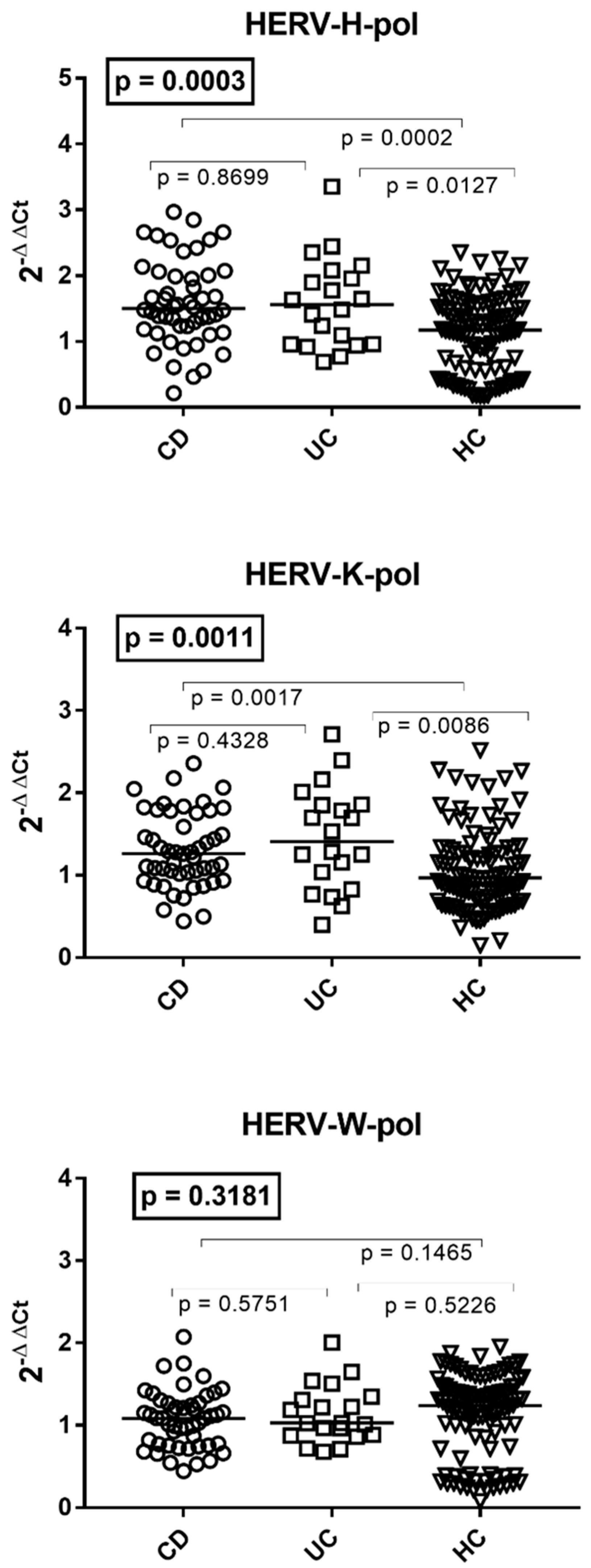
3.2. Transcription Levels of HERV-H-pol, HERV-K-pol, and HERV-W-pol in the Whole Blood of Patients with Crohn’s Disease, Ulcerative Colitis, and HC

The transcriptional levels of HERV-H-pol and HERV-K-pol were significantly higher in patients with CD or UC than in HC of comparable age, while no differences were found between the two subpopulations of IBD patients (Figure 1). The mRNA levels of HERV-W-pol were comparable between subjects with CD, UC, and HC. The medians and IQR 25–75% were as follows: HERV-H-pol CD 1.53, 1.22–2.02; UC 1.56, 0.96–1.99, HC 1.24, 0.63–1.63; HERV-K-pol CD 1.27, 1.00–1.76; UC 1.41, 0.99–1.85; HC 0.97, 0.71–1.34; HERV-W-pol CD 1.09, 0.78–1.27; UC 1.03, 0.88–1.32; HC 1.32, 0.86–1.59 (Figure 1).

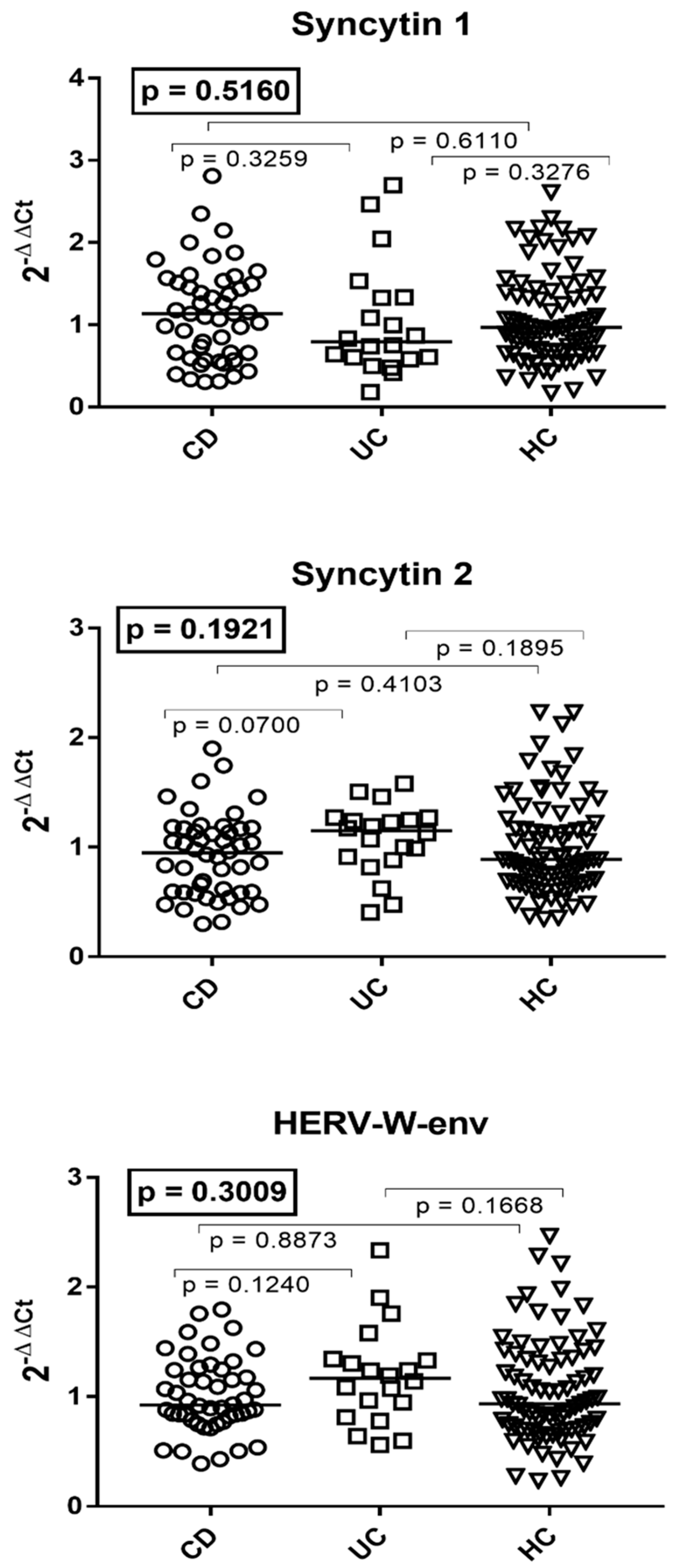
3.3. Transcription Levels of the env Genes of Syncytin 1, Syncytin 2, and HERV-W in the Whole Blood of Patients with Crohn’s Disease, Ulcerative Colitis, and HC

The median values of the env genes of SYN1, SYN2, and HERV-W were similar in patients with CD, UC, and HC of comparable age (Figure 2). The medians and IQR 25–75% were as follows: syncytin 1 CD 1.14, 0.66–1.52; UC 0.79, 0.6–1.33; HC 1.02, 0.70–1.48; syncytin 2 CD 0.95, 0.59–1.17; UC 1.15, 0.91–1.53; HC 0.93, 0.70–1.38. HERV-W CD 0.93, 0.81–1.25; UC 1.17, 0.9–1.34; HC 0.98, 0.73–1.44 (Figure 2).

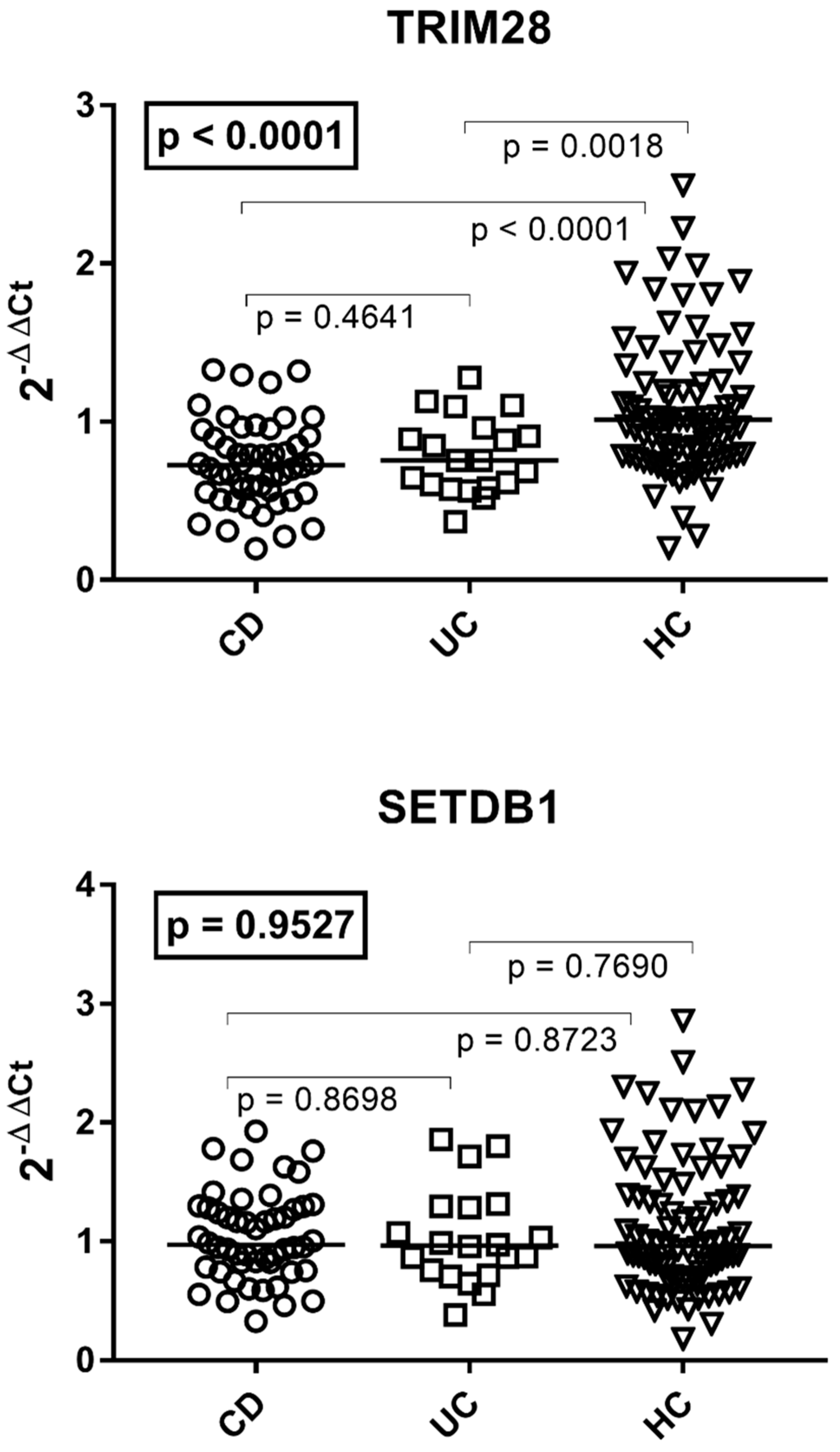
3.4. Transcription Levels of TRIM28 and SETDB1 in Patients with Crohn’s Disease, Ulcerative Colitis, and HC

As reported in Figure 3, the median transcript levels of TRIM28 were significantly lower in patients with CD or UC than in HC of comparable age, without a significant difference between the two groups of patients. The transcription levels of SETDB1 were comparable in the three groups of subjects. The medians and IQR 25–75% were as follows: TRIM28 CD 0.73, 0.56–0.92; UC 0.76, 0.60–0.92; HC 1.01, 0.79–1.25; SETDB1 CD 1.00, 0.81–1.29; UC 0.96, 0.75–1.29; HC 1.00, 0.74–1.40 (Figure 3).

3.5. Expressions of HERVs, TRIM28, and SETDB1 in IBD Patients According to Disease Activity

No significant differences emerged for HERVs, TRIM28, and SETDB1 between patients in remission (R) compared with those with active disease (including mild, moderate, and severe clinical disease activity, MMS) (Supplementary Figure S1). The medians and IQR 25–75% were as follows: HERV-H-pol R 1.58, 1.12–2.10; MMS 1.49, 1.22–1.91; HERV-K-pol R 1.29, 1.07–1.78; MMS 1.13, 0.90–1.79; HERV-W-pol R 1.11, 0.88–1.35; MMS 0.98, 0.81–1.24; syncytin 1 R 1.12, 0.65–1.50; MMS 0.98, 0.60–1.43; syncytin 2 R 1.03, 0.68–1.29; MMS 1.00, 0.59–1.17; HERV-W-env R 1.11, 0.84–1.27; MMS 0.95, 0.75–1.18; TRIM28 R 0.76, 0.58–0.92; MMS 0.72, 0.58–0.90; SETDB1 R 0.97, 0.86–1.30; MMS 0.98, 0.70–1.27 (Supplementary Figure S1).

3.6. Transcription Levels of HERVs, TRIM28, and SETDB1 in IBD Patients According to Mesalazine Treatment

The median mRNA levels of HERVs, TRIM28, and SETDB1 were comparable between patients treated with mesalazine (Mes) and those without mesalazine (No Mes) (Supplementary Figure S2). The medians and IQR 25–75% were as follows: HERV-H-pol Mes 1.48, 1.16–2.07; No Mes 1.64, 1.09–1.82; HERV-K-pol Mes 1.26, 1.03–1.79; No Mes 1.29, 0.91–1.70; HERV-W-pol Mes 1.09, 0.88–1.36; No Mes 0.97, 0.82–1.20; Syncytin 1 Mes 1.08, 0.59–1.48; No Mes 0.99, 0.66–1.51; Syncytin Mes 1.06, 0.66–1.29; No Mes 0.91, 0.58–1.05; HERV-W-env Mes 1.07, 0.85–1.33; No Mes 0.84, 0.78–1.09; TRIM28 Mes 0.76, 0.59–0.93; No Mes 0.67, 0.50–0.85; SETDB1 Mes 0.99, 0.84–1.29; No Mes 0.97, 0.71–1.28 (Supplementary Figure S2).

3.7. Transcription Levels of HERVs, TRIM28, and SETDB1 in IBD Patients According to Steroid Treatment

The median transcription levels of HERVs, TRIM28, and SETDB1 were comparable between patients treated with steroids and those untreated (Supplementary Figure S3). The medians and IQR 25–75% were as follows: HERV-H-pol steroids 1.56, 1.24–1.96; no steroids 1.48, 1.00–2.05; HERV-K-pol steroids 1.29, 1.08–1.73; no steroids 1.27, 0.87–1.79; HERV-W-pol steroids 1.12, 0.89–1.28; no steroids 1.06, 0.86–1.27; syncytin 1 steroids 1.23, 0.68–1.53; no steroids 0.99, 0.56–1.36; syncytin 2 steroids 0.89, 0.63–1.18; no steroids 1.06, 0.64–1.19; HERV-W-env steroids 1.00, 0.84–1.26; no steroids 1.02, 0.80–1.28; TRIM28 steroids 0.68, 0.57–0.83; no steroids 0.75, 0.58–0.96; SETDB1 steroids 1.01, 0.78–1.26; no steroids 0.96, 0.80–1.29 (Supplementary Figure S3).

3.8. Expressions of HERVs, TRIM28, and SETDB1 in IBD Patients with and without Anti-Tumor Necrosis Factor (TNF) Treatment

The expressions of HERVs, TRIM28, and SETB1 were comparable between IBD patients treated (anti-TNF POS) or untreated with anti-TNF (anti-TNF NEG) (Supplementary Figure S4). The medians and IQR 25–75% were as follows: HERV-H-pol anti-TNF POS 1.75, 1.27–2.31; anti-TNF NEG 1.48, 1.13–1.94; HERV-K-pol POS 1.31, 1.08–1.86; NEG 1.27, 0.93–1.70; HERV-W-pol anti-TNF POS 1.15, 0.97–1.26; anti-TNF NEG 1.03, 0.83–1.33; Syncytin 1 anti-TNF POS 0.97, 0.62–1.33; anti-TNF NEG 1.05, 0.62–1.53; Syncytin 2 anti-TNF POS 0.99, 0.84–1.18; anti-TNF NEG 1.03, 0.60–1.20; HERV-W-env anti-TNF POS 1.03, 0.84–1.32; anti-TNF NEG 1.00, 0.79–1.25; TRIM28 anti-TNF POS 0.87, 0.58–0.95; anti-TNF NEG 0.73, 0.58–0.88; SETDB1 anti-TNF POS 1.20, 0.94–1.30; anti-TNF NEG 0.95, 0.75–1.23 (Supplementary Figure S4).

4. Discussion

Our results document, for the first time, that patients affected by IBD exhibit significantly higher transcriptional levels of HERV-H-pol and HERV-K-pol in peripheral blood compared with healthy controls of similar age. The RNA levels of HERV-W-pol and the env genes of SYN1, SYN2, and HERV-W were comparable between patients and the control group. No significant difference in the expression of every HERV was observed between patients with CD and those with UC.
The HERV-H is the largest family of retroviruses in the human genome, while the HERV-K is the most recent component, present only in humans. The underlying biological mechanisms responsible for their aberrant expression in patients with IBD and their clinical significance remain to be elucidated. An extensive body of literature highlights that TRIM28 is implicated in maintaining endogenous retroviruses in a silent state [31,32]. The impaired transcriptional levels of TRIM28 in our patients may thus account for the enhanced expression of some HERV elements. SUMOylated TRIM8 is a scaffold protein recruiting SETDB1 to interact with KRAB-ZFPs to repress retroelements [32,49]. SETDB1 expression was normally represented in whole blood of our patients, in line with the preserved mucosal SETDB1 transcript levels in most patients with IBD [50], though others found a relative deficiency [28]. Rare missense variants of SETDB1 are over-represented in IBD and these have been suggested to participate in its pathogenesis [29]. It cannot be overlooked that functional interactions between TRIM28/SETDB1 and single HERVs may originate from post-translational events between the encoded proteins, while we assessed only their transcriptional landscape. Finally, in addition to the TRIM28/SETDB1/KRAB-ZFP complex, presumably a number of other genes are implicated in the control of HERV silencing; therefore, they could contribute to the abnormal expression of some retroviral elements in our patients.
Growing data document the increased release of pro-inflammatory cytokines in subjects with IBD [2]. Inflammatory cytokines give rise to the proteasome-driven activation of the NF-kB signaling pathway. The active isoform of NF-kB, after passage into the nucleus, binds to specific retroviral sequences that, along with inflammatory cytokines, lead to their enhanced transactivation [51]. It must be underlined that HERVs can, in turn, evoke robust inflammatory and immune responses [12,39] and exert several pathogenetic actions. It is worth mentioning that, recently, the gene enhancer ETS2, located in the non-coding desert zone of chr21q22, has been shown to play a major role in IBD, directing macrophage inflammation [5]. Interestingly, HERVs can be the promoters and enhancers of cellular genes [16,17]. HERV RNAs can be retrotranscribed and reintegrated into the DNA, causing possible mutations. The recognition of HERV RNAs by nucleic acid-sensing TLRs may lead to activation of the inflammasome [17,18]; for instance, HERV-K stimulates the NF-kB pathway through TLR8 [19]. Additionally, HERV antigens can trigger targeted responses, including specific and/or cross-reactive antibodies with tissue epitopes [52,53,54,55]. The final result may be a vicious circle leading to chronic inflammatory and immune reactions. In this context, it must be pointed out that enhanced HERV expressions have been documented in several autoimmune diseases [21,22,23,39].
There are mutual interactions between gut microbiota and endogenous retroviruses [25]. Germ-free mice lose intestinal expression of many retroviruses, while exposure to bacteria and their products can stimulate retroviral transcription [56]. The dysbiosis of the gut microbiota present in IBD patients might thus contribute to their enhanced HERV expression.
In contrast to our findings, the downregulation of syncytins has been reported in intestinal biopsies of patients affected by CD compared to those of normal subjects [38]. This discrepancy might be due to the sample size or the effect of certain treatments. On the other hand, the intestinal mucosa shows a regular expression of HERVs [57]. Diffuse staining of normal crypt cells was seen using an anti-HERV monoclonal antibody, whereas no background staining was observed in endothelial and infiltrating immune cells [58]. In CD, patients’ intestinal cells and stem cells [59], which are characterized by the highest levels of HERV expression [31,32], are reduced and replaced by the intense leukocyte infiltrate. These histologic changes might account for the defective syncytin expression in biopsies from CD patients.
There is wide consensus that abnormal epigenetic processes triggered by environmental factors contribute to the development of IBD [3,6,7,8], but the precise responsible pathways remain unknown. TRIM28 is highly implicated in the regulation of epigenetic mechanisms; its impaired transcription in our patients is the first specific molecular alteration documented in subjects affected by IBD. TRIM28 influences the differentiation, expansion and activation of T cells. Through a complex with KRAB-ZFPs and Foxp3, it modulates Treg suppressor activity [50,60]. Furthermore, TRIM28 represses the expression of inflammatory genes, while its deficiency increases the expansion of DCs and enhanced T cell priming toward inflammatory effector T cells via HERV activation [50]. Therefore, the reduced transcript levels of TRIM28 in IBD patients may contribute to their deficiency in T regulatory cells and the expansion of reactive T lymphocytes, ultimately giving rise to derailed local and systemic immune responses, with consequent autoimmune phenomena in the intestinal tract [36].
The expression levels of every HERV and TRIM28/SETDB1 were not associated with disease activity or treatment with mesalazine, steroids, or anti-TNF. These findings suggest that the variables here taken into consideration are independent from the disease state and are not influenced by these therapies. The prevalent number of patients in remission or with mild disturbances does not allow, however, for definitively ruling out the potential relationship of some variables with moderate/severe forms of disease in untreated or unresponsive patients. In contrast, all our patients were treated with one or more drugs, which played the main role in keeping the disease under control.
The high incidence and prevalence of IBD has reached a plateau in recent years in the Western world, whereas the disease burden is continuously increasing in newly industrialized Asian Pacific countries [61,62]. The reason(s) is ill-defined. Changes in diet and the addition of food additives [63], Western lifestyle, smoking, urban environment, and composition of enteric microbiome have been associated with an increased frequency of IBD in a genetically susceptible host [64]. Diet [65], smoking [66], and dysbiosis in commensal microorganisms [67] can act as epigenetic elements promoting CD and UC [68,69]. In addition, cigarette smoking [70], pollution [71], nutritional changes linked to lifestyle [72], and gut microbiota [56] are implicated in retrovirus expression too. Therefore, environmental factors, thought to play an essential role in the development of IBD, could exert their effects via HERV- and/or TRIM28-driven changes in specific biologic pathways.
Our findings raise further intriguing questions. Are the overexpression of HERVs and the downregulation of TRIM28 the biomarkers of IBD? Several anti-HERV therapeutic measures might be adopted, such as specific anti-RNAs, monoclonal antibodies, cytotoxic T lymphocytes against HERV antigens, and antiretroviral treatments [73,74,75]. Antiretroviral drugs inhibited both HIV viral burden and HERV expression in HIV+ subjects [76,77]. Combined antiretroviral treatment in patients with amyotrophic lateral sclerosis showed a better disease course in those with positive antiviral findings [15]. A novel anti-HIV product [78] induced positive effects in a phased II study in patients with UC [79]. Antiretroviral drugs inhibit proteasome activity [80], with a consequent blocking of NF-kB-driven HERV transcription [51]. Epigenetic variations observed in IBD are increasingly indicated as potential therapeutic targets [6,69,81], in particular the TRIM protein family [82].
In conclusion, our results suggest that HERVs and TRIM28 might be implicated in the pathophysiology of CD and UC, and may provide insights toward the development of innovative therapeutic interventions.

Supplementary Materials

The following supporting information can be downloaded at: https://www.mdpi.com/article/10.3390/v16101570/s1, Figure S1: Expression of HERVs, TRIM28 and SETDB1 according to disease activity; Figure S2: Expression of HERVs, TRIM28 and SETDB1 with and without Mesalazine; Figure S3: Expression of HERVs, TRIM28 and SETDB1 with and without Steroids; Figure S4: Expression of HERVs, TRIM28 and SETDB1 with and without anti-TNF; Table S1: Primers and probes used to assess expression of HERVs, TRIM28 and SETDB1.

Author Contributions

P.-A.T., D.G.R. and M.B.: conception and design of the study, data interpretation, first draft, and revision of the manuscript; G.P.C., V.V., D.P., S.F., E.T. and A.P.: collected and stored blood samples from patients and assessed their clinical data; V.V.: data interpretation and participation in the first draft of the article; M.D., C.C., P.M. and I.G. collected blood samples and data from healthy volunteers, performed quantitative PCR assays, and data analysis; I.G.: data interpretation and statistical analysis. All authors have read and agreed to the published version of the manuscript.

Funding

The research was supported by the research grant of the University of Turin (RIBD_RILO_22_01) and the research grant of the CRT bank (BERM_CRT_24_01).

Institutional Review Board Statement

The study protocol was approved by the Ethical Committee of the “Azienda Ospedaliera-Universitaria Città della Salute e della Scienza”, Turin, Italy (code n. 0005222, 16 January 2023).

Informed Consent Statement

All subjects enrolled in the study gave their written informed consent.

Data Availability Statement

The data underlying this article will be shared at the aggregate/population level upon reasonable request to the corresponding author.

Conflicts of Interest

The authors declare no conflicts of interest.

References

  1. Baumgart, D.C.; Carding, S.R. Inflammatory Bowel Disease: Cause and Immunobiology. Lancet 2007, 369, 1627–1640. [Google Scholar] [CrossRef] [PubMed]
  2. Neurath, M.F. Strategies for Targeting Cytokines in Inflammatory Bowel Disease. Nat. Rev. Immunol. 2024, 24, 559–576. [Google Scholar] [CrossRef] [PubMed]
  3. Jarmakiewicz-Czaja, S.; Zielińska, M.; Sokal, A.; Filip, R. Genetic and Epigenetic Etiology of Inflammatory Bowel Disease: An Update. Genes 2022, 13, 2388. [Google Scholar] [CrossRef] [PubMed]
  4. Kalla, R.; Adams, A.T.; Nowak, J.K.; Bergemalm, D.; Vatn, S.; Ventham, N.T.; Kennedy, N.A.; Ricanek, P.; Lindstrom, J.; IBD-Character Consortium; et al. Analysis of Systemic Epigenetic Alterations in Inflammatory Bowel Disease: Defining Geographical, Genetic and Immune-Inflammatory Influences on the Circulating Methylome. J. Crohns Colitis 2023, 17, 170–184. [Google Scholar] [CrossRef] [PubMed]
  5. Stankey, C.T.; Bourges, C.; Haag, L.M.; Turner-Stokes, T.; Piedade, A.P.; Palmer-Jones, C.; Papa, I.; Silva Dos Santos, M.; Zhang, Q.; Cameron, A.J.; et al. A Disease-Associated Gene Desert Directs Macrophage Inflammation through ETS2. Nature 2024, 630, 447–456. [Google Scholar] [CrossRef] [PubMed]
  6. Noble, A.J.; Nowak, J.K.; Adams, A.T.; Uhlig, H.H.; Satsangi, J. Defining Interactions Between the Genome, Epigenome, and the Environment in Inflammatory Bowel Disease: Progress and Prospects. Gastroenterology 2023, 165, 44–60.e2. [Google Scholar] [CrossRef]
  7. Ventham, N.T.; Kennedy, N.A.; Kalla, R.; Adams, A.T.; Noble, A.; Ennis, H.; TOPPIC Study Group; IBD-BIOM Consortium; Mowat, C.; Dunlop, M.G.; et al. Genome-Wide Methylation Profiling in 229 Patients With Crohn’s Disease Requiring Intestinal Resection: Epigenetic Analysis of the Trial of Prevention of Post-Operative Crohn’s Disease (TOPPIC). Cell Mol. Gastroenterol. Hepatol. 2023, 16, 431–450. [Google Scholar] [CrossRef]
  8. Bastida, G.; Mínguez, A.; Nos, P.; Moret-Tatay, I. Immunoepigenetic Regulation of Inflammatory Bowel Disease: Current Insights into Novel Epigenetic Modulations of the Systemic Immune Response. Genes 2023, 14, 554. [Google Scholar] [CrossRef]
  9. Yoo, J.Y.; Groer, M.; Dutra, S.V.O.; Sarkar, A.; McSkimming, D.I. Gut Microbiota and Immune System Interactions. Microorganisms 2020, 8, 1587. [Google Scholar] [CrossRef]
  10. Quaglio, A.E.V.; Grillo, T.G.; De Oliveira, E.C.S.; Di Stasi, L.C.; Sassaki, L.Y. Gut Microbiota, Inflammatory Bowel Disease and Colorectal Cancer. World J. Gastroenterol. 2022, 28, 4053–4060. [Google Scholar] [CrossRef]
  11. Liang, H.; Zhang, L.; Hoden, B.; Qu, B.; Derubeis, D.; Song, X.; Zhang, D. Delineating the Role of Toll-Like Receptors in Inflammatory Bowel Disease. Methods Mol. Biol. 2023, 2700, 221–228. [Google Scholar] [CrossRef] [PubMed]
  12. Johnson, W.E. Origins and Evolutionary Consequences of Ancient Endogenous Retroviruses. Nat. Rev. Microbiol. 2019, 17, 355–370. [Google Scholar] [CrossRef]
  13. Blond, J.L.; Lavillette, D.; Cheynet, V.; Bouton, O.; Oriol, G.; Chapel-Fernandes, S.; Mandrand, B.; Mallet, F.; Cosset, F.L. An Envelope Glycoprotein of the Human Endogenous Retrovirus HERV-W Is Expressed in the Human Placenta and Fuses Cells Expressing the Type D Mammalian Retrovirus Receptor. J. Virol. 2000, 74, 3321–3329. [Google Scholar] [CrossRef] [PubMed]
  14. Blaise, S.; de Parseval, N.; Bénit, L.; Heidmann, T. Genomewide Screening for Fusogenic Human Endogenous Retrovirus Envelopes Identifies Syncytin 2, a Gene Conserved on Primate Evolution. Proc. Natl. Acad. Sci. USA 2003, 100, 13013–13018. [Google Scholar] [CrossRef]
  15. Garcia-Montojo, M.; Rodriguez-Martin, E.; Ramos-Mozo, P.; Ortega-Madueño, I.; Dominguez-Mozo, M.I.; Arias-Leal, A.; García-Martínez, M.Á.; Casanova, I.; Galan, V.; Arroyo, R.; et al. Syncytin-1/HERV-W Envelope Is an Early Activation Marker of Leukocytes and Is Upregulated in Multiple Sclerosis Patients. Eur. J. Immunol. 2020, 50, 685–694. [Google Scholar] [CrossRef]
  16. Isbel, L.; Whitelaw, E. Endogenous Retroviruses in Mammals: An Emerging Picture of How ERVs Modify Expression of Adjacent Genes. Bioessays 2012, 34, 734–738. [Google Scholar] [CrossRef] [PubMed]
  17. Chuong, E.B.; Elde, N.C.; Feschotte, C. Regulatory Evolution of Innate Immunity through Co-Option of Endogenous Retroviruses. Science 2016, 351, 1083–1087. [Google Scholar] [CrossRef]
  18. Miyake, K.; Shibata, T.; Ohto, U.; Shimizu, T.; Saitoh, S.-I.; Fukui, R.; Murakami, Y. Mechanisms Controlling Nucleic Acid-Sensing Toll-like Receptors. Int. Immunol. 2018, 30, 43–51. [Google Scholar] [CrossRef]
  19. Dembny, P.; Newman, A.G.; Singh, M.; Hinz, M.; Szczepek, M.; Krüger, C.; Adalbert, R.; Dzaye, O.; Trimbuch, T.; Wallach, T.; et al. Human Endogenous Retrovirus HERV-K(HML-2) RNA Causes Neurodegeneration through Toll-like Receptors. JCI Insight 2020, 5, e131093. [Google Scholar] [CrossRef]
  20. Volkman, H.E.; Stetson, D.B. The Enemy within: Endogenous Retroelements and Autoimmune Disease. Nat. Immunol. 2014, 15, 415–422. [Google Scholar] [CrossRef]
  21. Perron, H.; Dougier-Reynaud, H.-L.; Lomparski, C.; Popa, I.; Firouzi, R.; Bertrand, J.-B.; Marusic, S.; Portoukalian, J.; Jouvin-Marche, E.; Villiers, C.L.; et al. Human Endogenous Retrovirus Protein Activates Innate Immunity and Promotes Experimental Allergic Encephalomyelitis in Mice. PLoS ONE 2013, 8, e80128. [Google Scholar] [CrossRef] [PubMed]
  22. Tovo, P.-A.; Rabbone, I.; Tinti, D.; Galliano, I.; Trada, M.; Daprà, V.; Cerutti, F.; Bergallo, M. Enhanced Expression of Human Endogenous Retroviruses in New-Onset Type 1 Diabetes: Potential Pathogenetic and Therapeutic Implications. Autoimmunity 2020, 53, 283–288. [Google Scholar] [CrossRef] [PubMed]
  23. Tovo, P.-A.; Opramolla, A.; Pizzol, A.; Calosso, G.; Daprà, V.; Galliano, I.; Calvi, C.; Pinon, M.; Cisarò, F.; Rigazio, C.; et al. Overexpression of Endogenous Retroviruses in Children with Celiac Disease. Eur. J. Pediatr. 2021, 180, 2429–2434. [Google Scholar] [CrossRef]
  24. Tovo, P.-A.; Galliano, I.; Parodi, E.; Calvi, C.; Gambarino, S.; Licciardi, F.; Dini, M.; Montanari, P.; Branca, M.; Ramenghi, U.; et al. Children with Chronic Immune Thrombocytopenia Exhibit High Expression of Human Endogenous Retroviruses TRIM28 and SETDB1. Genes 2023, 14, 1569. [Google Scholar] [CrossRef]
  25. Lima-Junior, D.S.; Krishnamurthy, S.R.; Bouladoux, N.; Collins, N.; Han, S.-J.; Chen, E.Y.; Constantinides, M.G.; Link, V.M.; Lim, A.I.; Enamorado, M.; et al. Endogenous Retroviruses Promote Homeostatic and Inflammatory Responses to the Microbiota. Cell 2021, 184, 3794–3811.e19. [Google Scholar] [CrossRef]
  26. Friedman, J.R.; Fredericks, W.J.; Jensen, D.E.; Speicher, D.W.; Huang, X.P.; Neilson, E.G.; Rauscher, F.J. KAP-1, a Novel Corepressor for the Highly Conserved KRAB Repression Domain. Genes Dev. 1996, 10, 2067–2078. [Google Scholar] [CrossRef]
  27. Schultz, D.C.; Ayyanathan, K.; Negorev, D.; Maul, G.G.; Rauscher, F.J. SETDB1: A Novel KAP-1-Associated Histone H3, Lysine 9-Specific Methyltransferase That Contributes to HP1-Mediated Silencing of Euchromatic Genes by KRAB Zinc-Finger Proteins. Genes Dev. 2002, 16, 919–932. [Google Scholar] [CrossRef]
  28. Wang, R.; Li, H.; Wu, J.; Cai, Z.-Y.; Li, B.; Ni, H.; Qiu, X.; Chen, H.; Liu, W.; Yang, Z.-H.; et al. Gut Stem Cell Necroptosis by Genome Instability Triggers Bowel Inflammation. Nature 2020, 580, 386–390. [Google Scholar] [CrossRef]
  29. Južnić, L.; Peuker, K.; Strigli, A.; Brosch, M.; Herrmann, A.; Häsler, R.; Koch, M.; Matthiesen, L.; Zeissig, Y.; Löscher, B.-S.; et al. SETDB1 Is Required for Intestinal Epithelial Differentiation and the Prevention of Intestinal Inflammation. Gut 2021, 70, 485–498. [Google Scholar] [CrossRef]
  30. Takikita, S.; Muro, R.; Takai, T.; Otsubo, T.; Kawamura, Y.I.; Dohi, T.; Oda, H.; Kitajima, M.; Oshima, K.; Hattori, M.; et al. A Histone Methyltransferase ESET Is Critical for T Cell Development. J. Immunol. 2016, 197, 2269–2279. [Google Scholar] [CrossRef]
  31. Matsui, T.; Leung, D.; Miyashita, H.; Maksakova, I.A.; Miyachi, H.; Kimura, H.; Tachibana, M.; Lorincz, M.C.; Shinkai, Y. Proviral Silencing in Embryonic Stem Cells Requires the Histone Methyltransferase ESET. Nature 2010, 464, 927–931. [Google Scholar] [CrossRef] [PubMed]
  32. Rowe, H.M.; Friedli, M.; Offner, S.; Verp, S.; Mesnard, D.; Marquis, J.; Aktas, T.; Trono, D. De Novo DNA Methylation of Endogenous Retroviruses Is Shaped by KRAB-ZFPs/KAP1 and ESET. Development 2013, 140, 519–529. [Google Scholar] [CrossRef] [PubMed]
  33. Turelli, P.; Castro-Diaz, N.; Marzetta, F.; Kapopoulou, A.; Raclot, C.; Duc, J.; Tieng, V.; Quenneville, S.; Trono, D. Interplay of TRIM28 and DNA Methylation in Controlling Human Endogenous Retroelements. Genome Res. 2014, 24, 1260–1270. [Google Scholar] [CrossRef] [PubMed]
  34. Groner, A.C.; Meylan, S.; Ciuffi, A.; Zangger, N.; Ambrosini, G.; Dénervaud, N.; Bucher, P.; Trono, D. KRAB-Zinc Finger Proteins and KAP1 Can Mediate Long-Range Transcriptional Repression through Heterochromatin Spreading. PLoS Genet. 2010, 6, e1000869. [Google Scholar] [CrossRef]
  35. Krischuns, T.; Günl, F.; Henschel, L.; Binder, M.; Willemsen, J.; Schloer, S.; Rescher, U.; Gerlt, V.; Zimmer, G.; Nordhoff, C.; et al. Phosphorylation of TRIM28 Enhances the Expression of IFN-β and Proinflammatory Cytokines During HPAIV Infection of Human Lung Epithelial Cells. Front. Immunol. 2018, 9, 2229. [Google Scholar] [CrossRef]
  36. Gehrmann, U.; Burbage, M.; Zueva, E.; Goudot, C.; Esnault, C.; Ye, M.; Carpier, J.-M.; Burgdorf, N.; Hoyler, T.; Suarez, G.; et al. Critical Role for TRIM28 and HP1β/γ in the Epigenetic Control of T Cell Metabolic Reprograming and Effector Differentiation. Proc. Natl. Acad. Sci. USA 2019, 116, 25839–25849. [Google Scholar] [CrossRef]
  37. Czerwinska, P.; Jaworska, A.M.; Wlodarczyk, N.A.; Mackiewicz, A.A. Melanoma Stem Cell-Like Phenotype and Significant Suppression of Immune Response within a Tumor Are Regulated by TRIM28 Protein. Cancers 2020, 12, 2998. [Google Scholar] [CrossRef]
  38. Klag, T.; Courth, L.; Ostaff, M.J.; Ott, G.; Stange, E.F.; Malek, N.P.; Seifarth, W.; Wehkamp, J. Human Endogenous Retroviruses: Residues of Ancient Times Are Differentially Expressed in Crohn’s Disease. Inflamm. Intestig. Dis. 2019, 3, 125–137. [Google Scholar] [CrossRef]
  39. Grandi, N.; Tramontano, E. Human Endogenous Retroviruses Are Ancient Acquired Elements Still Shaping Innate Immune Responses. Front. Immunol. 2018, 9, 2039. [Google Scholar] [CrossRef]
  40. Rolland, A.; Jouvin-Marche, E.; Viret, C.; Faure, M.; Perron, H.; Marche, P.N. The Envelope Protein of a Human Endogenous Retrovirus-W Family Activates Innate Immunity through CD14/TLR4 and Promotes Th1-like Responses. J. Immunol. 2006, 176, 7636–7644. [Google Scholar] [CrossRef]
  41. Maaser, C.; Sturm, A.; Vavricka, S.R.; Kucharzik, T.; Fiorino, G.; Annese, V.; Calabrese, E.; Baumgart, D.C.; Bettenworth, D.; Borralho Nunes, P.; et al. ECCO-ESGAR Guideline for Diagnostic Assessment in IBD Part 1: Initial Diagnosis, Monitoring of Known IBD, Detection of Complications. J. Crohns Colitis 2019, 13, 144–164. [Google Scholar] [CrossRef]
  42. Gambarino, S.; Galliano, I.; Clemente, A.; Calvi, C.; Montanari, P.; Pau, A.; Dini, M.; Bergallo, M. Characteristics of RNA Stabilizer RNApro for Peripheral Blood Collection. Diagnostics 2024, 14, 971. [Google Scholar] [CrossRef] [PubMed]
  43. Livak, K.J.; Schmittgen, T.D. Analysis of Relative Gene Expression Data Using Real-Time Quantitative PCR and the 2(-Delta Delta C(T)) Method. Methods 2001, 25, 402–408. [Google Scholar] [CrossRef] [PubMed]
  44. Spinsanti, G.; Zannolli, R.; Panti, C.; Ceccarelli, I.; Marsili, L.; Bachiocco, V.; Frati, F.; Aloisi, A.M. Quantitative Real-Time PCR Detection of TRPV1-4 Gene Expression in Human Leukocytes from Healthy and Hyposensitive Subjects. Mol. Pain 2008, 4, 51. [Google Scholar] [CrossRef] [PubMed]
  45. Biosafety and Biosecurity Policy. National Institutes of Health (NIH)—Office of Science Policy. Available online: https://osp.od.nih.gov/policies/biosafety-and-biosecurity-policy/ (accessed on 5 July 2024).
  46. Laboratory Biosafety Guidance Related to Coronavirus Disease (COVID-19): Interim Guidance. 28 January 2021. Available online: https://www.who.int/publications/i/item/WHO-WPE-GIH-2021.1 (accessed on 7 July 2024).
  47. Harvey, R.F.; Bradshaw, J.M. A Simple Index of Crohn’s-Disease Activity. Lancet 1980, 1, 514. [Google Scholar] [CrossRef]
  48. Schroeder, K.W.; Tremaine, W.J.; Ilstrup, D.M. Coated Oral 5-Aminosalicylic Acid Therapy for Mildly to Moderately Active Ulcerative Colitis. A Randomized Study. N. Engl. J. Med. 1987, 317, 1625–1629. [Google Scholar] [CrossRef]
  49. Fukuda, K.; Shinkai, Y. SETDB1-Mediated Silencing of Retroelements. Viruses 2020, 12, 596. [Google Scholar] [CrossRef]
  50. Chikuma, S.; Yamanaka, S.; Nakagawa, S.; Ueda, M.T.; Hayabuchi, H.; Tokifuji, Y.; Kanayama, M.; Okamura, T.; Arase, H.; Yoshimura, A. TRIM28 Expression on Dendritic Cells Prevents Excessive T Cell Priming by Silencing Endogenous Retrovirus. J. Immunol. 2021, 206, 1528–1539. [Google Scholar] [CrossRef]
  51. Manghera, M.; Ferguson-Parry, J.; Lin, R.; Douville, R.N. NF-κB and IRF1 Induce Endogenous Retrovirus K Expression via Interferon-Stimulated Response Elements in Its 5’ Long Terminal Repeat. J. Virol. 2016, 90, 9338–9349. [Google Scholar] [CrossRef]
  52. Brudek, T.; Christensen, T.; Aagaard, L.; Petersen, T.; Hansen, H.J.; Møller-Larsen, A. B Cells and Monocytes from Patients with Active Multiple Sclerosis Exhibit Increased Surface Expression of Both HERV-H Env and HERV-W Env, Accompanied by Increased Seroreactivity. Retrovirology 2009, 6, 104. [Google Scholar] [CrossRef]
  53. Nelson, P.N.; Roden, D.; Nevill, A.; Freimanis, G.L.; Trela, M.; Ejtehadi, H.D.; Bowman, S.; Axford, J.; Veitch, A.M.; Tugnet, N.; et al. Rheumatoid Arthritis Is Associated with IgG Antibodies to Human Endogenous Retrovirus Gag Matrix: A Potential Pathogenic Mechanism of Disease? J. Rheumatol. 2014, 41, 1952–1960. [Google Scholar] [CrossRef] [PubMed]
  54. Mameli, G.; Erre, G.L.; Caggiu, E.; Mura, S.; Cossu, D.; Bo, M.; Cadoni, M.L.; Piras, A.; Mundula, N.; Colombo, E.; et al. Identification of a HERV-K Env Surface Peptide Highly Recognized in Rheumatoid Arthritis (RA) Patients: A Cross-Sectional Case-Control Study. Clin. Exp. Immunol. 2017, 189, 127–131. [Google Scholar] [CrossRef]
  55. Perl, A.; Nagy, G.; Koncz, A.; Gergely, P.; Fernandez, D.; Doherty, E.; Telarico, T.; Bonilla, E.; Phillips, P.E. Molecular Mimicry and Immunomodulation by the HRES-1 Endogenous Retrovirus in SLE. Autoimmunity 2008, 41, 287–297. [Google Scholar] [CrossRef]
  56. Panova, V.; Attig, J.; Young, G.R.; Stoye, J.P.; Kassiotis, G. Antibody-Induced Internalisation of Retroviral Envelope Glycoproteins Is a Signal Initiation Event. PLoS Pathog. 2020, 16, e1008605. [Google Scholar] [CrossRef] [PubMed]
  57. Ko, E.-J.; Song, K.S.; Ock, M.S.; Choi, Y.H.; Kim, S.; Kim, H.-S.; Cha, H.-J. Expression Profiles of Human Endogenous Retrovirus (HERV)-K and HERV-R Env Proteins in Various Cancers. BMB Rep. 2021, 54, 368–373. [Google Scholar] [CrossRef] [PubMed]
  58. Mullins, C.S.; Hühns, M.; Krohn, M.; Peters, S.; Cheynet, V.; Oriol, G.; Guillotte, M.; Ducrot, S.; Mallet, F.; Linnebacher, M. Generation, Characterization and Application of Antibodies Directed against HERV-H Gag Protein in Colorectal Samples. PLoS ONE 2016, 11, e0153349. [Google Scholar] [CrossRef] [PubMed]
  59. Vanuytsel, T.; Senger, S.; Fasano, A.; Shea-Donohue, T. Major Signaling Pathways in Intestinal Stem Cells. Biochim. Biophys. Acta 2013, 1830, 2410–2426. [Google Scholar] [CrossRef]
  60. Huang, C.; Martin, S.; Pfleger, C.; Du, J.; Buckner, J.H.; Bluestone, J.A.; Riley, J.L.; Ziegler, S.F. Cutting Edge: A Novel, Human-Specific Interacting Protein Couples FOXP3 to a Chromatin-Remodeling Complex That Contains KAP1/TRIM28. J. Immunol. 2013, 190, 4470–4473. [Google Scholar] [CrossRef]
  61. Chen, X.; Xiang, X.; Xia, W.; Li, X.; Wang, S.; Ye, S.; Tian, L.; Zhao, L.; Ai, F.; Shen, Z.; et al. Evolving Trends and Burden of Inflammatory Bowel Disease in Asia, 1990–2019: A Comprehensive Analysis Based on the Global Burden of Disease Study. J. Epidemiol. Glob. Health 2023, 13, 725–739. [Google Scholar] [CrossRef]
  62. Caviglia, G.P.; Garrone, A.; Bertolino, C.; Vanni, R.; Bretto, E.; Poshnjari, A.; Tribocco, E.; Frara, S.; Armandi, A.; Astegiano, M.; et al. Epidemiology of Inflammatory Bowel Diseases: A Population Study in a Healthcare District of North-West Italy. J. Clin. Med. 2023, 12, 641. [Google Scholar] [CrossRef]
  63. Ananthakrishnan, A.N.; Khalili, H.; Konijeti, G.G.; Higuchi, L.M.; de Silva, P.; Fuchs, C.S.; Willett, W.C.; Richter, J.M.; Chan, A.T. Long-Term Intake of Dietary Fat and Risk of Ulcerative Colitis and Crohn’s Disease. Gut 2014, 63, 776–784. [Google Scholar] [CrossRef]
  64. Piovani, D.; Danese, S.; Peyrin-Biroulet, L.; Nikolopoulos, G.K.; Lytras, T.; Bonovas, S. Environmental Risk Factors for Inflammatory Bowel Diseases: An Umbrella Review of Meta-Analyses. Gastroenterology 2019, 157, 647–659.e4. [Google Scholar] [CrossRef] [PubMed]
  65. Marangoni, K.; Dorneles, G.; da Silva, D.M.; Pinto, L.P.; Rossoni, C.; Fernandes, S.A. Diet as an Epigenetic Factor in Inflammatory Bowel Disease. World J. Gastroenterol. 2023, 29, 5618–5629. [Google Scholar] [CrossRef] [PubMed]
  66. Zhang, H.; Kalla, R.; Chen, J.; Zhao, J.; Zhou, X.; Adams, A.; Noble, A.; Ventham, N.T.; Wellens, J.; Ho, G.-T.; et al. Altered DNA Methylation within DNMT3A, AHRR, LTA/TNF Loci Mediates the Effect of Smoking on Inflammatory Bowel Disease. Nat. Commun. 2024, 15, 595. [Google Scholar] [CrossRef] [PubMed]
  67. Ryan, F.J.; Ahern, A.M.; Fitzgerald, R.S.; Laserna-Mendieta, E.J.; Power, E.M.; Clooney, A.G.; O’Donoghue, K.W.; McMurdie, P.J.; Iwai, S.; Crits-Christoph, A.; et al. Colonic Microbiota Is Associated with Inflammation and Host Epigenomic Alterations in Inflammatory Bowel Disease. Nat. Commun. 2020, 11, 1512. [Google Scholar] [CrossRef] [PubMed]
  68. Rogler, G.; Vavricka, S. Exposome in IBD: Recent Insights in Environmental Factors That Influence the Onset and Course of IBD. Inflamm. Bowel Dis. 2015, 21, 400–408. [Google Scholar] [CrossRef]
  69. Ferenc, K.; Sokal-Dembowska, A.; Helma, K.; Motyka, E.; Jarmakiewicz-Czaja, S.; Filip, R. Modulation of the Gut Microbiota by Nutrition and Its Relationship to Epigenetics. Int. J. Mol. Sci. 2024, 25, 1228. [Google Scholar] [CrossRef]
  70. Gabriel, U.; Steidler, A.; Trojan, L.; Michel, M.S.; Seifarth, W.; Fabarius, A. Smoking Increases Transcription of Human Endogenous Retroviruses in a Newly Established in Vitro Cell Model and in Normal Urothelium. AIDS Res. Hum. Retroviruses 2010, 26, 883–888. [Google Scholar] [CrossRef]
  71. Azébi, S.; Batsché, E.; Michel, F.; Kornobis, E.; Muchardt, C. Expression of Endogenous Retroviruses Reflects Increased Usage of Atypical Enhancers in T Cells. EMBO J. 2019, 38, e101107. [Google Scholar] [CrossRef]
  72. Pathak, R.; Feil, R. Environmental Effects on Chromatin Repression at Imprinted Genes and Endogenous Retroviruses. Curr. Opin. Chem. Biol. 2018, 45, 139–147. [Google Scholar] [CrossRef]
  73. Hartung, H.-P.; Derfuss, T.; Cree, B.A.; Sormani, M.P.; Selmaj, K.; Stutters, J.; Prados, F.; MacManus, D.; Schneble, H.-M.; Lambert, E.; et al. Efficacy and Safety of Temelimab in Multiple Sclerosis: Results of a Randomized Phase 2b and Extension Study. Mult. Scler. 2022, 28, 429–440. [Google Scholar] [CrossRef] [PubMed]
  74. Giménez-Orenga, K.; Oltra, E. Human Endogenous Retrovirus as Therapeutic Targets in Neurologic Disease. Pharmaceuticals 2021, 14, 495. [Google Scholar] [CrossRef] [PubMed]
  75. Baldwin, E.T.; Götte, M.; Tchesnokov, E.P.; Arnold, E.; Hagel, M.; Nichols, C.; Dossang, P.; Lamers, M.; Wan, P.; Steinbacher, S.; et al. Human Endogenous Retrovirus-K (HERV-K) Reverse Transcriptase (RT) Structure and Biochemistry Reveals Remarkable Similarities to HIV-1 RT and Opportunities for HERV-K-Specific Inhibition. Proc. Natl. Acad. Sci. USA 2022, 119, e2200260119. [Google Scholar] [CrossRef]
  76. Laderoute, M.P.; Giulivi, A.; Larocque, L.; Bellfoy, D.; Hou, Y.; Wu, H.-X.; Fowke, K.; Wu, J.; Diaz-Mitoma, F. The Replicative Activity of Human Endogenous Retrovirus K102 (HERV-K102) with HIV Viremia. AIDS 2007, 21, 2417–2424. [Google Scholar] [CrossRef]
  77. Tyagi, R.; Li, W.; Parades, D.; Bianchet, M.A.; Nath, A. Inhibition of Human Endogenous Retrovirus-K by Antiretroviral Drugs. Retrovirology 2017, 14, 21. [Google Scholar] [CrossRef]
  78. Vautrin, A.; Manchon, L.; Garcel, A.; Campos, N.; Lapasset, L.; Laaref, A.M.; Bruno, R.; Gislard, M.; Dubois, E.; Scherrer, D.; et al. Both Anti-Inflammatory and Antiviral Properties of Novel Drug Candidate ABX464 Are Mediated by Modulation of RNA Splicing. Sci. Rep. 2019, 9, 792. [Google Scholar] [CrossRef] [PubMed]
  79. Vermeire, S.; Solitano, V.; Peyrin-Biroulet, L.; Tilg, H.; Danese, S.; Ehrlich, H.; Scherrer, D.; Gineste, P.; d’Agay, L.; Sands, B.E. Obefazimod: A First-in-Class Drug for the Treatment of Ulcerative Colitis. J. Crohns Colitis 2023, 17, 1689–1697. [Google Scholar] [CrossRef]
  80. Piccinini, M.; Rinaudo, M.T.; Chiapello, N.; Ricotti, E.; Baldovino, S.; Mostert, M.; Tovo, P.-A. The Human 26S Proteasome Is a Target of Antiretroviral Agents. AIDS 2002, 16, 693–700. [Google Scholar] [CrossRef]
  81. Lewis, A.; Humphreys, D.T.; Pan-Castillo, B.; Berti, G.; Felice, C.; Gordon, H.; Gadhok, R.; Nijhuis, A.; Mehta, S.S.; Eleid, L.; et al. Epigenetic and Metabolic Reprogramming of Fibroblasts in Crohn’s Disease Strictures Reveals Histone Deacetylases as Therapeutic Targets. J. Crohns Colitis 2024, 18, 895–907. [Google Scholar] [CrossRef]
  82. Chen, R.; Tie, Y.; Lu, J.; Li, L.; Zeng, Z.; Chen, M.; Zhang, S. Tripartite Motif Family Proteins in Inflammatory Bowel Disease: Mechanisms and Potential for Interventions. Cell Prolif. 2022, 55, e13222. [Google Scholar] [CrossRef]
Figure 1. Transcription levels of the pol genes of HERV-H, HERV-K, and HERV-W in the whole blood from 48 patients with Crohn’s disease, 20 with ulcerative colitis (UC), and 104 healthy controls (HC). 2−ΔΔCt = relative expression according to the 2−ΔΔCt method. Circles, squares, and triangles show the median of three individual measurements; horizontal lines represent the median values. The boxed p-values represent the result of a one-way ANOVA test, while the other p-values represent the result of the Mann–Whitney test.
Viruses 16 01570 g001
Figure 2. Transcription levels of the env genes of syncytin 1, syncytin 2, and HERV-W in the whole blood from 48 patients with Crohn’s disease (CD), 20 with ulcerative colitis (UC), and 81 healthy controls (HC). 2−ΔΔCt = relative expression according to the 2−ΔΔCt method. Circles, squares, and triangles show the median of three individual measurements; horizontal lines represent the median values. The boxed p-values represent the result of a one-way ANOVA test, while other p/values represent the result of the Mann–Whitney test.
Viruses 16 01570 g002
Figure 3. Transcription levels of TRIM28 and SETDB1 in the whole blood from 48 patients with Crohn’s disease (CD), 20 with ulcerative colitis (UC), and 81 healthy controls (HC). 2−ΔΔCt = relative expression according to the 2−ΔΔCt method. Circles, squares, and triangles show the median of three individual measurements; horizontal lines represent the median values. The boxed p-values represent the result of a one-way ANOVA test, while the other p-values represent the result of the Mann–Whitney test.
Viruses 16 01570 g003
Table 1. Demographics and clinical characteristics of patients with Crohn’s disease (CD) or ulcerative colitis (UC), and healthy volunteers (Groups B1 and B2).
Group A1 (CD)
n = 48
Group A2 (UC)
n = 20
Group B1 (HC)
n = 104
Group B2 (HC)
n = 81
Median age
(IQR)
46.3 years
(33.3–56.2)
55.9 years
(42.8–65.8)
41.5 years
(34.7–55.4)
41.0 years
(33.9–52.7)
Males n (%)26 (53.1)12 (60.0)62 (59.6)48 (59.3)
Duration of disease (yrs)
(IQR)
8
(3–20.3)
8.5
(5–22.3)
Resection n (%)19 (39.6)3 (15)
Clinical disease activity *
 Remission n (%)28 (58.3)12 (60)
 Mild n (%)13 (27.1)8 (40)
 Moderate n (%)6 (12.5)-
 Severe n (%)1 (2.1)-
Treatment
 Mesalazine n (%)33 (68.8)12 (60)
 Topic steroids n (%)11 (22.9)3 (15)
 Systemic steroids n (%)6 (12.5)3 (15)
 Anti-TNF n (%)14 (29.2)5 (25)
 Ustekinumab n (%)4 (8.3)-
 Vedolizumab n (%)1 (2.1)-
 Anti-Jak n (%)1 (2.1)1 (5)
n: number; IQR: interquartile range, expressed as 25 and 75 quartile values; TNF: tumor necrosis factor. * According to HBI score in CD [47] and pMAYO score in UC [48].
Disclaimer/Publisher’s Note: The statements, opinions and data contained in all publications are solely those of the individual author(s) and contributor(s) and not of MDPI and/or the editor(s). MDPI and/or the editor(s) disclaim responsibility for any injury to people or property resulting from any ideas, methods, instructions or products referred to in the content.

