Article

The Burden of Epstein–Barr Virus (EBV) and Its Determinants among Adult HIV-Positive Individuals in Ethiopia

1
Aklilu Lemma Institute of Pathobiology, Addis Ababa University, Addis Ababa 1176, Ethiopia
2
Ethiopian Public Health Institute, Addis Ababa 1242, Ethiopia
3
Department of Microbiology, Immunology and Parasitology, Addis Ababa University, Addis Ababa 9086, Ethiopia
4
Department of Internal Medicine, College of Health Sciences, Addis Ababa University, Addis Ababa 9086, Ethiopia
5
Global One Health Initiative, Addis Ababa 1000, Ethiopia
6
Centre for Innovative Drug Development and Therapeutic Trials for Africa (CDT-Africa), College of Health Sciences, Addis Ababa University, Addis Ababa 9086, Ethiopia
7
Center for Global Genomics and Health Equity, Department of Genetics, Perelman School of Medicine, University of Pennsylvania, Philadelphia, PA 19104, USA
8
Comprehensive Cancer Center, The James Cancer Hospital and Solove Research Institute, The Ohio State University, Columbus, OH 43210, USA
9
Division of Hematology, Department of Internal Medicine, College of Medicine, The Ohio State University, Columbus, OH 43210, USA
*
Authors to whom correspondence should be addressed.
Viruses 2023, 15(8), 1743; https://doi.org/10.3390/v15081743
Submission received: 6 July 2023 / Revised: 28 July 2023 / Accepted: 7 August 2023 / Published: 15 August 2023
(This article belongs to the Special Issue Epstein-Barr Virus and Associated Diseases)

Abstract

:
Epstein–Barr virus (EBV) is a well-known risk factor for the development of nasopharyngeal carcinoma, Hodgkin’s lymphoma (HL), and Non-Hodgkin’s lymphoma (NHL). People with HIV infection (PWH) are at increased risk for EBV-associated malignancies such as HL and NHL. Nevertheless, there are limited data on the burden of EBV among this population group in Ethiopia. Hence, this study aimed to determine the burden of EBV infection among adult HIV-positive individuals in Ethiopia and assess the determinants of EBV DNA positivity. We conducted a cross-sectional study at the Tikur Anbessa Specialised Hospital from March 2020 to March 2021. Two hundred and sixty individuals were enrolled in this study, including 179 HIV-positive and 81 HIV-negative individuals. A structured questionnaire was used to capture demographic and individual attributes. In addition, the clinical data of patients were also retrieved from clinical records. EBV viral capsid antigen (VCA) IgG antibody was measured by multiplex flow immunoassay, and EBV DNA levels were tested by quantitative real-time polymerase chain reaction (q-PCR) assays targeting the EBNA-1 open reading frame (ORF). Descriptive statistics were conducted to assess each study variable. A multivariable logistic regression model was applied to evaluate the determinants of EBV infection. Statistical significance was determined at a p-value < 0.05. Two hundred and fifty-three (97.7%) study participants were seropositive for the EBV VCA IgG antibody. Disaggregated by HIV status, 99.4% of HIV-positive and 93.8% of HIV-negative participants were EBV seropositive. In this study, 49.7% of HIV-positive and 24.7% of HIV-negative individuals were EBV DNA positive. PWH had a higher risk of EBV DNA positivity at 3.05 times (AOR: 3.05, 95% CI: 1.40–6.67). Moreover, among PWH, those with an HIV viral load greater than 1000 RNA copies/mL (AOR = 5.81, 95% CI = 1.40, 24.13) had a higher likelihood of EBV DNA positivity. The prevalence of EBV among PWH was significantly higher than among HIV-negative individuals. Higher HIV viral loads in PWH were associated with an increased risk of EBV DNA positivity. Since the increases in the viral load of EBV DNA among PWH could be related to the risk of developing EBV-associated cancers, it is necessary for more research on the role of EBV in EBV-associated cancer in this population group to be carried out.

1. Introduction

Viral co-infections in people with HIV infection (PWH) is a major global public health concern [1]. Co-infections with viruses have an increased association with the development of cancer in PWH [2]. Moreover, the natural history of these viral infections may be accelerated in PWH [3]. Epstein–Barr virus (EBV), hepatitis B virus (HBV), hepatitis C virus (HCV), human papillomavirus (HPV), and Kaposi’s sarcoma-associated herpesvirus (KSHV) are among the viruses that can lead to cancer in this population group [4]. The EBV, Human Gamma Herpesvirus 4, is a widely spread human virus carried as a life-long asymptomatic infection by most immune-competent individuals. Over 90% of the population is seropositive for EBV globally [5]. After primary infection, EBV establishes persistence in memory B lymphocytes, which can be detected in lymphoid tissue and peripheral blood [6]. The natural history of EBV infection in HIV-positive individuals is complex and varies depending on the severity of the HIV infection [7,8]. Studies have shown that EBV infection is more likely to be symptomatic and lead to serious complications in people with advanced HIV disease, such as EBV-associated lymphoproliferative disorders (LPD) [9]. LPDs are caused by the uncontrolled growth of EBV-infected B cells in people with HIV (PWH) [10]. Since the majority of LPDs contain EBV DNA and express viral gene products, such as EBV-encoded small RNAs (EBER), EBV plays a role in the pathogenesis of malignancies, including non-Hodgkin lymphoma (NHL) and Hodgkin lymphoma (HL) in PWH [7,10,11].
More than 70% of the world’s PWH are located in sub-Saharan Africa (SSA), the region that has been worst hit by the AIDS epidemic [12]. Ethiopia is one of the countries in SSA that has been most severely affected by HIV infections, with about 720,000 PWH and 27,104 newly diagnosed cases by 2020 [13]. The overall prevalence of HIV in Ethiopia is 0.96%, while the prevalence in urban areas is 3% [14,15]. In Ethiopia, cancer has become the second leading cause of death in the adult population [16].
Combination antiretroviral therapy (cART) has greatly modified the natural course of HIV infection, resulting in a decreased HIV RNA plasma viral load, increased absolute CD4 T cell count, and decreased HIV-associated opportunistic infections, indicating restoration of immune function [17]. However, the impact of cART seems less favorable regarding EBV-related malignancies than on other AIDS-defining cancers (ADCs), including EBV-positive Diffuse Large B Cell Lymphoma (DLBCL) and HL. NHL was the most common ADC during the cART era [18]. Discordant responses to cART with persisting HIV plasma viral load and chronic immune activation may contribute to sustaining B-cell activation and increasing EBV DNA levels [19].
EBV is a major hurdle in managing HIV/AIDS-infected individuals, since it is one of the most frequent causes of morbidity and mortality in this population [20]. In PWH, the early detection of EBV-related disease and intervention helped delay HIV/AIDS disease progression. However, minimal information is available on the prevalence of EBV and its determinants among PWH in Ethiopia. This study was therefore designed to determine the magnitude of EBV DNA positivity among adult PWH receiving cART at Tikur Anbessa Specialized Hospital Addis Ababa, Ethiopia.

2. Materials and Methods

2.1. Study Design, Time Period, and Setting

A cross-sectional study was conducted from March 2020 to March 2021 in Addis Ababa, Ethiopia, at the Tikur Anbessa Specialised Hospital (TASH). The hospital is a tertiary care teaching hospital of the Addis Ababa University located in Addis Ababa, the capital city of Ethiopia. The hospital offers inpatient, outpatient, and emergency services in twenty specialized clinics and units. The hospital has a bed capacity of 800, and more than 500,000 patients are treated as outpatients and inpatients annually.

2.2. Source and Study Population

The source population comprises adult PWH in Addis Ababa. The study population includes adult HIV-positive individuals on cART and outpatients in TASH who fulfilled all the inclusion criteria.

2.3. Inclusion and Exclusion Criteria

To be enrolled in the study, participants met the following inclusion criteria: adult HIV-positive individuals (18 years of age and older), who were willing to undergo testing for HIV (for the control group).

2.4. Study Variables

The dependent variable for this analysis was EBV DNA status among HIV-positive individuals in Addis Ababa, Ethiopia. The independent variables were socio-demographic, such as sex, age, place of residence, marital status, educational level, source of income, and average monthly payment, administered at the time of care, and clinical data (most recent HIV viral load, most recent CD4 cell count, ART, and regimen type) on patients obtained from clinical records.

2.5. Sample Collection

About 40 mL of venous blood was drawn from each participant using the acid citrate dextrose (ACD) anticoagulation-sterilized tubes. The sample was used for peripheral blood mononuclear cells (PBMC) and plasma isolation. A serum separator tube was used for serum collection. Blood samples were handled and stored following standard safety procedures and guidelines. All laboratory analysis was performed at the Ohio State University Comprehensive Cancer Center in the United States of America (Columbus, OH, USA).

2.6. Sample Preparation

Ficoll-Paque PLUS (Global Life Sciences Solutions USA LLC, Carlsbad, CA, USA) was used to isolate lymphocytes from blood samples (n = 260) collected in ACD tubes per the manufacturer’s instructions. Briefly, 1× phosphate-buffered saline (PBS) balanced salt solution (Life Technologies, Carlsbad, CA USA) was used for 1:1 dilution of the blood samples, and the diluted blood was layered over the Ficoll-Paque PLUS solution. After centrifugation at 350 rcf for 30 min at 20 °C, PBMCs were collected in a separate tube. Finally, PBMCs were subjected to short washing steps using a balanced salt solution to remove platelets, Ficoll-Paque PLUS, and remaining plasma. Isolated PBMCs were stored at −80 °C in 10% DMSO until DNA extraction was performed.

2.7. HIV Serology Test

For the control group, HIV-1/2 infection was screened by immune chromatographic or immune filtration format via rapid diagnostic test (RDT) as per the national testing algorithm. Counseling was provided to each study participant who consented to be enrolled in this study. Following the counseling, RDT was performed for HIV testing: STAT PACKTM (screening test), ABONTM (confirmatory test), and SDBIOLINETM (tie-breaker test). The results of the RDTs were disclosed to the participants, and as part of the routine procedure, all clients found to be HIV-positive per the national algorithm were referred to HIV care and treatment services (Figure 1).

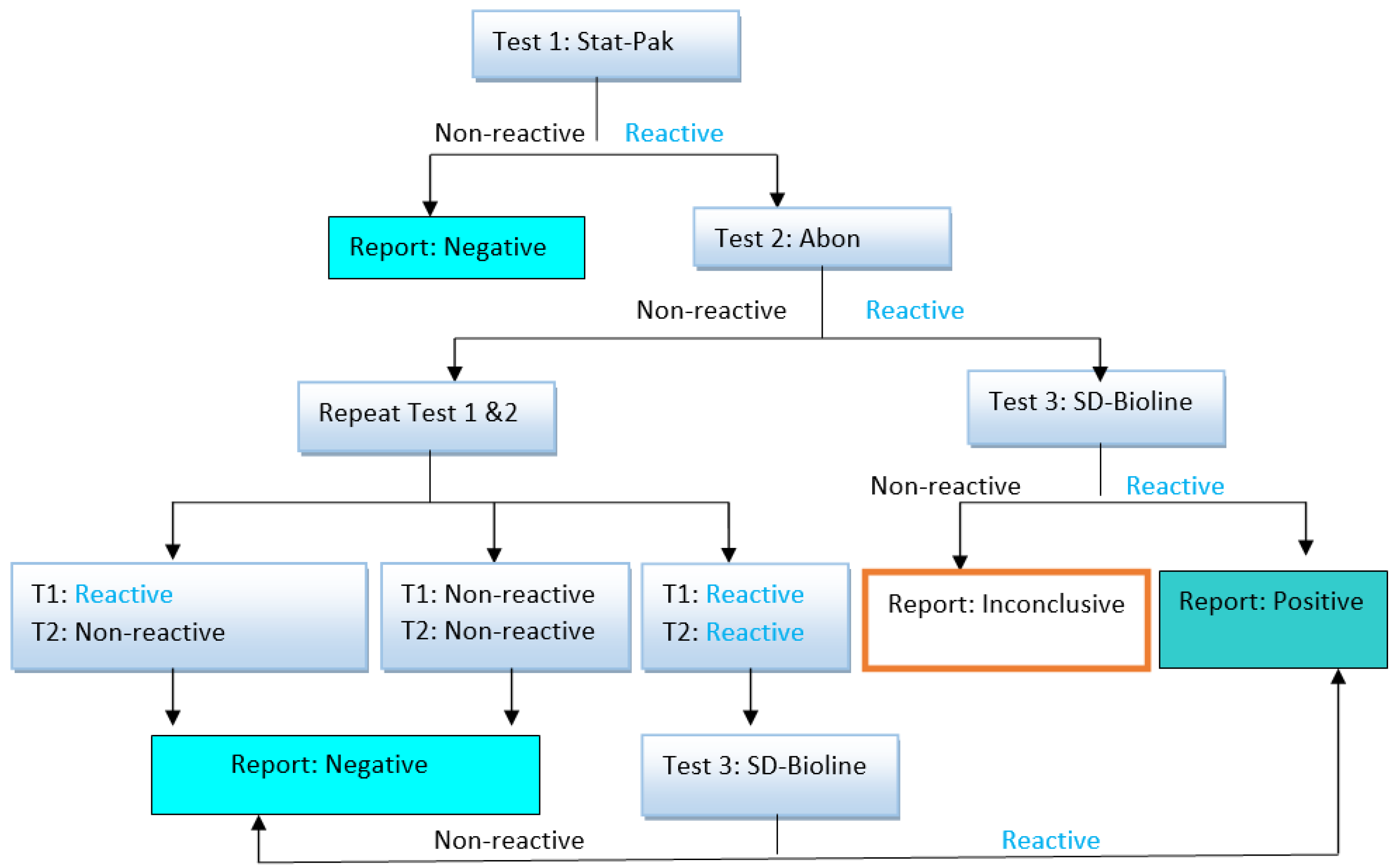
2.8. EBV Serology Test

Quantitative EBV-specific serology was performed on serum for all samples. EBV viral capsid antigen (VCA) IgG antibodies were determined by a multiplex flow immunoassay (MFI) using the BioPlex 2200 EBV IgG kits on a BioPlex 2200 Analyzer (Bio-Rad, Hercules, CA USA), according to the manufacturer’s instructions. Results were then classified, according to their antibody index values, as unfavorable (≤0.8), equivocal (0.9 to 1.0), or positive (≥1.1).

2.9. DNA Extraction

DNA was isolated from 5 × 10⁶ PBMCs in 200 μL using the QIAamp DNA mini kit (QIAGEN, Germantown, MD, USA), as per the manufacturer’s instructions. The concentration and purity of all extracted DNA samples were measured using a Qubit 3.0 fluorometer (Life Technologies, Waltham, MA, USA) and stored at −80 °C until used for quantitative real-time polymerase chain reaction (qPCR).

2.10. Real-Time Quantification PCR from Genomic DNA for EBV Load/Copy Number Determination

EBV was detected using the EBNA1 gene and performed with 10 ng of sample DNA as the initial concentration using the ViiA7 Real-time qPCR machine (Applied Biosystems, Waltham, MA USA). EBV DNA was quantified with primers specific to the EBV EBNA1 locus (forward: TCATCATCATCCGGGTCTCC, reverse: CCTACAGGGTGGAAAAATGGC), and signals were normalized to host genome DNA using primers specific for human ACTB (forward: CAGGCAGCTCGTAGCTCTTC, reverse: TCGTGCGTGACATTAAGGAG) [22]. With a total reaction volume of 10 µL, the reaction was carried out using 5 µL of 2× Fast SYBR Green Master Mix (Applied Biosystems, Waltham, MA USA), 0.25 µL of forward (10 µM) and 0.25 µL of reverse (10 µM) primers, 2.5 µL of PCR-grade water, and 2 µL of 5 ng/µL DNA concentration. The qPCR consisted of 40 cycles (95 °C for 1 s, 60 °C for 20 s, and 70 °C for 30 s). The positive control was the Raji cell line and the negative control was the K-562 cell line, which were included in each reaction. To calculate the EBV viral load, twelve standards of the known DNA copy number for the EBNA1 gene and ACTB loci at various concentrations (with 2-fold dilutions) were used to calculate EBV copies per ml. Each sample and the corresponding standard were run in triplicates on 384-well PCR plates. The EBNA1 gene’s CT value was calculated and converted into EBV copies/mL to obtain the EBV copy number. Genome copies per cellular genome elevated by more than two-fold above the negative control were considered EBV DNA positive. We used a no-template control (NTC) sample to detect qPCR reaction contamination in each run. All samples and controls were run in triplicate. The detection limit was determined using a purified PCR product in a dilution series and then determining the limit of detection with EBV-negative control DNAs.

2.11. Data Collection and Quality Assurance

The data were collected using a standardized data collection form. The form was pre-tested on 5% of the sample that was not used in the study. Under the direction of the principal investigator, a study nurse collected socio-demographics using a structured questionnaire and clinical data from a patient’s medical chart. Data collectors received training before data collection to help them incorporate the objective of the study, the method used, and the contents of the data extraction form.

2.12. Statistical Analysis

Data were entered into Epi Info 7, and statistical analyses were performed using IBM SPSS Statistics for Windows v. 26.0 (IBM).
Descriptive statistics were used to summarize the study variables. All variables were summarized using frequencies and proportions. The categorical data were compared using the chi-square test. Logistic regression models, such as bivariable and multivariable logistic regressions, were employed to determine the associations between variables. We assessed the independence, linearity, and normality tests before considering the multivariable regression analysis to ensure that our model was normally and randomly distributed. The odds ratio was determined along with the 95% CI, and those variables with a p-value <0.2 in the bivariable analysis were subjected to multivariable analysis to identify the independently associated variables. To account for the differences between the control and study groups, we used confounders related to demographic characteristics during the multivariable-adjusted odds ratio analysis. A p-value < 0.05 in the multivariable analysis was considered a statistically significant association.

3. Results

3.1. Socio-Demographic Characteristics

A total of 260 study participants, including 179 PWH on cART and 81 HIV-negative individuals, were included in the study. HIV infection was screened via the immune chromatographic or immune filtration format based on the national testing algorithm (Figure 1). Most participants (163, 62.7%) were female, and nearly one-half (127, 48.8%) were in the 18–38-year-old age group. More than one-third (105, 40.4%) of participants were married. About two-thirds (170, 65.4%) had primary and secondary school education, one-third (88, 33.8%) were government employees, and more than half (140, 53.8%) had a monthly income of ETB 3000 (USD 55.24) or less (Table 1).

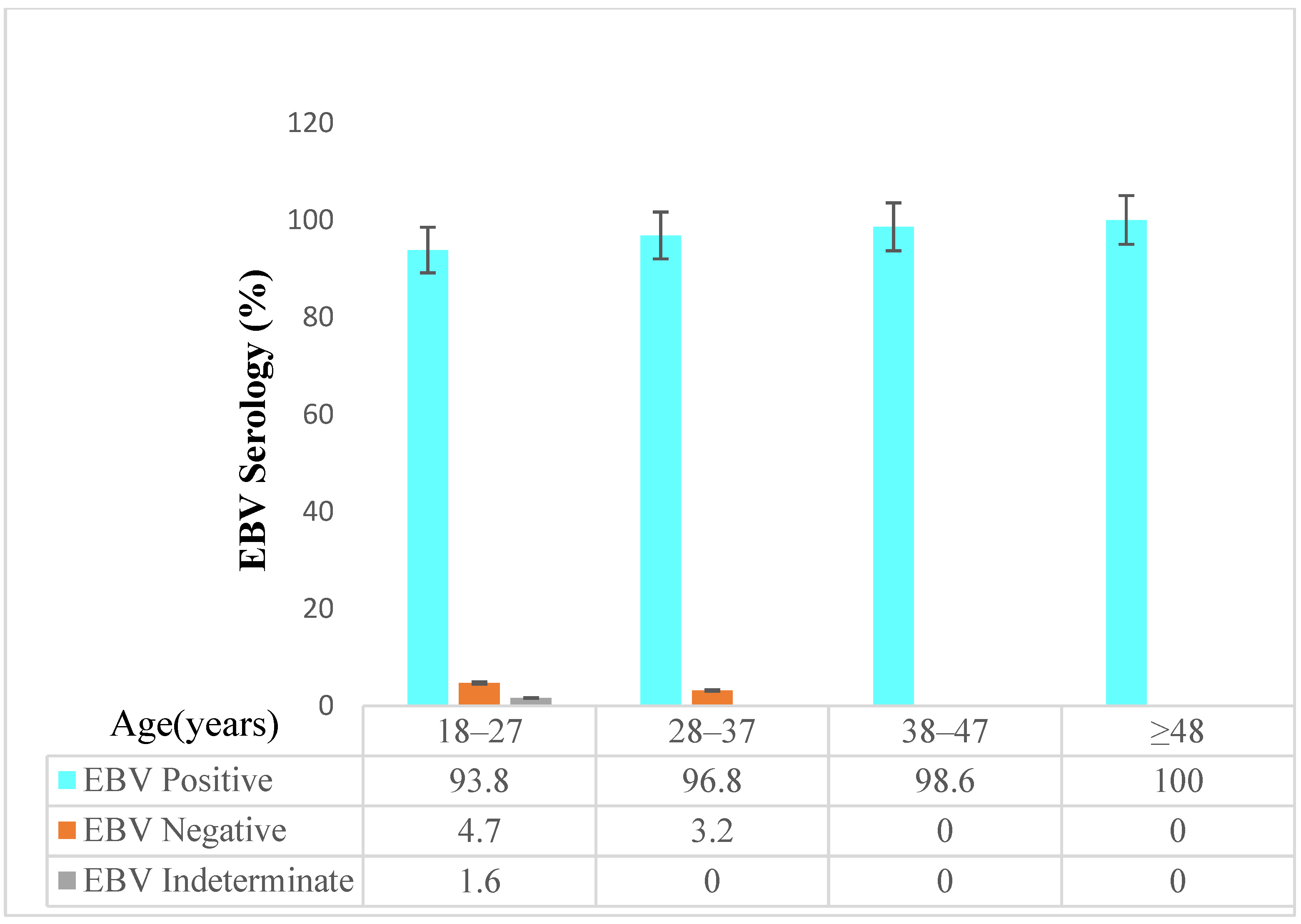
3.2. Detection of EBV Antibody

Of all the study participants, 253 (97.7%) were seropositive for the EBV VCA IgG antibody. Among the 179 HIV-positive individuals, 178 (99.4%) were seropositive, and 75 (93.8%) of the HIV-negative individuals were seropositive. There was a significant difference in seroprevalence between HIV-positive and -negative individuals (99.4% vs. 93.8%; p-value, 0.018) (Figure 2). EBV seroprevalence increased with age. Among the age group of 18–27 and 48 and above, EBV seropositivity was 93.8% and 100%, respectively (Figure 3).

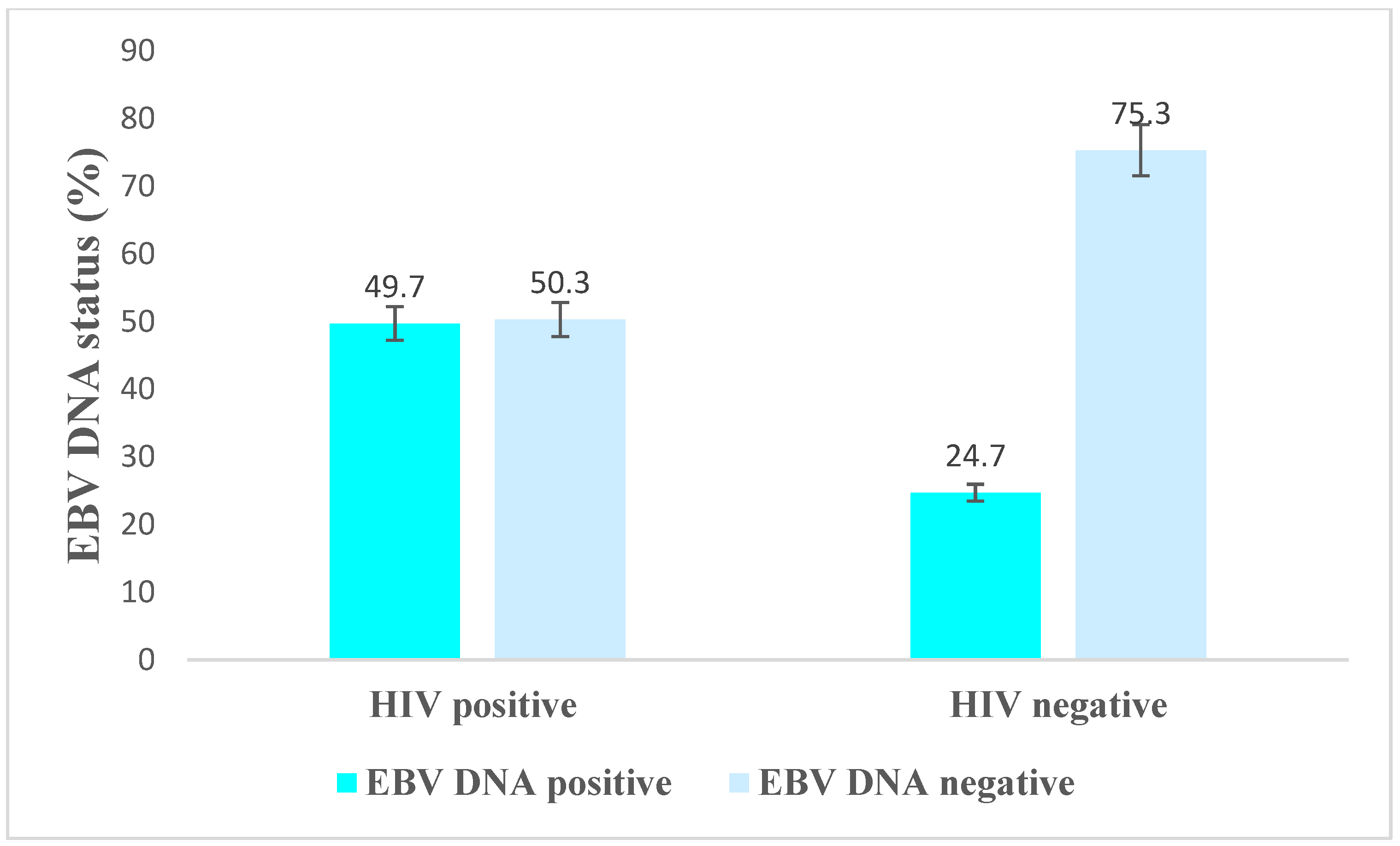
3.3. EBV DNA Positivity by Demographic and Clinical Characteristics among HIV-Positive Individuals

In this study, EBV DNA positivity was 49.7% (n = 89) among HIV-positive individuals and 24.7% (n = 20) among HIV-negative participants (Figure 4). Among HIV seropositive individuals, EBV DNA positivity correlated with some socio-demographic and clinical characteristics of the participants, as shown in Table 2. More specifically, participants of ≥35 years of age and with high (>1000 RNA copies/mL) HIV viral load demonstrated significant associations with EBV DNA positivity.

3.4. Associated Factors of EBV DNA Positivity among HIV-Positive and HIV-Negative Individuals

In the bivariable regression analysis, age group, marital status, educational status, average monthly income, and HIV status were risk factors for EBV DNA positivity at a p-value of less than 0.2. When all these variables were subjected to multivariable regression analysis, only being HIV-positive was found to be statistically significant. Accordingly, the odds of being EBV positive among HIV-positive individuals were three times as high as [AOR = 3.05; 95% CI: 1.40–6.67] those among HIV-negative individuals (Table 3).
We then further analyzed the factors associated with EBV DNA positivity among PWH. Our results show that age group, marital status, source income, average monthly income, HIV viral load, and regimen type were identified in the bivariable regression analysis as risk variables for EBV DNA positivity at a p-value of less than 0.2. Only HIV viral loads with more than 1000 RNA copies per ml were shown to be statistically significant when all these factors were examined through multivariable regression analysis. Among PWH, those with an HIV viral load of greater than 1000 RNA copies/mL (AOR = 5.81, 95% CI = 1.40, 24.13) had a higher likelihood of EBV DNA positivity compared to those who had an HIV viral load of less than 1000 RNA copies/mL (Table 4).

4. Discussion

Our study demonstrated that the EBV VCA IgG antibody seroprevalence was higher in HIV-positive individuals. The chi-square test (p values = 0.018) and Fisher’s exact test (p values = 0.012) showed that the two groups are statistically different. However, the antibody level between the two groups is almost equal. This might be due to the small sample size of the control group, and the statistically significant differences might not have biological meaning.
EBV DNA positivity was significantly higher in HIV-positive individuals in Ethiopia. Being HIV-positive is a risk factor for EBV DNA positivity. Patients with an HIV viral load greater than 1000 RNA copies/mL had a higher likelihood of EBV DNA positivity. This is the first study to report these findings in Ethiopia, where HIV and cancer remain significant public health problems.
EBV-associated lymphomas are a considerable threat to individuals infected with HIV and constitute an ADCs [23]. With more than 90% of the adult human population infected with the oncogenic herpesvirus EBV [24], coinfection with this virus is common for PWH [25]. Accordingly, knowing the burden of productive EBV infection among PWH is essential for controlling morbidity and mortality and preventing complications [26].
The findings of this study revealed that the overall seroprevalence of the EBV VCA IgG antibody was significantly higher in HIV-positive individuals compared to HIV-negative individuals. Our results are consistent with other studies that have found a higher seroprevalence of EBV infection in HIV disease [27,28]. A study conducted in Iran found that the seroprevalence of EBV infection was 100% in HIV-positive individuals and 91.9% in HIV-negative individuals [26]. Similarly, a study conducted in South Africa found that the seroprevalence of EBV infection was 100% in HIV-positive individuals [29]. Other studies have found seroprevalence rates of 87.2% in Ghana, 95% in Iran, and 97.9% in Qatar among healthy blood donors [30,31,32]. This high rate of infection could be due to immunodeficiency.
In the current study, the EBV DNA positivity was comparable with other studies conducted in Africa (South Africa, Ghana, Uganda, and Malawi), which ranged from 36% to 45% [33,34,35,36]. When the EBV DNA positivity among HIV-positive individuals compared with HIV-negative individuals, the findings were consistent with studies conducted in China (56% vs. 26%) [37], the UK (42.1% vs. 16.6%) [38], and Brazil (48.3% vs. 51.7%) [39]. On the other hand, our findings were less than those reported in a study in the United States (81% vs. 16%) [8]. In contrast to our findings, EBV DNA positivity was 30% among PWH in India [11], 39.5% among healthy individuals in Japan [40], and 37.2% in Portugal [41]. A possible reason for differences in EBV DNA detection could be due to a lack of standardization testing. These may include variations in the degree of EBV DNA detection in different laboratories due to differences in diagnostic kits, equipment, procedures, sample types [42], immunity [10], and geographic factors [43].
Other studies reported a high prevalence of EBV DNA in PWH and showed that HIV viral load was a key factor for EBV reactivation [39,44]. The prevalence of EBV DNA was significantly correlated with a detectable HIV viral load in the plasma, as observed in other studies, including in PWH without lymphoproliferative diseases [44,45]. A study suggested an interaction between EBV and HIV in the regulation of replication [46]. However, the mechanism of this regulation was not fully explored. Our findings demonstrate a significant association between EBV positivity and increased HIV viral loads in PWH in Ethiopia. This analysis strengthens the implications of an HIV–EBV interaction.
The major finding of our analysis indicated that being HIV-positive was one of the risk factors for EBV DNA positivity compared to HIV-negative individuals. Cancer is now the second most significant cause of death for adults in Ethiopia [16,47]. The HIV status of people affected by cancer has not been discussed in the literature. It would be interesting to follow these EBV DNA-positive patients, evaluate the rate of lymphoma development, and design appropriate control measures. PWH have a higher likelihood of detectable EBV DNA, which may account for the high prevalence of EBV-associated lymphomas in this population. Thus, our findings confirm that HIV infection a risk factor for EBV serostatus and infectious processes (the presence of EBV DNA), consistent with the results of other studies [7,10,26,37].

5. Conclusions

In conclusion, this study described the burden of EBV and its determinants among adult HIV-positive individuals in Ethiopia. The EBV load among PWH was significantly higher than that of HIV-negative individuals. Higher HIV viral loads in PWH were associated with an increased risk of EBV DNA positivity. The increased EBV viral load among PWH could be related to a higher risk of developing EBV-associated cancers. Hence, there is a need to do more research on the role of EBV in EBV-associated cancer in this population group.

Author Contributions

K.Z. designed the study, laboratory examinations, analyzed results, and wrote the manuscript. S.T. performed data collection, laboratory examinations and wrote the manuscript. C.W. carried out the laboratory examinations, analyzed the results and wrote the manuscript. E.H.A. carried out the laboratory examinations, analyzed the results and wrote the manuscript. R.B., T.A., N.B. and A.F.H. designed the study, analyzed the results, and wrote the manuscript. All the work performed in this study was carried out with the pertinent advice of E.A., G.Y. and W.A. All authors have read and agreed to the published version of the manuscript.

Funding

The Ohio State University and Addis Ababa University provided funding for the study. The funders had no role in study design, data collection and analysis, decision to publish, or preparation of the manuscript. Elshafa Hassan Ahmed is supported by NIH R01 (Grant No. 3R01AI141531) Administrative Supplements grant to Promote Diversity.

Institutional Review Board Statement

This study was approved by the Addis Ababa University, Aklilu Lemma Institute of Pathobiology-Institutional Review Board (protocol number: ALIPB-IRB #60/2013/21). Permission was also obtained from Tikur Anbesa Specialized Hospital. Prior to enrolment, all participants provided a written informed consent in accordance with the Helsinki Declaration.

Informed Consent Statement

Informed consent was obtained from all subjects involved in the study.

Data Availability Statement

The raw data can be obtained from the first author and corresponding author upon request.

Acknowledgments

The authors are grateful to the Ohio State University Jams Cancer Centre, Addis Ababa University, and the Ethiopian Public Health Institute for their support and coordination facilitation. The authors would like to express their special thanks to all data collectors and the study participants.

Conflicts of Interest

The authors declare no conflict of interest.

Abbreviations

ADCAIDS-defining cancer
AIDSAcquired immune deficiency syndrome
ARTAntiretroviral therapy
cARTCombined antiretroviral therapy
DNADeoxyribonucleic acid
DLBCLDiffuse large B cell lymphoma
DMSODimethyl sulfoxide
EBEREpstein–Barr virus (EBV)-encoded small RNAs
EBNA 1Epstein–Barr nuclear antigen 1
EBVEpstein–Barr virus
HBVHepatitis B virus
HCVHepatitis C virus
HHV-8Human herpesvirus-8
HIVHuman immunodeficiency viruses
HLHodgkin lymphoma
HPVHuman papillomavirus
IgGImmunoglobulin G
KSHVKaposi’s sarcoma-associated herpesvirus
NHLNon-Hodgkin lymphoma
PBMCsPeripheral blood mononuclear cells
PBSPhosphate buffer saline
PCRPolymerase chain reaction
PWHPeople living with HIV
qRT PCRQuantitative real-time polymerase chain reaction
RDTRapid diagnostic test
RNARibonucleic acid
SSASub-Saharan Africa
TASHTikur Anbessa Specialised Hospital
VCAViral capsid antigens

References

  1. Shahriar, S.; Araf, Y.; Ahmad, R.; Kattel, P.; Sah, G.S.; Rahaman, T.I.; Sadiea, R.Z.; Sultana, S.; Islam, M.S.; Zheng, C.; et al. Insights Into the Coinfections of Human Immunodeficiency Virus-Hepatitis B Virus, Human Immunodeficiency Virus-Hepatitis C Virus, and Hepatitis B Virus-Hepatitis C Virus: Prevalence, Risk Factors, Pathogenesis, Diagnosis, and Treatment. Front. Microbiol. 2022, 12, 780887. [Google Scholar] [CrossRef]
  2. Riedel, D.J.; Tang, L.S.; Rositch, A.F. The role of viral co-infection in HIV-associated non-AIDS-related cancers. Curr. HIV/AIDS Rep. 2015, 12, 362–372. [Google Scholar] [CrossRef] [PubMed] [Green Version]
  3. Calabresi, A.; Ferraresi, A.; Festa, A.; Scarcella, C.; Donato, F.; Vassallo, F.; Limina, R.M.; Castelli, F.; Quiros-Roldan, E.; Brescia HIV Cancer Study Group. Incidence of AIDS-defining cancers and virus-related and non-virus-related non-AIDS-defining cancers among HIV-infected patients compared with the general population in a large health district of Northern Italy, 1999–2009. HIV Med. 2013, 14, 481–490. [Google Scholar] [CrossRef] [PubMed]
  4. McLaughlin-Drubin, M.E.; Munger, K. Viruses associated with human cancer. Biochim. Biophys. Acta 2008, 1782, 127–150. [Google Scholar] [CrossRef] [Green Version]
  5. Shannon-Lowe, C.; Rickinson, A. The Global Landscape of EBV-Associated Tumors. Front. Oncol. 2019, 9, 713. [Google Scholar] [CrossRef] [Green Version]
  6. Thorley-Lawson, D.A. EBV persistence—Introducing the virus. In Epstein Barr Virus Volume 1; Springer: Berlin/Heidelberg, Germany, 2015; pp. 151–209. [Google Scholar]
  7. Bibas, M.; Antinori, A. EBV and HIV-related lymphoma. Mediterr. J. Hematol. Infect. Dis. 2009, 1, e2009032. [Google Scholar] [CrossRef] [PubMed]
  8. Ling, P.D.; Vilchez, R.A.; Keitel, W.A.; Poston, D.G.; Peng, R.S.; White, Z.S.; Visnegarwala, F.; Lewis, D.E.; Butel, J.S. Epstein-Barr virus DNA loads in adult human immunodeficiency virus type 1-infected patients receiving highly active antiretroviral therapy. Clin. Infect. Dis. 2003, 37, 1244–1249. [Google Scholar] [CrossRef] [PubMed]
  9. Merlo, A.; Turrini, R.; Dolcetti, R.; Martorelli, D.; Muraro, E.; Comoli, P.; Rosato, A. The interplay between Epstein-Barr virus and the immune system: A rationale for adoptive cell therapy of EBV-related disorders. Haematologica 2010, 95, 1769. [Google Scholar] [CrossRef] [PubMed]
  10. Shindiapina, P.; Ahmed, E.H.; Mozhenkova, A.; Abebe, T.; Baiocchi, R.A. Immunology of EBV-Related Lymphoproliferative Disease in HIV-Positive Individuals. Front. Oncol. 2020, 10, 1723. [Google Scholar] [CrossRef]
  11. Mandal, D.; Desai, D.; Sinha, S. High prevalence of plasma EBV DNA among the HIV positive individuals, with or without malignancies, attending the clinic at AIIMS, New Delhi. Virusdisease 2021, 32, 137–139. [Google Scholar] [CrossRef]
  12. Kharsany, A.B.; Karim, Q.A. HIV infection and AIDS in sub-Saharan Africa: Current status, challenges and opportunities. Open AIDS J. 2016, 10, 34. [Google Scholar] [CrossRef] [PubMed] [Green Version]
  13. The Ethiopian Public Health Institute, Ministry of Health. HIV Related Estimates and Projections in Ethiopia for the Year 2021–2022; The Ethiopian Public Health Institute: Addis Ababa, Ethiopia, 2022. [Google Scholar]
  14. Central Statistical Agency. Ethiopia Demographic and Health Survey; Central Statistical Agency: Addis Ababa, Ethiopia, 2016. [Google Scholar]
  15. Ethiopian Public Health Institute. Ethiopia Population-Based HIV Impact Assessment 2017–2018; Ethiopian Public Health Institute: Addis Ababa, Ethiopia, 2020. [Google Scholar]
  16. Memirie, S.T.; Habtemariam, M.K.; Asefa, M.; Deressa, B.T.; Abayneh, G.; Tsegaye, B.; Abraha, M.W.; Ababi, G.; Jemal, A.; Rebbeck, T.R. Estimates of cancer incidence in Ethiopia in 2015 using population-based registry data. J. Glob. Oncol. 2018, 4, 1–11. [Google Scholar] [CrossRef]
  17. Palella, F.J., Jr.; Delaney, K.M.; Moorman, A.C.; Loveless, M.O.; Fuhrer, J.; Satten, G.A.; Aschman, D.J.; Holmberg, S.D.; Investigators, H.O.S. Declining morbidity and mortality among patients with advanced human immunodeficiency virus infection. N. Engl. J. Med. 1998, 338, 853–860. [Google Scholar] [CrossRef] [PubMed]
  18. Simard, E.P.; Pfeiffer, R.M.; Engels, E.A. Cumulative incidence of cancer among individuals with acquired immunodeficiency syndrome in the United States. Cancer 2011, 117, 1089–1096. [Google Scholar] [CrossRef] [PubMed]
  19. Anselmi, A.; Vendrame, D.; Rampon, O.; Giaquinto, C.; Zanchetta, M.; De Rossi, A. Immune reconstitution in human immunodeficiency virus type 1-infected children with different virological responses to anti-retroviral therapy. Clin. Exp. Immunol. 2007, 150, 442–450. [Google Scholar] [CrossRef]
  20. Sachithanandham, J.; Kannangai, R.; Pulimood, S.; Desai, A.; Abraham, A.; Abraham, O.; Ravi, V.; Samuel, P.; Sridharan, G. Significance of Epstein-Barr virus (HHV-4) and CMV (HHV-5) infection among subtype-C human immunodeficiency virus-infected individuals. Indian J. Med. Microbiol. 2014, 32, 261–269. [Google Scholar] [CrossRef]
  21. Ministry of Health. National Guidelines for Comprehensive HIV Prevention, Care and Treatment; Ministry of Health: Addis Ababa, Ethiopia, 2018. [Google Scholar]
  22. Ahmed, E.H.; Lustberg, M.; Hale, C.; Sloan, S.; Mao, C.; Zhang, X.; Ozer, H.G.; Schlotter, S.; Smith, P.L.; Jeney, F. Follicular Helper and Regulatory T Cells Drive the Development of Spontaneous Epstein–Barr Virus Lymphoproliferative Disorder. Cancers 2023, 15, 3046. [Google Scholar] [CrossRef]
  23. Cesarman, E. Gammaherpesvirus and lymphoproliferative disorders in immunocompromised patients. Cancer Lett. 2011, 305, 163–174. [Google Scholar] [CrossRef] [Green Version]
  24. Luzuriaga, K.; Sullivan, J.L. Infectious Mononucleosis. N. Engl. J. Med. 2010, 362, 1993–2000. [Google Scholar] [CrossRef] [Green Version]
  25. Xu, G.J.; Kula, T.; Xu, Q.; Li, M.Z.; Vernon, S.D.; Ndung’u, T.; Ruxrungtham, K.; Sanchez, J.; Brander, C.; Chung, R.T. Comprehensive serological profiling of human populations using a synthetic human virome. Science 2015, 348, aaa0698. [Google Scholar] [CrossRef] [Green Version]
  26. Keramat, F.; Basir, H.R.G.; Ahmadpour, S.; Moradi, A. Seroprevalence of Epstein-Barr Virus in HIV-Infected Patients, A Case Control Study. J. Kermanshah Univ. Med. Sci. 2019, 23, e84967. [Google Scholar] [CrossRef] [Green Version]
  27. McClain, K.L.; Leach, C.T.; Jenson, H.B.; Joshi, V.V.; Pollock, B.H.; Parmley, R.T.; DiCarlo, F.J.; Chadwick, E.G.; Murphy, S.B. Association of Epstein–Barr virus with leiomyosarcomas in young people with AIDS. N. Engl. J. Med. 1995, 332, 12–18. [Google Scholar] [CrossRef] [PubMed]
  28. Kyaw, M.T.; Hurren, L.; Evans, L.; Moss, D.J.; Cooper, D.A.; Benson, E.; Esmore, D.; Sculley, T.B. Expression of B-type Epstein-Barr virus in HIV-infected patients and cardiac transplant recipients. AIDS Res. Hum. Retroviruses 1992, 8, 1869–1874. [Google Scholar] [CrossRef] [PubMed]
  29. Schaftenaar, E.; Verjans, G.M.; Getu, S.; McIntyre, J.A.; Struthers, H.E.; Osterhaus, A.D.; Peters, R.P. High seroprevalence of human herpesviruses in HIV-infected individuals attending primary healthcare facilities in rural South Africa. PLoS ONE 2014, 9, e99243. [Google Scholar] [CrossRef] [PubMed] [Green Version]
  30. Adjei, A.A.; Armah, H.B.; Gbagbo, F.; Boamah, I.; Adu-Gyamfi, C.; Asare, I. Seroprevalence of HHV-8, CMV, and EBV among the general population in Ghana, West Africa. BMC Infect. Dis. 2008, 8, 111. [Google Scholar] [CrossRef] [Green Version]
  31. Sharifipour, S.; Rad, K.D. Seroprevalence of Epstein–Barr virus among children and adults in Tehran, Iran. New Microbes New Infect. 2020, 34, 100641. [Google Scholar] [CrossRef]
  32. Smatti, M.K.; Yassine, H.M.; AbuOdeh, R.; AlMarawani, A.; Taleb, S.A.; Althani, A.A.; Nasrallah, G.K. Prevalence and molecular profiling of Epstein Barr virus (EBV) among healthy blood donors from different nationalities in Qatar. PLoS ONE 2017, 12, e0189033. [Google Scholar] [CrossRef] [Green Version]
  33. Corcoran, C.; Rebe, K.; van der Plas, H.; Myer, L.; Hardie, D.R. The predictive value of cerebrospinal fluid Epstein-Barr viral load as a marker of primary central nervous system lymphoma in HIV-infected persons. J. Clin. Virol. 2008, 42, 433–436. [Google Scholar] [CrossRef] [PubMed]
  34. Opintan, J.A.; Awadzi, B.K.; Biney, I.J.; Ganu, V.; Doe, R.; Kenu, E.; Adu, R.F.; Osei, M.M.; Akumwena, A.; Grigg, M.E. High rates of cerebral toxoplasmosis in HIV patients presenting with meningitis in Accra, Ghana. Trans. R. Soc. Trop. Med. Hyg. 2017, 111, 464–471. [Google Scholar] [CrossRef] [Green Version]
  35. Rajasingham, R.; Rhein, J.; Klammer, K.; Musubire, A.; Nabeta, H.; Akampurira, A.; Mossel, E.C.; Williams, D.A.; Boxrud, D.J.; Crabtree, M.B. Epidemiology of meningitis in an HIV-infected Ugandan cohort. Am. J. Trop. Med. Hyg. 2015, 92, 274. [Google Scholar] [CrossRef] [Green Version]
  36. Kelly, M.J.; Benjamin, L.A.; Cartwright, K.; Ajdukiewicz, K.M.; Cohen, D.B.; Menyere, M.; Galbraith, S.; Guiver, M.; Neuhann, F.; Solomon, T. Epstein-barr virus coinfection in cerebrospinal fluid is associated with increased mortality in Malawian adults with bacterial meningitis. J. Infect. Dis. 2012, 205, 106–110. [Google Scholar] [CrossRef] [PubMed] [Green Version]
  37. Pan, R.; Liu, X.; Zhou, S.; Ning, Z.; Zheng, H.; Gao, M.; Ding, Y.; Yao, W.; Liao, X.; He, N. Differential prevalence and correlates of whole blood Epstein–Barr virus DNA between HIV-positive and HIV-negative men who have sex with men in Shanghai, China. Epidemiol. Infect. 2017, 145, 2330–2340. [Google Scholar] [CrossRef] [PubMed] [Green Version]
  38. Ammatuna, P.; Campisi, G.; Giovannelli, L.; Giambelluca, D.; Alaimo, C.; Mancuso, S.; Margiotta, V. Presence of Epstein–Barr virus, cytomegalovirus and human papillomavirus in normal oral mucosa of HIV-infected and renal transplant patients. Oral Dis. 2001, 7, 34–40. [Google Scholar] [PubMed]
  39. Santos, L.; Azevedo, K.; Silva, L.; Oliveira, L. Epstein-Barr virus in oral mucosa from human immunodeficiency virus positive patients. Rev. Assoc. Médica Bras. 2014, 60, 262–269. [Google Scholar] [CrossRef] [PubMed] [Green Version]
  40. Nishiwaki, M.; Fujimuro, M.; Teishikata, Y.; Inoue, H.; Sasajima, H.; Nakaso, K.; Nakashima, K.; Sadanari, H.; Yamamoto, T.; Fujiwara, Y. Epidemiology of Epstein–Barr virus, cytomegalovirus, and kaposi’s sarcoma-associated herpesvirus infections in peripheral blood leukocytes revealed by a multiplex PCR assay. J. Med. Virol. 2006, 78, 1635–1642. [Google Scholar] [CrossRef]
  41. Sousa, H.; Silva, J.; Azevedo, L.; Pinto-Correia, A.L.; Catarino, R.; Pinto, D.; Lopes, C.; Medeiros, R. Epstein-Barr virus in healthy individuals from Portugal. Acta Médica Port. 2011, 24, 707–712. [Google Scholar]
  42. Abusalah, M.A.H.; Gan, S.H.; Al-Hatamleh, M.A.; Irekeola, A.A.; Shueb, R.H.; Yean Yean, C. Recent advances in diagnostic approaches for epstein–barr virus. Pathogens 2020, 9, 226. [Google Scholar] [CrossRef] [Green Version]
  43. Liao, H.M.; Liu, H.; Chin, P.J.; Li, B.; Hung, G.C.; Tsai, S.; Otim, I.; Legason, I.D.; Ogwang, M.D.; Reynolds, S.J.; et al. Epstein-Barr Virus in Burkitt Lymphoma in Africa Reveals a Limited Set of Whole Genome and LMP-1 Sequence Patterns: Analysis of Archival Datasets and Field Samples From Uganda, Tanzania, and Kenya. Front. Oncol. 2022, 12, 812224. [Google Scholar] [CrossRef]
  44. Lupo, J.; Germi, R.; Lancar, R.; Algarte-Genin, M.; Hendel-Chavez, H.; Taoufik, Y.; Mounier, N.; Partisani, M.; Bonnet, F.; Meyohas, M.-C. Prospective evaluation of blood Epstein–Barr virus DNA load and antibody profile in HIV-related non-Hodgkin lymphomas. AIDS 2021, 35, 861–868. [Google Scholar] [CrossRef]
  45. Wang, Y.; Yang, J.; Wen, Y. Lessons from Epstein-Barr virus DNA detection in cerebrospinal fluid as a diagnostic tool for EBV-induced central nervous system dysfunction among HIV-positive patients. Biomed. Pharmacother. 2022, 145, 112392. [Google Scholar] [CrossRef]
  46. Mbopi-Kéou, F.-X.; Bélec, L.; Teo, C.G.; Scully, C.; Porter, S.R. Synergism between HIV and other viruses in the mouth. Lancet Infect. Dis. 2002, 2, 416–424. [Google Scholar] [CrossRef] [PubMed]
  47. Timotewos, G.; Solomon, A.; Mathewos, A.; Addissie, A.; Bogale, S.; Wondemagegnehu, T.; Aynalem, A.; Ayalnesh, B.; Dagnechew, H.; Bireda, W. First data from a population based cancer registry in Ethiopia. Cancer Epidemiol. 2018, 53, 93–98. [Google Scholar] [CrossRef] [PubMed]
Figure 1. HIV diagnosis serial testing algorithm [21].
Viruses 15 01743 g001
Figure 2. EBV serology with HIV status.
Viruses 15 01743 g002
Figure 3. EBV serology with age.
Viruses 15 01743 g003
Figure 4. EBV DNA positivity among HIV-positive and -negative study participants.
Viruses 15 01743 g004
Table 1. Socio-demographic characteristics of the study participants.
CharacteristicsHIV StatusTotal (260)%
Positive
(n = 179)
%Negative
(n = 81)
%
SexMale6636.93138.39737.3
Female11363.15061.716362.7
Age 18–282514.03948.16424.6
29–383821.22530.96324.2
39–485933.01214.87127.3
>495731.856.26223.8
Marital statusUnmarried5128.54656.89737.3
Married7441.33138.310540.4
Divorced2312.844.92710.4
Widowed3117.300.03111.9
Educational statusIlliterate168.900.0166.2
Primary School5731.8911.16625.4
Secondary School7642.52834.610440.0
College diploma and above3016.84454.37428.5
Source of incomeGovernment employee2614.56276.58833.8
Stay-at-home spouse2715.100.02710.4
Private sector employee5329.600.05320.4
Self-employed4324.000.04316.5
Student1810.111.2197.3
Unemployed126.71822.23011.5
Average monthly income in ETB<10005229.111.25320.4
1001–20003720.71316.05019.2
2001–30002514.01214.83714.2
3001–4000158.42632.14115.8
>40001910.61113.63011.5
NA3117.31822.24918.8
HIV: human immunodeficiency virus; ETB: Ethiopian birr; NA: not applicable.
Table 2. Epstein–Barr virus (EBV) DNA positivity among HIV-1 positive individuals by demographic and clinical characteristics.
CharacteristicsEBV DNA StatusTotal%X2p-Value
Positive%Negative%
SexMale3045.5%3654.5%6637%0.760.383
Female5952.2%5447.8%11363%
Age <352036.4%3563.6%5531%0.570.017
≥356955.6%5544.4%12469%
Marital statusUnmarried2345.1%2854.9%5128%6.850.077
Married3344.6%4155.4%7441%
Divorced1147.8%1252.2%2313%
Widowed2271.0%929.0%3117%
Educational statusIlliterate956.3%743.8%169%1.530.676
Primary school3154.4%2645.6%5732%
Secondary school3444.7%4255.3%7642%
College diploma and above1550.0%1550.0%3017%
Source of incomeGovernment employee1557.7%1142.3%2615%12.620.027
Stay-at-home spouse1763.0%1037.0%2715%
Private sector employee2547.2%2852.8%5330%
Self-employed527.8%1372.2%1810%
Student216.7%1083.3%127%
Unemployed2558.1%1841.9%4324%
Average monthly income in ETB<10002955.8%2344.2%5229%2.670.751
1001–20002054.1%1745.9%3721%
2001–30001248.0%1352.0%2514%
3001–4000746.7%853.3%158%
>4000947.4%1052.6%1911%
NA1238.7%1961.3%3117%
Most recent VL<10007446.5%8553.5%15989%5.760.016
>10001575.0%525.0%2011%
Most recent CD4count<2001451.9%1348.1%2716%0.960.757
≥2007046.7%7451.4%14484%
ART1st line6450.0%6450.0%12872%2.570.277
2nd line1943.2%2556.8%4425%
3rd line480.0%120.0%53%
Regimen typeATV/r (PI based)1943.2%2556.8%4425%4.700.195
Darunavir (PI based)480.0%120.0%53%
DTG (INSTI based)6251.7%5848.3%12068%
EFV/NVP (NNRTI based)225.0%675.0%85%
NA = Not applicable.
Table 3. Bi-variable and multivariable regression analysis of EBV DNA positivity among HIV-positive and HIV-negative individuals in Addis Ababa, Ethiopia (n = 260).
CharacteristicsCOR (95% CI)AOR (95% CI)
Age <3511
≥351.75 (1.06–2.89)1.34 (0.65–2.72)
Marital statusUnmarried11
Married1.04 (0.59–1.84)0.78 (0.37–1.64)
Divorced1.16 (0.49–2.78)0.77 (0.28–2.12)
Widowed4.14 (1.72–9.97)2.71 (0.95–7.71)
Educational statusIlliterate1.43 (4.83–4.25)0.39 (0.11–1.50)
Primary school0.99 (0.51–1.92)0.49 (0.20–1.19)
Secondary school0.54 (0.29–1.00)0.32 (0.14–0.69)
College diploma and above11
Average monthly income<10001.75 (0.80–3.84)1.28 (0.503–3.24)
1001–20000.967 (0.43–2.16)1.03 (0.404–2.62)
2001–30000.79 (0.33–1.90)0.76 (0.285–2.03)
3001–40000.41 (0.16–1.04)0.49 (0.17–1.44)
>40002.17 (0.86–5.40)2.08 (0.72–6.01)
NA11
HIV statusNegative11
Positive3.02 (1.68–5.41)3.05 (1.4–6.67)
Crude odds ratio = COR; adjusted odds ratio = AOR, Not applicable = NA.
Table 4. Bi-variable and multivariable regression analysis of EBV DNA positivity among HIV-positive individuals in Addis Ababa, Ethiopia (n = 179).
CharacteristicsCOR (95% CI)AOR (95% CI)
Age<3511
≥352.20 (1.14–4.22)2.16 (0.89–5.30)
Marital statusUnmarried11
Married0.98 (0.48–2.01)0.49 (0.172–1.40)
Divorced1.12 (0.42–3.00)0.63 (1.90–2.11)
Widowed2.98 (1.15–7.71)1.37 (0.40–4.71)
Source of incomeGovernment employee11
Stay-at-home spouse1.25 (0.41–3.75)1.94 (0.504–7.50)
Private sector employee0.66(0.25–1.69)0.92(3.15–2.72)
Self-employed0.28 (0.08–1.03)0.35 (0.078–1.56)
Student0.147 (0.03–0.81)0.154 (0.014–1.64)
Unemployed1.02 (0.38–2.73)0.97 (0.27–3.50)
Average monthly income<10002.0 (0.81–4.94)1.31 (0.37–4.69)
1001–20001.86 (0.71–4.91)1.11 (0.28–4.47)
2001–30001.46 (0.503–4.25)0.94 (0.21–4.15)
3001–40001.38 (0.40–4.81)1.25 (0.21–7.48)
>40001.43 (0.45–4.52)1.11 (0.21–5.76)
NA11
Most recent VL<100011
>10003.45(1.20–9.94)5.81 (1.4–24.13)
Regimen typeATV/r based (PI based)11
Darunavir based (PI based)2.30 (0.41–12.60)2.4 (0.185–31.1)
DTG based (INSTI based)12.00 (0.80–180.9)1.5 (0.66–3.38)
EFV/NVP based (NNRTI based)3.21 (0.62–16.53)0.297 (0.039–2.28)
Crude odds ratio = COR; adjusted odds ratio = AOR, Not applicable = NA.
Disclaimer/Publisher’s Note: The statements, opinions and data contained in all publications are solely those of the individual author(s) and contributor(s) and not of MDPI and/or the editor(s). MDPI and/or the editor(s) disclaim responsibility for any injury to people or property resulting from any ideas, methods, instructions or products referred to in the content.

