Article

Risk Assessment of the Possible Intermediate Host Role of Pigs for Coronaviruses with a Deep Learning Predictor

1
College of Mathematics, Jilin University, Changchun, Jilin 130012, China
2
State Key Laboratory of Pathogen and Biosecurity, Beijing Institute of Microbiology and Epidemiology, AMMS, Beijing 100071, China
*
Authors to whom correspondence should be addressed.
These authors contributed equally to this work.
Viruses 2023, 15(7), 1556; https://doi.org/10.3390/v15071556
Submission received: 28 February 2023 / Revised: 13 July 2023 / Accepted: 13 July 2023 / Published: 15 July 2023
(This article belongs to the Collection Coronaviruses)

Abstract

:
Swine coronaviruses (CoVs) have been found to cause infection in humans, suggesting that Suiformes might be potential intermediate hosts in CoV transmission from their natural hosts to humans. The present study aims to establish convolutional neural network (CNN) models to predict host adaptation of swine CoVs. Decomposing of each ORF1ab and Spike sequence was performed with dinucleotide composition representation (DCR) and other traits. The relationship between CoVs from different adaptive hosts was analyzed by unsupervised learning, and CNN models based on DCR of ORF1ab and Spike were built to predict the host adaptation of swine CoVs. The rationality of the models was verified with phylogenetic analysis. Unsupervised learning showed that there is a multiple host adaptation of different swine CoVs. According to the adaptation prediction of CNN models, swine acute diarrhea syndrome CoV (SADS-CoV) and porcine epidemic diarrhea virus (PEDV) are adapted to Chiroptera, swine transmissible gastroenteritis virus (TGEV) is adapted to Carnivora, porcine hemagglutinating encephalomyelitis (PHEV) might be adapted to Primate, Rodent, and Lagomorpha, and porcine deltacoronavirus (PDCoV) might be adapted to Chiroptera, Artiodactyla, and Carnivora. In summary, the DCR trait has been confirmed to be representative for the CoV genome, and the DCR-based deep learning model works well to assess the adaptation of swine CoVs to other mammals. Suiformes might be intermediate hosts for human CoVs and other mammalian CoVs. The present study provides a novel approach to assess the risk of adaptation and transmission to humans and other mammals of swine CoVs.

1. Introduction

The ongoing global COVID-19 pandemic, caused by the pathogenic agent of severe acute respiratory syndrome coronavirus (CoV) 2 (SARS-CoV-2) has still been threatening human health. What is more worrisome is that little is known about how the virus crossed species barriers, adapting to human beings from its natural reservoir host of bat [1,2,3]. A paradigmatic cross-species transmission from natural reservoir host to an intermediate host and then to human beings has been widely accepted for human pandemic viruses, such influenza A viruses (IAVs) [4,5,6] and CoVs [7]. IAVs and CoVs with single-stranded RNA genomes mutate with much higher rates than double-stranded RNA viruses and DNA viruses (single- or double-stranded) [8]. Avian host-originated IAVs of H5N1 [9,10,11], H7N9 [12,13], and other subtypes [14,15] adapted to mammals or mammalian cells with or without mammalian passages. In particular, domestic pigs and probably other species in Suiformes played a key intermediate host role for all six historic influenza pandemics [6,16,17,18,19,20]. Suiformes are also one main family of mammalian hosts for CoVs [21,22,23,24,25,26]. Swine CoVs have been implied to replicate efficiently in human primary cells [27] and, more worryingly, cause infection in malnourished Haitian children [28] or calves and chickens [29]. Thus, it is urgent to pay more attention to the assessment of the adaptation and transmission risk to human beings or other mammals of swine CoVs.
CoVs not only directly infect a variety of mammals in the family of Chiroptera, Artiodactyla, Rodents, Lagomorphs, Carnivores, and Primates, but also cause trans-species infection among these mammals [7,30,31]. Such trans-species infection and transmission have been indicated to play important role for CoVs to adapt to human beings and cause prevalence [7]. Among human CoVs, SARS-CoV-2 is closely related to some bat CoVs [1]. HCoV-OC43 and HCoV-HKU1 originated from cattle [32] and rodents [33] respectively, and HCoV-NL63 originated from bats [34]. SARS-CoVs which originated from bats infected civets before infecting humans and causing an epidemic [35]. In the transmission of HCoV-229E and MERS-CoV from bats to humans, dromedary camels and camelids played the role of intermediate hosts, respectively [7]. Therefore, mammals might serve as intermediate hosts in the transmission of CoVs from their original hosts to humans.
Phylogenetic analysis and other traditional methods in bioinformatics have provided indicative clues to infer the homology to other mammalian CoVs and the potential infection/transmission risk to these hosts of Suiformes CoVs. Phylogenetic trees have indicated that swine CoVs, such as swine acute diarrhea syndrome CoV (SADS-CoV) [21] and porcine epidemic diarrhea virus (PEDV) [25] may originate from bats [26]. Swine transmissible gastroenteritis virus (TGEV) has very high homology to canine CoVs [36]. Results on multiple mammalian cell lines of different species showed susceptibility to SADS-CoV, implying possible multiple host adaptability of Suiformes CoVs [22]. Although biologically experimental results are very reliable, such experiments are time-consuming, resulting in a lag in research results. Furthermore, these methods can hardly quantify and intelligently assess the risk of infection and transmission of Suiformes CoVs to humans and other mammals.
Artificial intelligence (AI) methods have been found to be effective in solving these problems. Sequence compositions of nucleic acids and proteins are significantly associated with genome evolution and adaptation across all kingdoms of life [37]. Adaptive determinants have recently been widely identified at the nucleic acid level (genomic DNA, RNA, or mRNA) among pathogens such as parasites [38], bacteria [39], and viruses [40,41]. The dynamic homeostasis of genomic RNA sequences shapes the transcription, translation, and decay of mRNA [42], particularly for RNA viruses. These determinants regulate the replication of pathogens in hosts via the machinery related to codon usage bias [38,39,43], dinucleotide composition [44], tRNA abundance [39,41], mRNA decay [45], translation elongation speed [46], and translation efficiency [47]. Thus, the RNA sequence-based nucleotide composition is biologically meaningful and is closely related to the causal inference of the virus phenotype. Deep learning based on unlabeled amino acids (AAs) was utilized to extract statistical representation with rich semantics from fundamental features of a protein [48]. Viral escape was modeled using natural language processing (NLP) methods to predict the structural escape patterns [49,50]. Machine learning models based on compositional traits [36,40] such as codon, codon pair, and dinucleotide (DNT) [44,51] have enabled accurate predictions of virus adaptation to hosts. Several models have been utilized to predict SARS-CoV-2 variants on the basis of viral protein sequences, with a particular focus on key mutant AAs related to receptor binding [43,52,53], although easily falling into the trap of overfitting. Convolutional neural networks (CNNs) are widely used in the field of image recognition; however, in recent years, CNN has also performed well in predicting the adaptability of virus hosts. A CNN predictor based on the dinucleotide composition representation (DCR) [54] and AA [55] could provide real-time predictions of emerging SARS-CoV-2 variants. Thus, AI methods are expected to learn the adaptation of swine CoVs to other mammals, and to assess the possible intermediate host role of pigs for coronaviruses.
The present study aims to establish 3D-CNN classification models based on viral genomic DCR to predict the adaptation of Suiformes CoVs to the five types of hosts. The binding ability to specific receptors and the replication ability in host cells are considered key factors influencing viral host adaptation. Therefore, these two major viral proteins—receptor-binding glycoprotein (Gp; also named S for CoVs) and RNA-dependent RNA polymerase (RdRp; mainly ORF1ab-encoded)—were assessed by the classification traits. After ORF1ab and Spike genome decomposition by DCR and other traits, unsupervised learning was performed to analyze the distance between different types of Suiformes CoVs and CoVs from other adaptive hosts, and to filter out the traits with better interpretation of CoV sequences. On this basis, CNN models with five adaptation labels of ORF1ab and Spike were established to predict the host adaptation of each Suiformes CoV. The spatiotemporal distribution of the adaptation ratio was predicted by the models and through descriptive statistics. The rationality of the models was verified by establishment of phylogenetic trees. Our study predicts the adaptability of Suiformes CoVs to five types of hosts, and scientifically assesses the infection risk of existing or novel Suiformes CoVs to various mammalian hosts, especially humans.

2. Methods

2.1. Data Processing, Host Labeling, and Sequence Decomposition of CoVs

ORF1ab and Spike sequences were selected from full genome sequences of CoVs downloaded from NCBI nucleotide database (https://www.ncbi.nlm.nih.gov/nuccore) (accessed on 31 December 2019) after data cleaning. SARS-CoV-2 samples were downloaded from the GISAID CoV database (https://www.epicov.org/epi3/frontend) (accessed on 30 June 2021) [56], and then randomly sampled. Each CoV sample was marked with a collection date and continent, and then labeled according to the order or suborder of its adaptive hosts. Adaptive hosts of the samples were divided into Chiroptera (CHI), Artiodactyla (ART) (not including Suiformes CoVs), Rodent and Lagomorpha (ROD_LAG), Carnivora (CAR), Primates (PRI), and Suiformes (SUI). SUI CoV samples were further labeled with SADS-CoV, PEDV, PHEV, TGEV, and Porcine deltacoronavirus (PDCoV). A nucleotide counting script of python was utilized for genome sequence decomposition [54]. The frequency of six types of compositional traits (20 amino acids (AAs), 12 nucleotides (NTs), 48 dinucleotides (DNTs), 64 codons, 1536 dinucleotide composition representations (DCRs), and 3721 codon pairs were counted for ORF1ab or Spike sequence of each CoV sample with the following formula, where ‘count’ represents the quantity, and ‘seq_len’ represents the total length of the selected gene sequence.
F r e q N T = c o u n t N T × 4 × 3 s e q _ l e n ,
F r e q D N T = c o u n t D N T × 16 × 3 s e q _ l e n ,
F r e q D C R = c o u n t D C R × 256 × 3 s e q _ l e n 1 ,
F r e q C o d o n = c o u n t C o d o n × 64 × 3 s e q _ l e n ,
F r e q C o d o n   p a i r = c o u n t C o d o n   p a i r × 3721 × 3 s e q _ l e n 3 ,
F r e q A A = c o u n t A A × 20 × 3 s e q _ l e n .

2.2. Reduction, Visualization, and Clustering of Six Types of Compositional Traits of ORF1ab and Spike Sequences of CoVs

Considering the diversity of CHI CoVs, CHI CoV samples were screened on the basis of the two main components reduced from DCR, so as to eliminate some abnormal samples. After screening of the CHI CoVs, there were 301 CHI samples, 189 related to SARS-CoV and the remaining 112 related to other CHI CoVs. In the subsequent sampling, in order not to disrupt the sample distribution of CHI, these two types of CHI CoVs were sampled separately. The sample sizes for ART, ROD_LAG, CAR, PRI, and SUI (including 41 SADS-CoV samples, 530 PEDV samples, 13 PHEV samples, 58 TGEV samples, and 40 PDCoV samples) were 579, 48, 79, 485, and 682, respectively. Down-sampling (using the pandas.DataFrame.sample of python) was conducted to reduce the sample size of SUI CoVs to 65 (SADS-CoV, PEDV, PHEV, TGEV, and PDCoV each accounted for 20.0%). Due to the small number of PHEV samples in SUI CoVs, oversampling was not performed in order to prevent the generated data from deviating from the original distribution. Down-sampling was also performed to reduce sample size of CHI, ART, and PRI CoVs to about 97, and over-sampling (using imblearn.over_sampling.SMOTE of python) was also conducted to ensure that the numbers of samples from CHI, ART, ROD_LAG, CAR, and PRI were identical. After sampling, there were about 550 samples in total (CHI, ART, ROD_LAG, CAR and PRI CoVs each accounted for 17.6%, and SUI CoVs accounted for 11.8%).
The sklearn.decomposition.PCA and sklearn.manifold.TSNE packages (https://scikit-learn.org/stable/about.html#citing-scikit-learn) were utilized to perform t-distributed stochastic neighbor embedding (t-SNE) and principal component analysis (PCA) for dimensional reduction of six types of compositional traits of ORF1ab or Spike of five types of SUI CoVs and other CoVs, as well as their visualization. Two main components reduced from compositional traits by t-SNE or PCA were extracted. t-SNE and PCA were used to test whether samples from different adaptive hosts can be distinguished on the basis of various composition traits, and to observe the relationship among five types of SUI CoVs and CoVs from other adaptive hosts. To further study the distance and clustering five types of SUI CoVs and other CoVs, hierarchical clustering of each type of composition trait of ORF1ab and Spike was conducted using the sns.clustermap of python. In order to prevent samples from being hidden in hierarchical clustering results, fewer samples were further extracted from various types of CoVs. Down-sampling was performed to reduce samples of CHI, ART, ROD_LAG, CAR, and PRI to 20 each, and samples of SUI to 65 (SADS-CoV, PEDV, PHEV, TGEV, and PDCoV each accounted for 20.0%). After sampling, there were 165 samples in total (CHI, ART, ROD_LAG, CAR, and PRI CoVs each accounted for 12.1%, and SUI CoVs accounted for 39.4%).

2.3. Establishment of Convolutional Neural Network (CNN) Models of ORF1ab and Spike Sequences

Unsupervised learning was performed so as to select the compositional traits able to distinguish the samples from CHI, ART, ROD_LAG, CAR, and PRI, and to analyze the relationship among five types of Suiformes CoVs and samples from other adaptive hosts. On the basis of the results of unsupervised learning, 1536 DCRs were selected for supervised learning. The samples were labeled on the basis of their adaptation hosts. The adaptation labels of 0, 1, 2, 3, and 4 indicated adaptation to CHI, ART, ROD_LAG, CAR, and PRI. Down-sampling and over-sampling were performed to keep the number of samples from CHI, ART, ROD_LAG, CAR, and PRI balanced. Sampling in supervised learning was consistent with sampling through dimensionality reduction of unsupervised learning. Specifically, 25% of ORF1ab and Spike sequences of the samples other than SUI CoVs were randomly sampled for the test set, and the remaining sequences were used to train the 3D-CNN classification models based on the ORF1ab and Spike sequences. The adaptation of ORF1ab and Spike sequences of SUI CoV samples was predicted by the trained models. The 1536-dimensional DCR of ORF1ab and Spike sequences was respectively reshaped into an array of (6, 16, 16). Three-layer CNN models were established with a kernel size of convolution of (1, 3, 3), kernel size of average pooling of (1, 2, 2), stride of (1, 1, 1), padding of (0, 1, 1), and learning rate of 0.001. The ReLU and Sigmoid functions were selected for activation. The softmax function was used to output the adaptive probability for five types of hosts. The host with the maximum prediction value was considered to be the adapted host. Average pooling was selected for the models. The predictive effect of the CNN classification models was confirmed by plotting the training loss of the models with epochs from one to 50, as well as the confusion matrix, the receiver operating characteristic curve (ROC) and the area under curve (AUC), and pair-plotting of PCA1 and PCA2 of the fully connected layers (FC) data with epochs of 10, 20, 30, 40, and 50 of the ORF1ab and Spike models. Models with a training epoch of 50 were finally selected.
R e l u   f u n c t i o n : R e L u x = max 0 ,   x ,
S i g m o i d   f u n c t i o n : S i g m o i d x = 1 1 + e x ,
S o f t m a x   f u n c t i o n : S o f t m a x z i = e z i c = 1 C e z c .

2.4. Prediction of the Adaptation Ratio of Suiformes CoVs to Various Hosts and the Spatiotemporal Distribution of the Adaptation Ratio

In order to assess and visualize the adaptation of SUI CoVs to CHI, ART, ROD_LAG, CAR, and PRI, the adaptation ratio of SUI CoVs to these hosts was plotted according to the predicted results of both ORF1ab and Spike sequences. The temporal and spatial distributions of the adaptation ratios were also calculated via descriptive statistics. The SUI CoV samples were classified on the basis of the collection year and collection continent, and the adaptation ratios of SUI CoV samples to various hosts were also temporally and spatially plotted according to the predicted labels of ORF1ab and Spike sequences. The host adaptation of each type of SUI CoVs was also predicted.

2.5. Phylogenetic Analysis of ORF1ab and Spike Genes

In order to explore the phylogenetic relationship of the samples originating from PHEV, rabbitHKU14, and HCoV-OC43, phylogenetic trees were constructed on the basis of ORF1ab and Spike genes. The amino-acid sequences of all 12 PHEV and five rabbitHKU14 samples with known collection information, and the randomly sampled 15 HCoV-OC43 samples obtained from Section 2.1 were first aligned by MAFFT [57], and maximum likelihood (ML) trees were constructed using RAxML v8.2.12 [58] with 100 bootstrap iterations and other variables set to default. Phylogenetic trees were visualized using iTol [59]. Furthermore, in order to explore the phylogenetic relationship of CoVs originating from all species, proportional random sampling was carried out according to the number of samples. Some samples were first randomly sampled from the CoV samples with known collection information to constructed trees. On this basis, according to the genetic distance between samples on the tree, a portion of the representative samples (99 samples in total) were selected to rebuild the tree. The subsequent process was as described above.

2.6. Biolayer Interferometry (BLI) Assay for RBD of PHE-CoV Spike and NCAM Interaction

BLI binding experiments were performed in a Gator™ Label-Free Bioanalysis System (Gator Bio). The concentration of NCAM protein bound to the Anti-His probe was 5 µg/mL, while the Spike protein was double-gradient-diluted from 80 µg/mL to 1.25 µg/mL. Binding sensorgrams were aligned to dissociation, following subtraction of the reference well/sample, and globally fit to a 1:1 binding model. The equilibrium dissociation constant (KD) was calculated using the instrument’s software and visualized using GraphPad Prism 9.0 software.
The recombinant rat (80399-R08H) and human (10673-H08H) NCAM protein were purchased from Sino Biological. The rabbit NCAM protein (XP_051705223.1) and RBD of the PHEV Spike protein were expressed and purified by Sino Biological.

3. Results

3.1. Prediction Pipeline of Adaptive Hosts of SUI CoVs

The downloaded CoV samples were labeled with six hosts, namely, CHI, ART, ROD_LAG, CAR, PRI, and SUI. Among them, SUI CoV samples included SADS-CoV, PEDV, PHEV, TGEV, and PDCoV. The ORF1ab and Spike sequences of each sample were extracted and decomposed with six types of compositional traits, namely, 20 AAs, 12 NTs, 64 codons, 3721 codon pairs, 48 DNTs, and 1536 DCRs (Figure 1A). t-SNE, PCA, and hierarchical clustering were conducted for the reduction in and visualization of these traits, so as to analyze the distance between SUI CoVs and samples from other adaptive hosts of each compositional trait (Figure 1B). Deep learning based on DCR was performed to establish classification models for adaptive prediction of SUI CoVs. Models based on ORF1ab or Spike sequences were established (Figure 1C). Prediction of the adaptive hosts of each type of SUI CoV and the temporal and spatial distributions of the adaptation ratio were obtained using the models (Figure 1D).

3.2. Unsupervised Learning of SUI and Other CoVs

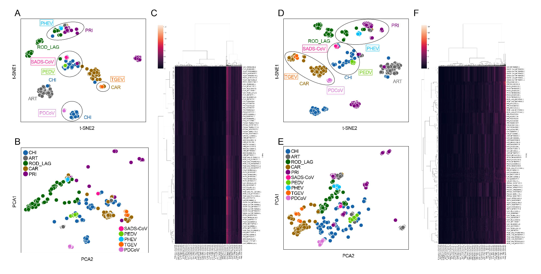
t-SNE and PCA were conducted for visualization and dimensional reduction of each type of compositional trait of ORF1ab and Spike sequences of CoVs. The samples from CHI, ART, ROD_LAG, CAR, and PRI were significantly separated on the basis of two main components reduced by both t-SNE (Figure 2A) and PCA (Figure 2B) of the 1536-dimentional-DCR of ORF1ab sequences. The sample size of ROD_LAG CoVs was relatively small, and some of these samples were generated through over-sampling; therefore, ROD_LAG CoV samples were relatively scattered in the dimensionality reduction result. According to the dimensional reduction results, SADS-CoV and PEDV gathered with the CoVs from CHI, PHEV gathered with the CoV samples from PRI and ROD_LAG, TGEV gathered with CAR CoV samples, and PDCoV gathered with CoVs from CHI based on the DCR of ORF1ab and CAR based on the DCR of Spike (Figure 2A,D). The distance between PDCoV and ART samples was relatively small for ORF1ab, and the distance between PDCoV and CHI samples was also relatively small for Spike (Figure 2B,E), suggesting that there might also be some homology among these gene sequences. The relationship between the samples obtained by hierarchical clustering based on the DCR of ORF1ab and Spike was similar to the distribution obtained by reduction (Figure 2C,F). t-SNE, PCA, and hierarchical clustering based on AAs (Supplementary Figure S1A–F), NTs (Supplementary Figure S1G–L), DNTs (Supplementary Figure S1M–R), codons (Supplementary Figure S2A–F), and codon pairs (Supplementary Figure S2G–L) indicated similar clustering or separation of these CoVs. DCR performed well in separating samples from various adaptive hosts. It can also be inferred that the distance among different types of Suiformes CoVs and CoVs from other hosts is different, making it necessary to establish new methods to predict and identify the host adaptability of each type of SUI CoVs.

3.3. Classification Effect of the DCR-Based CNN Models of ORF1ab and Spike

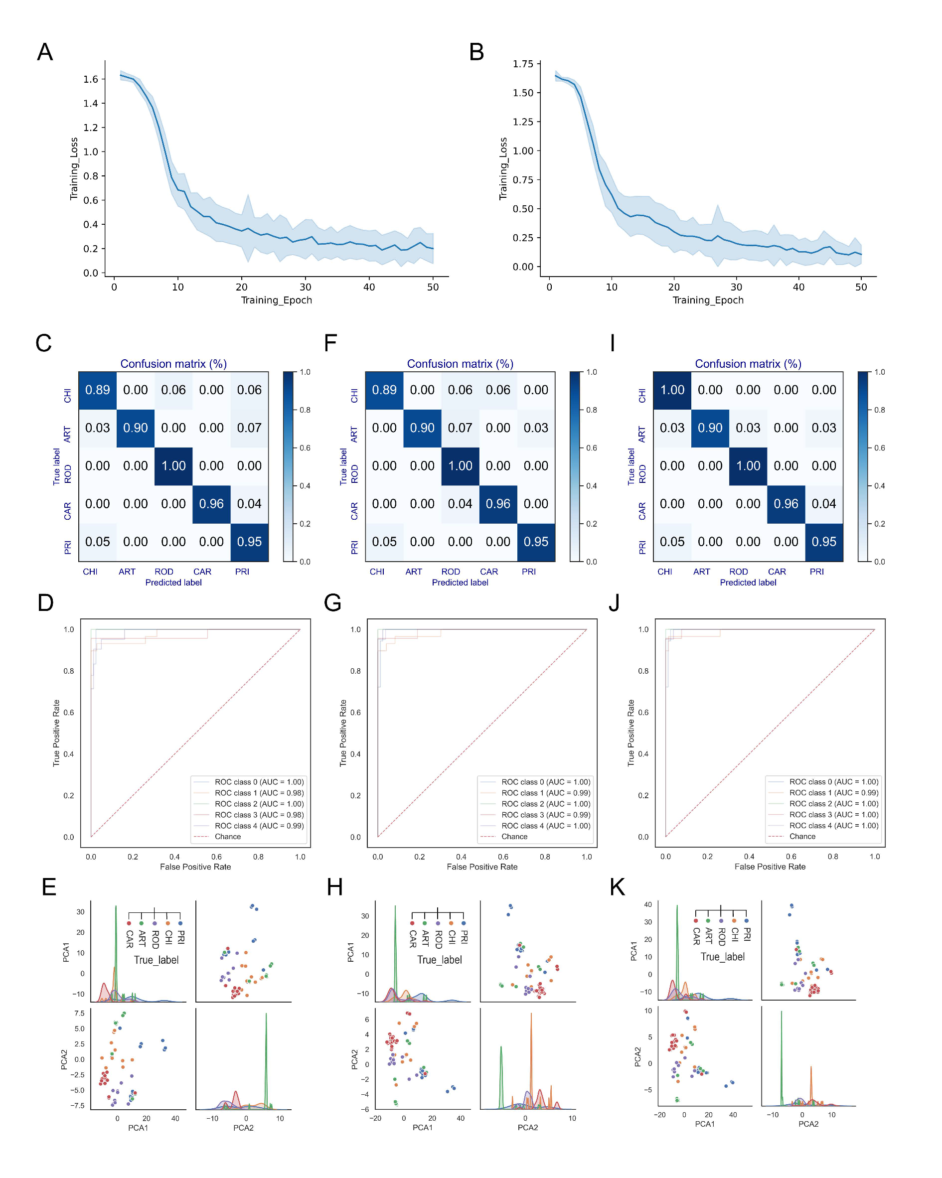
According to the distance between CoVs of various hosts, DCR-based CNN classification models were built to predict the adaptation of ORF1ab and Spike of SUI CoVs to different types of hosts. For both the ORF1ab (Figure 3A) and the Spike (Figure 3B) models, the training loss was relatively high with an epoch of 10. When the epoch value increased, the training loss decreased significantly, and the training loss was very close to 0 with an epoch of 50. The low values in the confusion matrices (Supplementary Figures S3A and S4A and Figure 3C), as well as the ROC and AUC (Supplementary Figures S3B and S4B and Figure 3D), showed the low accuracy of prediction of ORF1ab and Spike models with an epoch value of 10. The separation of samples with adaptation to CHI and those with adaptation to ART was not clearly indicated in the pair-plot of PCA1 and PCA2 of the FC data of Spike gene model (Figure 3E and Supplementary Figure S4C). The higher prediction accuracy was reflected in the confusion matrices (Supplementary Figures S3G and S4G and Figure 3F) and ROC and AUC (Supplementary Figures S3H and S4H and Figure 3G) of ORF1ab and Spike models with an epoch of 30. However, the pair-plotting of the Spike gene model was not able to distinguish the samples with adaptation to ART and samples with adaptation to CAR (Figure 3H and Supplementary Figure S4I). The pair-plotting of ORF1ab model with epochs of 10 and 30 indicated relatively clear separation of samples from different types of hosts (Supplementary Figure S3C,I). The high accuracy and low training loss value showed that the epoch of 50 should be selected for both ORF1ab and Spike models (Supplementary Figures S3M,N and S4M,N and Figure 3A,B,I,J). Separation of samples from various adaptive hosts was also illustrated in the pair-plotting of ORF1ab (Supplementary Figure S3O) and Spike (Figure 3K and Supplementary Figure S4O) models with an epoch of 50. The performances of both ORF1ab (Supplementary Figure S3D–F,J–L) and Spike (Supplementary Figure S4D–F,J–L) models with an epoch of 20 and 40 were also inferior to those with epoch of 50. Additionally, MERS-CoV is closely related to bat CoV HKU5, resulting in a relatively low prediction accuracy for samples adapted to ART (Supplementary Figure S3M). There are few CoVs adapted to ROD_LAG; therefore, many samples were obtained through over-sampling, which makes the distinction between these samples and the samples adapted to other hosts not very significant (Supplementary Figures S3O and S4O and Figure 3K).

3.4. Adaptation Prediction of Suiformes CoVs Based on the CNN Models

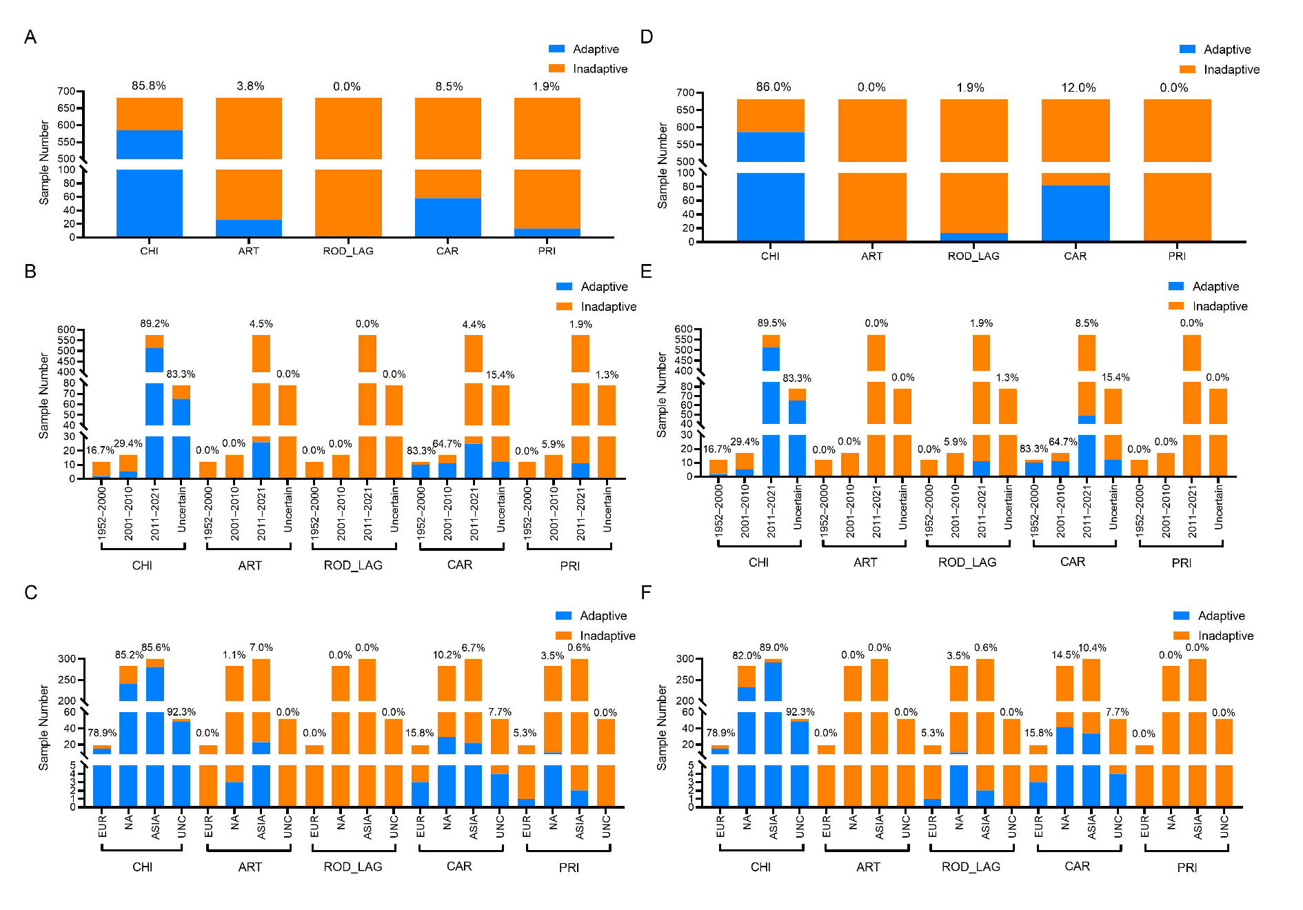
The adaptation of SUI CoVs to CHI, ART, ROD_LAG, CAR, and PRI was predicted by the DCR-based CNN models of ORF1ab and Spike sequences. The prediction results showed that, among the SUI CoV samples, 85.8% were predicted to be adaptive to CHI, along with 3.8% to ART, 8.5% to CAR, and 1.9% to PRI for ORF1ab, and 86.0% to CHI, 1.9% to ROD_LAG, and 12.0% to CAR for Spike (Figure 4A,D).
The spatiotemporal distribution of the adaptation ratio of SUI CoVs to five types of hosts was also obtained. Specifically, 16.7% of the SUI CoV samples collected from 1952 to 2000, 29.4% of the samples collected from 2001 to 2010, 89.2% of the samples collected from 2011 to 2021, and 83.3% of the samples with unknown collection date showed adaptation to CHI, 4.5% of the SUI CoV samples collected from 2011 to 2021 showed adaptation to ART, 83.3% of the SUI CoV samples collected from 1952 to 2000, 64.7% of the samples collected from 2001 to 2010, 4.4% of the samples collected from 2011 to 2021, and 15.4% of the samples with unknown collection date showed adaptation to CAR, and 5.9% of the samples collected from 2001 to 2010, 1.9% of the samples collected from 2011 to 2021, and 1.3% of the samples with unknown collection date showed adaptation to PRI for ORF1ab. On the other hand, 16.7% of the SUI CoV samples collected from 1952 to 2000, 29.4% of the samples collected from 2001 to 2010, 89.5% of the samples collected from 2011 to 2021, and 83.3% of the samples with unknown collection date showed adaptation to CHI, 5.9% of the samples collected from 2001 to 2010, 1.9% of the samples collected from 2011 to 2021, and 1.3% of the samples with unknown collection date showed adaptation to ROD_LAG, and 83.3% of the samples collected from 1952 to 2000, 64.7% of the samples collected from 2001 to 2010, 8.5% of the samples collected from 2011 to 2021, and 15.4% of the samples with unknown collection date showed adaptation to CAR for Spike (Figure 4B,E). Furthermore, 78.9% of the SUI CoV samples collected from Europe, 85.2% of the samples collected from North America, 85.6% of the samples collected from Asia, and 92.3% of the samples with unknown collection continent were predicted to be adaptive to CHI, 1.1% of the samples collected from North America and 7.0% of the samples collected from Asia were predicted to be adaptive to ART, 15.8% of the SUI CoV samples collected from Europe, 10.2% of the samples collected from North America, 6.7% of the samples collected from Asia, and 7.7% of the samples with unknown collection continent were predicted to be adaptive to CAR, and 5.3% of the SUI CoV samples collected from Europe, 3.5% of the samples collected from North America, and 0.6% of the samples collected from Asia were predicted to be adaptive to PRI for ORF1ab. On the other hand, 78.9% of the SUI CoV samples collected from Europe, 82.0% of the samples collected from North America, 89.0% of the samples collected from Asia, and 92.3% of the samples with unknown collection continent were predicted to be adaptive to CHI, 5.3% of the SUI CoV samples collected from Europe, 3.5% of the samples collected from North America, and 0.6% of the samples collected from Asia were predicted to be adaptive to ROD_LAG, and 15.8% of the SUI CoV samples collected from Europe, 14.5% of the samples collected from North America, 10.4% of the samples collected from Asia, and 7.7% of the samples with unknown collection continent were predicted to be adaptive to CAR for Spike (Figure 4C,F).

3.5. Adaptation Prediction, Phylogenetic Analysis, and Receptor Binding Verification of PHEV

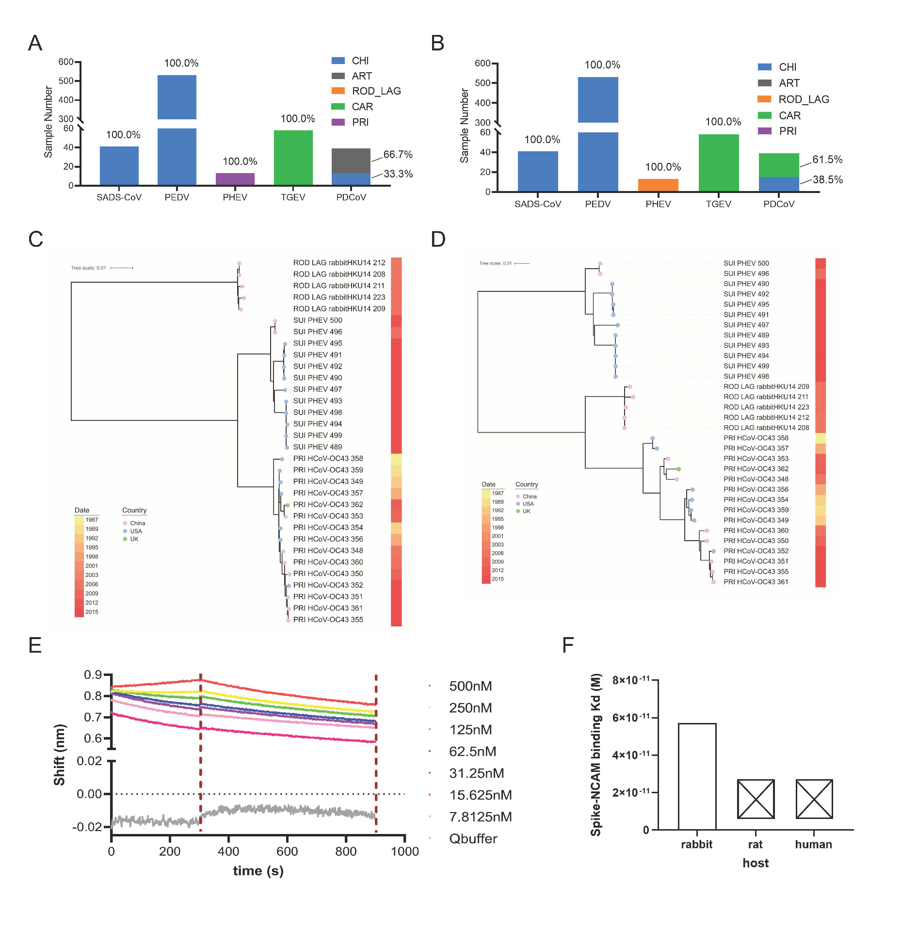
The host adaptation of each type of SUI CoVs was predicted in detail using the deep learning models with five adaptation labels of both ORF1ab and Spike sequences. Specifically, 100% of ORF1ab and Spike genes were predicted to be adaptive to CHI for SADS-CoVs and PEDVs, or to CAR for TGEVs (Figure 5A,B). However, 100% of PHEVs were predicted to be adaptive to PRI for ORF1ab and to ROD_LAG for Spike genes. Additionally, 66.7% of PDCoV was predicted to be adaptive to ART, and the remaining 33.3% was predicted to be adaptive to CHI for ORF1ab, while 61.5% of PDCoV was predicted to be adaptive to CAR, and the remaining 38.5% was predicted to be adaptive to CHI for Spike (Figure 5A,B). When both the ORF1ab and the Spike sequences of the Suiformes CoV samples are predicted to be adaptive to the same type of hosts, it can be inferred that the samples have significant adaptability to this type of host; otherwise, if the two genes of the samples are predicted to be adaptive to different types of hosts, the samples may have certain adaptability to those types of hosts. In order to elucidate the phylogenetic relationships between PHEVs and ROD_LAG or PRI CoV samples, two phylogeny trees were constructed on the basis of rgw ORF1ab and Spike genes of PHEV, rabbit CoVs (HKU14), and HCoV-OC43, with the closest distance in DCR traits. Interestingly, PHEV ORF1ab genes were also closer in phylogenetic relationship with those of HCoV-OC43 (Figure 5C), while Spike genes were in an independent branch from rabbit HKU14 CoVs and HCoV-OC43, relatively closer to the former (Figure 5D). These results were consistent with the predicted adaptation with the CNN model, confirming the accuracy of the adaptation prediction with deep learning methods. Then, after random sampling of CoV sequences from all five species, we similarly constructed another two phylogeny trees on the basis of ORF1ab and Spike genes. The results showed that the genetic distances of CoVs from the same species were not necessarily close in the phylogenetic trees, and some of them were even far away (Supplementary Figure S5A,B). This signified that our prediction model effectively complemented the traditional phylogenetic relationship, making it suitable for extensive application.
To further verify the analysis results of our models, we tried to explore the interaction between the PHEV Spike protein and cell lines of human, rat, and rabbit. According to previous studies, we determined that a fragment (258-amino-acid fragment, residues 291–548) located in Spike (277–794 is the RBD of the PHEV Spike protein) and a cellular neural cell adhesion molecule (NCAM) were the key receptor proteins [60,61]. Then, BLI binding experiments were performed to measure the binding ability of PHEV Spike RBD to NCAM of different host cells. We found that the PHEV Spike RBD could bind weakly to rabbit NCAM protein, with a KD of 5.73 × 10⁻¹¹ M, whereas it could hardly bind to human or rat NCAM protein (Figure 5E,F and Supplementary Figure S6A–C). This means that the PHEV Spike protein was more adaptive to rabbit, rather than rat or human, which was basically consistent with our prediction results that 100% of PHEV samples were predicted to be adaptive to ROD_LAG for Spike genes (Figure 5B). However, our prediction model could be further optimized, e.g., through refinement of host classification. Taken together, the DCR-based CNN models could accurately predict the host adaptability of SUI CoVs, and the results were basically consistent with the phylogenetic relationship and experiment results.

4. Discussion

This study evaluated the risk of SUI CoVs to infect and transmit in humans and other types of hosts, shedding light on whether SUI CoVs can spread across species, or whether pigs might be a possible intermediate host for CHI CoVs. Genomic DCR traits were qualified to distinguish the CoVs from various hosts. SUI SADS-CoV and PEDV samples were predicted to be adapted to CHI, and TGEV samples were predicted to be adapted to CAR according to DCR-based deep learning models with five adaptation labels for both ORF1ab and Spike. PHEV samples were predicted to be adapted to PRI according to ORF1ab and ROD_LAG according to Spike. Suiformes acted as intermediate hosts in H1N1 transmission [62], and it is predicted that SUI CoVs are also able to adapt to various hosts; therefore, they might also be intermediate hosts of various CoVs. PHEV is phylogenetically closely related to HCoV-OC43 [63] and is predicted to be adaptive to the PRI-based genomic DCR of ORF1ab. Therefore, there is a possibility that Suiformes can be intermediate hosts during the transmission of CoVs to humans.
Worryingly, it had been recently found that some strains of PDCoV are able to infect humans [28], implying evidence that pigs might be potential intermediate hosts for CoVs from CHI or other mammalian hosts. In order to further assess the risk of infection and transmission of PDCoV in the population, the host adaptation of the human-infected PDCoV was predicted using our deep learning models based on genomic DCR. It was found that these PDCoVs had a low probability of adapting to PRI, showing that, although Suiformes are potential intermediate hosts of PDCoV, these CoV strains may only cause spillover human infection in Haitian malnourished children, being unable to cause large-scale infection and epidemic in the human population. The existing studies have shown that SADS-CoV [23] and PEDV [25] originate from bat, and TGEV and PDCoV are respectively closely related to some CAR CoVs [36] and avian hosts [64] by comparing the homology of the sequences, which proves the rationality of our prediction. Compared with the existing research, the present method can quantify and compare the adaptability to different types of hosts in a timely manner.
Machine learning has been utilized to investigate the host adaptability of coronaviruses [65]. Liam et al. trained random forest models independently on genome composition biases of Spike protein and whole-genome sequences, including dinucleotide and codon usage biases in order to predict animal host. The combination of genetic resources with machine learning algorithms was consistent with our purpose; however, different modeling methods were used in our research. Moreover, codon usage and amino-acid preferences are other important factors that may influence the host adaptability of virus [66,67]. In essence, codon usage and amino-acid preferences are also based on nucleotide composition, which successfully explain the evolution of PEDV under the mutation pressure and natural selection. In our study, machine learning models provided a more systematic explanation for genome composition analysis and predicted the host adaptability of the virus from another point.
The binding ability of CoV Spike protein to a specific receptor can directly reflect the host adaptability of virus. To further verify this prediction model, we measured the binding ability of PHEV Spike to the NCAM of different host. More obvious binding activity was observed to the rabbit receptor, rather than rat or human, suggesting that PHEV might be more adaptive to rabbit, which is obviously consistent with our prediction results. However, the measured binding ability in our experiments was weak, whereby we thought that only the RBD region of Spike protein might be the main influencing factor. The main active domain of Spike protein is the RBD, as confirmed in previous research. However, the actual binding ability may be affected by other regions of Spike, and the RBD may not represent the whole native conformation of Spike; therefore, only using the RBD might be one limitation in this study.
However, studies such as ours and those of others based on a public CoV sequence database are sensitive to the quality and distribution of available sequence data. In this study, the imbalance in the number of various types of samples had a certain impact on the results. The few available samples of ROD_LAG CoV led to an imbalance in the number of CoVs from different hosts. Some ROD_LAG samples were generated during oversampling, resulting in a relatively scattered distribution of these samples in unsupervised learning results and the full connection layer data of trained deep learning models. Therefore, a greater accumulation of CoV samples may help to improve the accuracy of the models. In addition, the different receptor binding ability of PHEV Spike protein with receptors in rat and rabbit indicates that a refinement of host classification might also be helpful to establish more accurate prediction models.

5. Conclusions

In summary, the DCR trait was confirmed to be representative of the CoV genome, and the DCR-based deep learning model worked well to assess the adaptation of Suiformes CoVs to other mammals. Suiformes might be intermediate hosts for human CoVs and other mammalian CoVs. The present study provides a novel approach to assess the risk of adaptation and transmission to humans and other mammals of Suiformes CoVs.

Supplementary Materials

The following supporting information can be downloaded at https://www.mdpi.com/article/10.3390/v15071556/s1: Figure S1. Reduction, visualization and clustering of each type of SUI CoVs and other CoVs based on AAs, NTs and DNTs of ORF1ab and Spike sequences; Figure S2. Reduction, visualization and clustering of each type of SUI CoVs and other CoVs based on codons and codonpairs of ORF1ab and Spike sequences; Figure S3. Performance of the DCR-based CNN models of ORF1ab gene; Figure S4. Performance of the DCR-based CNN models of Spike gene; Figure S5. Phylogenetic tree of CoVs from different adaptive hosts for ORF1ab and Spike gene; Figure S6. Receptor binding verification of PHEV with different hosts.

Author Contributions

J.L., S.J., S.Z. (Sen Zhang) and T.J. conceptualized the study; S.Z. (Sen Zhang), X.K. and Y.F. contributed to the acquisition and interpretation of data; S.J., S.Z. (Sen Zhang), Y.L. (Yadan Li), M.N. and Y.L. (Yuchang Li) conducted the data cleaning and statistical analysis; J.L., S.J., S.Z. (Sen Zhang) and Y.C. performed genome parsing, unsupervised machine learning, and supervised deep learning with assistance from S.Z. (Shishun Zhao) and T.J.; J.L. drafted the manuscript; J.L., S.J. and S.Z. (Sen Zhang) drafted the manuscript, which was polished by X.K., S.Z. (Shishun Zhao) and T.J.; S.J., S.Z. (Sen Zhang) and J.L. coded all scripts for genome parsing, deep learning, and data visualization. All authors have read and agreed to the published version of the manuscript.

Funding

This research was supported by the grants from the National Key Research and Development Program of China (grant no. 2021YFC2302004) and the National Natural Science Foundation of China (grant no. 32070166, 2019-JCJQ-JJ-167).

Institutional Review Board Statement

Not applicable.

Informed Consent Statement

Not applicable.

Data Availability Statement

All original and cleaned CoV sequence data, and scripts for the project are available online from Github: https://github.com/Jamalijama/SwineCoVadaptation (accessed on 26 February 2023).

Acknowledgments

The authors gratefully acknowledge all data contributors, i.e., researchers and their originating laboratories responsible for obtaining the specimens, as well as the submitting laboratories for generating the genetic sequence and metadata, shared via the GISAID Initiative, on which this research is based.

Conflicts of Interest

The authors declare no conflict of interest.

References

  1. Zhou, P.; Yang, X.L.; Wang, X.G.; Hu, B.; Zhang, L.; Zhang, W.; Si, H.R.; Zhu, Y.; Li, B.; Huang, C.L.; et al. A pneumonia outbreak associated with a new coronavirus of probable bat origin. Nature 2020, 579, 270–273. [Google Scholar] [CrossRef] [PubMed] [Green Version]
  2. Zhou, H.; Chen, X.; Hu, T.; Li, J.; Song, H.; Liu, Y.; Wang, P.; Liu, D.; Yang, J.; Holmes, E.C.; et al. A Novel Bat Coronavirus Closely Related to SARS-CoV-2 Contains Natural Insertions at the S1/S2 Cleavage Site of the Spike Protein. Curr. Biol. 2020, 30, 2196–2203. [Google Scholar] [CrossRef] [PubMed]
  3. Lau, S.K.P.; Luk, H.K.H.; Wong, A.C.P.; Li, K.S.M.; Zhu, L.; He, Z.; Fung, J.; Chan, T.T.Y.; Fung, K.S.C.; Woo, P.C.Y. Possible Bat Origin of Severe Acute Respiratory Syndrome Coronavirus 2. Emerg. Infect. Dis. 2020, 26, 1542–1547. [Google Scholar] [CrossRef] [PubMed]
  4. Kobasa, D.; Jones, S.M.; Shinya, K.; Kash, J.C.; Copps, J.; Ebihara, H.; Hatta, Y.; Kim, J.H.; Halfmann, P.; Hatta, M.; et al. Aberrant innate immune response in lethal infection of macaques with the 1918 influenza virus. Nature 2007, 445, 319–323. [Google Scholar] [CrossRef] [PubMed]
  5. Ekiert, D.C.; Friesen, R.H.; Bhabha, G.; Kwaks, T.; Jongeneelen, M.; Yu, W.; Ophorst, C.; Cox, F.; Korse, H.J.; Brandenburg, B.; et al. A highly conserved neutralizing epitope on group 2 influenza A viruses. Science 2011, 333, 843–850. [Google Scholar] [CrossRef] [Green Version]
  6. Smith, G.J.; Vijaykrishna, D.; Bahl, J.; Lycett, S.J.; Worobey, M.; Pybus, O.G.; Ma, S.K.; Cheung, C.L.; Raghwani, J.; Bhatt, S.; et al. Origins and evolutionary genomics of the 2009 swine-origin H1N1 influenza A epidemic. Nature 2009, 459, 1122–1125. [Google Scholar] [CrossRef] [Green Version]
  7. Cui, J.; Li, F.; Shi, Z.L. Origin and evolution of pathogenic coronaviruses. Nat. Rev. Microbiol. 2019, 17, 181–192. [Google Scholar] [CrossRef] [Green Version]
  8. Sanjuan, R.; Domingo-Calap, P. Mechanisms of viral mutation. Cell. Mol. Life Sci. 2016, 73, 4433–4448. [Google Scholar] [CrossRef] [Green Version]
  9. Li, J.; Liu, B.; Chang, G.; Hu, Y.; Zhan, D.; Xia, Y.; Li, Y.; Yang, Y.; Zhu, Q. Virulence of H5N1 virus in mice attenuates after in vitro serial passages. Virol. J. 2011, 8, 93. [Google Scholar] [CrossRef] [PubMed] [Green Version]
  10. Han, P.; Hu, Y.; Sun, W.; Zhang, S.; Li, Y.; Wu, X.; Yang, Y.; Zhu, Q.; Jiang, T.; Li, J.; et al. Mouse lung-adapted mutation of E190G in hemagglutinin from H5N1 influenza virus contributes to attenuation in mice. J. Med. Virol. 2015, 87, 1816–1822. [Google Scholar] [CrossRef]
  11. Arai, Y.; Kawashita, N.; Daidoji, T.; Ibrahim, M.S.; El-Gendy, E.M.; Takagi, T.; Takahashi, K.; Suzuki, Y.; Ikuta, K.; Nakaya, T.; et al. Novel Polymerase Gene Mutations for Human Adaptation in Clinical Isolates of Avian H5N1 Influenza Viruses. PLoS Pathog. 2016, 12, e1005583. [Google Scholar] [CrossRef]
  12. Deng, Y.; Li, C.; Han, J.; Wen, Y.; Wang, J.; Hong, W.; Li, X.; Liu, Z.; Ye, Q.; Li, J.; et al. Phylogenetic and genetic characterization of a 2017 clinical isolate of H7N9 virus in Guangzhou, China during the fifth epidemic wave. Sci. China Life Sci. 2017, 60, 1331–1339. [Google Scholar] [CrossRef] [PubMed]
  13. Liang, L.; Jiang, L.; Li, J.; Zhao, Q.; Wang, J.; He, X.; Huang, S.; Wang, Q.; Zhao, Y.; Wang, G.; et al. Low Polymerase Activity Attributed to PA Drives the Acquisition of the PB2 E627K Mutation of H7N9 Avian Influenza Virus in Mammals. mBio 2019, 10, e1005583. [Google Scholar] [CrossRef] [PubMed] [Green Version]
  14. Song, H.; Qi, J.; Xiao, H.; Bi, Y.; Zhang, W.; Xu, Y.; Wang, F.; Shi, Y.; Gao, G.F. Avian-to-Human Receptor-Binding Adaptation by Influenza A Virus Hemagglutinin H4. Cell Rep. 2017, 20, 1201–1214. [Google Scholar] [CrossRef] [PubMed] [Green Version]
  15. Everest, H.; Billington, E.; Daines, R.; Burman, A.; Iqbal, M. The Emergence and Zoonotic Transmission of H10Nx Avian Influenza Virus Infections. mBio 2021, 12, e178521. [Google Scholar] [CrossRef]
  16. Anderson, T.K.; Chang, J.; Arendsee, Z.W.; Venkatesh, D.; Souza, C.K.; Kimble, J.B.; Lewis, N.S.; Davis, C.T.; Vincent, A.L. Swine Influenza A Viruses and the Tangled Relationship with Humans. Cold Spring Harb. Perspect. Med. 2021, 11, a038737. [Google Scholar] [CrossRef] [PubMed] [Green Version]
  17. Nelson, M.I.; Viboud, C.; Vincent, A.L.; Culhane, M.R.; Detmer, S.E.; Wentworth, D.E.; Rambaut, A.; Suchard, M.A.; Holmes, E.C.; Lemey, P. Global migration of influenza A viruses in swine. Nat. Commun. 2015, 6, 6696. [Google Scholar] [CrossRef] [Green Version]
  18. Ozawa, M.; Kawaoka, Y. Cross talk between animal and human influenza viruses. Annu. Rev. Anim. Biosci. 2013, 1, 21–42. [Google Scholar] [CrossRef] [Green Version]
  19. Yu, X.; Tsibane, T.; McGraw, P.A.; House, F.S.; Keefer, C.J.; Hicar, M.D.; Tumpey, T.M.; Pappas, C.; Perrone, L.A.; Martinez, O.; et al. Neutralizing antibodies derived from the B cells of 1918 influenza pandemic survivors. Nature 2008, 455, 532–536. [Google Scholar] [CrossRef] [Green Version]
  20. Hilleman, M.R. Serologic responses to split and whole swine influenza virus vaccines in light of the next influenza pandemic. J. Infect. Dis. 1977, 136, S683–S685. [Google Scholar] [CrossRef]
  21. Li, J.; Tian, F.; Zhang, S.; Liu, S.S.; Kang, X.P.; Li, Y.D.; Wei, J.Q.; Lin, W.; Lei, Z.; Feng, Y.; et al. Genomic representation predicts an asymptotic host adaptation of bat coronaviruses using deep learning. Front. Microbiol. 2023, 14. [Google Scholar] [CrossRef] [PubMed]
  22. Yang, Y.L.; Qin, P.; Wang, B.; Liu, Y.; Xu, G.H.; Peng, L.; Zhou, J.; Zhu, S.J.; Huang, Y.W. Broad Cross-Species Infection of Cultured Cells by Bat HKU2-Related Swine Acute Diarrhea Syndrome Coronavirus and Identification of Its Replication in Murine Dendritic Cells In Vivo Highlight Its Potential for Diverse Interspecies Transmission. J. Virol. 2019, 93, e01448-19. [Google Scholar] [CrossRef] [PubMed] [Green Version]
  23. Zhou, P.; Fan, H.; Lan, T.; Yang, X.L.; Shi, W.F.; Zhang, W.; Zhu, Y.; Zhang, Y.W.; Xie, Q.M.; Mani, S.; et al. Fatal swine acute diarrhoea syndrome caused by an HKU2-related coronavirus of bat origin. Nature 2018, 556, 255–258. [Google Scholar] [CrossRef] [PubMed] [Green Version]
  24. Gong, L.; Li, J.; Zhou, Q.; Xu, Z.; Chen, L.; Zhang, Y.; Xue, C.; Wen, Z.; Cao, Y. A New Bat-HKU2-like Coronavirus in Swine, China, 2017. Emerg. Infect. Dis. 2017, 23, 1607–1609. [Google Scholar] [CrossRef] [PubMed] [Green Version]
  25. He, W.T.; Bollen, N.; Xu, Y.; Zhao, J.; Dellicour, S.; Yan, Z.; Gong, W.; Zhang, C.; Zhang, L.; Lu, M.; et al. Phylogeography Reveals Association between Swine Trade and the Spread of Porcine Epidemic Diarrhea Virus in China and across the World. Mol. Biol. Evol. 2022, 39, msab364. [Google Scholar] [CrossRef] [PubMed]
  26. Wang, Q.; Vlasova, A.N.; Kenney, S.P.; Saif, L.J. Emerging and re-emerging coronaviruses in pigs. Curr. Opin. Virol. 2019, 34, 39–49. [Google Scholar] [CrossRef] [PubMed]
  27. Edwards, C.E.; Yount, B.L.; Graham, R.L.; Leist, S.R.; Hou, Y.J.; Dinnon, K.H., 3rd; Sims, A.C.; Swanstrom, J.; Gully, K.; Scobey, T.D.; et al. Swine acute diarrhea syndrome coronavirus replication in primary human cells reveals potential susceptibility to infection. Proc. Natl. Acad. Sci. USA 2020, 117, 26915–26925. [Google Scholar] [CrossRef]
  28. Lednicky, J.A.; Tagliamonte, M.S.; White, S.K.; Elbadry, M.A.; Alam, M.M.; Stephenson, C.J.; Bonnym, T.S.; Loeb, J.C.; Telisma, T.; Chavannes, S.; et al. Independent infections of porcine deltacoronavirus among Haitian children. Nature 2021, 600, 133–137. [Google Scholar] [CrossRef]
  29. Ye, X.; Chen, Y.; Zhu, X.; Guo, J.; Xie, D.; Hou, Z.; Xu, S.; Zhou, J.; Fang, L.; Wang, D.; et al. Cross-species transmission of deltacoronavirus and the origin of porcine deltacoronavirus. Evol. Appl. 2020, 13, 2246–2253. [Google Scholar] [CrossRef]
  30. Sit, T.H.C.; Brackman, C.J.; Ip, S.M.; Tam, K.W.S.; Law, P.Y.T.; To, E.M.W.; Yu, V.Y.T.; Sims, L.D.; Tsang, D.N.C.; Chu, D.K.W.; et al. Infection of dogs with SARS-CoV-2. Nature 2020, 586, 776–778. [Google Scholar] [CrossRef]
  31. de Wit, E.; van Doremalen, N.; Falzarano, D.; Munster, V.J. SARS and MERS: Recent insights into emerging coronaviruses. Nat. Rev. Microbiol. 2016, 14, 523–534. [Google Scholar] [CrossRef] [PubMed]
  32. Vijgen, L.; Keyaerts, E.; Moes, E.; Thoelen, I.; Wollants, E.; Lemey, P.; Vandamme, A.M.; Van Ranst, M. Complete genomic sequence of human coronavirus OC43: Molecular clock analysis suggests a relatively recent zoonotic coronavirus transmission event. J. Virol. 2005, 79, 1595–1604. [Google Scholar] [CrossRef] [PubMed] [Green Version]
  33. Corman, V.M.; Muth, D.; Niemeyer, D.; Drosten, C. Hosts and Sources of Endemic Human Coronaviruses. Adv Virus Res 2018, 100, 163–188. [Google Scholar] [CrossRef]
  34. Kiyuka, P.K.; Agoti, C.N.; Munywoki, P.K.; Njeru, R.; Bett, A.; Otieno, J.R.; Otieno, G.P.; Kamau, E.; Clark, T.G.; Van Der Hoek, L.; et al. Human Coronavirus NL63 Molecular Epidemiology and Evolutionary Patterns in Rural Coastal Kenya. J. Infect. Dis. 2018, 217, 1728–1739. [Google Scholar] [CrossRef] [PubMed] [Green Version]
  35. Li, W.; Shi, Z.; Yu, M.; Ren, W.; Smith, C.; Epstein, J.H.; Wang, H.; Crameri, G.; Hu, Z.; Zhang, H.; et al. Bats are natural reservoirs of SARS-like coronaviruses. Science 2005, 310, 676–679. [Google Scholar] [CrossRef] [PubMed]
  36. Wesseling, J.G.; Vennema, H.; Godeke, G.J.; Horzinek, M.C.; Rottier, P.J. Nucleotide sequence and expression of the spike (S) gene of canine coronavirus and comparison with the S proteins of feline and porcine coronaviruses. J. Gen. Virol. 1994, 75, 1789–1794. [Google Scholar] [CrossRef] [PubMed]
  37. Jiang, S.; Du, Q.; Feng, C.; Ma, L.; Zhang, Z. CompoDynamics: A comprehensive database for characterizing sequence composition dynamics. Nucleic Acids Res. 2021, 50, D962–D969. [Google Scholar] [CrossRef]
  38. Forsberg, R.; Christiansen, F.B. A codon-based model of host-specific selection in parasites, with an application to the influenza A virus. Mol Biol Evol. 2003, 20, 1252–1259. [Google Scholar] [CrossRef] [Green Version]
  39. Charles, H.; Calevro, F.; Vinuelas, J.; Fayard, J.M.; Rahbe, Y. Codon usage bias and tRNA over-expression in Buchnera aphidicola after aromatic amino acid nutritional stress on its host Acyrthosiphon pisum. Nucleic Acids Res. 2006, 34, 4583–4592. [Google Scholar] [CrossRef] [Green Version]
  40. Babayan, S.A.; Orton, R.J.; Streicker, D.G. Predicting reservoir hosts and arthropod vectors from evolutionary signatures in RNA virus genomes. Science 2018, 362, 577–580. [Google Scholar] [CrossRef] [Green Version]
  41. Chen, F.; Wu, P.; Deng, S.; Zhang, H.; Hou, Y.; Hu, Z.; Zhang, J.; Chen, X.; Yang, J.R. Dissimilation of synonymous codon usage bias in virus-host coevolution due to translational selection. Nat. Ecol. Evol. 2020, 4, 589–600. [Google Scholar] [CrossRef]
  42. Hausser, J.; Mayo, A.; Keren, L.; Alon, U. Central dogma rates and the trade-off between precision and economy in gene expression. Nat. Commun. 2019, 10, 68. [Google Scholar] [CrossRef] [Green Version]
  43. Chen, J.; Gao, K.; Wang, R.; Wei, G.W. Prediction and mitigation of mutation threats to COVID-19 vaccines and antibody therapies. Chem Sci. 2021, 12, 6929–6948. [Google Scholar] [CrossRef] [PubMed]
  44. Li, J.; Zhang, S.; Li, B.; Hu, Y.; Kang, X.P.; Wu, X.Y.; Huang, M.T.; Li, Y.C.; Zhao, Z.P.; Qin, C.F.; et al. Machine Learning Methods for Predicting Human-Adaptive Influenza A Viruses Based on Viral Nucleotide Compositions. Mol. Biol. Evol. 2020, 37, 1224–1236. [Google Scholar] [CrossRef] [PubMed]
  45. Contu, L.; Balistreri, G.; Domanski, M.; Uldry, A.C.; Mühlemann, O. Characterisation of the Semliki Forest Virus-host cell interactome reveals the viral capsid protein as an inhibitor of nonsense-mediated mRNA decay. PLoS Pathog. 2021, 17, e1009603. [Google Scholar] [CrossRef]
  46. Hershberg, R.; Petrov, D.A. Selection on codon bias. Annu Rev Genet. 2008, 42, 287–299. [Google Scholar] [CrossRef] [PubMed] [Green Version]
  47. Duret, L. tRNA gene number and codon usage in the C. elegans genome are co-adapted for optimal translation of highly expressed genes. Trends Genet. 2000, 16, 287–289. [Google Scholar] [CrossRef]
  48. Alley, E.C.; Khimulya, G.; Biswas, S.; AlQuraishi, M.; Church, G.M. Unified rational protein engineering with sequence-based deep representation learning. Nat. Methods 2019, 16, 1315–1322. [Google Scholar] [CrossRef]
  49. Hie, B.; Zhong, E.D.; Berger, B.; Bryson, B. Learning the language of viral evolution and escape. Science 2021, 371, 284–288. [Google Scholar] [CrossRef]
  50. Rao, R.; Bhattacharya, N.; Thomas, N.; Duan, Y.; Chen, X.; Canny, J.; Abbeel, P.; Song, Y.S. Evaluating Protein Transfer Learning with TAPE. Adv. Neural. Inf Process Syst. 2019, 32, 9689–9701. [Google Scholar]
  51. Xia, X. Extreme Genomic CpG Deficiency in SARS-CoV-2 and Evasion of Host Antiviral Defense. Mol. Biol. Evol. 2020, 37, 2699–2705. [Google Scholar] [CrossRef] [Green Version]
  52. Zahradník, J.; Marciano, S.; Shemesh, M.; Zoler, E.; Harari, D.; Chiaravalli, J.; Meyer, B.; Rudich, Y.; Li, C.; Marton, I.; et al. SARS-CoV-2 variant prediction and antiviral drug design are enabled by RBD in vitro evolution. Nat Microbiol. 2021, 6, 1188–1198. [Google Scholar] [CrossRef] [PubMed]
  53. Pucci, F.; Rooman, M. Prediction and Evolution of the Molecular Fitness of SARS-CoV-2 Variants: Introducing SpikePro. Viruses 2021, 13, 935. [Google Scholar] [CrossRef]
  54. Li, J.; Wu, Y.N.; Zhang, S.; Kang, X.P.; Jiang, T. Deep learning based on biologically interpretable genome representation predicts two types of human adaptation of SARS-CoV-2 variants. Brief. Bioinform. 2022, 23, bbac036. [Google Scholar] [CrossRef]
  55. Nan, B.G.; Zhang, S.; Li, Y.C.; Kang, X.P.; Chen, Y.H.; Li, L.; Jiang, T.; Li, J. Convolutional Neural Networks Based on Sequential Spike Predict the High Human Adaptation of SARS-CoV-2 Omicron Variants. Viruses 2022, 14, 1072. [Google Scholar] [CrossRef] [PubMed]
  56. Shu, Y.; McCauley, J. GISAID: Global initiative on sharing all influenza data-from vision to reality. Euro. Surveill. 2017, 22, 30494. [Google Scholar] [CrossRef] [Green Version]
  57. Katoh, K.; Misawa, K.; Kuma, K.; Miyata, T. MAFFT: A novel method for rapid multiple sequence alignment based on fast Fourier transform. Nucleic Acids Res. 2002, 30, 3059–3066. [Google Scholar] [CrossRef] [PubMed] [Green Version]
  58. Stamatakis, A. RAxML version 8: A tool for phylogenetic analysis and post-analysis of large phylogenies. Bioinformatics 2014, 30, 1312–1313. [Google Scholar] [CrossRef] [Green Version]
  59. Letunic, I.; Bork, P. Interactive tree of life (iTOL) v3: An online tool for the display and annotation of phylogenetic and other trees. Nucleic Acids Res. 2016, 44, W242-5. [Google Scholar] [CrossRef]
  60. Dong, B.; Gao, W.; Lu, H.; Zhao, K.; Ding, N.; Liu, W.; Zhao, J.; Lan, Y.; Tang, B.; Jin, Z.; et al. A small region of porcine hemagglutinating encephalomyelitis virus spike protein interacts with the neural cell adhesion molecule. Intervirology 2015, 58, 130–137. [Google Scholar] [CrossRef]
  61. Gao, W.; He, W.; Zhao, K.; Lu, H.; Ren, W.; Du, C.; Chen, K.; Lan, Y.; Song, D.; Gao, F. Identification of NCAM that interacts with the PHE-CoV spike protein. Virol. J. 2010, 7, 254. [Google Scholar] [CrossRef] [PubMed] [Green Version]
  62. Sun, H.; Xiao, Y.; Liu, J.; Wang, D.; Li, F.; Wang, C.; Li, C.; Zhu, J.; Song, J.; Sun, H.; et al. Prevalent Eurasian avian-like H1N1 swine influenza virus with 2009 pandemic viral genes facilitating human infection. Proc. Natl. Acad. Sci. USA 2020, 117, 17204–17210. [Google Scholar] [CrossRef] [PubMed]
  63. Vijgen, L.; Keyaerts, E.; Lemey, P.; Maes, P.; Van Reeth, K.; Nauwynck, H.; Pensaert, M.; Van Ranst, M. Evolutionary history of the closely related group 2 coronaviruses: Porcine hemagglutinating encephalomyelitis virus, bovine coronavirus, and human coronavirus OC43. J. Virol. 2006, 80, 7270–7274. [Google Scholar] [CrossRef] [Green Version]
  64. Jung, K.; Hu, H.; Saif, L.J. Porcine deltacoronavirus infection: Etiology, cell culture for virus isolation and propagation, molecular epidemiology and pathogenesis. Virus Res. 2016, 226, 50–59. [Google Scholar] [CrossRef]
  65. Brierley, L.; Fowler, A. Predicting the animal hosts of coronaviruses from compositional biases of spike protein and whole genome sequences through machine learning. PLoS Pathog. 2021, 17, e1009149. [Google Scholar] [CrossRef] [PubMed]
  66. Si, F.; Jiang, L.; Yu, R.; Wei, W.; Li, Z. Study on the Characteristic Codon Usage Pattern in Porcine Epidemic Diarrhea Virus Genomes and Its Host Adaptation Phenotype. Front. Microbiol. 2021, 12, 738082. [Google Scholar] [CrossRef]
  67. Bahir, I.; Fromer, M.; Prat, Y.; Linial, M. Viral adaptation to host: A proteome-based analysis of codon usage and amino acid preferences. Mol. Syst. Biol. 2009, 5, 311. [Google Scholar] [CrossRef]
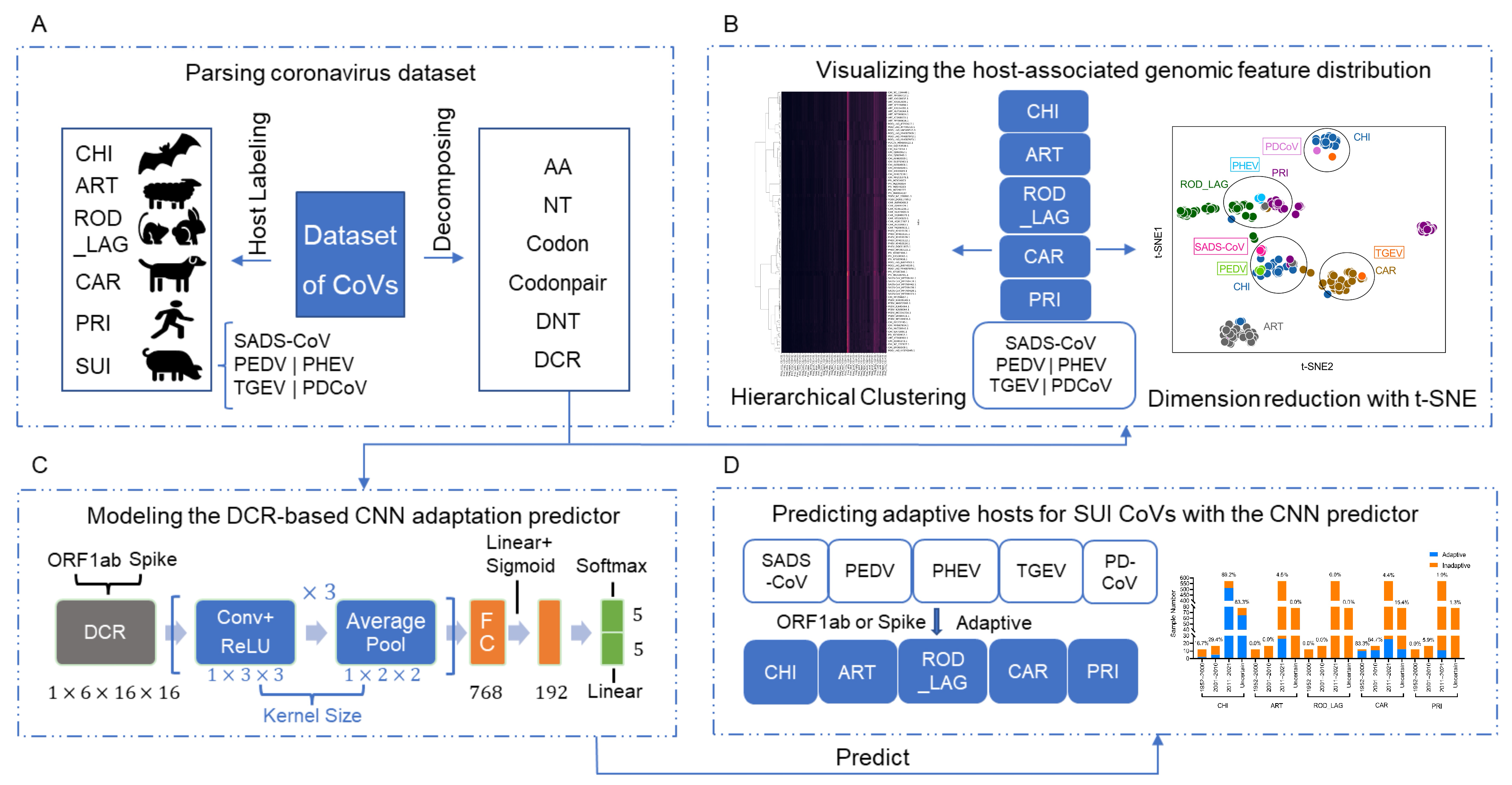
Figure 1. Pipeline of data processing, unsupervised learning, and prediction of adaptive hosts of SUI CoVs. The pipeline of this study can be divided into five parts. Host labeling (CHI, ART, ROD_LAG, CAR, PRI, and SUI (including SADS-CoV, PEDV, PHEV, TGEV, and PDCoV)), and ORF1ab and Spike sequences decomposition (six types of compositional traits: AAs, NTs, codons, codon pairs, DNTs, and DCRs) (A); reduction in and hierarchical clustering of each type of compositional trait and visualization (B); deep learning classification models established on the basis of DCR of ORF1ab and Spike sequences (C); prediction of adaptive hosts of each type of SUI CoV and temporal and spatial distributions of adaptation ratio (D).
Viruses 15 01556 g001
Figure 2. Reduction in, visualization of, and clustering of each type of SUI CoV and other CoVs based on DCR of ORF1ab and Spike sequences. Visualization of DCR reduced with t-SNE (A) and PCA (B), and hierarchical clustering of DCR of ORF1ab from each CoV sample (C); visualization of DCR reduced with t-SNE (D) and PCA (E), and hierarchical clustering of DCR of Spike from each CoV sample (F).
Viruses 15 01556 g002
Figure 3. Performance of the DCR-based CNN models of ORF1ab and Spike gene. The training loss plotted with the average training loss value and the standard deviation of every training epoch (1–50) of the ORF1ab (A) and Spike (B) models, the confusion matrices (C), ROC with AUC (D), and the pair-plotting of PCA1 and PCA2 of the FC data (E) of the Spike model with training epochs of 10, 30 (FH), and 50 (IK). ROD means ROD_LAG.
Viruses 15 01556 g003
Figure 4. The adaptation ratio of SUI CoVs to various hosts and the spatiotemporal distribution of the adaptation ratio. The adaptation ratio of SUI CoV samples to CHI, ART, ROD_LAG, CAR, and PRI (A), the adaptation ratio of SUI CoV samples collected from 1952 to 2000, 2001 to 2010, and 2011 to 2021, as well as the samples with unknown collection date (B), and the adaptation ratio of SUI CoV samples collected from Europe (EUR), North America (NA), and Asia (ASIA), as well as the samples with unknown collection continent (UNC) (C), of the ORF1ab and Spike (DF) models.
Viruses 15 01556 g004
Figure 5. The adaptation prediction, phylogenetic analysis, and receptor binding verification of PHEVs, PRI, and ROD_LAG CoVs. The adaptation ratio of SUI CoVs to CHI, ART, ROD_LAG, CAR, and PRI of ORF1ab (A) or Spike gene (B). The phylogenetic tree was constructed using iqtree with 100 bootstrap replicates for randomly sampled PHEVs, PRI HCoV-OC43 CoVs, and ROD_LAG HKU14 CoVs, which were close in DCR distance to PHEVs for ORF1ab (C) and Spike (D) genes. Different colored circles in the tree represent different sample locations (pink: China, cyan: USA, green: UK). Sampling date is indicated by the progressive color bar at right. (E) Biolayer interferometry (BLI) assay for the interaction between RBD of the PHEV Spike protein and NCAM of rabbit. (F) The equilibrium dissociation constant (KD) of PHEV Spike protein with NCAM of rat, human, and rabbit.
Viruses 15 01556 g005
Disclaimer/Publisher’s Note: The statements, opinions and data contained in all publications are solely those of the individual author(s) and contributor(s) and not of MDPI and/or the editor(s). MDPI and/or the editor(s) disclaim responsibility for any injury to people or property resulting from any ideas, methods, instructions or products referred to in the content.