Article

Surveillance, Isolation, and Genetic Characterization of Bat Herpesviruses in Zambia

1
Laboratory of Veterinary Public Health, Faculty of Agriculture, Tokyo University of Agriculture and Technology, Tokyo 183-8509, Japan
2
Management Department of Biosafety, Laboratory Animal and Pathogen Bank, National Institute of Infectious Diseases, Tokyo 162-8640, Japan
3
Department of Virology-I, National Institute of Infectious Diseases, Tokyo 162-8640, Japan
4
Division of Collaboration and Education, International Institute for Zoonosis Control, Hokkaido University, Sapporo 001-0020, Japan
5
International Collaboration Unit, International Institute for Zoonosis Control, Hokkaido University, Sapporo 001-0020, Japan
6
Division of International Research Promotion, International Institute for Zoonosis Control, Hokkaido University, Sapporo 001-0020, Japan
7
Department of Para-Clinical Studies, School of Veterinary Medicine, The University of Zambia, P.O. Box 32379, Lusaka 10101, Zambia
8
Department of Pathogenetic and Preventive Veterinary Science, Joint Faculty of Veterinary Medicine, Kagoshima University, Kagoshima 890-0065, Japan
9
Division of Global Epidemiology, International Institute for Zoonosis Control, Hokkaido University, Sapporo 001-0020, Japan
10
Department of Oral Microbiology, Faculty of Medicine, Dentistry and Pharmaceutical Sciences, Okayama University, Okayama 700-8558, Japan
11
Department of Virology, Faculty of Medicine, Dentistry and Pharmaceutical Sciences, Okayama University, Okayama 700-8558, Japan
12
One Health Research Center, Hokkaido University, Sapporo 060-0818, Japan
13
Division of Molecular Pathobiology, International Institute for Zoonosis Control, Hokkaido University, Sapporo 001-0020, Japan
14
Department of Disease Control, School of Veterinary Medicine, The University of Zambia, P.O. Box 32379, Lusaka 10101, Zambia
15
Macha Research Trust, Choma 20100, Zambia
16
Africa Center of Excellence for Infectious Diseases of Humans and Animals, The University of Zambia, P.O. Box 32379, Lusaka 10101, Zambia
17
Institute for Vaccine Research and Development, Hokkaido University, Sapporo 001-0021, Japan
*
Author to whom correspondence should be addressed.
Deceased 27 April 2019.
Viruses 2023, 15(6), 1369; https://doi.org/10.3390/v15061369
Submission received: 23 May 2023 / Revised: 12 June 2023 / Accepted: 12 June 2023 / Published: 13 June 2023
(This article belongs to the Special Issue Viruses and Bats 2023)

Abstract

:
Bats are of significant interest as reservoirs for various zoonotic viruses with high diversity. During the past two decades, many herpesviruses have been identified in various bats worldwide by genetic approaches, whereas there have been few reports on the isolation of infectious herpesviruses. Herein, we report the prevalence of herpesvirus infection of bats captured in Zambia and genetic characterization of novel gammaherpesviruses isolated from striped leaf-nosed bats (Macronycteris vittatus). By our PCR screening, herpesvirus DNA polymerase (DPOL) genes were detected in 29.2% (7/24) of Egyptian fruit bats (Rousettus aegyptiacus), 78.1% (82/105) of Macronycteris vittatus, and one Sundevall’s roundleaf bat (Hipposideros caffer) in Zambia. Phylogenetic analyses of the detected partial DPOL genes revealed that the Zambian bat herpesviruses were divided into seven betaherpesvirus groups and five gammaherpesvirus groups. Two infectious strains of a novel gammaherpesvirus, tentatively named Macronycteris gammaherpesvirus 1 (MaGHV1), were successfully isolated from Macronycteris vittatus bats, and their complete genomes were sequenced. The genome of MaGHV1 encoded 79 open reading frames, and phylogenic analyses of the DNA polymerase and glycoprotein B demonstrated that MaGHV1 formed an independent lineage sharing a common origin with other bat-derived gammaherpesviruses. Our findings provide new information regarding the genetic diversity of herpesviruses maintained in African bats.

1. Introduction

Bats are associated with several emerging zoonotic viruses such as Marburg virus, Hendra virus, and Nipah virus, and are thought to act as natural reservoirs for these emerging viruses [1]. Global viral surveillance aiming at identifying and mitigating future emergence of bat-borne zoonotic outbreaks has identified 12,986 bat-associated viruses as of August 2021 [2]. Bats (order Chiroptera) are the second most diverse mammalian order, comprising more than 20% of all known mammalian species [3]. The high viral diversity in bats relative to other mammals is reflective of the number of species [4]. Novel viral sequences are constantly discovered in various bat species owing to the development of next-generation sequencing technologies (NGS) [1].
Herpesviruses are enveloped, double-stranded DNA (dsDNA) viruses belonging to the family Herpesviridae that are classified into three subfamilies (Alphaherpesvirinae, Betaherpesvirinae, and Gammaherpesvirinae) on the basis of their genomic architectures, sequence similarities, and biological properties [5]. The dsDNA genome size of herpesviruses is approximately 120–240 kbp, including approximately 70–170 open reading frames (ORFs), and 40 of which encode core viral proteins conserved among all known herpesviruses [6,7]. The core viral proteins are generally involved in fundamental aspects of the viral life cycle such as nucleic acid metabolism, DNA synthesis, DNA packaging, and virion maturation, and are consequently often essential for viral replication [5]. In addition to ORFs, most herpesvirus genomes contain various reiterated sequences such as terminal, internal, and/or inverted repeats [5]. To date, 17 genera and 118 species have been classified into the Herpesviridae family by the International Committee on Taxonomy of Viruses (ICTV) [6]. In addition to these classified herpesviruses, many related and unclassified family members have been identified in various hosts, and the number of herpesvirus species is likely to exceed more than 200 [5]. In general, the host ranges of individual herpesviruses are restricted with most having evolved in association with their own host species [6].
Alpha-, beta-, and gammaherpesviruses have been discovered in various bat species worldwide, showing the high diversity of herpesviruses circulating in the hosts [8]. According to the database of zoonotic and vector-borne viruses (ZOVER), 348 complete or partial herpesvirus genomic sequences have been identified in 75 bat species as of 2022 [2]. In the last two decades, surveillance of herpesvirus infection in bats has been reported in African countries, including Cameroon, the Central African Republic, Ghana, Kenya, Madagascar, the Republic of the Congo, and South Africa [9,10,11,12,13]. In previous studies, herpesviruses were identified in a total of nine bat species in Africa: three species in the family Pteropodidae (frugivorous bats; Eidolon dupreanum, Eidolon helvum, and Rousettus aegyptiacus), four species in the family Vespertilionidae (insectivorous bats; Miniopterus natalensis, Neoromicia capensis, Neoromicia helios, and Pipistrellus nanulus), and two species in the family Rhinolophidae (insectivorous bats; Triaenops afer and Triaenops persicus). Although numerous partial herpesvirus genes have been detected by genetic screening, most of the detected herpesviruses remain to be isolated. To date, eleven herpesviruses have been isolated from bats. Six alphaherpesviruses were isolated from frugivorous bats (Eidolon dupreanum, Eidolon helvum, Pteropus lylei, and Pteropus hypomelanus-related bats), insectivorous bats (Lonchophylla thomasi), and an unidentified bat [13,14,15]; two betaherpesviruses were isolated from insectivorous bats (Miniopterus fuliginosus and Miniopterus schreibersii) [16,17], and three gammaherpesviruses were isolated from insectivorous bats (Myotis velifer incautus, Eptesicus fuscus, and Rhinolophus ferrumequinum) [18,19,20]. Of these, six isolates (two alphaherpesviruses, one betaherpesvirus, and three gammaherpesviruses) were analyzed to determine their complete or nearly complete genome sequences. The bat herpesviruses for which complete genome sequences are available have been identified for bats in Oceania (Australia), Asia (Vietnam, Indonesia, and Japan), and North America (United States of America and Canada), but no complete genome sequence of herpesviruses detected in African bats has been reported.
As part of the surveillance program of zoonotic virus infection in bats in Zambia, we screened frugivorous bats (Egyptian fruit bats, Rousettus aegyptiacus) and insectivorous bats (striped leaf-nosed bats, Macronycteris vittatus; Sundevall’s roundleaf bats, Hipposideros caffer) for herpesviruses, followed by phylogenetic analysis of the detected sequences. Here, we also report the isolation of a novel gammaherpesvirus named Macronycteris gammaherpesvirus 1 (MaGHV1) from Macronycteris vittatus bats in Zambia. We performed complete genome sequencing and genetically characterized the isolates.

2. Materials and Methods

2.1. Sample Collection

In 2018, 24 cave-dwelling Egyptian fruit bats (Rousettus aegyptiacus), 105 striped leaf-nosed bats (Macronycteris vittatus), and one Sundevall’s roundleaf bat (Hipposideros caffer) were captured in Chongwe (15.6° S, 28.7° E) with approval from the Department of National Parks and Wildlife, Ministry of Tourism and Arts, Zambia (DNPW8/27/1) [21]. Bat species were identified based on morphological characteristics and nucleotide sequence analysis of the mitochondrial 16S ribosomal RNA and the cytochrome b genes. We collected oral and rectal swabs from each captured bat and promptly placed them in Eagle’s minimum essential medium (MEM) supplemented with 1000 units/mL penicillin, 1000 µg/mL streptomycin, 25 µg/mL amphotericin B, 0.01 M HEPES, and 0.5% bovine serum albumin (BSA). After brief centrifugation, the supernatants were stored at −80 °C.

2.2. Amplification of the Herpesvirus DNA Polymerase Gene by PCR

Screening for the herpesvirus detection was conducted using pooled swab specimens. Oral and rectal swabs were pooled for each bat and nucleic acids were individually extracted from 130 pooled swab specimens using the QIAamp Viral RNA Mini Kit (QIAGEN, Hilden, Germany) according to the manufacturer’s instructions. Nucleic acids extracted by the kit include not only RNA but also DNA as previously described [22]. The herpesvirus DNA genome was detected by semi-nested PCR using Tks Gflex DNA Polymerase (TaKaRa, Shiga, Japan) with pan-herpesvirus primer sets targeting the DNA polymerase (DPOL) gene (Table S1) [23]. PCR products were subjected to direct sequencing using the Big Dye Terminator v3.1 Cycle Sequencing kit (Applied Biosystems, Foster City, CA). The 5′- and 3′-ends of the sequences derived from primers were trimmed and obtained sequences were analyzed through a BLAST search (https://blast.ncbi.nlm.nih.gov/Blast.cgi, accessed on 23 May 2023). The determined partial DPOL nucleotide sequences were deposited into the DNA Data Bank of Japan (DDBJ) under accession numbers LC762209-LC762247.

2.3. Virus Isolation

African green monkey kidney (Vero E6) cells were maintained in Dulbecco’s Modified Eagle’s Medium (DMEM) supplemented with 10% fetal bovine serum (FBS), 2 mM L-glutamine, 100 units/mL penicillin, 100 µg/mL streptomycin, 3.5 mg/mL D-glucose, and 1.0 mg/mL NaHCO3 at 37 °C with 5% CO2. The media from the swabs that tested positive for herpesvirus detection by genetic screening were inoculated onto Vero E6 cell cultures, followed by 1 h incubation at 37 °C in 5% CO2 for virus adsorption. After the inocula were removed, the cells were washed twice with PBS and maintained in DMEM containing 5 µg/mL trypsin, 0.3% BSA, 2 mM L-glutamine, 4% antibiotic–antimycotic solution (Gibco, Waltham, MA, USA), and 1.0 mg/mL NaHCO3 at 37 °C in 5% CO2 for 2 weeks. The supernatant of the inoculated cells was blindly passaged to fresh Vero E6 cells. Subsequently, the supernatant of the passaged culture from oral and rectal swabs was pooled for each bat and subjected to DNA extraction and NGS analysis as described below. Total DNA was extracted from the pool by using a DNeasy Blood & Tissue Kit (QIAGEN), and isolation of herpesviruses was confirmed by PCR.

2.4. Genome Sequencing, Construction, and Annotation

The complete genome sequences of isolated herpesviruses were determined by NGS analyses. The sequencing library was synthesized from total DNA of isolated viruses using a Nextera XT DNA Library Preparation Kit (Illumina, San Diego, CA, USA) according to the manufacturer’s instructions, and was then sequenced on a MiSeq instrument with a MiSeq Reagent Kit v3 (600 cycles) to generate 300 bp paired-end reads. Sequence reads were trimmed and analyzed by de novo assembly using CLC Genomics Workbench software (CLC bio, Hilden, Germany). Consensus sequences with coverage of over 20 reads were obtained from the herpesvirus genome contigs.
For filling the remaining gap, PCR was performed using a specific primer designed based on the upstream and downstream sequences of the gaps (Table S1). The obtained amplicons were sequenced using a Frongle and Ligation Sequencing Kit (SQK-Q20EA) (Oxford Nanopore Technologies, Oxford, United Kingdom). Base calling was performed using Guppy version 5.0.16+b9fcd7b. Extra sequences were trimmed using Porechop [24]. Then, consensus sequences were generated using Canu [25]. To compliment the terminal repeat sequences at both ends of the genome, the genome DNA was amplified using an illustra GenomiPhi V2 DNA Amplification Kit (GE Healthcare, Chicago, IL, USA), and their terminals were repaired using T7 Endonuclease I (New England Biolabs, Ipswich, MA, USA). Following sequencing, base calling and trimming were performed as described above. To select reads including the terminal sequence, they were aligned with one kb regions flanking each terminal repeat using BWA [26]. The selected reads for each terminal were assembled using Canu, and the most representative contig was manually selected. The obtained gap-filling and terminal repeat sequences were concatenated with the genome assembly and polished with Pilon [27]. The determined genome sequences were deposited in the DDBJ database under accession numbers LC763829-LC763830.
ORFs were predicted by the ORF finder tool provided by the National Center for Biotechnology Information (NCBI) (https://www.ncbi.nlm.nih.gov/orffinder/, accessed on 23 May 2023). ORFs were identified using the following criteria: (i) sequence similarity to known viral or cellular genes, (ii) the presence of an initiating methionine and a stop codon, and/or (iii) length greater than 100 amino acids. ORFs with more than two of these criteria were considered to be genes encoding viral proteins. The translated amino acid sequences of the ORFs were subjected to BLAST search to determine whether they matched known viral or cellular proteins. The nomenclature used for MaGHV1 ORFs is based on a similar strategy used for other gammaherpesviruses such as equid herpesvirus 2 [28]. The unified name of the ORF for viral genes conserved across the subfamily Gammaherpesvirinae is used in this study. Additional ORFs are numbered in order of the appearance in the genome from 5′ to 3′. The complete sequence of the isolated virus was analyzed in a tandem repeat finder program [29] to identify the repeat regions.

2.5. Genetic Comparison and Phylogenetic Analysis

Bioinformatic analyses were performed using herpesvirus sequences deposited in the DDBJ/EMBL-Bank/GenBank databases. Identity comparison analyses were conducted between isolated herpesviruses using GENETYX version 15 (GENETYX Corporation, Tokyo, Japan). Phylogenetic analyses based on the amino acid sequences of DPOL and glycoprotein B (gB) genes were performed using MEGA X software [30]. The MUSCLE protocol was used to align the sequences. Phylogenetic trees were constructed using the maximum likelihood method based on the model of LG+G+I for complete and partial DPOL genes and the model of LG+G+I+F for the gB gene as the best-fit models, with 1000 bootstrap replicates. The viruses and their accession numbers of the sequences used in the analysis are listed in Table S2 in the Supplementary Materials.

3. Results

3.1. Screening for Herpesviruses with DPOL Gene by Semi-Nested PCR

Swab specimens collected from 130 bats captured in Zambia were screened for herpesviruses by PCR. Of these, 69.2% (90/130) were positive for herpesvirus detection (Table 1), suggesting high prevalence of herpesvirus infection in bats in Zambia; 29.2% (7/24) of Rousettus aegyptiacus, 78.1% (82/105) of Macronycteris vittatus, and one Hipposideros caffer. BLAST analyses revealed that 26 of the 90 obtained sequences (181 or 184 bp in length) originated from betaherpesviruses and that 60 of the sequences (172 bp in length) originated from gammaherpesviruses. These sequences showed 45.6–100% and 68.4–100% amino acid identities with known betaherpesviruses and gammaherpesviruses, respectively (Table S3). Exceptionally, 4 sequences (172 bp in length of sample ID 3, 6, 114, and 126) were related to various alpha-, beta-, or gammaherpesviruses with 40–60% amino acid identities through BLAST analyses. Since these four sequences showed the highest identities with a bat-derived betaherpesvirus (Plecotus austriacus/Spain/psc-23/2004or2007), we tentatively classified them into the subfamily Betaherpesvirinae (Table 2).

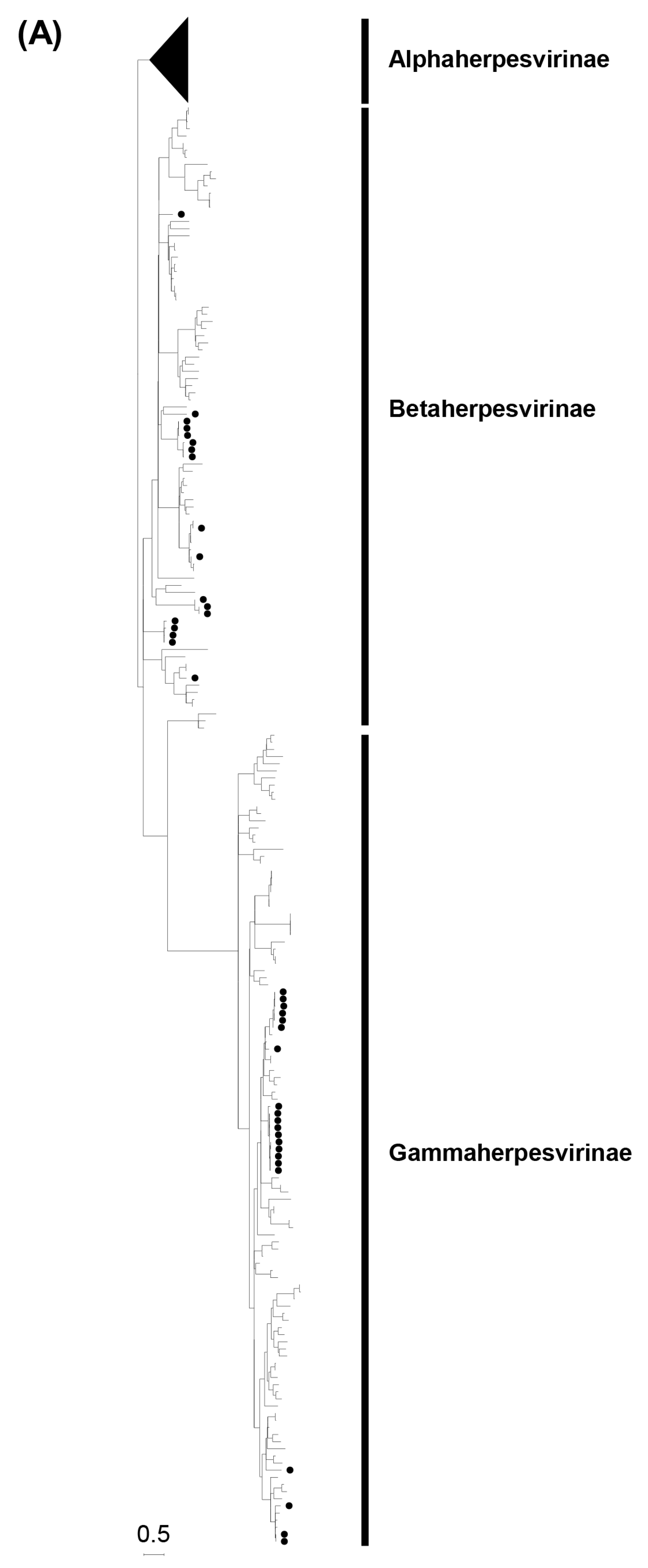
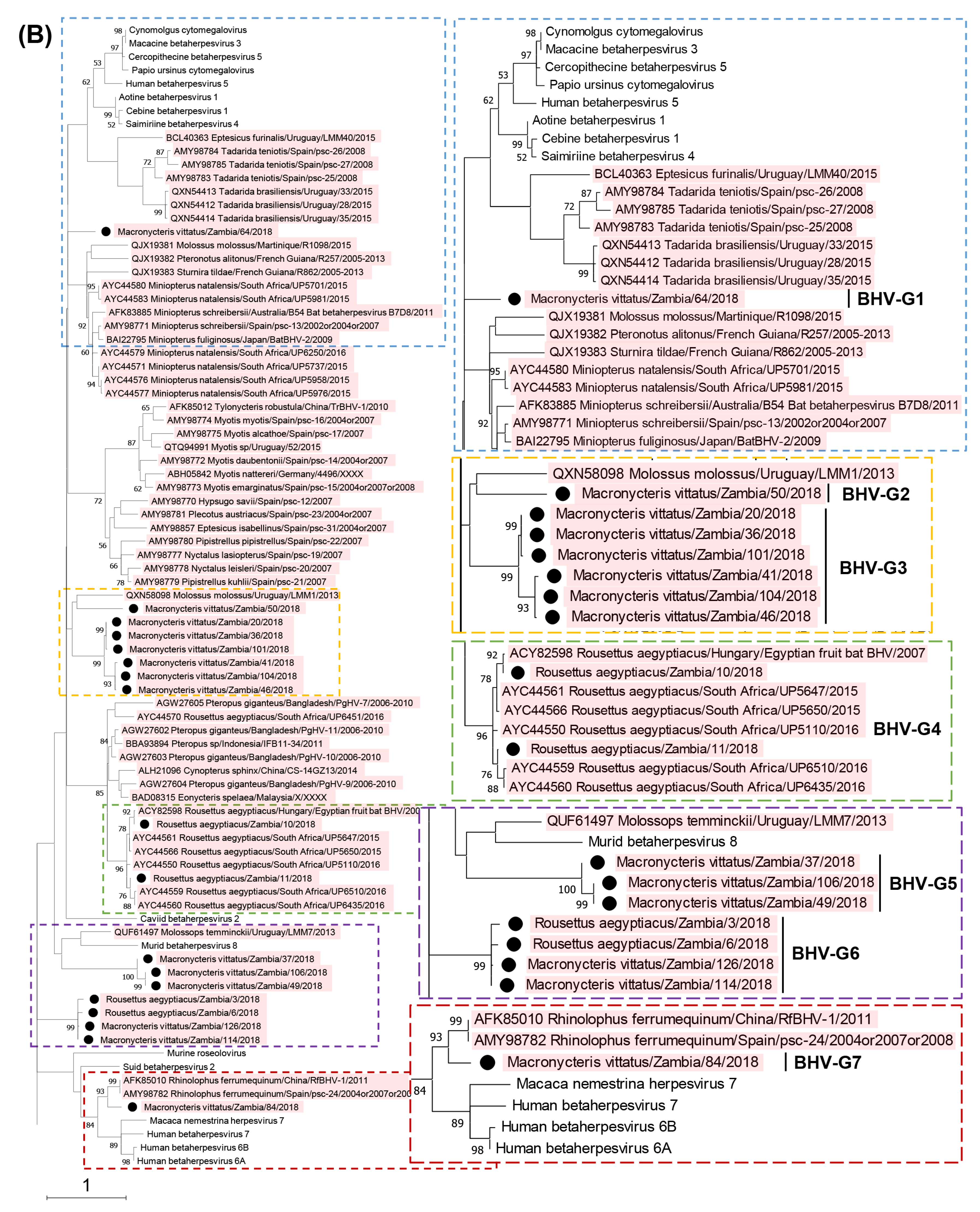
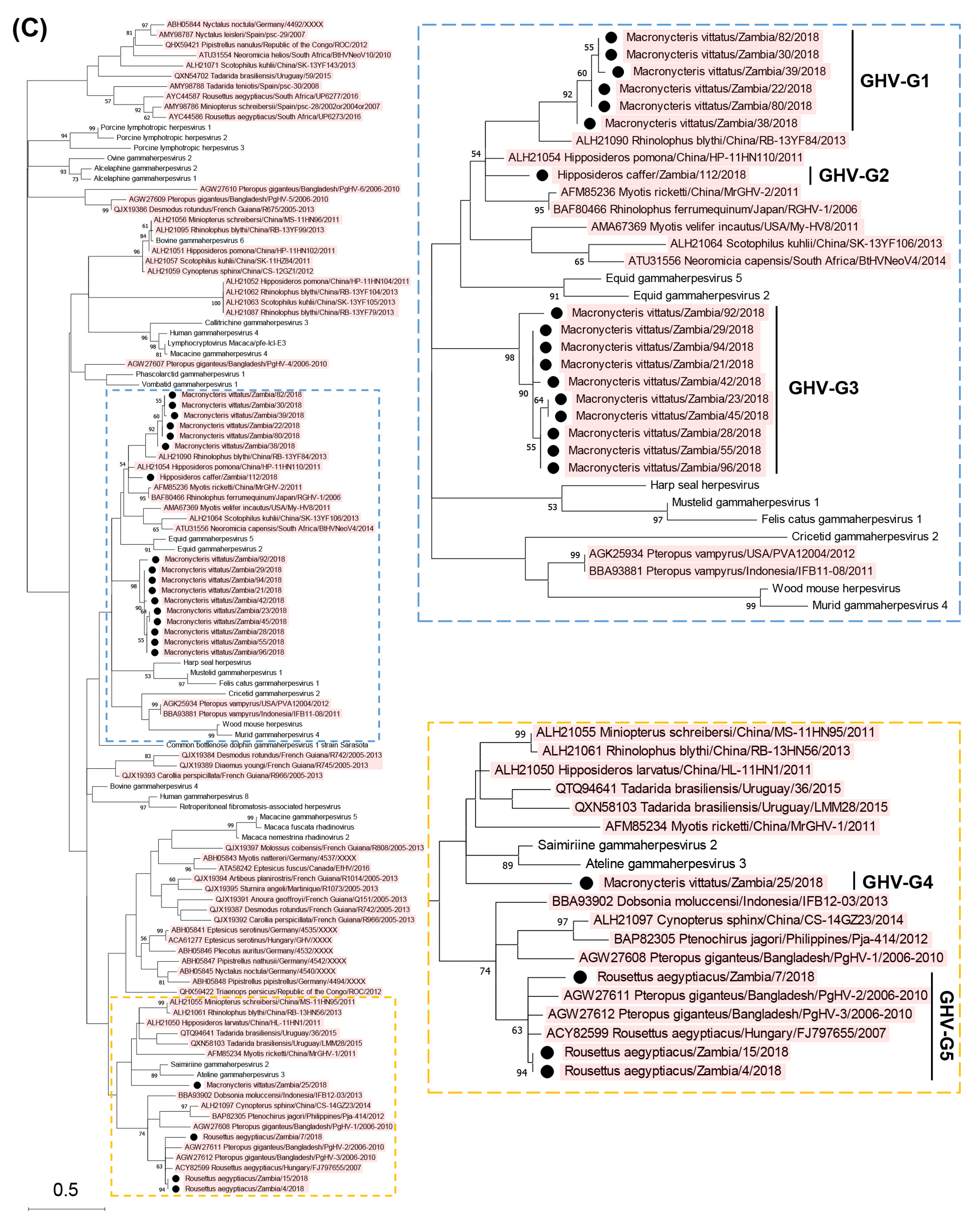
3.2. Phylogenetic Analyses Based on the Partial DPOL Gene

Phylogenetic trees of the partial DPOL gene were constructed using the amino acid sequences of the detected herpesviruses with other alpha-, beta-, and gammaherpesviruses identified in bats worldwide (Figure 1). The Zambian bat betaherpesviruses were phylogenetically divided into seven groups (BHV-G1 to -G7) as shown in Figure 1B. BHV-G1, -G2, and -G7 each consisted of a single virus identified in Macronycteris vittatus bats. BHV-G3, -G4, and -G7 were phylogenetically related to bat betaherpesviruses previously identified in various countries. BHV-G3 formed a cluster near the branches of BHV-G2 and a betaherpesvirus detected in Molossus molossus bats in Uruguay. BHV-G4 formed a cluster with betaherpesviruses from Rousettus aegyptiacus bats in South Africa and Hungary, and BHV-G7 was closely related to betaherpesviruses in Rhinolophus ferrumequinum bats in China and Spain. BHV-G5 formed a unique lineage near the branch of murid betaherpesvirus 8. Of note, BHV-G6 contained betaherpesviruses identified in Rousettus aegyptiacus and Macronycteris vittatus bats and formed an independent cluster under the subfamily Betaherpsvirinae. The Rousettus aegyptiacus/Zambia/3/2018 virus shared 100% nucleotide identity with the Macronycteris vittatus/Zambia/114/2018 virus, suggesting that the viruses were distributed in both frugivorous and insectivorous bats. On the other hand, the Zambian bat gammaherpesviruses were phylogenetically divided into five groups (GHV-G1 to -G5) as shown in Figure 1C. GHV-G1, -G2, -G3, and -G4 each formed independent clusters or branches. GHV-G1 were monophyletic along with a gammaherpesvirus detected in Rhinophus blythi bats in China. GHV-G2 belonged to a large clade including GHV-G1 and gammaherpesviruses identified in various insectivorous bats in China and Japan. GHV-G3 and G4 formed distinct lineages within the Betaherpsvirinae clade. GHV-G5 were phylogenetically related to previously described gammaherpesviruses detected in frugivorous bats in Hungary and Bangladesh. These results indicated that genetically diverse herpesviruses were maintained in bats in the cave in Zambia.

3.3. Isolation of a Novel Gammaherpesvirus from Macronycteris Vittatus Bats

To isolate the viruses detected by genetic screening, the herpesvirus-positive swab samples were inoculated onto Vero E6 cells. Gammaherpesviruses were isolated from seven Macronycteris vittatus bats (sample IDs 59, 80, 82, 86, 88, 89, and 106). After several passages, a cytopathic effect (CPE) was observed in Vero E6 cells infected with the isolates (Figure 2). The isolated viruses were genetically analyzed by PCR and sequencing targeting the partial DPOL gene which were used for screening. According to the sequencing analyses, the seven isolates were divided into two closely related groups (i.e., sample IDs 59/80/86/89/106 and IDs 82/88). The two sequences showed 98.3% similarity (169/172 bp). These newly isolated herpesvirus strains were tentatively named Macronycteris gammaherpesvirus 1 (MaGHV1). The two representative MaGHV1 strains from sample IDs 80 and 82 (strains 80 and 82, respectively) were used for further analyses.

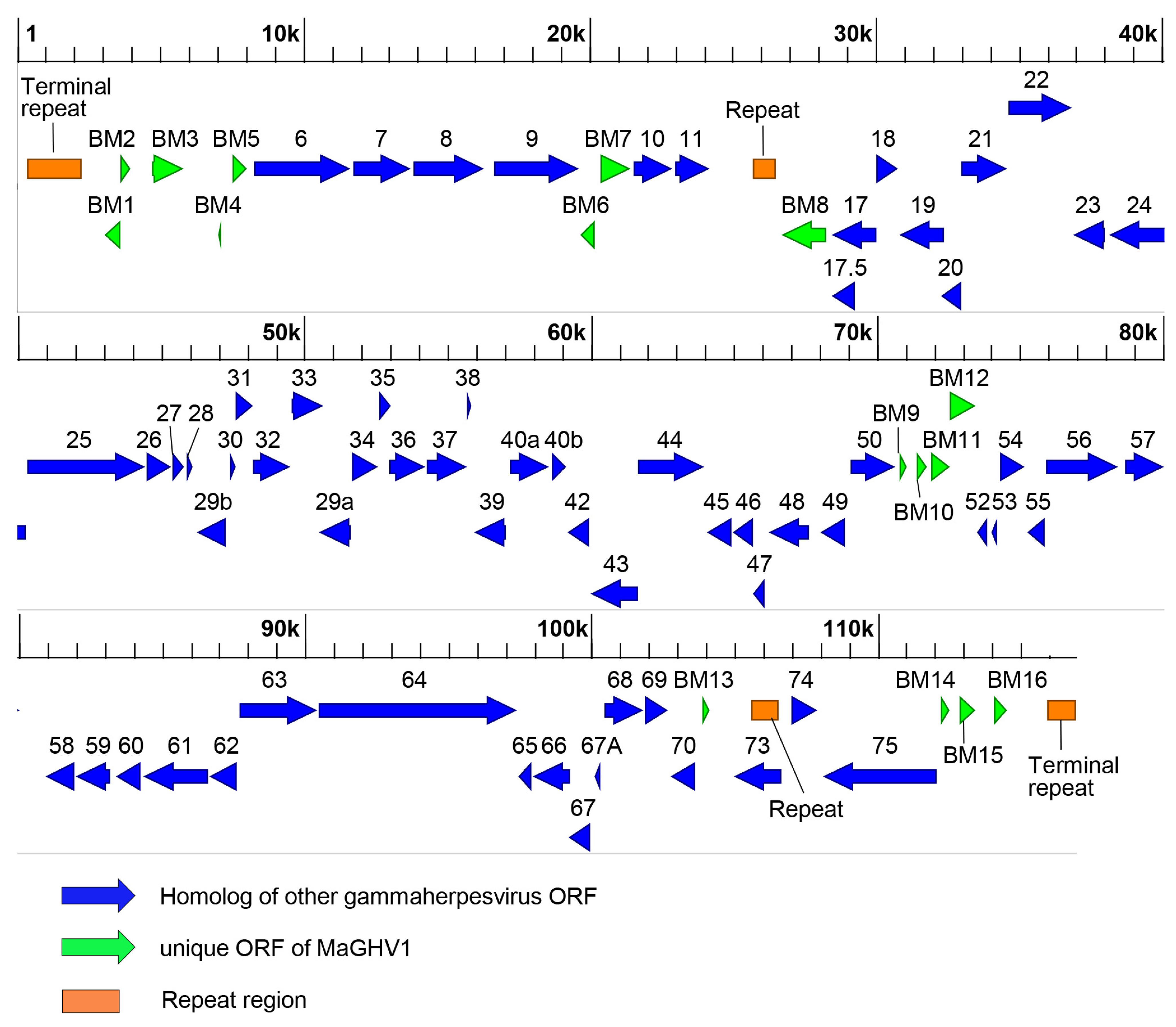
3.4. Complete Genome of MaGHV1 and Genetic Comparison between Strains

To determine the complete genome sequences of the MaGHV1 strains, we performed whole genome sequencing. The genome of MaGHV1 strain 82 was 116,972 bp in length with a GC content of 37.3% (Figure 3). The genome structure comprised a unique region that was flanked by tandem terminal repeats at each end (Table 3). The size of the consensus repeat sequences at both termini was 284 bp and the copy numbers were 7 and 3.8 at 5′ and 3′ termini, respectively. In the unique region, two repeat regions at coordinates 25,665 to 26,556 (repeat region 1) and 105,538 to 106,545 (repeat region 2) were found. A total of 79 ORFs were identified and most of the ORFs showed the highest identities to viral genes of rhinolophus gammaherpesvirus 1 (RGHV1) isolated from a greater horseshoe bat (Rhinolophus ferrumequinum) in Japan [20], followed by myotis gammaherpesvirus 8 isolated from a microbat (Myotis velifer incautus) cell line [18]. Notably, at least 67 ORFs showed homology with viral genes of equid herpesvirus 2, which is a well-known gammaherpesvirus, and one of them (ORF38) shared the highest identity (43.3%) with the myristylated tegument protein of equid gammaherpesvirus 2 rather than RGHV1. The BM1, BM4, and BM6 genes showed sequence similarity to cellular genes, CASP8 and FADD-like apoptosis regulator (32.5% identity), E3 ubiquitin-protein ligase (38.9% identity), and bcl-2 like protein (34.0% identity), respectively, and the BM4 and BM6 genes also had homologies to ORF 4 and ORF13 of RGHV1. Six ORFs (BM2, BM10, and BM13-16) showed no significant similarity to any known genes. In addition, ORF73, which encoded the homolog of nuclear antigen LANA-1, was mapped to a direct repeat (repeat region 2). To identify herpesviruses genetically related to MaGHV1, BLAST analyses were performed using the deduced amino acid sequences of viral proteins. The complete DPOL sequence (ORF9) of MaGHV1 strain 82 shared the highest identity (78.4%) with RGHV1. Among other ORFs, the deduced amino acid sequences of glycoprotein B (gB; ORF8), helicase-primase helicase subunit (ORF44), Uracil-DNA glycosylase (ORF46), deoxyribonuclease (ORF37), and major capsid protein (ORF25) showed 72.0, 78.6, 75.8, 68.0, and 78.1% identities to RGHV1 homologs, respectively (Table S4).
On the other hand, most of the 79 ORFs of MaGHV1 described above were also identified in the genome of strain 80: BM3–7 and ORF6–11 encoded in contig 1 (21,962 bp), BM8–13, ORF17–40, ORF42–50, and ORF52–70 in contig 2 (79,285 bp), and ORF73–75, BM14–16 in contig 3 (8979 bp) (Table S5). Identity comparison of each viral protein between the MaGHV1 strains demonstrated > 90% amino acid identity of 70 ORFs. Relatively low identities (52.6–89.5%) were found in the remaining viral proteins, including three membrane proteins (BM3, ORF47, and BM11), one tegument protein (ORF45), and two hypothetical proteins (BM12 and BM13).

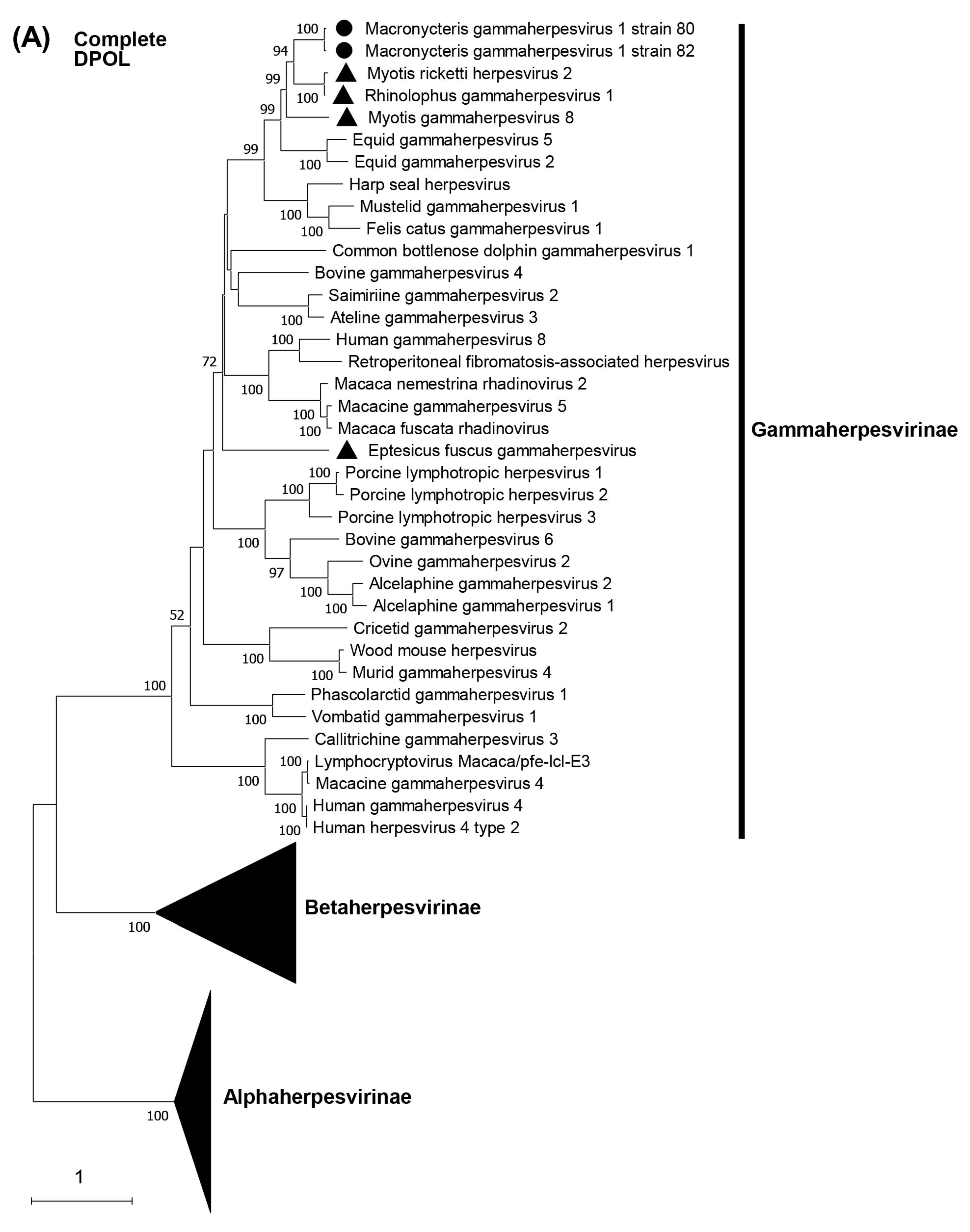
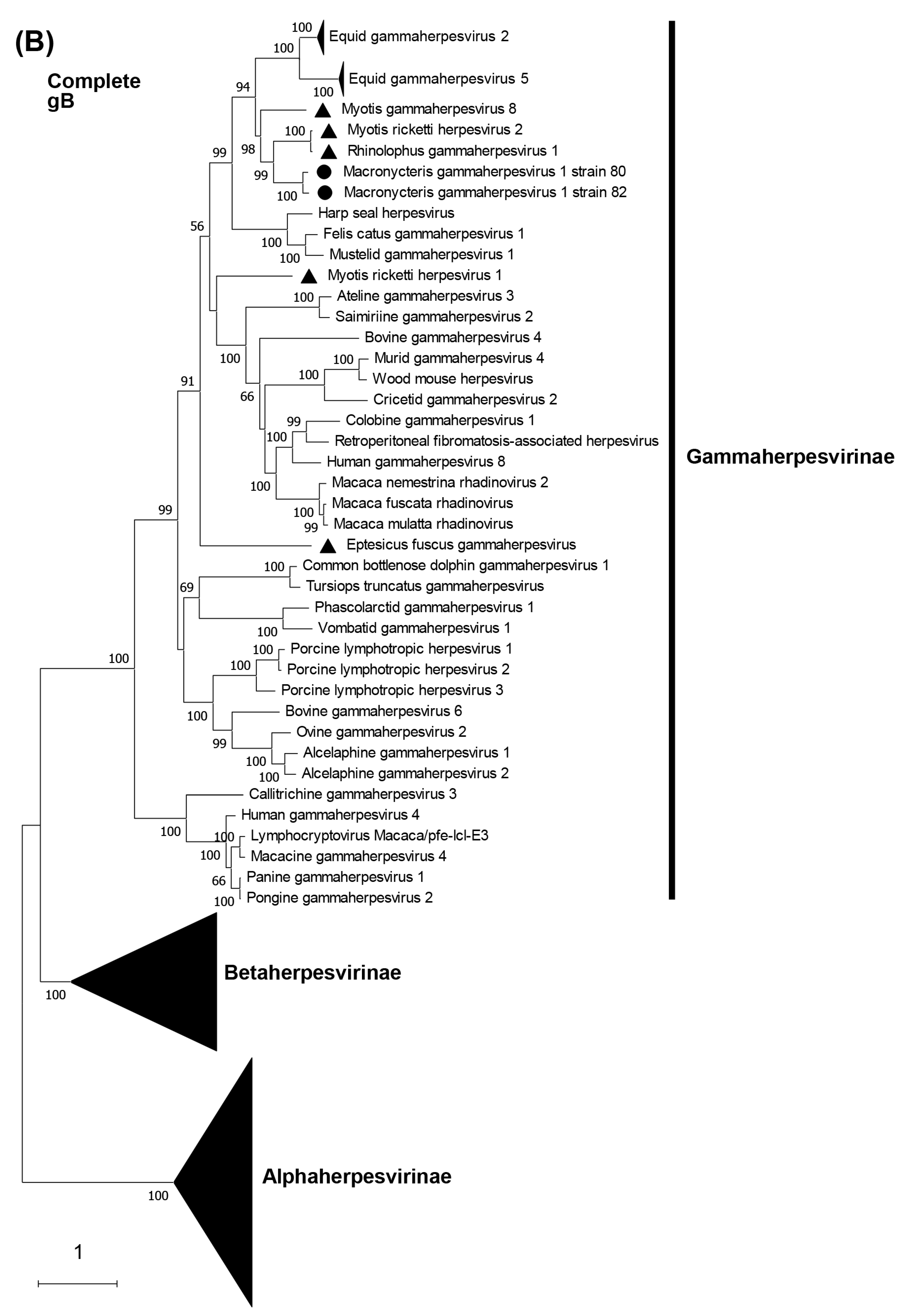
3.5. Phylogenetic Characterization of MaGHV1

Phylogenetic trees were constructed on the basis of DPOL and gB amino acid sequences. The phylogenetic analysis of DPOL demonstrated that two MaGHV1 strains were in the subfamily Gammaherpesvirinae and formed an independent lineage sharing a common origin with other bat-derived gammaherpesviruses such as RGHV1, myotis gammaherpesvirus 8, and myotis ricketti herpesvirus 2 detected by virome analyses in China [31] (Figure 4A). Similar results were obtained in the phylogenetic tree of gB (Figure 4B). In both the DPOL and gB phylogenetic trees, MaGHV1 strains were located near the branches of equid gammaherpesvirus 2 and equid gammaherpesvirus 5.

4. Discussion

In this study, we isolated a novel gammaherpesvirus, tentatively named MaGHV1, from Macronycteris vittatus bats in Zambia. We determined its complete genome sequence including the repeat sequences, and genetically characterized the encoded sequences. During the past two decades, many partial herpesvirus genes have been detected in various bats worldwide using NGS or PCR-based approaches [8,13,31,32,33]. However, few investigations isolated infectious herpesviruses. Isolated infectious viruses are often needed for downstream studies aiming at biological characterization of novel viruses, such as their host tropisms, transmissibility, and pathogenicity.
To date, three gammaherpesviruses (myotis gammaherpesvirus 8, eptesicus fuscus gammaherpesvirus, and RGHV1) have been isolated from Myotis velifer incautus, Eptesicus fuscus, and Rhinolophus ferrumequinum bats in the United States of America, Canada, and Japan, respectively [18,19,20]. Thus, MaGHV1 is the fourth infectious gammaherpesvirus strain isolated from bats. The deduced amino acid sequences of each MaGHV1 ORF showed less than 81% amino acid sequence identity to those of RGHV1, indicating that MaGHV1 is a distinct virus from other known herpesviruses. Phylogenetic analyses of full-length DPOL and gB amino acid sequences demonstrated that MaGHV1 was monophyletic with the other bat gammaherpesviruses except eptesicus fuscus gammaherpesvirus, and located near the branches of equid gammaherpesvirus 2 and equid gammaherpesvirus 5. The subfamily Gammaherpesvirinae is divided into seven genera: Percavirus, Rhadinovirus (e.g., Kaposi’s sarcoma-associated herpesvirus), Lymphocryptovirus (e.g., Epstein–Barr virus), Macavirus (e.g., ovine gammaherpesvirus 2), Bossavirus (e.g., delphinid gammaherpesvirus 1), Manticavirus (e.g., vombatid gammaherpesvirus 1), and Patagivirus (e.g., eptesicus fuscus gammaherpesvirus) [6]. To date, seven species have been classified into the genus Percavirus: Equid gammaherpesvirus 2, Equid gammaherpesvirus 5, Phocid gammaherpesvirus 3 (harp seal herpesvirus), Felid gammaherpesvirus 1 (felis catus gammaherpesvirus 1), Mustelid gammaherpesvirus 1, Rhinolophid gammaherpesvirus 1 (RGHV1), and Vespertilionid gammaherpesvirus 1 (myotis gammaherpesvirus 8). According to the species demarcation criteria of the ICTV [6], herpesviruses that have distinct epidemiological or biological characteristics and distinct genomes that represent independent replicating lineages differing from known species can be recognized as novel species. Therefore, we propose that MaGHV1 is a novel species belonging to the genus Percavirus.
Herpesviruses are highly disseminated in nature and most animal species have yielded at least one herpesvirus and frequently, several distinct herpesviruses [5]. By our PCR screening, at least seven betaherpesviruses and five gammaherpesviruses were detected in three Zambian bat species: Rousettus aegyptiacus, Macronycteris vittatus, and Hipposideros caffer. Our phylogenetic analyses indicate that diverse herpesviruses are distributed in the bat population in Zambia. It is generally believed that mammalian herpesviruses are mainly host-specific and have coevolved with their specific host species [34]. In a previous study, host–virus cophylogenetic analyses demonstrated that the phylogenetic tree of bat betaherpesviruses showed congruence with features in the phylogeny of corresponding host organisms, providing evidence for coevolutionary development of virus and host lineages [35]. Zambian bat herpesviruses were phylogenetically divided into several groups depending on their host species, except for BHV-G6. Interestingly, the nucleotide sequence of one betaherpesvirus from Rousettus aegyptiacus frugivorous bats was identical to that from Macronycteris vittatus insectivorous bats. Although evolutionary analyses of bat gammaherpesviruses have shown that host-switching events occur frequently [36], little information about the cross-species transmission of bat betaherpesviruses is available. In an epidemiological survey of herpesviruses in bats in Peru, betaherpesviruses detected in an Artibeus lituratus fruit bat and Desmodus rotundus vampire bats phylogenetically formed a cluster, suggesting a possible cross-species infection within the family Phyllostomidae (between the genera Artibeus and Desmodus) [35]. Our Zambian case may also show possible cross-species infection within order Chiroptera (between the families Hipposideridae and Pteropodidae), suggesting a unique and rare instance of cross-species transmission of betaherpesviruses. In our study, bats were captured using a harp trap, and trapped bats closely contacted each other in the collection chamber for a few hours. Considering this situation, bat saliva might be transferred to the oral cavities of other bats, implying possible contamination of the virus in an improper host without actual infection. To clarify the true frequency of betaherpesvirus transmission between the families Hipposideridae and Pteropodidae bat species, further investigation with virus isolation is required.
It is important to characterize herpesviruses in various host species for better understanding of the evolutionary history and host specificity of herpesviruses. Although partial herpesvirus genes have been detected in bats in various African countries [9,10,11,12,13], Macronycteris vittatus and Hipposideros caffer bats, which live in Africa, had not been analyzed for the detection of herpesviruses. In this study, we carried out the first genetic characterization of herpesviruses from these insectivorous bats living in Africa. Having a high Chiroptera (bat) diversity, there are currently 18 families (including 5 extinct), 84 genera (25 extinct), and 403 species (54 extinct) recognized as occurring in Africa [37]. Some of the bat species, including Macronycteris vittatus and Hipposideros caffer, were distributed only in Africa and the southwestern Arabian Peninsula, including Saudi Arabia and Yemen [37,38]. Therefore, surveillance of herpesviruses in bats in each country or region is necessary to fully understand the evolutionary host–virus relationship.
The prototype of gammaherpesvirus, Kaposi’s sarcoma-associated herpesvirus, is estimated to encode over 85 genes, of which about 58 are conserved in all gammaherpesvirus genomes, including 40 core genes for all herpesviruses [6,7,39]. Overall, the structure of the MaGHV1 genome is similar to other gammaherpesviruses such as RGHV1, equid herpesvirus 2, and equid herpesvirus 5, and MaGHV1 carries all the conserved ORFs among the subfamily Gammaherpesvirinae. The conserved genes in the MaGHV1 genome are arranged colinearly in direction and position, mostly as in other gammaherpesvirus genomes. We found unique putative ORFs of MaGHV1, some of which were defined as hypothetical proteins with no homology with any known proteins, and it would be of interest to analyze the functions of these unique viral proteins in future studies. The MaGHV1 genome was flanked by the terminal repeat region consisting of multiple copies of tandemly repeated sequences. According to the ICTV criteria for the classification of the herpesvirus genome [6], MaGHV1 could be classified into the class 2 genome structure, which is common in members of the subfamily Gammaherpesvirinae [6]. The genome structure of RGHV1, which is genetically related to MaGHV1, could also be classified into the class 2 genome [20]. On the other hand, myotis gammaherpesvirus 8, which might share a common origin with MaGHV1, is currently unclassified because its terminal repeat sequences were not determined. Therefore, complete genome sequences of herpesviruses, including the repeat regions, are necessary to clarify the genetic relationships among diverse herpesviruses.
In the present study, we showed the presence of multiple bat-derived herpesviruses in Zambia. Our findings provide new information regarding the genetic diversity of bat herpesviruses. Similar prevalence patterns were also observed in bats in other countries such as Indonesia and South Africa [11,32]. As the first investigation on bat-derived herpesviruses in Zambia, we only screened three bat species in the present study. However, since there are 65 known bat species in Zambia [40], more diverse herpesviruses might be associated with various bat species in the same manner as other bat herpesviruses that have been previously recognized. It would be of interest to further investigate the prevalence of herpesvirus infection of bats in Zambia and other African countries.

Supplementary Materials

The following supporting information can be downloaded at: https://www.mdpi.com/article/10.3390/v15061369/s1, Table S1: Primers used in this study; Table S2: Reference sequences used in this study; Table S3: Highest amino acid identities for partial DPOL genes detected by screening; Table S4: Identity comparison of each viral protein among Macronycteris gammaherpesvirus 1 strains and rhinolophus gammaherpesvirus 1; Table S5: Predicted protein coding regions in the genome of Macronycteris gammaherpesvirus 1 strain 80.

Author Contributions

Conceptualization, H.H.; Methodology, H.H., J.Y., M.K. and M.S.; Validation, H.H. and M.S.; Formal Analysis, H.H. and Y.Q.; Investigation, H.H. and Y.Q.; Resources, H.H., M.K., K.C., K.O., M.I., T.Y., H.O. and B.M.H.; Data Curation, H.H. and J.Y.; Writing—Original Draft Preparation, H.H. and J.Y.; Writing—Review and Editing, Y.Q., M.K., K.C., K.O., M.I., T.Y., H.O., N.N., M.S., E.S., H.S., K.I., B.M.H. and A.T.; Visualization, H.H.; Supervision, A.S.M. and A.T.; Project Administration, A.S.M., H.S., B.M.H. and A.T.; Funding Acquisition, H.S. and A.T. All authors, except A.S.M. (deceased 27 April 2019), have read and agreed to the published version of the manuscript.

Funding

This research was supported by the Japan Initiative for Global Research Network of Infectious Diseases (JP15fm0108008) from the Japan Agency for Medical Research and Development (AMED), the Japan Program for Infectious Diseases Research and Infrastructure (JP20wm0125008 and JP223fa627005) from AMED, and grants from AMED and the Japan International Cooperation Agency (JICA) within the framework of the Science and Technology Research Partnership for Sustainable Development (SATREPS) (JP22jm0110019).

Institutional Review Board Statement

Collections of bat samples used in this study were approved by the Department of National Parks and Wildlife, Ministry of Tourism and Arts of the Republic of Zambia (permit numbers DNPW8/27/1; guidelines Act No. 14 of 2015) and the Animal Care and Use Committee of Hokkaido University, following the Fundamental Guidelines for Proper Conduct of Animal Experiment and Related Activities in Academic Research Institutions under the jurisdiction of the Ministry of Education, Culture, Sports, Science and Technology in Japan (permit number 20-0124).

Informed Consent Statement

Not applicable.

Data Availability Statement

All relevant data are provided in the manuscript.

Acknowledgments

We would like to thank Aiko Ohnuma and Naoko Kawai from the International Institute for Zoonosis Control, Hokkaido University, and Sakiko Ueda from the Women’s Future Development Organization, Tokyo University of Agriculture and Technology, for technical support of analyses. We also would like to thank the staff members at the International Institute for Zoonosis Control, Hokkaido University, Japan, and Daniel Mwizabi of the Department of National Parks and Wildlife, Ministry of Tourism and Arts, Zambia, for collecting samples.

Conflicts of Interest

The authors declare no conflict of interest.

References

  1. Letko, M.; Seifert, S.N.; Olival, K.J.; Plowright, R.K.; Munster, V.J. Bat-borne virus diversity, spillover and emergence. Nat. Rev. Microbiol. 2020, 18, 461–471. [Google Scholar] [CrossRef] [PubMed]
  2. Zhou, S.; Liu, B.; Han, Y.; Wang, Y.; Chen, L.; Wu, Z.; Yang, J. ZOVER: The database of zoonotic and vector-borne viruses. Nucleic Acids Res. 2022, 50, D943–D949. [Google Scholar] [CrossRef] [PubMed]
  3. Burgin, C.J.; Colella, J.P.; Kahn, P.L.; Upham, N.S. How many species of mammals are there? J. Mammal. 2018, 99, 1–14. [Google Scholar] [CrossRef] [Green Version]
  4. Mollentze, N.; Streicker, D.G. Viral zoonotic risk is homogenous among taxonomic orders of mammalian and avian reservoir hosts. Proc. Natl. Acad. Sci. USA 2020, 117, 9423–9430. [Google Scholar] [CrossRef] [PubMed] [Green Version]
  5. Pellett, P.E.; Roizman, B. Herpesviridae. In Fields Virology, 6th ed.; Knipe, D.M., Howley, P.M., Cohen, J.I., Griffin, D.E., Lamb, R.A., Martin, M.A., Racaniello, V.R., Roizman, B., Eds.; Wolters Kluwer/Lippincott Williams & Winlkins: Philadelphia, PA, USA, 2013; Volume 2, pp. 1802–1822. [Google Scholar]
  6. Gatherer, D.; Depledge, D.P.; Hartley, C.A.; Szpara, M.L.; Vaz, P.K.; Benkő, M.; Brandt, C.R.; Bryant, N.A.; Dastjerdi, A.; Doszpoly, A.; et al. ICTV Virus Taxonomy Profile: Herpesviridae 2021. J. Gen. Virol. 2021, 102, 001673. [Google Scholar] [CrossRef]
  7. Fossum, E.; Friedel, C.C.; Rajagopala, S.V.; Titz, B.; Baiker, A.; Schmidt, T.; Kraus, T.; Stellberger, T.; Rutenberg, C.; Suthram, S.; et al. Evolutionarily conserved herpesviral protein interaction networks. PLoS Pathog. 2009, 5, e1000570. [Google Scholar] [CrossRef] [Green Version]
  8. James, S.; Donato, D.; de Thoisy, B.; Lavergne, A.; Lacoste, V. Novel herpesviruses in neotropical bats and their relationship with other members of the Herpesviridae family. Infect. Genet. Evol. 2020, 84, 104367. [Google Scholar] [CrossRef]
  9. Cameron, K.; Hayes, B.; Olson, S.H.; Smith, B.R.; Pante, J.; Laudisoit, A.; Goldstein, T.; Joly, D.O.; Bagamboula MPassi, R.; Lange, C.E. Detection of first gammaherpesvirus sequences in Central African bats. N. Microbes N. Infect. 2020, 36, 100705. [Google Scholar] [CrossRef]
  10. Kamau, J.; Ergunay, K.; Webala, P.W.; Justi, S.A.; Bourke, B.P.; Kamau, M.W.; Hassell, J.; Chege, M.N.; Mwaura, D.K.; Simiyu, C.; et al. A Novel Coronavirus and a Broad Range of Viruses in Kenyan Cave Bats. Viruses 2022, 14, 2820. [Google Scholar] [CrossRef]
  11. Dietrich, M.; Kearney, T.; Seamark, E.C.J.; Paweska, J.T.; Markotter, W. Synchronized shift of oral, faecal and urinary microbiotas in bats and natural infection dynamics during seasonal reproduction. R. Soc. Open Sci. 2018, 5, 180041. [Google Scholar] [CrossRef] [Green Version]
  12. Baker, K.S.; Leggett, R.M.; Bexfield, N.H.; Alston, M.; Daly, G.; Todd, S.; Tachedjian, M.; Holmes, C.E.; Crameri, S.; Wang, L.F.; et al. Metagenomic study of the viruses of African straw-coloured fruit bats: Detection of a chiropteran poxvirus and isolation of a novel adenovirus. Virology 2013, 441, 95–106. [Google Scholar] [CrossRef] [Green Version]
  13. Razafindratsimandresy, R.; Jeanmaire, E.M.; Counor, D.; Vasconcelos, P.F.; Sall, A.A.; Reynes, J.M. Partial molecular characterization of alphaherpesviruses isolated from tropical bats. J. Gen. Virol. 2009, 90, 44–47. [Google Scholar] [CrossRef]
  14. Sasaki, M.; Setiyono, A.; Handharyani, E.; Kobayashi, S.; Rahmadani, I.; Taha, S.; Adiani, S.; Subangkit, M.; Nakamura, I.; Sawa, H.; et al. Isolation and characterization of a novel alphaherpesvirus in fruit bats. J. Virol. 2014, 88, 9819–9829. [Google Scholar] [CrossRef] [Green Version]
  15. Inagaki, T.; Yamada, S.; Fujii, H.; Yoshikawa, T.; Shibamura, M.; Harada, S.; Fukushi, S.; Le, M.Q.; Nguyen, C.T.; Nguyen, T.T.T.; et al. Characterization of a Novel Alphaherpesvirus Isolated from the Fruit Bat. J. Virol. 2020, 94, e00673-20. [Google Scholar] [CrossRef]
  16. Watanabe, S.; Maeda, K.; Suzuki, K.; Ueda, N.; Iha, K.; Taniguchi, S.; Shimoda, H.; Kato, K.; Yoshikawa, Y.; Morikawa, S.; et al. Novel betaherpesvirus in bats. Emerg. Infect. Dis. 2010, 16, 986–988. [Google Scholar] [CrossRef]
  17. Zhang, H.; Todd, S.; Tachedjian, M.; Barr, J.A.; Luo, M.; Yu, M.; Marsh, G.A.; Crameri, G.; Wang, L.F. A novel bat herpesvirus encodes homologues of major histocompatibility complex classes I and II, C-type lectin, and a unique family of immune-related genes. J. Virol. 2012, 86, 8014–8030. [Google Scholar] [CrossRef] [Green Version]
  18. Shabman, R.S.; Shrivastava, S.; Tsibane, T.; Attie, O.; Jayaprakash, A.; Mire, C.E.; Dilley, K.E.; Puri, V.; Stockwell, T.B.; Geisbert, T.W.; et al. Isolation and Characterization of a Novel Gammaherpesvirus from a Microbat Cell Line. mSphere 2016, 1, e00070-15. [Google Scholar] [CrossRef] [Green Version]
  19. Subudhi, S.; Rapin, N.; Dorville, N.; Hill, J.E.; Town, J.; Willis, C.K.R.; Bollinger, T.K.; Misra, V. Isolation, characterization and prevalence of a novel Gammaherpesvirus in Eptesicus fuscus, the North American big brown bat. Virology 2018, 516, 227–238. [Google Scholar] [CrossRef]
  20. Noguchi, K.; Kuwata, R.; Shimoda, H.; Mizutani, T.; Hondo, E.; Maeda, K. The complete genomic sequence of Rhinolophus gammaherpesvirus 1 isolated from a greater horseshoe bat. Arch. Virol. 2019, 164, 317–319. [Google Scholar] [CrossRef]
  21. Kajihara, M.; Hang’ombe, B.M.; Changula, K.; Harima, H.; Isono, M.; Okuya, K.; Yoshida, R.; Mori-Kajihara, A.; Eto, Y.; Orba, Y.; et al. Marburgvirus in Egyptian Fruit Bats, Zambia. Emerg. Infect. Dis. 2019, 25, 1577–1580. [Google Scholar] [CrossRef] [Green Version]
  22. Qiu, Y.; Nakao, R.; Hang’ombe, B.M.; Sato, K.; Kajihara, M.; Kanchela, S.; Changula, K.; Eto, Y.; Ndebe, J.; Sasaki, M.; et al. Human Borreliosis Caused by a New World Relapsing Fever Borrelia-like Organism in the Old World. Clin. Infect. Dis. 2019, 69, 107–112. [Google Scholar] [CrossRef]
  23. Rose, T.M.; Strand, K.B.; Schultz, E.R.; Schaefer, G.; Rankin, G.W.; Thouless, M.E.; Tsai, C.C.; Bosch, M.L. Identification of two homologs of the Kaposi’s sarcoma-associated herpesvirus (human herpesvirus 8) in retroperitoneal fibromatosis of different macaque species. J. Virol. 1997, 71, 4138–4144. [Google Scholar] [CrossRef] [PubMed] [Green Version]
  24. Wick, R.R.; Judd, L.M.; Gorrie, C.L.; Holt, K.E. Completing bacterial genome assemblies with multiplex MinION sequencing. Microb. Genom. 2017, 3, e000132. [Google Scholar] [CrossRef] [PubMed]
  25. Koren, S.; Walenz, B.P.; Berlin, K.; Miller, J.R.; Bergman, N.H.; Phillippy, A.M. Canu: Scalable and accurate long-read assembly via adaptive. Genome Res. 2017, 27, 722–736. [Google Scholar] [CrossRef] [PubMed] [Green Version]
  26. Li, H.; Durbin, R. Fast and accurate short read alignment with Burrows-Wheeler transform. Bioinformatics 2009, 25, 1754–1760. [Google Scholar] [CrossRef] [Green Version]
  27. Walker, B.J.; Abeel, T.; Shea, T.; Priest, M.; Abouelliel, A.; Sakthikumar, S.; Cuomo, C.A.; Zeng, Q.; Wortman, J.; Young, S.K.; et al. Pilon: An integrated tool for comprehensive microbial variant detection and genome assembly improvement. PLoS ONE 2014, 9, e112963. [Google Scholar] [CrossRef]
  28. Telford, E.A.; Watson, M.S.; Aird, H.C.; Perry, J.; Davison, A.J. The DNA sequence of equine herpesvirus 2. J. Mol. Biol. 1995, 249, 520–528. [Google Scholar] [CrossRef]
  29. Benson, G. Tandem repeats finder: A program to analyze DNA sequences. Nucleic Acids Res. 1999, 27, 573–580. [Google Scholar] [CrossRef] [Green Version]
  30. Kumar, S.; Stecher, G.; Li, M.; Knyaz, C.; Tamura, K. MEGA X: Molecular Evolutionary Genetics Analysis across Computing Platforms. Mol. Biol. Evol. 2018, 35, 1547–1549. [Google Scholar] [CrossRef]
  31. Wu, Z.; Ren, X.; Yang, L.; Hu, Y.; Yang, J.; He, G.; Zhang, J.; Dong, J.; Sun, L.; Du, J.; et al. Virome analysis for identification of novel mammalian viruses in bat species from Chinese provinces. J. Virol. 2012, 86, 10999–11012. [Google Scholar] [CrossRef] [Green Version]
  32. Wada, Y.; Sasaki, M.; Setiyono, A.; Handharyani, E.; Rahmadani, I.; Taha, S.; Adiani, S.; Latief, M.; Kholilullah, Z.A.; Subangkit, M.; et al. Detection of novel gammaherpesviruses from fruit bats in Indonesia. J. Med. Microbiol. 2018, 67, 415–422. [Google Scholar] [CrossRef]
  33. Moreira Marrero, L.; Botto Nuñez, G.; Malta, L.; Delfraro, A.; Frabasile, S. Ecological and Conservation Significance of Herpesvirus Infection in Neotropical Bats. Ecohealth 2021, 18, 123–133. [Google Scholar] [CrossRef]
  34. Azab, W.; Dayaram, A.; Greenwood, A.D.; Osterrieder, N. How Host Specific Are Herpesviruses? Lessons from Herpesviruses Infecting Wild and Endangered Mammals. Annu. Rev. Virol. 2018, 5, 53–68. [Google Scholar] [CrossRef]
  35. Griffiths, M.E.; Bergner, L.M.; Broos, A.; Meza, D.K.; Filipe, A.D.S.; Davison, A.; Tello, C.; Becker, D.J.; Streicker, D.G. Epidemiology and biology of a herpesvirus in rabies endemic vampire bat populations. Nat. Commun. 2020, 11, 5951. [Google Scholar] [CrossRef]
  36. Escalera-Zamudio, M.; Rojas-Anaya, E.; Kolokotronis, S.O.; Taboada, B.; Loza-Rubio, E.; Méndez-Ojeda, M.L.; Arias, C.F.; Osterrieder, N.; Greenwood, A.D. Bats, Primates, and the Evolutionary Origins and Diversification of Mammalian Gammaherpesviruses. mBio 2016, 7, e01425-16. [Google Scholar] [CrossRef] [Green Version]
  37. ACR. African Chiroptera Report 2022; Cakenberghe, V.V., Seamark, E.C.J., Eds.; AfricanBats NPC: Pretoria, South Africa, 2022; pp. i–xvii + 8661. [Google Scholar]
  38. Wright, G.S. Hipposideros caffer (chiroptera: Hipposideridae). Mamm. Species 2009, 845, 1–9. [Google Scholar] [CrossRef] [Green Version]
  39. Russo, J.J.; Bohenzky, R.A.; Chien, M.C.; Chen, J.; Yan, M.; Maddalena, D.; Parry, J.P.; Peruzzi, D.; Edelman, I.S.; Chang, Y.; et al. Nucleotide sequence of the Kaposi sarcoma-associated herpesvirus (HHV8). Proc. Natl. Acad. Sci. USA 1996, 93, 14862–14867. [Google Scholar] [CrossRef] [Green Version]
  40. Hutson, A.M.; Mickleburgh, S.P.; Racey, P.A. Microchiropteran Bats: Global Status Survey and Conservation Action Plan; IUCN: Gland, Switzerland, 2001; Volume 56. [Google Scholar]
Figure 1. Phylogenetic analysis of the partial DPOL gene sequences obtained by screening. A phylogenetic tree based on the predicted amino acid sequences of the DPOL corresponding to positions 690–746 of the equid gammaherpesvirus 2 DNA polymerase catalytic subunit (GenBank accession no. NC_001650.2) was constructed using the maximum likelihood method with 1000 bootstrap replicates (A). Betaherpesviruses and gammaherpesviruses are magnified in panels (B) and (C), respectively. Bootstrap values greater than 50% are shown on the interior branch nodes and scale bars indicate the number of substitutions per site. Betaherpesvirus (BHV) and gammaherpesvirus (GHV) groups are indicated. Individual herpesviruses detected in bats are highlighted and named as follows: accession number/bat species of origin/country of identification/common name/year of collection. The black dots represent the bat herpesviruses detected in this study.
Viruses 15 01369 g001aViruses 15 01369 g001bViruses 15 01369 g001c
Figure 2. Isolation of MaGHV1. Phase-contrast images of MaGHV1 strain 82-infected (A) and mock-infected Vero E6 cells (B) at 12 days post infection. The cells infected with the virus exhibited cytopathic effects. Scale bar, 200 µm.
Viruses 15 01369 g002
Figure 3. Map of the MaGHV1 genome. Predicted ORFs in the genome of MaGHV1 strain 82 are indicated by arrows. Blue arrows represent homologs of other gammaherpesvirus ORFs. Green arrows represent unique ORFs of MaGHV1: ORF with homology to cellular genes and/or viral protein genes encoding in some gammaherpesviruses; ORF with no homology to known proteins. Repeat regions found by the tandem repeat finder program with the copy number > 2.0 and the score > 250 are indicated by orange rectangles. The total length of the genome is 116,972 bp. The scale is shown at the top.
Viruses 15 01369 g003
Figure 4. Phylogenetic analysis of the complete DPOL and gB. The complete ORF amino acid sequences of DPOL and gB of MaGHV1 were phylogenetically analyzed with corresponding proteins from the representative alpha-, beta-, and gammaherpesviruses. Bootstrap values greater than 50% are shown on the interior branch nodes and scale bars indicate the number of substitutions per site. The black dots represent MaGHV1 isolated in this study. The triangles represent herpesviruses identified in bats.
Viruses 15 01369 g004aViruses 15 01369 g004b
Table 1. Summary of bat samplings in 2018 in Zambia and screening results of herpesviruses.
Sampling DateBat (Species)Sample IDNo. of Positive/No. of Tested Samples (%)
19 NovemberEgyptian fruit bat
(Rousettus aegyptiacus)
1–177/17 (41.2)
Striped leaf-nosed bat
(Macronycteris vittatus)
18–7039/53 (73.6)
22 NovemberEgyptian fruit bat
(Rousettus aegyptiacus)
71–770/7 (0)
Striped leaf-nosed bat
(Macronycteris vittatus)
78–111
113–130
43/52 (82.7)
Sundevall’s roundleaf bat
(Hipposideros caffer)
1121/1 (100)
Total90/130 (69.2)
Table 2. Summary of herpesvirus DPOL-positive bats detected by PCR screening.
VirusAccession No.Sample IDSubfamilyGroup
Macronycteris vittatus/Zambia/64/2018LC76220964BetaherpesvirinaeBHV-G1
Macronycteris vittatus/Zambia/50/2018LC76221050, 69BetaherpesvirinaeBHV-G2
Macronycteris vittatus/Zambia/20/2018LC76221120BetaherpesvirinaeBHV-G3
Macronycteris vittatus/Zambia/36/2018LC76221236BetaherpesvirinaeBHV-G3
Macronycteris vittatus/Zambia/41/2018LC76221341BetaherpesvirinaeBHV-G3
Macronycteris vittatus/Zambia/46/2018LC76221446BetaherpesvirinaeBHV-G3
Macronycteris vittatus/Zambia/101/2018LC762215101, 111BetaherpesvirinaeBHV-G3
Macronycteris vittatus/Zambia/104/2018LC762216104BetaherpesvirinaeBHV-G3
Rousettus aegyptiacus/Zambia/10/2018LC76221710BetaherpesvirinaeBHV-G4
Rousettus aegyptiacus/Zambia/11/2018LC76221811BetaherpesvirinaeBHV-G4
Macronycteris vittatus/Zambia/37/2018LC76221937, 44BetaherpesvirinaeBHV-G5
Macronycteris vittatus/Zambia/49/2018LC76222049, 56, 61, 66, 89, 103, 121, 123, 128BetaherpesvirinaeBHV-G5
Macronycteris vittatus/Zambia/106/2018LC762221106, 109BetaherpesvirinaeBHV-G5
Rousettus aegyptiacus/Zambia/3/2018LC7622223BetaherpesvirinaeBHV-G6
Rousettus aegyptiacus/Zambia/6/2018LC7622236BetaherpesvirinaeBHV-G6
Macronycteris vittatus/Zambia/114/2018LC762224114BetaherpesvirinaeBHV-G6
Macronycteris vittatus/Zambia/126/2018LC762225126BetaherpesvirinaeBHV-G6
Macronycteris vittatus/Zambia/84/2018LC76222684BetaherpesvirinaeBHV-G7
Macronycteris vittatus/Zambia/22/2018LC76222722, 58, 65, 81, 83, 110, 130GammaherpesvirinaeGHV-G1
Macronycteris vittatus/Zambia/30/2018LC76222830, 60GammaherpesvirinaeGHV-G1
Macronycteris vittatus/Zambia/38/2018LC76222938GammaherpesvirinaeGHV-G1
Macronycteris vittatus/Zambia/39/2018LC76223039GammaherpesvirinaeGHV-G1
Macronycteris vittatus/Zambia/80/2018LC76223159, 70, 80, 86, 91, 97, 98, 107, 113GammaherpesvirinaeGHV-G1
Macronycteris vittatus/Zambia/82/2018LC76223218, 24, 26, 31, 78, 82, 85, 88, 124, 129GammaherpesvirinaeGHV-G1
Hipposideros caffer/Zambia/112/2018LC762233112GammaherpesvirinaeGHV-G2
Macronycteris vittatus/Zambia/21/2018LC76223421GammaherpesvirinaeGHV-G3
Macronycteris vittatus/Zambia/23/2018LC76223523, 43, 118GammaherpesvirinaeGHV-G3
Macronycteris vittatus/Zambia/28/2018LC76223628, 99, 100, 116, 120GammaherpesvirinaeGHV-G3
Macronycteris vittatus/Zambia/29/2018LC76223729GammaherpesvirinaeGHV-G3
Macronycteris vittatus/Zambia/42/2018LC76223842GammaherpesvirinaeGHV-G3
Macronycteris vittatus/Zambia/45/2018LC76223945GammaherpesvirinaeGHV-G3
Macronycteris vittatus/Zambia/55/2018LC76224055, 119GammaherpesvirinaeGHV-G3
Macronycteris vittatus/Zambia/92/2018LC76224192GammaherpesvirinaeGHV-G3
Macronycteris vittatus/Zambia/94/2018LC76224294GammaherpesvirinaeGHV-G3
Macronycteris vittatus/Zambia/96/2018LC76224396GammaherpesvirinaeGHV-G3
Macronycteris vittatus/Zambia/25/2018LC76224425, 33, 34, 47, 52, 95, 105, 108, 125GammaherpesvirinaeGHV-G4
Rousettus aegyptiacus/Zambia/4/2018LC7622454GammaherpesvirinaeGHV-G5
Rousettus aegyptiacus/Zambia/7/2018LC7622467GammaherpesvirinaeGHV-G5
Rousettus aegyptiacus/Zambia/15/2018LC76224715GammaherpesvirinaeGHV-G5
Abbreviations: BHV, betaherpesvirus; GHV, gammaherpesvirus.
Table 3. Predicted protein coding regions and repeat regions in the genome of Macronycteris gammaherpesvirus 1 strain 82.
GeneLocation
(Nucleotides)
StrandSize (No. of Amino Acids)Product or Predicted Function
BM13031–3630199homolog of CASP8 and FADD-like apoptosis regulator
BM23577–3990+137hypothetical protein
BM34693–5832+379homolog of EHV2 E3 membrane protein E3
BM46988–715254homolog of E3 ubiquitin-protein ligase and RGHV1 ORF4 MIR-like membrane protein
BM57520–8077+185homolog of EHV2 E4 apoptosis regulator BALF1
ORF68279–11,677+1132single-stranded DNA binding protein
ORF711,748–13,799+683DNA packaging terminase subunit 2
ORF813,816–16,362+848glycoprotein B
ORF916,633–19,638+1001DNA polymerase catalytic subunit
BM619,673–20,185170bcl-2-like protein
BM720,383–21,486+367homolog of EHV2 E6 membrane protein BILF1
ORF1021,530–22,927+465homolog of EHV2 ORF10 protein G10
ORF1122,977–24,209+410homolog of EHV2 ORF11 virion protein G11
BM826,676–28,304542homolog of bovine herpesvirus 6 Bov8 putative major envelope glycoprotein, RGHV1 ORF18 glycoprotein
ORF1728,449–30,029526capsid maturation protease
ORF17.528,449–29,300284capsid scaffold protein
ORF1830,022–30,798+258homolog of EHV2 ORF18 protein UL79
ORF1930,795–32,417540homolog of EHV2 ORF19 DNA packaging tegument protein UL25
ORF2032,281–32,985234homolog of EHV2 ORF20 nuclear protein UL24
ORF2132,984–34,600+538thymidine kinase
ORF2234,600–36,861+753glycoprotein H
ORF2336,858–38,051397homolog of EHV2 ORF23 tegument protein UL88
ORF2438,106–40,298730homolog of EHV2 ORF24 protein UL87
ORF2540,303–44,436+1377major capsid protein
ORF2644,456–45,358+300capsid triplex subunit 2
ORF2745,359–45,802+147homolog of EHV2 ORF27 envelope glycoprotein 48
ORF2845,867–46,118+83homolog of EHV2 ORF28 envelope glycoprotein 150
ORF29a50,460–51,623387DNA packaging terminase subunit 1
ORF29b46,207–47,250347DNA packaging terminase subunit 1
ORF3047,385–47,639+84homolog of EHV2 ORF30 protein UL91
ORF3147,552–48,235+227homolog of EHV2 ORF31 protein UL92
ORF3248,181–49,536+451homolog of EHV2 ORF32 DNA packaging tegument protein UL17
ORF3349,529–50,695+388homolog of EHV2 ORF33 tegument protein UL16
ORF3451,622–52,590+322homolog of EHV2 ORF34 protein UL95
ORF3552,577–53,047+156homolog of EHV2 ORF35 tegument protein UL14
ORF3652,944–54,245+433tegument serine/threonine protein kinase
ORF3754,248–55,702+484deoxyribonuclease
ORF3855,657–55,854+65myristylated tegument protein
ORF3955,918–57,051377glycoprotein M
ORF40a57,162–58,538+458helicase-primase subunit
ORF40b58,624–59,154+176helicase-primase subunit
ORF4259,149–59,964271homolog of EHV2 ORF42 tegument protein UL7
ORF4359,951–61,654567capsid portal protein
ORF4461,620–63,986+788helicase-primase helicase subunit
ORF4564,035–64,934299homolog of EHV2 ORF45 tegument protein G45
ORF4664,936–65,694252uracil-DNA glycosylase
ORF4765,657–66,115152glycoprotein L
ORF4866,213–67,646477homolog of EHV2 ORF48 tegument protein G48
ORF4968,010–68,894294homolog of EHV2 ORF49 tegument protein G49
ORF5069,039–70,646+535protein Rta
BM970,780–71,067+95hypothetical protein, homolog of RGHV1 ORF53
BM1071,349–71,759+136hypothetical protein
BM1171,849–72,568+239homolog of EHV2 E7A envelope glycoprotein 42
BM1272,526–73,479+317hypothetical protein, homolog of RGHV1 ORF57
ORF5273,493–73,897134homolog of EHV2 ORF52 virion protein G52
ORF5373,952–74,21286glycoprotein N
ORF5474,302–75,171+289dUTPase
ORF5575,240–75,893217homolog of EHV2 ORF55 tegument protein UL51
ORF5675,866–78,433+855helicase-primase primase subunit
ORF5778,642–80,066+474multifunctional expression regulator
ORF5880,927–81,964345homolog of EHV2 ORF58 envelope protein UL43
ORF5981,973–83,202409DNA polymerase processivity subunit
ORF6083,364–84,281305ribonucleotide reductase subunit 2
ORF6184,309–86,621770ribonucleotide reductase subunit 1
ORF6286,645–87,646333capsid triplex subunit 1
ORF6387,657–90,434+925homolog of EHV2 ORF63 tegument protein UL37
ORF6490,450–97,421+2323large tegument protein
ORF6597,435–97,935166small capsid protein
ORF6697,946–99,256436homolog of EHV2 ORF66 protein UL49
ORF6799,163–100,002279nuclear egress membrane protein
ORF67A100,065–100,33489homolog of EHV2 ORF67A DNA packaging protein UL33
ORF68100,437–101,819+460envelope glycoprotein, DNA packaging protein UL32
ORF69101,821–102,684+287nuclear egress lamina protein
ORF70102,750–103,628292thymidylate synthase
BM13103,853–104,161+102hypothetical protein
ORF73104,958–106,661567nuclear antigen LANA-1
ORF74106,940–107,914+324homolog of EHV2 ORF74 membrane protein G74
ORF75108,078–112,0911337homolog of EHV2 ORF75 tegument protein G75
BM14112,188–112,517+109hypothetical protein
BM15112,844–113,437+197hypothetical protein
BM16114,035–114,538+167hypothetical protein
Terminal
repeat
319–2295nonenonenone
Repeat
region 1
25,665–26,556nonenonenone
Repeat
region 2
105,538–106,545nonenonenone
Terminal
repeat
115,903–116,972nonenonenone
Disclaimer/Publisher’s Note: The statements, opinions and data contained in all publications are solely those of the individual author(s) and contributor(s) and not of MDPI and/or the editor(s). MDPI and/or the editor(s) disclaim responsibility for any injury to people or property resulting from any ideas, methods, instructions or products referred to in the content.

