Article

Chetomin, a SARS-CoV-2 3C-like Protease (3CLpro) Inhibitor: In Silico Screening, Enzyme Docking, Molecular Dynamics and Pharmacokinetics Analysis

by
Mahmoud A. A. Ibrahim
1,2,*,
Alaa H. M. Abdelrahman
1,
Dina E. M. Mohamed
1,
Khlood A. A. Abdeljawaad
1,
Mohamed Ahmed Naeem
3,
Gamal A. Gabr
4,5,
Ahmed M. Shawky
6,
Mahmoud E. S. Soliman
7,
Peter A. Sidhom
8,*,
Paul W. Paré
9 and
Mohamed-Elamir F. Hegazy
10
1
Computational Chemistry Laboratory, Chemistry Department, Faculty of Science, Minia University, Minia 61519, Egypt
2
School of Health Sciences, University of KwaZulu-Natal, Westville, Durban 4000, South Africa
3
Ain Shams University Specialized Hospital, Ain Shams University, Cairo 11588, Egypt
4
Department of Pharmacology and Toxicology, College of Pharmacy, Prince Sattam Bin Abdulaziz University, Al-Kharj 11942, Saudi Arabia
5
Agricultural Genetic Engineering Research Institute (AGERI), Agricultural Research Center, Giza 12619, Egypt
6
Science and Technology Unit (STU), Umm Al-Qura University, Makkah 21955, Saudi Arabia
7
Molecular Modelling and Drug Design Research Group, School of Health Sciences, University of KwaZulu-Natal, Westville, Durban 4000, South Africa
8
Department of Pharmaceutical Chemistry, Faculty of Pharmacy, Tanta University, Tanta 31527, Egypt
9
Department of Chemistry & Biochemistry, Texas Tech University, Lubbock, TX 79409, USA
10
Chemistry of Medicinal Plants Department, National Research Centre, 33 El-Bohouth St., Dokki, Giza 12622, Egypt
*
Authors to whom correspondence should be addressed.
Viruses 2023, 15(1), 250; https://doi.org/10.3390/v15010250
Submission received: 16 November 2022 / Revised: 10 January 2023 / Accepted: 13 January 2023 / Published: 15 January 2023
(This article belongs to the Special Issue Viral Enzyme Inhibitors: Structure and Dynamics)

Abstract

:
The emergence of the Coronavirus Disease 2019 (COVID-19) pandemic caused by severe acute respiratory syndrome coronavirus-2 (SARS-CoV-2) has led to over 6 million deaths. The 3C-like protease (3CLpro) enzyme of the SARS-CoV-2 virus is an attractive druggable target for exploring therapeutic drug candidates to combat COVID-19 due to its key function in viral replication. Marine natural products (MNPs) have attracted considerable attention as alternative sources of antiviral drug candidates. In looking for potential 3CLpro inhibitors, the MNP database (>14,000 molecules) was virtually screened against 3CLpro with the assistance of molecular docking computations. The performance of AutoDock and OEDocking software in anticipating the ligand-3CLpro binding mode was first validated according to the available experimental data. Based on the docking scores, the most potent MNPs were further subjected to molecular dynamics (MD) simulations, and the binding affinities of those molecules were computed using the MM-GBSA approach. According to MM-GBSA//200 ns MD simulations, chetomin (UMHMNP1403367) exhibited a higher binding affinity against 3CLpro than XF7, with ΔGbinding values of −55.5 and −43.7 kcal/mol, respectively. The steadiness and tightness of chetomin with 3CLpro were evaluated, revealing the high stabilization of chetomin (UMHMNP1403367) inside the binding pocket of 3CLpro throughout 200 ns MD simulations. The physicochemical and pharmacokinetic features of chetomin were also predicted, and the oral bioavailability of chetomin was demonstrated. Furthermore, the potentiality of chetomin analogues –namely, chetomin A-D– as 3CLpro inhibitors was investigated. These results warrant further in vivo and in vitro assays of chetomin (UMHMNP1403367) as a promising anti-COVID-19 drug candidate.

1. Introduction

COVID-19 is caused by severe acute respiratory syndrome coronavirus-2 (SARS-CoV-2), which has produced a global health emergency [1,2]. In December 2019, SARS-CoV-2 was first identified in Wuhan, China, and quickly spread around the world [3,4,5]. As of October 2022, over 623 million COVID-19 cases were confirmed, resulting in over 6.5 million deaths. COVID-19 has exposed the fragility of public health and healthcare systems in many nations [6]. Despite the predictions of viral abatement due to widespread vaccine production using next-generation technologies, rapidly evolving variants combined with vaccine hesitancy and limited vaccine availability have thwarted plans for viral control [7]. Indeed, now that the SARS-CoV-2 pandemic has been raging for two-and-a-half years, most people have either been vaccinated against and/or infected by the virus. The threats of the appearance of more genetic variations with greater virulence and the elevated rate of viral impedance to authorized therapeutics are driving an uninterrupted necessity for exploring efficient anti-COVID-19 therapies.
SARS-CoV-2 3C-like protease (3CLpro/Mpro/NSP5) is a vital enzyme in viral gene replication, transcription, and expression [8,9,10,11]. Since the outbreak of COVID-19, several experimental and computational trials have been conducted to repurpose various drugs to inhibit 3CLpro and combat this disease [12,13]. Among the repurposed drugs, remdesivir, lopinavir, umifenovir, ritonavir, and favipiravir, have been promoted for clinical investigation as COVID-19 medications [14,15]. Very recently, the FDA (US Food and Drug Administration) granted a contingency utilizing permission for a combination of nirmatrelvir and ritonavir as therapeutics for COVID-19 treatment.
Plant-based natural products (NPs) have been considered a substantial source of curative agents. Indeed, NPs have robust antiviral activity and have demonstrated inhibitory efficiency against numerous viral enzymes [16,17]; exploratory antiviral drugs include cyclosporine and cyanovirin-N [18,19].
An alternate source of natural products is marine secondary metabolites [20,21]. The marine environment is a rich source of chemically diverse and biologically active natural molecules. It has been reported that MNPs demonstrate antimalarial, antifungal, antibacterial, and antiviral activities [22,23]. The antiviral activity of MNPs has been investigated for dengue, influenza, SARS, and other viruses [24,25]. For instance, polyphenol and coumarin derivatives from MNP sources demonstrated high in vitro activities toward 3CLpro [26,27]. However, the potentiality of MNPs as 3CLpro inhibitors has yet to be examined extensively. Here, we explore MNPs as 3CLpro inhibitors by screening a database containing >14,000 compounds using in Silico molecular docking techniques. The most effective MNPs were submitted for molecular dynamics simulations. MNPs-3CLpro binding affinities were calculated utilizing the MM-GBSA approach. These in Silico calculations in combination with predicted physicochemical and pharmacokinetics characteristics, identified multiple drug candidates for COVID-19 treatment.

2. Materials and Methods

2.1. 3CLpro Preparation

The 3D structure of 3CLpro complexed with a noncovalent inhibitor ((5S)-5-(3-{3-chloro-5-[(2-chlorophenyl)methoxy]phenyl}-2-oxo [2H-[1,3’-bipyridine]]-5-yl)pyrimidine-2,4(3H,5H)-dione; XF7) was retrieved from the PDB database (PDB ID: 7L13 [28], resolution: 2.17 Å) and employed as a template for the current study. All heteroatoms, ligands, ions, and crystallographic water molecules were removed. The H++ server was utilized to identify residues’ protonation states, and missing hydrogen atoms were added [29].

2.2. Database Preparation

The marine natural product (MNP) database was downloaded in an SDF format from Prof. Encinar’s website (http://docking.umh.es/downloaddb (accessed on 11 February 2022)). The 3D chemical structures were generated using Omega2 software with a maximum of 200 conformers generated within a 10 kcal/mol energy window [30,31]; the lowest energy conformer was selected for further studies. The protonation state of each MNP was investigated using fixpka within QUACPAC software [32]. The Gasteiger–Marsili method was used to appoint the MNP atomic charges [33]. Based on the InChIKey (IUPAC international chemical identifier keys), duplicated molecules were eliminated [34]. The prepared MNP library is accessible via www.compchem.net/ccdb. The schematic diagram of the utilized in Silico computations for the filtration process of the MNP library is represented in Figure 1.

2.3. Molecular Docking

The initial molecular docking computations were performed using AutoDock4.2.6 [35] and OEDocking3.0.1 [36,37] software. For the AutoDock4.2.6 calculations, the pdbqt file of 3CLpro was created in accordance with the AutoDock protocol with the assistance of the molecular graphics laboratory (MGL) tools 1.5.7 [38]. The internal conformational search was executed by the LGA (Lamarckian Genetic Algorithm) [39]. The LGA population size and the maximum number of generations were 300 and 27,000, respectively. Three accuracy levels of docking computations were utilized, namely, fast, moderate, and expensive (i.e., more computational cost and time) calculations. The GA (number of genetic algorithms) run variables were 25, 100, and 250 for fast, moderate, and expensive molecular docking computations, respectively. In addition, the eval (maximum number of energy evaluations) was 2,500,000, 10,000,000, and 25,000,000 for fast, moderate, and expensive molecular docking computations, respectively. The grid dimensions (60 × 60 × 60 Å3) were able to embrace the complete binding pocket of 3CLpro. The coordinates of the grid center were x = −13.069, y = 9.740, z = 64.83. The employed grid is depicted in Figure S1. The predicted poses were clusterized using the internal conformational cluster engine with an RMSD tolerance value of 1.0 Å. The representative docking mode was chosen from the largest cluster with the lowest docking score. For OEDocking3.0.1 software, the resolution of the exhaustive search was set to high, and the best scoring pose was retained. Based on the complex of the co-crystallized XF7 ligand with 3CLpro, the active site was determined. XF7 was found to be one of the most potent noncovalent inhibitors toward 3CLpro with an IC50 value of 0.018 μM [28].

2.4. MD Simulations

MD (molecular dynamics) simulations were performed for the most potent MNPs complexed with 3CLpro with the assistance of AMBER16 software [40]. The details of the utilized protocol for the MD simulations are described elsewhere [41,42,43,44,45]. Briefly, the investigated MNPs were parameterized by a general AMBER force field (GAFF2) [46]. 3CLpro was characterized using the AMBER force field of 14SB [47]. In the current study, implicit and explicit water solvent MD simulations were performed.
In the context of implicit water solvent MD simulation, the investigated MNPs were optimized at MMFF94S, and the atomic charges of the MNP were assigned using the AM1-BCC approach [48]. For the nonbonded interactions, the cutoff value was adjusted to 999 Å. Additionally, non-periodic boundary conditions were utilized. Moreover, the solvent model (implicit generalized born (igb) = 1) was employed to estimate the solvation impact [49]. The docked MNPs complexed with 3CLpro were initially minimized based on 500 cycles. The minimized complexes were slowly heated from 0 to 310 K throughout 10 ps in six steps. After that, the investigated systems were equilibrated over a 50 ps simulation. Ultimately, the equilibrated systems underwent a production stage of 1 ns.
In the context of explicit water solvent MD, the investigated MNPs were optimized at HF/6-31G* level of theory by Gaussian09 software [50]. The RESP (restrained electrostatic potential) approach was subsequently applied to compute the charges of the MNPs [51]. All of the MNP-3CLpro complexes were solvated utilizing TIP3P water molecules in an octahedral box with a minimal distance of 12 Å. Using the tleap tool implemented inside AMBER16 software, the counter-ions (52 Na+ and/or 48 Cl) were inserted to attain 0.15 M NaCl concentration. Afterward, minimization was conducted using combined steepest descent and conjugate gradient algorithms for 5000 cycles to eliminate any unsuitable geometries or steric clashes. The minimized systems were progressively heated up to 310 K over 50 ps. Thereafter, the investigated systems were equilibrated for 10 ns. Eventually, the equilibrated systems were subjected to production runs over 5, 25, and 200 ns MD. For electrostatics, the Particle-Mesh Ewald (PME) algorithm was used [52]. An atomic cutoff distance of 12 Å was utilized for short-range nonbonded interactions [52]. All of the bonds that included hydrogen atoms were constrained using a SHAKE algorithm [53]. All of the MD simulations were conducted utilizing a GPU-accelerated MD engine in AMBER16 (pmemd.cuda). The CompChem GPU/CPU hybrid cluster was used to perform all in Silico computations. All graphical representations were visualized utilizing BIOVIA Discovery Studio Visualizer 2020 [54].

2.5. Binding Affinity Computations

The molecular mechanical-generalized Born surface area (MM-GBSA) approach within the AMBER16 software was used to compute the binding affinities for the most potent MNPs complexed with 3CLpro [55]. For MM-GBSA calculations, the frames were collected every 10 ps and the ΔGbinding was estimated according to the following equation:
Δ G binding = G Complex G MNP + G 3 C L p r o
where the G term is:
G = G GB + E ele + G SA + E vdw
GGB stands for electrostatic solvation-free energy. Eele is electrostatic energy. GSA refers to the nonpolar solvation-free energy that is evaluated from the SASA (solvent-accessible surface area) with the assistance of an LCPO algorithm [56]. Evdw is van der Waals energy. Because of its greater computational costs, the computation of entropy (S) was overlooked [57,58].

2.6. Physicochemical Features

The physicochemical characteristics of the most potent MNP were estimated utilizing the SwissADME server [59]. Under the framework of the Lipinski’s rule, five characteristics were considered, including the molecular weight (MWt ≤ 500 g/mol), hydrogen bond donors (HBD ≤ 5), log p value (log po/w ≤ 5), demonstrating good oral and intestinal absorption, topological polar surface area (TPSA ≤ 140 Å2), revealing that the compounds have eminent oral absorption or membrane permeability, and hydrogen bond acceptors (HBA ≤ 10). Passing these characteristics indicates that a given MNP is orally bioavailable.

2.7. Pharmacokinetic Characteristics

An online pkCSM tool was employed to anticipate the ADMET properties of the most potent MNPs [60]. Absorption (A) involves skin permeability, Caco2 permeability, HIA (human intestinal absorption), skin permeability, and P-glycoprotein substrate and inhibitor. Distribution (D) includes fraction unbound, CNS (central nervous system) permeability, VDss (steady-state volume of distribution), and BBB (blood–brain barrier) permeability. The metabolism (M) depends on factors such as CYP2D6/CYP3A4 substrate and cytochrome P450 inhibitors. The excretion (E) is estimated via inhibitor total clearance. The toxicity (T) is anticipated via AMES toxicity and skin sensitization.

3. Results and Discussion

The COVID-19 epidemic and its emerging variants underline the necessity to develop alternative treatments, as vaccines alone do not provide complete protection against illness. The 3C-like protease (3CLpro) has a vital function in viral replication and, as such, is deemed an effective druggable target. Moreover, marine natural products (MNPs) include several metabolites with antiviral properties and, as such, are potential drug candidates for COVID-19 treatment [61]. Here, we utilized in silico approaches to screen a chemical library containing > 14,000 MNP metabolites as potential SARS-CoV-2 3CLpro inhibitors.

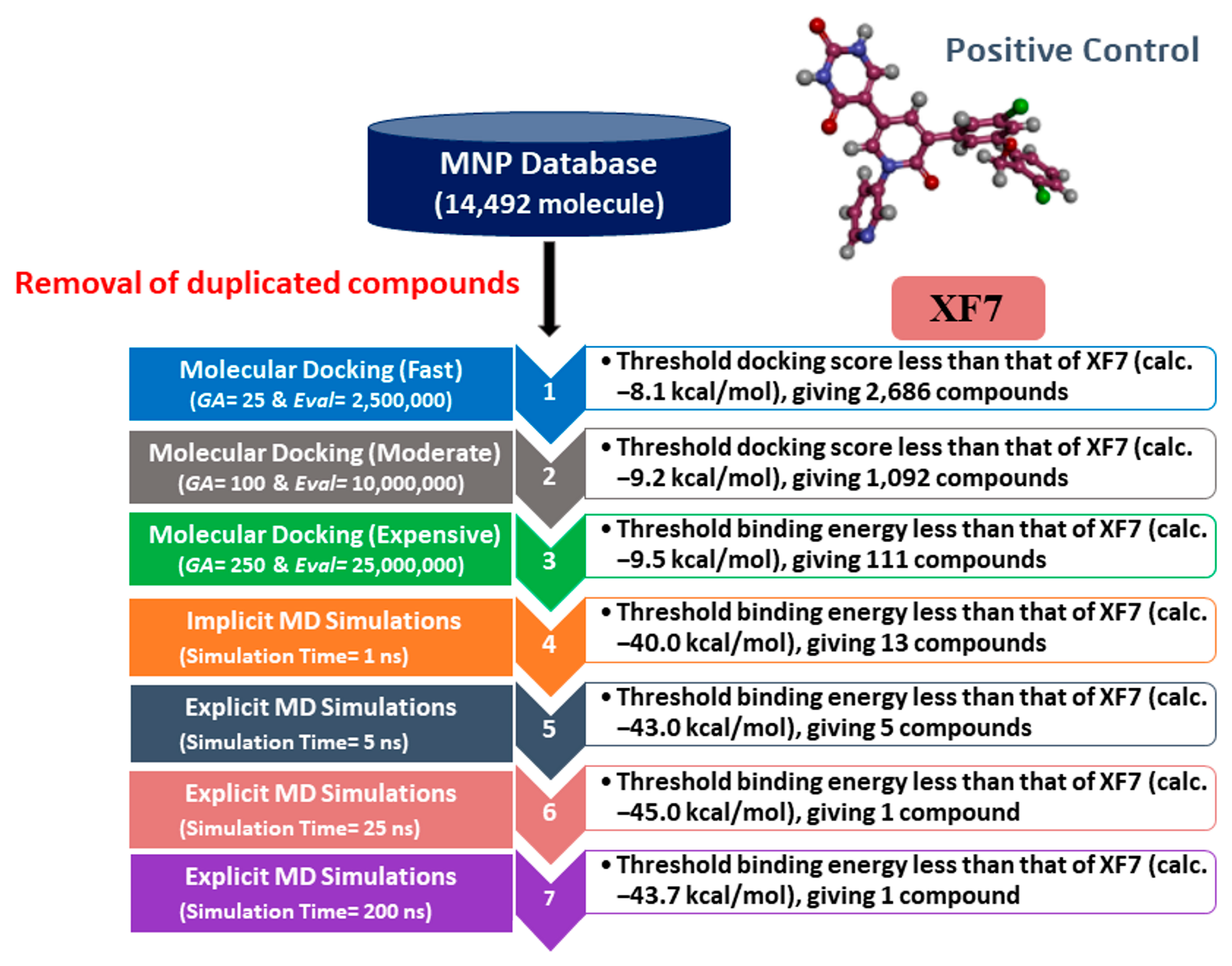
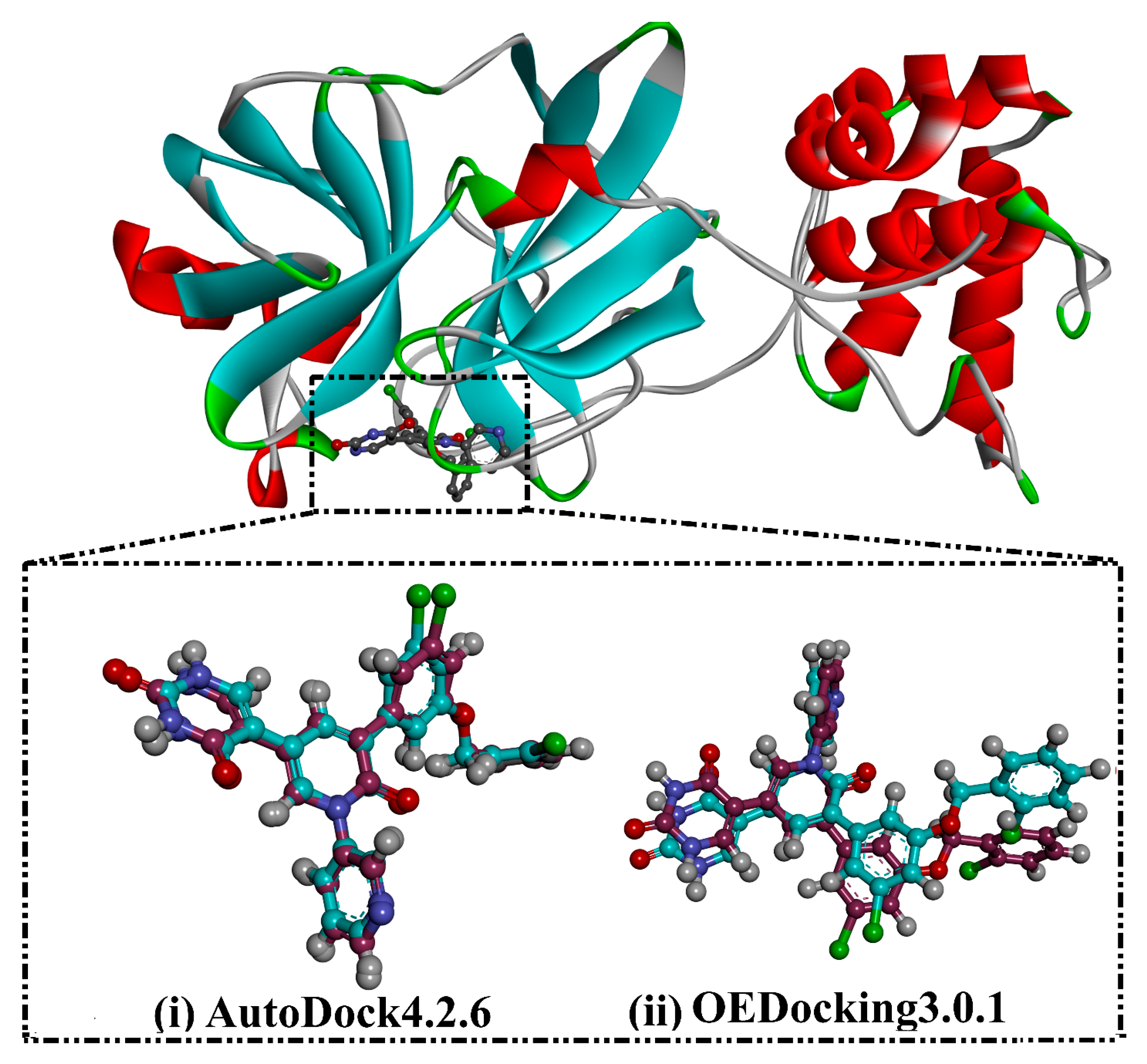
3.1. Docking Assessment

Two molecular docking engines, AutoDock and OEDocking, were used to validate the inhibitor-3CLpro binding mode. Initially, the binding mode of the XF7, a co-crystallized ligand in the 3CLpro binding pocket, was predicted and compared with the resolved native structure (PDB ID: 7L13 [28]) (Figure 2). The predicted docking pose was almost identical to the native binding mode with RMSD values of 0.20 and 0.43 Å for AutoDock and OEDocking, respectively (Figure 2). Comparing the RMSD values of AutoDock and OEDocking, it can be seen that AutoDock predicted the native inhibitor-3CLpro binding mode better than OEDocking. As a result, AutoDock was selected in the filtration process of the MNP database.

3.2. MNP Database Screening
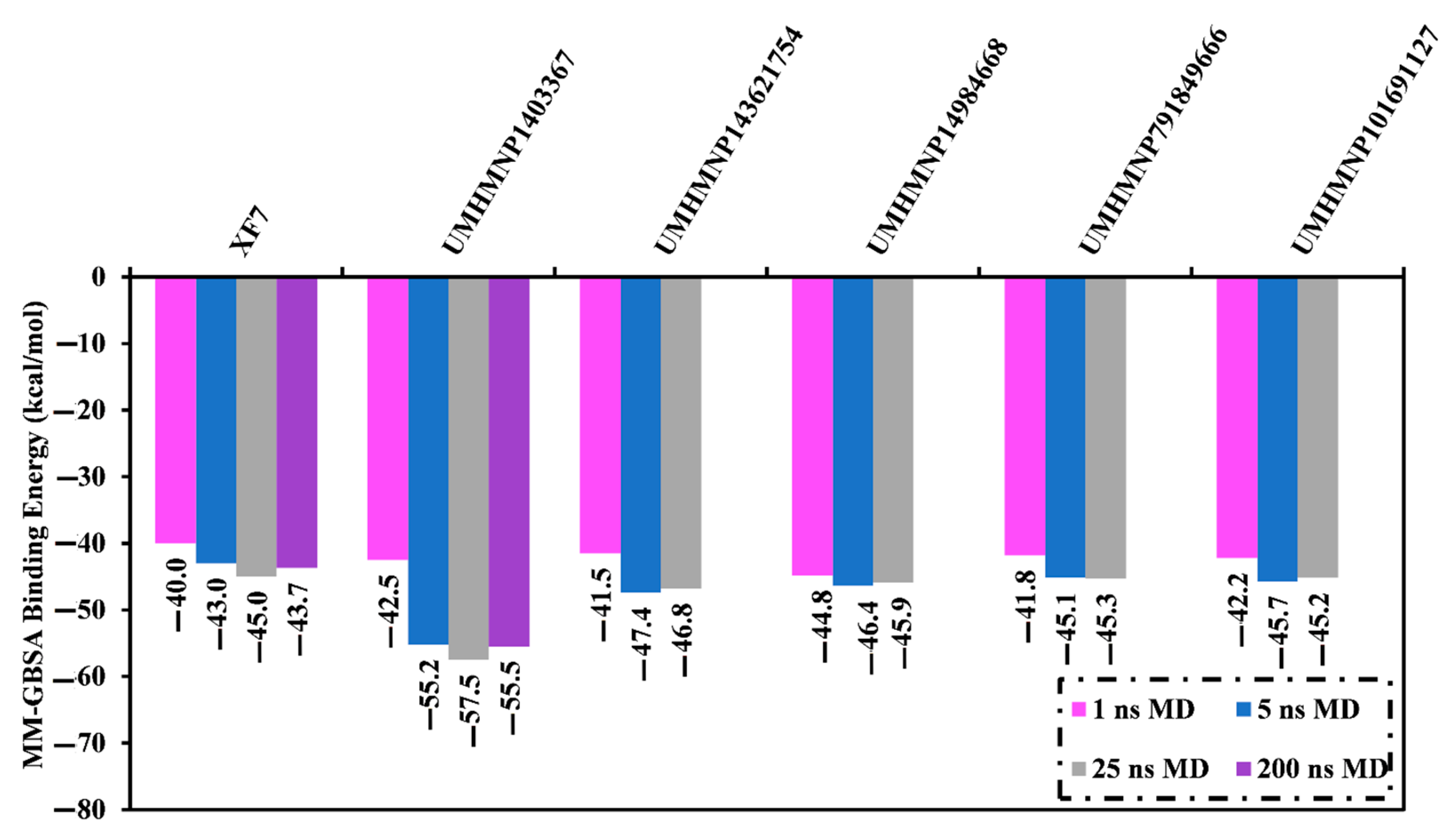
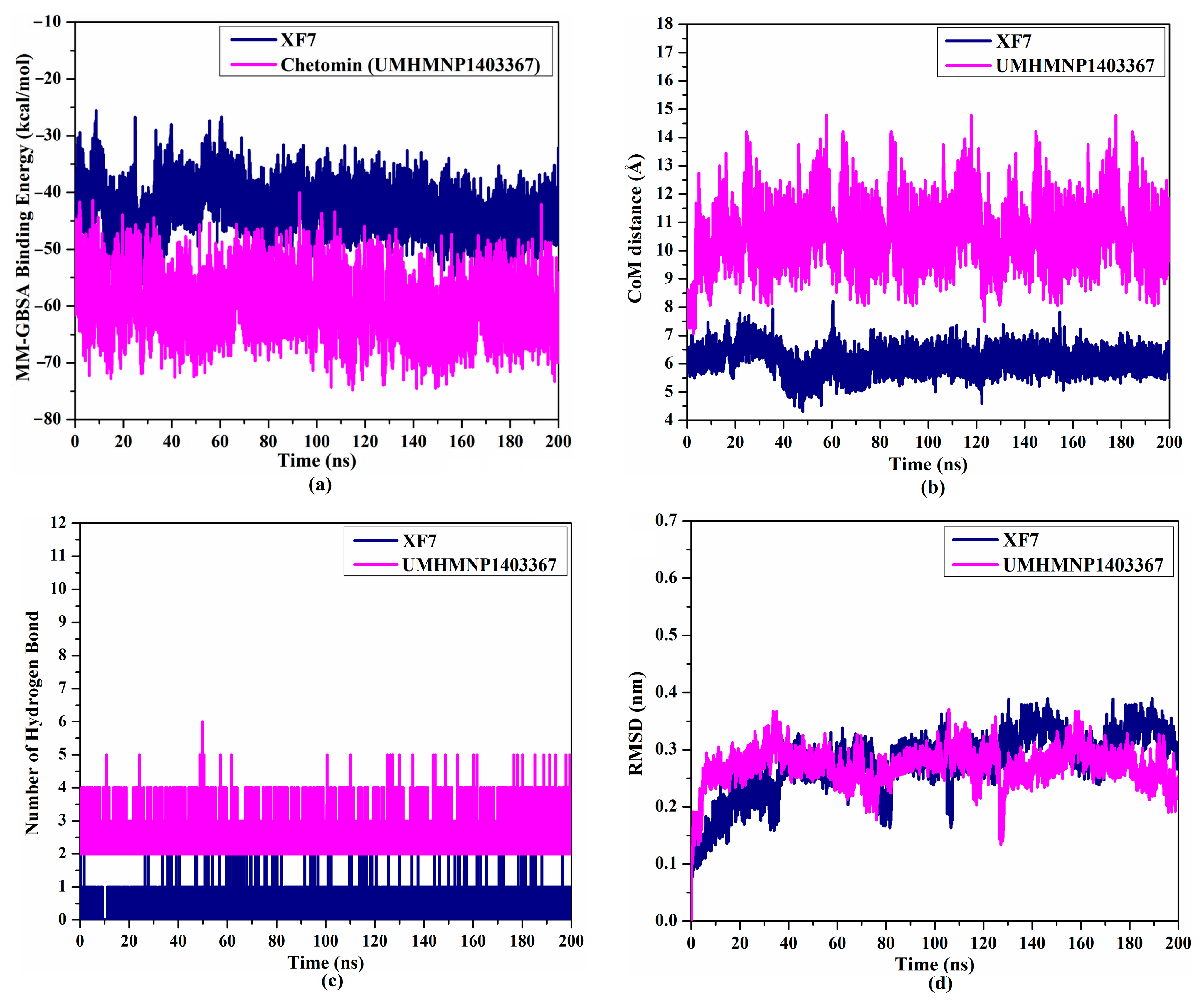
Initially, the MNP database was screened against 3CLpro using fast docking computation. On the basis of the anticipated docking scores, 2686 MNPs displayed docking scores lower than that of the XF7 ligand, with a value of −8.1 kcal/mol against 3CLpro. Consequently, those 2686 MNPs were subjected to moderate docking computations. The computed docking scores for the top 2686 MNPs are summarized in Table S1. According to the estimated moderate docking scores, 1092 MNPs showed docking scores less than that of the XF7 ligand, with a value of −9.2 kcal/mol. Thus, the 1092 MNPs were submitted to expensive docking computations, and the evaluated docking scores are listed in Table S2. Interestingly, about 10% of the selected MNPs (i.e., 111 MNPs) displayed lower docking scores than XF7 (calc. −9.5 kcal/mol). The predicted docking scores, binding features, and 2D chemical structures for the thirteen most potent MNPs for 3CLpro are shown in Table 1. The 2D docking poses for these selected MNPs are depicted in Figure S2. These MNPs were selected based on binding affinities over 1 ns implicit water solvent MD simulations, as described in Section 3.3. Generally, the 2D representations demonstrated the hydrogen bonding of those MNPs with GLN189, GLY143, and GLU166 residues in the 3CLpro binding pocket (Figure S2). π-based, hydrophobic, and vdW interactions were also monitored between the identified MNPs and key residues in the 3CLpro binding pocket (Figure S2).
Chetomin (UMHMNP1403367), an organic heteropentacyclic compound isolated from chaetomium globosum and farrowia seminuda, demonstrated the lowest docking score against 3CLpro with a value of −13.4 kcal/mol. Investigating the docking pose of UMHMNP1403367 within the 3CLpro’s active site disclosed that the CO and OH of the (1S,4S)-1-(hydroxymethyl)-2,3-dithia-5,7-diazabicyclo[2.2.2]octane-6,8-dione ring form two hydrogen bonds with the NH and CO of GLU166 with distances of 1.99 and 2.79 Å, respectively (Figure 3).
Compared to chetomin (UMHMNP1403367), XF7 complexed with 3CLpro demonstrated a competitive docking score with a value of −9.5 kcal/mol (Table 1). The robust binding of XF7 with 3CLpro is ascribed to the ability to form hydrogen bonds with THR26 (2.28, 2.49 Å), GLY143 (2.07 Å), SER144 (3.29 Å), HIS163 (1.91 Å), and GLU166 (1.81 Å) (Table 1).

3.3. MD Simulations

MD simulations are utilized to establish the stabilization of the ligand–target complex, structural specifics, conformational elasticities, and trustworthiness of ligand–target binding energy [62,63]. The most promising MNPs (111 molecules with docking scores <−9.5 kcal/mol) complexed with 3CLpro were submitted to MD simulations and pursued by binding energy computations. To diminish the time and computational costs, the simulations were executed for 1 ns MD in an implicit water solvent. The corresponding binding energies were evaluated, and thirteen MNPs manifested lower ΔGbinding compared to the native XF7 ligand (calc. −40.0 kcal/mol) (Table S3). These MNPs in complex with 3CLpro were then submitted to 5 ns MD in an explicit water solvent to gain more reliable binding energies (Table S4). Based on the estimated MM-GBSA binding energies over 5 ns MD simulations, only five MNPs unveiled lower ΔGbinding compared to the native XF7 ligand (calc. −43.0 kcal/mol) (Figure 4 and Table S4). These potent MNPs were then subjected to a 25 ns MD simulation, and the corresponding MM-GBSA binding energies were estimated (Figure 4). Interestingly, out of the five identified MNPs as potential 3CLpro inhibitors, only chetomin (UMHMNP1403367) displayed stationary binding energies with ΔGbinding values of −55.2 and −57.5 kcal/mol throughout 5 ns and 25 ns MD simulations. While UMHMNP143621754, UMHMNP14984668, UMHMNP791849666, and UMHMNP101691127 manifested a tenuous rise in binding energy (ΔGbinding) over the 25 ns MD simulation. This elucidates the importance of long MD simulation to foretell MNP-3CLpro binding energy. As well, the relative binding energy (ΔΔGbinding) values for the five identified MNPs compounds with respect to the XF7 were computed (Table S5). From Table S5, UMHMNP1403367 demonstrated a promising ΔΔGbinding value of −12.5 kcal/mol, while the other four MNPs showed insignificant ΔΔGbinding values of ≤−1.8 kcal/mol. Therefore, MD simulation for chetomin (UMHMNP1403367) complexed with 3CLpro was protracted to 200 ns. In addition, the corresponding binding affinity was computed (Figure 4).
The perceptible disproportion between the estimated binding energies for the chetomin-3CLpro complex over the 25 ns and 200 ns MD simulations was not observed. Compared with the native XF7 ligand (calc. −43.7 kcal/mol), chetomin was revealed to have a lower binding energy against 3CLpro throughout a 200 ns MD, with an average ΔGbinding of −55.5 kcal/mol (Figure 4). The 3D and 2D molecular interactions of average the structures of chetomin and XF7 inside the 3CLpro binding pocket over 200 ns are illustrated in Figure 5 and Figure S3, respectively. Inspecting the binding mode of chetomin inside the binding pocket of the 3CLpro showed that chetomin preserved its hydrogen bond with GLU166 and formed hydrogen bonds with HIS41, ASN142, and GLN192 residues (Figure 5 and Figure S3). Notably, those hydrogen bonds were absent in the docked pose of chetomin (Figure 3), demonstrating the importance of conducting MD simulation. More exactly, the two CO groups and OH of (1S,4S)-1-(hydroxymethyl)-2,3-dithia-5,7-diazabicyclo[2.2.2]octane-6,8-dione ring exhibit three hydrogen bonds with the CO and NH of GLU166 and NH of GLN192 with distances of 1.92, 2.67, and 2.59 Å, respectively (Figure 5). Moreover, the sulfur atom and CO of (1R,4R)-2,3-dithia-5,7-diazabicyclo[2.2.2]octane-6,8-dione ring demonstrated two hydrogen bonds with NH2 of ASN142 and NH of the imidazole ring of HIS41 with distances of 2.81 and 1.91 Å, respectively (Figure 5 and Figure S3).
For XF7 complexed with 3CLpro, the CO of pyridin-2(1H)-one and nitrogen of the pyridine ring interact with NH of GLU166 and SH of CYS145 by hydrogen bonds with lengths of 1.85 and 1.90 Å, respectively (Figure 5 and Figure S3).
The computed binding affinities were decomposed into individual components to explore the main driving forces in the binding of chetomin and XF7 with SARS-CoV-2 3CLpro (Figure 5). As shown in Figure 5, Eele was a favorable contributor in the binding affinities of chetomin and XF7 with 3CLpro, with values of −34.5 and −21.7 kcal/mol, respectively (Figure 5). The binding affinity of chetomin and XF7 with 3CLpro were dominated by Evdw interactions with an average value of −55.5 and −54.5 kcal/mol, respectively (Figure 5). Notably, Evdw is about one and a half fold stronger than Eele.
To inspect the participation of essential residues in the ligand–target complexes, the total ΔGbinding values were decomposed at the per-residue level. Only amino acids with ΔGbinding < −0.50 kcal/mol were demonstrated (Figure 6). It is apparent that GLU166, ASN142, HIS163, and GLN189 residues participated in the interactions of chetomin and XF7 with 3CLpro. Significant participation of the GLU166 residue to the total ΔGbinding was noticed with values of −4.1 and −2.3 kcal/mol for chetomin (UMHMNP1403367)- and XF7-3CLpro complexes, respectively (Figure 6).

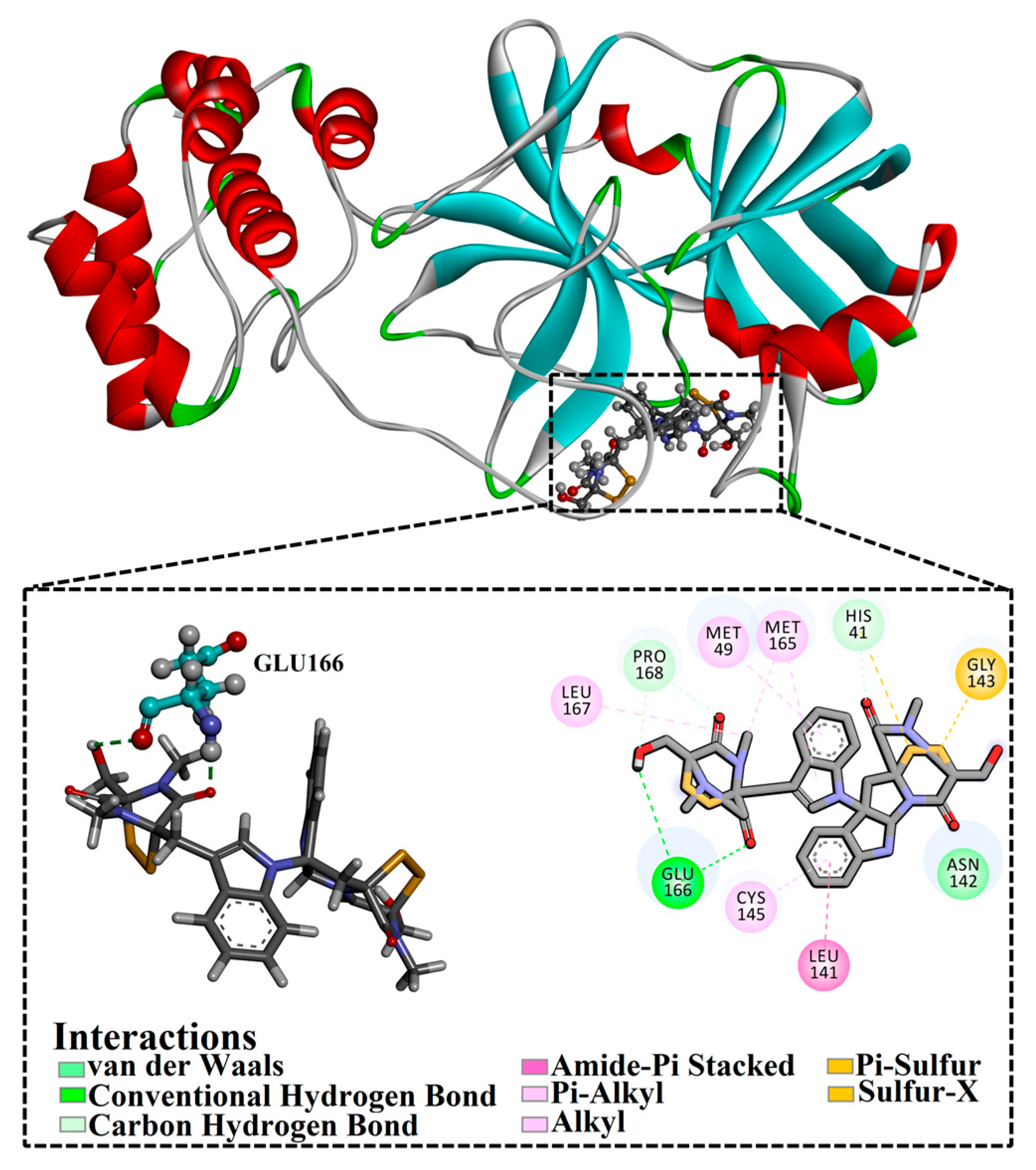
3.4. Post-MD Analyses

To further investigate the structural and energetical stability of chetomin and XF7 in the complex with 3CLpro, post-MD analyses were performed throughout the 200 ns MD simulations.

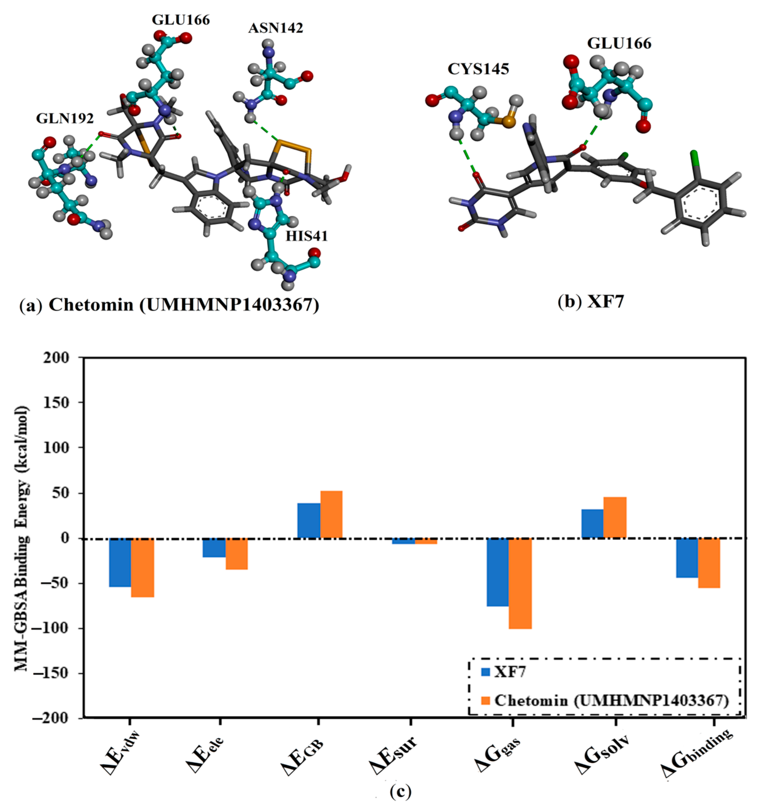
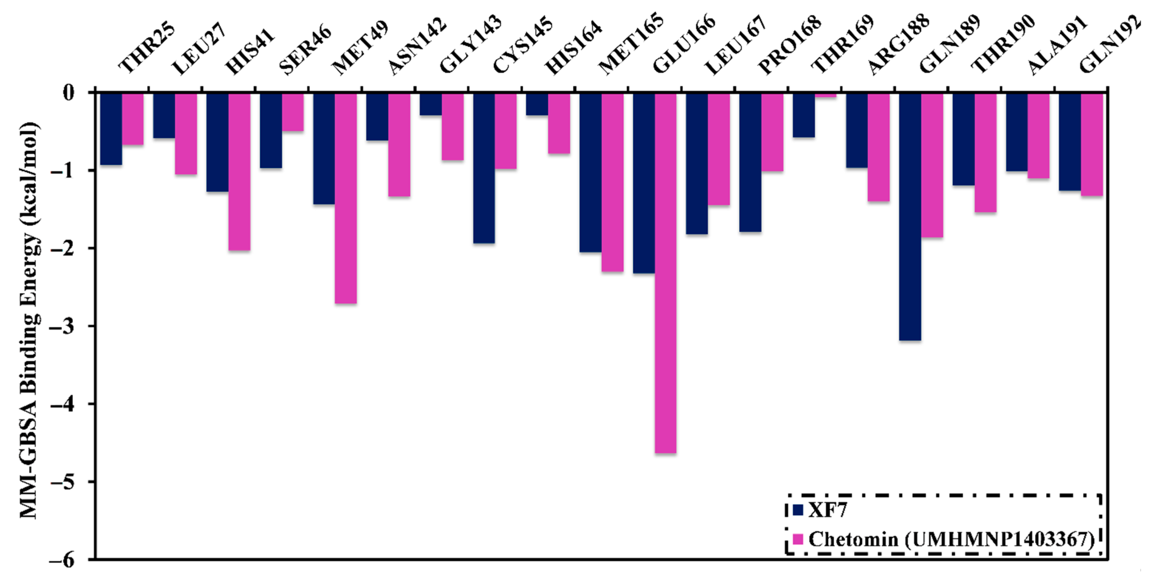
3.4.1. Binding Affinity Analysis

The structural stability of chetomin and XF7 in a complex with 3CLpro was inclusively estimated throughout the MD course of 200 ns by gauging the correlation between binding energy and time (Figure 7a). What stands out in Figure 7a is the general constancies of the binding affinities of chetomin and XF7 with 3CLpro, with ΔGbinding values of −55.5 ± 4.5 and −43.7 ± 3.9 kcal/mol, respectively. On the basis of the energetical analysis, all of the inspected complexes maintained stability throughout the 200 ns MD.

3.4.2. CoM Distance

To gain more in-depth insight into the stability of chetomin and XF7 complexed with 3CLpro over the 200 ns MD simulations, the center-of-mass (CoM) distance was evaluated between the investigated ligand and GLU166 (Figure 7b). The most exciting aspect of the CoM graph is the high steadiness of chetomin and XF7 in complex with 3CLpro with average CoM distances of 10.5 and 6.1 Å, respectively (Figure 7b). These findings revealed that the chetomin binds more tightly with the 3CLpro than XF7.

3.4.3. H-Bond Numbers

Furthermore, the steadiness of chetomin and XF7 in a complex with 3CLpro was evaluated by estimating the number of hydrogen bond interactions (H-bonds). The correlation between the number of H-bonds and simulation time was graphed in Figure 7c. As depicted in Figure 7c, the average number of H-bonds was three and one for chetomin- and XF7-3CLpro complexes. Notably, XF7 demonstrated the fewest number of hydrogen bonds with the fundamental residues inside the active site of 3CLpro. However, the good binding affinity of XF7 with an average ΔGbinding of −43.7 kcal/mol may be ascribed to other interactions, such as vdW, π-based, and hydrophobic interactions. The superiority of the vdW interactions of XF7 with 3CLpro conforms to the binding affinity decomposition results (Figure 5). The hydrogen bond analysis confirmed the presentence of considerable stability for the chetomin than XF7 complexed with 3CLpro.

3.4.4. Root-Mean-Square Deviation

Throughout the 200 ns MD course, the RMSD (root-mean-square deviation) of the C, Cα, N, and O of the entire system was evaluated to observe the conformational change of the chetomin- and XF7-3CLpro complexes (Figure 7d). As shown in Figure 7d, the investigated systems stabilized after the first 5 ns and maintained their stabilization until the end of the 200 ns simulation. The measured RMSD values with average values were 0.27 and 0.28 nm during the 200 ns MD for the chetomin- and XF7-3CLpro complexes. These findings assured that chetomin is tightly bonded and does not influence the structural steadiness of 3CLpro, in addition to preserving the structural integrity.

3.5. Physicochemical Features

The SwissADME server was employed to anticipate the potentiality of utilizing chetomin and the co-crystallized XF7 ligand as drugs by estimating the physicochemical characteristics. The physicochemical features involved the topological polar surface area (Å2), the MWt (g/mol), HBD, HBA, and Nrotb (number of rotatable bonds) (Table 2). As listed in Table 2, the MWt was 710.9 and 533.4 g/mol for chetomin and XF7, respectively (Table 2). Notably, the tenuous rise in MWt will not have tremendous leverage on drug diffusion and transmission, wherever it has been demonstrated that sundry FDA-approved drugs proceeded beyond the conventional low MWt of 500 g/mol [64]. The Log p values of chetomin and XF7 were auspicious, with values lower than 5 [65]. In addition, the number of HBD was less than 5, and the number of HBA was less than 10. These findings indicated that chetomin is a potential anti-COVID-19 drug candidate.

3.6. Pharmacokinetic Characteristics

The knowledge of pharmacokinetics and toxicity features offers worthy guidelines for starting-stage drug discovery. Caco2 permeability and HIA are absorption characteristics that must be considered in any medicine exploration process [66]. The investigated compounds demonstrated perfect absorption, with HIA values of 73.4% and 93.5% for chetomin and XF7, respectively (Table 3). The inspected compounds unveiled satisfactory skin permeability, with a value of −2.7 for both compounds (Table 3). The investigated compounds also demonstrated good Caco2 permeability (less than 0.9 cm/s). One of the most fundamental ADMET characteristics is the P-glycoprotein substrate/inhibitor. Chetomin and XF7 were characterized as inhibitors/substrates for P-glycoprotein (Table 3). The BBB membrane permeability, VDss, and CNS were estimated to check drug distribution. Significant distribution volumes were noticed for chetomin and XF7 with log BB values of −2.0 and −1.1, respectively, indicating that these compounds can readily pass the blood–brain barrier (BBB permeability) (Table 3). For VDss and CNS permeability, log VDss and log PS values were −0.1 and 0.3 and −3.9 and −2.5 for chetomin and XF7, respectively (Table 3). CYP450 has a fundamental function in the metabolism of the drug. The metabolism predictions exposed that chetomin and XF7 cannot inhibit CYP2D6 enzymes and cannot act as inhibitors for CYP1A2, CYP2C19, CYP2C9, and CYP3A4 enzymes (Table 3).
The total drug clearance was 0.3 and 0.8 mL/min/kg for chetomin and XF7, respectively (Table 3). Toxicity has an extraordinary role in opting for adequate drugs. Chetomin and XF7 did not expose AMES toxicity and skin sensitization (Table 3). According to the predicted ADMET features, these findings deduced that the investigated compounds might be used as potential anti-COVID-19 drug candidates.

3.7. Chetomin Derivatives as Prospective Anti-COVID-19 Drug Candidates

Based on the promising potentiality of chetomin as SARS-CoV-2 3CLpro, the study was extended to examine the perspectivity of the chetomin analogues, namely, chetomin A-D. The chemical structures of chetomin A-D were retrieved from the PubChem database and prepared for docking computations. The docking scores, binding features, and 2D chemical structures of the chetomin derivatives are listed in Table 4. As enrolled in Table 4, all of the chetomin derivatives demonstrated similar docking poses with 3CLpro, exhibiting hydrogen bonds with GLU166, ASN142, and GYS145. The anticipated docking scores for chetomin derivatives ranged from −11.1 to −12.2 kcal/mol. A comparison of the docking results revealed that chetomin (UMHMNP1403367/PubChem10417379) demonstrated the lowest docking score with a value of −13.4 kcal/mol (Table 4).

4. Conclusions

Currently, COVID-19 is spreading quickly worldwide, causing a high mortality rate and morbidity rate. The demand for COVID-19 treatments still very much exists. By in silico screening and characterization of MNPs as potential 3CLpro inhibitors, several candidate metabolites were identified. Included in that initial screening, chetomin was shown to have promising binding energy (ΔGbinding), with a value of −55.5 kcal/mol against 3CLpro over 200 ns MD simulation. Post-MD analyses throughout 200 ns MD simulations indicated the high stability of chetomin complexed with 3CLpro. Chetomin also showed favorable physicochemical and pharmacokinetic features. Comparing the docking scores of chetomin with its analogues, namely, chetomin A-D, demonstrated the superior binding affinity of chetomin against 3CLpro. These findings clearly clarify the suitability of chetomin as a promising drug candidate for further studies toward new COVID-19 treatments.

Supplementary Materials

The following supporting information can be downloaded at: https://www.mdpi.com/article/10.3390/v15010250/s1, Figure S1. The employed grid box in Autodock computations; Figure S2. 2D representation of binding modes and interactions for top thirteen scoring drugs with the key amino acid residues of the 3CLpro binding pocket; Figure S3. 2D molecular interaction pattern of binding modes of (a) UMHMNP1403367 and (b) XF7 complexed with 3CLpro based on the average structure over a 200 ns MD simulation; Table S1. Estimated fast and moderate docking scores (in kcal/mol) for top 2686 MNPs towards 3CLpro; Table S2. Estimated fast, moderate, and expensive docking scores for XF7 and the top 1092 potent MNPs compounds within the 3CLpro binding pocket; Table S3. Estimated fast, moderate, and expensive docking scores and MM-GBSA binding energies (in kcal/mol) over 1 ns implicit water solvent MD simulations for XF7 and the top 111 potent MNPs compounds within the 3CLpro binding pocket; Table S4. Estimated fast, moderate, and expensive docking scores and MM-GBSA binding energies (in kcal/mol) over 1 ns implicit-solvent and 5 ns explicit-solvent MD simulations for XF7 and the top 13 potent MNPs compounds within the 3CLpro binding pocket; Table S5. The estimated MM-GBSA binding energies (in kcal/mol) and relative binding energies (ΔΔGbinding) over 25 ns explicit water solvent MD simulations for XF7 and the top 5 potent MNPs compounds within the 3CLpro binding pocket.

Author Contributions

Conceptualization, M.A.A.I.; data curation, A.H.M.A. and D.E.M.M.; formal analysis, A.H.M.A. and K.A.A.A.; investigation, A.H.M.A. and M.A.N.; methodology, M.A.A.I. and M.-E.F.H.; project administration, M.A.A.I. and M.E.S.S.; resources, M.A.A.I.; software, M.A.A.I.; supervision, M.A.A.I.; visualization, A.H.M.A., G.A.G., A.M.S. and P.A.S.; writing—original draft, A.H.M.A.; writing—review and editing, M.A.A.I., D.E.M.M., K.A.A.A., M.A.N., G.A.G., A.M.S., M.E.S.S., P.A.S., P.W.P. and M.-E.F.H. All authors have read and agreed to the published version of the manuscript.

Funding

This research received no external funding.

Institutional Review Board Statement

Not applicable.

Informed Consent Statement

Not applicable.

Data Availability Statement

The data presented in this study are available in the Supplementary Materials.

Acknowledgments

Ahmed M. Shawky would like to thank the Deanship of Scientific Research at Umm Al-Qura University for supporting this work by Grant Code: (22UQU4331174DSR45). The computational work was completed with resources provided by the Science and Technology Development Fund (STDF-Egypt, Grants No. 5480 and 7972), and Bibliotheca Alexandrina (http://hpc.bibalex.org).

Conflicts of Interest

The authors declare no conflict of interest.

References

  1. Gorbalenya, A.E.; Baker, S.C.; Baric, R.S.; de Groot, R.J.; Drosten, C.; Gulyaeva, A.A.; Haagmans, B.L.; Lauber, C.; Leontovich, A.M.; Neuman, B.W.; et al. The species Severe acute respiratory syndrome-related coronavirus: Classifying 2019-nCoV and naming it SARS-CoV-2. Nat. Microbiol. 2020, 5, 536–544. [Google Scholar] [CrossRef] [Green Version]
  2. Sohrabi, C.; Alsafi, Z.; O’Neill, N.; Khan, M.; Kerwan, A.; Al-Jabir, A.; Iosifidis, C.; Agha, R. World Health Organization declares global emergency: A review of the 2019 novel coronavirus (COVID-19). Int. J. Surg. 2020, 76, 71–76. [Google Scholar] [CrossRef] [PubMed]
  3. Huang, C.; Wang, Y.; Li, X.; Ren, L.; Zhao, J.; Hu, Y.; Zhang, L.; Fan, G.; Xu, J.; Gu, X.; et al. Clinical features of patients infected with 2019 novel coronavirus in Wuhan, China. Lancet 2020, 395, 497–506. [Google Scholar] [CrossRef] [PubMed] [Green Version]
  4. She, J.; Jiang, J.; Ye, L.; Hu, L.; Bai, C.; Song, Y. 2019 novel coronavirus of pneumonia in Wuhan, China: Emerging attack and management strategies. Clin. Transl. Med. 2020, 9, 19. [Google Scholar] [CrossRef] [PubMed]
  5. Zhu, N.; Zhang, D.; Wang, W.; Li, X.; Yang, B.; Song, J.; Zhao, X.; Huang, B.; Shi, W.; Lu, R.; et al. A Novel Coronavirus from Patients with Pneumonia in China, 2019. N. Engl. J. Med. 2020, 382, 727–733. [Google Scholar] [CrossRef]
  6. Mishra, N.P.; Das, S.S.; Yadav, S.; Khan, W.; Afzal, M.; Alarifi, A.; Kenawy, E.R.; Ansari, M.T.; Hasnain, M.S.; Nayak, A.K. Global impacts of pre- and post-COVID-19 pandemic: Focus on socio-economic consequences. Sens. Int. 2020, 1, 100042. [Google Scholar] [CrossRef]
  7. Telenti, A.; Arvin, A.; Corey, L.; Corti, D.; Diamond, M.S.; Garcia-Sastre, A.; Garry, R.F.; Holmes, E.C.; Pang, P.S.; Virgin, H.W. After the pandemic: Perspectives on the future trajectory of COVID-19. Nature 2021, 596, 495–504. [Google Scholar] [CrossRef]
  8. Astuti, I.; Ysrafil. Severe Acute Respiratory Syndrome Coronavirus 2 (SARS-CoV-2): An overview of viral structure and host response. Diabetes Metab. Syndr. 2020, 14, 407–412. [Google Scholar] [CrossRef]
  9. Chen, Y.; Liu, Q.; Guo, D. Emerging coronaviruses: Genome structure, replication, and pathogenesis. J. Med. Virol. 2020, 92, 418–423. [Google Scholar] [CrossRef] [Green Version]
  10. Li, Q.; Kang, C. Progress in Developing Inhibitors of SARS-CoV-2 3C-Like Protease. Microorganisms 2020, 8, 1250. [Google Scholar] [CrossRef]
  11. Suarez, D.; Diaz, N. SARS-CoV-2 Main Protease: A Molecular Dynamics Study. J. Chem. Inf. Model. 2020, 60, 5815–5831. [Google Scholar] [CrossRef] [PubMed]
  12. Ibrahim, M.A.A.; Abdelrahman, A.H.M.; Hegazy, M.F. In-silico drug repurposing and molecular dynamics puzzled out potential SARS-CoV-2 main protease inhibitors. J. Biomol. Struct. Dyn. 2021, 39, 5756–5767. [Google Scholar] [CrossRef] [PubMed]
  13. Ibrahim, M.A.A.; Abdelrahman, A.H.M.; Allemailem, K.S.; Almatroudi, A.; Moustafa, M.F.; Hegazy, M.F. In Silico evaluation of prospective anti-COVID-19 drug candidates as potential SARS-CoV-2 main protease inhibitors. Protein J. 2021, 40, 296–309. [Google Scholar] [CrossRef] [PubMed]
  14. Pawar, A.Y. Combating devastating COVID-19 by drug repurposing. Int. J. Antimicrob. Agents 2020, 56, 105984. [Google Scholar] [CrossRef]
  15. Drozdzal, S.; Rosik, J.; Lechowicz, K.; Machaj, F.; Kotfis, K.; Ghavami, S.; Los, M.J. FDA approved drugs with pharmacotherapeutic potential for SARS-CoV-2 (COVID-19) therapy. Drug Resist. Updates 2020, 53, 100719. [Google Scholar] [CrossRef]
  16. Musarra-Pizzo, M.; Pennisi, R.; Ben-Amor, I.; Mandalari, G.; Sciortino, M.T. Antiviral Activity Exerted by Natural Products against Human Viruses. Viruses 2021, 13, 828. [Google Scholar] [CrossRef]
  17. Lin, L.T.; Hsu, W.C.; Lin, C.C. Antiviral natural products and herbal medicines. J. Tradit. Complement. Med. 2014, 4, 24–35. [Google Scholar] [CrossRef] [Green Version]
  18. Lichtiger, S.; Present, D.H.; Kornbluth, A.; Gelernt, I.; Bauer, J.; Galler, G.; Michelassi, F.; Hanauer, S. Cyclosporine in severe ulcerative colitis refractory to steroid therapy. N. Engl. J. Med. 1994, 330, 1841–1845. [Google Scholar] [CrossRef]
  19. Magnusson, I.; Ekman, L.; Wangdahl, M.; Wahren, J. N-Acetyl-L-Tyrosine and N-Acetyl-L-Cysteine as Tyrosine and Cysteine Precursors during Intravenous-Infusion in Humans. Metab. Clin. Exp. 1989, 38, 957–961. [Google Scholar] [CrossRef]
  20. Mia, M.M.; Hasan, M.; Miah, M.M.; Hossain, M.A.S.; Islam, S.; Shanta, V. Inhibitory Potentiality of Secondary Metabolites Extracted from Marine Fungus Target on Avian Influenza Virus-A Subtype H5N8 (Neuraminidase) and H5N1 (Nucleoprotein): A Rational Virtual Screening. Vet. Anim. Sci. 2022, 15, 100231. [Google Scholar] [CrossRef]
  21. Banerjee, P.; Mandhare, A.; Bagalkote, V. Marine natural products as source of new drugs: An updated patent review (July 2018–July 2021). Expert Opin. Ther. Pat. 2022, 32, 317–363. [Google Scholar] [CrossRef]
  22. Choudhary, A.; Naughton, L.M.; Montanchez, I.; Dobson, A.D.W.; Rai, D.K. Current Status and Future Prospects of Marine Natural Products (MNPs) as Antimicrobials. Mar. Drugs 2017, 15, 272. [Google Scholar] [CrossRef] [PubMed]
  23. Riccio, G.; Ruocco, N.; Mutalipassi, M.; Costantini, M.; Zupo, V.; Coppola, D.; de Pascale, D.; Lauritano, C. Ten-Year Research Update Review: Antiviral Activities from Marine Organisms. Biomolecules 2020, 10, 1007. [Google Scholar] [CrossRef] [PubMed]
  24. Yasuhara-Bell, J.; Lu, Y. Marine compounds and their antiviral activities. Antivir. Res. 2010, 86, 231–240. [Google Scholar] [CrossRef]
  25. Gogineni, V.; Schinazi, R.F.; Hamann, M.T. Role of Marine Natural Products in the Genesis of Antiviral Agents. Chem. Rev. 2015, 115, 9655–9706. [Google Scholar] [CrossRef] [PubMed] [Green Version]
  26. Park, J.Y.; Kim, J.H.; Kwon, J.M.; Kwon, H.J.; Jeong, H.J.; Kim, Y.M.; Kim, D.; Lee, W.S.; Ryu, Y.B. Dieckol, a SARS-CoV 3CL(pro) inhibitor, isolated from the edible brown algae Ecklonia cava. Bioorganic Med. Chem. 2013, 21, 3730–3737. [Google Scholar] [CrossRef]
  27. Lira, S.P.d.; Seleghim, M.H.R.; Williams, D.E.; Marion, F.; Hamill, P.; Jean, F.; Andersen, R.J.; Hajdu, E.; Berlinck, R.G.S. A SARS-coronovirus 3CL protease inhibitor isolated from the marine sponge Axinella cf. corrugata: Structure elucidation and synthesis. J. Braz. Chem. Soc. 2007, 18, 440–443. [Google Scholar] [CrossRef] [Green Version]
  28. Zhang, C.H.; Stone, E.A.; Deshmukh, M.; Ippolito, J.A.; Ghahremanpour, M.M.; Tirado-Rives, J.; Spasov, K.A.; Zhang, S.; Takeo, Y.; Kudalkar, S.N.; et al. Potent Noncovalent Inhibitors of the Main Protease of SARS-CoV-2 from Molecular Sculpting of the Drug Perampanel Guided by Free Energy Perturbation Calculations. ACS Cent. Sci. 2021, 7, 467–475. [Google Scholar] [CrossRef]
  29. Gordon, J.C.; Myers, J.B.; Folta, T.; Shoja, V.; Heath, L.S.; Onufriev, A. H++: A server for estimating pKas and adding missing hydrogens to macromolecules. Nucleic Acids Res. 2005, 33, W368–W371. [Google Scholar] [CrossRef]
  30. OMEGA, Version 2.5.1.4; OpenEye Scientific Software: Santa Fe, NM, USA, 2013.
  31. Hawkins, P.C.; Skillman, A.G.; Warren, G.L.; Ellingson, B.A.; Stahl, M.T. Conformer generation with OMEGA: Algorithm and validation using high quality structures from the Protein Databank and Cambridge Structural Database. J. Chem. Inf. Model. 2010, 50, 572–584. [Google Scholar] [CrossRef]
  32. QUACPAC, Version 1.7.0.2; OpenEye Scientific Software: Santa Fe, NM, USA, 2016.
  33. Gasteiger, J.; Marsili, M. Iterative Partial Equalization of Orbital Electronegativity—A Rapid Access to Atomic Charges. Tetrahedron 1980, 36, 3219–3228. [Google Scholar] [CrossRef]
  34. Heller, S.R.; McNaught, A.; Pletnev, I.; Stein, S.; Tchekhovskoi, D. InChI, the IUPAC International Chemical Identifier. J. Cheminformatics 2015, 7, 23. [Google Scholar] [CrossRef] [PubMed] [Green Version]
  35. Morris, G.M.; Huey, R.; Lindstrom, W.; Sanner, M.F.; Belew, R.K.; Goodsell, D.S.; Olson, A.J. AutoDock4 and AutoDockTools4: Automated docking with selective receptor flexibility. J. Comput. Chem. 2009, 30, 2785–2791. [Google Scholar] [CrossRef] [PubMed] [Green Version]
  36. McGann, M. FRED pose prediction and virtual screening accuracy. J. Chem. Inf. Model. 2011, 51, 578–596. [Google Scholar] [CrossRef]
  37. McGann, M. FRED and HYBRID docking performance on standardized datasets. J. Comput. Aided Mol. Des. 2012, 26, 897–906. [Google Scholar] [CrossRef]
  38. Forli, S.; Huey, R.; Pique, M.E.; Sanner, M.F.; Goodsell, D.S.; Olson, A.J. Computational protein-ligand docking and virtual drug screening with the AutoDock suite. Nat. Protoc. 2016, 11, 905–919. [Google Scholar] [CrossRef] [Green Version]
  39. Morris, G.M.; Goodsell, D.S.; Halliday, R.S.; Huey, R.; Hart, W.E.; Belew, R.K.; Olson, A.J. Automated docking using a Lamarckian genetic algorithm and an empirical binding free energy function. J. Comput. Chem. 1998, 19, 1639–1662. [Google Scholar] [CrossRef]
  40. Case, D.A.; Betz, R.M.; Cerutti, D.S.; Cheatham, T.E.; Darden, T.A.; Duke, R.E.; Giese, T.J.; Gohlke, H.; Goetz, A.W.; Homeyer, N.; et al. AMBER 2016; University of California: San Francisco, CA, USA, 2016. [Google Scholar]
  41. Ibrahim, M.A.A.; Abdeljawaad, K.A.A.; Abdelrahman, A.H.M.; Alzahrani, O.R.; Alshabrmi, F.M.; Khalaf, E.; Moustafa, M.F.; Alrumaihi, F.; Allemailem, K.S.; Soliman, M.E.S.; et al. Non-beta-lactam allosteric inhibitors target methicillin-resistant staphylococcus aureus: An in Silico drug discovery study. Antibiotics 2021, 10, 934. [Google Scholar] [CrossRef]
  42. Ibrahim, M.A.A.; Abdelrahman, A.H.M.; Badr, E.A.A.; Almansour, N.M.; Alzahrani, O.R.; Ahmed, M.N.; Soliman, M.E.S.; Naeem, M.A.; Shawky, A.M.; Sidhom, P.A.; et al. Naturally occurring plant-based anticancerous candidates as prospective ABCG2 inhibitors: An in silico drug discovery study. Mol. Divers. 2022, 26, 3255–3277. [Google Scholar] [CrossRef]
  43. Ibrahim, M.A.A.; Abdelrahman, A.H.M.; Hassan, A.M.A. Identification of novel Plasmodium falciparum PI4KB inhibitors as potential anti-malarial drugs: Homology modeling, molecular docking and molecular dynamics simulations. Comput. Biol. Chem. 2019, 80, 79–89. [Google Scholar] [CrossRef]
  44. Ibrahim, M.A.A.; Badr, E.A.A.; Abdelrahman, A.H.M.; Almansour, N.M.; Mekhemer, G.A.H.; Shawky, A.M.; Moustafa, M.F.; Atia, M.A.M. In Silico targeting human multidrug transporter ABCG2 in breast cancer: Database screening, molecular docking, and molecular dynamics study. Mol. Inform. 2022, 41, e2060039. [Google Scholar] [CrossRef] [PubMed]
  45. Ibrahim, M.A.A.; Badr, E.A.A.; Abdelrahman, A.H.M.; Almansour, N.M.; Shawky, A.M.; Mekhemer, G.A.H.; Alrumaihi, F.; Moustafa, M.F.; Atia, M.A.M. Prospective drug candidates as human multidrug transporter ABCG2 inhibitors: An in Silico drug discovery study. Cell Biochem. Biophys. 2021, 79, 189–200. [Google Scholar] [CrossRef] [PubMed]
  46. Wang, J.; Wolf, R.M.; Caldwell, J.W.; Kollman, P.A.; Case, D.A. Development and testing of a general amber force field. J. Comput. Chem. 2004, 25, 1157–1174. [Google Scholar] [CrossRef]
  47. Maier, J.A.; Martinez, C.; Kasavajhala, K.; Wickstrom, L.; Hauser, K.E.; Simmerling, C. ff14SB: Improving the accuracy of protein side chain and backbone parameters from ff99SB. J. Chem. Theory Comput. 2015, 11, 3696–3713. [Google Scholar] [CrossRef] [PubMed] [Green Version]
  48. Jakalian, A.; Jack, D.B.; Bayly, C.I. Fast, efficient generation of high-quality atomic charges. AM1-BCC model: II. Parameterization and validation. J. Comput. Chem. 2002, 23, 1623–1641. [Google Scholar] [CrossRef] [PubMed]
  49. Roux, B.; Simonson, T. Implicit solvent models. Biophys. Chem. 1999, 78, 1–20. [Google Scholar] [CrossRef] [PubMed]
  50. Frisch, M.J.; Trucks, G.W.; Schlegel, H.B.; Scuseria, G.E.; Robb, M.A.; Cheeseman, J.R.; Scalmani, G.; Barone, V.; Mennucci, B.; Petersson, G.A.; et al. Gaussian 09; Revision E01; Gaussian Inc.: Wallingford, CT, USA, 2009. [Google Scholar]
  51. Bayly, C.I.; Cieplak, P.; Cornell, W.D.; Kollman, P.A. A well-behaved electrostatic potential based method using charge restraints for deriving atomic charges-the RESP model. J. Phys. Chem. 1993, 97, 10269–10280. [Google Scholar] [CrossRef]
  52. Darden, T.; York, D.; Pedersen, L. Particle mesh Ewald: AnN⋅log(N) method for Ewald sums in large systems. J. Chem. Phys. 1993, 98, 10089–10092. [Google Scholar] [CrossRef] [Green Version]
  53. Miyamoto, S.; Kollman, P.A. Settle-An Analytical Version of the Shake and Rattle Algorithm for Rigid Water Models. J. Comput. Chem. 1992, 13, 952–962. [Google Scholar] [CrossRef]
  54. Dassault Systèmes BIOVIA. Discovery Studio Visualize, Version 2019; Dassault Systèmes BIOVIA: San Diego, CA, USA, 2019. [Google Scholar]
  55. Massova, I.; Kollman, P.A. Combined molecular mechanical and continuum solvent approach (MM-PBSA/GBSA) to predict ligand binding. Perspect. Drug Discov. 2000, 18, 113–135. [Google Scholar] [CrossRef]
  56. Weiser, J.; Shenkin, P.S.; Still, W.C. Approximate atomic surfaces from linear combinations of pairwise overlaps (LCPO). J. Comput. Chem. 1999, 20, 217–230. [Google Scholar] [CrossRef]
  57. Hou, T.; Wang, J.; Li, Y.; Wang, W. Assessing the performance of the molecular mechanics/Poisson Boltzmann surface area and molecular mechanics/generalized Born surface area methods. II. The accuracy of ranking poses generated from docking. J. Comput. Chem. 2011, 32, 866–877. [Google Scholar] [CrossRef] [PubMed] [Green Version]
  58. Wang, E.; Sun, H.; Wang, J.; Wang, Z.; Liu, H.; Zhang, J.Z.H.; Hou, T. End-point binding free energy calculation with MM/PBSA and MM/GBSA: Strategies and applications in drug design. Chem. Rev. 2019, 119, 9478–9508. [Google Scholar] [CrossRef] [PubMed]
  59. Daina, A.; Michielin, O.; Zoete, V. SwissADME: A free web tool to evaluate pharmacokinetics, drug-likeness and medicinal chemistry friendliness of small molecules. Sci. Rep. 2017, 7, 42717. [Google Scholar] [CrossRef] [Green Version]
  60. Pires, D.E.; Blundell, T.L.; Ascher, D.B. pkCSM: Predicting Small-Molecule Pharmacokinetic and Toxicity Properties Using Graph-Based Signatures. J. Med. Chem. 2015, 58, 4066–4072. [Google Scholar] [CrossRef]
  61. Geahchan, S.; Ehrlich, H.; Rahman, M.A. The Anti-Viral Applications of Marine Resources for COVID-19 Treatment: An Overview. Mar. Drugs 2021, 19, 409. [Google Scholar] [CrossRef]
  62. De Vivo, M.; Masetti, M.; Bottegoni, G.; Cavalli, A. Role of molecular dynamics and related methods in drug discovery. J. Med. Chem. 2016, 59, 4035–4061. [Google Scholar] [CrossRef]
  63. Kerrigan, J.E. Molecular dynamics simulations in drug design. In In Silico Models for Drug Discovery; Kortagere, S., Ed.; Humana Press: Totowa, NJ, USA, 2013; pp. 95–113. [Google Scholar]
  64. Mullard, A. Re-assessing the rule of 5, two decades on. Nat. Rev. Drug Discov. 2018, 17, 777. [Google Scholar] [CrossRef]
  65. Han, Y.; Zhang, J.; Hu, C.Q.; Zhang, X.; Ma, B.; Zhang, P. In silico ADME and toxicity prediction of ceftazidime and its impurities. Front. Pharmacol. 2019, 10, 434–443. [Google Scholar] [CrossRef]
  66. Dahlgren, D.; Lennernas, H. Intestinal Permeability and Drug Absorption: Predictive Experimental, Computational and In Vivo Approaches. Pharmaceutics 2019, 11, 411. [Google Scholar] [CrossRef]
Figure 1. A schematic diagram of the utilized in Silico computations in the filtration process of marine natural products (MNP) database.
Viruses 15 00250 g001
Figure 2. 3D molecular interaction of the experimental structure (in dark red) and the portended docking pose (in cyan) of XF7 in complex with the 3CLpro using (i) AutoDock and (ii) OEDocking software, respectively.
Viruses 15 00250 g002
Figure 3. Three- and two-dimensional molecular interaction patterns of the binding mode of chetomin (UMHMNP1403367) with SARS-CoV-2 3CLpro.
Viruses 15 00250 g003
Figure 4. Estimated binding affinities for the five most potent MNPs and the co-crystallized XF7 ligand in complex with 3CLpro over 1 ns in an implicit water solvent and 5, 25, and 200 ns in an explicit water solvent MD.
Viruses 15 00250 g004
Figure 5. 3D molecular interaction pattern of binding modes of (a) chetomin and (b) XF7 complexed with 3CLpro based on the average structure, and (c) components of the MM-GBSA binding energies throughout the MD course of 200 ns.
Viruses 15 00250 g005
Figure 6. Per-residue decomposition of the binding affinity (kcal/mol) of chetomin (UMHMNP1403367) and XF7 complexed with 3CLpro over 200 ns MD simulations.
Viruses 15 00250 g006
Figure 7. (a) Estimated binding energy per frame, (b) center-of-mass (CoM) distance, (c) the number of H-bonds, and (d) root-mean-square deviation (RMSD) of the backbone atoms from the starting structure of chetomin (pink) and XF7 (navy) towards the 3CLpro throughout 200 ns MD.
Viruses 15 00250 g007
Table 1. Evaluated fast, moderate, and expensive docking scores, binding features, and 2D chemical structures for most potent MNPs against 3CLpro a.
No.Compound IDDocking Score (kcal/mol)2D Chemical StructureBinding Feature b
FastModerateExpensive
XF7−8.1−9.2−9.5Viruses 15 00250 i001THR26
(2.28, 2.49 Å),
GLY143 (2.07 Å),
SER144 (3.29 Å),
HIS163 (1.91 Å),
GLU166 (1.81 Å)
1UMHMNP1403367
(Chemotin)
−11.7−12.2−13.4Viruses 15 00250 i002GLU166
(1.99, 2.79 Å)
2UMHMNP101691127−11.4−12.1−12.3Viruses 15 00250 i003LEU141
(2.01, 2.22 Å),
CYS145 (2.33 Å), GLU166
(2.01, 2.84 Å),
GLN189 (1.89 Å)
3UMHMNP791849666−11.4−11.9−12.3Viruses 15 00250 i004ASN142 (1.96 Å),
HIS163 (1.93 Å),
GLU166
(2.01, 2.09 Å),
ARG188 (1.78 Å)
4UMHMNP14984668−11.3−11.7−12.2Viruses 15 00250 i005HIS41 (2.41 Å),
GLY143 (2.31 Å), SER144 (2.28 Å), CYS145
(2.09, 2.17 Å), ARG188 (2.00 Å)
5UMHMNP143621754−11.3−11.7−12.2Viruses 15 00250 i006HIS41 (2.14 Å),
GLY143
(2.75, 2.91 Å), CYS145 (2.45 Å),
GLU166
(2.10, 2.78 Å)
6UMHMNP148839036−11.3−11.6−11.8Viruses 15 00250 i007HIS41 (2.20 Å), LEU141 (2.09 Å),
CYS145
(2.14, 2.27 Å)
7UMHMNP133056072−11.3−11.6−11.7Viruses 15 00250 i008GLU166 (2.30 Å)
8UMHMNP386274857−11.3−11.6−11.7Viruses 15 00250 i009SER144 (1.91 Å),
CYS145 (2.41 Å),
GLN192 (2.13 Å)
9UMHMNP133056094−11.2−11.5−11.5Viruses 15 00250 i010GLU166 (2.82 Å),
THR190 (2.56 Å),
GLN192 (2.48 Å)
10UMHMNP26195584−11.1−11.5−11.5Viruses 15 00250 i011HIS28 (2.28 Å), GLY143 (2.51 Å), CYS145
(2.15, 2.72 Å),
HIS163 (1.92 Å),
GLN189 (2.29 Å)
11UMHMNP874383707−11.1−11.5−11.4Viruses 15 00250 i012LEU141 (2.08 Å),
GLY143 (2.75 Å), SER144 (2.09 Å), CYS145 (2.46 Å),
GLU166 (1.75 Å)
12UMHMNP221163300−11.1−11.4−11.4Viruses 15 00250 i013PHE140 (2.04 Å), GLY143 (2.32 Å), CYS145 (2.87 Å), HIS163 (1.92 Å)
13UMHMNP109152387−11.1−11.4−11.4Viruses 15 00250 i014HIS41 (2.25 Å), PHE140 (1.93 Å), GLY143 (2.44 Å), CYS145
(2.35, 2.36 Å), HIS163 (2.00 Å), GLU166 (2.57 Å), HIS172 (2.23 Å)
a Data ranked on the basis of the expensive docking scores. b Conventional hydrogen bond only is displayed.
Table 2. Anticipated physiochemical characteristics of chetomin and XF7 as 3CLpro inhibitors.
Compound ID/NameLog po/wMWtHBDHBATPSANrotb
XF74.25533.425109.86
Chetomin1.05710.936239.95
Table 3. The ADMET anticipation for chetomin (UMHMNP1403367) and XF7 as anti-COVID-19 drug candidates.
ADMET CharacteristicsChetomin (UMHMNP1403367)XF7
Absorption (A)
Skin permeability (log Kp)−2.7−2.7
Caco2 permeability (log Papp, cm/s)−0.050.5
Intestinal absorption (human) (%)73.4%93.5%
P-glycoprotein substrate (Yes/No)YesYes
P-glycoprotein I inhibitor (Yes/No)YesYes
P-glycoprotein II inhibitor (Yes/No)YesYes
Distribution (D)
VDss (human) (log L/kg)−0.10.3
BBB permeability (log BB)−2.0−1.1
CNS permeability (log PS)−3.9−2.5
Metabolism (M)
CYP1A2 inhibitor (Yes/No)NoNo
CYP2C19 inhibitor (Yes/No)NoYes
CYP2C9 inhibitor (Yes/No)NoYes
CYP2D6 inhibitor (Yes/No)NoNo
CYP3A4 inhibitor (Yes/No)NoYes
Excretion (E)
Total Clearance (log mL/min/kg)0.30.8
Toxicity (T)
AMES toxicity (Yes/No)NoNo
Skin Sensitization (Yes/No)NoNo
Table 4. Predicted docking scores, binding features, and 2D chemical structures of chetomin analogs as SARS-CoV-2 3CLpro inhibitors.
No.Compound Name/PubChem IDDocking Score (kcal/mol)2D Chemical StructuresBinding Features a
1PubChem10417379 (Chetomin)−13.4Viruses 15 00250 i015GLU166
(1.99, 2.79 Å)
1PubChem139591137 (Chetomin B)−12.2Viruses 15 00250 i016GLU166 (2.69 Å), CYS145 (2.12 Å), HIS41 (1.71 Å)
2PubChem139591139 (Chetomin D)−12.0Viruses 15 00250 i017PRO168 (2.07 Å),
GLU166 (1.83 Å),
CYS145 (2.56, 2.95 Å),
LEU141 (1.97 Å)
3PubChem139591136 (Chetomin A)−11.5Viruses 15 00250 i018ASN142 (1.88, 2.93, 3.04 Å)
4PubChem139591138 (Chetomin C)−11.1Viruses 15 00250 i019GLN192 (2.46 Å), THR190 (1.88 Å), GLU166 (3.10 Å), ASN142 (2.17 Å)
a Conventional hydrogen bond only is displayed.
Disclaimer/Publisher’s Note: The statements, opinions and data contained in all publications are solely those of the individual author(s) and contributor(s) and not of MDPI and/or the editor(s). MDPI and/or the editor(s) disclaim responsibility for any injury to people or property resulting from any ideas, methods, instructions or products referred to in the content.

