Review

Human Retrovirus Genomic RNA Packaging

by
Heather M. Hanson
1,2,
Nora A. Willkomm
2,3,
Huixin Yang
2,4 and
Louis M. Mansky
1,2,3,4,5,6,*
1
Molecular, Cellular, Developmental Biology, and Genetics Graduate Program, University of Minnesota—Twin Cities, Minneapolis, MN 55455, USA
2
Institute for Molecular Virology, University of Minnesota—Twin Cities, Minneapolis, MN 55455, USA
3
DDS-PhD Dual Degree Program, University of Minnesota—Twin Cities, Minneapolis, MN 55455, USA
4
Comparative Molecular Biosciences Graduate Program, University of Minnesota—Twin Cities, St. Paul, MN 55455, USA
5
Masonic Cancer Center, University of Minnesota—Twin Cities, Minneapolis, MN 55455, USA
6
Division of Basic Sciences, School of Dentistry, University of Minnesota—Twin Cities, Minneapolis, MN 55455, USA
*
Author to whom correspondence should be addressed.
Viruses 2022, 14(5), 1094; https://doi.org/10.3390/v14051094
Submission received: 15 April 2022 / Revised: 12 May 2022 / Accepted: 14 May 2022 / Published: 19 May 2022
(This article belongs to the Special Issue 40 Years Anniversary of HTLV-1 Discovery)

Abstract

:
Two non-covalently linked copies of the retrovirus genome are specifically recruited to the site of virus particle assembly and packaged into released particles. Retroviral RNA packaging requires RNA export of the unspliced genomic RNA from the nucleus, translocation of the genome to virus assembly sites, and specific interaction with Gag, the main viral structural protein. While some aspects of the RNA packaging process are understood, many others remain poorly understood. In this review, we provide an update on recent advancements in understanding the mechanism of RNA packaging for retroviruses that cause disease in humans, i.e., HIV-1, HIV-2, and HTLV-1, as well as advances in the understanding of the details of genomic RNA nuclear export, genome translocation to virus assembly sites, and genomic RNA dimerization.

1. Introduction

Human retroviruses have infected millions of individuals worldwide. Human immunodeficiency virus type 1 (HIV-1, a lentivirus), the most prevalent human retroviral infection, and human immunodeficiency virus type 2 (HIV-2) cause AIDS, which can be treated with life-long antiretroviral therapy. Human T-cell leukemia virus type 1 (HTLV-1, a deltaretrovirus) infects an estimated 15 to 20 million individuals, and HTLV-1 infection can cause adult T-cell leukemia/lymphoma (ATLL) and HTLV-1 associated myelopathy/tropical spastic paraparesis (HAM/TSP). There are no successful antiviral treatments for HTLV-1 infection. Understanding the life cycle of human retroviruses on a molecular level has led to the successful development of the aforementioned antiretrovirals. A further understanding of retroviral replication can expand the development of treatments to combat human retroviral infections, and aid in the development of next-generation antiretroviral therapies.
Retroviruses encode for a positive sense, single-stranded RNA genome. The genome is relatively large, ranging in size from 9 kB to 10.2 kB for HIV-1, HIV-2, and HTLV-1. All human retroviral genomes contain a set of conserved genes, gag, pol, and env. Gag is the main structural protein of retroviruses, Pol is a polyprotein that includes the retroviral enzymes (i.e., protease, reverse transcriptase, integrase), and Env is the attachment protein. Human retroviruses encode a variety of accessory proteins that serve various functions. HIV-1 and HIV-2 encode Tat and Rev while HTLV-1 encodes Tax and Rex to control gene expression. Other accessory genes include those that counteract host restriction factors such as vif of both HIV-1 and HIV-2, vpu of HIV-1 and vpx of HIV-2. HIV-1 and HIV-2 also encode for the accessory genes nef and vpr. HTLV-1 expresses many accessory genes of various functions through the pX region by alternative splicing. HTLV-1 hbz gene encodes for HBZ which plays a major role in HTLV-1 pathogenesis.
Human retroviral genomes also contain noncoding sequences that serve various roles in retrovirus replication. Human retroviral genomes contain long terminal repeats at the 5′ and 3′ ends that contribute to genome replication and integration into the host genome. The 5′ untranslated region of retroviral genomes contains a region of high secondary structure and contains specific structures that mediate dimerization and genome packaging. Human retroviruses encode a secondary structural element that mediates export of retroviral unspliced genomic RNA from the nucleus, i.e., the Rev responsive element (RRE) (for HIV-1 and HIV-2) and the Rex responsive element (RxRE) (for HTLV-1).
The retroviral genome is converted to a double-stranded viral DNA. Recent evidence suggests that capsid core uncoating and reverse transcription primarily occur in the nucleus [1,2]. The cDNA is integrated into the host genome and host cell machinery transcribes the provirus to make new full-length genomic RNA (gRNA) copies and differently spliced transcripts. In order to assemble new viral particles containing the retroviral gRNA, these viral RNA transcripts must be exported from the nucleus. This requires a specialized mechanism to export the unspliced gRNA. The exported gRNA is then translocated to virus assembly sites at the plasma membrane. During virus particle assembly, two copies of noncovalently linked gRNA are packaged. The process of gRNA packaging remains incompletely understood for human retroviruses. Here, we review three key processes required for infectious particle assembly containing gRNA—(a) gRNA nuclear export, (b) gRNA translocation to particle assembly sites, and (c) gRNA packaging/encapsidation.

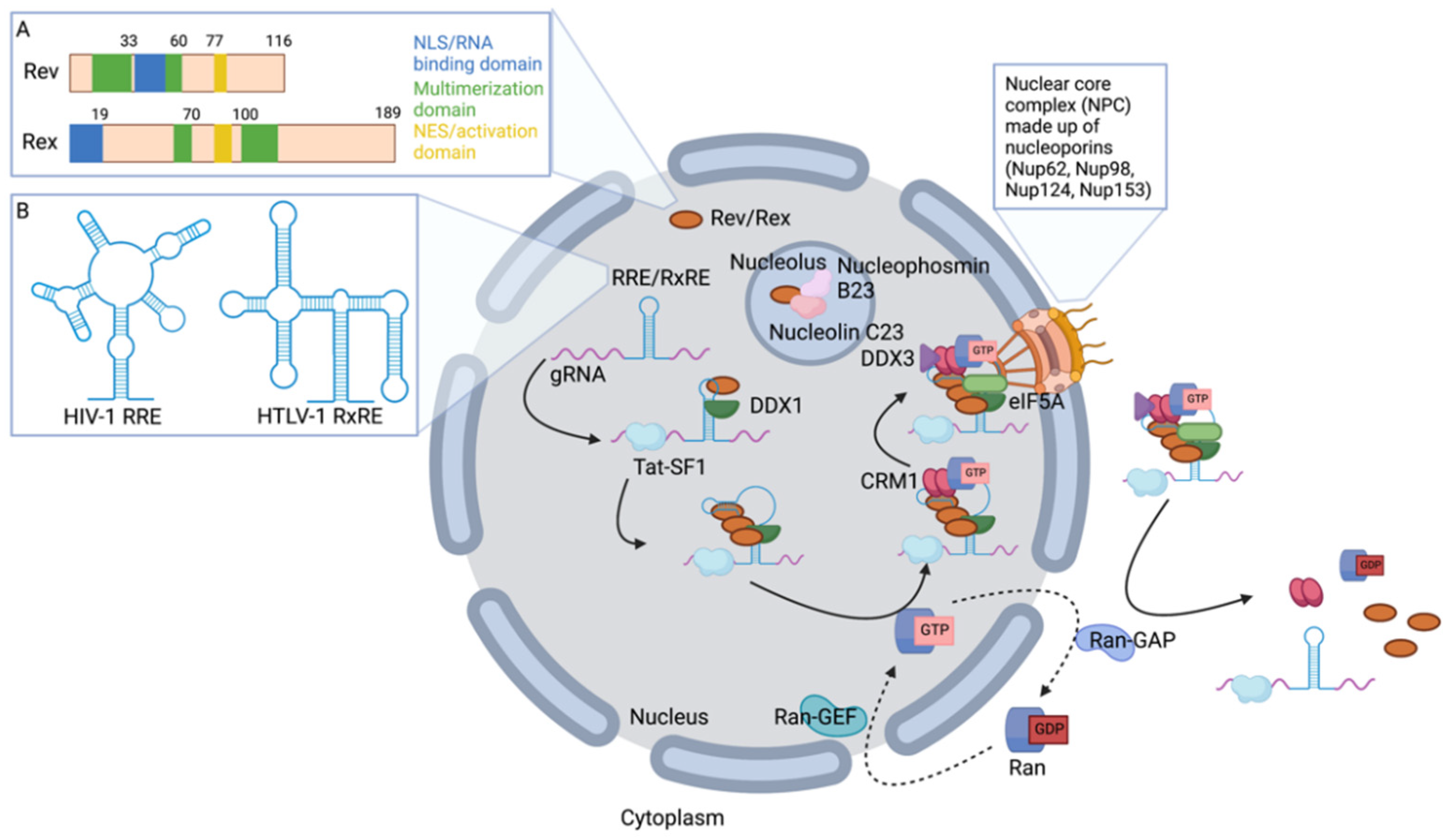
2. Genomic RNA (gRNA) Nuclear Export

In order to be packaged at the plasma membrane into virus particles, the unspliced retroviral gRNA must be exported from the nucleus despite not being recognized by standard RNA nuclear export machinery. Two cellular pathways have been identified to be hijacked by retroviruses to export unspliced and partially spliced viral RNAs from the nucleus. Some retroviruses utilize the TAP/NXF1 pathway, while other retroviruses have been found to use the CRM1/XPO1 nuclear export receptor. For human retroviruses, HIV-1, HIV-2 and HTLV-1, gRNA nuclear export utilizes the CRM1 pathway [3,4,5,6,7,8,9,10,11,12]. These complex human retroviruses encode accessory proteins that interface with the CRM1 export machinery. HIV encodes the accessory protein Rev, which mediates post-transcriptional gene expression [13,14,15]. HTLV-1 encodes for a homologous protein, Rex, that is required for post-transcriptional viral gene expression [16]. Both Rev and Rex permit retroviral gene expression by directly mediating the export of unspliced or partially spliced viral RNAs [15,17,18,19,20], therefore indirectly contributing to gRNA packaging.

2.1. Retroviral Rev and Rex Accessory Proteins Shuttle between the Nucleus and Cytoplasm

Rev and Rex are nucleocytoplasmic shuttle proteins. The shuttle function of the Rev and Rex proteins is mediated by a nuclear localization signal (NLS) and a nuclear export signal (NES) that is encoded within each protein; the NES is necessary for retroviral gRNA nuclear export [21,22,23]. The nuclear import of Rev and Rex allows for the interaction of these proteins with their respective unspliced gRNA in the nucleus. Rev nuclear import alone is not sufficient for HIV gRNA nuclear export, and retroviral gRNA nuclear export also depends on the nucleolar localization of the Rev [24,25] and Rex proteins [26] (Figure 1). Basic residues in the Rev and Rex RNA-binding domains mediate nucleolar localization [23,27,28]. While the significance of Rev and Rex nucleolar localization in facilitating gRNA nuclear export remains poorly understood, it is thought that nucleolar localization allows Rev/Rex interaction with host cell proteins involved in nuclear export of unspliced retroviral RNAs. For example, a recent study employing immunoprecipitation and mass spectrometry identified several nucleolar factors that interact with Rev only when Rev nucleolar localization sequences remain intact [29] (Figure 1). This observation suggests that proteins including nucleophosmin B23, nucleolin C23, and various cellular splicing factors interact with HIV-1 Rev in nucleoli and may impact gRNA nuclear export.
NES-mediated nuclear export of Rev (HIV-1, HIV-2) and Rex (HTLV-1) drives the export of retroviral RNAs. The NES is located within the activation domain of Rev and Rex proteins and contains numerous leucine residues associated with the nuclear export function [21,23,27,30,31]. Several studies have demonstrated that swapping homologous domains between Rev and Rex can result in the export of HIV or HTLV gRNAs [32,33,34,35,36]. These functional chimeras suggest a partially conserved mechanism of gRNA export. The observation of nonfunctional swaps between Rev and Rex nucleolar-targeting sequences [36] underscores potential differences in their mechanism of gRNA nuclear export. Investigation of these differences will help delineate the mechanism(s) of unspliced gRNA nuclear export.

2.2. Rev and Rex Interact Directly with gRNA

Retroviral accessory protein-mediated export of unspliced gRNAs occurs through direct RNA-protein interactions that form the export complex, and arise cotranscriptionally, before spliceosome activation [37]. The timing of complex formation allows for export of unspliced retroviral gRNA before it is processed. In the case of HIV-1, Rev specifically interacts with the RRE in the HIV-1 gRNA [38,39,40]. The HIV-1 RRE is a 530 base-pair sequence located in the env region of the genome and is retained in unspliced and partially spliced viral RNA species. The RRE has a complex RNA secondary structure comprised of a central stem and 5 stem-loop structures [17,41,42] (Figure 1). Several reports have indicated the requirement of the RRE secondary structure for Rev binding and have shown that the initial Rev-RRE interaction occurs at a high affinity binding site, i.e., stem-loop IIB [43,44,45,46]. Rev binds the RRE through an arginine-rich RNA binding domain contained within an alpha-helical structure [24,47,48,49,50]. This interaction between Rev and RRE is the foundation of the nuclear export complex.
Interactions between viral accessory proteins mediating nuclear export and the corresponding RNA sequence element are similar among the human retroviruses. The HIV-2 genome also contains a RRE (RRE-2) located in the env region of the genome that has complex secondary structure [42,51,52,53]. Like Rev, Rex also interacts with a specific RNA sequence to permit nuclear export, i.e., the RxRE [54,55,56,57]. Like the RRE, the RxRE has complex secondary structure, specifically it is made up of a long stem and four stem-loop structures [55,58,59] (Figure 1). The role of the stem-loop structures of the RxRE has been investigated in regards to their importance in RNA export efficiency [4,60]. For Rex, residues similar to those involved in Rev-RRE binding are required for RxRE binding. The RNA-binding domain of Rex is rich in basic residues, and specific residues important for binding have been mapped [32,57,61]. The interaction of HIV-2 Rev and HTLV-1 Rex with RRE-2 or RxRE containing RNAs, respectively, have not been studied as extensively as the interaction of HIV-1 Rev and RRE-1.
These similarities between Rev and Rex, and their corresponding target sequences, imply that they mediate nuclear export through a conserved mechanism. Rex has been shown to export RNAs containing RRE-1 and RRE-2 from the nucleus, but Rev cannot function in the export of RxRE-containing RNAs [62,63,64,65]. Some conservation of function has also been observed between HIV Rev and RRE sequences. For example, HIV-1 Rev can export unspliced viral RNAs containing the RRE-2, but HIV-2 Rev cannot mediate export of RRE-1 containing RNAs [52,64,65,66,67]. Further comparative analysis between human retroviruses that explore the incomplete conservation of Rev and Rex function is likely to reveal further details on the mechanism(s) of retroviral gRNA nuclear export.
The Rev-RRE interaction induces structural changes of RRE-containing RNAs that promote nuclear export. Specifically, initial Rev binding results in changes in the stem-loop structures of RRE-containing RNAs that allows for further Rev binding [68]. Sequential, cooperative binding of Rev to adjacent low affinity binding sites along the RRE results in Rev oligomerization [47,69,70,71,72,73] (Figure 1). Dissociation constants have been reported for Rev-RRE binding as 0.26–33 nM for Rev monomers and 0.77–92 nM for Rev dimers [71,72]. Up to 6–8 Rev molecules have been reported to bind the RRE in vitro [73]. Several studies have reported mutant Rev proteins that prevent oligomerization and do not bind RRE-containing RNAs, which impedes gRNA nuclear export [47,69,74,75,76]. This demonstrates the importance of Rev oligomerization in retroviral gRNA nuclear export and indicates the requirement of both protein-protein and protein-RNA interactions in this process. While Rev oligomerization is mediated by cooperative binding to RNA, residues of the arginine-rich RNA binding domain are not implicated in Rev oligomerization. In contrast, residues proximal to this region that comprise a hydrophobic patch on the surface of Rev are necessary for oligomerization [5,69,75,77]. Rev oligomerization is structurally linked to the specificity of the Rev-RRE interaction. This conclusion is supported by the solved structure of a Rev dimer that can bind the RRE, and by the extent to which HIV-1 and HIV-2 Rev can function with the opposite RRE—i.e., HIV-1 Rev can export HIV-2 gRNA, but HIV-2 Rev cannot export HIV-1 gRNA [66,78,79]. Rex oligomerization is required for export of HTLV-1 gRNA [80]. However, the HTLV-1 gRNA export mechanism is not as completely characterized as Rev oligomerization. Rev and Rex oligomerization are required for interaction with the CRM1 nuclear export pathway.

2.3. Host Proteins Involved in gRNA Nuclear Export

Cellular proteins have been shown to be involved in Rev binding to the RRE. CRM1 is a major nuclear export receptor that interfaces with the Rev-RRE complex and permits incompletely spliced RNA export from the nucleus. CRM1 interacts directly with Rev as a dimer [81] (Figure 1 and Table 1), and Rev serves as an adaptor to the cellular pathways that are hijacked by retroviruses to export gRNA. CRM1-mediated export of gRNA requires RanGTP interaction with the Rev-CRM1 complex [7,82]. Ran proteins are small G proteins that mediate translocation of cellular proteins and RNA through the nuclear pore complex (NPC). Ran proteins bind GTP in the nucleus and GDP in the cytoplasm, due to Ran GAPs that are primarily localized to the cytoplasm and Ran GEFs that are localized in the nucleus. RanGTP-bound CRM1 directs the Rev-RRE ribonucleoprotein complex out of the nucleus through direct interaction with the mRNA translocation machinery located at the nuclear envelope [3] (Figure 1 and Table 1). The GTP hydrolysis drives export of the viral gRNA utilizing the Ran cycle. The RanGTP hydrolysis in the cytoplasm dissociates the export complex.
DEAD box helicase (DDX) proteins interact with RRE-Rev-CRM1 complex to promote nuclear export (Figure 1 and Table 1). The DDX1 protein acts as a clamp on the RNA to nucleate oligomerization by promoting binding of the first Rev monomer to the RRE [83,84]. A recent study has further clarified the role of DDX1 in nucleating Rev oligomerization by showing that despite tighter binding to Rev, DDX1 nucleation of oligomerization is mediated through interaction with the RRE [84]. In particular, this study showed that a DDX1 mutant that cannot bind Rev can still nucleate oligomerization, and that DDX1 mutations that reduced the ability to bind RNA disrupted the ability of DDX1 to promote the first Rev monomer binding to gRNA [84]. The interaction of DDX1 with RRE RNA results in a structural change of the RRE RNA at stem IIB that enhances Rev binding and oligomerization [85]. DDX1 is hypothesized to either promote this structural change and/or stabilize it. DDX3 has been hypothesized to restructure retroviral gRNA in order to make it amenable to translocation through the nuclear pore by interaction with CRM1 [86,87] (Figure 1 and Table 1). The contribution of DDX1 and DDX3 is complicated by the conservation of other DEAD-box helicases which could permit interaction with the RRE. It is plausible that this effect could be the result of several DEAD-box helicases.
Additional cellular proteins interact with the Rev-CRM1 complex and have been shown to contribute to export of retroviral RNAs. Phosphofurin acid cluster sorting protein 1 (PACS1), which localizes furin to the trans-Golgi network, can also shuttle between the nucleus and cytoplasm, can associate with Rev and CRM1, and can contribute to nuclear export of viral transcripts (Table 1) [88]. Moreover, PACS1-mediated export of viral transcripts was discovered to be due to interaction with the Rev-CRM1 pathway [88]. Other proteins, such as acidic leucine-rich nuclear phosphoprotein 32 family member A and B (i.e., ANP32A, ANP32B), mediate the export of unspliced or partially-spliced viral mRNA via interactions with Rev and CRM1 (Table 1) [89]. In particular, a double knockout of ANP32A and ANP32B led to a significant decrease in Gag expression and a dramatic accumulation of unspliced viral mRNAs in the nucleus, while reconstitution of either ANP32A or ANP32B restored virus replication [89].
The Rev-RRE-CRM1 nuclear export complex interacts with the translocation machinery which is comprised of various cellular proteins. Some aspects of the NPC implicated in HIV-1 gRNA export have been characterized. Nup98, a nucleoporin, has been shown to interact with Rev, which implies it is likely part of the NPC responsible for Rev-mediated RNA export (Table 1) [90]. Other nucleoporins implicated in retroviral gRNA nuclear export include Nup124, Nup153, Nup98, and Nup62 (Table 1) [91]. Interaction of the Rev-RRE-CRM1 complex with the NPC is mediated by eIF5A [8,91,92] (Figure 1 and Table 1). It is known that eIF5A is essential for directing the retroviral export complex to the translocation machinery. This observation was made in cell-free studies, and it is possible that several DDX proteins may be contributing to restructuring of the retroviral gRNA that promotes nuclear export in cells. Part of the translocation machinery involved in export of HIV and HTLV gRNAs include nucleoporin-like protein Rab/hRIP, which interacts with CRM1 (Table 1) [31,93,94,95]. The specific function of Rab/hRIP in the nuclear export machinery of retroviral gRNA is not fully understood. Collectively, the interactions between cellular proteins and the export complex result in the transport of Rev and Rex with RRE- or RxRE-containing RNA out of the nucleus as a stable complex with CRM1. Studies with HIV-1 have provided much of the current understanding of the role of CRM1 and other associated cellular proteins in viral RNA transport. The previously described similarities in the viral components of nuclear export suggest potential overlap in the mechanisms of nuclear export among the human retroviruses. Comparable studies with HIV-2 and HTLV-1 regarding the involvement of other components of the translocation machinery are lacking.
Other cellular proteins that have been shown to be involved in the export of HIV-1 gRNA include UPF1, MOV10, Staufen2 and Tat-SF1. Host upframeshift protein 1 (UPF1) regulation of viral RNA nuclear export relies on the nucleocytoplasmic shuttling of UPF1 [96]. UPF1 exists in two essential viral ribonucleoprotein (RNP) complexes during the late phase of HIV-1 replication. The first type of RNP is nuclear export RNPs that contain Rev, CRM1, DDX3, and the nucleoporin p62; the second type of RNA is cytoplasmic RNPs that exclude these nuclear export markers but contain Gag (Table 1) [96]. UPF2 was found to be excluded from the UPF1-Rev-CRM1-DDX3 complex and was identified as a negative regulator of viral RNA nuclear export [96]. The RNA helicase MOV10, a member of the UPF1-like superfamily, was found to facilitate Rev/RRE-dependent nuclear export of viral RNAs, as a co-factor of HIV-1 Rev (Table 1) [97]. MOV10 interacts with Rev in an RNA-independent manner as determined by co-immunoprecipitation analysis [97]. The DEAG (Asp-Glu-Ala-Gly) box of MOV10 is required to enhance Rev/RRE-dependent nuclear export [97]. Staufen2 positively regulates Rev nuclear export, which is predicted to contribute to gRNA nuclear export [98]. Lastly, human Tat-specific factor 1 (Tat-SF1) was found to bind the HIV-1 genome at the trans-acting response (TAR) sequence and selectively transport HIV-1 RNAs by facilitating the nuclear export of unspliced gRNAs, while retaining singly spliced viral RNAs in the nucleus (Table 1) [99]. Cell-free studies have revealed some details of the Tat-SF1 interaction with the HIV-1 genome [99]. In particular, Tat-SF1 forms a complex with the TAR RNA independent of Tat. Tat-SF1 interacts with at least one additional location in the 5′ end of the HIV-1 gRNA, and this interaction is enhanced by Tat [99]. Taken together, these findings suggest that nuclear export of retroviral gRNA is a highly regulated process requiring several other cellular proteins. Future studies into how these cellular proteins interface to coordinate retroviral gRNA nuclear export will provide a more complete understanding of the mechanism(s) of RNA export. Such studies have yet to be fully extended to other human retroviruses.
A role for Gag in retroviral gRNA nuclear export has been suggested. For example, several retroviral Gag proteins, including those of Rous sarcoma virus (RSV) and HIV-1, have been observed in the nucleus, though the nuclear roles of Gag are not fully characterized. The RSV Gag protein interacts with unspliced viral RNA in the nuclei of infected cells to form viral RNPs [100]. RSV viral RNPs have been implicated in genomic RNA packaging [100]. HIV-1 Gag was also discovered to form viral RNP complexes with unspliced viral RNA at transcription sites [101]. The interaction of nuclear HIV-1 Gag with unspliced gRNA is specific and has been visualized in discrete foci in cell nuclei. Three-dimensional imaging analysis has revealed that HIV-1 Gag was localized to the perichromatin space and associated with both unspliced gRNA and Rev in a tripartite RNP complex. This observation supports a model where Gag interacts with newly transcribed gRNA to form nuclear RNP complexes which may aid in export and/or packaging.

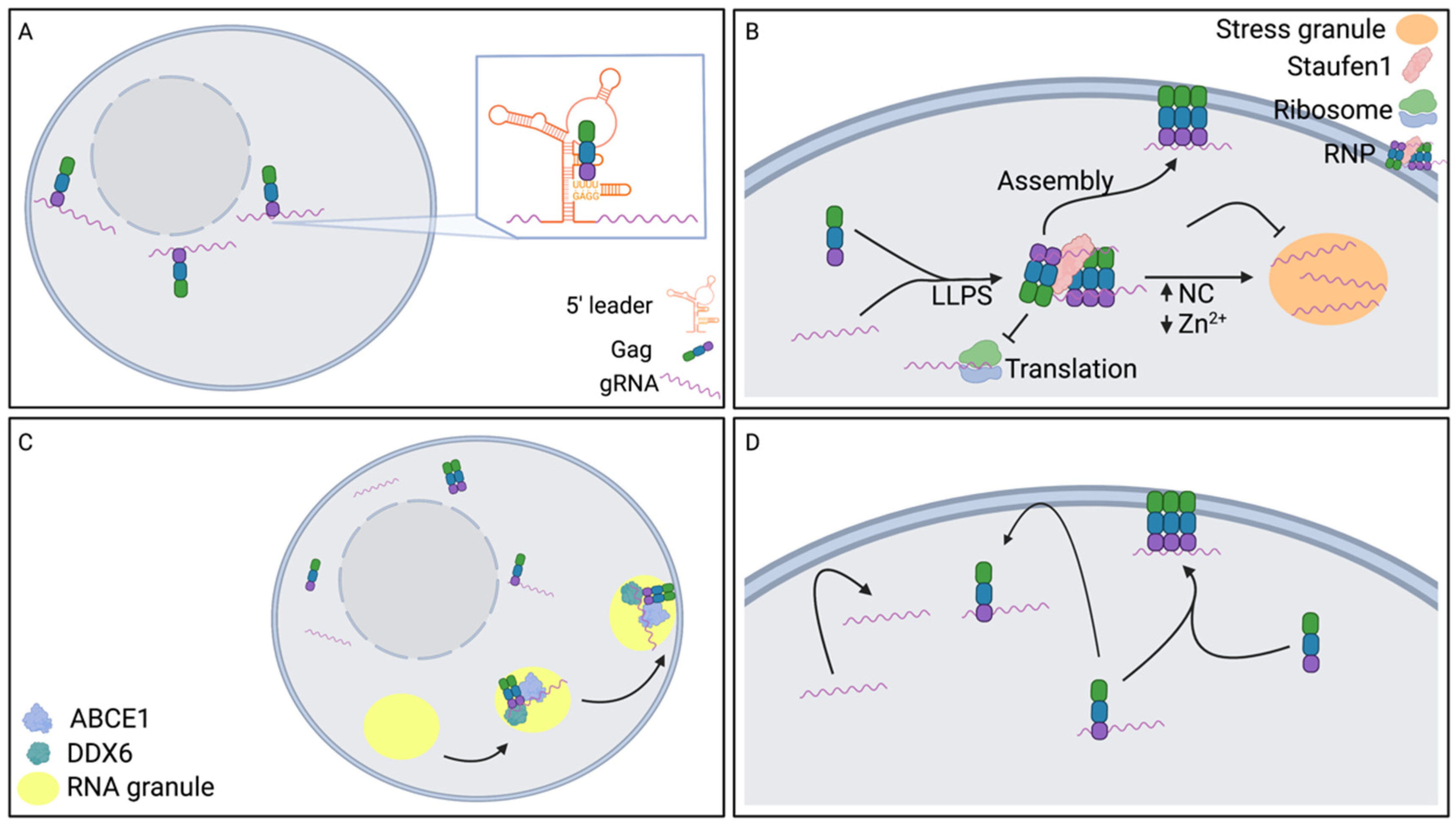
3. Translocation of gRNA to the Plasma Membrane

After the gRNA is exported out of the nucleus, the gRNA translocates through the cytoplasm to sites of virus particle assembly at the plasma membrane. One study reported tracking many individual HIV-1 gRNA molecules in the cytoplasm, and found most gRNAs moved in a manner that was ‘nondirectional’ and ‘random walk-like’, indicating that the HIV-1 gRNA moves by diffusion throughout the cytoplasm [102]. This type of gRNA movement is observed whether HIV-1 Gag is present or not [102]. Diffusive movement implies that Gag does not guide the movement of HIV-1 gRNA in a directed manner nor do cellular proteins. Furthermore, the observation of single HIV-1 gRNA molecules over time has revealed that at different time points, a given HIV-1 gRNA molecule could move at various speeds [102]. This change in gRNA movement implies that the local gRNA environment dictates gRNA behavior. These data, taken together, suggest that retroviral gRNA is not directly transported to virus assembly sites by a single mechanism and that several host cell components locally contribute to gRNA translocation to virus assembly sites. While the studies described in this review suggest various ways in which viral or host cell components may contribute to gRNA translocation to virus assembly sites, a cohesive model for retroviral gRNA translocation to assembly sites is lacking. The importance of all these factors in the context of retroviral infection events in infected individuals is unclear.

3.1. Influence of Nuclear Export Pathway on gRNA Translocation to the Plasma Membrane

Components of the nuclear export machinery have been implicated to influence gRNA translocation to virus particle assembly sites. For instance, changing the RNA export pathway from RRE-dependent trafficking via CRM1 to constitutive transport element (CTE)-dependent export via NXF1 was necessary for HIV-1 replication in murine cells [103]. The requirement of a specific nuclear export pathway in murine cells would imply a link between that pathway and gRNA translocation to virus assembly sites. In a system with HIV-1 gRNAs exported by different nuclear export pathways (i.e., CRM1 or NXF1), the gRNAs exported by different pathways were not copackaged [104]. Comparing retroviral gRNA exported by different pathways can provide further insights into potential linkage(s) of these processes. For example, HIV-1 gRNAs exported by the CRM1 or NXF1 pathway were found to have diffusive movement in the cytoplasm, while CTE-containing RNAs diffuse more slowly than RRE-containing RNAs [105]. While the biology causing observed differences in diffusion rates is unknown, one possible explanation is that RNAs exported by different pathways are associated with different proteins that alter diffusion rates to varying extents. In contrast, other studies using an HIV-1 RRE-containing RNA observed export to the cytoplasm that occurred in short bursts, while a CTE-containing Mason-Pfizer monkey virus (MPMV) gRNA, viral RNA export was observed to be continuous [106]. The subcellular localization of viral RNAs also differs depending upon the mode of RNA export, with HIV-1 RRE RNAs being exported throughout the cytoplasm with no specific localization, while MPMV gRNAs are specifically exported to the microtubule organizing center (MTOC) [106]. These observed differences in gRNA localization were reversed when the HIV-1 gRNA export pathway was reversed by replacement of the RRE with the CTE [106]. Taken together, the nuclear export pathway could, to some extent, impact viral gRNA localization. For example, components of either system could serve as a signal for a particular cytoplasmic fate. However, one study reported that changes in HIV-1 gRNA localization due to the nuclear export pathway do not change gRNA localization and that the HIV-1 gRNA was not targeted to centrosomes [105]. The differences in reported observations may involve the use of proviruses to express viral components versus transfected DNAs. To date, the precise role of nuclear export in retroviral gRNA translocation to virus particle assembly sites remains poorly understood for HIV-1, let alone HIV-2 and HTLV-1.

3.2. Influence of Gag on gRNA Translocation to the Plasma Membrane

Gag plays a major role in specific gRNA packaging; therefore, it is plausible that Gag may influence gRNA translocation to assembly sites. Genome recognition of gRNA by Gag represents the initial RNA-protein interaction. This process involves HIV-1 Gag specifically interacting with the gRNA in the 5′ region of the genome [107,108]. One study found that HIV-1 Gag and the gRNA colocalized at areas surrounding the nuclear envelope at early time points after the appearance of Gag expression, where this colocalization was shown to be driven by Gag [109,110]. These data, taken together, imply that HIV-1 genome recognition occurs in the perinuclear region of the cell (Figure 2A). However, it is technically challenging to detect single Gag molecules, and therefore the first Gag and gRNA interaction has not been observed in cells, though cell-free studies have detailed HIV-1 genome recognition. For example, a recent study demonstrated that out of more than two dozen binding sites in the 5′ leader sequence of the HIV-1 gRNA, the initial Gag-RNA contact determined by assessing NC binding to a portion of the 5′ leader sequence, occurred at high-affinity binding sites with the UUUU:GGAG motif [111] (Figure 2A). Initial binding of Gag to gRNA is dependent on the structural lability of this motif [111]. Gaining greater insights into the subcellular location of genome recognition will provide new details regarding the role of Gag in gRNA translocation to the plasma membrane.
Recent studies using transmission electron microscopy (TEM) to study HIV-1 Gag and gRNA localization at high resolution revealed colocalization mainly in the cytoplasm with few instances of colocalization in the nucleus [112]. The site of genome recognition was not defined, and clustering of HIV-1 Gag and gRNA in the cytoplasm was dependent on regions of Gag and gRNA previously reported to be involved in Gag and gRNA interactions [112]. Furthermore, amino acid substitutions at residues shown to reduce gRNA packaging were observed to also reduce Gag and gRNA clustering, implying that Gag and gRNA colocalization in the cytoplasm is important for genome recognition [112]. The relatively high resolution of the TEM approach allowed for measurements of the distance between Gag and gRNA and revealed that the distance between Gag and gRNA was reduced over time. This suggests that Gag and gRNA condense in the cytoplasm [112]. Additional high-resolution studies of Gag and gRNA in the cytoplasm have increased our understanding of translocation of gRNA to the plasma membrane. For example, one study that utilized 3D-super-resolution structured illumination microscopy detected gRNA dimers in the cytoplasm and found that Gag is involved in gRNA dimerization in the cytoplasm [113]. These results help to support the conclusion that Gag is involved in targeting gRNA dimers to the plasma membrane [113]. This study and another reported that Gag interaction with gRNA in the cytoplasm and translocation to the membrane is dependent on the NC domain of Gag and more specifically the zinc-coordinating finger (ZnF) domains [113,114]. Another study supports the model that Gag-gRNA interactions in the cytoplasm promote assembly given that reduction in Gag oligomerization in the cytoplasm occurred when the NC domain was mutated, and Gag-gRNA binding was inhibited [115].
Phase separation of Gag and gRNA in the cytoplasm has recently been reported, where the ZnF domains of HIV-1 Gag NC promote gRNA liquid-liquid phase separation (LLPS) into RNP complexes [116]. These RNPs were theorized to stimulate virus assembly by inhibiting translation and promoting formation of assembly intermediates (Figure 2B). HIV-2 and HTLV-1 Gag may create similar complexes as their respective NC domains also have the ability to phase separate in a Zn2+ dependent manner [116]. This study showed that Zn2+ concentration can regulate RNP formation through experiments in which high levels of HIV-1 NC resulted in a switch from RNP formation to Zn2+ dependent stress granule formation, and in which Zn2+ depletion led to gRNA localization to stress granules or retention in the nucleus [116] (Figure 2B). The switch between RNP and stress granule phase separation suggests a potential mechanism that indirectly regulates gRNA trafficking to virus assembly sites controlled by the NC domain of Gag and Zn2+. The level of Gag expression and Zn2+ containing NC maintains Gag and gRNA RNPs that promote translocation to assembly sites which likely occurs through assembly intermediates (Figure 2B). A key cellular protein, Staufen-1, plays a role in this switch mechanism. Staufen-1 has been shown to associate with HIV-1 gRNA and Gag RNPs as well as promote assembly and gRNA packaging [117,118]. Furthermore, Staufen-1 has been shown to have a role in dissociating HIV-1 stress granules and specifically preventing gRNA localization to stress granules [119] (Figure 2B). HIV-2 gRNA has also been shown to phase separate in RNPs and stress granules. However, the composition of the RNPs and the regulation of phase separation differ from that of HIV-1. While HIV-1 abrogates stress granule formation, HIV-2 infection has been shown to induce stress granule formation. HIV-2 gRNA phase separates into RNPs with a stress granule assembly protein, TIAR, that is found in the cytoplasm or in stress granules [120]. Phase separation of TIAR with HIV-2 gRNA was not dependent on the interaction with Gag, but increased Gag expression promoted stress granule formation [120]. The correlation between a threshold level of Gag expression and gRNA localization to stress granules suggests this may regulate the switch from translation to assembly, and the switch could promote gRNA translocation to assembly sites.
Similar to Gag and gRNA phase separation, Gag and gRNA colocalization in assembly intermediates suggests specific trafficking mechanisms of gRNA prior to localization to assembly sites at the plasma membrane. The HIV-1 gRNA has been reported to colocalize with Gag in complexes larger than 30S, such as Gag assembly intermediates [121]. In particular, HIV-1 gRNA was detected with Gag in an assembly intermediate derived from host RNA granules that contain two cellular proteins that facilitate assembly, ABCE1 and DDX6, an RNA granule protein [121] (Figure 2C). The localization of gRNA to Gag assembly intermediates may suggest a potential gRNA translocation mechanism in which after incorporation to assembly intermediates, the gRNA is translocated to plasma membrane assembly sites by the assembly intermediates.
Behavior of RNA near the plasma membrane can also provide insights into the mechanism of translocation to assembly sites. The HIV-1 gRNA can be localized to the plasma membrane in the absence of Gag and is only transiently near the membrane [107,122] (Figure 2D). The retention of gRNA at the plasma membrane requires Gag and Gag-gRNA interactions via the RNA packaging signal [107,110,122] (Figure 2D). Not all RNAs localized near the plasma membrane are packaged into particles [122]. As Gag expression level increases, the frequency of RNA packaging also increased [122], suggesting that Gag likely plays an important role late in the translocation to a virus assembly site. Gag membrane binding and multimerization are required to retain gRNA at the membrane [107]. It is believed that Gag oligomers can readily recruit gRNA to the plasma membrane for gRNA packaging [107] (Figure 2D). It remains unclear how all the observations described above contribute to gRNA translocation to virus assembly sites in the formation of infectious virus particles.
At the plasma membrane, the gRNA promotes particle assembly. Not only can the HIV-1 gRNA stimulate particle assembly, but the gRNA can stimulate particle production by specific Gag-gRNA interactions [123,124,125] and highlights the importance of specific genome recognition in virus particle production [125]. This further supports studies that have shown that RNA incorporation in virus particles is necessary for virion integrity [126,127,128] and that specific interactions between dimeric HIV-1 gRNA and Gag act to nucleate the process of HIV-1 particle assembly. Retroviral gRNA is particularly important for nucleation at low Gag levels in cells [129], which is thought to be driven by the decrease in activation energy needed for Gag interactions with the gRNA packaging signal that allow for selective packaging.

3.3. Role of Microtubule Organizing Center (MTOC) on gRNA Translocation to the Plasma Membrane

HIV-1 gRNA sequences suggest a role for a cellular protein in translocating gRNA via the MTOC. For example, the HIV-1 gRNA contains two sequences predicted to be compatible for interaction with host protein hnRNP A2, similar to the hnRNP A2 response element (A2RE) found in the human genome [130]. The A2RE-1 site in the HIV-1 gRNA is located in the gag gene, while the A2RE-2 site is located in the overlap between the tat and vpr genes [130]. These sequences are involved in RNA transport in oligodendrocytes [130]. However, the extent of the function of the A2RE-like sequences in the HIV-1 genome is poorly understood. Depletion of hnRNP A2 in HIV-1 expressing cells was found to result in HIV-1 gRNA accumulation at the MTOC [131]. Other studies have found HIV-1 Gag and gRNA colocalization at the MTOC [109]. The accumulation at the MTOC in the absence of hnRNP A2 implies that hnRNP A2 may aid in translocating gRNA to virus assembly sites along microtubules. Alternatively, translocation to the MTOC could provide an opportunity for interaction with other proteins that coordinate trafficking to assembly sites and not via microtubules. Treatment of cells with microtubule depolymerizing drugs was observed to not alter the random-walk like motion of HIV-1 gRNA in the cytoplasm [102], suggesting that the role of hnRNP A2 may be in promoting colocalization with other proteins that permit trafficking to the plasma membrane. No reports of hnRNP A2 in gRNA translocation have been reported for HIV-2 or HTLV-1. The function of gRNA localization to the MTOC remains unclear.

3.4. Role of Endosomal Vesicles on gRNA Translocation to the Plasma Membrane

Endosomal vesicles are another component of the cell that have been reported to influence retroviral gRNA translocation. In a Gag dependent manner, HIV-1 gRNA has been reported to be transported via endosomal vesicles, where the RNA displayed directed movement instead of diffusive cytoplasmic movement for a minority of the gRNA molecules that were tracked [102,132]. It has also been suggested that Gag and gRNA may be transported to the perinuclear region by late endosomes in a dynein dependent manner [133]. This localization and transport via endosomes were not the result of endocytosis of either gRNA or Gag at the plasma membrane [133], suggesting a specific population of gRNA and Gag associates with late endosomes and is packaged into virus particles.

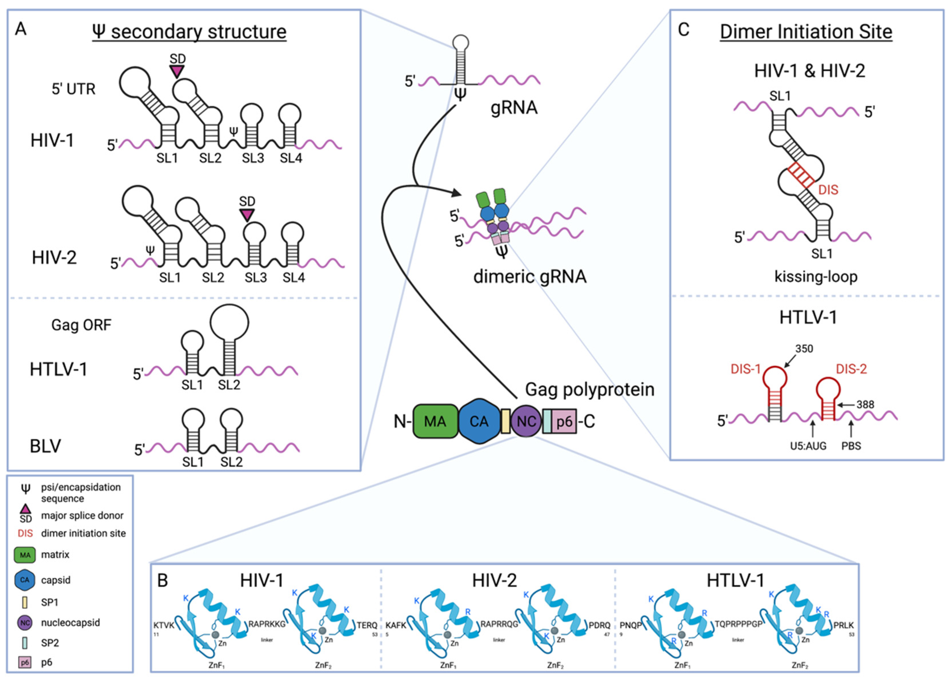
4. Packaging of Human Retroviral gRNA

Retroviral gRNA is specifically packaged as a noncovalently linked dimer. Compared to spliced viral RNAs, HIV-1 gRNA is enriched in virions by a factor of 20 [134]. It has been reported that dimerized gRNA is the RNA unit recognized for gRNA packaging [135,136]. This is supported by studies that show that mutations that promote dimerization between RNAs promote the packaging of those RNAs [135,137]. Inversely, it has been reported that gRNA mutations that prevent dimerization will inhibit gRNA packaging [138,139]. However, one study that made different mutations to prevent dimerization did not find a reduction in packaging, while another study uncoupled gRNA dimerization and packaging and observed robust packaging of gRNA monomers [140,141]. The apparent discrepancies are likely due to the use of different dimerization mutants. For HTLV-1 mutations that disrupted HTLV-1 gRNA dimerization in vitro had no effect on gRNA packaging into released particles [142].
Two key interactions are required for specific packaging of dimerized gRNA: 1. A high-affinity protein-RNA interaction between Gag and gRNA and 2. A specific interaction of structural motifs in two gRNA molecules (dimerization).

4.1. gRNA Sequences Involved in the Gag-gRNA Interaction

In order to package retroviral gRNA, the Gag protein interacts with a specific cis-acting element of the gRNA. This element has been referred to by different names, including the RNA packaging signal (psi) or the RNA encapsidation signal (E). Interaction of Gag with the RNA packaging signal promotes selective packaging of retroviral gRNA over other virus-derived RNAs and cellular RNAs [126,129,143,144]. The RNA packaging signal for retroviruses is comprised of a complex secondary (and tertiary) structure commonly involving several RNA stem-loops. This RNA packaging signal is conserved on a structural level [145,146]. The RNA secondary structure of the stem-loops is important for gRNA packaging [141]. It has been hypothesized that selective packaging by the RNA packaging signal is due to Gag nucleation occurring at a faster rate on these sequences.
Retroviral RNA packaging sequences are located in the 5′ leader sequence of the gRNA. The HIV-1 RNA packaging signal is located in the 5′ UTR after the major splice donor site and before the gag gene (Figure 3A) [134,147,148,149,150,151]. Because the sequences necessary for packaging are downstream of the splice donor site these sequences are only in unspliced RNA molecules; only the gRNA is specifically packaged over other RNAs. For HIV-2, the key sequences required for RNA packaging are located upstream of the major splice site (Figure 3A) [152,153]. Sequences between the major splice donor and the beginning of the Gag ORF have also been shown to play a role in HIV-2 gRNA packaging, but the effect on packaging when these sequences are deleted or mutated is less than when the sequences upstream of the splice donor are mutated [152,154]. The sequences downstream of the splice donor site have been reported to bind to HIV-2 Gag [155]. This RNA-protein interaction likely permits selectivity between spliced RNAs and the unspliced gRNA that is not encoded solely by sequences upstream of the splice donor site. The differences in the location of RNA packaging sequences in HIV-1 and HIV-2 relative to the splice donor site suggest potential differences in the mechanistic details of gRNA packaging, which could have implications for particle assembly and infectivity.
For HIV-1 and HIV-2, the RNA packaging signal contains four stem-loop structures (i.e., SL1, SL2, SL3, and SL4). These structures are the key sites for Gag-gRNA interactions [156,157]. All 4 stem-loops can independently bind Gag, and all four of the stem-loops have been shown to be important for RNA packaging [158,159,160,161,162,163]. Evidence supports the conclusion that SL3 has the most significant contribution to gRNA packaging, followed next by SL1 [108,134,160,164,165,166,167]. In particular, a 110 nt sequence in the gRNA from position 227–337 represents the core Gag binding domain [168]. The stem-loop structure of this region is mediated by a long-range interaction between a CU-rich region and nucleotides proximal to the AUG start codon [168]. In addition to RNA stem-loop structures, it has more recently become apparent that unpaired guanosine bases in the packaging signal contribute to specific Gag binding of gRNA, perhaps even more than SL1 and SL3 for HIV-1 as determined in cell-free studies using a shortened RNA containing the packaging signal [129,169]. Similar observations have been made for unpaired guanosine nucleotides in the full-length genome [161]. Unpaired guanosine nucleotides are also present at several locations in the packaging signal that have previously been identified to be primary HIV-1 Gag binding sites [161]. Mutation of a few unpaired guanosine nucleotides in one Gag binding site was found to cause a minor defect in gRNA packaging, but the combination of mutations at unpaired guanosine nucleotides of multiple Gag binding sites resulted in a synergistic reduction in gRNA packaging [163]. This suggests that multiple Gag molecules binding to gRNA are required for packaging. Furthermore, these observations imply functional redundancy in the RNA packaging mechanism. Unpaired guanosines in the HIV-2 RNA packaging signal have also been implicated in gRNA packaging [170], which suggests conservation of the RNA packaging mechanism. Other nucleotides have been identified that play a more secondary role in specific gRNA packaging, as they are not Gag binding sites. For the HIV-1 RNA packaging signal, the nucleotides at positions 226 and 227 have been reported to promote gRNA packaging efficiency by regulating RNA secondary structure of the packaging signal [171].
The HTLV-1 packaging signal has not been as extensively characterized. Bovine leukemia virus (BLV), a closely related deltaretrovirus, has provided insight into 5′ leader sequences involved in HTLV-1 RNA packaging. Two RNA stem-loop structures in the HTLV-1 gag gene that are homologous to stem-loops in BLV that were shown to be involved in gRNA packaging were able to be substituted in the BLV genome and allowed for RNA packaging (Figure 3A) [172]. This implies that these stem-loop structures are likely involved in HTLV-1 gRNA packaging. The contributions of other sequences involved in gRNA packaging for HTLV-1 such as those in the 5′ UTR are not well characterized.

4.2. Gag NC Determinants of Gag-gRNA Interaction

The RNA-protein interaction mediating packaging of gRNA is known to involve the NC domain of Gag. The NC domain of HIV-1 and HIV-2 Gag has been shown to bind the viral gRNA (Figure 3) [125,126,127,128,129,130,131,132,133,134]. The specific motifs in NC important for interaction with the gRNA include two ZnFs, each of which is formed by a cysteine-histidine (Cys-His) box, (Figure 3B) [144,158,169,173,174,175,176]. These similar yet distinct ZnFs are thought to contribute differently to viral gRNA packaging. While both are required, the N-terminal ZnF is thought to be more dominant and the two ZnFs are known to not be interchangeable [158,177,178,179,180,181]. A recent study more completely characterized the role of ZnFs in gRNA packaging. This report suggests that both ZnFs mediate Gag-gRNA interaction in the cytoplasm equally, but each ZnF contributed individually to Gag-gRNA accumulation at the plasma membrane with the more C-terminal ZnF being more crucial to this activity [114]. Specific NC residues involved in gRNA binding have been characterized, and include positively charged, basic amino acids within the ZnFs [178,179,182,183,184]. However, mutation of both ZnFs did not completely inhibit HIV-1 gRNA packaging, providing evidence that the Gag-gRNA interaction is mediated by other residues outside of the ZnFs [185]. Taken together, these observations suggest a model for gRNA packaging in which basic residues and ZnFs of HIV-1 Gag NC cooperate to select gRNA to be packaged into assembling viral particles.
The mechanism by which ZnFs promote binding to gRNA has been of great interest. An NMR spectroscopy study of the structure of HIV-1 NC in complex with SL3 of the HIV-1 gRNA packaging signal revealed that the ZnFs form hydrophobic clefts that bind to unpaired guanosines [186]. More recent work has described how ZnF metalation may also support that structure. Gag variants containing two Zn2+ molecules were shown to have a higher binding affinity to and selectivity for model gRNA when compared to Gag variants with one Zn2+ molecule [181]. These findings support that both ZnFs of Gag must be metallated by Zn2+ for gRNA packaging. It has been hypothesized that this permits gRNA packaging, as the structure of metallated ZnFs brings basic residues flanking these motifs closer together to form a basic region that could serve to select RNA. The binding of Gag to a model gRNA compared to binding to other viral RNAs has revealed how ZnFs may mediate specific interactions. In particular, it was observed that Gag binds to gRNA in a specific salt-independent manner selecting for gRNA over other RNAs in the cell [169]. ZnF metallation may also be important in differentiating the function of the NC domain within full-length Gag from that of NCp7, mature cleavage product of Gag encoded by the NC region. This is supported by a decreased affinity and selectivity of NCp7 for gRNA regions that promote packaging [181]. Notably, cleavage of Gag that creates NCp7 favors nonspecific nucleic acid interactions that allow NCp7 to coat the entire genome in mature virus particles. HIV-2 and HTLV-1 Gag have ZnF motifs similar to that of HIV-1 Gag (Figure 3B). Further study of the specific interaction of HIV-2 and HTLV-1 Gag and the respective gRNAs is needed to understand the mechanistic similarities in packaging retroviral gRNAs.

4.3. Role of Other Gag Domains in Gag-gRNA Interaction

Other domains of Gag have been shown to contribute to Gag-gRNA interactions involved in retroviral gRNA packaging. HIV-1 NC, not in the context of the full-length Gag protein, can bind RNA with high-affinity, but without specificity, though NC is required for specificity and binding affinity in full-length Gag [157,181]. This suggests a role for other domains of Gag in packaging specificity. For HIV-1, the Gag domains SP1 and p6 have both been shown to be involved in selectively binding gRNA. The p6 domain is required for specific high-affinity binding to gRNA, while mutations in SP1 reduced HIV-1 gRNA packaging through a mechanism that is not fully understood [187,188,189]. It has been suggested that the p6 domain indirectly supports selective RNA binding through interaction with Gag NC ZnFs. This has been suggested to promote steric selection of gRNA by masking the positive charges of Gag NC. The deltaretroviral MA domain of Gag has been shown to potentially play an important role in gRNA packaging. The HTLV-1 and HTLV-2 MA proteins have higher nucleic acid binding affinity than HTLV-1 NC and can act as a nucleic acid chaperone [142,190]. Furthermore, the HTLV-2 MA specifically binds HTLV-2 gRNA with high-affinity [190]. For other deltaretroviruses, specific residues in MA have been shown to be required for gRNA packaging. The HIV-2 Gag MA domain also has been reported to have nucleic acid chaperone activity [191]. It is plausible that both MA and NC domains contribute to HIV-2 gRNA packaging. There is a paucity of virological studies to fully validate cell-free studies to date and it is an important direction for future investigations. The function of other Gag domains in gRNA packaging, as well as their roles in direct or indirect gRNA recognition, need further clarification as the role of Gag domains in gRNA packaging varies among retroviruses. These differences can provide deeper insights into the various mechanisms evolved by retroviruses.
A recent study reported that HIV-1 CA and MA domains have a functional role in genome packaging as Gag multimerization and membrane binding, mediated by both the CA and MA domains, are required for genome packaging [192]. These observations support a model for HIV-1 gRNA packaging in which Gag proteins interact with the gRNA at the plasma membrane, multimerize, and lead to nucleation of the particle assembly complex.

4.4. Dimerization Initiation Site/Dimer Initiation Signal (DIS) Mediates gRNA-gRNA Interaction

The interaction of two gRNA molecules, or dimerization, is the second key interaction in gRNA packaging. Where gRNA dimerization occurs in the cell has been studied. For HIV-1, studies have suggested that dimerization occurs after the gRNA is exported from the nucleus, likely at or near the plasma membrane, but prior to gRNA packaging [104,193,194]. For example, a recent study tracked individual HIV-1 RNA molecules and found that individual gRNAs dimerize at the plasma membrane and do not arrive as dimers [194]. The RNA secondary structure can impact gRNA dimerization, given that the RNA structure involved in dimerization is located within the packaging signal [150,195]. In particular, HIV-1 has a palindromic sequence in the SL1 of the packaging signal that mediates dimerization, i.e., the dimerization initiation site (DIS) [196,197,198,199]. The key nucleotides mediating this interaction are located at the top of the SL1 hairpin loop (i.e., the kissing loop) (Figure 3C). Dimerization relies on the guanine quartets in the hairpin loop [200], where dimerization occurs through annealing of the palindromic sequences by a loop-loop interaction between two monomers with no extended duplex [196,201,202]. A second aspect of the dimerization mechanism involves intrastrand base pairs that form the stems of the hairpin loops and dissociate and reanneal to form a stable interstrand duplex [196]. The extended dimer conformation of dimerized HIV-1 gRNA molecules has been shown to be similar among various HIV-1 strains [203], suggesting a consistent model of dimerization among HIV-1 strains.
Similarities exist in the dimerization mechanisms for other human retroviruses. The HIV-2 DIS is highly similar to HIV-1 and is also a 6-mer palindromic sequence located in a hairpin loop within SL1 (Figure 3C) [204,205,206]. Another HIV-2 hairpin loop that may mediate dimerization is located in the trans-activation response (TAR) element [206,207]. Furthermore, a sequence in the HIV-2 primer binding site (PBS) is involved in dimerization [205,208,209]. Higher RNA concentrations were used in these studies, so the identified role of TAR and PBS in dimerization could be artifacts due to nonspecific interactions. During virus particle assembly, the tRNAlys3 binds the PBS, which changes the RNA structure and likely inhibits RNA dimerization. Though the PBS may permit gRNA dimerization, it would not do so in the context of gRNA packaging. More recent findings suggest it is also possible that the PBS was identified as a DIS because tRNAlys3 binding changes the overall gRNA structure in a way that promotes dimerization through the DIS identified in SL1 [210]. The HIV-1 and HIV-2 DIS hairpin loops are too distinct from one another to permit heterodimerization [211], suggesting that the likelihood of HIV-1 and HIV-2 recombination would be low. There have been two DIS sequences identified for HTLV-1, with both being palindrome-like sequences located within loop structures in the 5′ leader sequence upstream of the PBS (Figure 3C) [142,212,213,214]. One of these sites contains a structural motif that is different than the hairpin loops involved in HIV-1 and HIV-2 dimerization. The RNA forms a trinucleotide RNA loop that has been implicated in dimerization [215]. The differences in dimerization among deltaretroviruses and lentiviruses, and their implications, remain to be determined.

4.5. Regulation of gRNA Dimerization by RNA Conformation

The conformation of the gRNA is important for mediating the interaction between two gRNA molecules. Two major RNA structural conformations, i.e., a long-distance interaction (LDI) conformation in which the DIS is inaccessible, and a branched molecular hairpin (BMH) conformation in which the DIS is exposed, have been observed [216]. For HIV-1, the LDI conformation is prompted by interactions between the polyA sequence and DIS hairpin motifs [216]. For HIV-2, the LDI structure forms through interaction of nucleotide bases upstream of the PBS and sequences in the vicinity of the Gag start codon [198,217,218]. For both HIV-1 and HIV-2, the BMH structure, which is composed of several intramolecular interactions, promotes dimerization by altering the gRNA structure such that the palindromic sequence of the hairpin loop is exposed [219,220,221,222,223]. Both the HIV-1 and HIV-2 BMH structures promote gRNA packaging, which implies a linkage in these two processes [146,220,224]. Taken together, these observations suggest a mechanism in which changes in RNA structure may serve as a molecular switch between processes associated with gRNA packaging—i.e., dimerization, NC binding, and viral protein translation. Among lentiviruses, this molecular switch, appears conserved, while little is known for deltaretroviruses like HTLV-1.
Other sequences forming intramolecular interactions have been proposed to contribute to dimerization. In particular, sequences located within the major splice donor site and downstream sequences have been predicted to form intramolecular interactions that promote dimerization [196,225]. For HIV-2, an additional palindromic sequence (pal) located in the stem B region of SL1 was found to be necessary for dimerization [206,226,227,228]. This pal sequence has been shown to permit intramolecular interactions with upstream and downstream sequences in the 5′ UTR that are predicted to alter how the DIS is exposed [162,229,230,231]. A recent study reported a role for a key intramolecular interaction that is present only in the BMH conformation between sequences in the U5 region and in the vicinity of the Gag start codon [231]. Specifically, when sequences near the Gag start codon were replaced with sequences that form a stable hairpin structure, dimer stabilization was maintained. However, when the sequence was altered to sequester the sequence surrounding the Gag AUG, the U5:AUG interaction was prevented, which inhibited both dimerization and gRNA packaging [231]. This and other studies have confirmed the importance of intramolecular interactions in stabilizing gRNA dimers.

4.6. Role of Gag in Promoting gRNA Dimerization

Gag has been implicated in facilitating gRNA dimerization. A positive correlation between HIV-1 Gag concentration in cells and RNA dimer formation has been reported [194]. A link between NC binding and RNA dimerization has also been observed [195,218]. The mechanism by which NC binding promotes dimerization is presently unknown, but it is plausible that Gag binding to gRNA could stabilize dimers, or that the chaperone activity of NC may promote gRNA dimerization. In support of this, it has been observed that NC binding may enhance gRNA dimerization by stabilizing the BMH conformation [216]. However, it has also been demonstrated that gRNA can adopt a stable conformation with 5′ UTR intermolecular base pairing with the AUG start codon of the gag gene in the absence of NC domain of Gag as well as any RNA chaperone [232]. The mechanism for how Gag promotes dimerization remains an outstanding question in the field. It has also been observed that dimerization may occur between two Gag-gRNA duplexes, based on the observation that gRNA behavior is influenced by Gag near the plasma membrane [122]. This suggests that gRNA near the membrane interacts with Gag that is at low copy numbers [122].

4.7. Other RNA Sequences Contribute to gRNA Packaging: Secondary Packaging Signals

Other regions of retroviral genomes have been reported to contribute to gRNA packaging. While not required for packaging, the HIV-1 RRE sequence has been shown to enhance gRNA packaging, as has HIV-1 Rev, beyond the effect of exporting gRNA from the nucleus [231,233,234,235,236]. The effect of Rev-mediated enhancement of packaging was also demonstrated when a similar RNA was exported by NXF1, suggesting that Rev has a role in gRNA packaging beyond its role in mediating gRNA nuclear export [104,234]. The exact mechanism by which Rev enhances gRNA packaging is poorly understood, though it has been indicated to be indirect [230]. The interaction of the gRNA with Rev has been suggested to alter the RNP complex, or in general, make the gRNA more accessible for selection by Gag. The RRE sequence may contribute to long-distance interactions that stabilize RNA structures necessary for Gag-gRNA interactions. To date, there have not been any reports of a contribution of Rev/RRE in HIV-2 gRNA packaging [237]. Other genomic sequences potentially involved in gRNA packaging include a sequence in the HIV-1 Gag P1-P6 domain, as well as the Gag-Pol ribosomal frameshift site [238]. However, subsequent work has shown that the frameshift site is not required for packaging [239]. Differences in the experimental systems explain the differences in observations made.
The observed differences in gRNA packaging mechanisms among human retroviruses are further underscored by the differential ability to cross-package or co-package gRNAs. In gRNA cross packaging, the gRNA of one virus is incorporated into the virions of other viruses; gRNA co-packaging is the incorporation of a gRNA from one virus with a gRNA from another virus. Retrovirus gRNA cross packaging is not universal [240]. In particular, non-reciprocal gRNA packaging between HIV-1 and HIV-2 has been observed, where HIV-1 Gag-Pol can package HIV-2 gRNA, but HIV-2 cannot cross package HIV-1 gRNA [241]. The cross packaging and co-packaging of HTLV-1 with human lentiviruses have not been reported to date. Nonreciprocal cross packaging and co-packaging imply differences in gRNA packaging for retroviruses, which may include differences in binding sites or other aspects of gRNA packaging, including gRNA dimerization and/or translocation to particle assembly sites.

4.8. RNA Selection for gRNA Packaging Is Regulated on a Transcriptional Level

Whether a retroviral gRNA is utilized for packaging or for protein translation is a key aspect of the retrovirus lifecycle that has attracted great interest for many years. One favored model has been that there is a functional separation of gRNA for packaging and translation (i.e., separate pools). Inhibition of transcription does not reduce HIV-1 or HIV-2 gRNA packaging more rapidly than reductions that occur in cytoplasmic RNA levels [242,243,244]. Furthermore, inhibition of nuclear export by leptomycin B was not found to affect HIV-1 and HIV-2 gRNA packaging [243]. Together, these observations suggest that there is no functional separation of gRNAs into pools because the rate of decrease in gRNA packaging and translation of viral proteins matched the decrease in abundance of cytoplasmic RNAs. More recent studies have indicated that gRNA packaging is regulated at a transcriptional level, based on the observation that several versions of the same transcript are observed with varying numbers of 5′ capped guanosines [245]. Transcripts with a single capped guanosine are enriched in virions, while transcripts with two or three capped guanosines are enriched on polysomes [245,246]. This differential capping mechanism has been shown to alter the transcript structure in a way that correlates with changes in function. In particular, for gRNA with a single capped guanosine, the dimeric multi-hairpin conformation was promoted, resulting in an inaccessible cap that prevents interaction with eIF4E [247]. The addition of two or three capped guanosines forms a structure that will expose the cap, which favors RNA translation [247]. Another study supported this model by demonstrating that HIV-1 gRNA cap exposure can inhibit gRNA packaging [248]. Whether gRNA is selected for packaging in cis or in trans by Gag has been studied, and it was found that HIV-1 gRNA that cannot be translated was still packaged into virions [230,249]. Furthermore, it was reported that HIV-1 Gag selectively packages non-translated gRNA [250]. These observations imply that cis packaging is not a requirement for HIV-1 gRNA packaging. The HIV-2 gRNA packaging has also been shown to be random, implying that cis packaging is not required [249,251,252,253]. The fact that cis packaging is not required for gRNA packaging supports the conclusion for functional separation of gRNA for human retroviruses.

5. Conclusions

Retroviral gRNA packaging represents a crucial step in the production of infectious virus particles and studies of this process provide greater insights into retrovirus replication. To date, knowledge regarding retrovirus gRNA packaging has come mainly from studies of HIV-1. Many details of gRNA packaging known for HIV-1 are poorly understood for closely related human retroviruses, i.e., HIV-2 and HTLV-1. Thus, comparative analyses of HIV-2 and HTLV-1 gRNA packaging would provide deeper insights into the details of gRNA packaging by revealing the conserved or novel mechanisms. The mechanism of gRNA nuclear export represents an important aspect of unspliced RNA molecule translocation to the cytoplasm. Viral elements essential for gRNA nuclear export have been defined for all three human retroviruses, but cellular proteins involved in gRNA nuclear export have, to date, only been characterized for HIV-1. Viral gRNA-protein interactions required for gRNA encapsidation and dimerization are well characterized, particularly for HIV-1 and HIV-2. A particular knowledge gap in the field involves the dynamics of gRNA packaging—particularly how the gRNA translocates to the plasma membrane. While various studies have identified cellular components that influence gRNA translocation to the plasma membrane, the mechanistic details remain unclear. The temporospatial dynamics of the gRNA and Gag are lacking beyond those studies at limited timescales and at various subcellular localizations. To date, studies of gRNA translocation to virus assembly sites have almost exclusively focused on HIV-1. Addressing these knowledge gaps will improve our understanding of the complex process of genome recognition and gRNA packaging.

Author Contributions

Conceptualization, H.M.H. and L.M.M.; writing—original draft preparation, H.M.H., N.A.W. and H.Y.; writing—review and editing, H.M.H., N.A.W., H.Y. and L.M.M.; visualization, H.M.H. and N.A.W. All authors have read and agreed to the published version of the manuscript.

Funding

This research was funded by National Institutes of Health, grant number R01 AI150468 (L.M.M.) and the University of Minnesota Masonic Cancer Center (L.M.M.). H.M.H. was supported by NIH grants T32 AI083196 and F31 AI147805; N.A.W. was supported by NIH grants T90 DE022732 and F30 DE031829.

Institutional Review Board Statement

Not applicable.

Informed Consent Statement

Not applicable.

Acknowledgments

We would like to acknowledge the assistance of Sarah Aitken for providing input on an early version of the manuscript.

Conflicts of Interest

The authors declare no conflict of interest.

References

  1. Burdick, R.C.; Li, C.; Munshi, M.; Rawson, J.M.O.; Nagashima, K.; Hu, W.S.; Pathak, V.K. HIV-1 uncoats in the nucleus near sites of integration. Proc. Natl. Acad. Sci. USA 2020, 117, 5486–5493. [Google Scholar] [CrossRef] [PubMed]
  2. Li, C.; Burdick, R.C.; Nagashima, K.; Hu, W.S.; Pathak, V.K. HIV-1 cores retain their integrity until minutes before uncoating in the nucleus. Proc. Natl. Acad. Sci. USA 2021, 118, e2019467118. [Google Scholar] [CrossRef] [PubMed]
  3. Pfeifer, K.; Weiler, B.E.; Ugarkovic, D.; Bachman, M.; Schroder, H.C.; Muller, W.E.G. Evidence for specific nucleocytoplasmic transport pathways used by leucine-rich nuclear export signals. Eur. J. Biochem. 1991, 199, 53–64. [Google Scholar] [CrossRef]
  4. Bogerd, H.; Greene, W.C. Dominant Negative Mutants of Human T-Cell Leukemia Virus Type I Rex and Human Immunodeficiency Virus Type 1 Rev Fail To Multimerize In Vivo. J. Virol. 1993, 67, 2496–2502. [Google Scholar] [CrossRef] [PubMed] [Green Version]
  5. Madore, S.J.; Tiley, L.S.; Malim, M.H.; Cullen, B.R. Sequence Requirements for Rev Multimerization in Vivo. Virology 1994, 202, 186–194. [Google Scholar] [CrossRef] [PubMed]
  6. Fritz, C.C.; Green, M.R. HIV Rev uses a conserved cellular protein export pathway for the nucleocytoplasmic transport of viral RNAs. Curr. Biol. 1996, 6, 848–854. [Google Scholar] [CrossRef] [Green Version]
  7. Fornerod, M.; Ohno, M.; Yoshida, M.; Mattaj, I.W. CRM1 Is an Export Receptor for Leucine-Rich Nuclear Export Signals. Cell 1997, 90, 1051–1060. [Google Scholar] [CrossRef] [Green Version]
  8. Elfgang, C.; Rosorius, O.; Hofer, L.; Jaksche, H.; Hauber, J.; Bevec, D. Evidence for specific nucleocytoplasmic transport pathways used by leucine-rich nuclear export signals. Proc. Natl. Acad. Sci. USA 1999, 96, 6229–6234. [Google Scholar] [CrossRef] [Green Version]
  9. Yi, R.; Bogerd, H.P.; Cullen, B.R. Recruitment of the Crm1 nuclear export factor is sufficient to induce cytoplasmic expression of incompletely spliced human immunodeficiency virus mRNAs. J. Virol. 2002, 76, 2036–2042. [Google Scholar] [CrossRef] [Green Version]
  10. Stade, K.; Ford, C.S.; Guthrie, C.; Weis, K. Exportin 1 (Crm1p) Is an Essential Nuclear Export Factor. Cell 1997, 90, 1041–1050. [Google Scholar] [CrossRef] [Green Version]
  11. Weichselbraun, I.; Farrington, G.K.; Rusche, J.R.; Bohnlein, E.; Hauber, J. Definition of the Human Immunodeficiency Virus Type 1 Rev and Human T-Cell Leukemia Virus Type I Rex Protein Activation Domain by Functional Exchange. J. Virol. 1992, 66, 2583–2587. [Google Scholar] [CrossRef] [PubMed] [Green Version]
  12. Hakata, Y.; Yamada, M.; Shida, H. A multifunctional domain in human CRM1 (exportin 1) mediates RanBP3 binding and multimerization of human T-cell leukemia virus type 1 Rex protein. Mol. Cell. Biol. 2003, 23, 8751–8761. [Google Scholar] [CrossRef] [PubMed] [Green Version]
  13. Sodrowski, J.; Goh, W.C.; Rosen, C.; Dayton, A.; Terwilliger, E.; Haseltine, W. A second post-transcriptional trans-activator gene required for HTLV-III replication. Nature 1986, 321, 412–417. [Google Scholar] [CrossRef] [PubMed]
  14. Terwilliger, E.; Burghoff, R.; Sia, R.; Sodroski, J.; Haseltine, W.; Rosen, G. The art Gene Product of Human Immunodeficiency Virus Is Required for Replication. J. Virol. 1988, 62, 655–658. [Google Scholar] [CrossRef] [PubMed] [Green Version]
  15. Emerman, M.; Vazeux, R.; Peden, K. The rev Gene Product of the Human Immunodeficiency Virus Affects Envelope-Specific RNA Localization. Cell 1989, 57, 1155–1165. [Google Scholar] [CrossRef]
  16. Hidaka, M.; Inoue, J.; Yoshida, M.; Seiki, M. Post-transcriptional regulator (rex) of HTLV-1 initiates expression of viral structural proteins but supresses expression of regulatory proteins. EMBO J. 1988, 7, 519–523. [Google Scholar] [CrossRef]
  17. Malim, M.H.; Hauber, J.; Le, S.-Y.; Maizel, J.V.; Cullen, B.R. The HIV-1 rev trans-activator acts through a structured target sequence to activate nuclear export of unspliced viral mRNA. Nature 1989, 338, 254–257. [Google Scholar] [CrossRef]
  18. Kalland, K.-H.; Szilvay, A.M.; Brokstad, K.A.; Saetrevik, W.; Haukenes, G. The Human Immunodeficiency Virus Type 1 Rev Protein Shuttles between the Cytoplasm and Nuclear Compartments. Mol. Cell. Biol. 1994, 14, 7436–7444. [Google Scholar]
  19. Fischer, U.; Meyer, S.; Teufel, M.; Heckel, C.; Luhrmann, R.; Rautmann, G. Evidence that HIV-1 Rev directly promotes the nuclear export of unspliced RNA. EMBO J. 1994, 13, 4105–4112. [Google Scholar] [CrossRef]
  20. Palmeri, D.; Malim, M.H. The Human T-Cell Leukemia Virus Type 1 Posttranscriptional trans-Activator Rex Contains a Nuclear Export Signal. J. Virol. 1996, 70, 6442–6445. [Google Scholar] [CrossRef] [Green Version]
  21. Meyer, B.E.; Malim, M.H. The HIV-1 Rev trans-activator shuttles between the nucleus and the cytoplasm. Genes Dev. 1994, 8, 1538–1547. [Google Scholar] [CrossRef] [PubMed] [Green Version]
  22. Richard, N.; Iacampo, S.; Cochrane, A. HIV-1 Rev Is Capable fo Shuttling between the Nucleus and Cytoplasm. Virology 1994, 204, 123–131. [Google Scholar] [CrossRef] [PubMed]
  23. Szilvay, A.M.; Brokstad, K.A.; Kopperud, R.; Haukenes, G.; Kalland, K.-H. Nuclear Export of the Human Immunodeficiency Virus Type 1 Nucleocytoplasmic Shuttle Protein Rev Is Mediated by Its Activation Domain and Is Blocked by Transdominant Negative Mutants. J. Virol. 1995, 69, 3315–3323. [Google Scholar] [CrossRef] [PubMed] [Green Version]
  24. Berger, J.; Aepinus, C.; Dobrovnik, M.; Fleckenstein, B.; Hauber, J.; Bohnlein, E. Mutational Analysis of Functional Domains in the HIV-1 Rev trans-Regulatory Protein. Virology 1991, 183, 630–635. [Google Scholar] [CrossRef]
  25. Dillon, P.J.; Nelbock, P.; Perkins, A.; Rosen, C.A. Structural and Functional ANalysis of the Human Immunodeficiency Virus Type 2 Rev Protein. J. Virol. 1991, 65, 445–449. [Google Scholar] [CrossRef] [Green Version]
  26. Nosaka, T.; Siomi, H.; Adachi, Y.; Ishibashi, M.; Kubota, S.; Maki, M.; Hatanaka, M. Nucleolar targeting signal of human T-cell leukemia virus type I rex-encoded protein is essential for cytoplasmic accumulation of unspliced viral mRNA. Proc. Natl. Acad. Sci. USA 1989, 86, 9798–9802. [Google Scholar] [CrossRef] [Green Version]
  27. Malim, M.H.; Bohnlein, S.; Hauber, J.; Cullen, B.R. Functional Dessiction of the HIV-1 Rev Trans-Activator- Derivation of a Trans-Dominant Repressor of Rev Function. Cell 1989, 58, 205–214. [Google Scholar] [CrossRef]
  28. Siomi, H.; Shida, H.; Nam, S.H.; Nosaka, T.; Maki, M.; Hatanaka, M. Sequence Requirements for Nucleolar Localization of Human T Cell Leukemia Virus Type I pX Protein Which Regulates Viral RNA Processing. Cell 1988, 55, 197–209. [Google Scholar] [CrossRef]
  29. Arizala, J.A.C.; Chomchan, P.; Li, H.; Moore, R.; Ge, H.; Ouellet, D.L.; Rossi, J.J. Identification of Nucleolar Factors During HIV-1 Replication through Rev Immunoprecipitation and Mass Spectrometry. J. Vis. Exp. 2019, 148, e59329. [Google Scholar] [CrossRef]
  30. Fischer, U.; Huber, J.; Boelens, W.C.; Mattaj, I.W.; Luhrmann, R. The HIV-1 Rev Activation Domain Is a Nuclear Export Signal That Accesses an Export Pathway Used by Specific Cellular RNAs. Cell 1995, 82, 475–483. [Google Scholar] [CrossRef] [Green Version]
  31. Bogerd, H.P.; Fridell, R.A.; Benson, R.E.; Hua, J.; Cullen, B.R. Protein Sequence Requirements for Function of the Human T-Cell Leukemia Virus Type 1 Rex Nuclear Export Signal Delineated by a Novel In Vivo Randomization-Selection Assay. Mol. Cell. Biol. 1996, 16, 4207–4214. [Google Scholar] [CrossRef] [Green Version]
  32. Hofer, L.; Weichselbraun, I.; Quick, S.; Farrington, G.K.; Bohnlein, E.; Hauber, J. Mutational Analysis of the Human T-Cell Leukemia Virus Type I trans-Acting rex Gene Product. J. Virol. 1991, 65, 3379–3383. [Google Scholar] [CrossRef] [PubMed] [Green Version]
  33. Hope, T.J.; Bond, B.L.; McDonald, C.; Klein, N.P.; Parslow, T.G. Effector DOmains of Human Immunodeficiency Virus Type 1 Rev and Human T-Cell Leukemia Virus Type I Rex Are Functionally Interchangeable and Share an Essential Peptide Motif. J. Virol. 1991, 65, 6001–6007. [Google Scholar] [CrossRef] [PubMed] [Green Version]
  34. Kesic, M.; Doueiri, R.; Ward, M.; Semmes, O.J.; Green, P.L. Phosphorylation regulates human T-cell leukemia virus type 1 Rex function. Retrovirology 2009, 6, 105. [Google Scholar] [CrossRef] [PubMed] [Green Version]
  35. Kim, F.J.; Beeche, A.A.; Hunter, J.J.; Chin, D.J.; Hope, T.J. Characterization of the Nuclear Export Signal of Human T-Cell Lymphotropic Virus Type 1 Rex Reveals that Nuclear Export Is Mediated by Position-Variable Hydrophobic Interactions. Mol. Cell. Biol. 1996, 16, 5147–5155. [Google Scholar] [CrossRef] [Green Version]
  36. Kubota, S.; Nosaka, T.; Cullen, B.R.; Maki, M.; Hatanaka, M. Effects of Chimeric Mutants of Human Immunodeficiency Virus Type 1 Rev and Human T-Cell Leukemia Virus Type I Rex on Nucleolar Targeting Signals. J. Virol. 1991, 65, 2452–2456. [Google Scholar] [CrossRef] [Green Version]
  37. Nawroth, I.; Mueller, F.; Basyuk, E.; Beerens, N.; Rahbek, U.L.; Darzacq, X.; Bertrand, E.; Kjems, J.; Schmidt, U. Stable assembly of HIV-1 export complexes occurs cotranscriptionally. RNA 2014, 20, 1–8. [Google Scholar] [CrossRef] [Green Version]
  38. Rosen, C.A.; Terwilliger, E.; Dayton, A.; Sodroski, J.G.; Haseltine, W.A. Intragenic cis-acting art gene-responsive sequences of the human immunodeficiency virus. Proc. Natl. Acad. Sci. USA 1988, 85, 2071–2075. [Google Scholar] [CrossRef] [Green Version]
  39. Felber, B.K.; Hadzopoulou-Cladaras, M.; Cladaras, C.; Copeland, T.; Pavlakis, G.N. rev protein of human immunodeficiency virus type 1 affects the stability and transport of the viral mRNA. Proc. Natl. Acad. Sci. USA 1989, 86, 1495–1499. [Google Scholar] [CrossRef] [Green Version]
  40. Daly, T.J.; Cook, K.S.; Gray, G.S.; Maione, T.E.; Rusche, J.R. Specific binding of HIV-1 recombinant Rev protein to the Rev-responsive element in vitro. Nature 1989, 342, 816–819. [Google Scholar] [CrossRef]
  41. Hadzopoulou-Cladaras, M.; Felber, B.K.; CLadaras, C.; Athanassopoulos, A.; Tse, A.; Pavlakis, G.N. The rev (trs/art) Protein of Human Immunodeficiency Virus Type 1 Affects Viral mRNA and Protein Expression via a cis-Acting Sequence in the env Region. J. Virol. 1989, 63, 1265–1274. [Google Scholar] [CrossRef] [PubMed] [Green Version]
  42. Le, S.-Y.; Malim, M.H.; Cullen, B.R.; Maizel, J.V. A highly conserved RNA folding region coincident with the Rev response element of primate immunodeficiency viruses. Nucleic Acids Res. 1990, 18, 1613–1623. [Google Scholar] [CrossRef] [PubMed] [Green Version]
  43. Heaphy, S.; Finch, J.T.; Gait, M.J.; Karn, J.; Singh, M. Human immunodeficiency virus type 1 regulator of virion expression, rev, forms nucleoprotein filaments after binding to a purine-rich “bubble” located within the rev-responsive region of viral mRNAs. Proc. Natl. Acad. Sci. USA 1991, 88, 7366–7370. [Google Scholar] [CrossRef] [PubMed] [Green Version]
  44. Bartel, D.P.; Zapp, M.L.; Green, M.R.; Szostak, J.W. HIV-1 rev regulation involves recognition of non-Watson-Crick base pairs in viral RNA. Cell 1991, 67, 529–536. [Google Scholar] [CrossRef]
  45. Tiley, L.S.; Malim, M.H.; Tewary, H.K.; Stockley, P.G.; Cullen, B.R. Identification of a high-affinity RNA-binding site for the human immunodeficiency virus type 1 Rev protein. Proc. Natl. Acad. Sci. USA 1992, 89, 758–762. [Google Scholar] [CrossRef] [Green Version]
  46. Jayaraman, B.; Crosby, D.C.; Homer, C.; Ribeiro, I.; Mavor, D.; Frankel, A.D. RNA-directed remodeling of the HIV-1 protein Rev orchestrates assembly of the Rev-Rev response element complex. Elife 2014, 3, e04120. [Google Scholar] [CrossRef]
  47. Olsen, H.S.; Cochrane, A.W.; Dillon, P.J.; Nalin, C.M.; Rosen, C.A. Interaction of the human immunodeficiency virus type 1 Rev protein with a structured reigon in env mRNA is dependent on multimer formation mediated through a basic stretch of amino acids. Genes Dev. 1990, 4, 1357–1364. [Google Scholar] [CrossRef] [Green Version]
  48. Bohnlein, E.; Berger, J.; Hauber, J. Functional Mapping of the Human Immunodeficiency Virus Type 1 Rev RNA Binding Domain:New Insights into the Domain Structure of Rev and Rex. J. Virol. 1991, 65, 7051–7055. [Google Scholar] [CrossRef] [Green Version]
  49. Jensen, T.H.; Leffers, H.; Kjems, J. Intermolecular Binding Sites of Human Immunodeficiency Virus Type 1 Rev Protein Determined by Protein Footprinting. J. Biol. Chem. 1995, 270, 13777–13784. [Google Scholar] [CrossRef] [Green Version]
  50. Jain, C.; Belasco, J.G. A Structural Model for the HIV-1 Rev-RRE Complex Deduced from Altered-Specificity Rev Variants Isolated by a Rapid Genetic Strategy. Cell 1996, 87, 115–125. [Google Scholar] [CrossRef] [Green Version]
  51. Keller, R.; Montagnier, L.; Cordonnier, A. Characterization of a Nuclear Retention Sequence within the 3′ Region of the HIV-2 Envelope Gene. Virology 1992, 192, 33–37. [Google Scholar] [CrossRef] [PubMed]
  52. Paulous, S.; Emerman, M.; Keller, R.; Montagnier, L.; Cordonnier, A. Functional mapping of the rev-responsive element of human immunodeficiency virus type 2 (HIV02): Influence of HIV-2 envelope encoding sequences on HIV-1 gp120 expression in the presence or absence of Rev. J. Gen. Virol. 1992, 73, 1773–1780. [Google Scholar] [CrossRef] [PubMed]
  53. Lusvarghi, S.; Sztuba-Solinska, J.; Purzycka, K.J.; Pauly, G.T.; Rausch, J.W.; Grice, S.F. The HIV-2 Rev-response element: Determining secondary structure and defining folding intermediates. Nucleic Acids Res. 2013, 41, 6637–6649. [Google Scholar] [CrossRef] [PubMed]
  54. Seiki, M.; Inoue, J.-I.; Hidaka, M.; Yoshida, M. Two cis-acting elements responsible for posttranscriptional trans-regulation of gene expression of human T-cell leukemia virus type I. Proc. Natl. Acad. Sci. USA 1988, 85, 7124–7128. [Google Scholar] [CrossRef] [PubMed] [Green Version]
  55. Ballaun, C.; Farrington, G.K.; Dobrovnik, M.; Rusche, J.; Hauber, J.; Bohnlein, E. Function analysis of Human T-cell Leukemia Virus Type I rex-Response Element: Direct RNA Binding of Rex Protein Correlates with In Vivo Activity. J. Virol. 1991, 65, 4408–4413. [Google Scholar] [CrossRef] [PubMed] [Green Version]
  56. Bogerd, H.P.; Huckaby, G.L.; Ahmed, Y.F.; Hanly, S.M.; Greene, W.C. The type I human T-cell leukemia virus (HTLV-I) Rex trans-activator binds directly to the HTLV-I Rex and the type 1 human immunodeficiency virus Rev RNA response elements. Proc. Natl. Acad. Sci. USA 1991, 88, 5704–5708. [Google Scholar] [CrossRef] [Green Version]
  57. Grassmann, R.; Berchtold, S.; Aepinus, C.; Callaun, C.; Boehnlein, E.; Fleckenstein, B. In Vitro Binding of Human T-Cell Leukemia Virus rex Proteins to the rex-Response Elelment of Viral Transcripts. J. Virol. 1991, 65, 3721–3727. [Google Scholar] [CrossRef] [Green Version]
  58. Toyoshima, H.; Itoh, M.; Inoue, J.-I.; Seiki, M.; Takaku, F.; Yoshida, M. Secondary Structure of the Human T-Cell Leukemia Virus Type 1 rex-Responsive Element Is Essential for rex Regulation of RNA Processing and Transport of Unspliced RNAs. J. Virol. 1990, 64, 2825–2832. [Google Scholar] [CrossRef] [Green Version]
  59. Baskerville, S.; Zapp, M.; Ellington, A. High-Resolution Mapping of the Human T-Cell Leukemia Virus Type 1 Rex-Binding Element by In Vitro Selection. J. Virol. 1995, 69, 7559–7569. [Google Scholar] [CrossRef] [Green Version]
  60. Grone, M.; Hoffman, E.; Berchtold, S.; Cullen, B.R.; Grassmann, R. A Single Stem-Loop Structure within the HTLV-1 Rex Response Element Is Sufficient to Mediate Rex Activity in Vivo. Virology 1994, 204, 144–152. [Google Scholar] [CrossRef]
  61. Weichselbraun, I.; Berger, J.; Dobrovnik, M.; Bogerd, H.; Grassmann, R.; Greene, W.C.; Hauber, J.; Bohnlein, E. Dominant-Negative Mutants Are Clustered in a Domain of the Human T-Cell Leukemia Virus Type I Rex Protein: Implications for trans Dominance. J. Virol. 1992, 66, 4540–4545. [Google Scholar] [CrossRef] [PubMed] [Green Version]
  62. Rimsky, L.; Hauber, J.; Dukovich, M.; Malim, M.H.; Langlois, A.; Cullen, B.R.; Greene, W.C. Functional replacement of the HIV-1 rev protein by the HTLV-1 rex protein. Nature 1988, 335, 738–740. [Google Scholar] [CrossRef] [PubMed]
  63. Hanly, S.M.; Rimsky, L.T.; Malim, M.H.; Kim, J.H.; Hauber, J.; Dodon, M.D.; Le, S.-Y.; Maizel, J.N.; Cullen, B.R.; Greene, W.C. Comparative analysis of the HTLV-I Rex and HIV-1 Rev trans-regulatory proteins and their RNA response elements. Genes Dev. 1989, 3, 1534–1544. [Google Scholar] [CrossRef] [Green Version]
  64. Sakai, H.; Siomi, H.; Shida, H.; Kiyomasu, T.; Adachi, A. Functional Comparison of Transactivation by Human Retrovirus rec and rex Genes. J. Virol. 1990, 64, 5833–5839. [Google Scholar] [CrossRef] [PubMed] [Green Version]
  65. Lewis, N.; Williams, J.; Rekosh, D.; Hammarskjold, M.-L. Identification of a cis-Acting Element in Human Immunodeficiency Virus Type 2 (HIV-2) That is Responsive to the HIV-1 rev and Human T-Cell Leukemia Virus Types I and II rex Proteins. J. Virol. 1990, 64, 1690–1697. [Google Scholar] [CrossRef] [Green Version]
  66. Garrett, E.D.; Cullen, B.R. Comparative Analysis of Rev Function in Human Immunodeficiency Virus Types 1 and 2. J. Virol. 1992, 66, 4288–4294. [Google Scholar] [CrossRef] [Green Version]
  67. Dillon, P.J.; Nelbock, P.; Perkins, A.; Rosen, C.A. Function of the Human Immunodeficiency Virus Types 1 and 2 Rev Proteins Is Dependent on Their Ability To Interact with a Structured Region Present in env Gene mRNA. J. Virol. 1990, 64, 4428–4437. [Google Scholar] [CrossRef] [Green Version]
  68. Bai, Y.; Tambe, A.; Zhou, K.; Doudna, J.A. RNA-guided assembly of Rev-RRE nuclear export complexes. Elife 2014, 3, e03656. [Google Scholar] [CrossRef]
  69. Malim, M.H.; Cullen, B.R. HIV-1 Structural Gene Expression Requires the Binding of Multiple Rev Monomers to the Viral RRE: Implications for HIV-1 Latency. Cell 1991, 65, 241–248. [Google Scholar] [CrossRef]
  70. Mann, D.A.; Mikaelian, I.; Zemmel, R.W.; Green, S.M.; Lowe, A.D.; Kimura, T.; Singh, M.; Butler, P.J.G.; Gait, M.H.; Karn, J. Co-operative Rev Binding to Stem I of the Rev-response Element Modulates Human Immunodeficiency Virus Type-I Late Gene Expression. J. Mol. Biol. 1994, 241, 193–207. [Google Scholar] [CrossRef]
  71. Jain, C.; Belasco, J.G. Structural Model for the Cooperative Assembly of HIV-1 Rev Multimers on the RRE as Deduced from Analysis of Assembly-Defective Mutants. Mol. Cell 2001, 7, 602–614. [Google Scholar] [CrossRef]
  72. Pond, S.J.; Ridgeway, W.K.; Robertson, R.; Wang, J.; Millar, D.P. HIV-1 Rev protein assembles on viral RNA one molecule at a time. Proc. Natl. Acad. Sci. USA 2009, 106, 1404–1408. [Google Scholar] [CrossRef] [PubMed] [Green Version]
  73. Daugherty, M.D.; Booth, D.S.; Jayaraman, B.; Cheng, Y.; Frankel, A.D. HIV Rev response element (RRE) directs assembly of the Rev homooligomer into discrete asymmetric complexes. Proc. Natl. Acad. Sci. USA 2010, 107, 12481–12486. [Google Scholar] [CrossRef] [PubMed]
  74. Zapp, M.L.; Hope, T.J.; Parslow, T.G.; Green, M.R. Oligomerization and RNA binding domains of the type 1 human immunodeficiency virus Rev protein: A dual function for an arginie-rich binding motif. Proc. Natl. Acad. Sci. USA 1991, 88, 7734–7738. [Google Scholar] [CrossRef] [Green Version]
  75. Thomas, S.L.; Oft, M.; Jaksche, H.; Casari, G.; Heger, P.; Dobrovnik, M.; Bevec, D.; Hauber, J. Functional Analysis of the Human Immunodeficiency Virus Type 1 Rev Protein Oligomerization Interface. J. Virol. 1998, 72, 2935–2944. [Google Scholar] [CrossRef] [PubMed] [Green Version]
  76. Trikha, R.; Brighty, D.W. Phenotypic analysis of human immunodeficiency virus type 1 Rev trimerization-interface mutants in human cells. J. Gen. Virol. 2005, 86, 1509–1513. [Google Scholar] [CrossRef] [PubMed]
  77. Daly, T.J.; Rennert, P.; Lynch, P.; Barry, J.K.; Dundas, M.; Rusche, J.R.; Doten, R.C.; Auer, M.; Farrington, G.K. Perturbation of the carboxy terminus of HIV-1 Rev affects multimerization on the Rev responsive element. Biochemistry 1993, 32, 8945–8954. [Google Scholar] [CrossRef]
  78. Furuta, R.A.; Sakai, H.; Kawamura, M.; Tokunaga, K.; Satanaka, M.; Adachi, A. Functionality of Chimeric Rev Proteins of HIV/SIV. Virus Genes 1995, 11, 11–14. [Google Scholar] [CrossRef]
  79. Daugherty, M.D.; Liu, B.; Frankel, A.D. Structural basis for cooperative RNA binding and export complex assembly by HIV Rev. Nat. Struct Mol. Biol. 2010, 17, 1337–1342. [Google Scholar] [CrossRef] [Green Version]
  80. Heger, P.; Rosorius, O.; Koch, C.; Casari, G.; Grassmann, R.; Hauber, J. Multimer Formation Is Not Essential for Nuclear Export of Human T-Cell Leukemia Virus Type 1 Rex trans-Activator Protein. J. Virol. 1998, 72, 8659–8668. [Google Scholar]
  81. Booth, D.S.; Cheng, Y.; Frankel, A.D. The export receptor Crm1 forms a dimer to promote nuclear export of HIV RNA. Elife 2014, 3, e04121. [Google Scholar] [CrossRef] [PubMed] [Green Version]
  82. Askjaer, P.; Jensen, T.H.; Nilsson, J.; Englmeier, L.; Kjems, J. The Specificty of the CRM1-Rev Nuclear Export Signal Interaction Is Mediated by RanGTP. J. Biol. Chem. 1998, 273, 33414–33422. [Google Scholar] [CrossRef] [PubMed] [Green Version]
  83. Edgcomb, S.P.; Carmel, A.B.; Naji, S.; Ambrus-Aikelin, G.; Reyes, J.R.; Saphire, A.C.; Gerace, L.; Williamson, J.R. DDX1 is an RNA-dependent ATPase involved in HIV-1 Rev function and virus replication. J. Mol. Biol. 2012, 415, 61–74. [Google Scholar] [CrossRef] [PubMed]
  84. Lamichhane, R.; Hammond, J.A.; Pauszek, R.F., 3rd; Anderson, R.M.; Pedron, I.; van der Schans, E.; Williamson, J.R.; Millar, D.P. A DEAD-box protein acts through RNA to promote HIV-1 Rev-RRE assembly. Nucleic Acids Res. 2017, 45, 4632–4641. [Google Scholar] [CrossRef] [PubMed] [Green Version]
  85. Hammond, J.A.; Lamichhane, R.; Millar, D.P.; Williamson, J.R. A DEAD-Box Helicase Mediates an RNA Structural Transition in the HIV-1 Rev Response Element. J. Mol. Biol. 2017, 429, 697–714. [Google Scholar] [CrossRef] [PubMed] [Green Version]
  86. Yedavalli, V.S.; Neuveut, C.; Chi, Y.H.; Kleiman, L.; Jeang, K.T. Requirement of DDX3 DEAD box RNA helicase for HIV-1 Rev-RRE export function. Cell 2004, 119, 381–392. [Google Scholar] [CrossRef] [Green Version]
  87. Mahboobi, S.H.; Javanpour, A.A.; Mofrad, M.R. The interaction of RNA helicase DDX3 with HIV-1 Rev-CRM1-RanGTP complex during the HIV replication cycle. PLoS ONE 2015, 10, e0112969. [Google Scholar] [CrossRef] [Green Version]
  88. Liu, H.; Hu, P.W.; Budhiraja, S.; Misra, A.; Couturier, J.; Lloyd, R.E.; Lewis, D.E.; Kimata, J.T.; Rice, A.P. PACS1 is an HIV-1 cofactor that functions in Rev-mediated nuclear export of viral RNA. Virology 2020, 540, 88–96. [Google Scholar] [CrossRef]
  89. Wang, Y.; Zhang, H.; Na, L.; Du, C.; Zhang, Z.; Zheng, Y.H.; Wang, X. ANP32A and ANP32B are key factors in the Rev-dependent CRM1 pathway for nuclear export of HIV-1 unspliced mRNA. J. Biol. Chem. 2019, 294, 15346–15357. [Google Scholar] [CrossRef]
  90. Kiss, A.; Li, L.; Gettemeier, T.; Venkatesh, L.K. Functional analysis of the interaction of the human immunodeficiency virus type 1 Rev nuclear export signal with its cofactors. Virology 2003, 314, 591–600. [Google Scholar] [CrossRef] [Green Version]
  91. Hofmann, W.; Reichart, B.; Ewald, A.; Muller, E.; Schmitt, I.; Stauber, R.H.; Lottspeich, F.; Jockusch, B.M.; Scheer, U.; Hauber, J.; et al. Cofactor Requirements for Nuclear Export of Rev Response Element (RRE)– and Constitutive Transport Element (CTE)–containing Retroviral RNAs- An Unexpected Role for Actin. J. Cell Biol. 2001, 152, 896–910. [Google Scholar] [CrossRef] [PubMed] [Green Version]
  92. Ruhl, M.; Himmelspach, M.; Bahr, G.M.; Hammerschmid, F.; Jaksche, H.; Wolff, B.; Sschauer, H.; Farrington, G.K.; Probst, H.; Bevec, D.; et al. Eukaryotic Initiation Factor 5A Is a Cellular Target of the Human Immunodeficiency Virus Type 1 Rev Activation Domain Mediating Trans-Activation. J. Cell Biol. 1993, 123, 1309–1320. [Google Scholar] [CrossRef] [PubMed] [Green Version]
  93. Fritz, C.C.; Zapp, M.L.; Green, M.R. A human nucleoporin-like protein that specifically interacts with HIV Rev. Nature 1995, 376, 530–533. [Google Scholar] [CrossRef] [PubMed]
  94. Stutz, F.; Neville, M.; Rosbash, M. Identification of a Novel Nuclear Pore-Associated Protein as a Functional Target of the HIV-1 Rev Protein in Yeast. Cell 1995, 82, 495–506. [Google Scholar] [CrossRef] [Green Version]
  95. Bogerd, H.P.; Fridell, R.A.; Madore, S.; Cullen, B.R. Identification of a Novel Cellular Cofactor for the Rev/Rex Class of Retroviral Regulatory Proteins. Cell 1995, 82, 485–494. [Google Scholar] [CrossRef] [Green Version]
  96. Ajamian, L.; Abel, K.; Rao, S.; Vyboh, K.; Garcia-de-Gracia, F.; Soto-Rifo, R.; Kulozik, A.E.; Gehring, N.H.; Mouland, A.J. HIV-1 Recruits UPF1 but Excludes UPF2 to Promote Nucleocytoplasmic Export of the Genomic RNA. Biomolecules 2015, 5, 2808–2839. [Google Scholar] [CrossRef]
  97. Huang, F.; Zhang, J.; Zhang, Y.; Geng, G.; Liang, J.; Li, Y.; Chen, J.; Liu, C.; Zhang, H. RNA helicase MOV10 functions as a co-factor of HIV-1 Rev to facilitate Rev/RRE-dependent nuclear export of viral mRNAs. Virology 2015, 486, 15–26. [Google Scholar] [CrossRef] [Green Version]
  98. Banerjee, A.; Benjamin, R.; Balakrishnan, K.; Ghosh, P.; Banerjee, S. Human protein Staufen-2 promotes HIV-1 proliferation by positively regulating RNA export activity of viral protein Rev. Retrovirology 2014, 11, 18. [Google Scholar] [CrossRef] [Green Version]
  99. Hulver, M.J.; Trautman, J.P.; Goodwin, A.P.; Roszczenko, S.K.; Fogarty, K.H.; Miller, H.B. Human Tat-specific factor 1 binds the HIV-1 genome and selectively transports HIV-1 RNAs. Mol. Biol. Rep. 2020, 47, 1759–1772. [Google Scholar] [CrossRef]
  100. Gudleski, N.; Flanagan, J.M.; Ryan, E.P.; Bewley, M.C.; Parent, L.J. Directionality of nucleocytoplasmic transport of the retroviral gag protein depends on sequential binding of karyopherins and viral RNA. Proc. Natl. Acad. Sci. USA 2010, 107, 9358–9363. [Google Scholar] [CrossRef] [Green Version]
  101. Tuffy, K.M.; Maldonado, R.J.K.; Chang, J.; Rosenfeld, P.; Cochrane, A.; Parent, L.J. HIV-1 Gag Forms Ribonucleoprotein Complexes with Unspliced Viral RNA at Transcription Sites. Viruses 2020, 12, 1281. [Google Scholar] [CrossRef] [PubMed]
  102. Chen, J.; Grunwald, D.; Sardo, L.; Galli, A.; Plisov, S.; Nikolaitchik, O.A.; Chen, D.; Lockett, S.; Larson, D.R.; Pathak, V.K.; et al. Cytoplasmic HIV-1 RNA is mainly transported by diffusion in the presence or absence of Gag protein. Proc. Natl. Acad. Sci. USA 2014, 111, E5205–E5213. [Google Scholar] [CrossRef] [PubMed] [Green Version]
  103. Swanson, C.M.; Puffer, B.A.; Ahmad, K.M.; Doms, R.W.; Malim, M.H. Retroviral mRNA nuclear export elements regulate protein function and virion assembly. EMBO J. 2004, 23, 2632–2640. [Google Scholar] [CrossRef] [Green Version]
  104. Moore, M.D.; Nikolaitchik, O.A.; Chen, J.; Hammarskjold, M.L.; Rekosh, D.; Hu, W.S. Probing the HIV-1 genomic RNA trafficking pathway and dimerization by genetic recombination and single virion analyses. PLoS Pathog. 2009, 5, e1000627. [Google Scholar] [CrossRef] [Green Version]
  105. Chen, J.; Umunnakwe, C.; Sun, D.Q.; Nikolaitchik, O.A.; Pathak, V.K.; Berkhout, B.; Das, A.T.; Hu, W.S. Impact of Nuclear Export Pathway on Cytoplasmic HIV-1 RNA Transport Mechanism and Distribution. mBio 2020, 11, e01578-20. [Google Scholar] [CrossRef] [PubMed]
  106. Pocock, G.M.; Becker, J.T.; Swanson, C.M.; Ahlquist, P.; Sherer, N.M. HIV-1 and M-PMV RNA Nuclear Export Elements Program Viral Genomes for Distinct Cytoplasmic Trafficking Behaviors. PLoS Pathog. 2016, 12, e1005565. [Google Scholar] [CrossRef] [Green Version]
  107. Jouvenet, N.; Simon, S.M.; Bieniasz, P.D. Imaging the interaction of HIV-1 genomes and Gag during assembly of individual viral particles. Proc. Natl. Acad. Sci. USA 2009, 106, 19114–19119. [Google Scholar] [CrossRef] [PubMed] [Green Version]
  108. Bernacchi, S.; Abd El-Wahab, E.W.; Dubois, N.; Hijnen, M.; Smyth, R.P.; Mak, J.; Marquet, R.; Paillart, J.C. HIV-1 Pr55(Gag) binds genomic and spliced RNAs with different affinity and stoichiometry. RNA Biol. 2017, 14, 90–103. [Google Scholar] [CrossRef] [Green Version]
  109. Poole, E.; Strappe, P.; Mok, H.P.; Hicks, R.; Lever, A.M. HIV-1 Gag-RNA interaction occurs at a perinuclear/centrosomal site; analysis by confocal microscopy and FRET. Traffic 2005, 6, 741–755. [Google Scholar] [CrossRef]
  110. Kemler, I.; Meehan, A.; Poeschla, E.M. Live-cell coimaging of the genomic RNAs and Gag proteins of two lentiviruses. J. Virol. 2010, 84, 6352–6366. [Google Scholar] [CrossRef] [Green Version]
  111. Ding, P.; Kharytonchyk, S.; Waller, A.; Mbaekwe, U.; Basappa, S.; Kuo, N.; Frank, H.M.; Quasney, C.; Kidane, A.; Swanson, C.; et al. Identification of the initial nucleocapsid recognition element in the HIV-1 RNA packaging signal. Proc. Natl. Acad. Sci. USA 2020, 117, 17737–17746. [Google Scholar] [CrossRef] [PubMed]
  112. Durand, S.; Seigneuret, F.; Burlaud-Gaillard, J.; Lemoine, R.; Tassi, M.F.; Moreau, A.; Mougel, M.; Roingeard, P.; Tauber, C.; de Rocquigny, H. Quantitative analysis of the formation of nucleoprotein complexes between HIV-1 Gag protein and genomic RNA using transmission electron microscopy. J. Biol. Chem. 2021, 298, 101500. [Google Scholar] [CrossRef] [PubMed]
  113. Ferrer, M.; Clerte, C.; Chamontin, C.; Basyuk, E.; Laine, S.; Hottin, J.; Bertrand, E.; Margeat, E.; Mougel, M. Imaging HIV-1 RNA dimerization in cells by multicolor super-resolution and fluctuation microscopies. Nucleic Acids Res. 2016, 44, 7922–7934. [Google Scholar] [CrossRef] [PubMed] [Green Version]
  114. Boutant, E.; Bonzi, J.; Anton, H.; Nasim, M.B.; Cathagne, R.; Real, E.; Dujardin, D.; Carl, P.; Didier, P.; Paillart, J.C.; et al. Zinc Fingers in HIV-1 Gag Precursor Are Not Equivalent for gRNA Recruitment at the Plasma Membrane. Biophys. J. 2020, 119, 419–433. [Google Scholar] [CrossRef] [PubMed]
  115. Hendrix, J.; Baumgartel, V.; Schrimpf, W.; Ivanchenko, S.; Digman, M.A.; Gratton, E.; Krausslich, H.G.; Muller, B.; Lamb, D.C. Live-cell observation of cytosolic HIV-1 assembly onset reveals RNA-interacting Gag oligomers. J. Cell Biol. 2015, 210, 629–646. [Google Scholar] [CrossRef]
  116. Monette, A.; Niu, M.; Chen, L.; Rao, S.; Gorelick, R.J.; Mouland, A.J. Pan-retroviral Nucleocapsid-Mediated Phase Separation Regulates Genomic RNA Positioning and Trafficking. Cell Rep. 2020, 31, 107520. [Google Scholar] [CrossRef]
  117. Mouland, A.J.; Mercier, J.; Luo, M.; Bernier, L.; DesGroseillers, L.; Cohen, E.A. The Double-Stranded RNA-Binding Protein Staufen Is Incorporated in Human Immunodeficiency Virus Type 1: Evidence for a ROle in Genomic RNA Encapsidation. J. Virol. 2000, 74, 5441–5451. [Google Scholar] [CrossRef] [Green Version]
  118. Abrahamyan, L.G.; Chatel-Chaix, L.; Ajamian, L.; Milev, M.P.; Monette, A.; Clement, J.F.; Song, R.; Lehmann, M.; DesGroseillers, L.; Laughrea, M.; et al. Novel Staufen1 ribonucleoproteins prevent formation of stress granules but favour encapsidation of HIV-1 genomic RNA. J. Cell Sci 2010, 123, 369–383. [Google Scholar] [CrossRef] [Green Version]
  119. Rao, S.; Hassine, S.; Monette, A.; Amorim, R.; Desgroseillers, L.; Mouland, A.J. HIV-1 requires Staufen1 to dissociate stress granules and to produce infectious viral particles. RNA 2019, 25, 727–736. [Google Scholar] [CrossRef]
  120. Soto-Rifo, R.; Valiente-Echeverria, F.; Rubilar, P.S.; Garcia-de-Gracia, F.; Ricci, E.P.; Limousin, T.; Decimo, D.; Mouland, A.J.; Ohlmann, T. HIV-2 genomic RNA accumulates in stress granules in the absence of active translation. Nucleic Acids Res. 2014, 42, 12861–12875. [Google Scholar] [CrossRef]
  121. Barajas, B.C.; Tanaka, M.; Robinson, B.A.; Phuong, D.J.; Chutiraka, K.; Reed, J.C.; Lingappa, J.R. Identifying the assembly intermediate in which Gag first associates with unspliced HIV-1 RNA suggests a novel model for HIV-1 RNA packaging. PLoS Pathog. 2018, 14, e1006977. [Google Scholar] [CrossRef] [PubMed] [Green Version]
  122. Sardo, L.; Hatch, S.C.; Chen, J.; Nikolaitchik, O.; Burdick, R.C.; Chen, D.; Westlake, C.J.; Lockett, S.; Pathak, V.K.; Hu, W.S. Dynamics of HIV-1 RNA Near the Plasma Membrane during Virus Assembly. J. Virol. 2015, 89, 10832–10840. [Google Scholar] [CrossRef] [PubMed] [Green Version]
  123. Crist, R.M.; Datta, S.A.; Stephen, A.G.; Soheilian, F.; Mirro, J.; Fisher, R.J.; Nagashima, K.; Rein, A. Assembly properties of human immunodeficiency virus type 1 Gag-leucine zipper chimeras: Implications for retrovirus assembly. J. Virol. 2009, 83, 2216–2225. [Google Scholar] [CrossRef] [Green Version]
  124. Kutluay, S.B.; Bieniasz, P.D. Analysis of the initiating events in HIV-1 particle assembly and genome packaging. PLoS Pathog. 2010, 6, e1001200. [Google Scholar] [CrossRef] [PubMed]
  125. Dilley, K.A.; Nikolaitchik, O.A.; Galli, A.; Burdick, R.C.; Levine, L.; Li, K.; Rein, A.; Pathak, V.K.; Hu, W.-S. Interactions between HIV-1 Gag and Viral RNA Genome Enhance Virion Assembly. J. Virol. 2017, 91, e02319-16. [Google Scholar] [CrossRef] [PubMed] [Green Version]
  126. Clavel, F.; Orenstein, J.M. A Mutant of Human Immunodeficiency Virus with Reduced RNA Packaging and Abnormal Particle Morphology. J. Virol. 1990, 64, 5230–5234. [Google Scholar] [CrossRef] [PubMed] [Green Version]
  127. Wang, S.W.; Aldovini, A. RNA incorporation is critical for retroviral particle integrity after cell membrane assembly of Gag complexes. J. Virol. 2002, 76, 11853–11865. [Google Scholar] [CrossRef] [PubMed] [Green Version]
  128. Wang, S.W.; Noonan, K.; Aldovini, A. Nucleocapsid-RNA interactions are essential to structural stability but not to assembly of retroviruses. J. Virol. 2004, 78, 716–723. [Google Scholar] [CrossRef] [Green Version]
  129. Comas-Garcia, M.; Kroupa, T.; Datta, S.A.; Harvin, D.P.; Hu, W.S.; Rein, A. Efficient support of virus-like particle assembly by the HIV-1 packaging signal. Elife 2018, 7, e38438. [Google Scholar] [CrossRef]
  130. Mouland, A.J.; Xu, H.; Cui, H.; Krueger, W.; Munro, T.P.; Prasol, M.; Mercier, J.; Rekosh, D.; Smith, R.; Barbarese, E.; et al. RNA trafficking signals in human immunodeficiency virus type 1. Mol. Cell. Biol. 2001, 21, 2133–2143. [Google Scholar] [CrossRef] [Green Version]
  131. Levesque, K.; Halvorsen, M.; Abrahamyan, L.; Chatel-Chaix, L.; Poupon, V.; Gordon, H.; DesGroseillers, L.; Gatignol, A.; Mouland, A.J. Trafficking of HIV-1 RNA is mediated by heterogeneous nuclear ribonucleoprotein A2 expression and impacts on viral assembly. Traffic 2006, 7, 1177–1193. [Google Scholar] [CrossRef] [PubMed]
  132. Molle, D.; Segura-Morales, C.; Camus, G.; Berlioz-Torrent, C.; Kjems, J.; Basyuk, E.; Bertrand, E. Endosomal trafficking of HIV-1 gag and genomic RNAs regulates viral egress. J. Biol. Chem. 2009, 284, 19727–19743. [Google Scholar] [CrossRef] [PubMed] [Green Version]
  133. Lehmann, M.; Milev, M.P.; Abrahamyan, L.; Yao, X.J.; Pante, N.; Mouland, A.J. Intracellular transport of human immunodeficiency virus type 1 genomic RNA and viral production are dependent on dynein motor function and late endosome positioning. J. Biol. Chem. 2009, 284, 14572–14585. [Google Scholar] [CrossRef] [PubMed] [Green Version]
  134. Luban, J.; Goff, S.P. Mutationsal Analysis of cis-Acting Packaging Signals in Human Immunodeficiency Virus Type 1 RNA. J. Virol. 1994, 68, 3784–3793. [Google Scholar] [CrossRef] [Green Version]
  135. Chen, J.; Nikolaitchik, O.; Singh, J.; Wright, A.; Bencsics, C.E.; Coffin, J.M.; Ni, N.; Lockett, S.; Pathak, V.K.; Hu, W.S. High efficiency of HIV-1 genomic RNA packaging and heterozygote formation revealed by single virion analysis. Proc. Natl. Acad. Sci. USA 2009, 106, 13535–13540. [Google Scholar] [CrossRef] [Green Version]
  136. Nikolaitchik, O.A.; Dilley, K.A.; Fu, W.; Gorelick, R.J.; Tai, S.H.; Soheilian, F.; Ptak, R.G.; Nagashima, K.; Pathak, V.K.; Hu, W.S. Dimeric RNA recognition regulates HIV-1 genome packaging. PLoS Pathog. 2013, 9, e1003249. [Google Scholar] [CrossRef]
  137. Dilley, K.A.; Ni, N.; Nikolaitchik, O.A.; Chen, J.; Galli, A.; Hu, W.S. Determining the frequency and mechanisms of HIV-1 and HIV-2 RNA copackaging by single-virion analysis. J. Virol. 2011, 85, 10499–10508. [Google Scholar] [CrossRef] [Green Version]
  138. Berkhout, B.; Wamel, J.L.B.V. Role of the DIS Hairpin in Replication of Human Immunodeficiency Virus Type 1. J. Virol. 1996, 70, 6723–6732. [Google Scholar] [CrossRef] [Green Version]
  139. Laughrea, M.; Jette, L.; Mak, J.; Kleiman, L.; Liang, C.; Wainberg, M.A. Mutations in the Kissing-Loop Hairpin of Human Immunodeficiency Virus Type 1 Reduce Viral Infectivity as well as Genomic RNA Packaging and Dimerization. J. Virol. 1997, 71, 3397–3406. [Google Scholar] [CrossRef] [Green Version]
  140. Sakuragi, J.; Iwamoto, A.; Shioda, T. Dissociation of genome dimerization from packaging functions and virion maturation of human immunodeficiency virus type 1. J. Virol. 2002, 76, 959–967. [Google Scholar] [CrossRef] [Green Version]
  141. CLever, J.L.; Parslow, T.G. Mutant Human Immunodeficiency Virus Type 1 Genomes with Defects in RNA Dimerization or Encapsidation. J. Virol. 1997, 71, 3407–3414. [Google Scholar] [CrossRef] [PubMed] [Green Version]
  142. Wu, W.; Hatterschide, J.; Syu, Y.C.; Cantara, W.A.; Blower, R.J.; Hanson, H.M.; Mansky, L.M.; Musier-Forsyth, K. Human T-cell leukemia virus type 1 Gag domains have distinct RNA-binding specificities with implications for RNA packaging and dimerization. J. Biol. Chem. 2018, 293, 16261–16276. [Google Scholar] [CrossRef] [PubMed] [Green Version]
  143. Lever, A.; Gottlinger, H.; Haseltine, W.; Sodroski, J. Identification of a Sequence Required for Efficient Packaging of Human Immunodeficiency Virus Type 1 RNA into Virions. J. Virol. 1989, 63, 4085–4087. [Google Scholar] [CrossRef] [PubMed] [Green Version]
  144. Aldovini, A.; Young, R.A. Mutations of RNA and Protein Sequences Involved in Human Immunodeficiency Virus Type 1 Packaging Result in Production of Noninfectious Virus. J. Virol. 1990, 64, 1920–1926. [Google Scholar] [CrossRef] [Green Version]
  145. Garzino-Demo, A.; Gallo, R.C.; Arya, S.K. Human Immunodeficiency Virus Type 2 (HIV-2)—Packaging Signal and Associated Negative Regulatory Element. Hum. Gene Ther. 1995, 6, 177–184. [Google Scholar] [CrossRef]
  146. Damgaard, C.K.; Andersen, E.S.; Knudsen, B.; Gorodkin, J.; Kjems, J. RNA interactions in the 5′ region of the HIV-1 genome. J. Mol. Biol. 2004, 336, 369–379. [Google Scholar] [CrossRef]
  147. Luban, J.; Goff, S.P. Binding of Human Immunodeficiency Virus Type 1 (HIV-1) RNA to Recombinant HIV-1 gag Polyprotein. J. Virol. 1991, 65, 3203–3212. [Google Scholar] [CrossRef] [Green Version]
  148. Berkowitz, R.D.; Luban, J.; Goff, S.P. Specific Binding of Human Immunodeficiency Virus Type 1 gag Polyprotein and Nucleocapsid Protein to Viral RNAs Detected by RNA Mobility Shift Assays. J. Virol. 1993, 67, 7190–7200. [Google Scholar] [CrossRef] [Green Version]
  149. McBride, M.S.; Panganiban, A.R. The Human Immunodeficiency Virus Type 1 Encapsidation Site Is a Multipartite RNA Element Composed of Functional Hairpin Structures. J. Virol. 1996, 70, 2963–2973. [Google Scholar] [CrossRef] [Green Version]
  150. Heng, X.; Kharytonchyk, S.; Garcia, E.L.; Lu, K.; Divakaruni, S.S.; LaCotti, C.; Edme, K.; Telesnitsky, A.; Summers, M.F. Identification of a minimal region of the HIV-1 5′-leader required for RNA dimerization, NC binding, and packaging. J. Mol. Biol. 2012, 417, 224–239. [Google Scholar] [CrossRef] [Green Version]
  151. Liu, Y.; Nikolaitchik, O.A.; Rahman, S.A.; Chen, J.; Pathak, V.K.; Hu, W.S. HIV-1 Sequence Necessary and Sufficient to Package Non-viral RNAs into HIV-1 Particles. J. Mol. Biol. 2017, 429, 2542–2555. [Google Scholar] [CrossRef] [PubMed]
  152. McCann, E.M.; Lever, A.M.L. Location of cis-Acting Signals Important for RNA Encapsidation in the Leader Sequence of Human Immunodeficiency Virus Type 2. J. Virol. 1997, 71, 4133–4137. [Google Scholar] [CrossRef] [PubMed] [Green Version]
  153. Griffin, S.D.; Allen, J.F.; Lever, A.M. The major human immunodeficiency virus type 2 (HIV-2) packaging signal is present on all HIV-2 RNA species: Cotranslational RNA encapsidation and limitation of Gag protein confer specificity. J. Virol. 2001, 75, 12058–12069. [Google Scholar] [CrossRef] [PubMed] [Green Version]
  154. Poeschla, E.; Gilbert, J.; Li, X.; Huang, S.; Ho, A.; Wong-Staal, F. Identification of a Human Immunodeficiency Virus Type 2 (HIV-2) Encapsidation Determinant and Transduction of Nondividing Human Cells by HIV-2-Based Lentivirus Vectors. J. Virol. 1998, 72, 6527–6536. [Google Scholar] [CrossRef] [Green Version]
  155. Tsukahara, T.; Komatsu, H.; Kubo, M.; Obata, F.; Tozawa, H. Binding Properties of Human Immunodeficiency Virus Type-2 (HIV-2) RNA Corresponding to the Packaging Signal to its Nucleocapsid Protein. Biochem. Mol. Biol. Int. 1996, 40, 33–42. [Google Scholar] [CrossRef]
  156. Clever, J.; Sassetti, C.; Parslow, T.G. RNA Secondary Structure and Binding Sites for gag Gene Products in the 59 Packaging Signal of Human Immunodeficiency Virus Type 1. J. Virol. 1995, 69, 2101–2109. [Google Scholar] [CrossRef] [Green Version]
  157. Bacharach, E.; GOff, S.P. Binding of the Human Immunodeficiency Virus Type 1 Gag Protein to the Viral RNA Encapsidation Signal in the Yeast Three-Hybrid System. J. Virol. 1998, 72, 6944–6949. [Google Scholar] [CrossRef] [Green Version]
  158. Berkowitz, R.D.; Goff, S.P. Analysis of Binding Elements in the Human Immunodeficiency Virus Type 1 Genomic RNA and Nucleocapsid Protein. Virology 1994, 202, 233–246. [Google Scholar] [CrossRef]
  159. McBride, M.S.; Panganiban, A.T. Position Dependence of Functional Hairpins Important for Human Immunodeficiency Virus Type 1 RNA Encapsidation In Vivo. J. Virol. 1997, 71, 2050–2058. [Google Scholar] [CrossRef] [Green Version]
  160. Rong, L.; Russell, R.S.; Hu, J.; Laughrea, M.; Wainberg, M.A.; Liang, C. Deletion of stem-loop 3 is compensated by second-site mutations within the Gag protein of human immunodeficiency virus type 1. Virology 2003, 314, 221–228. [Google Scholar] [CrossRef] [Green Version]
  161. Wilkinson, K.A.; Gorelick, R.J.; Vasa, S.M.; Guex, N.; Rein, A.; Mathews, D.H.; Giddings, M.C.; Weeks, K.M. High-throughput SHAPE analysis reveals structures in HIV-1 genomic RNA strongly conserved across distinct biological states. PLoS Biol. 2008, 6, e96. [Google Scholar] [CrossRef] [PubMed] [Green Version]
  162. Kim, S.H.; Jun, H.J.; Jang, S.I.; You, J.C. The determination of importance of sequences neighboring the Psi sequence in lentiviral vector transduction and packaging efficiency. PLoS ONE 2012, 7, e50148. [Google Scholar] [CrossRef] [PubMed]
  163. Nikolaitchik, O.A.; Somoulay, X.; Rawson, J.M.O.; Yoo, J.A.; Pathak, V.K.; Hu, W.S. Unpaired Guanosines in the 5’ Untranslated Region of HIV-1 RNA Act Synergistically To Mediate Genome Packaging. J. Virol. 2020, 94, e00439-20. [Google Scholar] [CrossRef] [PubMed]
  164. Sakaguchi, K.; Zambrano, N.; Baldwin, E.T.; Shapiro, B.A.; Erickson, J.W.; Omichinski, J.G.; CLore, M.; Gronenborn, A.M.; Appella, E. Identification of a binding site for the human immunodeficiency virus type 1 nucleocapsid protein. Proc. Natl. Acad. Sci. USA 1993, 90, 5219–5223. [Google Scholar] [CrossRef] [PubMed] [Green Version]
  165. Houzet, L.; Paillart, J.C.; Smagulova, F.; Maurel, S.; Morichaud, Z.; Marquet, R.; Mougel, M. HIV controls the selective packaging of genomic, spliced viral and cellular RNAs into virions through different mechanisms. Nucleic Acids Res. 2007, 35, 2695–2704. [Google Scholar] [CrossRef]
  166. Lu, K.; Heng, X.; Summers, M.F. Structural determinants and mechanism of HIV-1 genome packaging. J. Mol. Biol. 2011, 410, 609–633. [Google Scholar] [CrossRef] [Green Version]
  167. Abd El-Wahab, E.W.; Smyth, R.P.; Mailler, E.; Bernacchi, S.; Vivet-Boudou, V.; Hijnen, M.; Jossinet, F.; Mak, J.; Paillart, J.C.; Marquet, R. Specific recognition of the HIV-1 genomic RNA by the Gag precursor. Nat. Commun. 2014, 5, 4304. [Google Scholar] [CrossRef] [Green Version]
  168. Smyth, R.P.; Despons, L.; Huili, G.; Bernacchi, S.; Hijnen, M.; Mak, J.; Jossinet, F.; Weixi, L.; Paillart, J.C.; von Kleist, M.; et al. Mutational interference mapping experiment (MIME) for studying RNA structure and function. Nat. Methods 2015, 12, 866–872. [Google Scholar] [CrossRef]
  169. Webb, J.A.; Jones, C.P.; Parent, L.J.; Rouzina, I.; Musier-Forsyth, K. Distinct binding interactions of HIV-1 Gag to Psi and non-Psi RNAs: Implications for viral genomic RNA packaging. RNA 2013, 19, 1078–1088. [Google Scholar] [CrossRef] [Green Version]
  170. Umunnakwe, C.N.; Duchon, A.; Nikolaitchik, O.A.; Rahman, S.A.; Liu, Y.; Chen, J.; Tai, S.; Pathak, V.K.; Hu, W.S. Specific Guanosines in the HIV-2 Leader RNA are Essential for Efficient Viral Genome Packaging. J. Mol. Biol. 2021, 433, 166718. [Google Scholar] [CrossRef]
  171. Sakuragi, S.; Kotani, O.; Yokoyama, M.; Shioda, T.; Sato, H.; Sakuragi, J.I. Identification of a Novel Cis-Acting Regulator of HIV-1 Genome Packaging. Int. J. Mol. Sci. 2021, 22, 3435. [Google Scholar] [CrossRef] [PubMed]
  172. Mansky, L.M.; Gajary, L.C. The primary nucleotide sequence of the bovine leukemia virus RNA packaging signal can influence efficient RNA packaging and virus replication. Virology 2002, 301, 272–280. [Google Scholar] [CrossRef] [PubMed] [Green Version]
  173. Gorelick, R.J.; Jr, S.M.N.; Jr, J.W.B.; Arthur, L.O.; Henderson, L.E.; Rein, A. Noninfectious Human Immunodeficiency Virus Type 1 Mutants Deficient in Genomic RNA. J. Virol. 1990, 64, 3207–3211. [Google Scholar] [CrossRef] [PubMed] [Green Version]
  174. Jowett, H.B.M.; Hockley, D.J.; Nermut, M.V.; Jones, I.M. Distinct signals in human immunodeficiency virus type 1 Pr55 necessary for RNA binding and particle formation. J. Gen. Virol. 1992, 73, 3079–3086. [Google Scholar] [CrossRef] [PubMed]
  175. Gorelick, R.J.; Gargliardi, T.D.; Bosche, W.J.; Wiltrout, T.A.; Coren, L.V.; Chabot, D.J.; Lifson, J.D.; Henderson, L.E.; Aurthur, L.O. Strict Conservation of the Retroviral Nucleocapsid Protein Zinc Finger Is Strongly Influenced by Its Role in Viral Infection Processes: Characterization of HIV-1 Particles Containing Mutant Nucleocapsid Zinc-Coordinating Sequences. Virology 1999, 256, 92–104. [Google Scholar] [CrossRef] [Green Version]
  176. Mark-Danieli, M.; Laham, N.; Kenan-Eichler, M.; Castiel, A.; Melamed, D.; Landau, M.; Bouvier, N.M.; Evans, M.J.; Bacharach, E. Single point mutations in the zinc finger motifs of the human immunodeficiency virus type 1 nucleocapsid alter RNA binding specificities of the gag protein and enhance packaging and infectivity. J. Virol. 2005, 79, 7756–7767. [Google Scholar] [CrossRef] [Green Version]
  177. Gorelick, R.J.; Chabot, D.J.; Rein, A.; Henderson, L.E.; Aurthur, L.O. The Two Zinc FIngers in the Human Immunodeficiency VIrus Type 1 Nucleocapsid Protein Are Not FUnctionally Equivalent. J. Virol. 1993, 67, 4027–4036. [Google Scholar] [CrossRef] [Green Version]
  178. Dorfman, T.; Luban, J.; Goff, S.P.; Haseltine, W.A.; Gottlinger, H.G. Mapping of Functionally Important Residues of a Cysteine-Histidine Box in the Human Immunodeficiency Virus Type 1 Nucleocapsid Protein. J. Virol. 1993, 67, 6159–6169. [Google Scholar] [CrossRef] [Green Version]
  179. Dannull, J.; Surovoy, A.; Jung, G.; Moelling, K. Specific binding of HIV-1 nucleocapsid proteins to PSI RNA in vitro requires N-terminal zinc finger and flanking basic amino acid residues. EMBO J. 1994, 13, 1525–1533. [Google Scholar] [CrossRef]
  180. Schwartz, M.D.; Fiore, D.; Panganiban, A.T. Distinct Functions and Requirements for the Cys-His Boxes of the Human Immunodeficiency Virus Type 1 Nucleocapsid Protein during RNA Encapsidation and Replication. J. Virol. 1997, 71, 9295–9305. [Google Scholar] [CrossRef] [Green Version]
  181. Guo, C.; Yao, X.; Wang, K.; Wang, J.; Wang, Y. Comparison of HIV-1 Gag and NCp7 in their selectivity for package signal, affinity for stem-loop 3, and Zn2+ content. Biochimie 2020, 179, 135–145. [Google Scholar] [CrossRef] [PubMed]
  182. Rocquigny, H.D.; Gabus, C.; Vincent, A.; Fournie-Zaluski, M.-C.; Roques, B.; Darlix, J.-L. Viral RNA annealing activities of human immunodeficiency virus type 1 nucleocapsid protein require only peptide domains outside the zinc fingers. Proc. Natl. Acad. Sci. USA 1992, 89, 6472–6476. [Google Scholar] [CrossRef] [PubMed] [Green Version]
  183. Poon, D.T.K.; Wu, J.; Aldovini, A. Charged Amino Acid Residues of Human Immunodeficiency Virus Type 1 Nucleocapsid p7 Protein Involved in RNA Packaging and Infectivity. J. Virol. 1996, 70, 6607–6616. [Google Scholar] [CrossRef] [PubMed] [Green Version]
  184. Schmalzbauer, E.; Strack, B.; Dannull, J.; Guehmann, S.; Moelling, K. Mutations of Basic Amino Acids of MCp7 of Human Immunodeficiency Virus Type 1 Affect RNA Binding In Vitro. J. Virol. 1996, 70, 771–777. [Google Scholar] [CrossRef] [Green Version]
  185. Zhang, Y.; Barklis, E. Nucleocapsid Protein Effects on the Specificity of Retrovirus RNA Encapsidation. J. Virol. 1995, 69, 5716–5722. [Google Scholar] [CrossRef] [Green Version]
  186. Guzman, R.N.D.; Wu, Z.R.; Stalling, C.C.; Pappalardo, L.; Borer, P.N.; Summers, M.F. Structure of the HIV-1 Nucleocapsid Protein Bound to the SL3 Ψ-RNA Recognition Element. Science 1998, 279, 384–388. [Google Scholar] [CrossRef]
  187. Zhang, Y.; Barklis, E. Effects of Nucleocapsid Mutations on Human Immunodeficiency Virus Assembly and RNA Encapsidation. J. Virol. 1997, 71, 6765–6776. [Google Scholar] [CrossRef] [Green Version]
  188. Russell, R.S.; Roldan, A.; Detorio, M.; Hu, J.; Wainberg, M.A.; Liang, C. Effects of a single amino acid substitution within the p2 region of human immunodeficiency virus type 1 on packaging of spliced viral RNA. J. Virol. 2003, 77, 12986–12995. [Google Scholar] [CrossRef] [Green Version]
  189. Dubois, N.; Khoo, K.K.; Ghossein, S.; Seissler, T.; Wolff, P.; McKinstry, W.J.; Mak, J.; Paillart, J.C.; Marquet, R.; Bernacchi, S. The C-terminal p6 domain of the HIV-1 Pr55(Gag) precursor is required for specific binding to the genomic RNA. RNA Biol. 2018, 15, 923–936. [Google Scholar] [CrossRef] [Green Version]
  190. Sun, M.; Grigsby, I.F.; Gorelick, R.J.; Mansky, L.M.; Musier-Forsyth, K. Retrovirus-specific differences in matrix and nucleocapsid protein-nucleic acid interactions: Implications for genomic RNA packaging. J. Virol. 2014, 88, 1271–1280. [Google Scholar] [CrossRef] [Green Version]
  191. Pachulska-Wieczorek, K.; Blaszczyk, L.; Biesiada, M.; Adamiak, R.W.; Purzycka, K.J. The matrix domain contributes to the nucleic acid chaperone activity of HIV-2 Gag. Retrovirology 2016, 13, 18. [Google Scholar] [CrossRef] [PubMed] [Green Version]
  192. Duchon, A.; Santos, S.; Chen, J.; Brown, M.; Nikolaitchik, O.A.; Tai, S.; Chao, J.A.; Freed, E.O.; Pathak, V.K.; Hu, W.-S. Plasma Membran Anchoring and Gag:Gag Multimerization on Viral RNA are Critical Properties of HIV-1 Gag Required to Mediate Efficient Genome Packaging. mBio 2021, 12, e0325421. [Google Scholar] [CrossRef] [PubMed]
  193. Moore, M.D.; Fu, W.; Nikolaitchik, O.; Chen, J.; Ptak, R.G.; Hu, W.S. Dimer initiation signal of human immunodeficiency virus type 1: Its role in partner selection during RNA copackaging and its effects on recombination. J. Virol. 2007, 81, 4002–4011. [Google Scholar] [CrossRef] [PubMed] [Green Version]
  194. Chen, J.; Rahman, S.A.; Nikolaitchik, O.A.; Grunwald, D.; Sardo, L.; Burdick, R.C.; Plisov, S.; Liang, E.; Tai, S.; Pathak, V.K.; et al. HIV-1 RNA genome dimerizes on the plasma membrane in the presence of Gag protein. Proc. Natl. Acad. Sci. USA 2016, 113, E201–E208. [Google Scholar] [CrossRef] [Green Version]
  195. Darlix, J.-L.; Gabus, C.; Nugeyre, M.-T.; Clavel, F.; Barre-Sinoussi, F. Cis Elements and Trans-acting Factors Involved in the RNA Dimerization of the Human Immunodeficiency Virus HIV-1. J. Mol. Biol. 1990, 216, 689–699. [Google Scholar] [CrossRef]
  196. Skripkin, E.; Paillart, J.-C.; Marquet, R.; Ehresmann, B.; Ehresmann, C. Identification of the primary site of the human immunodeficiency virus type 1 RNA dimerization in vitro. Proc. Natl. Acad. Sci. USA 1994, 91, 4945–4949. [Google Scholar] [CrossRef] [Green Version]
  197. Paillart, J.-C.; Berthoux, L.; Ottmann, M.; Darlix, J.-L.; Marquet, R.; Ehresmann, B.; Ehresmann, C. A Dual Role of the Putative RNA Dimerization Initiation Site of Human Immunodeficiency Virus Type 1 in Genomic RNA Packaging and Proviral DNA Synthesis. J. Virol. 1996, 70, 8348–8354. [Google Scholar] [CrossRef] [Green Version]
  198. Lanchy, J.M.; Ivanovitch, J.D.; Lodmell, J.S. A structural linkage between the dimerization and encapsidation signals in HIV-2 leader RNA. RNA 2003, 9, 1007–1018. [Google Scholar] [CrossRef] [Green Version]
  199. Sakuragi, J.; Ueda, S.; Iwamoto, A.; Shioda, T. Possible role of dimerization in human immunodeficiency virus type 1 genome RNA packaging. J. Virol. 2003, 77, 4060–4069. [Google Scholar] [CrossRef] [Green Version]
  200. Awang, G.; Sen, D. Mode of Dimerization of HIV-1 Genomic RNA. Biochemistry 1993, 32, 11453–11457. [Google Scholar] [CrossRef]
  201. Clever, J.L.; Wong, M.L.; Parslow, T.G. Requirements for Kissing-Loop-Mediated DImerization of Human Immunodeficiency Virus RNA. J. Virol. 1996, 70, 5902–5908. [Google Scholar] [CrossRef] [PubMed] [Green Version]
  202. Paillart, J.-C.; Skripkin, E.; Ehresmann, B.; Ehresmann, C.; Marquet, R. A loop-loop “kissing” complex is the essential part of the dimer linkage of genomic HIV-1 RNA. Proc. Natl. Acad. Sci. USA 1996, 93, 5572–5577. [Google Scholar] [CrossRef] [PubMed] [Green Version]
  203. Blakemore, R.J.; Burnett, C.; Swanson, C.; Kharytonchyk, S.; Telesnitsky, A.; Munro, J.B. Stability and conformation of the dimeric HIV-1 genomic RNA 5’UTR. Biophys. J. 2021, 120, 4874–4890. [Google Scholar] [CrossRef] [PubMed]
  204. Dirac, A.M.G.; Huthoff, H.; Kjems, J.; Berkhout, B. The Dimer Initiation Site Hairpin Mediates Dimerization of the Human Immunodeficiency Virus, Type 2 RNA Genome. J. Biol. Chem. 2001, 276, 32345–32352. [Google Scholar] [CrossRef] [Green Version]
  205. Lanchy, J.-M.; Stephen Lodmell, J. Alternate Usage of Two Dimerization Initiation Sites in HIV-2 Viral RNA In Vitro. J. Mol. Biol. 2002, 319, 637–648. [Google Scholar] [CrossRef]
  206. Purzycka, K.J.; Pachulska-Wieczorek, K.; Adamiak, R.W. The in vitro loose dimer structure and rearrangements of the HIV-2 leader RNA. Nucleic Acids Res. 2011, 39, 7234–7248. [Google Scholar] [CrossRef] [Green Version]
  207. Berkhout, B.; Essink, B.B.; Schoneveld, I. In vitro dimerization of HIV-2 leader RNA in the absence of PuGGAPuA motifs. FASEB J. 1993, 7, 181–187. [Google Scholar] [CrossRef]
  208. Jossinet, F.; Lodmell, J.S.; Ehresmann, C.; Ehresmann, B.; Marquet, R. Identification of the in vitro HIV-2/SIV RNA dimerization site reveals striking differences with HIV-1. J. Biol. Chem. 2001, 276, 5598–5604. [Google Scholar] [CrossRef] [Green Version]
  209. Deer, E.L.; Douk, B.; Lanchy, J.-M.; Lodmell, J.S. Elucidation and characterization of oligonucleotide-accessible sites on HIV-2 leader region RNA. Antisense Nucleic Acid Drug Dev. 2003, 13, 45–55. [Google Scholar] [CrossRef] [Green Version]
  210. Seif, E.; Niu, M.; Kleiman, L. Annealing to sequences within the primer binding site loop promotes an HIV-1 RNA conformation favoring RNA dimerization and packaging. RNA 2013, 19, 1384–1393. [Google Scholar] [CrossRef] [Green Version]
  211. Dirac, A.M.G.; Huthoff, H.; Kjems, J.; Berkhout, B. Requirements for RNA heterodimerization of the human immunodeficiency virus type 1 (HIV-1) and HIV-2 genomes. J. Gen. Virol. 2002, 83, 2533–2542. [Google Scholar] [CrossRef] [PubMed]
  212. Greatorex, J.S.; Laisse, V.; Dokhelar, M.-C.; Lever, A.M.L. Sequences involved in the dimerisation of human T cell leukaemia virus type-1 RNA. Nucleic Acids Res. 1996, 24, 2919–2923. [Google Scholar] [CrossRef] [PubMed] [Green Version]
  213. Blanc, I.L.; Greatorex, J.; Dokhelar, M.-C.; Lever, A.M. A 37 base sequence in the leader region of human T-cell leukemia virus type I is a high affinity dimerization site but is not essential for virus replication. J. Gen. Virol. 2000, 81, 105–108. [Google Scholar]
  214. Monie, T.; Greatorex, J.; Lever, A.M.L. Oligonucleotide mapping of the core genomic RNA dimer linkage in human T-cell leukaemia virus type-1. Virus Res. 2001, 78, 45–56. [Google Scholar] [CrossRef]
  215. Monie, T.P.; Greatorex, J.S.; Zacharias, M.; Lever, A.M.L. The Human T-Cell Lymphotropic Virus Type-I Dimerization Initiation Site Forms a Hairpin Loop, Unlike Previously Characterized Retroviral Dimerization Motifs. Biochemistry 2004, 43, 6085–6090. [Google Scholar] [CrossRef] [PubMed]
  216. Huthoff, H.; Berkhout, B. Two alternating structures of the HIV-1 leader RNA. RNA 2001, 7, 143–157. [Google Scholar] [CrossRef] [PubMed] [Green Version]
  217. Dirac, A.M.G.; Huthoff, H.; Kjems, J.; Berkhout, B. Regulated HIV-2 RNA dimerization by means of alternative RNA conformations. Nucleic Acids Res. 2002, 30, 2647–2655. [Google Scholar] [CrossRef] [PubMed]
  218. Lanchy, J.-M.; Rentz, C.A.; Ivanovitch, J.D.; Lodmell, J.S. Elements located upstream and downstream of the major splice donor site incluence the ability of HIV-2 leader RNA to dimerize in vitro. Biochemistry 2003, 42, 2634–2642. [Google Scholar] [CrossRef] [Green Version]
  219. Huthoff, H.; Berkhout, B. Mutations in the TAR hairpin affect the equilibrium between alternative conformations of the HIV-1 leader RNA. Nucleic Acids Res. 2001, 29, 2594–2600. [Google Scholar] [CrossRef]
  220. Abbink, T.E.; Berkhout, B. A novel long distance base-pairing interaction in human immunodeficiency virus type 1 RNA occludes the Gag start codon. J. Biol. Chem. 2003, 278, 11601–11611. [Google Scholar] [CrossRef] [Green Version]
  221. Ooms, M.; Huthoff, H.; Russell, R.; Liang, C.; Berkhout, B. A riboswitch regulates RNA dimerization and packaging in human immunodeficiency virus type 1 virions. J. Virol. 2004, 78, 10814–10819. [Google Scholar] [CrossRef] [PubMed] [Green Version]
  222. Lu, K.; Heng, X.; Garyu, L.; Monti, S.; Garcia, E.L.; Kharytonchyk, S.; Dorjsuren, B.; Kulandaivel, G.; Jones, S.; Hiremath, A.; et al. NMR detection of structures in the HIV-1 5′-leader RNA that regulate genome packaging. Science 2011, 334, 242–245. [Google Scholar] [CrossRef] [PubMed] [Green Version]
  223. Tran, T.; Liu, Y.; Marchant, J.; Monti, S.; Seu, M.; Zaki, J.; Yang, A.L.; Bohn, J.; Ramakrishnan, V.; Singh, R.; et al. Conserved determinants of lentiviral genome dimerization. Retrovirology 2015, 12, 83. [Google Scholar] [CrossRef] [Green Version]
  224. Strong, C.L.; Lanchy, J.M.; Lodmell, J.S. Viral SELEX reveals individual and cooperative roles of the C-box and G-box in HIV-2 replication. RNA 2011, 17, 1307–1320. [Google Scholar] [CrossRef] [PubMed] [Green Version]
  225. Deforges, J.; Chamond, N.; Sargueil, B. Structural investigation of HIV-1 genomic RNA dimerization process reveals a role for the Major Splice-site Donor stem loop. Biochimie 2012, 94, 1481–1489. [Google Scholar] [CrossRef]
  226. L’Hernault, A.; Greatorex, J.S.; Crowther, R.A.; Lever, A.M. Dimerisation of HIV-2 genomic RNA is linked to efficient RNA packaging, normal particle maturation and viral infectivity. Retrovirology 2007, 4, 90. [Google Scholar] [CrossRef] [PubMed] [Green Version]
  227. Lanchy, J.M.; Lodmell, J.S. An extended stem-loop 1 is necessary for human immunodeficiency virus type 2 replication and affects genomic RNA encapsidation. J. Virol. 2007, 81, 3285–3292. [Google Scholar] [CrossRef] [Green Version]
  228. Baig, T.T.; Lanchy, J.M.; Lodmell, J.S. Randomization and in vivo selection reveal a GGRG motif essential for packaging human immunodeficiency virus type 2 RNA. J. Virol. 2009, 83, 802–810. [Google Scholar] [CrossRef] [Green Version]
  229. Baig, T.T.; Lanchy, J.M.; Lodmell, J.S. HIV-2 RNA dimerization is regulated by intramolecular interactions in vitro. RNA 2007, 13, 1341–1354. [Google Scholar] [CrossRef] [Green Version]
  230. McBride, M.S.; Schwartz, M.D.; Panganiban, A.T. Efficient Encapsidation of Human Immunodeficiency VIrus Type 1 Vectors and Further Characterization of cis Elements Required for Encapsidation. J. Virol. 1997, 71, 4544–4554. [Google Scholar] [CrossRef] [Green Version]
  231. Kharytonchyk, S.; Brown, J.D.; Stilger, K.; Yasin, S.; Iyer, A.S.; Collins, J.; Summers, M.F.; Telesnitsky, A. Influence of gag and RRE Sequences on HIV-1 RNA Packaging Signal Structure and Function. J. Mol. Biol. 2018, 430, 2066–2079. [Google Scholar] [CrossRef] [PubMed]
  232. Keane, S.C.; Van, V.; Frank, H.M.; Sciandra, C.A.; McCowin, S.; Santos, J.; Heng, X.; Summers, M.F. NMR detection of intermolecular interaction sites in the dimeric 5′-leader of the HIV-1 genome. Proc. Natl. Acad. Sci. USA 2016, 113, 13033–13038. [Google Scholar] [CrossRef] [PubMed] [Green Version]
  233. Kaye, J.F.; Richardson, J.H.; Lever, A.M.L. cis-Acting Sequences Involved in Human Immunodeficiency Virus Type 1 RNA Packaging. J. Virol. 1995, 69, 6588–6592. [Google Scholar] [CrossRef] [PubMed] [Green Version]
  234. Blissenbach, M.; Grewe, B.; Hoffmann, B.; Brandt, S.; Uberla, K. Nuclear RNA export and packaging functions of HIV-1 Rev revisited. J. Virol. 2010, 84, 6598–6604. [Google Scholar] [CrossRef] [PubMed] [Green Version]
  235. Cockrell, A.S.; van Praag, H.; Santistevan, N.; Ma, H.; Kafri, T. The HIV-1 Rev/RRE system is required for HIV-1 5′ UTR cis elements to augment encapsidation of heterologous RNA into HIV-1 viral particles. Retrovirology 2011, 8, 51. [Google Scholar] [CrossRef] [Green Version]
  236. Grewe, B.; Ehrhardt, K.; Hoffmann, B.; Blissenbach, M.; Brandt, S.; Uberla, K. The HIV-1 Rev protein enhances encapsidation of unspliced and spliced, RRE-containing lentiviral vector RNA. PLoS ONE 2012, 7, e48688. [Google Scholar] [CrossRef]
  237. D’Costa, J.; Brown, H.M.; Kundra, P.; Davis-Warren, A.; Arya, S.K. Human immunodeficiency virus type 2 lentiviral vectors: Packaging signal and splice donor in expression and encapsidation. J. Gen. Virol. 2001, 82, 425–434. [Google Scholar] [CrossRef] [Green Version]
  238. Chamanian, M.; Purzycka, K.J.; Wille, P.T.; Ha, J.S.; McDonald, D.; Gao, Y.; Le Grice, S.F.; Arts, E.J. A cis-acting element in retroviral genomic RNA links Gag-Pol ribosomal frameshifting to selective viral RNA encapsidation. Cell Host Microbe 2013, 13, 181–192. [Google Scholar] [CrossRef] [Green Version]
  239. Nikolaitchik, O.A.; Hu, W.S. Deciphering the role of the Gag-Pol ribosomal frameshift signal in HIV-1 RNA genome packaging. J. Virol. 2014, 88, 4040–4046. [Google Scholar] [CrossRef] [Green Version]
  240. Kaye, J.F.; Lever, A.M.L. Nonreciprocal Packaging of Human Immunodeficiency Virus Type 1 and Type 2 RNA- a Possible Role for the p2 Domain of Gag in RNA Encapsidation. J. Virol. 1998, 72, 5877–5885. [Google Scholar] [CrossRef] [Green Version]
  241. Strappe, P.M.; Hampton, D.W.; Brown, D.; Cachon-Gonzalez, B.; Caldwell, M.; Fawcett, J.W.; Lever, A.M. Identification of unique reciprocal and non reciprocal cross packaging relationships between HIV-1, HIV-2 and SIV reveals an efficient SIV/HIV-2 lentiviral vector system with highly favourable features for in vivo testing and clinical usage. Retrovirology 2005, 2, 55. [Google Scholar] [CrossRef] [Green Version]
  242. Butsch, M.; Boris-Lawrie, K. Translation Is Not Required To Generate Virion Precursor RNA in Human Immunodeficiency Virus Type 1-Infected T Cells. J. Virol. 2000, 74, 11531–11537. [Google Scholar] [CrossRef] [PubMed] [Green Version]
  243. Dorman, N.; Lever, A. Comparison of Viral Genomic RNA Sorting Mechanisms in Human Immunodeficiency Virus Type 1 (HIV-1), HIV-2, and Moloney Murine Leukemia Virus. J. Virol. 2000, 74, 11413–11417. [Google Scholar] [CrossRef] [PubMed] [Green Version]
  244. Nikolaitchik, O.; Rhodes, T.D.; Ott, D.; Hu, W.S. Effects of mutations in the human immunodeficiency virus type 1 Gag gene on RNA packaging and recombination. J. Virol. 2006, 80, 4691–4697. [Google Scholar] [CrossRef] [PubMed] [Green Version]
  245. Kharytonchyk, S.; Monti, S.; Smaldino, P.J.; Van, V.; Bolden, N.C.; Brown, J.D.; Russo, E.; Swanson, C.; Shuey, A.; Telesnitsky, A.; et al. Transcriptional start site heterogeneity modulates the structure and function of the HIV-1 genome. Proc. Natl. Acad. Sci. USA 2016, 113, 13378–13383. [Google Scholar] [CrossRef] [Green Version]
  246. Masuda, T.; Sato, Y.; Huang, Y.L.; Koi, S.; Takahata, T.; Hasegawa, A.; Kawai, G.; Kannagi, M. Fate of HIV-1 cDNA intermediates during reverse transcription is dictated by transcription initiation site of virus genomic RNA. Sci Rep. 2015, 5, 17680. [Google Scholar] [CrossRef] [Green Version]
  247. Brown, J.D.; Kharytonchyk, S.; Chaudry, I.; Iyer, A.S.; Carter, H.; Becker, G.; Desai, Y.; Glang, L.; Choi, S.H.; Singh, K.; et al. Structural basis for transcriptional start site control of HIV-1 RNA fate. Science 2020, 368, 413–417. [Google Scholar] [CrossRef]
  248. Ding, P.; Kharytonchyk, S.; Kuo, N.; Cannistraci, E.; Flores, H.; Chaudhary, R.; Sarkar, M.; Dong, X.; Telesnitsky, A.; Summers, M.F. 5′-Cap sequestration is an essential determinant of HIV-1 genome packaging. Proc. Natl. Acad. Sci. USA 2021, 118, e2112475118. [Google Scholar] [CrossRef]
  249. Sakuragi, S.; Shioda, T.; Sakuragi, J.I. Relationship between genome packaging and Gag translation/AUG of primate lentiviruses. Microbes Infect. 2019, 21, 119–123. [Google Scholar] [CrossRef]
  250. Chen, J.; Liu, Y.; Wu, B.; Nikolaitchik, O.A.; Mohan, P.R.; Chen, J.; Pathak, V.K.; Hu, W.S. Visualizing the translation and packaging of HIV-1 full-length RNA. Proc. Natl. Acad. Sci. USA 2020, 117, 6145–6155. [Google Scholar] [CrossRef]
  251. Ni, N.; Nikolaitchik, O.A.; Dilley, K.A.; Chen, J.; Galli, A.; Fu, W.; Prasad, V.V.; Ptak, R.G.; Pathak, V.K.; Hu, W.S. Mechanisms of human immunodeficiency virus type 2 RNA packaging: Efficient trans packaging and selection of RNA copackaging partners. J. Virol. 2011, 85, 7603–7612. [Google Scholar] [CrossRef] [PubMed] [Green Version]
  252. Kaye, J.F.; Lever, A.M.L. Human Immunodeficiency Virus Types 1 and 2 Differ in the Predominant Mechanism Used for Selection of Genomic RNA for Encapsidation. J. Virol. 1999, 73, 3023–3031. [Google Scholar] [CrossRef] [PubMed] [Green Version]
  253. Poon, D.T.; Chertova, E.N.; Ott, D.E. Human immunodeficiency virus type 1 preferentially encapsidates genomic RNAs that encode Pr55(Gag): Functional linkage between translation and RNA packaging. Virology 2002, 293, 368–378. [Google Scholar] [CrossRef] [PubMed] [Green Version]
Figure 1. Nuclear export of human retroviral gRNA. (A). The mapped domains in the HIV-1 Rev and HTLV-1 Rex proteins. Nuclear localization signal (NLS) Nuclear export signal (NES) (B). The secondary structure of the HIV-1 RRE and HTLV-1 RxRE. Rev responsive element (RRE) Rex responsive element (RxRE) See text for further details. Note that the RRE and RxRE structures are not drawn to scale. DEAD box helicase 1 (DDX1), DEAD box helicase 3 (DDX3), HIV Tat-specific factor 1 (Tat-SF1), Chromosomal Maintenance 1, also known as Exportin 1 (CRM1), Ras-related Nuclear protein (Ran), Ran Guanine nucleotide exchange factor (Ran-GEF), Ran GTPase activating protein (Ran-GAP), Eukaryotic translation initiation factor 5A (eIF5A). Created with BioRender.com software (accessed on 4 May 2022).
Viruses 14 01094 g001
Figure 2. HIV-1 gRNA interaction with Gag and translocation to assembly sites. (A). Genome recognition occurs in the perinuclear region of the cell. (B). Phase separation of gRNA regulates translocation to virus particle assembly sites. Liquid-liquid phase separation (LLPS), Ribonucleoprotein complex (RNP), Gag nucleocapsid domain (NC). (C). Localization of gRNA to assembly intermediates promotes virus particle assembly. DEAD box Helicase 6 (DDX6), ATP Binding Cassette Subfamily E Member 1 (ABCE1) (D). Retention of gRNA at the plasma membrane is dependent upon interaction with Gag. Created with BioRender.com (accessed 4 May 2022).
Viruses 14 01094 g002
Figure 3. Schematic representation of the human retroviral gRNA packaging process. Gag stabilization of retroviral genome dimers which occurs between two Gag-gRNA duplexes. (A) HIV-1 and HIV-2 RNA secondary structure models of the 5-untranslated region (5′ UTR) containing the psi (Ψ) sequence with indication of the major splice donor site (SD) either upstream to the Ψ sequences necessary for packaging, as in HIV-1 or downstream to the Ψ packaging sequences as in the case of HIV-2; HTLV-1 and BLV RNA secondary structure models of the gRNA packaging sequence are located in the Gag open reading frame (ORF). The RNA structural elements are marked as indicated: SL1, stem-loop 1; SL2, stem-loop 2; SL3, stem-loop 3; SL4, stem-loop 4; gRNA, viral genomic RNA. Stem-loops are not drawn to scale. (B) Model of the structural motifs in the retroviral Gag polyprotein nucleocapsid (NC) domain mediating the specific Gag-gRNA interaction. Shown are the two zinc-finger (ZnF1 and ZnF2) motifs in HIV-1, HIV-2, and HTLV-1 NC (amino acid residues 11–53, 5–47, and 9–53 respectively). During HIV-1 gRNA packaging, both ZnFs are necessary as the predominant interacting partners with the gRNA, yet the N-terminal ZnF has a greater impact on gRNA packaging. Highlighted in blue are the specific, positively charged, basic amino acid residues within the two ZnF motifs that have been identified to be involved in binding the gRNA. (C) Representative kissing-loop structure induced by NC of HIV-1 and HIV-2 at the gRNA dimer initiation site (DIS), a palindromic sequence located in SL1 of the Ψ sequence secondary structure in the 5′ UTR; representative structure of the two DIS sites identified for HTLV-1 containing palindrome-like sequences located within loop structures in the 5’ leader sequence upstream of the primer binding site (PBS). Created with BioRender.com software (accessed 4 May 2022).
Viruses 14 01094 g003
Table 1. Summary of cellular proteins involved in export of retroviral gRNA from the nucleus.
ProteinFunction in gRNA Nuclear Export
CRM1Major nuclear export receptor
RanG protein, Ran GTP hydrolysis cycle drives export of CRM1 complex
DDX1Nucleates Rev oligomerization on RRE
DDX3Restructures gRNA for translocation through NPC
Nup62, Nup98, Nup124, Nup153Nucleoporins that have been identified as part of the NPC involved in gRNA nuclear export
PACS1Nucleocytoplasmic shuttle protein that interacts with Rev-RRE-CRM1 complex
ANP32A/BMediate export of viral RNAs via interaction with Rev-RRE-CRM1 complex
eIF5aMediates Rev-RRE-CRM1 interaction with NPC
Rab/hRIPInteracts with CRM1
UPF1Nucleocytoplasmic shuttle protein that interacts with Rev-RRE-CRM1 complex
MOV10Rev cofactor
Staufen2Regulates Rev nuclear export
Tat-SF1Interacts with gRNA and promotes nuclear export
Publisher’s Note: MDPI stays neutral with regard to jurisdictional claims in published maps and institutional affiliations.

