Review

HIV UTR, LTR, and Epigenetic Immunity

Department of Medicine, Beth Israel Deaconess Medical Center, Harvard Medical School, Boston, MA 02215, USA
*
Authors to whom correspondence should be addressed.
Viruses 2022, 14(5), 1084; https://doi.org/10.3390/v14051084
Submission received: 22 March 2022 / Revised: 5 May 2022 / Accepted: 13 May 2022 / Published: 18 May 2022
(This article belongs to the Special Issue Regulatory Mechanisms of Viral UTRs)

Abstract

:
The duel between humans and viruses is unending. In this review, we examine the HIV RNA in the form of un-translated terminal region (UTR), the viral DNA in the form of long terminal repeat (LTR), and the immunity of human DNA in a format of epigenetic regulation. We explore the ways in which the human immune responses to invading pathogenic viral nucleic acids can inhibit HIV infection, exemplified by a chromatin vaccine (cVaccine) to elicit the immunity of our genome—epigenetic immunity towards a cure.

1. Introduction

The onset of HIV/AIDS in the 1980s is a naturally occurred loss of function model in studying of the human immunity to find the prevention and cure. More than four decades of research has contributed multidisciplinary knowledge ranging from the viral infection to human immunity, including, but not limited to, the HIV lifecycle and the role of CD4 T-cells in the host immunity, as well as how HIV maneuvers the host transcriptional machinery, specially releasing a pause of RNA polymerase II (RNAPII) in transcription of the viral RNA [1,2,3].
The study of HIV/AIDS has taught us that from a molecular to a systemic level, human DNA has evolved a defense system against the infection of pathogenic nuclei acids. Herein, we review the contributions of many investigators, aiming to assemble the knowledge in a reminiscing and holistic manner [4,5,6,7,8,9], and to provide some foresight in production of effective immunogens such as the chromatin vaccines (cVaccines) for eliciting the host genetic immunity against HIV infection.
Since structure determines function in biology, we first review the studies on the HIV UTRs, which are the un-translated regions of HIV RNA. Both HIV 5′ and 3′ UTR have regulatory roles in the AIDS pathogenesis. Next, we will follow with studies on the HIV LTRs, the DNA elements where the HIV enhancer and promoter are located. It is well recognized that the HIV 5′ and 3′ LTR play indispensable roles in HIV replication that is called the viral load, as well as in the latent infection that is dubbed the viral reservoir. Finally, we discuss these findings in a context of host epigenetic regulation, which embodies a host genetic immune system against the infection of HIV. We have termed this immunity of DNA genome as the epigenetic immunity [10].
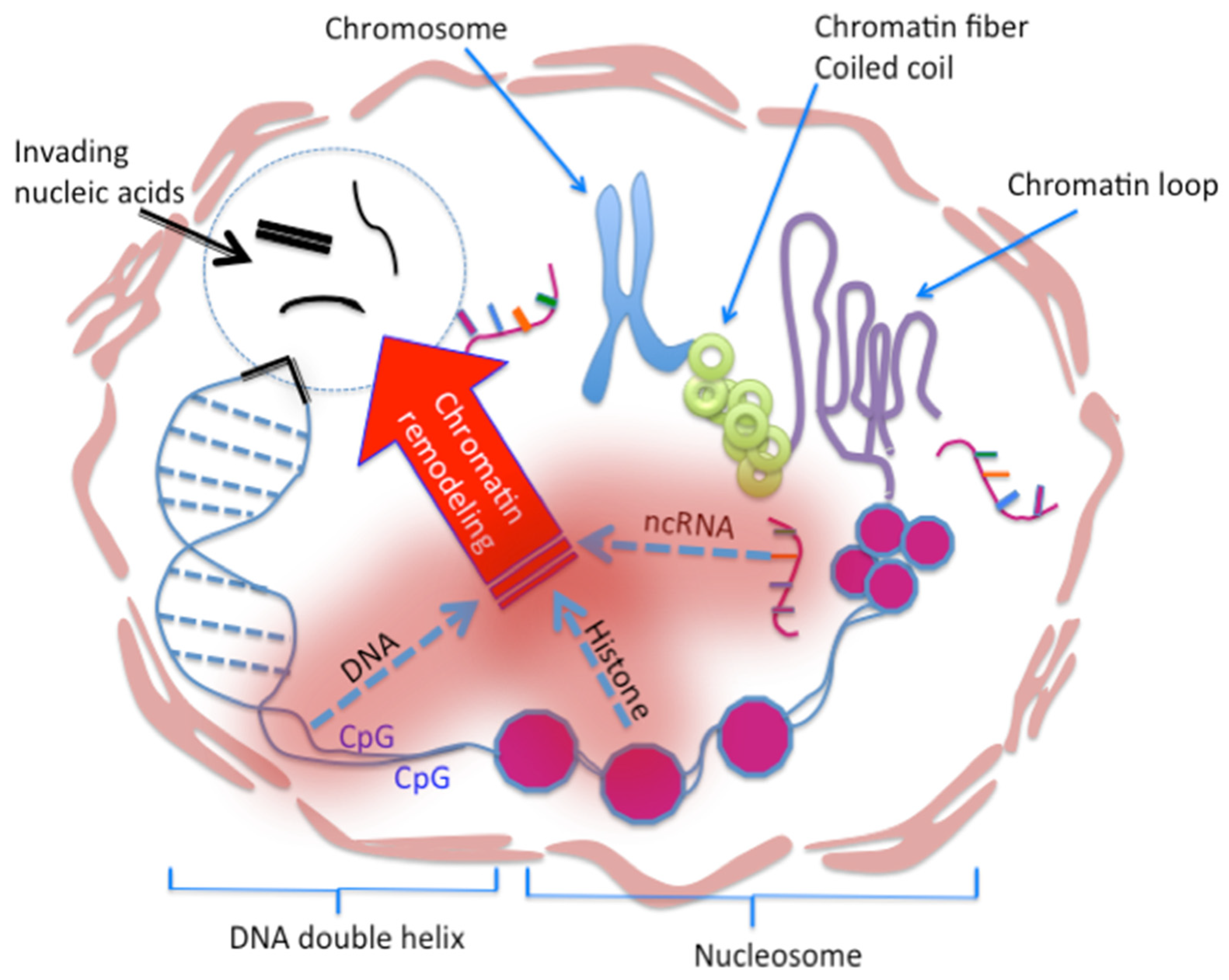
Differing from innate, adaptive, and trained immunities, epigenetic immunity occurs in every eukaryotic cell that has a nucleus. Epigenetic immunity consists of three elements protecting our DNA genome: DNA methylation, histone modification, and ncRNA activity. Upon antigen stimulations, which are usually foreign nucleic acids, a host genome launches immune responses via epigenetic regulations consisting of DNA methylation, histone modification and noncoding RNA (ncRNA) function to protect the DNA integrity.

2. HIV UTRs

HIV is a retrovirus, and its replication cycle consists of viral RNA and viral DNA. The viral DNA embedded in the human DNA is called the provirus. HIV RNA is a positive, genomic RNA (gRNA), and it encodes all the viral proteins but needs a reverse transcription to integrate into our genome, where it becomes a provirus, i.e., as a part of our DNA.
Similar to the classic cellular mRNA, HIV RNA has both 5′UTR and 3′UTR. The differences, however, are the following: (1) both HIV 5′ and 3′UTR are longer than their cellular mRNA counterparts. (2) The average length of human mRNA is 3.4 kb. HIV-1 mRNAs lie in a range from 9.3 kb of gRNA to the 2 kb of spliced mRNA, which encode the HIV structural and accessory proteins. All the HIV mRNAs possess a similar 5′ and 3′ UTR region.

2.1. 5′UTR

The 5′UTR of HIV-1 gRNA is known to form specific structures and has important functions besides those of classic cellular mRNA [4]. These include TAR, the elongating sequences required for gRNA replication [11,12,13,14], site of gRNA dimerization (in kissing or linear form) [15], packaging signal (ψ) [16,17,18], site of reverse transcription initiation (tRNALys3 annealing) [19], and a cap- or an internal ribosome entry site (IRES) for cap-independent translational initiation [20,21,22,23].
Mutations in the sequence of 5’UTR disrupt the structure of this region and affect the transcription of HIV RNA, reverse transcription, and packaging, as well as the fundamental role of mRNA in protein translation, i.e., the viral protein production. Note that starting at transcription, more than 23 copies of HIV mRNA are produced, which include alternatively spliced isoforms of env mRNA for translation of the regulatory, accessory, and Env proteins, and the gRNA for Gag-Pol structural polyproteins [24,25,26,27].
The multiple splice donor (SD) and splice acceptor (SA) sites in the HIV-1 gRNA support alternative splicing for a pool of 4-kb and 2-kb mRNAs that differ in their 5′UTR. These mRNAs contain partly overlapping open reading frames (ORFs), and collectively encode the nine HIV-1 proteins and polyproteins [24,25,26,27]. In addition, the IRES-dependent translational initiation is a cap-independent translational process. An internal AUG codon found near the amino terminus of the Pr55(gag) capsid domain drives translation of a 40-kDa Gag isoform [20,21,22,23,27].
The 5′UTR has been found to be the most conserved part in the HIV gRNA. In simple terms, its 335-nucleotide residues form the regulatory motifs, which initiate multiple steps in the HIV lifecycle. For example, the HIV-1 5’UTR reveals a 3D tRNA mimicry and is important for the viral reverse transcription. The 3D structure, but not the RNA sequence, is conserved across the distinct HIV-1 subtypes such as in C, B and A [28]. The tertiary motif is correlated to the region in 5′LTR after a reverse transcription for the formation subtypes of HIV [28,29,30].

2.2. 3′UTR

HIV-1 has a longer 3′UTR compared to the cellular mRNA. The HIV-1 encodes a polyadenylation (polyA) signal (AAUAAA) within both its highly conserved 5′ and 3’ UTR sites [31,32]. Note that in the polyadenylation process, an RNA transcript is cleaved and then elongated with the polyA. With repression of the 5’ polyA signal, the utilization of the 3’ polyA signal occurs. Studies suggest that polyA signals in the HIV RNA show metastable features to switch into different structures that regulate the viral gRNA function [31].
In addition to the classic role of 3′UTR in translational termination and the stability of RNA, HIV 3′UTR plays a cardinal role in formation of the HIV provirus. This is a separate topic, however, and will not be addressed in this review. Besides the role in HIV provirus formation, the 3′UTR takes part in the HIV packaging, a process of virion formation.
Studies have shown that among pools of cellular mRNAs, those with a long 3’UTR, including the HIV RNA, are selectively packaged into the virion [33]. It seems plausible that the 3’UTR, a stretch of RNA not occupied by ribosomes, offers a favorable binding site for Gag, through encapsidation to the virion packaging. Moreover, Rev protein has been shown to function at the HIV-1 3′UTR. The Rev-responsive element (RRE) overcomes the inhibitory effects of a 5’ splice site located within the 3’UTR, to transport the HIV RNA from nucleoplasm to the cytoplasm [34].
Previous studies report that the host factors, e.g., eukaryotic translation initiation factor 3 subunit F (elF3f), restrict the HIV mRNA expression and target in the 3′UTR region, binding here blocks 3′ end processing [35,36]. Cellar miRNAs and HIV-encoded miRNAs, both mediating the repression of HIV RNA, are also targeting on the 3′UTR [37,38]. Recent discoveries on the epigenetic regulation of 3′UTR have further broadened our knowledge of host immunity in control of viral RNA expression via the 3′UTR [39,40].
Beside the role of miRNA in repression of the HIV RNA expression, the modification on viral 3’UTR m(6)A sites, analogous the cellular m(6)A sites, strongly enhances mRNA expression in cis by recruiting the cellular YTHDF m(6)A “reader” proteins. This protein-RNA interaction has shown a cell type specific manner. For example, studies show that over expression of YTHDF, originally found in Drosophila where m6A recruits YTHDF to the mRNA, enhances the HIV RNA, protein expression and the viral replication in the CD4 T-cells [39]. YTHDF2 has recently been shown to increase HIV mRNA stability while YTHDC1 reader recruitment helps regulate HIV alternative splicing [41].
Studies on viruses other than HIV have also shown the cell type specific expression of a virus. In other words, a virus replicates only in a specific type of cells—the viral or cell tropism. These studies unveil that the cell type specific viral replication is also driven by the viral mRNA 3′UTR, where the host cell factors interact with the viral 3′UTR motifs to stabilize viral protein expression [40,42].
Furthermore, ncRNA has been shown to act on the 3′UTR with the anti-viral effect. An artificial miR-30a-3’-untranslated region (miR-3-UTR) obtained from a single RNA polymerase II (RNAP II) was used to simultaneously target all HIV transcripts. The study reports that HIV-1 replication was significantly inhibited in the cells with the miR-3-UTR construct that acts as the RNA interference (RNAi) [43]. A highly abundant miRNA, miR-29a, is also reported to specifically target the HIV-1 3’UTR region, and enhances the viral mRNA association with RNAi machinery RISC (RNA-induced silencing complexes) and P body proteins to inhibit the HIV RNA expression [44].
To summarize, besides its important biological function in the viral lifecycle, the 3′UTR is a target of host antiviral immunity that functions at the epigenetic level. Of note, some of the ncRNA are transcribed directly from the viral 3′UTR, and target this region to induce HIV specific RNAi. The ncRNA, exemplified by miRNAs, bind via a protein complex to the HIV 3’UTR. RNAi is one of the epigenetic silencing mechanisms.
The ncRNA function is one of the three elements of epigenetic immunity, and the 3′UTR, either of viral RNA or ncRNA, has a paramount role in the modulation of host immunity against the HIV infection. Note that for simplicity, the ncRNA includes miRNA, siRNA, shRNA, snRNA, lncRNA, and eRNA (enhancer RNA).

3. HIV LTRs

After reverse transcription, the 9.3 kb gRNA becomes the HIV DNA to be integrated into the host DNA with an integration process. Of note, through reverse transcription, the 5′UTR becomes the 5′LTR, and 3′UTR becomes the 3′LTR. Upon a successful integration, the provirus is produced.
Provirus not only keeps all the regulatory regions highly conserved in the 5′ and the 3′UTR, but also a new U3 region is in the front of the 5′LTR and a new U5 is at the end of the 3′LTR (Figure 1). This results in a longer DNA sequence than the 5′ and 3′UTR of gRNA and confers the long terminal repeat (LTR) to the provirus. A provirus contains the duplicated LTR sequences, meaning two identical enhancer-promoter regions, flanking the proviral genome with a 5’ and 3’ LTR symmetry. The LTR has one specific role—to transcribe the HIV RNA.
It is easily understood that 5′ and 3′ LTR govern HIV RNA expression. The provirus is a parasite, and the transcription of gRNA relies on the host. In fact, provirus is a metastable stage in the HIV lifecycle, and the break of a metastable equilibrium depends on the host, not the parasite. In other words, a competition occurs between the host immunity and the virus on the viral gene expression—the transcription of the viral RNA. In this competition, HIV has 5′ and 3′ LTR, but the host has an entire genomic DNA, which has ~3.2 × 10⁹ nucleotide base pairs deploying DNA methylation, histone modification, and ncRNA function against the viral RNA replication.
After more than 40-years, HIV/AIDS research has led to an explosion of knowledge on human immunity, based on the fact that HIV/AIDS is a naturally occurring model of loss of function in the human immune response. The study of HIV/AIDS has provided an unprecedented platform to understand our immunity, specifically the role of CD4 T-cells in eukaryotic vs. prokaryotic immunity against viral infections, and in prevention as well as cure, including the vaccination (immunization).
In essence, research on HIV/AIDS has unveiled that HIV targets the immune cells, specifically CD4 T-cells. HIV evolves in the human body with a rate unseen in other retroviruses, exemplified from R5 (M tropic) to X4 (T tropic) virus. Under cART (Highly Effective Anti-Retroviral Treatment, or Combined Anti-Retroviral Therapy), there are founder virus and quasispecies [45,46]. Moreover, ample molecular studies have revealed that HIV 5′LTR governs the gRNA expression by the viral enhancer and promoter. Herein, we refresh our knowledge of HIV 5′LTR, to delineate the arsenal of the virus, and armamentaria of the host in control of HIV gRNA expression towards a cure.

3.1. 5′LTR

HIV 5′LTR contains both enhancer and promoter elements [3,5,6,7,47,48,49,50,51,52,53,54,55,56,57,58,59,60,61,62,63,64,65,66,67,68,69]. Upon receiving cell signals and within a host DNA niche, the provirus can transcript its gRNA. If there is no cellular signal and there is no niche, there is no gRNA expression, or at the most, an aborted gRNA expression. Therefore, not every provirus is equal, and the host genome’s immunity dictates the outcome.
HIV enhancer. More than 30 years ago, the NFkappaB core element was identified as the HIV enhancer [6]. Continuing studies have identified other core enhancer elements, such as PMA, the most common and potent phorbol ester, inducible GGGACTTTCC core enhancer element [48,49,50,51], and the core enhancer element CCAAT/(C/EBP) all in HIV LTR [60,61,62,63,64,65,66,67,68,69,70,71,72,73,74,75]. These studies unveil a trajectory that shows how HIV replicates in CD4 T-cells, in monocytes/microphages, and in central nervous system cells [6,58,59,60,61,62,63,64,65,66,67,68,69,70,71,72,73,74]. The HIV enhancer, located in the HIV LTR, has played an indispensable role in determining the viral tropism. In other words, the HIV enhancer has dictated a range of cell types to host the viral RNA expression, therefore the viral tropism and the subtype formation [29,30,45,46,47,48,60,61,62,63,64,65,66,67,68,69,70,71,72,73,74,75].
Based on studies of cellular enhancers, the HIV enhancers arranged in the 5′LTR should be considered as the super enhancer and the intragenic enhancer [55,76]. Super enhancer is a region comprising multiple enhancers that is collectively bound by an array of transcription factor proteins to drive transcription of genes involved in cell identity. Note that enhancer functions at both directions of 5′ and 3′ in cis, which is different from the promoters.
The studies of HIV/AIDS have shown that HIV enhancer differs from cellular enhancer in its intragenic way, meaning HIV super enhancer drives only a gRNA expression from its 5′UTR and ends in the 3′UTR, for both the gRNA and its spliced mRNA. In contrast to the host cellular enhancers, which activate their target gene expressions by topological-spatial contact, usually, several kilo- to megabases away from the target genes to be activated.
Finally, HIV enhancer confers a rate of evolution to a given HIV. This highly conserved region establishes a trajectory of a given HIV species on a successful infection of the host cells, starting at the attachment, entry, reverse transcription, integration, transcription, and, thereby, encapsidation, packaging, virion releasing, and a repeat of this lifecycle.
In HIV infection, the dual LTR is capable of breaching the genome’s immunity to replicate HIV within intestinal macrophages and CD4 T-cells, from peripheral monocytes and CD4 T-cells to the neural microglial cells, astrocytes, and CD4 T-cells. The HIV enhancer may be the most effective weapon in the viral arsenal. In fact, the HIV super enhancer provides a 3D-platform to recruit not only cellular proteins/factors, but also viral proteins/factors, cooperating and overlapping with those on the HIV promoter to drive the viral replication in different tissues and cells. This prevalent tactic of HIV has breached the immunity of the host genome and allowed a parasite to live in our DNA, and its replication occurs at the expense of our lives.
HIV promoter. The HIV promoter contains the basic sequences for RNA transcription. Similar to its cellular counterpart, the HIV promoter has basal level transcriptional activity. The proteins binding in this region interact with those in the super enhancer to transcribe the gRNA. The transcription of gRNA, however, needs the enhancer function [3,6,52,57,58,59,60,61,62,63,64,65,66,67,68,69,72,74,75].
As stated previously, the HIV super enhancer competes with the host genome’s immunity in RNA transcription. Reports have shown that a R5 virus has different arrangements of the main and other core enhancer elements than a X4 virus in the 5′LTR super enhancer region [6,47,58,60,62,64,65,70,71,72,73,77,78]. Of note, the interaction of protein–nucleic acid complex between enhancer and promoter responds, and regulates the host cell signals. Enhancer binding proteins usually accelerate or magnify the cellular signal, and drive the promoter to generate a transcription niche in the DNA genome via binding and interacting with transcription factor complexes at both enhancer-promoter sites to transcribe the gRNA. After this activation point, the rest of viral replication is like a ball falling on ground. The metastable equilibrium between host and provirus is broken. The ball has fallen on ground rather than remaining in a biologically metastable state. This is similar to a system that returns to equilibrium after small (but not large) displacements in the physical chemistry, and may be represented by a ball resting in a small hole on top a hill. If the ball is only slightly pushed, it will settle back into the hole. But a stronger push may start the ball rolling down the hill.

3.2. 3′LTR

Although in symmetry with and being a duplicate of 5′LTR, the 3′LTR guides the effective gRNA expression by its polyA signal, or responds to cellular stimuli to stop the gRNA transcription [79,80,81], and, finally, functions as the 5′LTR when 5′LTR is defective due to the integration [82].
Note that the 3′LTR polyA signal, which shows a metastable feature in 3′UTR, is reported to form a transcription-dependent gene loop with the 5’LTR occluded polyA signal. Activation of 5’LTR polyA signal or inactivation of 3’LTR polyA signal abolishes the gene loop formation [83].
Recent studies reveal that the 3′LTR serves as the 5′LTR in the DNA negative strand, and transcribes an antisense ncRNA named ASP (the ncRNA codes the antisense protein ASP) [84,85]. The ASP regulates the HIV latency by RNA:DNA base pairing to 5′LTR with Watson–Crick and Hoogsteen specificity. After the pairing, the provirus is back in business: latency. This epigenetic silencing differs from RNAi in the molecular mechanism: the ASP associates with polycomb repressor complex and promotes nucleosome assembly for the transcriptional-epigenetic silencing.

3.3. Impact of LTR and UTR in Viral Tropism

Both LTR and UTR play important roles in determining HIV infection of CD4 T cells or macrophages. The HIV LTR, via the enhancer that binds the host cellular transcription factors, transcribes the viral RNA. The 3′UTR is responsible for the viral RNA stability and then the viral protein expression. Together, they drive an active viral replication and the production of CXCR4 (X4) or CCR5 (R5) viruses.
The interaction of host cellular transcription factors with LTR, i.e., HIV enhancer, determines the viral mRNA and gRNA expression. Despite the fact that the repeats of the enhancer elements vary in different HIV strains, such as R5 or X4, or a dual-tropic HIV [66,69,70,71,72,73], the NFkB core elements are considered as the main enhancer, and other enhancers add to the tissue and cell specific expression of gRNA [6,58,59,60,61,62,63,64,65,66,67,68,69,70,71,72,73,74,86].
Similar to its cellular counterpart, HIV enhancer is a driver for the cell and tissue specific expressions of HIV RNA, and, therefore, controls the HIV tropism [29,30,45,46,47,48,55,76]. This underlines a molecular mechanism that adds onto the viral tropism, which used to refer an expression of cell surface co-receptors for the entry of R5 or X4 viruses.
The interaction of host cellular proteins with 3′UTR allows a given virus to express viral proteins after entry. Cellular miRNAs and host proteins in a particular cell host have been shown to function on the viral 3′UTR [35,36,37,38,39,40,42,43,44]. The immune pressure exerted on HIV 3′UTR, in the form of miRNAs and cellular factors, determines whether the HIV protein is translated or not and, therefore, determines an HIV protein function, and, hence, the viral tropism—a host cell that HIV can make an active replication within [39,40,42,43,44].
Note here is that the interaction of viral RNA-host protein underpins the molecular mechanism of viral tropism, at the step targeting the function of viral mRNA on the viral protein translation. Hence, the interaction of RNA-protein at the viral 3′UTR determines the functions of viral mRNA and viral protein, determines the viral tropism, and embodies that a virus can make an active replication within a cell or not. This clearly differs from a viral entry via a cell surface co-receptor and differs in an intrinsic mechanism that regulates the viral protein expression.
Lastly, the expression and function of ncRNA have been known to be the cell and tissue specific [87,88,89]. Taken together, studies have shown that the HIV 3′UTR, its ncRNA, and the cellular ncRNA not only play roles in formation the so-called T tropic, M tropic, R5, X4, and the founder virus or quasispecies of HIV, but also on the host immunity against these HIV strains [29,30,45,46].

4. Armamentaria of the Human Host

The study of how HIV RNA is expressed in the nucleosome in vitro was conducted in the same period as that defining the HIV enhancers and promoters: in the DNA era [90,91,92], during the NIH launched Human Genome Project (HGP) that began in 1990 and completed in 2003. The technology and knowledge on nucleosome, i.e., the study of chromatin, however, has developed rapidly in this era of epigenetics (the transcriptome era) [10,93,94,95,96,97,98], symbolized by the Encyclopedia of DNA Elements (ENCODE) project started in 2003 and concluded in 2012. ENCODE analyzes and identifies the functional elements in the human genome sequence after the HGP. We refresh the knowledge of HIV/AIDS research to harness an unprecedented model on the loss of function in humans to define not only the cause but also the effect—a cure of HIV/AIDS. We need to pay back this unparalleled model in the study of the human immune diseases, and we urgently need to harness what this model has provided and further the discovery of a cure of HIV/AIDS and other immune maladies.

4.1. Epigenetic Immunity

Built on the HIV/AIDS model, we have proposed that the host DNA has an evolutionally developed immunity against pathogenic nucleic acid invasion. We have named this immunity the epigenetic immunity, grounded on our and others studies that p21 (p21Cip1Waf1Sdi1) restricts the HIV infection of the human primitive hematopoietic cells, macrophages, and the CD4 T-cells of elite controllers [9,99,100,101,102]. Our study, showing that p21 prevented HIV infection in human primitive hematopoietic cells, establishes that human cellular mechanisms have an essential role to play in inhibition of HIV replication [9].
The epigenetic research on the pathogenesis of viral infections has, in fact, proceeded with the study of HIV/AIDS. The seminal studies in this area include, but are not limit to, the following: (1) the studies on the human cytomegalovirus, Epstein–Barr Virus, HIV, and foreign DNA insertion [97,103,104,105]; (2) the epigenetics of infectious diseases [106,107,108]; (3) the epigenetic regulation of immune cell memory [109,110]; (4) the epigenetic molecular study of CD4 T-cells [111,112,113,114,115,116,117,118,119,120]; (5) super enhancers [117,121,122]; (6) the function of non-coding RNAs (ncRNAs) [123,124]; and (7) the epigenetic repression or imprinting (DNA methylation) on the human endogenous retroviruses (HERVs) [98,125,126].
Building on the contributions above, we conclude that the epigenetic immunity consists of three elements: DNA methylation, histone modification, and ncRNA function (Figure 2). We believe that this immunity exists in every cell that has a genomic DNA, but not the cells without the genomic DNA, such as the mature red blood cells [9,10,127]. Similar to the known innate, adaptive, or trained immunity, the immunogens such as the pathogenic RNA and DNA can elicit the host epigenetic immunity. We have proposed a cVaccine, not only as a tool to study the epigenetic immunity, but also the immunogen to elicit the host epigenetic immunity.

4.2. Chromatin Vaccine (cVaccine)

cVaccine is a functional gene transcription unit with enhancer, in nucleosome format that resists nuclease degradation while mediating epigenetic silencing of viral RNA by ncRNA function and the enhancer decommissioning process [128,129,130,131].
It is well recognized that HIV infects naïve CD4 T-cells. CD4 T-cells have the stem cell property of self-renewal and differentiating into effector cells. The naïve CD4 T-cells analogize embryonic stem cell asymmetric division upon an antigen stimulation, and differentiate into memory and immune effector cells. Most importantly, these cells are distributed into all body tissues and embody antigen specific lineage of CD4 cytotoxic T-cells (CTL), CD4 regulatory T-cells (Treg), CD4 follicular helper T-cells (Tfh), etc., to directly attack pathogen, regulate CD8 T-cell, B cell, and other immune cell functions.
The enhancer decommissioning occurs routinely in stem cells, specifically in lineage differentiation into tissue specific effector cells by regulating the exact same genomic DNA. cVaccine is an immunogen (antigen), and functions as a primer of naïve CD4 T-cell enhancer, by triggering the signaling pathways of previously identified Toll Like Receptor (TLR), interferon and NFkB, to load the TF (transcription factor)/RNAPII to the CD4 T-cell enhancer. Such a primed cell enhancer allows CD4 T-cells to rapidly differentiate into effector cells upon encounter with the HIV. The antigen specific differentiation of immune cells by primed enhancers or promoters is dubbed the poised ones, and is well studied in the immunology [110,111,112,113,114,115,116,117,118,121,122].
The enhancer decommissioning between HIV and CD4 T-cell is the ectopic or cognate binding the ration (limited supply) of TF/RNAPII either to a viral super enhancer or a cell linage enhancer. Note that the integration of intrinsic signals and TF/RNAPII binding to the CD4 gene (Cd4) enhancer allows the CD4 lineage enhancer to become a super enhancer, and differentiates into the antigen specific immune cells [118,121,122]. Therefore, cVaccine in an enhancer decommissioning process elicits the cell genomic determined fate for CD4 T-cells by epigenetic regulations, ushers CD4 T-cell lineage development by its lineage enhancer transiting into the supper enhancer, which superiorly allows the naïve CD4 T-cells to differentiate into the immune effector cells.
In other words, cVaccine serves as a primer to the CD4 T-cell lineage enhancer, and to prime the CD4 T-cell enhancer into an active and poised state, ready to differentiate to antigen specific CD4 T-effector cells upon reencounter with the HIV. The vaccinated CD4 T-cells counteract the HIV proviral enhancer (usually one provirus per a cell) not only by competitively loaded TF/RNAPII on their enhancers, but also by the eRNA (enhancer RNA) function. Both converge to silence the HIV RNA expression while allow CD4 T-cells to differentiate into anti-HIV effector cells to establish a long lasting anti-HIV immunity. This embodies the genomic immunity—epigenetic immunity. We consider this is a molecular mechanism of the low viral load post cART, in the HIV controllers and in the HIV latent infection.
Compared to the current mRNA vaccine, the cVaccines stops the viral RNA expression rather than expressing a viral protein. cVaccine aims to stop the viral RNA expression and, therefore, stops all the viral protein expressions, cuts down the viral mutagenesis, and ceases toxicity of viral RNA and proteins from damaging our body. Viral RNA and proteins are the culprits causing DNA mutation, immune inflammation and autoimmunity. We term these deviations from anti-viral immunity as the hypersensitivity V herein [132].
Since the cVaccine resists the nuclease degradations, it could be administered by the routes of viral infections, from entry and all the way to the target cells per se, aiming to elicit the IgA, IgM, and IgG, as well as innate, adaptive, trained, and epigenetic immunity in a systemic manner. This results from the study of HIV/AIDS, which is a naturally occurred model of loss-of-function in human immunity. Such a model has empowered the research on human immunodeficiency without initiating any ethical disputes, at a scale from bench, in silico, to bedside, from molecular to systemic mechanis ms that unveil the human immune response in etiology, pathogenesis, and up to a cure.

5. Conclusions

We have stated four of the achievements that multidisciplinary investigators have contributed from the HIV/AIDS and epigenetics research fields: 1. HIV super enhancer is essential for the HIV RNA expression. 2. Host genome has an immune system protecting our DNA against the viral infection, embodying the epigenetic immunity of our DNA. 3. The epigenetic regulation has forced the appearance of different HIV tropisms via an LTR-UTR circuit governed by LTR—the HIV super intragenic enhancer. 4. Host immunity and cART have kept the HIV in a metastable stage—the provirus. Further studies on epigenetic silencing can enforce HIV to become a new endogenous retrovirus that is unable to cause disease for generations.

Funding

This research received no specific grant from any funding agency in the public, commercial, or not-for-profit sector.

Institutional Review Board Statement

Not applicable.

Informed Consent Statement

Not applicable.

Data Availability Statement

Data sharing is not applicable to this article as no datasets were generated or analyzed during the current study.

Acknowledgments

We thank Stephen Goff, Philip Askenase, and Joseph Sodroski for reading, editing the manuscript, and providing helpful suggestions. The cited articles in this review are from publications of PubMed in 1981 to 2021, and selected under the key words of UTR, LTR, virus, HIV, epigenetics, and immunity. We apologize for numerous seminal work and publications that are not included in this review as the review has a focus on HIV UTR, LTR, and the entailed effects towards an HIV cure.

Conflicts of Interest

The authors declare no conflict of interest. This research received no specific grant from any funding agency in the public, commercial, or not-for-profit sector. The authors plan to have provisional patent applications on some of the concepts discussed in this review.

Abbreviations

AIDS: Acquired Immunodeficiency Syndrome, cART: Combined Antiretroviral Therapy, HIV: Human Immunodeficiency Virus, TAR: Trans-Activation Response element, Tat: Trans-Activator of Transcription protein, YTHDF: YT521-B homology (YTH) domain family protein, YTHDF2: YT521-B homology (YTH) domain family 2 protein, YTHDC1: YT521-B homology (YTH) domain containing protein 1.

References

  1. Sadaie, M.R.; Kalyanaraman, V.S.; Mukopadhayaya, R.; Tschachler, E.; Gallo, R.C.; Wong-Staal, F. Biological characterization of noninfectious HIV-1 particles lacking the envelope protein. Virology 1992, 187, 604–611. [Google Scholar] [CrossRef]
  2. Ribeiro, R.M.; Mohri, H.; Ho, D.D.; Perelson, A.S. In vivo dynamics of T cell activation, proliferation, and death in HIV-1 infection: Why are CD4+ but not CD8+ T cells depleted? Proc. Natl. Acad. Sci. USA 2002, 99, 15572–15577. [Google Scholar] [CrossRef] [PubMed] [Green Version]
  3. Lu, X.; Welsh, T.M.; Peterlin, B.M. The human immunodeficiency virus type 1 long terminal repeat specifies two different transcription complexes, only one of which is regulated by Tat. J. Virol. 1993, 67, 1752–1760. [Google Scholar] [CrossRef] [PubMed] [Green Version]
  4. Al-Harthi, L.; Roebuck, K.A. Human immunodeficiency virus type-1 transcription: Role of the 5’-untranslated leader region (review). Int. J. Mol. Med. 1998, 1, 875–881. [Google Scholar] [CrossRef] [PubMed]
  5. Sodroski, J.; Rosen, C.; Wong-Staal, F.; Salahuddin, S.Z.; Popovic, M.; Arya, S.; Gallo, R.C.; Haseltine, W.A. Trans-acting transcriptional regulation of human T-cell leukemia virus type III long terminal repeat. Science 1985, 227, 171–173. [Google Scholar] [CrossRef] [Green Version]
  6. Nabel, G.; Baltimore, D. An inducible transcription factor activates expression of human immunodeficiency virus in T cells. Nature 1987, 326, 711–713, Erratum in Nature 1990, 344, 178. [Google Scholar] [CrossRef]
  7. Kaufman, P.A.; Weinberg, J.B.; Greene, W.C. Nuclear expression of the 50- and 65-kD Rel-related subunits of nuclear factor-kappa B is differentially regulated in human monocytic cells. J. Clin. Investig. 1992, 90, 121–129. [Google Scholar] [CrossRef]
  8. Henderson, A.J.; Connor, R.I.; Calame, K.L. C/EBP activators are required for HIV-1 replication and proviral induction in monocytic cell lines. Immunity 1996, 5, 91–101. [Google Scholar] [CrossRef] [Green Version]
  9. Zhang, J.; Scadden, D.T.; Crumpacker, C.S. Primitive hematopoietic cells resist HIV-1 infection via p21. J. Clin. Investig. 2007, 117, 473–481. [Google Scholar] [CrossRef] [Green Version]
  10. Zhang, J.L.; Crumpacker, C.S. Towards a Cure, does host immunity play a role? mSphere 2017, 2, e00138-17. [Google Scholar] [CrossRef] [Green Version]
  11. Song, R.; Kafaie, J.; Laughrea, M. Role of the 5’ TAR stem--loop and the U5-AUG duplex in dimerization of HIV-1 genomic RNA. Biochemistry 2008, 47, 3283–3293. [Google Scholar] [CrossRef]
  12. Soto-Rifo, R.; Limousin, T.; Rubilar, P.S.; Ricci, E.P.; Décimo, D.; Moncorgé, O.; Trabaud, M.A.; André, P.; Cimarelli, A.; Ohlmann, T. Different effects of the TAR structure on HIV-1 and HIV-2 genomic RNA translation. Nucleic Acids Res. 2012, 40, 2653–2667. [Google Scholar] [CrossRef] [PubMed] [Green Version]
  13. Pallesen, J. Structure of the HIV-1 5’ untranslated region dimer alone and in complex with gold nanocolloids: Support of a TAR-TAR-containing 5’ dimer linkage site (DLS) and a 3’ DIS-DIS-containing DLS. Biochemistry 2011, 50, 6170–6177. [Google Scholar] [CrossRef] [PubMed]
  14. Charbonneau, J.; Gendron, K.; Ferbeyre, G.; Brakier-Gingras, L. The 5’ UTR of HIV-1 full-length mRNA and the Tat viral protein modulate the programmed -1 ribosomal frameshift that generates HIV-1 enzymes. RNA 2012, 18, 519–529. [Google Scholar] [CrossRef] [PubMed] [Green Version]
  15. Balakrishnan, M.; Fay, P.J.; Bambara, R.A. The kissing hairpin sequence promotes recombination within the HIV-I 5’ leader region. J. Biol. Chem. 2001, 276, 36482–36492. [Google Scholar] [CrossRef] [Green Version]
  16. Cockrell, A.S.; van Praag, H.; Santistevan, N.; Ma, H.; Kafri, T. The HIV-1 Rev/RRE system is required for HIV-1 5’ UTR cis elements to augment encapsidation of heterologous RNA into HIV-1 viral particles. Retrovirology 2011, 8, 51. [Google Scholar] [CrossRef] [Green Version]
  17. Didierlaurent, L.; Racine, P.J.; Houzet, L.; Chamontin, C.; Berkhout, B.; Mougel, M. Role of HIV-1 RNA and protein determinants for the selective packaging of spliced and unspliced viral RNA and host U6 and 7SL RNA in virus particles. Nucleic Acids Res. 2011, 39, 8915–8927. [Google Scholar] [CrossRef]
  18. Ka, W.H.; Jeong, Y.Y.; You, J.C. Identification of the HIV-1 packaging RNA sequence (Ψ) as a major determinant for the translation inhibition conferred by the HIV-1 5’ UTR. Biochem. Biophys. Res. Commun. 2012, 417, 501–507. [Google Scholar] [CrossRef]
  19. Barraud, P.; Gaudin, C.; Dardel, F.; Tisné, C. New insights into the formation of HIV-1 reverse transcription initiation complex. Biochimie 2007, 89, 1204–1210. [Google Scholar] [CrossRef] [Green Version]
  20. Buck, C.B.; Shen, X.; Egan, M.A.; Pierson, T.C.; Walker, C.M.; Siliciano, R.F. The human immunodeficiency virus type 1 gag gene encodes an internal ribosome entry site. J. Virol. 2001, 75, 181–191. [Google Scholar] [CrossRef] [Green Version]
  21. Waysbort, A.; Bonnal, S.; Audigier, S.; Estève, J.P.; Prats, A.C. Pyrimidine tract binding protein and La autoantigen interact differently with the 5’ untranslated regions of lentiviruses and oncoretrovirus mRNAs. FEBS Lett. 2001, 490, 54–58. [Google Scholar] [CrossRef]
  22. Abbink, T.E.; Berkhout, B. A novel long distance base-pairing interaction in human immunodeficiency virus type 1 RNA occludes the Gag start codon. J. Biol. Chem. 2003, 278, 11601–11611. [Google Scholar] [CrossRef] [PubMed] [Green Version]
  23. Balvay, L.; Soto Rifo, R.; Ricci, E.P.; Decimo, D.; Ohlmann, T. Structural and functional diversity of viral IRESes. Biochim. Biophys. Acta 2009, 1789, 542–557. [Google Scholar] [CrossRef] [PubMed]
  24. Anderson, J.L.; Johnson, A.T.; Howard, J.L.; Purcell, D.F. Both linear and discontinuous ribosome scanning are used for translation initiation from bicistronic human immunodeficiency virus type 1 env mRNAs. J. Virol. 2007, 81, 4664–4676. [Google Scholar] [CrossRef] [Green Version]
  25. Obayashi, C.M.; Shinohara, Y.; Masuda, T.; Kawai, G. Influence of the 5′-terminal sequences on the 5′-UTR structure of HIV-1 genomic RNA. Sci. Rep. 2021, 11, 10920. [Google Scholar] [CrossRef]
  26. Yamamoto, M.; Harada, K.; Shoji, S.; Misumi, S.; Takamune, N. Identification of essential cis element in 5’UTR of Nef mRNA for Nef translation. Curr. HIV Res. 2014, 12, 213–219. [Google Scholar] [CrossRef]
  27. Barrera, A.; Olguín, V.; Vera-Otarola, J.; López-Lastra, M. Cap-independent translation initiation of the unspliced RNA of retro. Viruses Biochim. Biophys. Acta Gene Regul. Mech. 2020, 1863, 194583. [Google Scholar] [CrossRef]
  28. Comandur, R.; Olson, E.D.; Musier-Forsyth, K. Conservation of tRNA mimicry in the 5’-untranslated region of distinct HIV-1 subtypes. RNA 2017, 23, 1850–1859. [Google Scholar] [CrossRef] [Green Version]
  29. Sampathkumar, R.; Scott-Herridge, J.; Liang, B.; Kimani, J.; Plummer, F.A.; Luo, M. HIV-1 Subtypes and 5’LTR-Leader Sequence Variants Correlate with Seroconversion Status in Pumwani Sex Worker Cohort. Viruses 2017, 10, 4. [Google Scholar] [CrossRef] [Green Version]
  30. Mbondji-Wonje, C.; Dong, M.; Zhao, J.; Wang, X.; Nanfack, A.; Ragupathy, V.; Sanchez, A.M.; Denny, T.N.; Hewlett, I. Genetic variability of the U5 and downstream sequence of major HIV-1 subtypes and circulating recombinant forms. Sci. Rep. 2020, 10, 13214. [Google Scholar] [CrossRef]
  31. Gee, A.H.; Kasprzak, W.; Shapiro, B.A. Structural differentiation of the HIV-1 polyA signals. J. Biomol. Struct. Dyn. 2006, 23, 417–428. [Google Scholar] [CrossRef] [PubMed]
  32. Spriggs, S.; Garyu, L.; Connor, R.; Summers, M.F. Potential intra- and intermolecular interactions involving the unique-5’ region of the HIV-1 5’-UTR. Biochemistry 2008, 47, 13064–13073. [Google Scholar] [CrossRef] [PubMed] [Green Version]
  33. Comas-Garcia, M.; Davis, S.R.; Rein, A. On the Selective Packaging of Genomic RNA by HIV-1. Viruses 2016, 8, 246. [Google Scholar] [CrossRef] [PubMed] [Green Version]
  34. Barksdale, S.K.; Baker, C.C. The human immunodeficiency virus type 1 Rev protein and the Rev-responsive element counteract the effect of an inhibitory 5’ splice site in a 3’ untranslated region. Mol. Cell Biol. 1995, 15, 2962–2971. [Google Scholar] [CrossRef] [PubMed] [Green Version]
  35. Valente, S.T.; Gilmartin, G.M.; Venkatarama, K.; Arriagada, G.; Goff, S.P. HIV-1 mRNA 3’ end processing is distinctively regulated by eIF3f, CDK11, and splice factor 9G8. Mol. Cell 2009, 36, 279–289. [Google Scholar] [CrossRef] [PubMed] [Green Version]
  36. De Breyne, S.; Chamond, N.; Décimo, D.; Trabaud, M.A.; André, P.; Sargueil, B.; Ohlmann, T. In vitro studies reveal that different modes of initiation on HIV-1 mRNA have different levels of requirement for eukaryotic initiation factor 4F. FEBS J. 2012, 279, 3098–3111. [Google Scholar] [CrossRef] [PubMed]
  37. Schopman, N.C.; Willemsen, M.; Liu, Y.P.; Bradley, T.; van Kampen, A.; Baas, F.; Berkhout, B.; Haasnoot, J. Deep sequencing of virus-infected cells reveals HIV-encoded small RNAs. Nucleic Acids Res. 2012, 40, 414–427. [Google Scholar] [CrossRef]
  38. Ebert, M.S.; Sharp, P.A. Emerging roles for natural microRNA sponges. Curr. Biol. 2010, 20, R858–R861. [Google Scholar] [CrossRef] [Green Version]
  39. Kennedy, E.M.; Bogerd, H.P.; Kornepati, A.V.; Kang, D.; Ghoshal, D.; Marshall, J.B.; Poling, B.C.; Tsai, K.; Gokhale, N.S.; Horner, S.M.; et al. Posttranscriptional m(6)A Editing of HIV-1 mRNAs Enhances Viral Gene Expression. Cell Host Microbe 2016, 19, 675–685, Erratum in Cell Host Microbe 2017, 22, 830. [Google Scholar] [CrossRef] [Green Version]
  40. Garcia-Moreno, M.; Sanz, M.A.; Carrasco, L. A Viral mRNA Motif at the 3’-Untranslated Region that Confers Translatability in a Cell-Specific Manner. Implications for Virus Evolution. Sci. Rep. 2016, 6, 19217. [Google Scholar] [CrossRef]
  41. Tsai, K.; Bogerd, H.P.; Kennedy, E.M.; Emery, A.; Swanstrom, R.; Cullen, B.R. Epitranscriptomic addition of m6A regulates HIV-1 RNA stability and alternative splicing. Genes Dev. 2021, 35, 992–1004. [Google Scholar] [CrossRef] [PubMed]
  42. Carrasco, L.; Sanz, M.A.; González-Almela, E. The Regulation of Translation in Alphavirus-Infected Cells. Viruses 2018, 10, 70. [Google Scholar] [CrossRef] [PubMed] [Green Version]
  43. Nejati, A.; Shahmahmoodi, S.; Arefian, E.; Shoja, Z.; Marashi, S.M.; Tabatabaie, H.; Mollaei-Kandelous, Y.; Soleimani, M.; Nategh, R. Efficient inhibition of human immunodeficiency virus replication using novel modified microRNA-30a targeting 3′-untranslated region transcripts. Exp. Ther. Med. 2016, 11, 1833–1838. [Google Scholar] [CrossRef] [PubMed] [Green Version]
  44. Nathans, R.; Chu, C.Y.; Serquina, A.K.; Lu, C.C.; Cao, H.; Rana, T.M. Cellular microRNA and P bodies modulate host-HIV-1 interactions. Mol. Cell. 2009, 34, 696–709. [Google Scholar] [CrossRef] [PubMed] [Green Version]
  45. Shaw, G.M.; Hunter, E. HIV transmission. Cold Spring Harb. Perspect. Med. 2012, 2, a006965. [Google Scholar] [CrossRef]
  46. Musich, T.; Jones, J.C.; Keele, B.F.; Jenkins, L.M.M.; Demberg, T.; Uldrick, T.S.; Yarchoan, R.; Robert-Guroff, M. Flow virometric sorting and analysis of HIV quasispecies from plasma. JCI Insight 2017, 2, e90626. [Google Scholar] [CrossRef] [Green Version]
  47. Franza, B.R., Jr.; Josephs, S.F.; Gilman, M.Z.; Ryan, W.; Clarkson, B. Characterization of cellular proteins recognizing the HIV enhancer using a microscale DNA-affinity precipitation assay. Nature 1987, 330, 391–395. [Google Scholar] [CrossRef]
  48. Tong-Starksen, S.E.; Luciw, P.A.; Peterlin, B.M. Human immunodeficiency virus long terminal repeat responds to T-cell activation signals. Proc. Natl. Acad. Sci. USA 1987, 84, 6845–6849. [Google Scholar] [CrossRef] [Green Version]
  49. Kaufman, J.D.; Valandra, G.; Roderiquez, G.; Bushar, G.; Giri, C.; Norcross, M.A. Phorbol ester enhances human immunodeficiency virus-promoted gene expression and acts on a repeated 10-base-pair functional enhancer element. Mol. Cell Biol. 1987, 7, 3759–3766. [Google Scholar] [CrossRef]
  50. Dinter, H.; Chiu, R.; Imagawa, M.; Karin, M.; Jones, K.A. In vitro activation of the HIV-1 enhancer in extracts from cells treated with a phorbol ester tumor promoter. EMBO J. 1987, 6, 4067–4071. [Google Scholar] [CrossRef]
  51. Böhnlein, E.; Lowenthal, J.W.; Siekevitz, M.; Ballard, D.W.; Franza, B.R.; Greene, W.C. The same inducible nuclear proteins regulates mitogen activation of both the interleukin-2 receptor-alpha gene and type 1 HIV. Cell 1988, 53, 827–836. [Google Scholar] [CrossRef]
  52. Jakobovits, A.; Smith, D.H.; Jakobovits, E.B.; Capon, D.J. A discrete element 3’ of human immunodeficiency virus 1 (HIV-1) and HIV-2 mRNA initiation sites mediates transcriptional activation by an HIV trans activator. Mol. Cell Biol. 1988, 8, 2555–2561. [Google Scholar] [CrossRef] [PubMed]
  53. Hazan, U.; Thomas, D.; Alcami, J.; Bachelerie, F.; Israel, N.; Yssel, H.; Virelizier, J.L.; Arenzana-Seisdedos, F. Stimulation of a human T-cell clone with anti-CD3 or tumor necrosis factor induces NF-kappa B translocation but not human immunodeficiency virus 1 enhancer-dependent transcription. Proc. Natl. Acad. Sci. USA 1990, 87, 7861–7865. [Google Scholar] [CrossRef] [PubMed] [Green Version]
  54. Nomura, N.; Zhao, M.J.; Nagase, T.; Maekawa, T.; Ishizaki, R.; Tabata, S.; Ishii, S. HIV-EP2, a new member of the gene family encoding the human immunodeficiency virus type 1 enhancer-binding protein. Comparison with HIV-EP1/PRDII-BF1/MBP-1. J. Biol. Chem. 1991, 266, 8590–8594. [Google Scholar] [CrossRef]
  55. Verdin, E. DNase I-hypersensitive sites are associated with both long terminal repeats and with the intragenic enhancer of integrated human immunodeficiency virus type 1. J. Virol. 1991, 65, 6790–6799. [Google Scholar] [CrossRef] [Green Version]
  56. Berkhout, B.; Jeang, K.T. Functional roles for the TATA promoter and enhancers in basal and Tat-induced expression of the human immunodeficiency virus type 1 long terminal repeat. J. Virol. 1992, 66, 139–149. [Google Scholar] [CrossRef] [Green Version]
  57. Proudfoot, N.J.; Lee, B.A.; Monks, J. Multiple SP1 binding sites confer enhancer-independent, replication-activated transcription of HIV-1 and globin gene promoters. New Biol. 1992, 4, 369–381. [Google Scholar]
  58. Fiume, G.; Vecchio, E.; De Laurentiis, A.; Trimboli, F.; Palmieri, C.; Pisano, A.; Falcone, C.; Pontoriero, M.; Rossi, A.; Scialdone, A.; et al. Human immunodeficiency virus-1 Tat activates NF-κB via physical interaction with IκB-α and p65. Nucleic Acids Res. 2012, 40, 3548–3562. [Google Scholar] [CrossRef] [Green Version]
  59. Stein, B.; Baldwin, A.S., Jr.; Ballard, D.W.; Greene, W.C.; Angel, P.; Herrlich, P. Cross-coupling of the NF-kappa B p65 and Fos/Jun transcription factors produces potentiated biological function. EMBO J. 1993, 12, 3879–3891. [Google Scholar] [CrossRef]
  60. Mondal, D.; Alam, J.; Prakash, O. NF-kappa B site-mediated negative regulation of the HIV-1 promoter by CCAAT/enhancer binding proteins in brain-derived cells. J. Mol. Neurosci. 1994, 5, 241–258. [Google Scholar] [CrossRef]
  61. Vacca, A.; Farina, M.; Maroder, M.; Alesse, E.; Screpanti, I.; Frati, L.; Gulino, A. Human immunodeficiency virus type-1 tat enhances interleukin-2 promoter activity through synergism with phorbol ester and calcium-mediated activation of the NF-AT cis-regulatory motif. Biochem. Biophys. Res. Commun. 1994, 205, 467–474. [Google Scholar] [CrossRef] [PubMed]
  62. Henderson, A.J.; Calame, K.L. CCAAT/enhancer binding protein (C/EBP) sites are required for HIV-1 replication in primary macrophages but not CD4(+) T cells. Proc. Natl. Acad. Sci. USA 1997, 94, 8714–8719. [Google Scholar] [CrossRef] [PubMed] [Green Version]
  63. Sawaya, B.E.; Khalili, K.; Amini, S. Transcription of the human immunodeficiency virus type 1 (HIV-1) promoter in central nervous system cells: Effect of YB-1 on expression of the HIV-1 long terminal repeat. J. Gen. Virol. 1998, 79, 239–246. [Google Scholar] [CrossRef] [PubMed]
  64. Popik, W.; Hesselgesser, J.E.; Pitha, P.M. Binding of human immunodeficiency virus type 1 to CD4 and CXCR4 receptors differentially regulates expression of inflammatory genes and activates the MEK/ERK signaling pathway. J. Virol. 1998, 72, 6406–6413. [Google Scholar] [CrossRef] [PubMed] [Green Version]
  65. Honda, Y.; Rogers, L.; Nakata, K.; Zhao, B.Y.; Pine, R.; Nakai, Y.; Kurosu, K.; Rom, W.N.; Weiden, M. Type I interferon induces inhibitory 16-kD CCAAT/ enhancer binding protein (C/EBP)beta, repressing the HIV-1 long terminal repeat in macrophages: Pulmonary tuberculosis alters C/EBP expression, enhancing HIV-1 replication. J. Exp. Med. 1998, 188, 1255–1265. [Google Scholar] [CrossRef] [Green Version]
  66. Blackwell, T.S.; Yull, F.E.; Chen, C.L.; Venkatakrishnan, A.; Blackwell, T.R.; Hicks, D.J.; Lancaster, L.H.; Christman, J.W.; Kerr, L.D. Multiorgan nuclear factor kappa B activation in a transgenic mouse model of systemic inflammation. Am. J. Respir. Crit. Care Med. 2000, 162, 1095–1101. [Google Scholar] [CrossRef] [Green Version]
  67. Rosati, M.; Valentin, A.; Patenaude, D.J.; Pavlakis, G.N. CCAAT-enhancer-binding protein beta (C/EBP beta) activates CCR5 promoter: Increased C/EBP beta and CCR5 in T lymphocytes from HIV-1-infected individuals. J. Immunol. 2001, 167, 1654–1662. [Google Scholar] [CrossRef]
  68. Lee, E.S.; Zhou, H.; Henderson, A.J. Endothelial cells enhance human immunodeficiency virus type 1 replication in macrophages through a C/EBP-dependent mechanism. J. Virol. 2001, 75, 9703–9712. [Google Scholar] [CrossRef] [Green Version]
  69. Fang, G.; Burger, H.; Chappey, C.; Rowland-Jones, S.; Visosky, A.; Chen, C.H.; Moran, T.; Townsend, L.; Murray, M.; Weiser, B. Analysis of transition from long-term nonprogressive to progressive infection identifies sequences that may attenuate HIV type 1. AIDS Res. Hum. Retroviruses 2001, 17, 1395–1404. [Google Scholar] [CrossRef]
  70. Hoshino, Y.; Nakata, K.; Hoshino, S.; Honda, Y.; Tse, D.B.; Shioda, T.; Rom, W.N.; Weiden, M. Maximal HIV-1 replication in alveolar macrophages during tuberculosis requires both lymphocyte contact and cytokines. J. Exp. Med. 2002, 195, 495–505. [Google Scholar] [CrossRef] [Green Version]
  71. Reed-Inderbitzin, E.; Maury, W. Cellular specificity of HIV-1 replication can be controlled by LTR sequences. Virology 2003, 314, 680–695. [Google Scholar] [CrossRef]
  72. Dandekar, D.H.; Ganesh, K.N.; Mitra, D. HIV-1 Tat directly binds to NFkappaB enhancer sequence: Role in viral and cellular gene expression. Nucleic Acids Res. 2004, 32, 1270–1278. [Google Scholar] [CrossRef] [PubMed]
  73. Bates, D.L.; Barthel, K.K.; Wu, Y.; Kalhor, R.; Stroud, J.C.; Giffin, M.J.; Chen, L. Crystal structure of NFAT bound to the HIV-1 LTR tandem kappaB enhancer element. Structure 2008, 16, 684–694. [Google Scholar] [CrossRef] [PubMed] [Green Version]
  74. Kogan, M.; Haine, V.; Ke, Y.; Wigdahl, B.; Fischer-Smith, T.; Rappaport, J. Macrophage colony stimulating factor regulation by nuclear factor kappa B: A relevant pathway in human immunodeficiency virus type 1 infected macrophages. DNA Cell Biol. 2012, 31, 280–289. [Google Scholar] [CrossRef] [Green Version]
  75. Bhargavan, B.; Woollard, S.M.; Kanmogne, G.D. Toll-like receptor-3 mediates HIV-1 transactivation via NFκB and JNK pathways and histone acetylation, but prolonged activation suppresses Tat and HIV-1 replication. Cell Signal. 2016, 28, 7–22. [Google Scholar] [CrossRef] [Green Version]
  76. Li, W.; Notani, D.; Rosenfeld, M.G. Enhancers as non-coding RNA transcription units: Recent insights and future perspectives. Nat. Rev. Genet. 2016, 17, 207–223. [Google Scholar] [CrossRef]
  77. Yi, H.; Liu, S.; Kashiwagi, Y.; Ikegami, D.; Huang, W.; Kanda, H.; Iida, T.; Liu, C.H.; Takahashi, K.; Lubarsky, D.A.; et al. Phosphorylated CCAAT/Enhancer Binding Protein β Contributes to Rat HIV-Related Neuropathic Pain: In Vitro and In Vivo Studies. J. Neurosci. 2018, 38, 555–574. [Google Scholar] [CrossRef] [Green Version]
  78. Acchioni, C.; Remoli, A.L.; Marsili, G.; Acchioni, M.; Nardolillo, I.; Orsatti, R.; Farcomeni, S.; Palermo, E.; Perrotti, E.; Barreca, M.L.; et al. Alternate NF-κB-Independent Signaling Reactivation of Latent HIV-1 Provirus. J. Virol. 2019, 93, e00495-19. [Google Scholar] [CrossRef] [Green Version]
  79. Böhnlein, S.; Hauber, J.; Cullen, B.R. Identification of a U5-specific sequence required for efficient polyadenylation within the human immunodeficiency virus long terminal repeat. J. Virol. 1989, 63, 421–424. [Google Scholar] [CrossRef] [Green Version]
  80. Eggermont, J.; Proudfoot, N.J. Tat-dependent occlusion of the HIV poly(A) site. EMBO J. 1993, 12, 2119–2128. [Google Scholar]
  81. Ashe, M.P.; Griffin, P.; James, W.; Proudfoot, N.J. Poly(A) site selection in the HIV-1 provirus: Inhibition of promoter-proximal polyadenylation by the downstream major splice donor site. Genes Dev. 1995, 9, 3008–3025. [Google Scholar] [CrossRef] [PubMed] [Green Version]
  82. Katano, H.; Sato, Y.; Hoshino, S.; Tachikawa, N.; Oka, S.; Morishita, Y.; Ishida, T.; Watanabe, T.; Rom, W.N.; Mori, S.; et al. Integration of HIV-1 caused STAT3-associated B cell lymphoma in an AIDS patient. Microbes. Infect. 2007, 9, 1581–1589. [Google Scholar] [CrossRef] [PubMed] [Green Version]
  83. Perkins, K.J.; Lusic, M.; Mitar, I.; Giacca, M.; Proudfoot, N.J. Transcription-dependent gene looping of the HIV-1 provirus is dictated by recognition of pre-mRNA processing signals. Mol. Cell. 2008, 29, 56–68. [Google Scholar] [CrossRef] [PubMed]
  84. Zapata, J.C.; Campilongo, F.; Barclay, R.A.; DeMarino, C.; Iglesias-Ussel, M.D.; Kashanchi, F.; Romerio, F. The Human Immunodeficiency Virus 1 ASP RNA promotes viral latency by recruiting the Polycomb Repressor Complex 2 and promoting nucleosome assembly. Virology 2017, 506, 34–44. [Google Scholar] [CrossRef]
  85. Ma, G.; Yasunaga, J.I.; Shimura, K.; Takemoto, K.; Watanabe, M.; Amano, M.; Nakata, H.; Liu, B.; Zuo, X.; Matsuoka, M. Human retroviral antisense mRNAs are retained in the nuclei of infected cells for viral persistence. Proc. Natl. Acad. Sci. USA 2021, 118, e2014783118. [Google Scholar] [CrossRef]
  86. Canchi, S.; Swinton, M.K.; Rissman, R.A.; Fields, J.A. Transcriptomic analysis of brain tissues identifies a role for CCAAT enhancer binding protein β in HIV-associated neurocognitive disorder. J. Neuroinflamm. 2020, 17, 112. [Google Scholar] [CrossRef] [Green Version]
  87. Roy, S.; Ganguly, N.; Banerjee, S. Exploring clinical implications and role of non-coding RNAs in lung carcinogenesis. Mol. Biol. Rep. 2022, 1–13. [Google Scholar] [CrossRef]
  88. Wilusz, J.E.; Sunwoo, H.; Spector, D.L. Long noncoding RNAs: Functional surprises from the RNA world. Genes Dev. 2009, 23, 1494–1504. [Google Scholar] [CrossRef] [Green Version]
  89. Wadley, G.D.; Lamon, S.; Alexander, S.E.; McMullen, J.R.; Bernardo, B.C. Noncoding RNAs regulating cardiac muscle mass. J. Appl. Physiol. 2019, 127, 633–644. [Google Scholar] [CrossRef]
  90. Widlak, P.; Gaynor, R.B.; Garrard, W.T. In vitro chromatin assembly of the HIV-1 promoter. ATP-dependent polar repositioning of nucleosomes by Sp1 and NFkappaB. J. Biol. Chem. 1997, 272, 17654–17661. [Google Scholar] [CrossRef] [Green Version]
  91. Van Lint, C.; Amella, C.A.; Emiliani, S.; John, M.; Jie, T.; Verdin, E. Transcription factor binding sites downstream of the human immunodeficiency virus type 1 transcription start site are important for virus infectivity. J. Virol. 1997, 71, 6113–6127. [Google Scholar] [CrossRef] [PubMed] [Green Version]
  92. Angelov, D.; Charra, M.; Seve, M.; Côté, J.; Khochbin, S.; Dimitrov, S. Differential remodeling of the HIV-1 nucleosome upon transcription activators and SWI/SNF complex binding. J. Mol. Biol. 2000, 302, 315–326. [Google Scholar] [CrossRef] [PubMed]
  93. Weber, S.; Weiser, B.; Kemal, K.S.; Burger, H.; Ramirez, C.M.; Korn, K.; Anastos, K.; Kaul, R.; Kovacs, C.; Doerfler, W. Epigenetic analysis of HIV-1 proviral genomes from infected individuals: Predominance of unmethylated CpG’s. Virology 2014, 449, 181–189. [Google Scholar] [CrossRef] [PubMed] [Green Version]
  94. Sen, D.R.; Kaminski, J.; Barnitz, R.A.; Kurachi, M.; Gerdemann, U.; Yates, K.B.; Tsao, H.W.; Godec, J.; LaFleur, M.W.; Brown, F.D.; et al. The epigenetic landscape of T cell exhaustion. Science 2016, 354, 1165–1169. [Google Scholar] [CrossRef] [PubMed] [Green Version]
  95. Miklík, D.; Šenigl, F.; Hejnar, J. Proviruses with Long-Term Stable Expression Accumulate in Transcriptionally Active Chromatin Close to the Gene Regulatory Elements: Comparison of ASLV-, HIV- and MLV-Derived Vectors. Viruses 2018, 10, 116. [Google Scholar] [CrossRef] [PubMed] [Green Version]
  96. Chao, T.C.; Zhang, Q.; Li, Z.; Tiwari, S.K.; Qin, Y.; Yau, E.; Sanchez, A.; Singh, G.; Chang, K.; Kaul, M.; et al. The Long Noncoding RNA HEAL Regulates HIV-1 Replication through Epigenetic Regulation of the HIV-1 Promoter. mBio 2019, 10, e02016-19. [Google Scholar] [CrossRef] [Green Version]
  97. Doerfler, W. Epigenetic consequences of foreign DNA insertions: De novo methylation and global alterations of methylation patterns in recipient genomes. Rev. Med. Virol. 2011, 21, 336–346. [Google Scholar] [CrossRef]
  98. Weber, S.; Jung, S.; Doerfler, W. DNA methylation and transcription in HERV (K, W, E) and LINE sequences remain unchanged upon foreign DNA insertions. Epigenomics 2016, 8, 157–165. [Google Scholar] [CrossRef]
  99. Zhang, J.; Attar, E.; Cohen, K.; Crumpacker, C.; Scadden, D. Silencing p21(Waf1/Cip1/Sdi1) expression increases gene transduction efficiency in primitive human hematopoietic cells. Gene Ther. 2005, 12, 1444–1452. [Google Scholar] [CrossRef] [Green Version]
  100. Bergamaschi, A.; David, A.; Le Rouzic, E.; Nisole, S.; Barré-Sinoussi, F.; Pancino, G. The CDK inhibitor p21Cip1/WAF1 is induced by FcgammaR activation and restricts the replication of human immunodeficiency virus type 1 and related primate lentiviruses in human macrophages. J. Virol. 2009, 83, 12253–12265. [Google Scholar] [CrossRef] [Green Version]
  101. Chen, H.; Li, C.; Huang, J.; Cung, T.; Seiss, K.; Beamon, J.; Carrington, M.F.; Porter, L.C.; Burke, P.S.; Yang, Y.; et al. CD4+ T cells from elite controllers resist HIV-1 infection by selective upregulation of p21. J. Clin. Investig. 2011, 121, 1549–1560. [Google Scholar] [CrossRef] [PubMed] [Green Version]
  102. Zhang, J.; Crumpacker, C. Eradication of HIV and Cure of AIDS, Now and How? Front. Immunol. 2013, 4, 337. [Google Scholar] [CrossRef] [PubMed] [Green Version]
  103. Reichel, A.; Stilp, A.C.; Scherer, M.; Reuter, N.; Lukassen, S.; Kasmapour, B.; Schreiner, S.; Cicin-Sain, L.; Winterpacht, A.; Stamminger, T. Chromatin-Remodeling Factor SPOC1 Acts as a Cellular Restriction Factor against Human Cytomegalovirus by Repressing the Major Immediate Early Promoter. J. Virol. 2018, 92, e00342-18. [Google Scholar] [CrossRef] [PubMed] [Green Version]
  104. Niller, H.H.; Banati, F.; Salamon, D.; Minarovits, J. Epigenetic Alterations in Epstein-Barr Virus-Associated Diseases. Adv. Exp. Med. Biol. 2016, 879, 39–69. [Google Scholar] [CrossRef] [PubMed]
  105. Doerfler, W.; Hohlweg, U.; Müller, K.; Remus, R.; Heller, H.; Hertz, J. Foreign DNA integration--perturbations of the genome--oncogenesis. Ann. N. Y. Acad. Sci. 2001, 945, 276–288. [Google Scholar] [CrossRef] [PubMed]
  106. Doerfler, W.; Casadesús, J. (Eds.) Epigenetics of Infectious Diseases; Springer: Cham, Switzerland, 2016; ISBN 978-3-319-55021-3. [Google Scholar] [CrossRef]
  107. Doerfler, W.; Böhm, P. (Eds.) Epigenetics—A Different Way of Looking at Genetics; Springer: Cham, Switzerland, 2016; ISBN 978-3-319-27186-6. Available online: https://link.springer.com/book/10.1007/978-3-319-27186-6 (accessed on 20 March 2022).
  108. Minarovits, J.; Banati, F.; Szenthe, K.; Niller, H.H. Epigenetic Regulation. Adv. Exp. Med. Biol. 2016, 879, 1–25. [Google Scholar] [CrossRef]
  109. Chang, H.D.; Radbruch, A. Targeting pathogenic T helper cell memory. Ann. Rheum. Dis. 2011, 70 (Suppl. S1), i85–i87. [Google Scholar] [CrossRef]
  110. Chang, H.D.; Tokoyoda, K.; Hoyer, B.; Alexander, T.; Khodadadi, L.; Mei, H.; Dörner, T.; Hiepe, F.; Burmester, G.R.; Radbruch, A. Pathogenic memory plasma cells in autoimmunity. Curr. Opin. Immunol. 2019, 61, 86–91. [Google Scholar] [CrossRef]
  111. Hirahara, K.; Vahedi, G.; Ghoreschi, K.; Yang, X.P.; Nakayamada, S.; Kanno, Y.; O’Shea, J.J.; Laurence, A. Helper T-cell differentiation and plasticity: Insights from epigenetics. Immunology 2011, 134, 235–245. [Google Scholar] [CrossRef]
  112. Nakayamada, S.; Kanno, Y.; Takahashi, H.; Jankovic, D.; Lu, K.T.; Johnson, T.A.; Sun, H.W.; Vahedi, G.; Hakim, O.; Handon, R.; et al. Early Th1 cell differentiation is marked by a Tfh cell-like transition. Immunity 2011, 35, 919–931. [Google Scholar] [CrossRef] [Green Version]
  113. Nakayamada, S.; Takahashi, H.; Kanno, Y.; O’Shea, J.J. Helper T cell diversity and plasticity. Curr. Opin. Immunol. 2012, 24, 297–302. [Google Scholar] [CrossRef] [PubMed] [Green Version]
  114. Hirahara, K.; Poholek, A.; Vahedi, G.; Laurence, A.; Kanno, Y.; Milner, J.D.; O’Shea, J.J. Mechanisms underlying helper T-cell plasticity: Implications for immune-mediated disease. J. Allergy Clin. Immunol. 2013, 131, 1276–1287. [Google Scholar] [CrossRef] [PubMed] [Green Version]
  115. Wei, G.; Wei, L.; Zhu, J.; Zang, C.; Hu-Li, J.; Yao, Z.; Cui, K.; Kanno, Y.; Roh, T.Y.; Watford, W.T.; et al. Global mapping of H3K4me3 and H3K27me3 reveals specificity and plasticity in lineage fate determination of differentiating CD4+ T cells. Immunity 2009, 30, 155–167. [Google Scholar] [CrossRef] [Green Version]
  116. Vahedi, G.; Kanno, Y.; Sartorelli, V.; O’Shea, J.J. Transcription factors and CD4 T cells seeking identity: Masters, minions, setters and spikers. Immunology 2013, 139, 294–298. [Google Scholar] [CrossRef] [PubMed] [Green Version]
  117. Johnson, J.L.; Georgakilas, G.; Petrovic, J.; Kurachi, M.; Cai, S.; Harly, C.; Pear, W.S.; Bhandoola, A.; Wherry, E.J.; Vahedi, G. Lineage-Determining Transcription Factor TCF-1 Initiates the Epigenetic Identity of T Cells. Immunity 2018, 48, 243–257.e10. [Google Scholar] [CrossRef] [Green Version]
  118. Durek, P.; Nordström, K.; Gasparoni, G.; Salhab, A.; Kressler, C.; De Almeida, M.; Bassler, K.; Ulas, T.; Schmidt, F.; Xiong, J.; et al. Epigenomic profiling of human CD4+ T cells supports a linear differentiation model and highlights molecular regulators of memory development. Immunity 2016, 45, 1148–1161. [Google Scholar] [CrossRef] [Green Version]
  119. Issuree, P.D.; Day, K.; Au, C.; Raviram, R.; Zappile, P.; Skok, J.A.; Xue, H.H.; Myers, R.M.; Littman, D.R. Stage-specific epigenetic regulation of CD4 expression by coordinated enhancer elements during T cell development. Nat. Commun. 2018, 9, 3594. [Google Scholar] [CrossRef] [Green Version]
  120. Maqbool, M.A.; Pioger, L.; El Aabidine, A.Z.; Karasu, N.; Molitor, A.M.; Dao, L.T.M.; Charbonnier, G.; van Laethem, F.; Fenouil, R.; Koch, F.; et al. Alternative Enhancer Usage and Targeted Polycomb Marking Hallmark Promoter Choice during T Cell Differentiation. Cell Rep. 2020, 32, 108048. [Google Scholar] [CrossRef]
  121. Witte, S.; O’Shea, J.J.; Vahedi, G. Super-enhancers: Asset management in immune cell genomes. Trends Immunol. 2015, 36, 519–526. [Google Scholar] [CrossRef] [Green Version]
  122. Paiano, J.J.; Johnson, J.L.; Vahedi, G. Enhancing our understanding of enhancers in T-helper cells. Eur. J. Immunol. 2015, 45, 2998–3001. [Google Scholar] [CrossRef] [Green Version]
  123. Haftmann, C.; Riedel, R.; Porstner, M.; Wittmann, J.; Chang, H.D.; Radbruch, A.; Mashreghi, M.F. Direct uptake of Antagomirs and efficient knockdown of miRNA in primary B and T lymphocytes. J. Immunol. Methods 2015, 426, 128–133. [Google Scholar] [CrossRef] [PubMed] [Green Version]
  124. Lalevée, S.; Feil, R. Long noncoding RNAs in human disease: Emerging mechanisms and therapeutic strategies. Epigenomics 2015, 7, 877–879. [Google Scholar] [CrossRef] [PubMed] [Green Version]
  125. Sanli, I.; Feil, R. Chromatin mechanisms in the developmental control of imprinted gene expression. Int. J. Biochem. Cell Biol. 2015, 67, 139–147. [Google Scholar] [CrossRef] [PubMed] [Green Version]
  126. Pathak, R.; Feil, R. Environmental effects on chromatin repression at imprinted genes and endogenous retroviruses. Curr. Opin. Chem. Biol. 2018, 45, 139–147. [Google Scholar] [CrossRef] [PubMed]
  127. Busslinger, M.; Tarakhovsky, A. Epigenetic control of Immunity. Cold Spring Harb. Perspect. Biol. 2014, 6, a019307. [Google Scholar] [CrossRef] [Green Version]
  128. Whyte, W.A.; Bilodeau, S.; Orlando, D.A.; Hoke, H.A.; Frampton, G.M.; Foster, C.T.; Cowley, S.M.; Young, R.A. Enhancer decommissioning by LSD1 during embryonic stem cell differentiation. Nature 2012, 482, 221–225. [Google Scholar] [CrossRef] [Green Version]
  129. Smith, E.; Shilatifard, A. Enhancer biology and enhanceropathies. Nat. Struct. Mol. Biol. 2014, 21, 210–219. [Google Scholar] [CrossRef]
  130. Carter, A.C.; Xu, J.; Nakamoto, M.Y.; Wei, Y.; Zarnegar, B.J.; Shi, Q.; Broughton, J.P.; Ransom, R.C.; Salhotra, A.; Nagaraja, S.D.; et al. Spen links RNA-mediated endogenous retrovirus silencing and X chromosome inactivation. eLife 2020, 9, e54508. [Google Scholar] [CrossRef]
  131. Sheng, W.; LaFleur, M.W.; Nguyen, T.H.; Chen, S.; Chakravarthy, A.; Conway, J.R.; Li, Y.; Chen, H.; Yang, H.; Hsu, P.H.; et al. LSD1 Ablation Stimulates Anti-tumor Immunity and Enables Checkpoint Blockade. Cell 2018, 174, 549–563.e19. [Google Scholar] [CrossRef] [Green Version]
  132. Zhang, J.L.; Crumpacker, C.S. An integrative immunobiology and inflammation study on cytomegalovirus. Integr. Immunobiol. Inflamm. 2016. [Google Scholar]
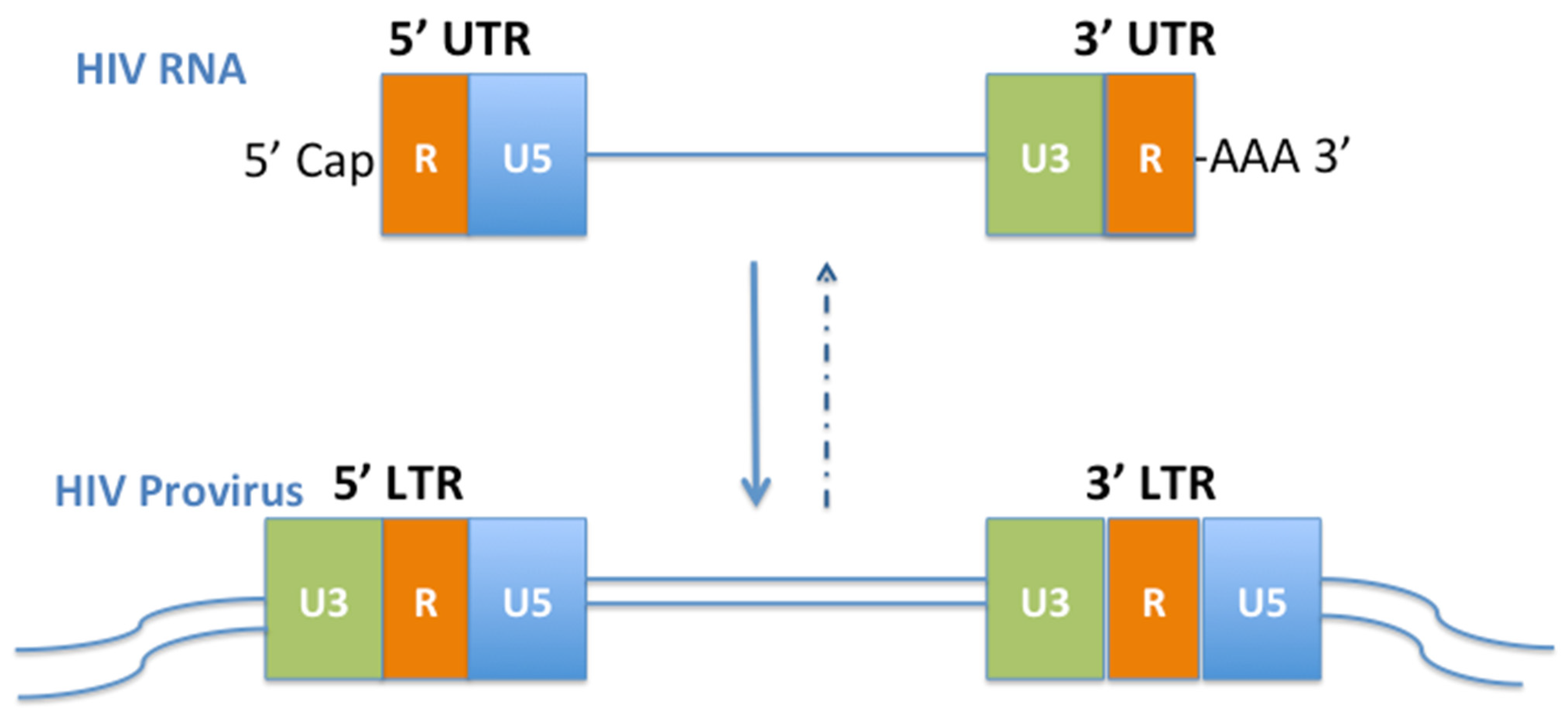
Figure 1. HIV UTR and LTR. U: unique element. R: repeat element. Provirus is a metastable stage in the retroviral lifecycle. A breach of the metastable equilibrium depends on the host cell signals not the virus. Based on the knowledge of X-chromosome inactivation and the stem cell features of CD4 T-cells, the host epigenetic silencing and cART can force the provirus into a stable state—a permanent silencing, similar to the ancient human endogenous retroviruses (HERVs) resided in our DNA.
Viruses 14 01084 g001
Figure 2. Epigenetic Immunity—a genetic immunity. A cell defends its genome against a foreign agent invasion by epigenetic immune regulations [9,10,127]. In HIV infection, the host cell protects its DNA from the viral nucleic acid attack by the epigenetic immunity, consisting of DNA methylation, histone acetylation/methylation, and non-coding RNA (ncRNA) activity. DNA methylation affects the function of double stranded DNA, histone modification affects the function of nucleosome, and ncRNA affects the function of chromatin. Each works independently but synergistically with the other two, guarding the topological structure and function of the DNA, and acting as a writer, reader, eraser, adaptor, modifier, organizer, or programmer [10,118,127]. The epigenetic factors reprogram genetic immune responses embodying gene activation, silencing, epigenetic memory, and chromatin remodeling in responding to environmental stimuli, specifically pathogenic foreign nucleic acids such as HIV.
Viruses 14 01084 g002
Publisher’s Note: MDPI stays neutral with regard to jurisdictional claims in published maps and institutional affiliations.