Next Article in Journal
An Improved αvβ6-Receptor-Expressing Suspension Cell Line for Foot-and-Mouth Disease Vaccine Production
Previous Article in Journal
Lack of Evidence of Chikungunya Virus Infection among Blood Donors during the Chikungunya Outbreak in Lazio Region, Italy, 2017
 
 
Correction published on 20 February 2024, see Viruses 2024, 16(3), 319.
Article

Efficacy Assessment of Phage Therapy in Treating Staphylococcus aureus-Induced Mastitis in Mice

1
Heilongjiang Key Laboratory for Animal Disease Control and Pharmaceutical Development, Department of Preventive, Veterinary Medicine, College of Veterinary Medicine, Northeast Agricultural University, Harbin 150038, China
2
Branch of Animal Husbandry and Veterinary of Heilongjiang Academy of Agricultural Sciences, Qiqihar 161000, China
3
Department for Radiological Protection, Heilongjiang Province Center for Disease Control and Prevention, Harbin 150030, China
4
Promotion Demonstration Department of Heilongjiang Fishery Technology Extension Station, Harbin 150030, China
*
Authors to whom correspondence should be addressed.
Viruses 2022, 14(3), 620; https://doi.org/10.3390/v14030620
Submission received: 11 February 2022 / Revised: 11 March 2022 / Accepted: 14 March 2022 / Published: 16 March 2022 / Corrected: 20 February 2024
(This article belongs to the Topic Veterinary Infectious Diseases)

Abstract

:
The primary aim of this study was to evaluate the efficacy of phage against mastitis induced by drug-resistant S. aureus in a mouse model. In this study, five S. aureus phages—4086-1, 4086-2, 4086-3, 4086-4, and 4086-6—were isolated from milk samples secreted by mastitis cows. Transmission electron microscopy showed that all the five phages had icosahedral heads and short non-contractile tails, which are typical characteristics of the family Podoviridae. All these phages were species-specific against S. aureus. The one-step growth curve showed a short latency period (10–20 min) and high burst size (up to 400 PFU/infected cell). To evaluate the effectiveness of the phage 4086-1 in the treatment against mastitis, a mouse model of mastitis was challenged with drug-resistant S. aureus. The results showed the proliferation of S. aureus in the mammary glands was significantly inhibited after treating by phage 4086-1. The concentrations of TNF-α and IL-6 decreased significantly, which demonstrated the phages could effectively alleviate the inflammatory responses. Furthermore, the histopathological analysis showed that inflammatory infiltration in the mammary glands was significantly reduced. These results demonstrate that phage may be a promising alternative therapy against mastitis caused by drug-resistant S. aureus.

1. Introduction

Bovine mastitis is a major cause of economic losses in the dairy industry as well as a public health concern [1]. Mastitis may be caused by several microorganisms, while S. aureus is one of the most frequent causative agents. In some countries, the prevalence is even as high as 50% [2]. S. aureus is a gram-positive bacterium involved in various infectious diseases of humans and animals, which is difficult to treat [3,4]. S. aureus infections can lead to a variety of symptoms with severity ranging from mild to life-threatening [5]. Currently, diseases caused by S. aureus are widely treated with antibiotics [6].
Fleming’s discovery of penicillin in the 1940s pioneered the era of antibiotics for the treatment of infections [7]. With the widespread use of penicillin, infectious diseases caused by S. aureus were well controlled at the time. However, many drug-resistant bacteria, such as methicillin-resistant S. aureus (MRSA) and vancomycin-resistant enterococci, have emerged [8]. In recent decades, MRSA has rapidly become the most common drug-resistant pathogen discovered worldwide [9,10]. In addition, MRSA have a strong ability for biofilm formation, which can further complicate eradication by increasing resistance to antimicrobial agents and the host immune response. Currently, vancomycin is the most effective antibiotic for treating MRSA infection and can inhibit the synthesis of cell walls. However, the sensitivity of MRSA to vancomycin is gradually decreasing, and the number of multidrug-resistant S. aureus strains is increasing sharply. Therefore, finding an effective treatment for S. aureus infection is an urgent problem that needs to be solved.
Phages are viruses that infect bacteria and are host specific. In recent years, antibiotic therapy for bacterial infections has been facing unprecedented challenges due to rising rates of drug-resistant infections worldwide [11]. The emergence of a series of drug-resistant strains has led to increased attention towards phage research [12]. Some phages have been proven to be effective in treating bacterial infections in animals. Compared with antibiotics, high specificity and fast self-proliferation are the unique advantages of phages [13]. Moreover, phages must recognize and bind to specific bacterial receptors so they cannot infect human or animal cells. Therefore, the side effects of phage therapy applied to humans and animals are considered to be minimal [14]. In this regard, phage therapy may be an alternative approach to the treatment of bacterial infections in animals, which has already been successfully demonstrated in several animal models [15,16].
Antibiotic resistance greatly challenges clinical treatment of Bovine mastitis. Therefore, new strategies to control S. aureus infection are required in the dairy industry. The primary aim of this study was to evaluate the efficacy of phage against mastitis induced by drug-resistant S. aureus in a mouse model. In the present study, five strains of S. aureus phages were isolated from milk samples secreted by mastitis cows and their biological characteristics were identified, which provided experimental data required for in vivo study application. Furthermore, the efficacy of phage against mastitis compared to a commercial antibiotic was evaluated in mice with induced mastitis, which provides preclinical evidence for the feasibility of phage therapy against mastitis.

2. Materials and Methods

2.1. Bacterial Strains

S. aureus CVCC 546, Staphylococcus gallinarum CVCC 528, and Escherichia coli CVCC 10,141 were purchased from the China Veterinary Microbial Culture Management Center (CVCC, Beijing, China). S. aureus 4086, S. aureus Ben, S. aureus ATCC 43,300, Staphylococcus xylosus 17, Micrococcus luteus 26,003, Staphylococcus saparophytics 17, Staphylococcus saparophytics E4, Staphylococcus saparophytics X4, Staphylococcus haemolyticus 13, Enterococcus faecalis 13, Bacillus subtilis, and Pasteurella P-1059 were all preserved in our laboratory.

2.2. Phage Isolation and Culture Conditions

The milk samples secreted by cows with mastitis were collected from cattle farms in Heilongjiang Province. S. aureus CVCC 546 was used as the indicator strain for phage isolation from milk samples. The milk samples were centrifuged at 6500× g for 20 min. The supernatants were collected and filtered with a 0.22-μm pore size filter [17,18]. Each sample filtrate (0.5 mL) and 0.2 mL of fresh culture of the indicator strain (108 colony-forming units [CFU]/mL) was added to 7 mL of LB semi-solid medium and incubated at 37 °C. Single plaques were selected and inoculated in 5 mL of LB broth, and 100 μL indicator bacteria were added and cultured overnight at 37 °C at 200 rpm. The phages were purified by five cycles of resuspension, dilution, and replating individual lysed plaque, respectively. Phage enumeration (plaque-forming units (PFU)/mL) was performed using the double-layer plaque titration method [19], and the operation was repeated three times.

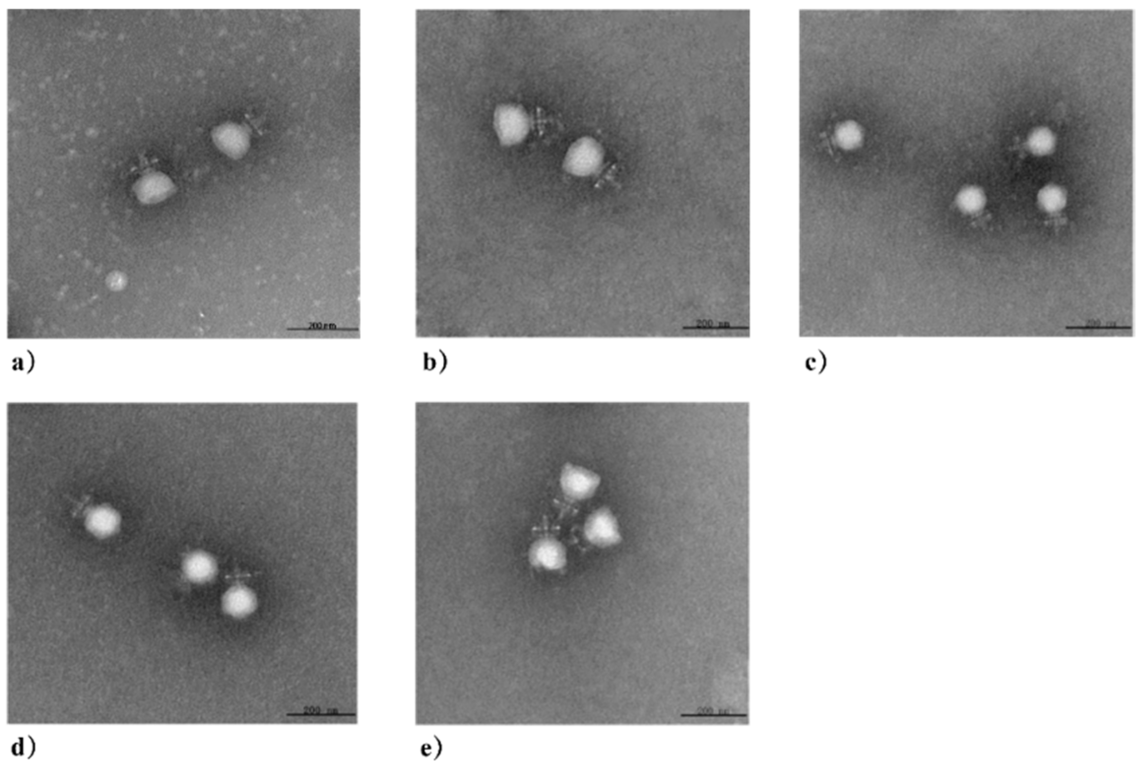
2.3. Transmission Electron Microscopy
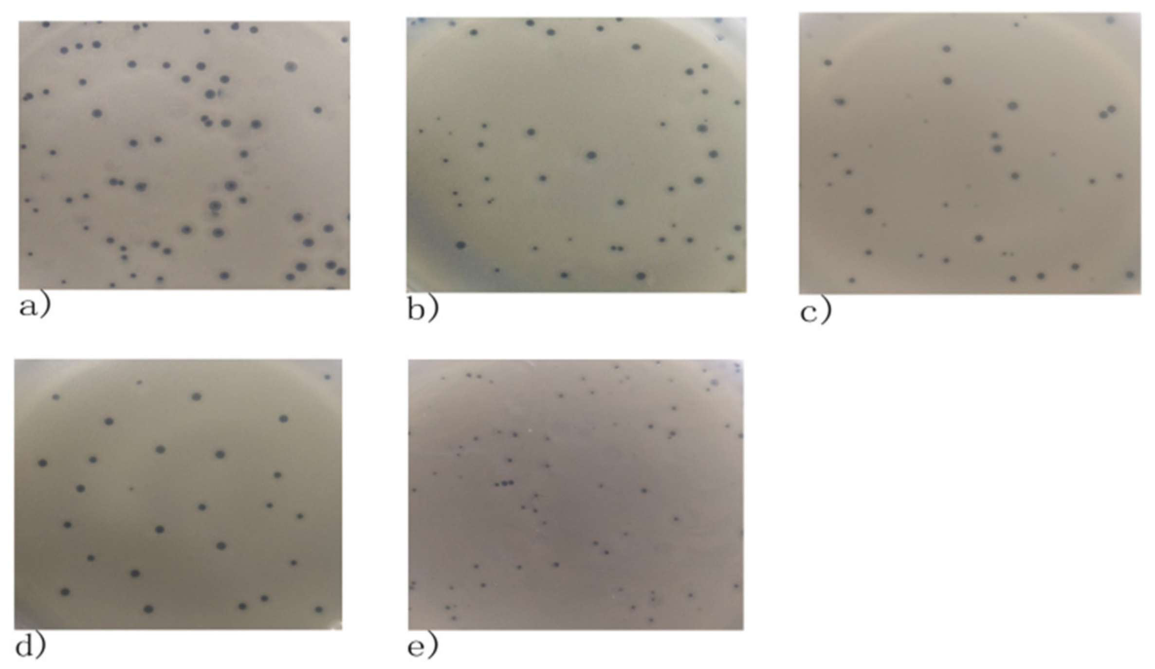
The morphology particles of phages were examined by transmission electron microscopy (TEM). A total of 10 μL of purified phage particles (1 × 10⁸ PFU/mL) were dropped onto a copper acid grid for about 10 min and stained with 2% (w/v) phosphotungstic acid. After drying, the phages were observed under an H-7650 TEM (Hitachi Co., Tokyo, Japan).

2.4. Spot Test

S. aureus CCVC 546, S. aureus 4086, S. aureus Ben, S. aureus ATCC 43,300, S. xylosus 17, M. luteus 26,003, S. saparophytics 17, S. saparophytics E4, S. saparophytics X4, S. haemolyticus 13, E. faecalis 13, B. subtilis, Pasteurella p 1059, S. gallinarum CVCC 528, E. coli BL21, E. coli DH 5 α, and E. coli CVCC 10,141 were used to test the infectivity of the isolated phages. Then, 200 μL of fresh bacterial culture was spread on an LB agar plate. Then, 4 μL of phage cultures (1 × 10⁹ PFU/mL) were added to the center of the plate respectively after it was completely dried. After incubation at 37 °C for 12 h, the S. aureus culture without phage was incubated at 37 °C overnight as the control.

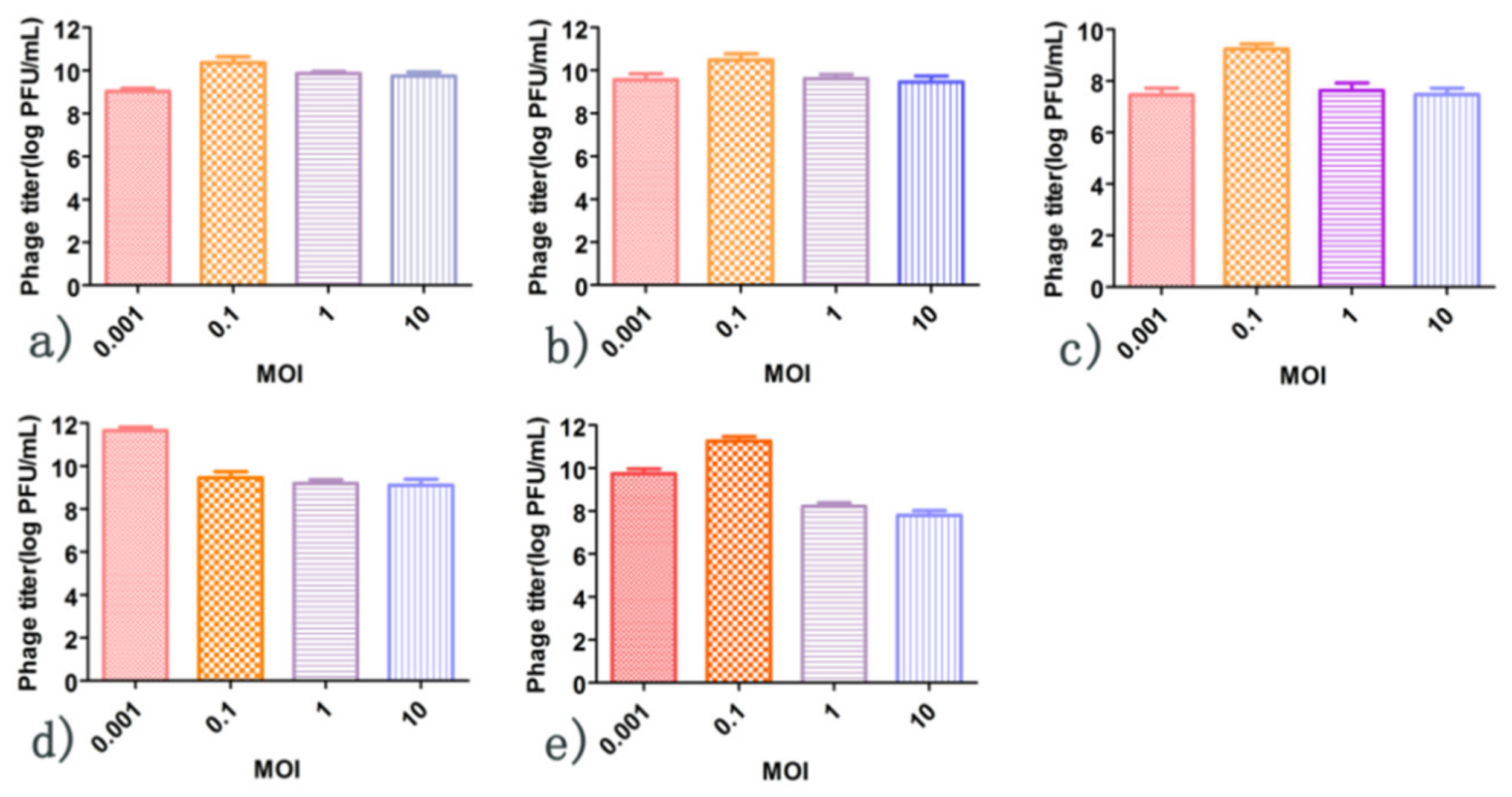
2.5. Optimal Multiplicity of Infection

The host bacteria (S. aureus CVCC 546) were cultured in LB medium at 37 °C with shaking at 200 rpm until the growth reached an exponential phase (optical density at 600 nm [OD600] = 0.6). S. aureus cultures were then diluted 1:100 in fresh LB medium and cultured in a shaking incubator at 37 °C. Then, the corresponding phages were added into the host cultures according to the multiplicity of infection (MOI) ratios of 0.001, 0.01, 0.1, 1, and 10. All samples were cultured in LB medium at 37 °C for 4 h. The phage lysates were centrifuged at 12,000× g for 4 min.
The phage titers were determined using the double-layer plate method, and the operation was repeated three times. The optimal MOI was determined when the phage titer reached its highest value. The assay was performed in triplicate.

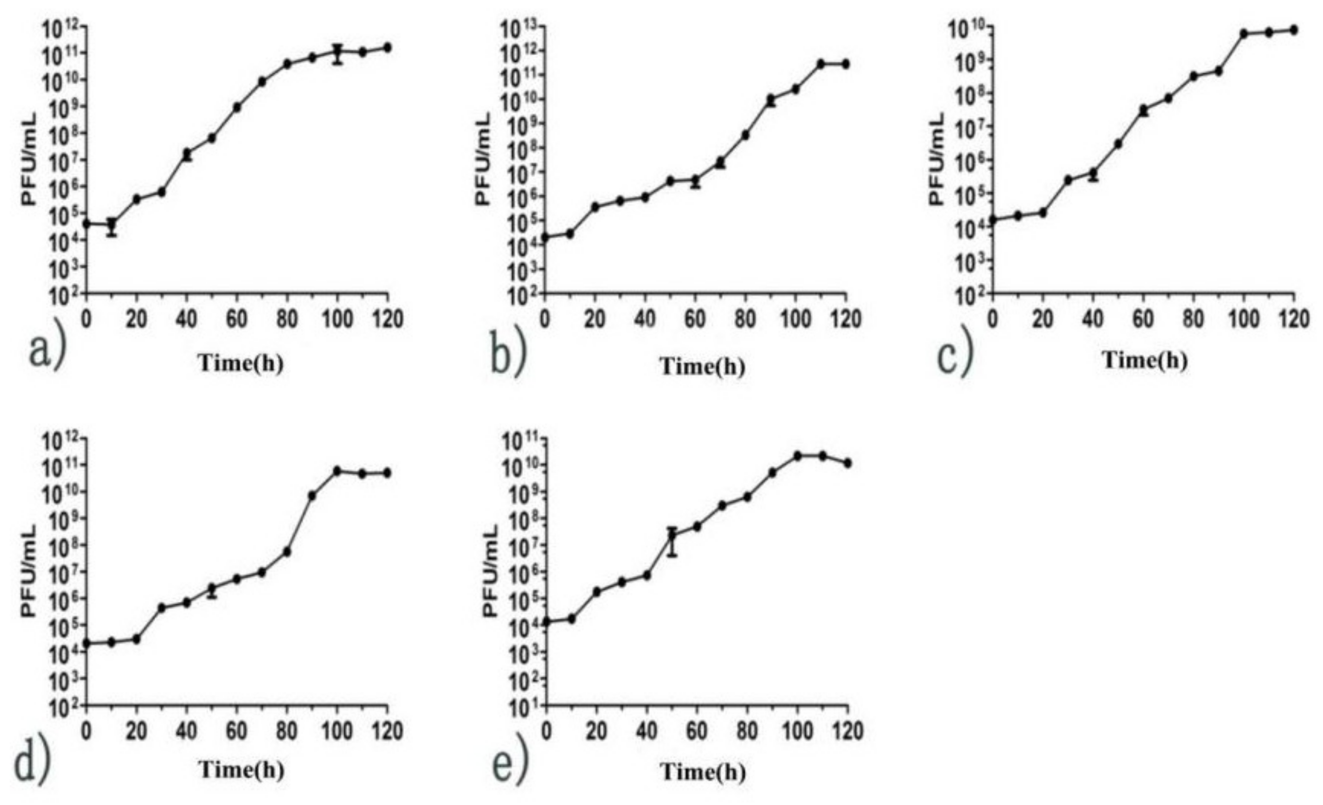
2.6. One-Step Growth Curve

S. aureus CVCC 546 was inoculated at a rate of 2% into 20 mL LB broth and cultured at 37 °C with shaking (200 rpm) until its growth reached the exponential growth early phase (OD600 = 0.1–0.2). The cells were then centrifuged at 3000× g for 10 min. The pellets were resuspended in 2.5 mL LB broth. The isolated phages were added into these suspensions, respectively, according to their optimal MOI and cultured at 37 °C with shaking (200 rpm). Cultures (500 μL) were sampled every 15 min for 2 h. The phage titers at each time point were determined by the double-layer plate method to obtain one-step growth curves of the phages. The assay was performed in triplicate.

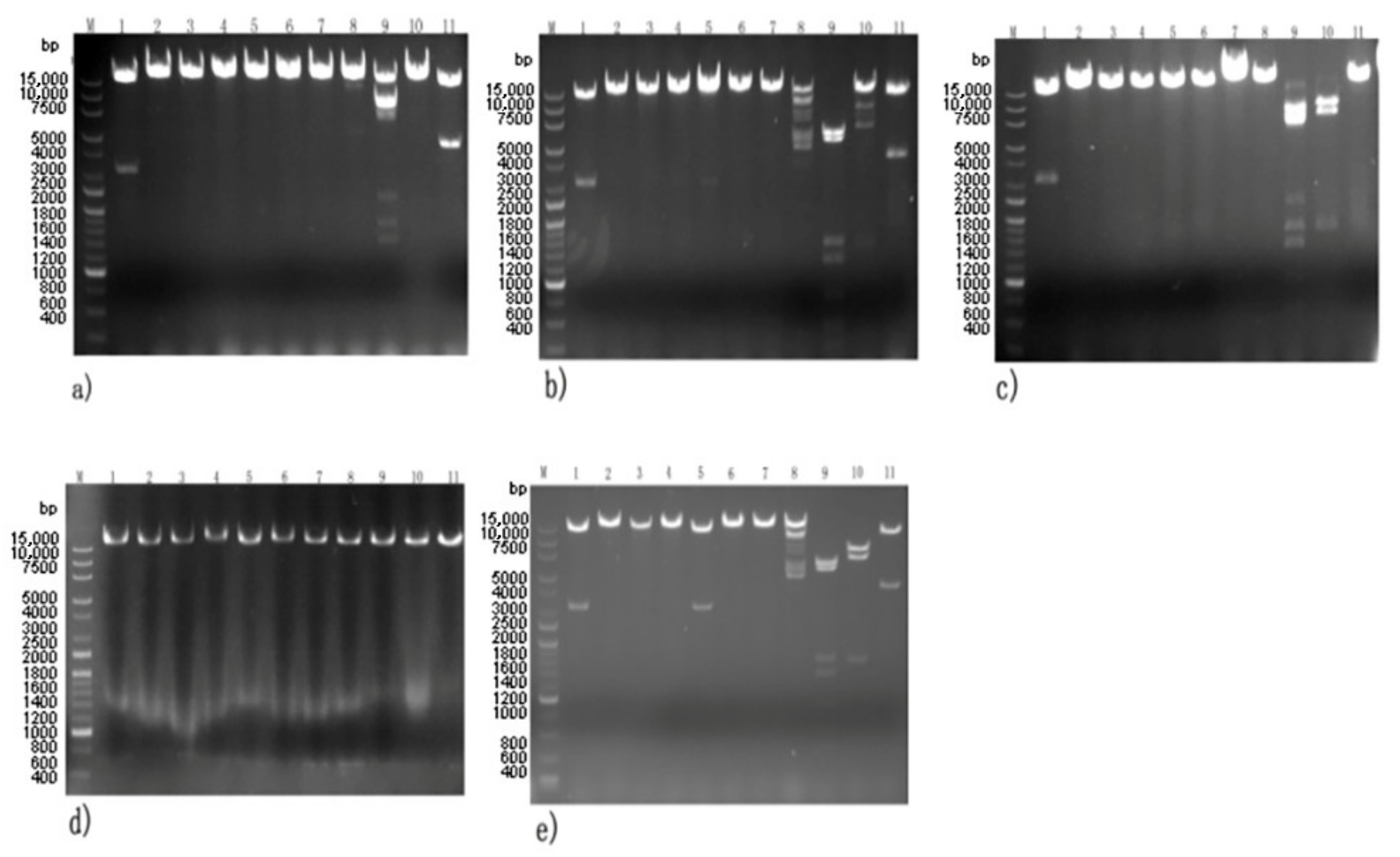
2.7. DNA Extraction and Identification

Overnight cultures of all phages were harvested for DNA extraction. Chloroform was added to the phage cultures at a final concentration of 0.5%. The cultures were incubated at 37 °C for 1 h to make it lyse completely. Lysates obtained were centrifugated at 8000× g for 10 min. The supernatants were collected and then filtered. The filtrates were treated with DNase I (a final concentration of 50 U/mL, Sigma, St. Louis, MO, USA) and RNase A (a final concentration of 50 U/mL, Sigma, St. Louis, MO, USA) at 37 °C for 1 h. Phage particles were treated with 1 mol/L NaCl in ice for 1 h, then concentrated with 10% (w/v) polyethylene glycol 8000. After centrifuging at 8000× g for 15 min, the pellets were collected and resuspended in 1 mL SM buffer (0.05 M Tris-HCl, pH 7.5, 0.1 M NaCl, 0.017 M MgSO4, 0.01% gelatin). Phage DNAs were obtained by the phenol chloroform-extraction method, as previously reported [20,21].
The phage genomes were digested with the endonucleases EcoRI, HpaI, RsrII, SacI, Sph I, Bgl II, Sma I, Sac II, SnaB I, Avr II, and Pst I. The products were identified using agarose gel electrophoresis.

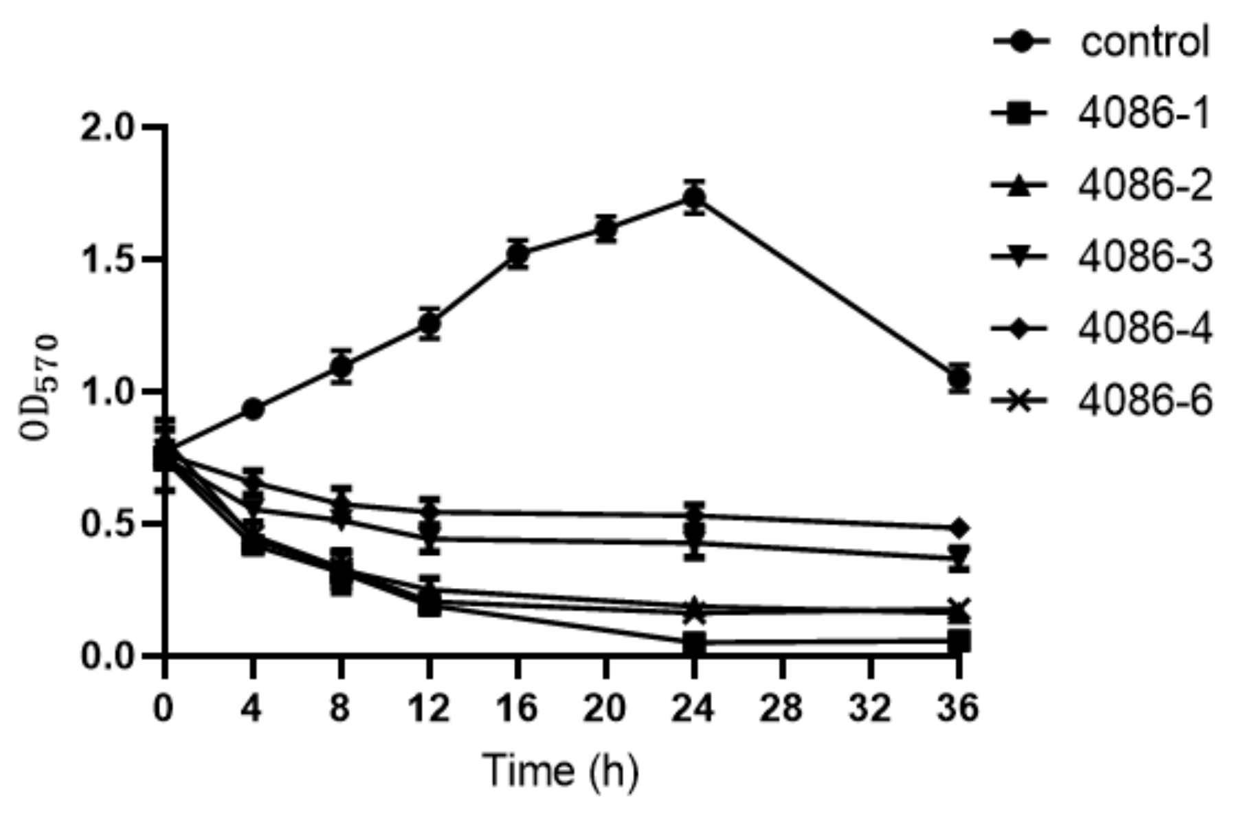
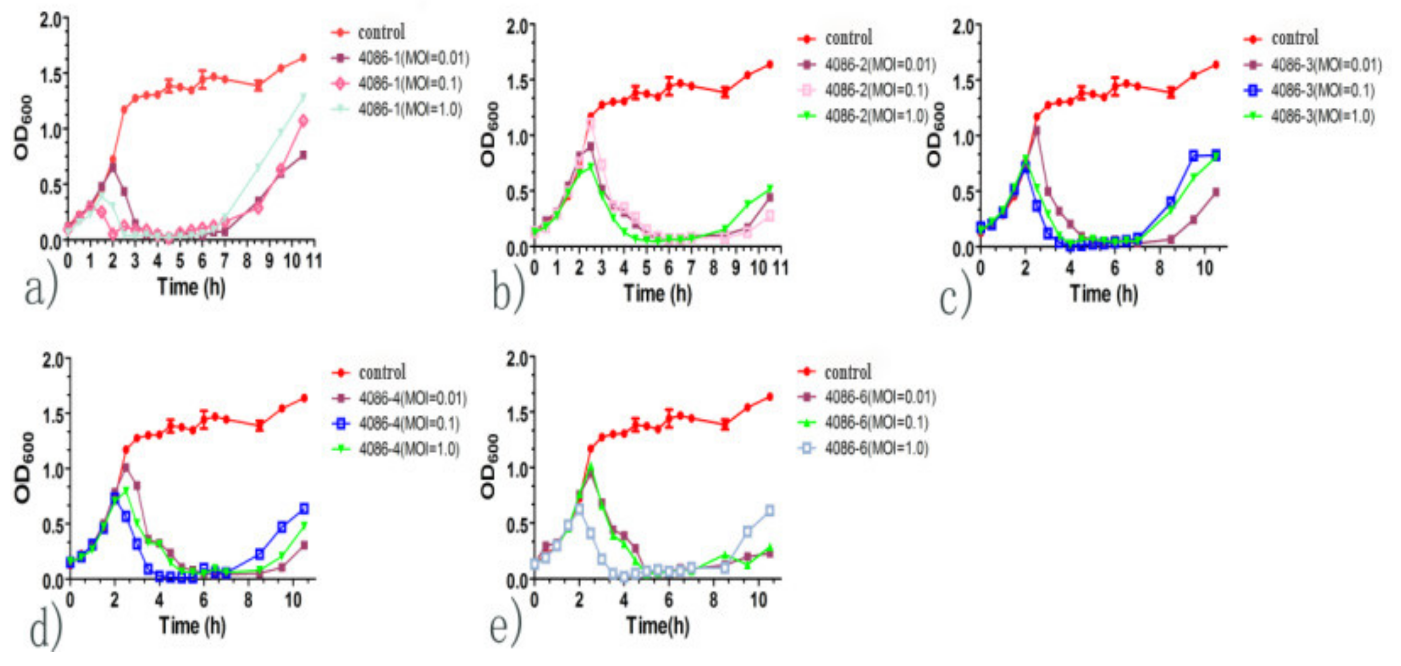
2.8. Growth Kinetic Curve of In Vitro Lysis

S. aureus CVCC 546 was inoculated at a rate of 2% into 20 mL LB broth and cultured at 37 °C with shaking (200 rpm) until its growth reached the exponential growth early phase (OD600 = 0.1–0.2). Then, the phages were inoculated into the medium at MOI ratios of 0.01, 0.1, 1, and 10. One milliliter of culture was continuously sampled every 30 min within a period of 10.5 h. The OD600 was measured using a spectrophotometer (Thermo Fisher Scientific, Waltham, MA, USA), and growth dynamics curves were constructed based on this value. The assay was performed in triplicate.

2.9. Detection of Phages to Remove Biofilms

To explore the ability of phages to remove S. aureus biofilms, S. aureus CVCC 546 was streaked on LB agar and cultured at 37 °C for 12 h. Then, a single colony was picked and inoculated in a 5 mL LB broth and cultured at 37 °C for 10 h with shaking (200 rpm). The cultures were centrifuged at 12,000× g for 10 min. The pellets were collected, and the concentration of the bacteria was adjusted to approximately 1 × 10⁸ CFU/mL with phosphate-buffered saline (PBS). Then, 50 μL of bacterial suspension and 150 μL of LB medium (with a final glucose concentration of 5%) were added to 96-well plates. After treatment at 37 °C for 24 h, the supernatant was discarded. PBS (250 μL) was added to each well, and the wells were washed twice to remove unbound bacteria. Then, 200 μL of phage suspensions (1 × 10⁹ PFU/mL) were added to each well respectively. After treatment for 4, 8, 12, 24, and 36 h, the supernatants in the wells were discarded. The 96-well plates were washed twice and then dried at 60 °C for 10 min. Next, 200 μL of 0.1% crystal violet solution was added to each well and incubated for 10 min. After discarding the crystal violet solution, 250 μL of PBS was added to each well and washed three times to remove the unbound crystal violet. The optical density was measured at 570 nm using a spectrophotometer (Thermo Fisher Scientific, Waltham, MA, USA).

2.10. Infectious Inoculum Preparation

A total of 600 μL of S. aureus CVCC 546 (1 × 10⁸ CFU/mL) was inoculated into a 30 mL LB broth and cultured at 37 °C overnight. The culture (2 mL) was sampled and centrifuged at 4000× g for 15 min. The pellets were collected and washed three times with 1 mL PBS and resuspended in PBS to a concentration of 1 × 10⁸ CFU/mL.

2.11. In Vivo Phage Therapy (Mouse Model)

To evaluate the effects of phages against S. aureus CVCC 546 in vivo, an experimental S. aureus-induced mastitis mouse model was used, as previously described, with some modifications [22,23]. The mice were anesthetized with 10% chloral hydrate with an injection of 0.05 µL/g. The fourth pair of nipples and the surrounding skin were disinfected with 75% alcohol. Then, a 2 mm incision was made using sterilized scissors to expose the mammary duct, and 100 µL of the bacterial suspension (1 × 10⁸ CFU/mL) or sterilized PBS was slowly injected into the mouse mammary duct.
Female BALB/C mice (13-week-old) lactating for seven days were randomly divided into four groups (A–D) with four mice per group. Mice in group A were maintained as healthy mice (blank control). Mice in groups B–D were injected with S. aureus CVCC 546 (1 × 10⁸ CFU/mL) to induce mastitis. Two hours after administration, the mice in the B-D groups were treated with 100 µL of PBS, phage 4086-1 (1 × 10⁸ PFU/mL), and ceftiofur sodium (5 mg/kg), respectively. The animals were euthanized, and the mammary tissue samples were collected aseptically.

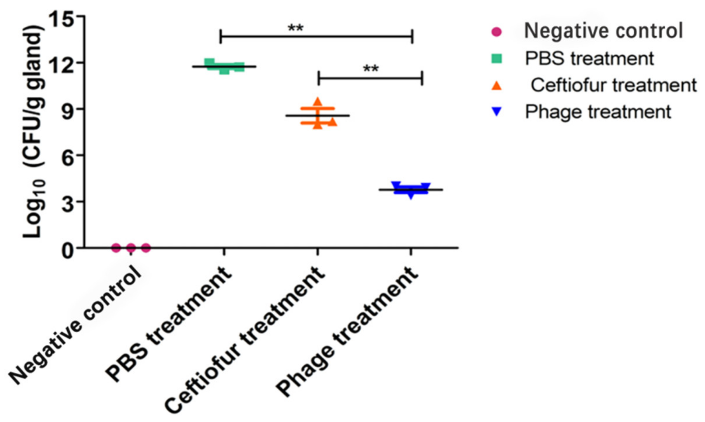
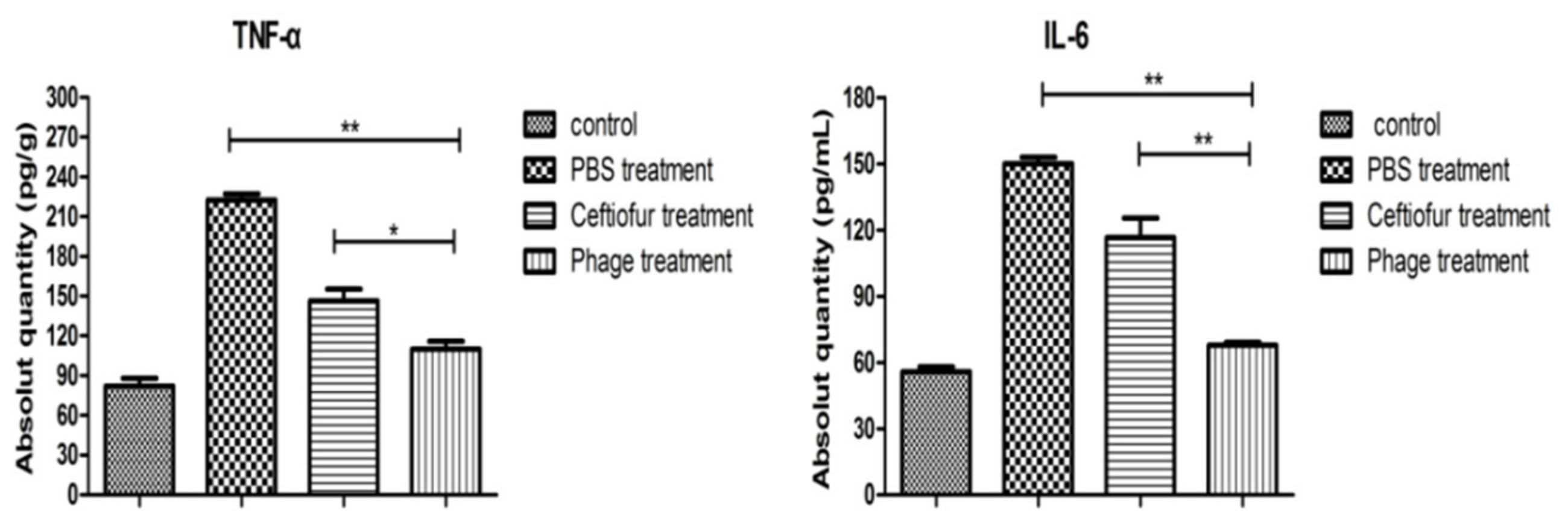
2.12. Bacterial Cell Counting and Detection of Cytokines

The minced mammary glands (100 mg) were homogenized in 1 mL of PBS with TissueLyser II (QIAGEN, Hilden, Germany). The homogenate was serially diluted (1:10) with PBS. Bacterial colonies (CFUs) were counted using the plate method [24].
The homogenate was centrifuged at 4 °C (10,000× g) for 10 min. The supernatant was used to measure tumor necrosis factor (TNF-α) and interleukin 6 (IL-6) concentrations. ELISA was performed using a kit (MEIMIAN, Jiangsu, China) according to the manufacturer’s protocol.

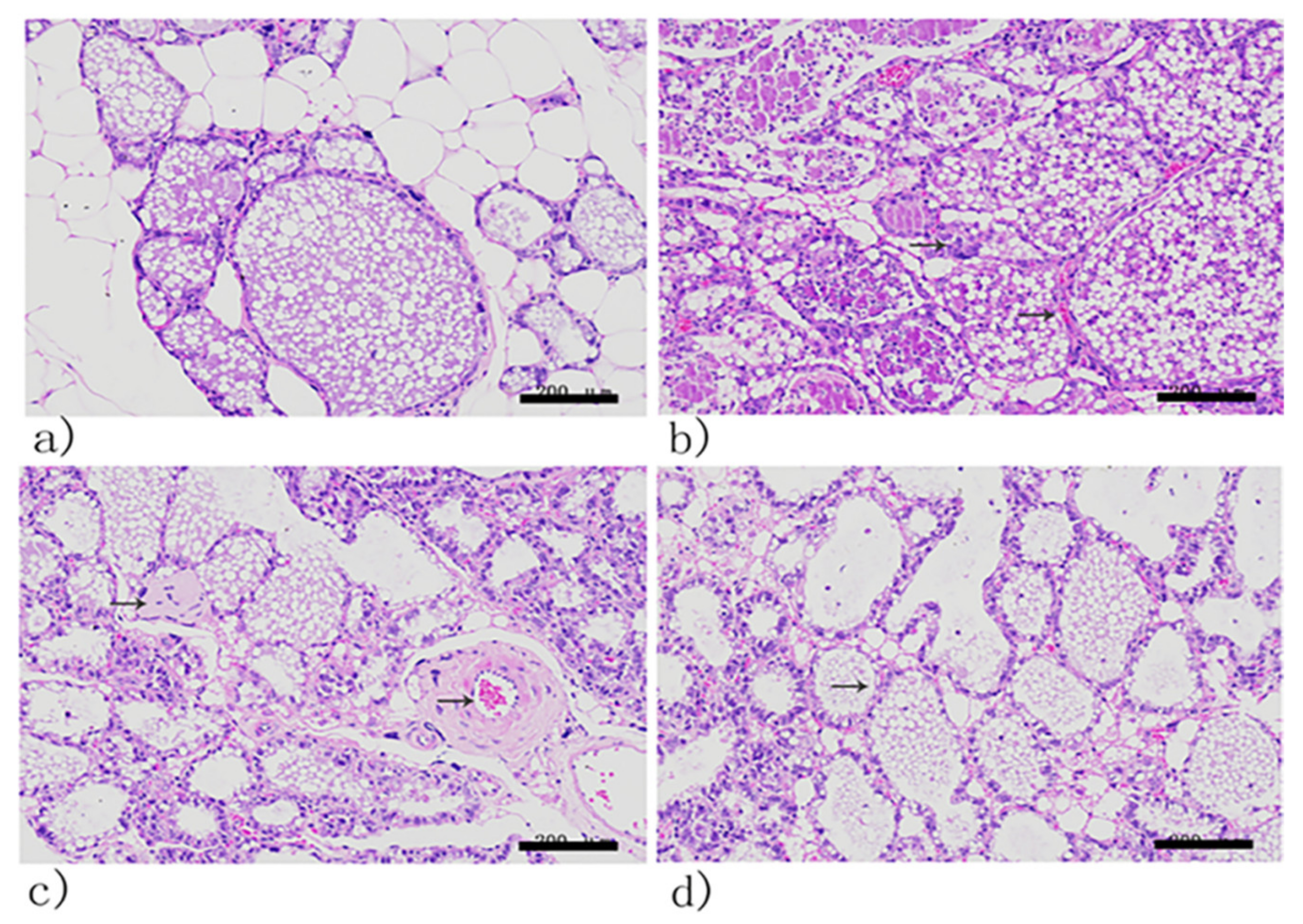
2.13. Histopathological Analysis

For histological analysis, the dissected mammary glands were fixed in 10% neutral buffered formalin, processed into paraffin wax, and sectioned at a nominal thickness of 5 μm. The slides were stained with hematoxylin and eosin (HE) and morphological changes were analyzed using light microscopy.

2.14. Statistical Analysis

Data are presented as mean ± standard deviation (SD). Statistical significance was determined using SPSS 22.0 software (IBM) for Windows, version 22.0. Results within samples were determined using Tukey’s multiple comparison statistical test. Statistical significance was set at p < 0.001 or p < 0.05.

3. Results

3.1. Morphology of Phages

Five phages were isolated and designated as 4086-1, 4086-2, 4086-3, 4086-4, and 4086-6, respectively. All five phages produced clear plaques, indicating that they could lyse S. aureus CVCC 546. The plaque diameters of 4086-1, 4086-2, 4086-3, and 4086-4 were all about 2–3 mm, while that of 4086-6 were approximately 1 mm (Figure 1). All the phages had an isometric head of 37.5 ± 3 nm in diameter, a non-contractile tail with a length of 15 ± 3 nm, and a baseplate structure at the tip of the tail (Figure 2). Based on these properties, the five phages should be classified in the family Podoviridae.

3.2. Phage Host Range

The host ranges of the five phages were evaluated using a spot test. Phages 4086-1, 4086-2, and 4086-3 could lyse 4 of the 17 strains tested, while phage 4086-4 and 4086-6 could only lyse 2 of 17 strains (Table 1). Overall, the five phages were species-specific, attacking only S. aureus. The phages 4086-1, 4086-2, and 4086-3 were found to have a wider host range, which can lyse all four S. aureus strains. Phage 4086-4 and phage 4086-6 could only lyse two strains of S. aureus.

3.3. Optimal Multiplicity of Infection

When the MOI was 0.1, the titers of phages 4086-1, 4086-2, 4086-3, and 4086-6 were the highest, which were 1.46 × 10¹⁰ PFU/mL, 3.21 × 10¹⁰ PFU/mL, 7.66 × 10¹⁰ PFU/mL, and 2.35 × 10¹¹ PFU/mL, respectively. While the MOI was 0.001, the titer of phage 4086-4 was the highest at 2.41 × 10¹² PFU/mL (Figure 3).

3.4. One-Step Growth Curve

Based on the MOI of the phages, the one-step growth curve showed a latent period (time interval between the absorption and the start of the first burst) of phages that was approximately 10–20 min. The burst period of the five phages was 80–100 min. Phages 4086-1 and 4086-2 had a larger burst size that was 365.7 PFU/infected cell and 400 PFU/infected cell, respectively (Figure 4).

3.5. Phage Genome Identification

Digestion was performed with different endonucleases, and the results are shown in Figure 5. Phage 4086-1 could be digested by EcoRI, SnaBI, and Pst I. Phage 4086-2 could be digested by EcoRI, SacII, SnaB I, Avr II, and Pst I. Phage 4086-3 could be digested by EcoRI, SnaB I, and Avr II. Phage 4086-6 could be digested by EcoRI, SphI, SacII, SnaB I, Avr II, and Pst I. Phage 4086-4 could not be digested by any of these endonucleases. This suggested that phages 4086-1, 4086-2, 4086-3, 4086-4, and 4086-6 were different from each other (Figure 5).

3.6. Growth Kinetic Curve of In Vitro Lysis

When S. aureus CVCC 546 was inoculated and cultured to the exponential growth early phase, the phages were inoculated at an MOI of 0.01, 0.1, 1, and 10. Two hours after infection, the cultures showed a significant decrease in OD600 compared to that of the control. The results showed that the phages effectively inhibited the growth of S. aureus CVCC 546. After 8 h of treatment, phage-resistant bacteria appeared (Figure 6).

3.7. The Ability of Phages to Remove Biofilms

The effects of phages on the removal of the S. aureus biofilms were investigated. Bacterial biofilm formation was measured at various time points. The extent of an incremental decrease in biomass in the biofilms was characterized by the OD570 value after crystal violet staining and extraction of the dye with ethanol. The biofilms decreased effectively after 24 h of treatment (Figure 7). In particular, phages 4086-1, 4086-2, and 4086-6 had stronger abilities to remove biofilms.

3.8. Colony-Forming Units

The S. aureus counts of the infected mammary glands were measured using the plate-counting method. S. aureus CFUs were greatly reduced after phage treatment (Figure 8), indicating that phages could effectively reduce the number of S. aureus.

3.9. Changes in TNF-α and IL-6 Concentrations

The infected mammary glands were treated with the phage 4086-1. The concentrations of TNF-α and IL-6 were determined using ELISA kits. Compared with the PBS-treated group, the ceftiofur sodium-treatment group and the phage-treatment group both showed significant decreases in the concentrations of TNF-α and IL-6. The phage-treated group displayed the most significant reduction in the concentrations of TNF-α and IL-6 (p < 0.01) (Figure 9). The above results demonstrated that phages could effectively alleviate inflammatory responses after phage treatment.

3.10. Histopathological Analysis

Histopathological analysis of mammary glands is shown in Figure 8. Tissue sections of the mammary glands of healthy mice revealed no abnormal histopathological changes (Figure 10a). In contrast, a massive proliferation of bacteria could be observed in the PBS-treated group. The acinar epithelial cells were partially necrotic and apoptotic, and inflammatory infiltrates were observed around the acinar space and acini (Figure 10b).
The mammary glands of the ceftiofur sodium-treated groups showed relatively fewer pathological changes. Mammary epithelial tissues were relatively intact with a variable, minimal to moderate degree of necrotic (or apoptotic) epithelial cells and a small amount of inflammatory cell infiltration (Figure 10c). Mice treated with phages display normal mammary glands. The cuboidal epithelial cells and acini were intact (Figure 10d). Furthermore, inflammatory cell infiltration was reduced.

4. Discussion

In the present study, five lytic phages were isolated from milk samples secreted by mastitis cows and characterized. After characterization of the phages in vitro, a mouse model for staphylococcal mastitis was established to evaluate the effect of the phage 4086-1 in vivo. The results showed that the phage 4086-1 was a promising therapeutic antibacterial agent as it exhibited an outstanding efficacy for treating S. aureus-induced mastitis.
Taxonomically, most S. aureus phages belong to the Siphoviridae family, and only a few belong to the Podoviridae and Myoviridae families, which are mainly virulent phages [25]. The five phages isolated in this experiment belonged to the Podoviridae family and presented favorable characteristics, such as species-specificity, short latent periods, and large burst sizes. Especially, the burst sizes of phages 4086-1 and 4086-2 are 365.7 and 400 PFU/infected cell, respectively. The burst sizes were much larger than that of phage CSA13 (230 PFU/infected cell) and phage LM12 (52 PFU/infected cell) isolated by Yoyeon Cha [26] and Joana Barros [27].
Many antibiotic-resistant bacteria can form biofilms, which can protect the bacteria in the membrane from physical, chemical, and biological factors. Once the biofilm has formed, the bacteria can evade the host immune response and limit the therapeutic effect of antibiotics. The formation of biofilm causes the infection to alternate between the static and acute stages, which is difficult to cure [28]. Therefore, the search for methods that can suppress biofilm formation is an immensely important and relevant issue. Many studies have shown that phages have significant effects on biofilm removal. Phages phiIBB-PAA2 and phiIBB-PAP21 could both reduce the Pseudomonas aeruginosa biofilm population by approximately 1–2 log after 2 h of infection, and the reduction was further enhanced after 6 h [29]. It has been reported that a single phage or phage cocktail has a good therapeutic effect on biofilm formation by E. coli, A. baumannii, and P. aeruginosa in the chronically infected skin of pigs, highlighting its potential as an alternative or complementary treatment agent to control wound infections [30]. Many strains of S. aureus can form biofilms during growth and reproduction, making the infection more difficult to cure. In this study, five isolated phages were used to remove biofilms formed by S. aureus CVCC 546. Bacterial biofilm formation was measured at different time points after treatment with the isolated phages. The amount of biofilm decreased effectively after 24 h of treatment. In particular, phages 4086-1, 4086-2, and 4086-6 had stronger abilities to remove biofilms.
S. aureus is one of the major causes of mastitis. Most of the S. aureus mastitis isolates displayed antibiotic insensitivity in vitro [6], and they are more difficult to eliminate in vivo with the established clinical treatments [31,32]. Moreover, it has been reported that multiple multidrug-resistant strains present a serious challenge in the treatment of S. aureus-induced mastitis infections [33,34]. This study evaluated the efficacy of the S. aureus phage 4086-1 in treating mastitis using mouse models.
Phage therapy studies with mouse models have shown that the phage 4086-1 was efficient in reducing the amount of S. aureus in the mammary glands over time. Increased concentrations of TNF-α and IL-6 were detected in mice following the induction of mastitis, which indicated an ongoing inflammatory process. Similarly, after treatment with phages, the concentrations of TNF-α and IL-6 in the mammary glands decreased significantly. Both the cytokines could be involved in cell proliferation, inflammation, and immunity at the local and systemic concentrations and were often used to assess the degree of inflammation in mouse mammary glands [35,36]. The results showed that phages could effectively alleviate humoral and cell-mediated immune responses. It has also been reported that the concentrations of TNF-α and IL-6 could be downregulated after treatment with a single-dose T4 phage in an E. coli-induced mastitis mouse model [2]. This indicated that E. coli phages could effectively alleviate inflammatory responses, which corresponds to the results described in this study.
In addition, the pathological analysis also showed that the mammary glands in the phage-treatment groups were relatively intact, with significantly less inflammatory cell infiltration. This demonstrated that phages were efficient in reducing the density of the infecting bacterial population to a level that may allow the host immune response to defend and clear the infections, which provides evidence for the application of phage therapy for the treatment of mastitis.
In summary, the therapeutic effects of S. aureus phages on mastitis caused by MRSA strains were evaluated. Based on the data obtained, phage therapy could be considered an innovative approach to replace antibiotics in the treatment of drug-resistant strains.

5. Conclusions

In this study, we isolated five lytic phages from the milk of cows suffering from mastitis, which could lyse S. aureus CVCC 546 (MRSA strain) and belong to the family Podoviridae. The one-step growth curve showed a short latency period (10–20 min) and high burst size (up to 400 PFU/infected cell). In a mouse model of mastitis, phage treatment effectively alleviated inflammatory responses and significantly reduced inflammatory infiltration in mammary glands. These results demonstrated that phage treatment may be a potential therapy against mastitis caused by drug-resistant S. aureus.

Author Contributions

Conceptualization, X.Q.; methodology, F.T., X.X., S.Z. and J.L.; validation, X.W. and J.L.; formal analysis, X.X. and S.Z.; investigation, Y.J., H.Z. and W.C.; samples, G.L., L.Z. and R.W.; data curation, F.T., X.X. and S.Z.; writing—original draft preparation, F.T.; writing—review and editing, F.T., X.Q., L.W., L.T. and Y.L.; supervision, X.Q., L.W., L.T. and Y.L.; project administration, F.T., Y.J. and W.C.; funding acquisition, X.Q. All authors have read and agreed to the published version of the manuscript.

Funding

This research was funded by the Natural Science Foundation of Heilongjiang Province (Grant no: LH2020C022), Key Research and Development Program of Heilongjiang Province (Grant no: GA21B004), and the National Natural Science Foundation of China (NSFC) (No. 32072876).

Institutional Review Board Statement

The study was conducted according to the guidelines of the Declaration of Helsinki and approved by the Ethical Committee for Animal Experiments of Northeast Agricultural University, China (protocol code NEAU2018024).

Informed Consent Statement

Not applicable.

Data Availability Statement

De-identified data are available through personal email requests to the authors, Fei Teng ([email protected]) and Xinyuan Qiao ([email protected]).

Conflicts of Interest

The authors declare no conflict of interest. The funders had no role in the design of the study; in the collection, analyses, or interpretation of data; in the writing of the manuscript; or in the decision to publish the results.

References

  1. Da Silva Duarte, V.; Dias, R.S.; Kropinski, A.M.; Campanaro, S.; Treu, L.; Siqueira, C.; Vieira, M.S.; Da Silva Paes, I.; Santana, G.; Martins, F.; et al. Genomic analysis and immune response in a murine mastitis model of vB_EcoM-UFV13, a potential biocontrol agent for use in dairy cows. Sci. Rep. 2018, 8, 6845. [Google Scholar] [CrossRef] [PubMed]
  2. Leitner, G.; Lubashevsky, E.; Trainin, Z. Staphylococcus aureus vaccine against mastitis in dairy cows, composition and evaluation of its immunogenicity in a mouse model. Vet. Immunol. Immunopathol. 2003, 93, 159–167. [Google Scholar] [CrossRef]
  3. Avedissian, S.N.; Rhodes, N.J.; Shaffer, C.L.; Tran, L.; Bradley, J.S.; Le, J. Antimicrobial prescribing for treatment of serious infections caused by Staphylococcus aureus and methicillin-resistant Staphylococcus aureus in pediatrics: An expert review. Expert Rev. Anti-Infect. Ther. 2021, 19, 1107–1116. [Google Scholar] [CrossRef] [PubMed]
  4. Toledo-Silva, B.; de Souza, F.N.; Mertens, K.; Piepers, S.; Haesebrouck, F.; De Vliegher, S. Bovine-associated non-aureus staphylococci suppress Staphylococcus aureus biofilm dispersal in vitro yet not through agr regulation. Vet. Res. 2021, 52, 114. [Google Scholar] [CrossRef] [PubMed]
  5. Tong, S.; Davis, J.; Eichenberger, E.; Holland, T.; Fowler, V. Staphylococcus aureus infections: Epidemiology, pathophysiology, clinical manifestations, and management. Clin. Microbiol. Rev. 2015, 28, 603–661. [Google Scholar] [CrossRef] [PubMed]
  6. Breyne, K.; Honaker, R.; Hobbs, Z.; Richter, M.; Zaczek, M.; Spangler, T.; Steenbrugge, J.; Lu, R.; Kinkhabwala, A.; Marchon, B.; et al. Efficacy and Safety of a Bovine-Associated Staphylococcus aureus Phage Cocktail in a Murine Model of Mastitis. Front. Microbiol. 2017, 8, 2348. [Google Scholar] [CrossRef]
  7. Guo, Y.; Song, G.; Sun, M.; Wang, J.; Wang, Y. Prevalence and Therapies of Antibiotic-Resistance in Staphylococcus aureus. Front. Cell. Infect. Microbiol. 2020, 10, 107. [Google Scholar] [CrossRef]
  8. Menichetti, F.; Tagliaferri, E. Antimicrobial resistance in internal medicine wards. Intern. Emerg. Med. 2012, 7, 271–281. [Google Scholar] [CrossRef]
  9. Mediavilla, J.R.; Chen, L.; Mathema, B.; Kreiswirth, B.N. Global epidemiology of community-associated methicillin resistant Staphylococcus aureus (CA-MRSA). Curr. Opin. Microbiol. 2012, 15, 588–595. [Google Scholar] [CrossRef]
  10. Lakhundi, S.; Zhang, K. Methicillin-Resistant Staphylococcus aureus: Molecular Characterization, Evolution, and Epidemiology. Clin. Microbiol. Rev. 2018, 31, e00020-18. [Google Scholar] [CrossRef]
  11. Lin, S.; Koh, J.-J.; Aung, T.; Sin, W.L.W.; Lim, F.; Wang, L.; Lakshminarayanan, R.; Zhou, L.; Tan, D.T.H.; Cao, D.; et al. Semisynthetic Flavone-Derived Antimicrobials with Therapeutic Potential against Methicillin-Resistant Staphylococcus aureus (MRSA). J. Med. Chem. 2017, 60, 6152–6165. [Google Scholar] [CrossRef]
  12. Shlezinger, M.; Khalifa, L.; Houri-Haddad, Y.; Coppenhagen-Glazer, S.; Resch, G.; Que, Y.-A.; Beyth, S.; Dorfman, E.; Hazan, R.; Beyth, N. Phage Therapy: A New Horizon in the Antibacterial Treatment of Oral Pathogens. Curr. Top. Med. Chem. 2017, 17, 1199–1211. [Google Scholar] [CrossRef]
  13. Krut, O.; Bekeredjian-Ding, I. Contribution of the Immune Response to Phage Therapy. J. Immunol. 2018, 200, 3037–3044. [Google Scholar] [CrossRef]
  14. Rios, A.C.; Moutinho, C.; Pinto, F.C.; Fiol, F.D.S.D.; Jozala, A.; Chaud, M.V.; Vila, M.M.; Teixeira, J.; Balcão, V.M. Alternatives to overcoming bacterial resistances: State-of-the-art. Microbiol. Res. 2016, 191, 51–80. [Google Scholar] [CrossRef]
  15. Schmelcher, M.; Powell, A.M.; Camp, M.J.; Pohl, C.S.; Donovan, D.M. Synergistic streptococcal phage λSA2 and B30 endolysins kill streptococci in cow milk and in a mouse model of mastitis. Appl. Microbiol. Biotechnol. 2015, 99, 8475–8486. [Google Scholar] [CrossRef]
  16. Gill, J.J.; Pacan, J.C.; Carson, M.E.; Leslie, K.E.; Griffiths, M.W.; Sabour, P.M. Efficacy and Pharmacokinetics of Bacteriophage Therapy in Treatment of Subclinical Staphylococcus aureus Mastitis in Lactating Dairy Cattle. Antimicrob. Agents Chemother. 2006, 50, 2912–2918. [Google Scholar] [CrossRef]
  17. Moscoso, M.; Suárez, J. Characterization of the DNA replication module of bacteriophage A2 and use of its origin of replication as a defense against infection during milk fermentation by Lactobacillus casei. Virology 2000, 273, 101–111. [Google Scholar] [CrossRef]
  18. Zago, M.; Lanza, B.; Rossetti, L.; Muzzalupo, I.; Carminati, D.; Giraffa, G. Selection of Lactobacillus plantarum strains to use as starters in fermented table olives: Oleuropeinase activity and phage sensitivity. Food Microbiol. 2013, 34, 81–87. [Google Scholar] [CrossRef]
  19. Gutiérrez, D.; Rodríguez-Rubio, L.; García, P.; Billington, C.; Premarante, A.; Rodríguez, A.; Martínez, B. Phage sensitivity and prophage carriage in Staphylococcus aureus isolated from foods in Spain and New Zealand. Int. J. Food Microbiol. 2016, 230, 16–20. [Google Scholar] [CrossRef]
  20. Ung, P.; Peng, C.; Yuk, S.; Ann, V.; Mith, H.; Tan, R.; Miyanaga, K.; Tanji, Y. Fate of Escherichia coli in dialysis device exposed into sewage influent and activated sludge. J. Water Health 2018, 16, 380–390. [Google Scholar] [CrossRef]
  21. Peng, C.; Hanawa, T.; Azam, A.H.; LeBlanc, C.; Ung, P.; Matsuda, T.; Onishi, H.; Miyanaga, K.; Tanji, Y. Silviavirus phage ΦMR003 displays a broad host range against methicillin-resistant Staphylococcus aureus of human origin. Appl. Microbiol. Biotechnol. 2019, 103, 7751–7765. [Google Scholar] [CrossRef]
  22. Geng, H.; Zou, W.; Zhang, M.; Xu, L.; Liu, F.; Li, X.; Wang, L.; Xu, Y. Evaluation of phage therapy in the treatment of Staphylococcus aureus-induced mastitis in mice. Folia Microbiol. 2020, 65, 339–351. [Google Scholar] [CrossRef]
  23. Iwano, H.; Inoue, Y.; Takasago, T.; Kobayashi, H.; Furusawa, T.; Taniguchi, K.; Fujiki, J.; Yokota, H.; Usui, M.; Tanji, Y.; et al. Bacteriophage ΦSA012 Has a Broad Host Range against Staphylococcus aureus and Effective Lytic Capacity in a Mouse Mastitis Model. Biology 2018, 7, 8. [Google Scholar] [CrossRef]
  24. Naghili, H.; Tajik, H.; Mardani, K.; Rouhani, S.M.R.; Ehsani, A.; Zare, P. Validation of drop plate technique for bacterial enumeration by parametric and nonparametric tests. Veter. Res. Forum Int. Q. J. 2013, 4, 179–183. [Google Scholar]
  25. Deghorain, M.; Van Melderen, L. The Staphylococci Phages Family: An Overview. Viruses 2012, 4, 3316–3335. [Google Scholar] [CrossRef]
  26. Cha, Y.; Chun, J.; Son, B.; Ryu, S. Staphylococcus aureus Characterization and Genome Analysis of Podovirus CSA13 and Its Anti-Biofilm Capacity. Viruses 2019, 11, 54. [Google Scholar] [CrossRef]
  27. Barros, J.; Melo, L.; Poeta, P.; Igrejas, G.; Ferraz, M.P.; Azeredo, J.; Monteiro, F. Lytic bacteriophages against multidrug-resistant Staphylococcus aureus, Enterococcus faecalis and Escherichia coli isolates from orthopaedic implant-associated infections. Int. J. Antimicrob. Agents 2019, 54, 329–337. [Google Scholar] [CrossRef]
  28. Lewis, R.; Clooney, A.G.; Stockdale, S.R.; Buttimer, C.; Draper, L.A.; Ross, R.P.; Hill, C. Isolation of a Novel Jumbo Bacteriophage Effective against Klebsiella aerogenes. Front. Med. 2020, 7, 67. [Google Scholar] [CrossRef]
  29. Azeredo, F.J. Use of newly isolated phages for control of Pseudomonas aeruginosa PAO1 and ATCC 10145 biofilms. Res. Microbiol. 2011, 162, 798–806. [Google Scholar]
  30. Milho, C.; Andrade, M.; Boas, D.V.; Alves, D.; Sillankorva, S. Antimicrobial assessment of phage therapy using a porcine model of biofilm infection. Int. J. Pharm. 2019, 557, 112–123. [Google Scholar] [CrossRef]
  31. Barkema, H.; Schukken, Y.; Zadoks, R. Invited Review: The Role of Cow, Pathogen, and Treatment Regimen in the Therapeutic Success of Bovine Staphylococcus aureus Mastitis. J. Dairy Sci. 2006, 89, 1877–1895. [Google Scholar] [CrossRef]
  32. Notebaert, S.; Meyer, E. Mouse models to study the pathogenesis and control of bovine mastitis. A review. Veter. Q. 2006, 28, 2–13. [Google Scholar] [CrossRef] [PubMed]
  33. Omwenga, I.; Aboge, G.O.; Mitema, E.S.; Obiero, G.; Ngaywa, C.; Ngwili, N.; Wamwere, G.; Wainaina, M.; Bett, B. Antimicrobial Usage and Detection of Multidrug-Resistant Staphylococcus aureus, Including Methicillin-Resistant Strains in Raw Milk of Livestock from Northern Kenya. Microb. Drug Resist. 2020, 27, 843–854. [Google Scholar] [CrossRef] [PubMed]
  34. De Lencastre, H.; Oliveira, D.; Tomasz, A. Antibiotic resistant Staphylococcus aureus: A paradigm of adaptive power. Curr. Opin. Microbiol. 2007, 10, 428–435. [Google Scholar] [CrossRef] [PubMed]
  35. Zhao, L.; Guo, Z.; Liu, J.; Wang, Z.; Wang, R.; Li, Y.; Wang, L.; Xu, Y.; Tang, L.; Qiao, X. Recombinant Lactobacillus casei expressing Clostridium perfringens toxoids α, β2, ε and β1 gives protection against Clostridium perfringens in rabbits. Vaccine 2017, 35, 4010–4021. [Google Scholar] [CrossRef]
  36. Maskrey, B.H.; Megson, I.L.; Whitfield, P.D.; Rossi, A.G. Mechanisms of resolution of inflammation: A focus on cardiovascular disease. Arterioscler. Thromb. Vasc. Biol. 2011, 31, 1001–1006. [Google Scholar] [CrossRef] [PubMed]
Figure 1. The plaques formed by S. aureus phages isolated from clinical mastitis. All the five phages could produce clear plaques. The plaque diameters of 4086-1, 4086-2, 4086-3, 4086-4 were about 2–3 mm, while that of 4086-6 was about 1 mm. (a) Phage 4086-1; (b) Phage 4086-2; (c) Phage 4086-3; (d) Phage 4086-4; (e) Phage 4086-6.
Viruses 14 00620 g001
Figure 2. Electron micrograph of S. aureus phages isolated from clinical mastitis. All the phages have an isometric head of 37.5 ± 3 nm in diameter, a non-contractile tail with a length of 15 ± 3 nm, and a baseplate structure at the tip of the tail: (a) Phage 4086-1; (b) Phage 4086-2; (c) Phage 4086-3; (d) Phage 4086-4; (e) Phage 4086-6.
Viruses 14 00620 g002
Figure 3. The optimal multiplicity of infection of S. aureus phages isolated from clinical mastitis. When the MOI was 0.1, the titers of phages 4086-1, 4086-2, 4086-3, and 4086-6 were the highest, which were 1.46 × 10¹⁰ PFU/mL, 3.21 × 10¹⁰ PFU/mL, 7.66 × 10¹⁰ PFU/mL, and 2.35 × 10¹¹ PFU/mL, respectively. While the MOI was 0.001, the titer of phage 4086-4 was the highest at 2.41 × 10¹² PFU/mL. (a) Phage 4086-1; (b) Phage 4086-2; (c) Phage 4086-3; (d) Phage 4086-4; (e) Phage 4086-6.
Viruses 14 00620 g003
Figure 4. One-step growth curve of S. aureus phages isolated from clinical mastitis. The results are shown as the mean ± standard deviation from three independent experiments. The one-step growth curve showed a short latency period (10–20 min) and high burst size (up to 400 PFU/infected cells). (a) Phage 4086-1; (b) Phage 4086-2; (c) Phage 4086-3; (d) Phage 4086-4; (e) Phage 4086-6.
Viruses 14 00620 g004
Figure 5. Agarose gel electrophoresis of generated DNA fragments of the isolated phages by digestion with 11 restriction enzymes. Lane M, DL 15,000 marker. Restriction endonuclease digestion map of phage 4086-1 (a), phage 4086-2 (b), phage 4086-3 (c), phage 4086-4 (d), phage 4086-6 (e) digested by restriction enzymes EcoR I (1), Hpa I (2), Rsr II (3), Sac I (4), Sph I (5), Bgl II (6), Sma I (7), Sac II (8), SnaB I (9), Avr II (10), and Pst I (11), respectively.
Viruses 14 00620 g005
Figure 6. Effect of the isolated phages on bacterial growth. Host bacteria S. aureus CVCC 546 were infected with phage 4086-1, phage 4086-2, phage 4086-3, phage 4086-4, and phage 4086-6 to determine the OD600 at different times. Phages could effectively inhibit the growth of S. aureus CVCC 546 when the MOI was 0.01, 0.1, 1, and 10. (a) Phage 4086-1; (b) Phage 4086-2; (c) Phage 4086-3; (d) Phage 4086-4; (e) Phage 4086-6.
Viruses 14 00620 g006
Figure 7. The effects of the isolated phages on the removal of the S. aureus biofilms. Bacterial biofilm formation was measured at various time points.
Viruses 14 00620 g007
Figure 8. S. aureus CFUs in the mammary glands after phage treatment. Each point on the graph corresponds to the log10 CFU/g value of an individual gland. The bar represents the median value for each group. **, p < 0.01.
Viruses 14 00620 g008
Figure 9. Concentration of TNF-α and IL-6 after phage treatment using an induced mastitis model in mice. The concentrations of IL-6 and TNF-α in mice mammary tissues decreased significantly. The results are reported as the mean titer ± SD for four mice per group. *, p < 0.05; **, p < 0.01.
Viruses 14 00620 g009
Figure 10. Histopathological sections of mammary glands after phage treatment. (a) Tissue sections of the mammary glands of the control group revealed no abnormal histopathological changes. (b) In the PBS-treatment group, the acinar epithelial cells were partially necrotic and apoptotic, with a large number of inflammatory cells infiltrating the acinar space and acini (as indicated by the arrows). (c) The mammary glands of the ceftiofur sodium-treated groups showed relatively fewer inflammatory cells infiltrating (as indicated by the arrows). (d) Mice treated with phages exhibited normal mammary glands. The cuboidal epithelial cells and acini were intact (as indicated by the arrow). Sections were stained with hematoxylin and eosin and photographed at 40× magnification.
Viruses 14 00620 g010
Table 1. Host range of S. aureus phages isolated from clinical mastitis by spot test.
Phage
4086-1
Phage
4086-2
Phage
4086-3
Phage
4086-4
Phage
4086-6
S. aureus CVCC 546+++++
S. aureus 4086+++++
S. aureus Ben+++
S. aureus ATCC 43,300+++
Micrococcus luteus 26,003
Staphylococcus saparophytics 17
Staphylococcus saparophytics E4
Staphylococcus saparophytics X4
Staphylococcus haemolyticus 13
Staphylococcus xylosus 17
Enterococcus faecalis 13
Bacillus subtilis
Pasteurella p−1059
Staphylococcus gallinarum CVCC 528
Escherichia coli BL 21
Escherichia coli DH5α
Escherichia coli CVCC 10,141
Total44422
(+) sensitive (clear plaque); (−): insensitive (no plaque).
Publisher’s Note: MDPI stays neutral with regard to jurisdictional claims in published maps and institutional affiliations.

