Article

Downregulation of the Protein C Signaling System Is Associated with COVID-19 Hypercoagulability—A Single-Cell Transcriptomics Analysis

by
Bruna Rafaela Santos Silva
1,2,
Carlos Poblete Jara
3,
Davi Sidarta-Oliveira
2,4,
Licio A. Velloso
2,4,
William H. Velander
3 and
Eliana P. Araújo
1,2,*
1
Nursing School, University of Campinas, Tessalia Vieira de Camargo, 126, Campinas 13084-970, Brazil
2
Laboratory of Cell Signalling, Obesity and Comorbidities Center, OCRC, University of Campinas, Carl Von Linnaeus, s/n, Campinas 13084-864, Brazil
3
Department of Chemical and Biomolecular Engineering, University of Nebraska, Lincoln, NE 68588-0643, USA
4
School of Medical Sciences, University of Campinas, Tessalia Vieira de Camargo, 126, Campinas 13083-887, Brazil
*
Author to whom correspondence should be addressed.
Viruses 2022, 14(12), 2753; https://doi.org/10.3390/v14122753
Submission received: 12 October 2022 / Revised: 14 November 2022 / Accepted: 5 December 2022 / Published: 9 December 2022
(This article belongs to the Special Issue Cytokines in SARS-CoV-2 Infection)

Abstract

:
Because of the interface between coagulation and the immune response, it is expected that COVID-19-associated coagulopathy occurs via activated protein C signaling. The objective was to explore putative changes in the expression of the protein C signaling network in the liver, peripheral blood mononuclear cells, and nasal epithelium of patients with COVID-19. Single-cell RNA-sequencing data from patients with COVID-19 and healthy subjects were obtained from the COVID-19 Cell Atlas database. A functional protein–protein interaction network was constructed for the protein C gene. Patients with COVID-19 showed downregulation of protein C and components of the downstream protein C signaling cascade. The percentage of hepatocytes expressing protein C was lower. Part of the liver cell clusters expressing protein C presented increased expression of ACE2. In PBMC, there was increased ACE2, inflammatory, and pro-coagulation transcripts. In the nasal epithelium, PROC, ACE2, and PROS1 were expressed by the ciliated cell cluster, revealing co-expression of ACE-2 with transcripts encoding proteins belonging to the coagulation and immune system interface. Finally, there was upregulation of coagulation factor 3 transcript in the liver and PBMC. Protein C could play a mechanistic role in the hypercoagulability syndrome affecting patients with severe COVID-19.

1. Introduction

Coronavirus disease 2019 (COVID-19)-associated coagulopathy is defined as a systemic activation of coagulation that is characterized by endotheliopathy, with activation of the innate and adaptive immune response and the complement system [1]. Indeed, blood coagulation disorder was described during the 2003 outbreak of severe acute respiratory syndrome coronavirus (SARS-CoV) infection [2]. In affected patients, there is acute-phase diffuse alveolar damage with the formation of hyaline membranes and a fibrin-rich exudate in the alveolar space with hemostatic alterations [3]. In addition to alveolar destruction and the presence of blood exudate, the capillaries present dilation, congestion, mononuclear cell infiltration, and thrombosis [4]. There are macroscopic thrombi and fibrin microthrombi inside alveolar capillaries in patients with severe COVID-19. These changes suggest that effective therapies should not only target the virus itself but also the thrombotic and angiopathic effects resulting from the infection [5]. The prothrombotic status of critical patients with extrapulmonary thrombotic manifestations [6,7,8] implies that severe COVID-19 is a multisystem syndrome. First, the disease affects the vascular endothelium [7,8], with a reduction in the fibrinolytic response that potentiates microthrombus events, which then contribute to the progression of the disease toward the fibroproliferative phase.
Currently, there is an ongoing effort to develop strategies that could improve the pharmacological treatment of COVID-19 complications. This has led to the testing of antiviral agents [9], convalescent plasma [10], monoclonal antibodies [11], steroids [12], and inhibitors of bradykinin [13]. In general, they have failed to protect patients from the development of serious thrombotic complications. Even vaccination, which has reduced the number of cases in the United States [14], has not proved to be effective in reducing the risk of thrombotic complications in patients who develop severe COVID-19 [15]. Based on the functional interface between coagulation and the immune response [16], researchers have hypothesized that targeting coagulation pathways ameliorates the dysregulated immune response observed in patients with COVID-19. In fact, anticoagulant treatment in patients with COVID-19 has proved to be effective in critical stages of the disease [17]; however, there is still no consensus about the mechanisms underlying this threatening condition [18]. In this study, we explored the hypothesis that COVID-19 is related to the dysregulation of tissue factors and to the downregulation of the innate anticoagulation dependent on the activated protein C (APC) signaling.
Protein C (PC) synthesis occurs mainly in hepatocytes; it is then released into the bloodstream as a zymogen [19]. Previous studies have shown that PC is expressed in human tissues such as the male reproductive organs [20], kidneys, and urinary bladder [21]. In addition, recombinant human PC has been produced in the porcine mammary gland [22]. APC exerts cytoprotective effects via anti-inflammatory and anti-apoptotic activities, as well as protecting endothelial barrier function [23]. Zymogen PC is present in the blood and is proteolytically activated by the thrombin-thrombomodulin (TM) complex on the surface of endothelial cells. This activation occurs in the presence of the membrane receptor endothelial protein C receptor (EPCR). EPCR facilitates the interaction between PC with the TM complex, therefore increasing the generation of APC [24]. When binding to EPCR, APC exerts most of its cytoprotective effects through the cleavage of protease-activated receptor-1 (PAR-1) [25]. PAR-1 cleavage in the N-terminal exodomain triggers the activation of endothelial cytoprotective molecules such as Rac1. Additionally, cleavage of PAR-1 triggers the inhibition of RhoA, a small GTPase that regulates endothelial barrier permeability [26]. The interface between PC and the inflammatory system occurs mostly through interleukin 6 (IL-6), which is secreted by endothelial cells [23], affecting the immune response, cell proliferation and differentiation [27]. Here, we used bioinformatics to analyse single-cell transcriptomes from patients with COVID-19 (Figure 1A). We explored components of the PC signaling system. We identified changes in the expression of the PC signaling network in the liver, peripheral blood mononuclear cells (PBMC) and nasal epithelium of patients with COVID-19. This descriptive study suggests the PC system is involved in the local and systemic derangement of haemostatic mechanisms in COVID-19.

2. Methods

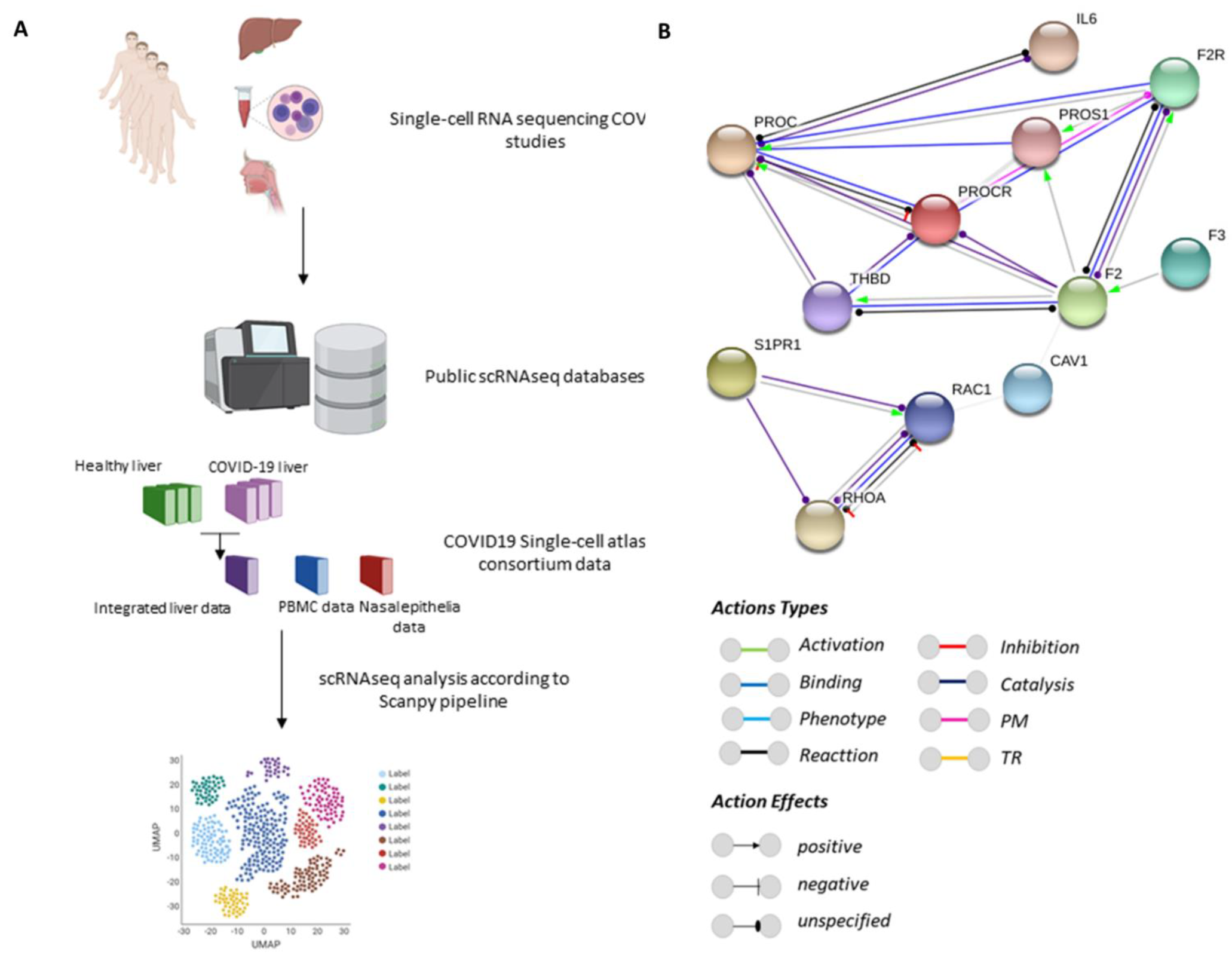
2.1. Protein–Protein Interaction Networks

Protein functional interaction networks for PC signaling proteins were prepared using the STRING v11 database, a software toolkit that performs data mining on a large number of databases and on individually published high-throughput datasets to build putative protein interactome maps. The association evidence in the STRING database comes from independent sources (‘channels’): genomic context information (neighborhood, fusion, gene co-occurrence), co-expression, text-mining, biochemical/genetic data (‘experiments’), and previously curated pathways and protein complex datasets (‘databases’) [28]. The default settings for functional interaction networks were queried for PC signaling proteins in Homo sapiens. The association’s sources used for functional interactions were neighborhood, experiments, gene fusion, databases, co-occurrence, and co-expression, and visualized by the molecular actions. The network nodes represent proteins and edges represent protein–protein associations. A permalink webpage of the PROC protein–protein interaction network is accessible at https://version-11-0.string-db.org/cgi/network.pl?networkId=1bGTXH59glgq, accessed on 16 April 2022.

2.2. Analysis of Single-Cell RNA-Sequencing (scRNA-seq) Data

For the expression of receptors and PC-related genes, datasets were retrieved from published studies containing data from multiple human tissues, including the liver [29,30], nasal epithelium [31,32], and peripheral blood [32,33]. These datasets are available and can be visualized and assessed through the website portal www.COVID-19cellatlas.org, accessed on 1 February 2022 [32]. In addition, a folder within all data analyzed is available on https://drive.google.com/drive/folders/12nmcAwphN8Zl8as8qgFiiJBafVcbsK6o?usp=sharing, accessed on 20 October 2022. The processed .h5ad files were loaded by using read_h5ad and matrix plots were illustrated using sc.pl.matrixplot in Scanpy 1.7.2, which is a model for single-cell analysis in Python [34]. For the scRNA-seq analysis of liver cells in patients with COVID-19, a dataset reported by Delorey et al. [29] was retrieved from Gene Expression Omnibus (GEO) with accession number GSE171668; control patients were retrieved from the GEO accession number GSE115469, a dataset reported by MacParland et al. [30]. The raw data with the .h5 format were loaded for analysis through the toolkit Scanpy 1.7.2 in Python igraph 0.9.6. For each sample, cells were filtered out if they contained fewer than 200 genes, more than 1500 genes or if mitochondrial genes were >15% of the total. The remaining cells were integrated into a gene-barcode matrix and then normalized and log transformed. The highly variable genes were identified by using the pp.highly_variable_genes function for the downstream principal component analysis (PCA) [35]. For the neighborhood graph of cells, the PCA representation of the data matrix was used and clustering was performed by Leiden graph-clustering method [36]. We chose the top 50 principal components for the Uniform Manifold Approximation and Projection (UMAP) and graph-based clustering. The cell type identity was manually annotated and the differentially expressed genes (DEG) for a cluster and all remaining cells calculated by the two-sided Wilcoxon rank-sum test [37]. The marker genes were then ranked by their log fold change of expression in particular cell types. The Supplementary Material for all tissues analyzed is available with marker genes for the cells type (Supplementary Tables S1–S3).

2.3. Statistical Analyses

The DEGs of patients with COVID-19 compared with healthy controls were determined with Scanpy’s implementation of the two-sided Wilcoxon rank-sum test [37]. The p_val_cutoff = 0.05 and logfc_cutoff = 0.1 were established by the Scanpy utils function.

2.4. Data Availability

Publicly available processed data used in this study with identified metadata and embeddings are available for download from the COVID-19 Cell Atlas (https://www.COVID-19cellatlas.org/, accessed on 1 February 2022) [32] hosted by the Wellcome Sanger Institute. The raw sequencing data from Delorey et al. [29] and MacParland et al. [30] are available at the NCBI Gene Expression Omnibus, accession numbers GSE171668 and GSE115469, respectively, and also available to download at https://drive.google.com/drive/folders/12nmcAwphN8Zl8as8qgFiiJBafVcbsK6o?usp=sharing, accessed on 20 October 2022.

2.5. Code Availability

All scripts used for data analysis are available from GitHub (https://github.com/OCRC/BRSilva_2022, accessed on 20 October 2022).

2.6. Generation of Diagrams and Illustrations

A licensed version of BioRender was employed to generate the diagrams present in most figures.

3. Results

3.1. PROC Interacts with Proteins Belonging to Barrier Integrity and the Inflammatory and Coagulation Systems

The protein–protein interaction network (Figure 1B) shows the reactions and binding interactions between PC (PROC) and its receptor, EPCR (PROCR), and the effects (unspecified, negative, and positive). PROC has a binding interaction with protein S (PROS1) and protease-activated receptor 1 (F2R), evidenced by the Experimental/Biochemical Association in Curated Databases. In addition, PROCR has a post-translational modification interaction with F2R with an unspecified effect. THBD shows a catalytic interaction with an unspecified effect with PROC and PROCR and a mutual interaction with an unspecified effect with F2. F2 shows binding, reaction, and catalytic interactions with an unspecified and positive effect with F2R and a positive effect with PROS1. PC also interacts with IL-6 through a reaction and catalytic interaction with an unspecified effect. F3 shows a positive effect with F2. Finally, S1PR1 interacts by catalysis with RAC1 and RHOA, with an unspecified and positive effect. RAC1 and RHOA present a mutual negative effect and interact by binding, catalysis, and reaction interactions with an unspecified effect.

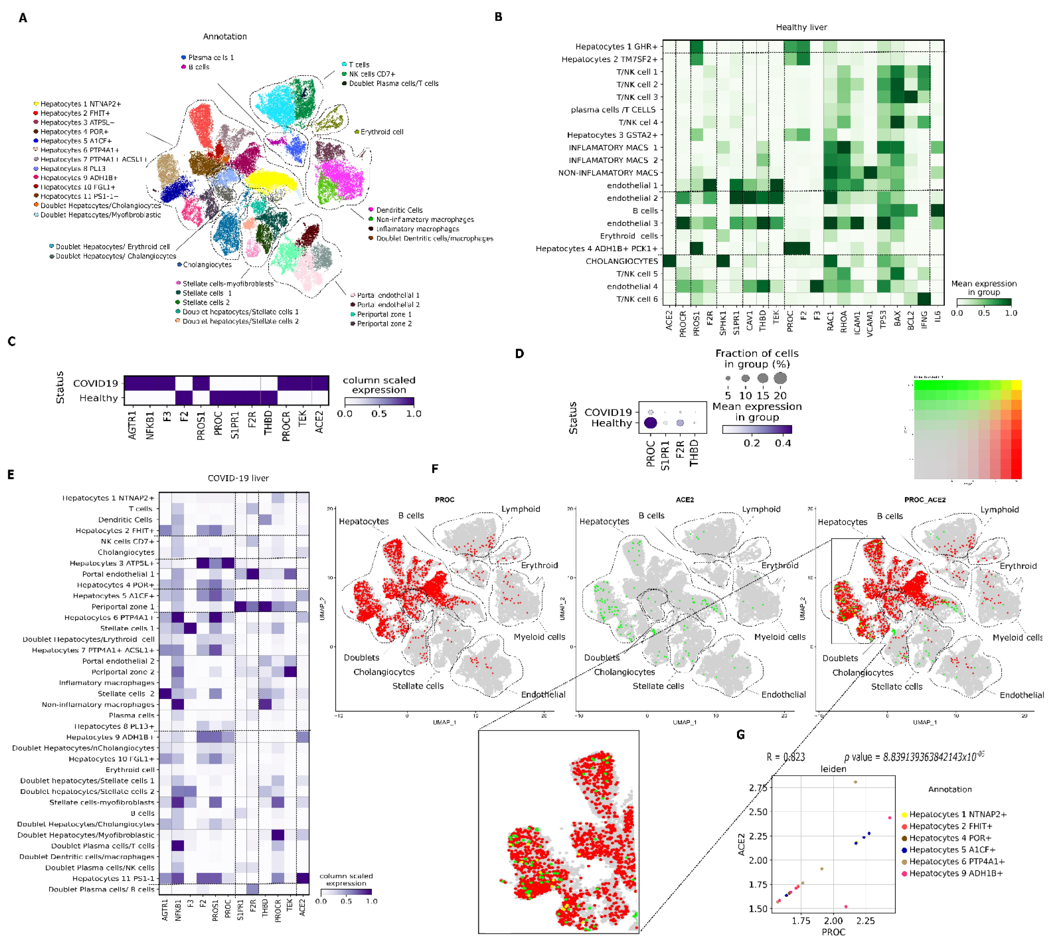
3.2. PC pathway Landscape at the Single-Cell Level in Tissues from Patients with COVID-19
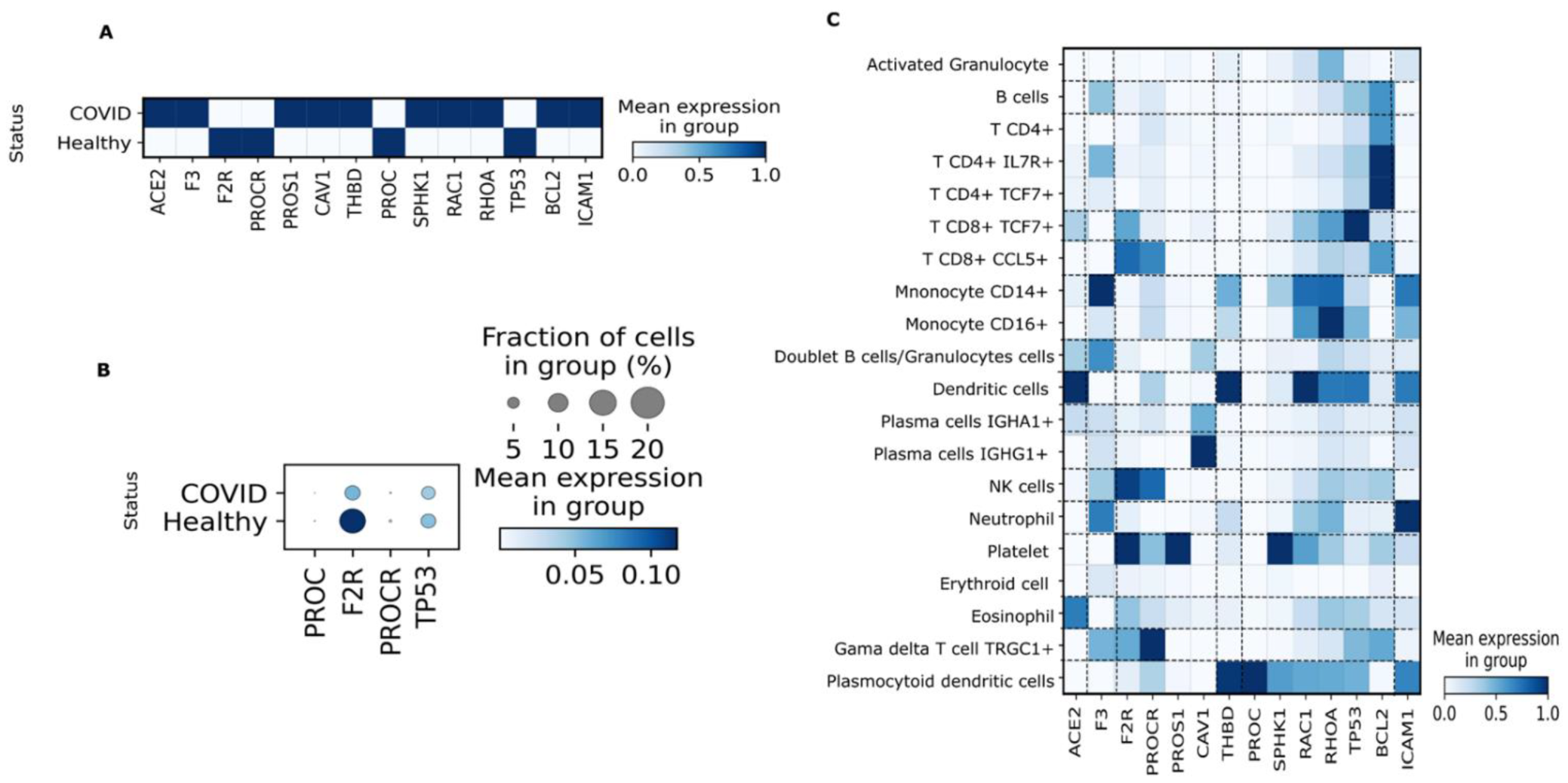
To study transcriptional changes in PC signaling in patients with COVID-19, gene expression of PC signaling-associated genes was explored in liver samples (Figure 2A–G), PBMC (Figure 3A–C), and nasal epithelia (Figure 4A–F) from patients with severe COVID-19 compared with healthy controls.
Differentially expressed genes between patients with COVID-19 and healthy controls were determined with Scanpy’s implementation of the two-sided Wilcoxon rank-sum test. Genes lacking a value (denoted as ‘-’) did not reach the logFC threshold of 0.1. COVID-19 %: percentage of cells in the COVID-19 group that expresses the gene. Healthy %: percentage of cells in the healthy group that expresses the gene. Abbreviations—logFC: log fold change; PBMC: peripheral blood mononuclear cells.
Liver. PC synthesis occurs mainly in hepatocytes, and it is released into the bloodstream as a zymogen [19]. First, we determined the expression of the PROC gene in healthy liver parenchyma and then compared the expression to samples collected from autopsies of patients who died because of severe COVID-19 complications. PROC was expressed throughout the liver parenchyma of healthy individuals, particularly in hepatocyte 4 (ADH1B+/PCK1+), which displayed the highest expression (Figure 2B). ACE2 expression was restricted to cholangiocytes in the healthy liver; the main receptors involved in PC signaling—PROCR, F2R, S1PR1, THBD, and TEK—were detected in endothelial cells (Figure 2B). In liver autopsy samples from patients who died because of COVID-19 complications, PC activation signaling receptors were significantly decreased compared with control as follows: 2.7-fold for S1PR1, 2.5-fold for F2R and 1.7-fold for THBD (p < 0.001), and less than 5% of cells expressed these genes (Table 1; Figure 2C,D). Conversely, liver samples from patients with COVID-19 presented a significant 1.9-fold increase in AGTR1, a 1.8-fold increase in NFKB1, and a 1.2-fold increase in F3 (p < 0.001) (Table 1; Figure 2C). Additionally, there was a significant 2.192-fold decrease in PROC in liver samples from individuals who died from COVID-19 complications (p < 0.001) (Table 1; Figure 2C). The relative number of hepatocytes from patients with COVID-19 expressing PROC was lower (about 5%) than in healthy individuals (about 20%) (Table 1; Figure 2D). The highest expression of PROC occurred in hepatocyte 3 (ATP5L+), followed by hepatocytes 9 (ADH1B+), 5 (A1CF+), and 6 (PTP4A1+) (Figure 2E). ACE2 gene expression was 2.868-fold higher in patients with COVID-19 (p = 0.000) (Table 1), and the expression was no longer restricted to cholangiocytes (as in the healthy liver) but extended to the liver parenchyma. ACE2 was expressed by hepatocytes 9 (ADH1B+), 5 (A1CF+), 6 (PTP4A1+), and 2 (FHIT+) with low PROC co-expression (Figure 2E,F). Moreover, there was a very strong association between ACE2 and PROC gene expression in samples from patients with COVID-19 between the cells that co-expressed these two genes (Figure 2G).

3.3. PBMC

Considering the effects of APC on the inflammatory response that leads to reduced immune cell nuclear factor κB (NF-κB)-related protein syntheses [38], we determined the expression of genes related to the PC signaling pathway in PBMC of patients with COVID-19 within 10 days of the onset of symptoms. In COVID-19 samples, there were significant decreases in the following genes compared with healthy individuals: 1.127-fold for F2R, 1.097-fold for PROCR, 1.040-fold for PROC, and 0.193-fold for TP53 (p < 0.05) (<10% of PBMC expressed these genes compared with healthy controls) (Table 1; Figure 3A,B). Conversely, there was a significant 2.943-fold increase in ACE2 expression and significantly altered expression of pro-inflammatory and pro-coagulation genes, including 1.306-fold for F3 and 0.548-fold for ICAM1 (p < 0.05) in patients with COVID-19 compared with healthy controls (Table 1; Figure 3A). ACE2 expression was detected in dendritic cells, eosinophils, IGHA1+ plasma cells, and CD8+ TCF7+ T cells. The F3 gene was detected in CD14+ monocytes, neutrophils, TRGC1+ gamma-delta T cells, CD4+ T cells, and B cells. ICAM1 expression was detected in neutrophils, dendritic cells, CD14+ monocytes, plasmacytoid dendritic cells, and CD16+ monocytes (Figure 3C). Moreover, there was a heterogeneous expression of PC activation receptors in other cell populations, such as platelets, natural killer (NK) cells, CD8+ CCL5+ T cells, TRGC1+ gamma-delta T cells, plasmacytoid dendritic cells, dendritic cells and IGHG1+ plasma cells, each expressing distinct PC activation receptors including F2R, PROCR, THBD, and CAV1 (Figure 3C).

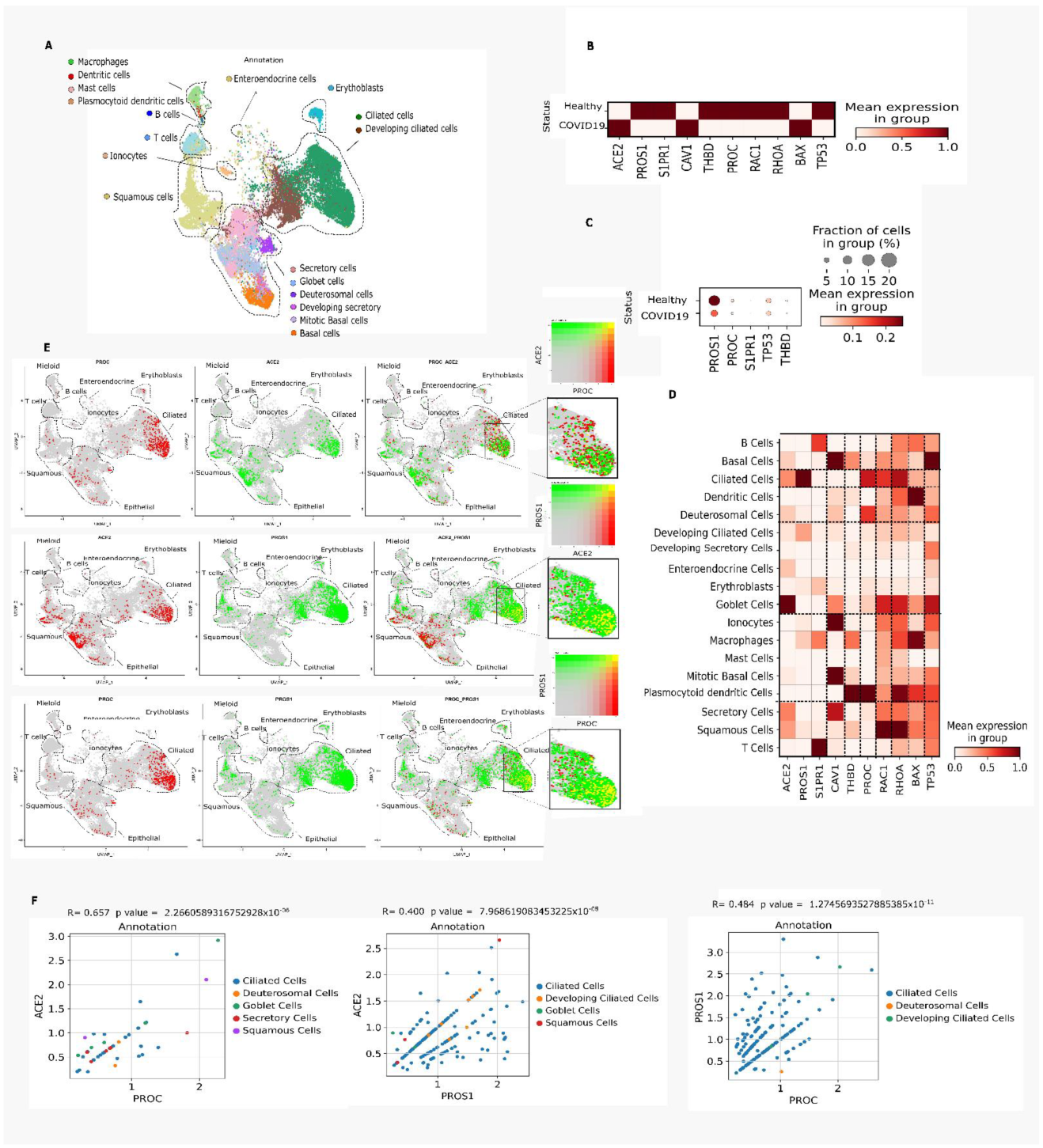
3.4. Nasal Epithelium

In the early stages of COVID-19, the upper respiratory tract is the primary target for severe acute respiratory syndrome coronavirus 2 (SARS-CoV-2) replication [39]. Hence, we looked for transcriptional changes in the PC activation and signaling-associated genes in nasal epithelial cells of patients with COVID-19 who had been hospitalized for 1–3 days. In COVID-19 samples, there was a significant decrease in the following genes compared with healthy subjects: 0.985-fold for PROS1, 1.108-fold for S1PR1, 1.323-fold for THBD, 0.502-fold for PROC, 0.767-fold for RAC1, 0.841-fold for RHOA, and 0.207-fold for TP53 (p < 0.05) (Table 1; Figure 4B). Less than 5% of nasal epithelial cells from patients with COVID-19 expressed PROS1 compared with healthy controls (Figure 4C). PROC expression was detected in plasmacytoid dendritic, ciliated, deuterosomal, basal, and goblet cells (Figure 4D). Basal cells expressed the most APC signaling-associated genes as well as those regulated in response to APC activation (CAV1, THBD, PROC, RAC1, RHOA, BAX, and TP53) (Figure 4D). Analysis of nasal epithelium showed that the PROC, ACE2, and PROS1 genes were expressed by the same ciliated cells, showing PROS1ACE2 and PROCPROS1 co-expression (Figure 4E,F). In addition, there was a strong association (p < 0.05) between ACE2 and PROC gene expression in ciliated cells from patients with COVID-19 (Figure 4F).

4. Discussion

The inflammation and coagulation axis has emerged as one of the main mechanisms controlling the host response to invading microorganisms, and the imbalance in this mechanism is responsible for the poor prognosis in patients with severe sepsis, including the risk of death [40]. Sepsis is a complex syndrome that results from infection [41] mainly by bacteria; however, studies suggest that the prevalence of viral sepsis is often underestimated [42,43]. Respiratory viral sepsis is a highly heterogeneous and multifaceted syndrome that can cause extrapulmonary organ dysfunction resulting from respiratory virus infections [41]. Acute kidney and cardiac injuries have been reported in influenza virus infection; infection caused by Middle East respiratory syndrome-related coronavirus (MERS-CoV) is related to acute kidney injury and thrombocytopenia. Hepatic dysfunction has been reported for respiratory syncytial virus infection. Furthermore, among fatal cases of SARS-CoV infection, high viral loads in the intestine and liver and moderate viral loads in the kidney have been reported [44,45,46,47]. Since the initial cases of COVID-19, there have been patients with a severe form of the disease, including sepsis, who have died as a result of the infection. They showed decreased platelet counts and elevated bilirubin and creatinine, reflecting clear signs of clotting disorder in addition to liver and kidney dysfunction [48,49]. In this study, we used the opportunity to assess several different vascularized tissues to detail the expression of one of the main pathways involved in hemostasis and endogenous immune responses in patients with severe COVID-19. We identified that the PC system may be involved in local and systemic derangement of hemostatic mechanisms in COVID-19.
Complications related to hyper-inflammation have been described in critical patients with COVID-19 [50]. Angiotensin-converting enzyme 2 (ACE2) is an important vasoconstriction enzyme that regulates vascular tone through its effector peptide angiotensin 1–7 (Ang (1–7)), and receptor Mas1 induces vasodilation and attenuates Ang II-induced vasoconstriction [51,52]. The coagulation cascade is intimately tied to ACE2 responses via the FXII contact activation pathway [53,54]. SARS-CoV-2 may contribute to Ang II-induced thrombosis by an imbalance in ACE2 levels, causing angiotensin-induced coagulopathy, with a high risk of thrombosis, comprising platelet activation, thrombin generation, and endothelial damage via the type 1 angiotensin II receptor [54]. Previous studies have shown that modulation of ACE2 is correlated to the time of infection and the tissue affected [55]. We found that patients with COVID-19 had elevated ACE2 messenger RNA (mRNA) levels throughout the liver parenchyma and decreased PROC expression. In silico analysis of patients with COVID-19 showed that high ACE2 expression enhances the expression of genes involved in replication, assembly and prolonging the virus life cycle [55]. Here, we showed that in COVID-19 samples, ACE2 was also expressed by the hepatocytes responsible for PROC expression. We suggest that the lower PROC mRNA levels found in the liver may be related to ACE2 expression in those hepatocytes.
The renin-angiotensin system (RAS) has been identified as a potential driver of host pulmonary and systemic immune responses, where the physiological balance between pro-inflammatory, vasoconstrictive, and pro-fibrotic effects of angiotensinogen through the angiotensin II receptor-like receptor 1 (AGTR1) are maintained in healthy subjects by equilibrium with AGTR2 and by the engagement of the Mas receptor by Ang (1–7) [52,56]. Here, we observed increased hepatic expression of F3, AGTR1, and NFKB1 in COVID-19 individuals. Previous studies have shown that hepatocyte tissue factor contributes to the hypercoagulable state in a mouse model of chronic liver injury [57]. After binding AngII to AT1R, the receptor mediates several important systemic effects in the liver, including vascular, proliferative, and inflammatory reactions [58]. In addition, injury-induced activation of hepatic NF-κB is observed in a variety of non-parenchymal and parenchymal liver cells, indicating that NF-κB plays a central role in coordinating the inflammatory response and wound healing by stimulating gene transcription in several key cellular players [59]. A computer model identified potentially beneficial drugs for critically COVID-19 patients that act by inhibiting the tumor necrosis factor (TNF)-induced NFkB1 signaling pathway [60], which is inhibited by APC activity [61].
The change in transcription of PROC and the activator receptors PROCR, FR2, and THBD in patients with COVID-19 suggests impaired cytoprotection and progression to the hypercoagulable state [62,63]. This progression starts with pathogen-induced cellular stress, a concurrent increase in tissue factor expression, and ensuing vascular cell death. Tissue factor has been shown to be upregulated in bronchial epithelial cells infected with SARS-CoV-2 [63]. The chief role of PC is to counteract the cell stress and apoptosis induced by cytokines and tissue factors released as part of the inflammatory response of bacterial and viral sepsis. Indeed, recent studies have shown that decreased plasma PC levels in patients with COVID-19 at hospital admission were associated with a worse clinical prognosis [64,65]. Our analysis revealed low PROC expression in the nasal epithelium during days 1–3 of hospitalization and in PBMC within 10 days of symptom onset. However, in the analysis of patients admitted to the intensive care unit (ICU), two independent cohorts revealed high PC plasma levels [65] but low functional activity [66]. Here, we explored PROC mRNA expression in patients who did not overcome the infection. We observed that the expression of hepatic PROC was decreased in these patients.
We found that PROC, S1PR1, and THBD were downregulated in the nasal epithelium of patients with COVID-19. Human nasal respiratory epithelial cells are primary targets for SARS-CoV-2 replication in the early stages of COVID-19 infection [16,39]. Compared with the oral squamous epithelium, viral tropism is restricted to the cells of the nasal epithelium [67] because they strongly express SARS-CoV-2 entry factors along with innate immunity genes, highlighting the role of these cells in early viral infection, dissemination, and elimination. Our analyses revealed that these cells also harbor intrinsic anticoagulation mechanisms and express genes correlated with inflammation that co-express with ACE2 (the virus receptor) in nasal ciliated cells. The co-expression between ACE2 and genes that facilitate the infection and that may contribute to the evolution of disease has been previously described in corneal tissue [68].
Previous studies have shown important dysregulation in the immune system of patients with severe COVID-19 [69,70]. Our PBMC analyses showed lower expression of PROC, F2R, and PROCR in patients with COVID-19 compared with controls. These genes are already known to be key to the activation and signaling of PC [71]. In addition, immature neutrophils and immunosuppressant cells in patients with COVID-19, followed by high levels of inflammatory cytokines, could be linked to PC inhibition, leading to the activation of the coagulation cascade. In addition, inflammatory factors can increase the formation of plasminogen activator inhibitor-1 (PAI-1), the synthesis of which increases during COVID-19 [72], and prevent fibrinolysis, thus promoting coagulation and even the formation of microthrombi.
In addition to decreased PROC and PROCR expression, we identified heterogeneous expression of genes associated with PC activators in immune cells. This finding suggests an important aspect of endogenous PC activation in COVID-19 pathogenesis. We know that circulating zymogen PC is activated by interacting with the thrombin-thrombomodulin complex in the presence of EPCR expressed on the surfaces of endothelial and epithelial cells [71,73]. However, our analysis revealed that different immune cells expressed distinct PC activation receptors, suggesting that PC activation might occur dynamically by zymogen PC interacting with ‘migratory’ receptors expressed on the surfaces of infiltrating immune cells in the injury site (Figure 5). PC activation might not be restricted to endothelial cells; it could also be extended to an ‘on-demand extra-vessel activation’ in an immune-mediated PC activation process. These findings highlight the importance of studying the expression of PROC and PC activation-related receptors in different cellular subsets.

5. Conclusions

Considering the fact that thromboembolic complications are commonly observed in patients with severe COVID-19, as well as our findings showing that the expression of APC signaling-related genes is downregulated in patients with COVID-19, we propose that APC dysfunction in severe COVID-19 could be involved in the development of COVID-19-associated coagulopathy. In addition, considering that THBD, PROCR, and F2R are also involved in the regulation of the inflammatory response [74,75,76], we suggest another important interface that supports the involvement of APC in the pathogenesis of the COVID-19-associated coagulopathy. Furthermore, when visualizing the expression of PC activation-related components and PROC in different cell types, we did not identify co-expression of these components, suggesting that PC activation might occur dynamically through the migration and interaction of these circulating cells at the site of the injured tissue. It is possible that SARS-CoV-2 replication occurs in hepatocytes due to their ACE2 expression, and this replication reprogrammes the hepatocyte transcriptome and affects PROC expression. These changes could then contribute to decreased PC synthesis; however, experimental studies are necessary to validate these hypotheses.
There are a few limitations to this study. We used an observational approach and thus could not draw conclusions about causative mechanisms. In addition, we only examined alterations in gene expression. It would be interesting to observe whether there are changes at the protein level.

Supplementary Materials

The following supporting information can be downloaded at: https://www.mdpi.com/article/10.3390/v14122753/s1, Table S1: Liver DE genes used to annotate cluster identity; Table S2: PBMC DE genes used to annotate cluster identity; Table S3: Nasal Epithelia DE genes used to annotate cluster identity.

Author Contributions

Conceptualization, E.P.A.; methodology, B.R.S.S., D.S.-O. and C.P.J.; validation, L.A.V. and W.H.V.; investigation, B.R.S.S.; data curation E.P.A., L.A.V. and W.H.V., writing, B.R.S.S.; writing—review and editing, E.P.A. All authors have read and agreed to the published version of the manuscript.

Funding

This study was carried out with the support of the Sao Paulo Research Foundation and the Coordination for the Improvement of Higher Education Personnel—Brazil (CAPES)—Financing Code 001 and by CEPID—FAPESP—OCRC—Obesity and Comorbidities Research Center, Brazil. Process No.2013/07607-8. Additionally, the Nebraska DHHS Stem Cell Grant 2019 (C.P.J. and W.H.V.) supported this research.

Institutional Review Board Statement

Not applicable.

Informed Consent Statement

Not applicable.

Data Availability Statement

Data will be available upon request.

Conflicts of Interest

The authors declare that they have no conflict of interest.

References

  1. Iba, T.; Warkentin, T.E.; Thachil, J.; Levi, M.; Levy, J.H. Proposal of the definition for COVID-19-associated coagulopathy. J. Clin. Med. 2021, 10, 191. [Google Scholar] [CrossRef] [PubMed]
  2. Ng, K.; Wu, A.; Cheng, V.; Tang, B.; Chan, C.; Yung, C.; Luk, S.; Lee, T.; Chow, L.; Yuen, K. Pulmonary artery thrombosis in a patient with severe acute respiratory syndrome. Postgrad. Med. J. 2005, 81, e3. [Google Scholar] [CrossRef] [PubMed] [Green Version]
  3. Xu, Z.; Shi, L.; Wang, Y.; Zhang, J.; Huang, L.; Zhang, C.; Liu, S.; Zhao, P.; Liu, H.; Zhu, L.; et al. Pathological findings of COVID-19 associated with acute respiratory distress syndrome. Lancet Respir. Med. 2020, 8, 420–422. [Google Scholar] [CrossRef] [PubMed]
  4. Dolhnikoff, M.; Duarte-Neto, A.N.; de Almeida Monteiro, R.A.; da Silva, L.F.F.; de Oliveira, E.P.; Saldiva, P.H.N.; Mauad, T.; Negri, E.M. Pathological evidence of pulmonary thrombotic phenomena in severe COVID-19. J. Thromb. Haemost. 2020, 18, 1517–1519. [Google Scholar] [CrossRef] [PubMed] [Green Version]
  5. Pestka, S.B. Old drug, new Trick? The rationale for the treatment of COVID-19 with activated protein C. Med. Hypotheses 2021, 149, 110537. [Google Scholar] [CrossRef]
  6. Gupta, A.; Madhavan, M.V.; Sehgal, K.; Nair, N.; Mahajan, S.; Sehrawat, T.S.; Bikdeli, B.; Ahluwalia, N.; Ausiello, J.C.; Wan, E.Y.; et al. Extrapulmonary manifestations of COVID-19. Nat. Med. 2020, 26, 1017–1032. [Google Scholar] [CrossRef]
  7. Bonaventura, A.; Vecchie, A.; Dagna, L.; Martinod, K.; Dixon, D.L.; Van Tassell, B.W.; Dentali, F.; Montecucco, F.; Massberg, S.; Levi, M.; et al. Endothelial dysfunction and immunothrombosis as key pathogenic mechanisms in COVID-19. Nat. Rev. Immunol. 2021, 21, 319–329. [Google Scholar] [CrossRef]
  8. Jin, Y.; Ji, W.; Yang, H.; Chen, S.; Zhang, W.; Duan, G. Endothelial activation and dysfunction in COVID-19: From basic mechanisms to potential therapeutic approaches. Signal Transduct. Target. 2020, 5, 293. [Google Scholar] [CrossRef]
  9. Pan, H.; Peto, R.; Karim, Q. WHO, Solidarity trial consortium. As the members of the writing committee. Repurposed antiviral drugs for COVID-19–interim WHO SOLIDARITY trial results. medRxiv 2020, 10, 15.20209817. [Google Scholar]
  10. Gharbharan, A.; Jordans, C.C.; GeurtsvanKessel, C.; den Hollander, J.G.; Karim, F.; Mollema, F.P.; Stalenhoef, J.E.; Dofferhoff, A.; Ludwig, I.; Koster, A. Convalescent plasma for COVID-19. A randomized clinical trial. medRxiv 2020. [Google Scholar] [CrossRef]
  11. Wang, D.; Fu, B.; Peng, Z.; Yang, D.; Han, M.; Li, M.; Yang, Y.; Yang, T.; Sun, L.; Li, W. Tocilizumab in patients with moderate or severe COVID-19: A randomized, controlled, open-label, multicenter trial. Front. Med. 2021, 15, 486–494. [Google Scholar] [CrossRef] [PubMed]
  12. Russell, C.D.; Millar, J.E.; Baillie, J.K. Clinical evidence does not support corticosteroid treatment for 2019-nCoV lung injury. Lancet 2020, 395, 473–475. [Google Scholar] [CrossRef] [PubMed] [Green Version]
  13. Mansour, E.; Palma, A.C.; Ulaf, R.G.; Ribeiro, L.C.; Bernardes, A.F.; Nunes, T.A.; Agrela, M.V.; Bombassaro, B.; Monfort-Pires, M.; Camargo, R.L. Safety and outcomes associated with the pharmacological inhibition of the kinin–kallikrein system in severe COVID-19. Viruses 2021, 13, 309. [Google Scholar] [CrossRef] [PubMed]
  14. Suthar, A.B.; Wang, J.; Seffren, V.; Wiegand, R.E.; Griffing, S.; Zell, E. Public health impact of covid-19 vaccines in the US: Observational study. BMJ 2022, 377, e069317. [Google Scholar] [CrossRef] [PubMed]
  15. Fiolet, T.; Kherabi, Y.; MacDonald, C.-J.; Ghosn, J.; Peiffer-Smadja, N. Comparing COVID-19 vaccines for their characteristics, efficacy and effectiveness against SARS-CoV-2 and variants of concern: A narrative review. Clin. Microbiol. Infect. 2021, 12, 202–221. [Google Scholar] [CrossRef]
  16. Huang, C.; Wang, Y.; Li, X.; Ren, L.; Zhao, J.; Hu, Y.; Zhang, L.; Fan, G.; Xu, J.; Gu, X.; et al. Clinical features of patients infected with 2019 novel coronavirus in Wuhan, China. Lancet 2020, 395, 497–506. [Google Scholar] [CrossRef] [Green Version]
  17. ATTACC, A.-a.; Investigators, R.-C. Therapeutic anticoagulation with heparin in noncritically ill patients with Covid-19. N. Engl. J. Med. 2021, 385, 790–802. [Google Scholar]
  18. Willyard, C. Coronavirus blood-clot mystery intensifies. Nature 2020, 581, 250. [Google Scholar] [CrossRef]
  19. Esmon, C.; Stenflo, J.; Suttie, J. A new vitamin K-dependent protein. A phospholipid-binding zymogen of a serine esterase. J. Biol. Chem. 1976, 251, 3052–3056. [Google Scholar] [CrossRef]
  20. He, X.; Shen, L.; Bjartell, A.; Malm, J.; Lilja, H.; Dahlbäck, B. The gene encoding vitamin K-dependent anticoagulant protein C is expressed in human male reproductive tissues. J. Histochem. Cytochem. 1995, 43, 563–570. [Google Scholar] [CrossRef] [Green Version]
  21. Uhlén, M.; Björling, E.; Agaton, C.; Szigyarto, C.A.-K.; Amini, B.; Andersen, E.; Andersson, A.-C.; Angelidou, P.; Asplund, A.; Asplund, C. A human protein atlas for normal and cancer tissues based on antibody proteomics. Mol. Cell. Proteom. 2005, 4, 1920–1932. [Google Scholar] [CrossRef] [PubMed] [Green Version]
  22. Van Cott, K.E.; Williams, B.; Velander, W.H.; Gwazdauskas, F.; Lee, T.; Lubon, H.; Drohan, W.N. Affinity purification of biologically active and inactive forms of recombinant human protein C produced in porcine mammary gland. J. Mol. Recognit. 1996, 9, 407–414. [Google Scholar] [CrossRef]
  23. Riewald, M.; Petrovan, R.J.; Donner, A.; Ruf, W. Activated protein C signals through the thrombin receptor PAR1 in endothelial cells. J. Endotoxin Res. 2003, 9, 317–321. [Google Scholar] [CrossRef] [Green Version]
  24. Stearns-Kurosawa, D.J.; Kurosawa, S.; Mollica, J.S.; Ferrell, G.L.; Esmon, C.T. The endothelial cell protein C receptor augments protein C activation by the thrombin-thrombomodulin complex. Proc. Natl. Acad. Sci. USA 1996, 93, 10212–10216. [Google Scholar] [CrossRef] [Green Version]
  25. Schuepbach, R.A.; Feistritzer, C.; Brass, L.F.; Riewald, M. Activated protein C-cleaved protease activated receptor-1 is retained on the endothelial cell surface even in the presence of thrombin. Blood 2008, 111, 2667–2673. [Google Scholar] [CrossRef] [PubMed] [Green Version]
  26. Baxter, A.A.; Lay, F.T.; Poon, I.K.; Kvansakul, M.; Hulett, M.D. Tumor cell membrane-targeting cationic antimicrobial peptides: Novel insights into mechanisms of action and therapeutic prospects. Cell. Mol. Life Sci. 2017, 74, 3809–3825. [Google Scholar] [CrossRef]
  27. Whetstone, W.D.; Walker, B.; Trivedi, A.; Lee, S.; Noble-Haeusslein, L.J.; Hsu, J.C. Protease-Activated Receptor-1 Supports Locomotor Recovery by Biased Agonist Activated Protein C after Contusive Spinal Cord Injury. PLoS ONE 2017, 12, e0170512. [Google Scholar] [CrossRef] [Green Version]
  28. Szklarczyk, D.; Gable, A.L.; Lyon, D.; Junge, A.; Wyder, S.; Huerta-Cepas, J.; Simonovic, M.; Doncheva, N.T.; Morris, J.H.; Bork, P.; et al. STRING v11: Protein-protein association networks with increased coverage, supporting functional discovery in genome-wide experimental datasets. Nucleic Acids Res. 2019, 47, D607–D613. [Google Scholar] [CrossRef] [Green Version]
  29. Delorey, T.M.; Ziegler, C.G.; Heimberg, G.; Normand, R.; Yang, Y.; Segerstolpe, Å.; Abbondanza, D.; Fleming, S.J.; Subramanian, A.; Montoro, D.T. COVID-19 tissue atlases reveal SARS-CoV-2 pathology and cellular targets. Nature 2021, 595, 107–113. [Google Scholar] [CrossRef]
  30. MacParland, S.A.; Liu, J.C.; Ma, X.Z.; Innes, B.T.; Bartczak, A.M.; Gage, B.K.; Manuel, J.; Khuu, N.; Echeverri, J.; Linares, I.; et al. Single cell RNA sequencing of human liver reveals distinct intrahepatic macrophage populations. Nat. Commun. 2018, 9, 4383. [Google Scholar] [CrossRef] [Green Version]
  31. Ziegler, C.G.; Miao, V.N.; Owings, A.H.; Navia, A.W.; Tang, Y.; Bromley, J.D.; Lotfy, P.; Sloan, M.; Laird, H.; Williams, H.B. Impaired local intrinsic immunity to SARS-CoV-2 infection in severe COVID-19. Cell 2021, 184, 4713–4733.e4722. [Google Scholar] [CrossRef]
  32. COVID CZIS-C; Ballestar, E.; Farber, D.L.; Glover, S.; Horwitz, B.; Meyer, K.; Nikolić, M.; Ordovas-Montanes, J.; Sims, P.; Shalek, A. Single cell profiling of COVID-19 patients: An international data resource from multiple tissues. MedRxiv 2020. [Google Scholar] [CrossRef]
  33. Wilk, A.J.; Rustagi, A.; Zhao, N.Q.; Roque, J.; Martínez-Colón, G.J.; McKechnie, J.L.; Ivison, G.T.; Ranganath, T.; Vergara, R.; Hollis, T. A single-cell atlas of the peripheral immune response in patients with severe COVID-19. Nat. Med. 2020, 26, 1070–1076. [Google Scholar] [CrossRef]
  34. Wolf, F.A.; Angerer, P.; Theis, F.J. SCANPY: Large-scale single-cell gene expression data analysis. Genome Biol. 2018, 19, 15. [Google Scholar] [CrossRef] [Green Version]
  35. Luecken, M.D.; Theis, F.J. Current best practices in single-cell RNA-seq analysis: A tutorial. Mol. Syst. Biol. 2019, 15, e8746. [Google Scholar] [CrossRef]
  36. Traag, V.A.; Waltman, L.; Van Eck, N.J. From Louvain to Leiden: Guaranteeing well-connected communities. Sci. Rep. 2019, 9, 5233. [Google Scholar] [CrossRef] [PubMed] [Green Version]
  37. Zhu, X.; Garmire, L.X. Data Analysis in Single-Cell RNA-Seq. In Single-Cell Omics; Debmalya Barh, V.A., Ed.; Elsevier: Amsterdam, The Netherlands, 2019; pp. 419–432. [Google Scholar]
  38. White, B.; Schmidt, M.; Murphy, C.; Livingstone, W.; O’toole, D.; Lawler, M.; O’Neill, L.; Kelleher, D.; Schwarz, H.; Smith, O. Activated protein C inhibits lipopolysaccharide-induced nuclear translocation of nuclear factor κB (NF-κB) and tumour necrosis factor α (TNF-α) production in the THP-1 monocytic cell line. Br. J. Haematol. 2000, 110, 130–134. [Google Scholar] [CrossRef] [PubMed]
  39. Sungnak, W.; Huang, N.; Bécavin, C.; Berg, M.; Queen, R.; Litvinukova, M.; Talavera-López, C.; Maatz, H.; Reichart, D.; Sampaziotis, F. SARS-CoV-2 entry factors are highly expressed in nasal epithelial cells together with innate immune genes. Nat. Med. 2020, 26, 681–687. [Google Scholar] [CrossRef] [PubMed] [Green Version]
  40. Della Valle, P.; Pavani, G.; D’Angelo, A. The protein C pathway and sepsis. Thromb. Res. 2012, 129, 296–300. [Google Scholar] [CrossRef]
  41. Gu, X.; Zhou, F.; Wang, Y.; Fan, G.; Cao, B. Respiratory viral sepsis: Epidemiology, pathophysiology, diagnosis and treatment. Eur. Respir. Rev. 2020, 29, 200038. [Google Scholar] [CrossRef] [PubMed]
  42. Phua, J.; Ngerng, W.J.; See, K.C.; Tay, C.K.; Kiong, T.; Lim, H.F.; Chew, M.Y.; Yip, H.S.; Tan, A.; Khalizah, H.J. Characteristics and outcomes of culture-negative versus culture-positive severe sepsis. Crit. Care 2013, 17, R202. [Google Scholar] [CrossRef] [PubMed] [Green Version]
  43. Ljungström, L.R.; Jacobsson, G.; Claesson, B.E.; Andersson, R.; Enroth, H. Respiratory viral infections are underdiagnosed in patients with suspected sepsis. Eur. J. Clin. Microbiol. Infect. Dis. 2017, 36, 1767–1776. [Google Scholar] [CrossRef]
  44. Bakalli, I. Liver dysfunction in severe sepsis from respiratory syncytial virus. J. Pediatr. Intensive Care 2018, 7, 110–114. [Google Scholar] [CrossRef] [PubMed]
  45. Farcas, G.A.; Poutanen, S.M.; Mazzulli, T.; Willey, B.M.; Butany, J.; Asa, S.L.; Faure, P.; Akhavan, P.; Low, D.E.; Kain, K.C. Fatal severe acute respiratory syndrome is associated with multiorgan involvement by coronavirus. J. Infect. Dis. 2005, 191, 193–197. [Google Scholar] [CrossRef] [Green Version]
  46. Nin, N.; Lorente, J.; Soto, L.; Ríos, F.; Hurtado, J.; Arancibia, F.; Ugarte, S.; Echevarría, E.; Cardinal, P.; Saldarini, F. Acute kidney injury in critically ill patients with 2009 influenza A (H1N1) viral pneumonia: An observational study. Intensive Care Med. 2011, 37, 768–774. [Google Scholar] [CrossRef]
  47. Gao, C.; Wang, Y.; Gu, X.; Shen, X.; Zhou, D.; Zhou, S.; Huang, J.-a.; Cao, B.; Guo, Q. Association between cardiac injury and mortality in hospitalized patients infected with avian influenza A (H7N9) virus. Crit. Care Med. 2020, 48, 451. [Google Scholar] [CrossRef]
  48. Yang, X.; Yu, Y.; Xu, J.; Shu, H.; Liu, H.; Wu, Y.; Zhang, L.; Yu, Z.; Fang, M.; Yu, T. Clinical course and outcomes of critically ill patients with SARS-CoV-2 pneumonia in Wuhan, China: A single-centered, retrospective, observational study. Lancet Respir. Med. 2020, 8, 475–481. [Google Scholar] [CrossRef] [Green Version]
  49. Iba, T.; Levy, J.H. Sepsis-induced coagulopathy and disseminated intravascular coagulation. Anesthesiology 2020, 132, 1238–1245. [Google Scholar] [CrossRef]
  50. Manson, J.J.; Crooks, C.; Naja, M.; Ledlie, A.; Goulden, B.; Liddle, T.; Khan, E.; Mehta, P.; Martin-Gutierrez, L.; Waddington, K.E. COVID-19-associated hyperinflammation and escalation of patient care: A retrospective longitudinal cohort study. Lancet Rheumatol. 2020, 2, e594–e602. [Google Scholar] [CrossRef]
  51. Samavati, L.; Uhal, B.D. ACE2, much more than just a receptor for SARS-COV-2. Front. Cell. Infect. Microbiol. 2020, 10, 317. [Google Scholar] [CrossRef] [PubMed]
  52. Kuriakose, J.; Montezano, A.C.; Touyz, R.M. ACE2/Ang-(1-7)/Mas1 axis and the vascular system: Vasoprotection to COVID-19-associated vascular disease. Clin. Sci. 2021, 135, 387–407. [Google Scholar] [CrossRef] [PubMed]
  53. Meini, S.; Zanichelli, A.; Sbrojavacca, R.; Iuri, F.; Roberts, A.T.; Suffritti, C.; Tascini, C. Understanding the pathophysiology of COVID-19: Could the contact system be the key? Front. Immunol. 2020, 11, 2014. [Google Scholar] [CrossRef] [PubMed]
  54. Gando, S.; Wada, T. Thromboplasminflammation in COVID-19 coagulopathy: Three viewpoints for diagnostic and therapeutic strategies. Front. Immunol. 2021, 12, 649122. [Google Scholar] [CrossRef]
  55. Li, G.; He, X.; Zhang, L.; Ran, Q.; Wang, J.; Xiong, A.; Wu, D.; Chen, F.; Sun, J.; Chang, C. Assessing ACE2 expression patterns in lung tissues in the pathogenesis of COVID-19. J. Autoimmun. 2020, 112, 102463. [Google Scholar] [CrossRef]
  56. Capettini, L.S.A.; Montecucco, F.; Mach, F.; Stergiopulos, N.; Santos, R.A.S.; da Silva, R.F. Role of renin-angiotensin system in inflammation, immunity and aging. Curr. Pharm. Des. 2012, 18, 963–970. [Google Scholar] [CrossRef]
  57. Rautou, P.-E.; Tatsumi, K.; Antoniak, S.; Owens III, A.P.; Sparkenbaugh, E.; Holle, L.A.; Wolberg, A.S.; Kopec, A.K.; Pawlinski, R.; Luyendyk, J.P. Hepatocyte tissue factor contributes to the hypercoagulable state in a mouse model of chronic liver injury. J. Hepatol. 2016, 64, 53–59. [Google Scholar] [CrossRef] [Green Version]
  58. Shim, K.Y.; Eom, Y.W.; Kim, M.Y.; Kang, S.H.; Baik, S.K. Role of the renin-angiotensin system in hepatic fibrosis and portal hypertension. Korean J. Intern. Med. 2018, 33, 453. [Google Scholar] [CrossRef] [Green Version]
  59. Elsharkawy, A.M.; Mann, D.A. Nuclear factor-κB and the hepatic inflammation-fibrosis-cancer axis. Hepatology 2007, 46, 590–597. [Google Scholar] [CrossRef]
  60. Rehan, M.; Ahmed, F.; Howladar, S.M.; Refai, M.Y.; Baeissa, H.M.; Zughaibi, T.A.; Kedwa, K.M.; Jamal, M.S. A Computational Approach Identified Andrographolide as a Potential Drug for Suppressing COVID-19-Induced Cytokine Storm. Front. Immunol. 2021, 12, 648250. [Google Scholar] [CrossRef]
  61. Okajima, K. Prevention of Endothelial Cell Injury by Activated Protein C: The Molecular Mechanism(s) and Therapeutic Implications. Curr. Vasc. Pharmacol. 2004, 2, 125–133. [Google Scholar] [CrossRef] [PubMed]
  62. Corrêa, T.D.; Cordioli, R.L.; Campos Guerra, J.C.; Caldin da Silva, B.; dos Reis Rodrigues, R.; De Souza, G.M.; Midega, T.D.; Campos, N.S.; Carneiro, B.V.; Campos, F.N.D. Coagulation profile of COVID-19 patients admitted to the ICU: An exploratory study. PLoS ONE 2020, 15, e0243604. [Google Scholar] [CrossRef] [PubMed]
  63. FitzGerald, E.S.; Jamieson, A.M. Unique transcriptional changes in coagulation cascade genes in SARS-CoV-2-infected lung 3 epithelial cells: A potential factor in COVID-19 coagulopathies 4. bioRxiv 2020. [Google Scholar] [CrossRef]
  64. Stanne, T.M.; Pedersén, A.; Gisslén, M.; Jern, C. Low admission protein C levels is a risk factor for disease worsening and mortality in hospitalized patients with COVID-19. Thromb. Res. 2021, 204, 13–15. [Google Scholar] [CrossRef]
  65. Panigada, M.; Bottino, N.; Tagliabue, P.; Grasselli, G.; Novembrino, C.; Chantarangkul, V.; Pesenti, A.; Peyvandi, F.; Tripodi, A. Hypercoagulability of COVID-19 patients in intensive care unit: A report of thromboelastography findings and other parameters of hemostasis. J. Thromb. Haemost. 2020, 18, 1738–1742. [Google Scholar] [CrossRef]
  66. Tabatabai, A.; Rabin, J.; Menaker, J.; Madathil, R.; Galvagno, S.; Menne, A.; Chow, J.H.; Grazioli, A.; Herr, D.; Tanaka, K. Factor VIII and functional protein C activity in critically ill patients with coronavirus disease 2019: A case series. Aa Pract. 2020, 14, e01236. [Google Scholar] [CrossRef]
  67. Ahn, J.H.; Kim, J.; Hong, S.P.; Choi, S.Y.; Yang, M.J.; Ju, Y.S.; Kim, Y.T.; Kim, H.M.; Rahman, M.T.; Chung, M.K. Nasal ciliated cells are primary targets for SARS-CoV-2 replication in early stage of COVID-19. J. Clin. Investig. 2021, 131, e148517. [Google Scholar] [CrossRef]
  68. Yuan, J.; Fan, D.; Xue, Z.; Qu, J.; Su, J. Co-expression of mitochondrial genes and ACE2 in cornea involved in COVID-19. Investig. Ophthalmol. Vis. Sci. 2020, 61, 13. [Google Scholar] [CrossRef]
  69. Liao, M.; Liu, Y.; Yuan, J.; Wen, Y.; Xu, G.; Zhao, J.; Chen, L.; Li, J.; Wang, X.; Wang, F. The landscape of lung bronchoalveolar immune cells in COVID-19 revealed by single-cell RNA sequencing. MedRxiv 2020. [Google Scholar] [CrossRef] [Green Version]
  70. Schulte-Schrepping, J.; Reusch, N.; Paclik, D.; Baßler, K.; Schlickeiser, S.; Zhang, B.; Krämer, B.; Krammer, T.; Brumhard, S.; Bonaguro, L. Severe COVID-19 is marked by a dysregulated myeloid cell compartment. Cell 2020, 182, 1419–1440.e1423. [Google Scholar] [CrossRef]
  71. Willis Fox, O.; Preston, R.J. Molecular basis of protease-activated receptor 1 signaling diversity. J. Thromb. Haemost. 2020, 18, 6–16. [Google Scholar] [CrossRef] [PubMed]
  72. Zou, H.; Xiong, W.-F. Advances in the relationship between coronavirus infection and coagulation function. Chin. Med. J. 2020, 133, 1489. [Google Scholar] [CrossRef] [PubMed]
  73. Russo, A.; Soh, U.J.; Paing, M.M.; Arora, P.; Trejo, J. Caveolae are required for protease-selective signaling by protease-activated receptor–1. Proc. Natl. Acad. Sci. USA 2009, 106, 6393–6397. [Google Scholar] [CrossRef] [PubMed]
  74. Wenzel, J.; Assmann, J.; Schwaninger, M. Thrombomodulin–a new target for treating stroke at the crossroad of coagulation and inflammation. Curr. Med. Chem. 2014, 21, 2025–2034. [Google Scholar] [CrossRef] [PubMed]
  75. Kishi, Y.; Kondo, T.; Xiao, S.; Yosef, N.; Gaublomme, J.; Wu, C.; Wang, C.; Chihara, N.; Regev, A.; Joller, N. Protein C receptor (PROCR) is a negative regulator of Th17 pathogenicity. J. Exp. Med. 2016, 213, 2489–2501. [Google Scholar] [CrossRef] [Green Version]
  76. Schoergenhofer, C.; Schwameis, M.; Gelbenegger, G.; Buchtele, N.; Thaler, B.; Mussbacher, M.; Schabbauer, G.; Wojta, J.; Jilma-Stohlawetz, P.; Jilma, B. Inhibition of protease-activated receptor (PAR1) reduces activation of the endothelium, coagulation, fibrinolysis and inflammation during human endotoxemia. Thromb. Haemost. 2018, 118, 1176–1184. [Google Scholar] [CrossRef]
Figure 1. Experimental design and protein–protein interaction network. (A). Public data from single-cell RNA sequencing (scRNA-seq) studies on the human tissues (liver, peripheral blood mononuclear cells (PBMC), and nasal epithelia) were retrieved from the website portal www.COVID-19cellatlas.org accessed on 16 April 2022 and Gene Expression Omnibus (GEO) accession numbers GSE171668 and GSE115469. (B). The PROC interactome was retrieved with the string-db data mining toolkit (https://version-11-0.string-db.org/cgi/network.pl?taskId=lu4XDH7F7cOZ, accessed on 16 April 2022). The sources used for functional interactions were neighborhood, experiments, gene fusion, databases, co-occurrence and co-expression, and visualized by the ‘molecular actions’. The network nodes represent proteins and the edges represent protein–protein associations. The edges indicate the known molecular action of a protein node relative to another protein node. PM: post-translational modification; TR: transcriptional regulation.
Viruses 14 02753 g001
Figure 2. Expression of genes involved in activated protein C (APC) signaling in the liver from healthy individuals and patients with coronavirus disease 2019 (COVID-19) based on single-cell transcriptomics. (A). Uniform Manifold Approximation and Projection (UMAP) embedding of liver cells via single-cell RNA sequencing (scRNA-Seq) droplet-based single-cell platform, showing single liver parenchymal cells grouped into 36 clusters. (B). Matrix plot showing expression of genes related to APC signaling according to the cell type in a healthy liver. (C). Matrix plot showing expression of genes related to APC signaling in patients with COVID-19 versus healthy individuals. (D). Dot plot representation of downregulated genes in patients with COVID-19. (E). Matrix plot showing expression of genes related to APC signaling according to the cell type in an autopsy liver sample from a patient with COVID-19. (F). Visualization of co-expression (yellow) by color overlap. PROC (red) is barely co-expressed with ACE2 (green) in hepatocyte clusters. (G). Spearman’s correlation of subset of cells that co-expressed PROC and ACE2.
Viruses 14 02753 g002
Figure 3. Expression of genes involved in activated protein C (APC) signaling in human peripheral blood mononuclear cells (PBMC) from patients with coronavirus disease 2019 (COVID-19) based on single-cell transcriptomics. (A). Matrix plot showing expression of genes related to APC signaling in patients with COVID-19 versus healthy individuals. (B). Dot plot representation of downregulated genes in patients with COVID-19. (C). Matrix plot showing expression of the main genes related to APC signaling according to cell types.
Viruses 14 02753 g003
Figure 4. Expression of genes involved in activated protein C (APC) signaling in nasal epithelial cells from patients with coronavirus disease 2019 (COVID-19) based on single-cell transcriptomics. (A). Uniform manifold approximation and projection (UMAP) embedding of nasal epithelial cells via single-cell RNA sequencing (scRNA-Seq) droplet-based single-cell platform grouped into 18 clusters. (B). Matrix plot showing expression of genes related to APC signaling in patients with COVID-19 versus uninfected individuals. (C). Dot plot representation of downregulated genes in patients with COVID-19. (D). Matrix plot showing expression of the main genes related to APC signaling according to cell types retrieved from Nasal epithelial samples. (E). Visualization of co-expression (yellow) by color overlap. PROC (red) is barely co-expressed with ACE2 (green) in ciliated cell clusters. ACE2 (red) is co-expressed with PROS1 (green) in ciliated cell clusters. PROC (red) is co-expressed with PROS1 (green) in ciliated cell clusters. (F). Spearman’s correlation of a subset of cells that expressed PROC, PROS1, and ACE2.
Viruses 14 02753 g004
Figure 5. PC activation and signaling in the endothelial cell. When thrombin binds to TM on endothelial cell surfaces, PC is converted to its active form (APC), and EPCR is necessary for optimal PC activation. The co-localization of PAR-1 and EPCR in caveolin-1-rich membrane microdomains specialized in the cell membrane is essential for PAR-1-mediated cytoprotective APC signaling. Furthermore, the heterogeneous expression of PC activation receptors (EPCR, THBD, CAV-1, and PAR-1) in other cell populations suggests that PC activation might occur dynamically by zymogen PC interacting with ‘migratory’ receptors expressed on the surfaces of infiltrating immune cells in the injury site. The anti-inflammatory and anticoagulant actions of APC occur mainly through PAR-1, which, when activated, propagates pro-inflammatory and anti-inflammatory responses according to the protease that cleaved it. PAR-1 is sensitive to proteolysis at its N-terminal exodomain by multiple plasma proteases, including thrombin, APC, plasmin, factor VIIa, factor Xa, and matrix metalloproteases. Cleavage of PAR-1 at the R41/46 sites at the N-terminal exodomain by APC initiates signaling, promoting structural reorganization of the receptor and coupling to the G protein complex (GRK-5) to phosphorylate the intracellular region of PAR-1 and recruitment of β-arrestin 2, which serves as a framework for cytoprotective signaling. This cascade initiates a series of intracellular signaling pathways, including upregulation of SPHK1 activity and, therefore, S1P production, leading to activation of signaling through its S1PR1 receptor, which stimulates a marked increase in Rac1 activation to perform a protective barrier function by stabilizing the endothelial cytoskeleton and inhibiting RhoA signaling. The Tie2 receptor transmembrane domain can initiate downstream signaling pathways and maintain vascular integrity through direct APC binding, which results in increased ZO-1 phosphorylation of Akt via PI3K and ERK inhibition, contributing to increasing the integrity of the barrier. Protein S cofactor-linked APC can intervene at various points during the systemic response to infection. It exerts an antithrombotic effect by inactivating factors Va and VIIIa, limiting thrombin generation. Finally, PAR-3 cleavage by APC can initiate PAR1-dependent cytoprotective signaling in endothelial cells in the presence of EPCR. APC: activated protein C; EPCR: endothelial protein receptor; ERK: extracellular signal-regulated protein kinase; PAR-1: protease-activated receptor 1; PAR-3: protease-activated receptor 3; PC: protein C; PI3K: phosphoinositide 3-kinase; S1P: sphingosine-1-phosphate; SPHK1: sphingosine kinase-1; TH: thrombin; Tie2: transmembrane receptor tyrosine kinase; TM: thrombomodulin.
Viruses 14 02753 g005
Table 1. Differentially expressed genes related to protein C (PC) activation and signaling in samples from patients with coronavirus disease 2019 (COVID-19). Differentially expressed genes between patients with COVID-19 and healthy controls were determined with Scanpy’s implementation of the two-sided Wilcoxon rank-sum test. Genes lacking a value (denoted as ‘-’) did not reach the logFC threshold of 0.1. COVID-19%: percentage of cells in the COVID-19 group that expresses the gene. Healthy%: percentage of cells in the healthy group that expresses the gene. Abbreviations—logFC: log fold change; PBMC: peripheral blood mononuclear cells.
LiverPBMCNasal Epithelium
GeneProtein ProductLogFCCOVID-19%Healthy
%
Adj. p-Value LogFCCOVID-19%Healthy
%
Adj. p-Value LogFCCOVID-19%Healthy
%
Adj. p-Value
ACE2Angiotensin-converting enzyme 22.8680.4340.0681.54 × 10−142.9430.0460.0060.0130.4293.3362.5830.000161
AGTR1Angiotensin II receptor type 11.9376.5341.8622.349 × 10−98--------
BAXBCL2-associated X protein−4.1500.95814.2682.285 × 10−168----0.2116.7785.5870.009326
BCL2BCL2 apoptosis regulator2.1112.9034.3237.662 × 10−2310.21120.76219.0538.444 × 10−10----
CAV1Caveolin 1−27.1806.1342.662 × 10−802.6850.8890.1443.332 × 10−31----
F2Prothrombin−1.68512.57229.6131.753 × 10−167-------
F2RCoagulation factor II Thrombin receptor−2.5331.7519.0059.583 × 10−78−1.1277.09014.3261.136 × 10−118----
F3Tissue factor1.2270.7220.3243.121 × 10−061.3090.2490.1020.0004----
ICAM1Intercellular adhesion molecule 1−0.2843.5274.716 0.9063.6371.9489.690 × 10−260.5487.8684.9050.000189
NFKB1Nuclear factor kappa B subunit 11.81216.6486.0159.235 × 10−2090.12116.03914.6500.0024−0.26113.71616.1285.76 × 10−05
PROCProtein C−2.1925.48719.4809.039 × 10−149−1.0400.0850.1740.0462−0.5021.6592.4320.000246
PROCREndothelial protein C receptor0.4471.9312.3760.0012−1.0970.3020.6435.658 × 10−06----
PROS1Protein S30.59318.4120.161.731 × 10−081.0260.3980.1980.00034−0.9856.89412.9937.8006 × 10−34
RAC1Rac family small GTPase 1−30.680042.94200.25928.82824.8454.315 × 10−21−0.76734.80338.8973.00078 × 10−33
RHOARho family of small GTPases,−30.570041.16500.11354.26051.5307.641 × 10−09−0.84142.30046.8411.98516 × 10−36
S1PR1Sphingosine-1-phosphate receptor 1−2.6860.6854.3063.276 × 10−39----−1.1080.1320.2540.024404
SPHK1Sphingosine kinase 1----1.1942.0300.8727.036 × 10−23----
TEKTEK receptor Tyrosine kinase1.0835.7353.0934.331 × 10−32--------
THBDThrombomodulin−1.6360.5971.8283.007 × 10−111.8812.3920.6613.457 × 10−52−1.3230.9071.6051.18302 × 10−07
TP53Tumor protein P53−1.0131.9264.2721.501 × 10−13−0.1936.0766.8500.00198−0.2074.2054.7190.036667
Publisher’s Note: MDPI stays neutral with regard to jurisdictional claims in published maps and institutional affiliations.

