Abstract
Using reverse genetics, we analyzed a chikungunya virus (CHIKV) isolate of the Indian Ocean lineage lacking direct repeat (DR) elements in the 3′ untranslated region, namely DR1a and DR2a. While this deletion mutant CHIKV-∆DR exhibited growth characteristics comparable to the wild-type virus in Baby Hamster Kidney cells, replication of the mutant was reduced in Aedes albopictus C6/36 and Ae. aegypti Aag2 cells. Using oral and intrathoracic infection of mosquitoes, viral infectivity, dissemination, and transmission of CHIKV-∆DR could be shown for the well-known CHIKV vectors Ae. aegypti and Ae. albopictus. Oral infection of Ae. vexans and Culex pipiens mosquitoes with mutant or wild-type CHIKV showed very limited infectivity. Dissemination, transmission, and transmission efficiencies as determined via viral RNA in the saliva were slightly higher in Ae. vexans for the wild-type virus than for CHIKV-∆DR. However, both Ae. vexans and Cx. pipiens allowed efficient viral replication after intrathoracic injection confirming that the midgut barrier is an important determinant for the compromised infectivity after oral infection. Transmission efficiencies were neither significantly different between Ae. vexans and Cx. pipiens nor between wild-type and CHIKV-∆DR. With a combined transmission efficiency of 6%, both Ae. vexans and Cx. pipiens might serve as potential vectors in temperate regions.
1. Introduction
Chikungunya virus (CHIKV) is a re-emerging arbovirus that is transmitted to humans through anthropophilic mosquitoes of the Aedes genus [1]. The term chikungunya means ‘to walk bent over’ in some east African languages and is attributed to the joint pains which occur during infection [2]. Besides myalgia and arthralgia, high fever, headache, and rash are common symptoms of chikungunya disease [2].
CHIKV was first isolated in 1953 in the Newala district of Tanzania [3]. The virus came back into focus due to an urban epidemic in 1999–2000 in Kinshasa, Democratic Republic of the Congo, with about 50,000 infected people [4]. In 2004 an outbreak was reported on the island Lamu, Kenya, which affected almost 75% of the island population [5] before the virus spread in 2005 to further islands in the Indian Ocean including the Grande Comoro Island, La Réunion, Mayotte, Mauritius, and the Seychelles [6,7,8]. The virus subsequently reached India where over 1.3 million infected people were reported during 2005–2006 [9,10]. The epidemic on La Réunion which affected almost 250,000 people is particularly noteworthy as it is the first documented report of a CHIKV outbreak in which Aedes albopictus was the main vector in Africa. A mutation in the CHIKV E1 protein (E1-A226V) was found to yield a higher fitness and better transmission of CHIKV in Ae. albopictus mosquitoes [11,12]. Subsequently, CHIKV outbreaks involving Ae. albopictus were also reported in temperate regions. In 2007, first local outbreaks with the E1-A226V variant were reported in the province of Ravenna, Italy [13]. The adaptive mutation was also present during an autochthonic outbreak in south-east France in 2017 [14]. In late 2013 CHIKV emerged in the Caribbean where transmission occurred by Ae. aegypti resulting in over 2.5 million cases until the end of 2017 in the Americas [15,16]. Phylogenetic analyses revealed that the virus causing the Caribbean epidemics belongs to the Asian genotype [17].
CHIKV is an enveloped, positive-strand RNA virus of the genus Alphavirus within the family Togaviridae. The genome is capped at the 5′ end and polyadenylated at the 3′ end. It has a length of about 12 kilobases and encodes two open reading frames (ORF) [18]. The first ORF, encompassing the 5′ two-thirds of the genome, encodes the nonstructural proteins nsP1-nsP4. The second ORF, present in the last third of the genome, encodes the structural proteins (capsid, E1, and E2) and two small cleavage products (E3 and 6k). The ORFs are flanked by two untranslated regions (UTRs). The alphavirus 5′ UTR contains cis-acting elements involved in regulating minus and plus strand RNA synthesis [19]. The 3′ UTRs are characterized by several repeated sequence elements and a conserved sequence element (CSE) directly upstream of the poly(A) tail [20,21,22].
CHIKV can be classified into three main genotypes: East, Central, and South African (ECSA), West African (WA), and Asian genotype [23]. Within the ECSA genotype, the Indian Ocean (IO) lineage emerged [23], whereas CHIKV isolates from the 2013 Caribbean outbreak from a novel American lineage within the Asian genotype [24]. For CHIKV, the number of direct repeats (DRs 1, 2, and 3) and their arrangement differs between CHIKV lineages [22,25]. Compared to the WA and ECSA genotypes, members of the Asian genotype have a longer 3′ UTR, encompassing more DR elements. The emergence of the different UTRs has been described to be an evolutionary process shaped by fitness trade-offs and population bottlenecks [22,26]. Experimental deletion of some DR elements within the CHIKV 3′ UTR of a Caribbean strain resulted in reduced replication in mosquito cells and delayed replication in Ae. aegypti and Ae. albopictus mosquitoes leading to a longer extrinsic incubation period [27]. This extrinsic incubation time describes the viral incubation period between the time when a mosquito takes a viremic bloodmeal and the time when the mosquito becomes infectious; it is positively correlated with temperature, but mainly depends on how good and fast the virus is able to cross tissue barriers within the mosquito. Tissue barriers at the midgut and salivary gland levels also play an important role with regard to vector competence in general [28]. While the midgut infection barrier (MIB) regulates whether a virus is able to enter and establish an infection in the midgut, the midgut escape barrier (MEB) affects the ability to escape from the midgut to allow dissemination in the mosquito secondary tissues [28]. Similarly, salivary gland infection and escape barriers (SGIB and SGEB) exist that must be overcome in order for the virus to be secreted in saliva and transmitted to the host. The CHIKV E1-A226V mutation mentioned before allowed the virus to cross more easily the MIB in Ae. albopictus [12,29]. The latter mosquito species is able to adapt very well to different climatic conditions. This ability, climate change, and globalization enabled an enormous spread of Ae. albopictus also in Europe [30,31,32,33,34]. This carries the risk that CHIKV will spread further, especially considering that global warming is playing an increasingly important role. Already in 2016, a successful overwintering of Ae. albopictus in Germany was reported [35].
Altogether, these findings also raise the question as to what extent other mosquitoes in temperate regions are able to transmit CHIKV. We therefore aimed to investigate on the one hand whether mosquitoes from the same genus as the main vectors Ae. aegypti and Ae. albopictus as well as also being present in temperature regions are able to serve as vector. One such mosquito species is Aedes vexans, which is distributed almost worldwide and therefore also present in Europe [34]. On the other hand, we were interested to analyze whether a further distantly related mosquito genus, namely Culex, is competent to transmit CHIKV. Since the 3′ UTR has been described to impact mosquito transmission [28], the respective studies were performed for a CHIKV with deletion of DR elements in the 3′ UTR as well as CHIKV wild-type virus.
2. Materials and Methods
2.1. Mosquitoes
Four laboratory reared mosquito species were used in the experiments (Table 1).

Table 1.
Taxa and origin of mosquitoes used in the infection experiments.
2.2. Rearing Mosquitoes
The mosquitoes were kept in 50 × 50 × 45 cm (length/width/height) cages at room temperature of 25 ± 1 °C and 65 ± 10% relative humidity. The following light-regime was used: 0.5 h twilight—16 h daylight—0.5 h twilight—7 h darkness. An 8% fructose solution was used as a maintaining food and liquid source for the mosquitoes. Feeding was carried out using cotton pads moistened with fructose solution, which were placed on the mosquito cages. For blood feeding we used expired erythrocyte concentrate from the transfusion medicine of the University Hospital Leipzig. For feeding the mosquitoes a parafilm-covered polyoxymethylene (POM) plate [36] was filled with 4 mL of erythrocyte concentrate-fetal bovine serum (FBS, Sigma-Aldrich, St. Louis, MO, USA) solution (1:1). The loaded POM plate was placed on the mosquito cage and kept at 37 °C using a size-fitting hot plate.
2.3. Cell Culture
Baby Hamster Kidney cells (BHK-21/J, kindly provided by Charles M. Rice, Rockefeller University, New York, NY, USA) were grown in MEM (Gibco, Thermo Fisher Scientific, Waltham, MA, USA) containing 7.5% FBS (Sigma, St. Louis, MO, USA), 1% L- glutamine and 1% non-essential amino acids (NEAA) (Gibco). Vero B4 cells were cultured in DMEM (Gibco) containing 1% L-glutamine and 10% FBS. Leibovitz’s L-15 Medium (Gibco) with 10% FBS (Sigma, St. Louis, MO, USA) was used to grow C6/36 cells (ATCC). Aag2 cells (CCL/FLI, Greifswald, Germany) were cultured in Leibovitz’s L-15 Medium (Gibco) supplemented with 10% FBS (Sigma, St. Louis, MO, USA), 10% tryptose phosphate broth solution (Sigma, St. Louis, MO, USA) and 1% NEAA. BHK and Vero cells were kept at 37 °C with 5% CO2. The insect cells were cultured at 28 °C without CO2.
2.4. Construction of CHIKV cDNA Clones
The CHIKV-WT used in this study was derived from a previously described Mauritius infectious cDNA clone, rCHIKV [37], in which a mutation for the E1-A226V exchange was introduced. To this end, two PCR fragments were amplified from the rCHIKV plasmid using primers Bo946 (5′-GAAGTGCTGCGGTACAGCAGA-3′) and Bo1082 (5′-TACCGTACCCACAGCCGGTCTCT-3′) or Bo1081 (5′-AGAGACCGGCTGTGGGTACGGTACA-3′) and Bo1083 (5′-CAGTCCAGTTACGCTGGAGTCTGAG-3′), respectively. The obtained fragments were fused via PCR amplification using the outer primers Bo946 and Bo1083. The resulting fragment was cut with SgrAI and NotI and inserted into the infectious rCHIKV clone cut with the same restriction enzymes. CHIKV-∆DR corresponds to a virus established by first amplifying two PCR fragments using primers Bo946 and Bo1474 (5′-TGTCTCTTAGGGGACACGTATACCTTCATACTTAATTGTCAAGTTAGTGCCT-3′) or Bo1473 (5′-AGGTATACGTGTCCCCTAAGAGACA-3′) and Bo1083, respectively from rCHIKV-WT (E1-A226V), followed by fusion PCR using the outer primers and thereafter replacing the SgrAI-NotI fragment in the rCHIKV-WT plasmid against the amplified PCR fragment cut with the same enzymes. Changes introduced into the plasmids were verified via Sanger sequencing. Using primers Bo1392 (5′-GCCATACTCTCAGGCACCAT-3′) and Bo1393 (5′-ATTAAAAACAAAATAACATCTCCTACGTCCCTGTG-3′), the 3′ UTR regions of the recombinant viruses were amplified via RT-PCR and subsequently sequenced to confirm the genotypes of CHIKV-WT and CHIKV-∆DR in this region. The deletion encompasses nucleotides (nt) 11332–11461 (according to GenBank number FJ959103).
2.5. In Vitro Transcription and Recovery of Recombinant Virus
Infectious cDNA clones were linearized with NotI, and in vitro transcription was performed using the T7 mMessage Machine Kit (InvitrogenTM, Thermo Fisher Scientific, Waltham, MA, USA) according to the manufacturer’s instruction. The integrity and amount of the in vitro transcribed RNA were analyzed by electrophoresis in ethidium bromide agarose gels. To recover recombinant viruses, the in vitro transcribed RNA was electroporated into BHK cells as described previously [38]. For mosquito infection experiments virus was passaged once on Vero B4 cells.
2.6. Growth Kinetic Studies in Cell Culture
Cells seeded the day before at 8 × 10⁴ (BHK) or 2.5 × 10⁵ (C6/36, Aag2) cells per 24 well were infected in triplicate at a multiplicity of infection (MOI) of 0.1 with wild-type or mutant CHIKV for 1 h at 37 °C or 28 °C, respectively. After infection, cells were washed once with PBS and once with media without supplements before adding the corresponding cell culture media. Aliquots of the supernatants were harvested at 0 h, 8 h, 20 h, 30 h, and 44 h (BHK and C6/36 cells) or 0 h, 24 h, 48 h, 72 h, and 96 h (Aag2 cells) and used to determine viral titers via plaque assay on BHK cells.
2.7. Plaque Assay
Titration of virus stocks used for mosquito infection was performed on Vero B4 cells. Viral titers for assessment of growth curves were determined on BHK cells. Cells seeded the day before at 3 × 10⁵ cells (BHK) or 5 × 10⁵ cells (Vero B4) per 6-well were infected with 200 µL of ten-fold dilutions of the virus solution and incubated for 1 h at 37 °C. Thereafter, cells were covered with 3 mL overlay obtained by mixing 1.2% agarose with 2× MEM, 4% FBS, 2% Pen/Strep at a ratio of 1:1. After incubation for two days at 37 °C, cells were fixed with 6% formaldehyde and stained with crystal violet solution to visualize plaques.
2.8. Infection of Mosquitoes
Infection of mosquitoes with CHIKV was performed in a Biosafety Level 3 laboratory at the University of Bonn Medical Centre. Prior to oral infection 5–10 days old female mosquitoes were starved for 24 h. Feeding was performed in groups of up to 20 individuals. Virus-spiked blood meal for infection was prepared essentially as described [39]. Briefly, human expired erythrocyte concentrate was mixed with 8% fructose solution, FBS and virus stock in a ratio of 5:3:1:1 so that the final concentration of virus solution was 1 × 10⁶ PFU/mL. This viral titer was chosen as it is in the range of viremic titers found in CHIKV patients [40,41]. Depending on the type of mosquito, two different oral infection methods were used. Oral infection of Ae. aegypti mosquitoes was performed using a Hemotek membrane feeding device (Hemotek Ltd., Blackburn, UK). For Ae. vexans, Ae. albopictus, and Culex pipiens (Cx. pipiens) oral infection was done with cotton swabs each soaked with 200 μL of blood-virus solution. Feeding was allowed for two to three hours. To determine blood fed mosquitoes, the insects were anesthetized on ice and visually inspected for blood uptake. In total, 27 Ae. aegypti, 20 Ae. albopictus, 78 Ae. vexans, and 62 Cx. pipiens engorged and were included in the experiments. Intrathoracic injection was performed according to Rosen and Gubler [42]. Female mosquitoes were anesthetized by cooling on ice and intrathoracally injected with 200 plaque forming units (PFU) of virus in 27.6 nL 0.9% sodium chloride solution (Braun, Melsungen, Germany). Injection was performed with a fine glass capillary (Drummond Scientific Company, Broomall, PA, USA) using a Nanoject II device (Drummond Scientific Company). Infected mosquitoes were kept in climate incubators at 28 °C and 80% humidity with a 12 h light–12 h dark photocycle.
2.9. Mosquito Processing
Infected mosquitoes were either sampled as a whole or dissected anesthetized on ice into legs, wings, and body using a Leica DMS 1000stereomicroscope (Leica, Wetzlar, Germany). After removal of legs and wings, the proboscides of the mosquitoes were inserted into a 10 µL pipette tip filled with 5 µL of FBS. Then 1 µL of eye drops with 1% pilocarpine hydrochloride (Pilomann 1%, Bausch-Lomb, Rochester, NY, USA) was applied to the thorax of the mosquito to force salivation [43]. Salivation was allowed for 20–30 min before the content of the pipet tip was mixed with 15 µL of MEM cell culture media and stored at −80 °C until further processing. Whole mosquitoes or mosquito body parts were stored at −80 °C as well.
2.10. Nucleic Acid Extraction from Mosquitoes and Real-Time RT-PCR
To extract viral RNA from whole mosquitoes or mosquito body parts, 300 µL MEM media without FBS and 8–10 Precellys zirconium oxide beads of 1.4 mm diameter (Bertin Technologies, Montigny-le-Bretonneux, France) were added to the respective samples. The tubes were placed into a tissue lyser adaptor and stored at −20 °C for 4–8 min before the samples were shredded twice for 30 s with a frequency of 30 Hz using a TissueLyser (Qiagen, Hilden, Germany). To remove remaining body parts, the samples were centrifuged at 4 °C for 10 min at 2500 rpm. Then, 140 µL of the supernatant was added to 600 µL RAV1 buffer (NucleoSpin RNA virus kit, Macherey-Nagel, Macherey-Nagel, Düren, Germany), heated for 10 min at 70 °C and proceeded for RNA isolation according to the manufacturer’s instructions (Macherey-Nagel). For real-time RT-PCR analyses of saliva, 10 µL of the stored saliva solution (see above) was mixed with 130 µL MEM media and processed using the Macherey-Nagel kit as described before.
Viral genome copy numbers from processed mosquito samples were quantified by real-time RT-PCR in a One Step RT-PCR reaction (SuperScript™ III One-Step RT-PCR System with Platinum™ Taq DNA Polymerase, InvitrogenTM) using primers Bo1171 (5′-TGACCCCGACTCAACCATCCT-3′) and Bo1172 (5′-GGCAGACGCAGTGGTACTTCCT-3′) and probe Bo1179 (5′-6-carboxyfluorescein [FAM]-TCCGACATC/ZEN/ATCCTCCT TGCTGGC-lowa Black®FQ-3′). RT-PCR involved reverse transcription at 50 °C for 30 min, initial denaturation at 94 °C for 2 min, and 45 cycles with 94 °C for 15 s, 58 °C for 30 s and 72 °C for 30 s. In vitro transcribed RNA from the target region served as standard for each separate run. A representative standard curve used for quantification is shown in Figure S1. Samples were run in a LightCycler® 480 Instrument II (Roche, Basel, Switzerland).
2.11. Vector Competence Indices
The infection rate after oral feeding is the number of CHIKV real-time RT-PCR positive carcasses in relation to the total number of mosquitoes examined. The dissemination rate refers to the number of real-time RT-PCR positive wings (or wings and legs) in relation to the numbers of positive carcasses. The transmission rate is the percentage of mosquitoes with positive carcasses and wings (or wings and legs) that were also positive in the saliva. The transmission efficiency corresponds to the number of mosquitoes that were tested positive in the saliva in relation to the total number of mosquitoes examined.
2.12. Statistics
Data were analyzed using GraphPad Prism 6 (GraphPad Software Inc., San Diego, CA, USA). If needed, values were normalized to d0 values prior to calculation. Differences of viral replication in Ae. vexans and Cx. pipiens after intrathoracic infection with either CHIKV-WT or CHIKV-∆DR were calculated using the Mann–Whitney test. Comparison of transmission efficiencies in these mosquito species after oral infection was calculated with Fisher’s exact test. For both tests a p < 0.05 was considered significant (* p < 0.05; ** p < 0.01; *** p < 0.001; **** p < 0.0001).
3. Results
3.1. Characterization of CHIKV Lacking DR Elements in the 3′ UTR in Cell Culture
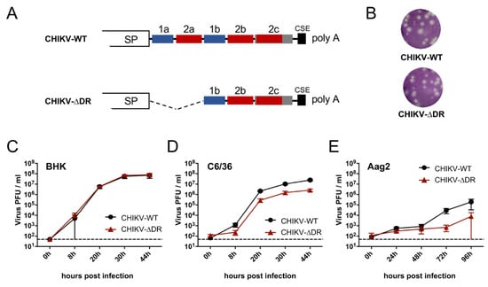
The 3′ UTRs of CHIKV belonging to the IO lineage contain two DR elements, namely DR1 (two copies) and DR2 (three copies) [22,26]. To analyze the importance of DR elements in viral replication and vector competence, we rescued CHIKV encompassing deletion of the DR elements 1a + 2a using reverse genetics (CHIKV-∆DR) (Figure 1A). As backbone the CHIKV E1-A226V variant was chosen. This variant is known to better infect Ae. albopictus which has also invaded European regions [12,44], and we aimed to analyze especially the vector competence of mosquito species prevalent in temperate regions.

Figure 1.
Growth kinetics of wild-type and ∆DR mutant CHIKV in different cells. (A) Schematic drawing of the 3′ UTRs of wild-type and mutant viruses. For CHIKV-∆DR, 130 nt encompassing the DR elements 1a and 2a were deleted. SP = structural protein coding region: CSE = 19 nt long conserved sequence element directly adjacent to the poly A tail. (B) Plaque morphology of CHIKV-WT and CHIKV-∆DR in Vero cells. Infected cells were overlaid with agarose. At two days post infection, cells were fixed and subjected to crystal violet staining. (C–E) Growth kinetics of CHIKV-WT and CHIKV-∆DR in vertebrate (BHK) and mosquito (C6/36, Aag2) cells. Cells were infected at an MOI of 0.1. Quantification of virus released into the supernatant was performed by titration in BHK cells. Data represent Mean ± SD of triplicate infection experiments. Dashed lines: detection limit.
Plaques formed by CHIKV-∆DR were similar to those of CHIKV-WT (Figure 1B). To further analyze the growth of CHIKV-∆DR in cell culture, we infected different cells with mutant and wild-type CHIKV for growth kinetic analyses. Both viruses readily replicated in vertebrate BHK cells with comparable growth characteristics reaching a titer at 44 h post infection of around 5 × 10⁸ (Figure 1C). Efficient growth was also observed in Ae. albopictus-derived C6/36 cells with CHIKV-WT growing nearly one log higher than CHIKV-∆DR (Figure 1D). Although CHIKV grew less efficiently in Ae. aegypti-derived Aag2 cells, viral titers were also reduced for CHIKV-∆DR compared to CHIKV-WT at later time points post infection (Figure 1E). These growth characteristics could also be demonstrated using reporter viruses of both variants expressing mCherry within the nsP3 protein (Figure S2) [37].
3.2. Vector Competence of CHIKV-∆DR in Ae. aegypti and Ae. albopictus
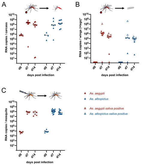
Ae. aegypti and Ae. albopictus represent known vectors for CHIKV. To confirm that the CHIKV-∆DR mutant can also replicate in these mosquitoes, we first infected female Ae. aegypti and Ae. albopictus orally with the mutant virus using blood meal containing 1 × 10⁶ PFU/mL virus (Figure 2A,B). Mosquitoes were dissected either within three hours after feeding (d0) or at d7 or d14 post infection, respectively. Analyses of mosquito carcasses revealed that viral RNA could be detected in all analyzed mosquitoes resulting in infection rates of 100% (Figure 2A, Table 2). The median viral titers increased between d0 and d7 for both Ae. aegypti and Ae. albopictus indicating that both mosquito species can efficiently be infected orally with the CHIKV-∆DR virus (Figure 2A). High median viral titers were sustained up to d14.

Figure 2.
Infection of Ae. aegypti and Ae. albopictus with CHIKV-∆DR. (A) Infection of mosquitoes after oral feeding with blood meal containing 1 × 10⁶ PFU/mL CHIKV-∆DR. At 0, 7 and 14 days post infection, mosquitoes were dissected and viral RNA copies were determined in the carcasses of the mosquitoes by real-time RT-PCR. (B) Dissemination of virus into secondary tissues after oral infection. At the indicated time points, viral RNA copies in wings and legs (Ae. aegypti) or wings (Ae. albopictus) were determined by real-time RT-PCR. Open symbols indicate disseminated mosquitoes for which transmission into saliva was also detected. (C) Intrathoracic infection of mosquitoes using 200 PFU of CHIKV-∆DR. At the indicated time points viral load in the whole mosquitoes was determined using real-time RT-PCR. Each point represents a single female mosquito. The black line indicates the median of the viral RNA copies in each group.

Table 2.
Infection, dissemination, transmission rates, and transmission efficiencies for CHIKV-∆DR at d7 and d14 post infection in Ae. aegypti and Ae. albopictus.
To analyze for dissemination, the wings and legs of Ae. aegypti were pooled and viral RNA copies were measured. For Ae. aegypti, the dissemination rate was at least 90% at d7 or d14, respectively (Table 2). Since Ae. albopictus were orally fed using cotton swabs instead of a Hemotek, the legs might have been potentially contaminated with virus from the blood meal. Hence, for Ae. albopictus only wings were taken to measure the ability of dissemination, which was determined to be 85.7% at d7 or 100% at d14, respectively (Figure 2B, Table 2). To determine whether disseminated virus was also able to reach the salivary glands, we collected the saliva of the mosquitoes and analyzed the samples again via RT-PCR. Transmission could be observed at d7 for 4 out of 10 disseminated Ae. aegypti (transmission rate 40%) or at d14 for 3 out of 9 disseminated Ae. aegypti (transmission rate 33.3%), respectively (Figure 2B, Table 2). Overall, this yielded transmission efficiencies of 36.4% (d7) or 30% (d14). For Ae. albopictus the transmission rates of 50% (d7) and 66.6% (d14), respectively, were obtained resulting in transmission efficiencies of 42.9% (d7) and 66.7% (d14) (Table 2).
While the median infectivity titers increased after oral infection for both Ae. aegypti and Ae. albopictus, individual Ae. aegypti showed only a low viral titer at d7 or d14 post infection in the carcasses (Figure 2A). We therefore also analyzed for Ae. aegypti as well as for Ae. albopictus as to which extent virus replication occurs when circumventing the midgut barrier. To this end Ae. aegypti and Ae. albopictus were intrathoracally infected with 200 PFU of CHIKV-∆DR. Viral RNA levels were analyzed at d0 from whole mosquitoes to verify the viral input level and at d7 and d14 post infection to analyze for viral replication. As can be seen in Figure 2C, the median titers increased for all d7 and d14 samples compared to the d0 time point demonstrating that CHIKV-∆DR replicates efficiently in Ae. aegypti and Ae. albopictus after circumventing the midgut barrier. This suggests that individual Ae. aegypti with low viral titers in the carcasses had insufficient oral virus uptake.
3.3. Vector Competence of CHIKV-∆DR and CHIKV-WT in Ae. vexans and Cx. pipiens
Ae. albopictus is known to have invaded areas of Europe [44]. To analyze whether other mosquito species in temperate regions are also competent for CHIKV and whether DR elements in the 3′ UTR have an impact on possible infection, dissemination and transmission rates, we performed infection experiments with Ae. vexans and Cx. pipiens (Figure 3).

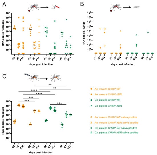
Figure 3.
Infection of Ae. vexans and Cx. pipiens with CHIKV-WT and CHIKV-∆DR. (A) Infection of mosquitoes after oral feeding with blood meal containing 1 × 10⁶ PFU/mL CHIKV-WT or CHIKV-∆DR. At d0, d7, and d14 post infection, mosquitoes were dissected and viral RNA copies were determined in the carcasses of the mosquitoes by real-time RT-PCR. (B) Dissemination of virus into secondary tissues after oral infection. At the indicated time points, viral RNA copies in the wings were determined. Open symbols indicate disseminated mosquitoes for which also transmission into saliva was detected. (C) Intrathoracic infection of mosquitoes using 200 PFU of CHIKV-WT or CHIKV-∆DR. At the indicated time points viral load in the whole mosquitoes was determined. Each symbol represents a single female mosquito. The black line indicates the median of the viral RNA copies in each group (** p < 0.01; *** p < 0.001; **** p < 0.0001).
Female Ae. vexans and Cx. pipiens were orally feed with a blood meal containing 1 × 10⁶ PFU/mL CHIKV-WT or CHIKV-∆DR, respectively (Figure 3A,B). Analysis of the viral titers in the Ae. vexans carcasses at d7 and d14 post infection revealed infection rates for CHIKV-WT of 83.3% (d7) and 65% (d14), and for CHIKV-∆DR of 80% (d7) and 47.8% (d14), respectively (Figure 3A, Table 3). To assess whether infected Ae. vexans are able to disseminate the viruses, wings were analyzed for genomic RNA of CHIKV (Figure 3B). At the early time point of 7 days post infection, the dissemination rate for CHIKV-WT was 20% at d7 and increased to 30.8% at d14. An increase of the dissemination rate over time was also observed for CHIKV-∆DR (0%, d7 and 36.4%, d14) (Table 3). Transmission rates in Ae. vexans were in the range of 50–75% at d7 or d14 for CHIKV-WT, whereas only a transmission rate up to 25% was observed for CHIKV-∆DR at d14 post infection. Altogether, this resulted in transmission efficiencies of 8.3% (d7) and 15% (d14) for CHIKV-WT in Ae. vexans, which however were not significantly higher than the transmission efficiencies of CHIKV-∆DR with 0% (d7) and 4.3% (d14).

Table 3.
Infection, dissemination, transmission rates, and transmission efficiencies for CHIKV-WT and CHIKV-∆DR at d7 and d14 post infection in Ae. vexans and Cx. pipiens.
In case of Cx. pipiens, viral RNA could be detected after oral feeding in the mosquito carcasses to 0% (d7) and 30% (d14) for CHIKV-WT and 75% (d7) and 28.5% (d14) for CHIKV-∆DR (Figure 3A and Table 3). Dissemination occurred rather sporadically and was found in three mosquitoes infected with CHIKV-WT at d14 and one mosquito infected with CHIKV-∆DR at d7 post infection (Figure 3B, Table 3). Viral RNA in the saliva could be detected for the two mosquitoes where the viral load in the wings was above 1 × 10⁴ RNA copies, namely once for CHIK-WT (d14) and once for CHIKV-∆DR (d7) suggesting a low potential for transmission (Figure 3B, Table 3).
For both Ae. vexans as well as Cx. pipiens it was observed that once increased viral titers were found in the carcasses, the virus was usually also able to disseminate and reach the salivary glands. This suggested that the midgut barrier represents the main barrier with regard to vector competence. To further support this finding, we infected Ae. vexans and Cx. pipiens intrathoracally using again 200 PFU with CHIKV-WT or CHIKV-∆DR, respectively. As can be seen in Figure 3C, analyses of the whole mosquitoes revealed efficient virus replication. CHIKV-WT replicated significantly higher in Ae. vexans compared to CHIKV-∆DR at both d7 and d14 post infection (p = 0.0002 or p = 0.0003, respectively). For Cx. pipiens a significant difference for CHIKV-WT in comparison to CHIKV-∆DR was only observed at d7 post infection (p = 0.0037). Furthermore, differences were obtained comparing mosquito species. CHIKV-WT replicated significantly higher in Ae. vexans compared to Cx. pipiens (p < 0.0001 for d7 and d14) and also replication of CHIKV-∆DR resulted in significant differences at d7 (p = 0.0023) and d14 (p = 0.0043). Furthermore, significant differences were obtained comparing the viral loads of CHIKV-∆DR after intrathoracic injection in Ae. aegypti (Figure 2) to the ones obtained in Cx. pipiens (p = 0.0012, d7 and p = 0.0043, d14). The differences were even more significant comparing the replication rates of CHIKV-∆DR after intrathoracic injection of Ae. albopictus (Figure 2) with the ones of Cx. pipiens (p < 0.0001, d7 and p < 0.0003, d14) suggesting that replication rates in the secondary tissues differ among both mosquito species and genus.
4. Discussion
The presence of repetitive elements in alphaviruses was already described over 20 years ago [20,21,45]. The number and arrangement of these direct repeat elements varies not only among different alphavirus species but also within species. In the Venezuelan equine encephalitis virus complex as well as for Ross River virus, isolates from mosquitoes in nature with various numbers of repeat elements have been described but their impact on ecology and pathogenesis is not well understood [21,46]. Comparison of the CHIKV genotypes likewise revealed various arrangements of repeat elements within the 3′ UTR [22]. The authors suggested that these differences were a result of an evolutionary process in order to especially adapt to mosquitoes rather than to host species. We investigated whether deletion of the DR elements DR1a and DR2a in CHIKV Mauritius belonging to the IO lineage affects viral replication in cell cultures or mosquitoes. While growth in vertebrate BHK cells was not affected for the mutant, the deletion resulted in reduced growth in Ae. albopictus mosquito cells. Similar results were already described many years ago for a Sindbis virus (SINV) deletion mutant and more recently for CHIKV deletion mutants of the Asian genotype as well as the IO lineage [22,26,47,48]. For SINV, viable virus was still recovered after removing up to 293 nt in the 3′ UTR so that only 25 nt from the 3′ terminus of the genome including the CSE element and one nucleotide 3′ of the UGA termination codon of the structural proteins was retained [47]. The resulting SINV mutant grew less efficiently in mosquito cells than in vertebrate cells and was in general more impaired than variants with smaller 3′ UTR deletions. Deletion of different numbers and sets of DR elements based on a Caribbean CHIKV isolate also resulted in reduced growth in mosquito C6/36 cells, whereby the growth was more restricted for the mutant with the larger deletion [26]. The same authors also deleted both the DR1ab and DR2ab motifs in CHIKV La Reunion belonging also to the IO lineage. This decreased the growth in C6/36 cells of about 3–4 logs while deletion of only DR1a and DR2a in our Mauritius isolate resulted in a reduction of about only 1 log supporting the finding that the size of deletion correlates with the level of growth reduction.
Although Ae. aegypti is known to be a main vector for CHIKV, Ae. aegypti derived Aag2 cells did not support the replication of CHIKV Mauritius as efficiently as C6/36 cells. After infection with the same MOI of 0.1, increase of viral titers was delayed and only peaked around 105 PFU/mL. Other studies also describe a comparatively low replication of CHIKV in Aag2 cells [49]. The efficiency of CHIKV release from Aag2 cells in comparison to C6/36 cells also seems to vary between different CHIKV isolates. Viral RNA copies in the supernatant of Aag2 cells increased faster within 24 h for a CHIKV belonging to the Asian genotype compared to CHIKV of the IO lineage belonging to the ECSA genotype [50]. Nevertheless, as observed for C6/36 cells, replication of CHIKV-∆DR was also impaired in Aag2 cells. As discussed previously, the phenotypic differences observed between vertebrate and mosquito cells might be attributed to different 3′ UTR binding proteins [47]. In line with this hypothesis, functional differences between 3′ UTRs in vertebrate and insect cells were described with regard to translation. A motif of repetitive sequence elements in the 3′ UTR was found to be involved in translation of SINV genomic and subgenomic mRNAs in mosquito cells but not in vertebrate cells [51]. It was discussed that such repetitive motifs might have been acquired during evolution to extend the host range towards mosquitoes [51].
Despite the fact that the deletion mutant showed reduced growth in mosquito cells compared to its wild-type counterpart, it readily could infect and disseminate in the well-known mosquito vectors Ae. aegypti and Ae. albopopictus both after oral and intrathoracic infection. Recent studies based on a Caribbean isolate compared a 3′ UTR mutant with several DR elements deleted to the wild-type virus with regard to infection, dissemination and transmission rates in Ae. aegypti and Ae. albopictus mosquitoes [27]. These analyses showed that the wild-type virus had a shorter extrinsic incubation period and it was found that the replication kinetics determine the ability to escape from the midgut. The slower replicating viruses encompassing the 3′ UTR deletion were less able to cross the midgut barrier as was shown in competition experiments with the wild-type virus [27]. Increased viral titers were also observed for the wild-type virus compared to the deletion mutant after intrathoracic infection but only at the earlier time points after infection (d2 and d4) [27]. This is in accordance with our findings that no significant differences between mutant and wild-type virus were obtained after intrathoracic injection in Ae. vexans or Cx. pipiens, respectively, when analyzing samples from d7 and d14 post infection.
However, statistically significant differences were obtained when comparing one type of virus in different mosquito species. After intrathoracic infection with CHIKV-∆DR the viral titers increased by 4 logs in both Ae. aegypti and Ae. albopictus (Figure 2C) whereas an increase of only 3 or 2 logs was observed for Ae. vexans or Cx. pipiens, respectively (Figure 3C). Similarly, the intrathoracically injected CHIKV-WT replicated to higher titers in Ae. vexans compared to Cx. pipiens. Hence, the midgut escape barrier does not seem to be the only factor influencing vector competence of mosquitoes. Also, replication kinetics in the secondary tissues seem to differ between mosquito species.
Both factors would also result in a longer extrinsic incubation period of CHIKV in Ae. vexans and Cx. pipiens compared to Ae. aegypti and Ae. albopictus after oral infection. This is supported by the fact that for Ae. vexans high viral titers within the body were not observed until d14, the time point where also the majority of mosquitoes with disseminated virus was observed (Figure 3A,B). Furthermore, detection of virus in the saliva was mainly observed in samples with high viral load in the wings suggesting that efficient replication in the secondary tissues to reach a certain threshold is critical for transmission. Although less Cx. pipiens mosquitoes were infected after oral feeding compared to Ae. vexans, dissemination as well as transmission was observed when 104 or more RNA copies were detected in the wings. This indicates that Cx. pipiens potentially could serve as a vector. To our knowledge this is the first study analyzing the vector competence of Cx. pipiens from Germany for CHIKV. Previous studies investigated the vector competence of Ae. vexans and Cx. pipiens using mosquitoes hatched from larvae or pupae sampled in France or Italy [52,53]. Whereas Ae. vexans from France as well as Cx. pipiens from both France and Italy were refractory to CHIKV infection, a low disseminated infection rate was observed in Ae. vexans from Italy based on immunofluorescence analyses of head squashes [52,53]. Analyses of another Aedes species, namely Aedes geniculatus originating from Albania revealed that they are susceptible to CHIKV and could transmit the virus although a longer extrinsic incubation period was observed compared to Ae. albopictus [54]. CHIKV positive Cx. pipiens were also detected during an outbreak in the Amazon region of Bazil. Since only mosquito pools were analyzed using PCR, no statement on the transmission rate could be made [55]. Nevertheless, our results indicate that other mosquito species than the well-established Ae. aegypti and Ae. albopictus may serve as competent mosquito vectors not only under laboratory conditions.
5. Conclusions
Our results demonstrate that the well-known CHIKV vectors Ae. aegypti and Ae. albopictus are also competent vectors for CHIKV exhibiting a two DR element deletion in the 3′ UTR. Testing two mosquito species, Ae. vexans and Cx. pipiens, for which little data on vector competence is currently available, demonstrated single mosquito individuals for which both CHIKV-WT and CHIKV-∆DR were able to overcome the midgut infection barrier and appeared in the saliva 7 or 14 days after oral infection. Together with the finding that these mosquito species replicated CHIKV-WT and CHIKV-∆DR in secondary tissue very efficiently after intrathoracic infection, both mosquito species may eventually transmit CHIKV also in nature.
Supplementary Materials
The following are available online at https://www.mdpi.com/1999-4915/13/3/403/s1, Figure S1: CHIKV real-time PCR; Figure S2: Expression of mCherry from CHIKV-WT and CHIKV-ΔDR reporter viruses at different time points after infection.
Author Contributions
Conceptualization, M.P. and B.M.K.; methodology, Y.K., A.v.H., J.W. M.P., and B.M.K.; validation, Y.K., A.v.H., J.W., M.P. and B.M.K.; formal analysis, A.v.H. and M.P.; investigation, Y.K., A.v.H., J.W., M.P. and B.M.K.; resources, Y.K., M.P. and B.M.K.; writing—original draft preparation, B.M.K.; writing—review and editing, Y.K., A.v.H. and M.P.; visualization, A.v.H. and B.M.K.; supervision, M.P. and B.M.K.; project administration, M.P. and B.M.K.; funding acquisition, M.P. and B.M.K. All authors have read and agreed to the published version of the manuscript.
Funding
This research was funded by the Deutsche Forschungsgemeinschaft (DFG, German Research Foundation)—KU 1201/4-2 (SPP 1596/1) and Project-ID 369799452-TRR237 to B.M.K; PF 723/1-1 (SPP 1596/1) to M.P.
Acknowledgments
We thank Romeo Bellini (Centro Agricoltura Ambiente, Italy) for kindly providing Ae. albopictus and Ramona Dahl, Federal Environment Agency, Berlin, Germany, for providing Ae. aegypti. Furthermore, we are grateful to Roland Kuhn (formerly Johannes Gutenberg University, Mainz, Germany) for kindly giving us Ae. vexans ‘TAMU’ and Cx. pipiens molestus. A big thank you goes to Cornelia Silaghi and Marlene Hausner for support in rearing Ae. vexans and Cx. pipiens mosquitoes. All members of the Institute of Animal Hygiene and Veterinary Public Health are warmly acknowledged for their help in maintaining the mosquito colonies.
Conflicts of Interest
The authors declare no conflict of interest.
References
- Weaver, S.C.; Barrett, A.D. Transmission cycles, host range, evolution and emergence of arboviral disease. Nat. Rev. Microbiol. 2004, 2, 789–801. [Google Scholar] [CrossRef]
- Pialoux, G.; Gaüzère, B.A.; Jauréguiberry, S.; Strobel, M. Chikungunya, an epidemic arbovirosis. Lancet Infect Dis. 2007, 7, 319–327. [Google Scholar] [CrossRef]
- Ross, R.W. The Newala epidemic. III. The virus: Isolation, pathogenic properties and relationship to the epidemic. J. Hyg. (Lond.) 1956, 54, 177–191. [Google Scholar] [CrossRef] [PubMed]
- Pastorino, B.; Muyembe-Tamfum, J.J.; Bessaud, M.; Tock, F.; Tolou, H.; Durand, J.P.; Peyrefitte, C.N. Epidemic resurgence of Chikungunya virus in democratic Republic of the Congo: Identification of a new central African strain. J. Med. Virol. 2004, 74, 277–282. [Google Scholar] [CrossRef] [PubMed]
- Sergon, K.; Njuguna, C.; Kalani, R.; Ofula, V.; Onyango, C.; Konongoi, L.S.; Bedno, S.; Burke, H.; Dumilla, A.M.; Konde, J.; et al. Seroprevalence of Chikungunya virus (CHIKV) infection on Lamu Island, Kenya, October 2004. Am. J. Trop. Med. Hyg. 2008, 78, 333–337. [Google Scholar] [CrossRef] [PubMed]
- Sergon, K.; Yahaya, A.A.; Brown, J.; Bedja, S.A.; Mlindasse, M.; Agata, N.; Allaranger, Y.; Ball, M.D.; Powers, A.M.; Ofula, V.; et al. Seroprevalence of Chikungunya virus infection on Grande Comore Island, union of the Comoros, 2005. Am. J. Trop. Med. Hyg. 2007, 76, 1189–1193. [Google Scholar] [CrossRef] [PubMed]
- Renault, P.; Solet, J.L.; Sissoko, D.; Balleydier, E.; Larrieu, S.; Filleul, L.; Lassalle, C.; Thiria, J.; Rachou, E.; de Valk, H.; et al. A major epidemic of chikungunya virus infection on Reunion Island, France, 2005–2006. Am. J. Trop. Med. Hyg. 2007, 77, 727–731. [Google Scholar] [CrossRef]
- Chastel, C. Chikungunya virus: Its recent spread to the southern Indian Ocean and Reunion Island (2005–2006). Bull. Acad. Natl. Med. 2005, 189, 1827–1835. [Google Scholar] [PubMed]
- Lahariya, C.; Pradhan, S.K. Emergence of chikungunya virus in Indian subcontinent after 32 years: A review. J. Vector Borne Dis. 2006, 43, 151–160. [Google Scholar] [PubMed]
- Naresh Kumar, C.V.; Sai Gopal, D.V. Reemergence of Chikungunya virus in Indian Subcontinent. Indian J. Virol. 2010, 21, 8–17. [Google Scholar] [CrossRef]
- Tsetsarkin, K.A.; Vanlandingham, D.L.; McGee, C.E.; Higgs, S. A single mutation in chikungunya virus affects vector specificity and epidemic potential. PLoS Pathog. 2007, 3, e201. [Google Scholar] [CrossRef] [PubMed]
- Vazeille, M.; Moutailler, S.; Coudrier, D.; Rousseaux, C.; Khun, H.; Huerre, M.; Thiria, J.; Dehecq, J.S.; Fontenille, D.; Schuffenecker, I.; et al. Two Chikungunya isolates from the outbreak of La Reunion (Indian Ocean) exhibit different patterns of infection in the mosquito, Aedes albopictus. PLoS ONE 2007, 2, e1168. [Google Scholar] [CrossRef] [PubMed]
- Rezza, G.; Nicoletti, L.; Angelini, R.; Romi, R.; Finarelli, A.C.; Panning, M.; Cordioli, P.; Fortuna, C.; Boros, S.; Magurano, F.; et al. Infection with chikungunya virus in Italy: An outbreak in a temperate region. Lancet 2007, 370, 1840–1846. [Google Scholar] [CrossRef]
- Calba, C.; Guerbois-Galla, M.; Franke, F.; Jeannin, C.; Auzet-Caillaud, M.; Grard, G.; Pigaglio, L.; Decoppet, A.; Weicherding, J.; Savaill, M.C.; et al. Preliminary report of an autochthonous chikungunya outbreak in France, July to September 2017. Euro Surveill. 2017, 22. [Google Scholar] [CrossRef]
- Leparc-Goffart, I.; Nougairede, A.; Cassadou, S.; Prat, C.; de Lamballerie, X. Chikungunya in the Americas. Lancet 2014, 383, 514. [Google Scholar] [CrossRef]
- Pan American Health Organization/World Health Organization (PAHO/WHO). Geographic Spread of Chikungunya in the Americas, December 2013–December 2017. Available online: https://ais.paho.org/phip/viz/ed_chikungunya_amro.asp (accessed on 25 January 2021).
- Lanciotti, R.S.; Valadere, A.M. Transcontinental movement of Asian genotype chikungunya virus. Emerg. Infect Dis. 2014, 20, 1400–1402. [Google Scholar] [CrossRef]
- Kuhn, R.J. Togaviridae. In Fields Virology, 6th ed.; Knipe, D.M., Howley, P.M., Eds.; Lippincott Williams & Wilkons: Philadelphia, PA, USA, 2013; Volume 1, pp. 629–650. [Google Scholar]
- Frolov, I.; Hardy, R.; Rice, C.M. Cis-acting RNA elements at the 5′ end of Sindbis virus genome RNA regulate minus- and plus-strand RNA synthesis. RNA 2001, 7, 1638–1651. [Google Scholar] [CrossRef] [PubMed]
- Ou, J.H.; Trent, D.W.; Strauss, J.H. The 3′-non-coding regions of alphavirus RNAs contain repeating sequences. J. Mol. Biol. 1982, 156, 719–730. [Google Scholar] [CrossRef]
- Pfeffer, M.; Kinney, R.M.; Kaaden, O.R. The alphavirus 3′-nontranslated region: Size heterogeneity and arrangement of repeated sequence elements. Virology 1998, 240, 100–108. [Google Scholar] [CrossRef]
- Chen, R.; Wang, E.; Tsetsarkin, K.A.; Weaver, S.C. Chikungunya virus 3′ untranslated region: Adaptation to mosquitoes and a population bottleneck as major evolutionary forces. PLoS Pathog. 2013, 9, e1003591. [Google Scholar] [CrossRef]
- Volk, S.M.; Chen, R.; Tsetsarkin, K.A.; Adams, A.P.; Garcia, T.I.; Sall, A.A.; Nasar, F.; Schuh, A.J.; Holmes, E.C.; Higgs, S.; et al. Genome-scale phylogenetic analyses of chikungunya virus reveal independent emergences of recent epidemics and various evolutionary rates. J. Virol. 2010, 84, 6497–6504. [Google Scholar] [CrossRef] [PubMed]
- Sahadeo, N.; Mohammed, H.; Allicock, O.M.; Auguste, A.J.; Widen, S.G.; Badal, K.; Pulchan, K.; Foster, J.E.; Weaver, S.C.; Carrington, C.V. Molecular Characterisation of Chikungunya Virus Infections in Trinidad and Comparison of Clinical and Laboratory Features with Dengue and Other Acute Febrile Cases. PLoS Negl. Trop. Dis. 2015, 9, e0004199. [Google Scholar]
- Hyde, J.L.; Chen, R.; Trobaugh, D.W.; Diamond, M.S.; Weaver, S.C.; Klimstra, W.B.; Wilusz, J. The 5′ and 3′ ends of alphavirus RNAs—Non-coding is not non-functional. Virus Res. 2015, 206, 99–107. [Google Scholar] [CrossRef] [PubMed]
- Filomatori, C.V.; Bardossy, E.S.; Merwaiss, F.; Suzuki, Y.; Henrion, A.; Saleh, M.C.; Alvarez, D.E. RNA recombination at Chikungunya virus 3’UTR as an evolutionary mechanism that provides adaptability. PLoS Pathog. 2019, 15, e1007706. [Google Scholar] [CrossRef] [PubMed]
- Merwaiss, F.; Filomatori, C.V.; Susuki, Y.; Bardossy, E.S.; Alvarez, D.E.; Saleh, M.C. Chikungunya virus replication rate determines the capacity of crossing tissue barriers in mosquitoes. J. Virol. 2021. [Google Scholar] [CrossRef]
- Filomatori, C.V.; Merwaiss, F.; Bardossy, E.S.; Alvarez, D.E. Impact of alphavirus 3’UTR plasticity on mosquito transmission. Semin. Cell Dev. Biol. 2020, 111, 148–155. [Google Scholar] [CrossRef]
- Arias-Goeta, C.; Mousson, L.; Rougeon, F.; Failloux, A.B. Dissemination and transmission of the E1-226V variant of chikungunya virus in Aedes albopictus are controlled at the midgut barrier level. PLoS ONE 2013, 8, e57548. [Google Scholar] [CrossRef]
- Roiz, D.; Neteler, M.; Castellani, C.; Arnoldi, D.; Rizzoli, A. Climatic factors driving invasion of the tiger mosquito (Aedes albopictus) into new areas of Trentino, northern Italy. PLoS ONE 2011, 6, e14800. [Google Scholar] [CrossRef]
- Medlock, J.M.; Hansford, K.M.; Schaffner, F.; Versteirt, V.; Hendrickx, G.; Zeller, H.; Van Bortel, W. A review of the invasive mosquitoes in Europe: Ecology, public health risks, and control options. Vector Borne Zoonotic. Dis. 2012, 12, 435–447. [Google Scholar] [CrossRef]
- Sherpa, S.; Blum, M.G.B.; Capblancq, T.; Cumer, T.; Rioux, D.; Després, L. Unravelling the invasion history of the Asian tiger mosquito in Europe. Mol. Ecol. 2019, 28, 2360–2377. [Google Scholar] [CrossRef]
- Goiri, F.; González, M.A.; Goikolea, J.; Oribe, M.; Castro, V.; Delacour, S.; Lucientes, J.; Ortega-Araiztegi, I.; Barandika, J.F.; García-Pérez, A.L. Progressive Invasion of Aedes albopictus in Northern Spain in The Period 2013-2018 and A Possible Association with the Increase in Insect Bites. Int. J. Environ. Res. Public Health 2020, 17, 1678. [Google Scholar] [CrossRef]
- ECDC. Mosquito Maps. 2020. Available online: https://ecdc.europa.eu/en/disease-vectors/surveillance-and-disease-data/mosquito-maps (accessed on 25 January 2021).
- Pluskota, B.; Jöst, A.; Augsten, X.; Stelzner, L.; Ferstl, I.; Becker, N. Successful overwintering of Aedes albopictus in Germany. Parasitol. Res. 2016, 115, 3245–3247. [Google Scholar] [CrossRef]
- Modlmaier, M.; Kuhn, R.; Kaaden, O.R.; Pfeffer, M. Transmission studies of a European Sindbis virus in the floodwater mosquito Aedes vexans (Diptera: Culicidae). Int. J. Med. Microbiol. 2002, 291 (Suppl. 33), 164–170. [Google Scholar] [CrossRef]
- Kümmerer, B.M.; Grywna, K.; Gläsker, S.; Wieseler, J.; Drosten, C. Construction of an infectious Chikungunya virus cDNA clone and stable insertion of mCherry reporter genes at two different sites. J. Gen. Virol. 2012, 93 Pt 9, 1991–1995. [Google Scholar] [CrossRef]
- Voßmann, S.; Wieseler, J.; Kerber, R.; Kümmerer, B.M. A basic cluster in the N terminus of yellow fever virus NS2A contributes to infectious particle production. J. Virol. 2015, 89, 4951–4965. [Google Scholar] [CrossRef]
- Heitmann, A.; Jansen, S.; Lühken, R.; Leggewie, M.; Schmidt-Chanasit, J.; Tannich, E. Forced Salivation as a Method to Analyze Vector Competence of Mosquitoes. J. Vis. Exp. 2018. [Google Scholar] [CrossRef]
- Riswari, S.F.; Ma’roef, C.N.; Djauhari, H.; Kosasih, H.; Perkasa, A.; Yudhaputri, F.A.; Artika, I.M.; Williams, M.; van der Ven, A.; Myint, K.S.; et al. Study of viremic profile in febrile specimens of chikungunya in Bandung, Indonesia. J. Clin. Virol. 2016, 74, 61–65. [Google Scholar] [CrossRef]
- Waggoner, J.J.; Gresh, L.; Vargas, M.J.; Ballesteros, G.; Tellez, Y.; Soda, K.J.; Sahoo, M.K.; Nuñez, A.; Balmaseda, A.; Harris, E.; et al. Viremia and Clinical Presentation in Nicaraguan Patients Infected With Zika Virus, Chikungunya Virus, and Dengue Virus. Clin. Infect Dis. 2016, 63, 1584–1590. [Google Scholar] [CrossRef]
- Rosen, L.; Gubler, D. The use of mosquitoes to detect and propagate dengue viruses. Am. J. Trop. Med. Hyg. 1974, 23, 1153–1160. [Google Scholar] [CrossRef]
- Boorman, J. Induction of salivation in biting midges and mosquitoes, and demonstration of virus in the saliva of infected insects. Med. Vet. Entomol. 1987, 1, 211–214. [Google Scholar] [CrossRef]
- Kraemer, M.U.G.; Reiner, R.C., Jr.; Brady, O.J.; Messina, J.P.; Gilbert, M.; Pigott, D.M.; Yi, D.; Johnson, K.; Earl, L.; Marczak, L.B.; et al. Past and future spread of the arbovirus vectors Aedes aegypti and Aedes albopictus. Nat. Microbiol. 2019, 4, 854–863. [Google Scholar] [CrossRef]
- Strauss, J.H.; Strauss, E.G. The alphaviruses: Gene expression, replication, and evolution. Microbiol. Rev. 1994, 58, 491–562. [Google Scholar] [CrossRef]
- Faragher, S.G.; Dalgarno, L. Regions of conservation and divergence in the 3’ untranslated sequences of genomic RNA from Ross River virus isolates. J. Mol. Biol. 1986, 190, 141–148. [Google Scholar] [CrossRef]
- Kuhn, R.J.; Hong, Z.; Strauss, J.H. Mutagenesis of the 3’ nontranslated region of Sindbis virus RNA. J. Virol. 1990, 64, 1465–1476. [Google Scholar] [CrossRef]
- Morley, V.J.; Noval, M.G.; Chen, R.; Weaver, S.C.; Vignuzzi, M.; Stapleford, K.A.; Turner, P.E. Chikungunya virus evolution following a large 3’UTR deletion results in host-specific molecular changes in protein-coding regions. Virus Evol. 2018, 4, vey012. [Google Scholar] [CrossRef]
- Roberts, G.C.; Zothner, C.; Remenyi, R.; Merits, A.; Stonehouse, N.J.; Harris, M. Evaluation of a range of mammalian and mosquito cell lines for use in Chikungunya virus research. Sci. Rep. 2017, 7, 14641. [Google Scholar] [CrossRef]
- Zhang, X.; Huang, Y.; Wang, M.; Yang, F.; Wu, C.; Huang, D.; Xiong, L.; Wan, C.; Cheng, J.; Zhang, R. Differences in genome characters and cell tropisms between two chikungunya isolates of Asian lineage and Indian Ocean lineage. Virol. J. 2018, 15, 130. [Google Scholar] [CrossRef]
- Garcia-Moreno, M.; Sanz, M.A.; Carrasco, L. A Viral mRNA Motif at the 3’-Untranslated Region that Confers Translatability in a Cell-Specific Manner. Implications for Virus Evolution. Sci. Rep. 2016, 6, 19217. [Google Scholar] [CrossRef]
- Vazeille, M.; Jeannin, C.; Martin, E.; Schaffner, F.; Failloux, A.B. Chikungunya: A risk for Mediterranean countries? Acta Trop. 2008, 105, 200–202. [Google Scholar] [CrossRef]
- Talbalaghi, A.; Moutailler, S.; Vazeille, M.; Failloux, A.B. Are Aedes albopictus or other mosquito species from northern Italy competent to sustain new arboviral outbreaks? Med. Vet. Entomol. 2010, 24, 83–87. [Google Scholar] [CrossRef]
- Prudhomme, J.; Fontaine, A.; Lacour, G.; Gantier, J.C.; Diancourt, L.; Velo, E.; Bino, S.; Reiter, P.; Mercier, A. The native European Aedes geniculatus mosquito species can transmit chikungunya virus. Emerg. Microbes Infect 2019, 8, 962–972. [Google Scholar] [CrossRef] [PubMed]
- Ribeiro Cruz, A.C.; Pinto Nunes Neto, J.; Patroca da Silva, S.; Vieira Pinto da Silva, E.; Juscely Galvão Pereira, G.; Maia Santos, M.; Antônio de Oliveira Monteiro, H.; Barreto Dos Santos, F.; José de Paula Souza, E.G.R.; Fortes Aragão, C.; et al. Chikungunya virus Detection in Aedes aegypti and Culex quinquefasciatus during an Outbreak in the Amazon Region. Viruses 2020, 12, 853. [Google Scholar] [CrossRef]
Publisher’s Note: MDPI stays neutral with regard to jurisdictional claims in published maps and institutional affiliations. |
© 2021 by the authors. Licensee MDPI, Basel, Switzerland. This article is an open access article distributed under the terms and conditions of the Creative Commons Attribution (CC BY) license (http://creativecommons.org/licenses/by/4.0/).