Article

Phylogenomic Analysis of Global Isolates of Canid Alphaherpesvirus 1

1
Veterinary Clinical Sciences, School of Veterinary Medicine, Louisiana State University, Baton Rouge, LA 70803, USA
2
Center for Computation and Technology, Louisiana State University, Baton Rouge, LA 70808, USA
3
Department of Clinical Sciences, College of Veterinary Medicine, Cornell University, Ithaca, NY 14853, USA
*
Author to whom correspondence should be addressed.
Viruses 2020, 12(12), 1421; https://doi.org/10.3390/v12121421
Submission received: 19 November 2020 / Revised: 7 December 2020 / Accepted: 8 December 2020 / Published: 10 December 2020
(This article belongs to the Special Issue Animal Herpesviruses Pathogenesis and Immunity)

Abstract

:
Canid alphaherpesvirus 1 (CHV-1) is a widespread pathogen of dogs with multiple associated clinical signs. There has been limited prior investigation into the genomics and phylogeny of this virus using whole viral genome analysis. Fifteen CHV-1 isolates were collected from animals with ocular disease based in the USA. Viral DNA was extracted for Illumina MiSeq full genome sequencing from each isolate. These data were combined with genomes of previously sequenced CHV-1 isolates obtained from hosts in the UK, Australia and Brazil. Genomic, recombinational and phylogenetic analysis were performed using multiple programs. Two isolates were separated into a clade apart from the remaining isolates and accounted for the majority of genomic distance (0.09%): one was obtained in 2019 from a USA-based host (ELAL-1) and the other in 2012 from a host in Brazil (BTU-1). ELAL-1 was found to contain variants previously reported in BTU-1 but also novel variants in the V57 gene region. Multiple non-synonymous variants were found in USA-based isolates in regions associated with antiviral resistance. Evidence of recombination was detected between ELAL-1 and BTU-1. Collectively, this represents evidence of trans-boundary transmission of a novel form of CHV-1, which highlights the importance of surveillance for this pathogen in domestic dog populations.

1. Introduction

Disease caused by CHV-1 is widespread in canine populations [1,2,3] and affects both neonatal and adult animals. The virus is a significant cause of sudden death in neonates [4], ocular disease [5], respiratory disease [6,7] and reproductive disease [8,9]. Despite this, there has been minimal genomic assessment of the virus to date.
CHV-1 is a varicellovirus of the herpesviridae family, which establishes latency following an initial infection period [10,11]. The virus has a worldwide distribution [12,13,14] and a high seroprevalence among various canine populations [1,2,3]. Diagnosis of disease is currently based on viral isolation, PCR or serology. Although symptomatic treatment of certain forms of CHV-1 (ocular disease) has progressed in recent years [15], minimal options are available for effectively treating the disease in neonatal animals. Vaccines are available but do not offer complete protection against infection [16,17]. Together, this means that disease surveillance and prevention are essential for control of this virus.
The structure of CHV-1 is similar to other herpesviruses, with unique long (UL) and short (US) sequences flanked by terminal (TRS/TRL) and internal inverted (IRS/IRL) repeats. The overall length is approximately 125 kbp with 76 open reading frames [18]. Several complete/near-complete CHV-1 isolates have been sequenced and analyzed in the past [18,19,20]. Phylogenetic analysis of complete CHV-1 genomes has been previously been performed using a very limited number of isolates [18,20]. While most CHV-1 genomes were previously found to be mostly homogenous (0.005% distance in UK isolates), one isolate (BTU-1) obtained from a Brazilian host canid in 2012, was noted to be 0.34% distant from the others, mostly due to variants in the CHV-1 UL50 protein sequence region [20].
Our objective was to characterize the genomes of circulating CHV-1 isolates in the USA and to relate their sequence characteristics to global isolates. We hypothesized that CHV-1 isolates obtained in the USA would be genomically homogenous but would separate into clades based on geography, as is the case for the related varicellovirus, feline herpesvirus 1 (FHV-1) [21].

2. Materials and Methods

2.1. Cells and Viruses

Virus isolation was performed in a routine manner using diagnostic samples collected from naturally infected host canids (with informed owner consent). Sample collection was performed by vigorously swabbing the conjunctival fornices of host animals using a flocked swab, which was then immediately placed into universal viral transport media (BD, NJ) for transport. Tubes containing swabs and viral transport medium were vortexed. Approximately 1.5 cc of viral transport medium was inoculated onto cells and allowed to absorb for 1 h with periodic rocking. Isolations were performed with A-72 canine cells (ATCC, CRL-1542, VA), Madin-Darby Canine Kidney (MDCK) cells (ATCC, CRL-2935, VA), and a laboratory-developed canine skin cell line in minimum essential medium-E with 10% fetal bovine serum, 5% serum replacement solution, 2% penicillin-streptomycin solution, 1% amphotericin B solution and 1% gentamicin sulfate solution. Cultures were incubated at 37 °C (5% CO2), checked at 24-h intervals for cytopathic effect (CPE), subcultured every 5–7 days, and held for up to 21 days. Cell cultures with CPE typical of CHV-1 were stained with anti-CHV-1 polyclonal antiserum conjugated to fluorescein isothiocyanate (CHV-1 direct fluorescent antibody conjugate, CJ-F-CHV-10ML, VMRD Inc, Pullman, WA, USA) [22] to confirm viral identification. At this point, each flask was subjected to 3 freeze/thaw cycles before the contents were transferred to a 15 cc conical tube and centrifuged at 600× g for 5 min at 4 °C. The supernatant was then removed and aliquoted for storage at −80 °C prior to bulk processing for sequencing.
Using a 500 µL volume of previously stored supernatant, a T25 flask (Thermo Fisher, Waltham, MA, USA) containing a monolayer of MDCK cells was subsequently infected in a similar manner for each isolate using Dulbecco’s Modified Eagle Media (DMEM) (Thermo Fisher, Waltham, MA, USA) containing 2% fetal bovine serum (Thermo Fisher, Waltham, MA, USA) and 1% penicillin/streptomycin (Thermo Fisher, Waltham, MA, USA). Following observation of 100% CPE, 3 freeze/thaw cycles and centrifugation (600× g for 5 min at 4°C), 200µL of the resulting supernatant was used for viral DNA extraction. Viral DNA was prepared using a commercial kit according to the manufacturer’s instructions (Purelink Viral RNA/DNA Mini Kit, Invitrogen, Carlsbad, CA, USA).

2.2. Sequencing

Extracted DNA was submitted to the Louisiana State University School of Veterinary Medicine GeneLab. DNA purity and concentration were assessed using a NanoDrop One Microvolume Spectrophotometer (Thermo Scientific, Waltham, MA, USA) and then processed using the Nextera DNA Flex Library Prep Kit (Illumina Inc., San Diego, CA, USA) with modifications specific for the 100–500 ng DNA input range. Quality and quantity of the finished libraries were assessed using a Fragment Analyzer Instrument (Advanced Analytical) and dsDNA HS Assay Kit, respectively. Libraries were pooled, standardized to 10 μM and paired end sequencing was performed using the Illumina MiSeq Sequencer and a MiSeq 500 bp (v2) sequencing kit (MS-102-2003).

2.3. Genome Assembly

Reference-based assembly was performed using Geneious Prime ver 2020.2.4. Paired end reads were trimmed using BBDuk adapter/quality trimming ver 38.84 (right end, Kmer length = 27, maximum substitutions = 1, minimum quality = 20, minimum overlap = 20, minimum length = 20). Trimmed paired end reads were then assembled to the reference sequence for CHV-1 (0194, Genbank accession NC_030117). A consensus sequence was extracted from the aligned reads with gaps filled with “N’s”. Genomes were annotated and submitted to Genbank using Geneious Prime ver 2020.2.4.

2.4. Viral Genome Alignments

Alignments were created using MAFFT ver 7.450 [23] within Geneious Prime ver 2020.2.4. In all instances, the default parameters were used; a scoring matrix of 1 PAM/k = 2, gap penalty of 1.53 and offset value of 0.123. Alignments created included all whole CHV-1 genomes (USA + UK + Brazil + Australia), all whole CHV-1 genomes plus a Feline herpesvirus (FHV-1) outgroup (C-27, Genbank accession NC_013590.2) and the isolated CHV-1 V57 gene region from 3 CHV-1 genomes (0194, ELAL-1 and BTU-1). An additional alignment using the same 3 complete CHV-1 genomes (0194, ELAL-1 and BTU-1) was also created.

2.5. Variant Analysis

Variant analysis was performed using the Geneious variant finder (Geneious Prime ver 2020.2.4) in regions with a minimum coverage = 100, minimum variant frequency (proportion of reads matching variant) = 0.25, maximum variant p-value = 10−6 and minimum strand-bias (disagreement between the forward and reverse strand) p-value =10−5 when exceeding 65% bias. Variants were called by comparing each sequenced isolate to the reference genome (CHV-1 0194). Linear regression with viral gene length as the repressor and the total number of the variants per gene as the outcome (with 99% confidence limits) was constructed using JMP Pro 15.0.0 (SAS Institute Inc., Cary, NC, USA).
Regions of genomic distance between 3 CHV-1 isolates (0194, ELAL-1 and BTU-1) were visualized using RDP ver 4.100 [24] using the manual distance plot function (window = 1200, step = 500, transition:transversion rate ratio = 2, coefficient of variation = 1, Jin and Nei model [25]).

2.6. Phylogenetic and Recombination Analysis

An alignment containing all whole CHV-1 genomes plus a Feline herpesvirus (FHV-1) outgroup was processed using ModelFinder [26] via IQ-Tree 2 ver 1.6.12 [27] to automatically select the best-fit model (TVM+F+G4). The resultant maximum likelihood tree was viewed using Splitstree ver 4.16.1 [28]. Recombination analysis was performed using RDP ver 4.100 [24] on an aligned set of CHV-1 genomes using a manual bootscan (window = 1200, step = 500, replicates = 100, 70% cutoff, Jin and Nei model [25]), RDP, GENECONV, MaxChi and Chimaera.
Pairwise genomic distances were calculated using MEGAX [29] with the gamma distribution model (5), partial deletion of gaps and 1000 bootstrap replicates.

2.7. Sequence Accession Numbers

All isolates used for analysis are shown in Table 1 and are available on the GenBank website (https://www.ncbi.nlm.nih.gov/genbank/).

3. Results

3.1. Sequencing and Genome Assembly

A total of 15 isolates of CHV-1 were sequenced using Illumina MiSeq. A further five previously sequenced CHV-1 genomes were included in the analysis (Table 1). The majority of isolates sequenced for this study were obtained from animals in the NY, USA region and all were taken from animals with ocular disease (conjunctivitis and keratitis). A variety of host breeds were represented, with most (7/15) being beagles. Almost all (6/7) of these beagles were housed together in New York State and had samples taken as part of an investigation into a CHV-1 disease outbreak. Both juvenile and adult host animals are represented for the 15 isolates sequenced for this study, with the youngest being approximately 8 weeks old (ELAL-13) and the oldest being 12 years old (ELAL-12).
The details of sequencing are shown in Table 2. The total number of reads ranged from 1,292,262 (ELAL-3) to 3,533,658 (ELAL-10). The number of reads mapped ranged from 155,016 (ELAL-1) to 856,992 (ELAL-14). The mean mapped read length ranged from 201.1 (ELAL-12) to 228.6 (ELAL-2). Mean coverage across the genomes ranged from 290× (ELAL-1) to 1509× (ELAL-6). Coverage was excellent throughout the length of the genomes, with rare regions of less than 100× coverage in several isolates (Figure S1). The average G-C content was 31.6%, and this value was fairly consistent along the length of all genomes (with one elevation 36 kb from the left end of the genome of approximately 74% G-C content). No relationship between G-C content and coverage was observed.

3.2. Variant Analysis

Variants were found to be evenly distributed throughout the length of the genomes and are summarized in Table 3 and Table S1. A total of 237 unique synonymous and 233 unique non-synonymous variants were found across the genomes. The most unique synonymous variants were located in the RS1 gene region (22) and the most unique non-synonymous variants (42) were located in the UL36 gene region. The number of unique synonymous variants within each isolate ranged from 4 (ELAL-2, ELAL-8) to 188 (ELAL-1). The number of unique non-synonymous variants ranged from 17 (ELAL-8) to 154 (ELAL-1). The total number of variants for each gene is plotted against gene length in a regression analysis in Figure S2. A single gene (UL50/53 variants/918 bp) was found to contain a higher-than-expected number of variants based on gene length.
Non-synonymous variants were detected in UL23 (thymidine kinase), UL30 (DNA polymerase catalytic subunit) and UL42 (DNA polymerase processivity subunit), all regions which are targeted by commonly used antiviral medications [30]. All sequenced isolates apart from 4 (ELAL-6, ELAL-7, ELAL-12, ELAL-15) contained non-synonymous variants in UL30, whereas only two isolates contained non-synonymous variants in UL42 (ELAL-1, ELAL-6). Only one isolate (ELAL-1) contained non-synonymous variants in UL23, UL30 and UL42.

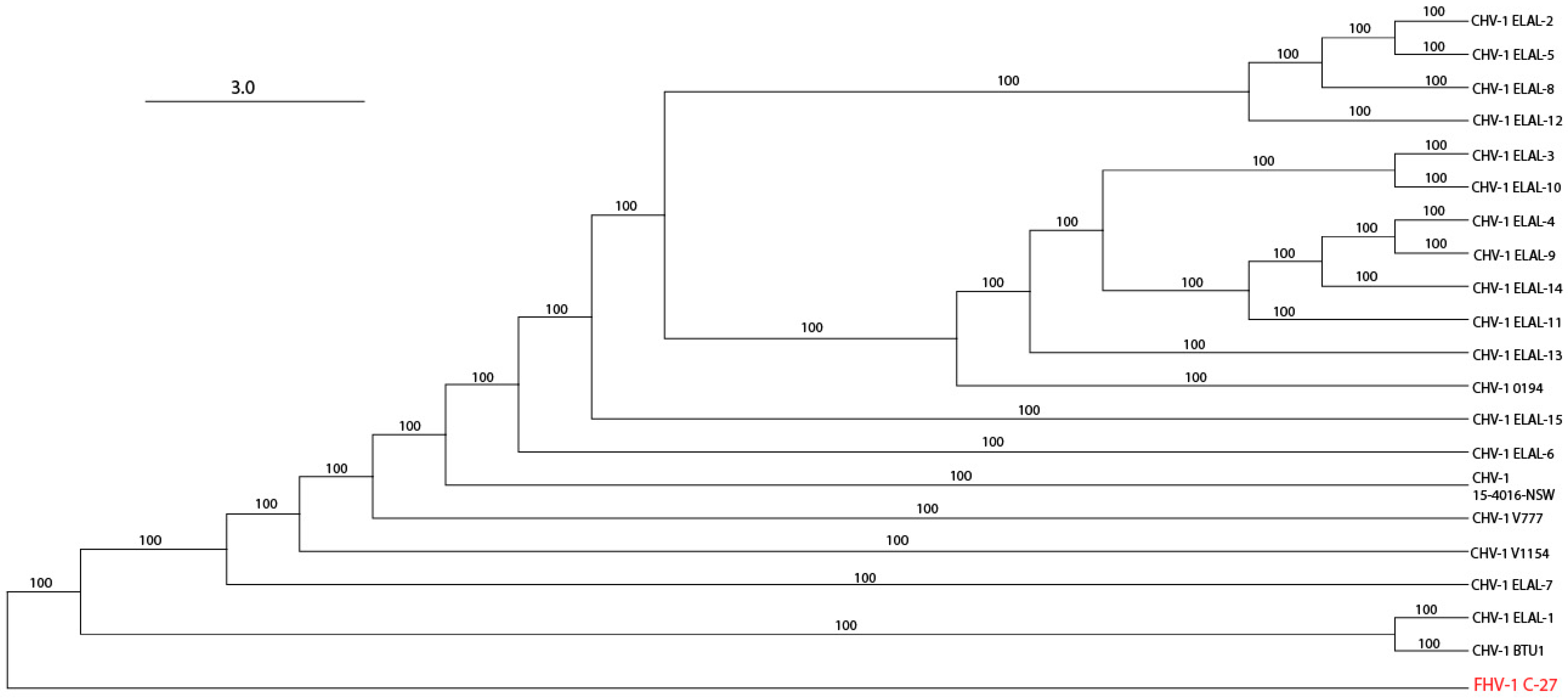
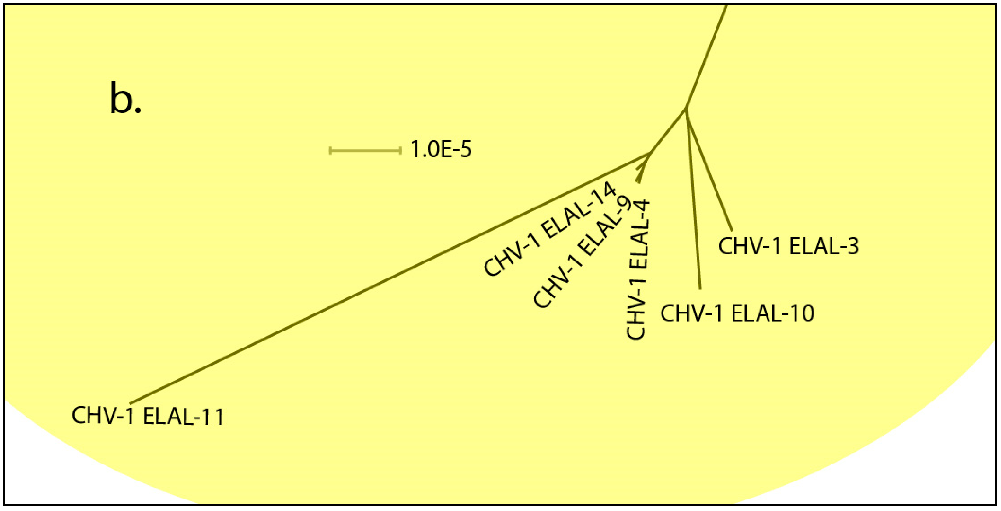
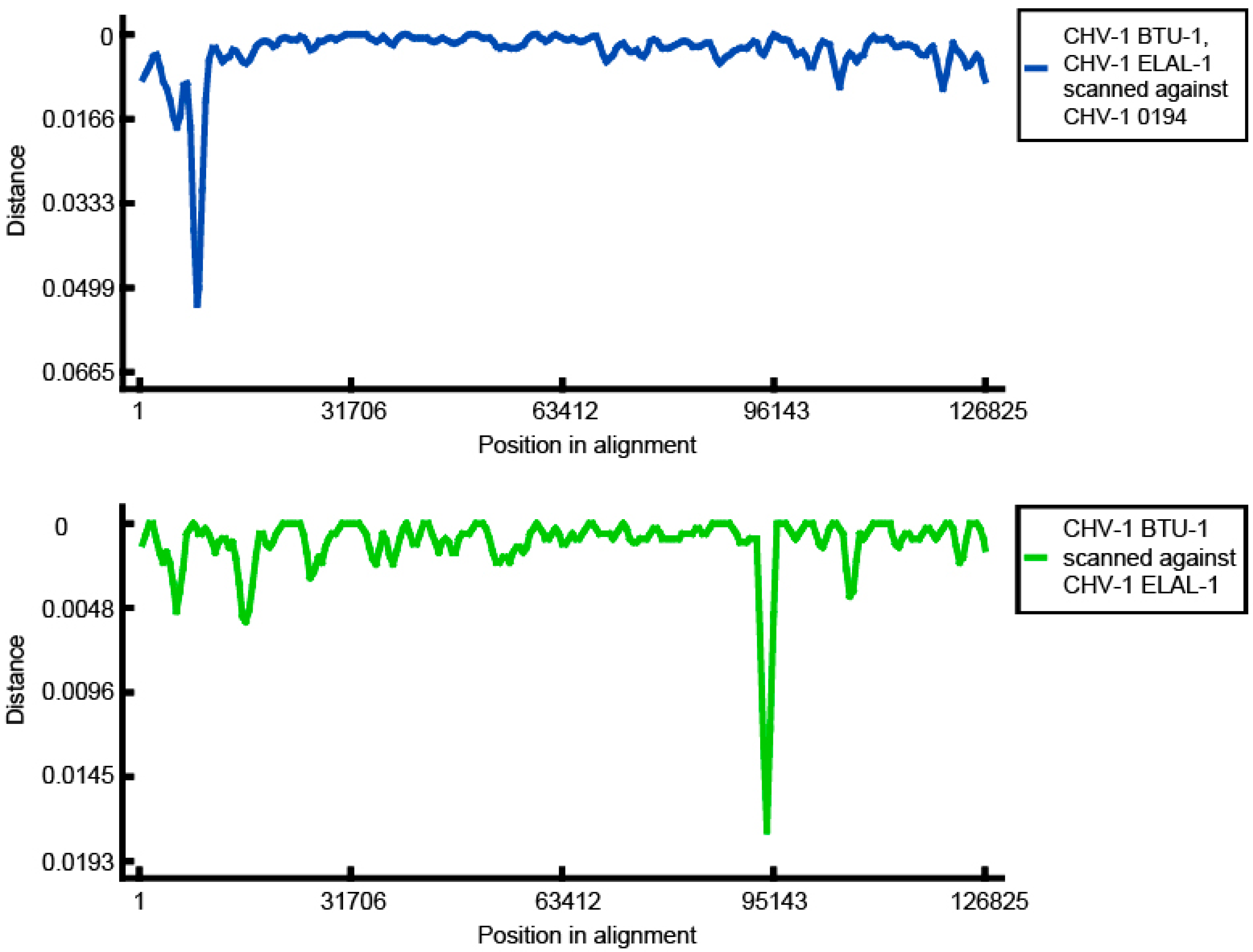
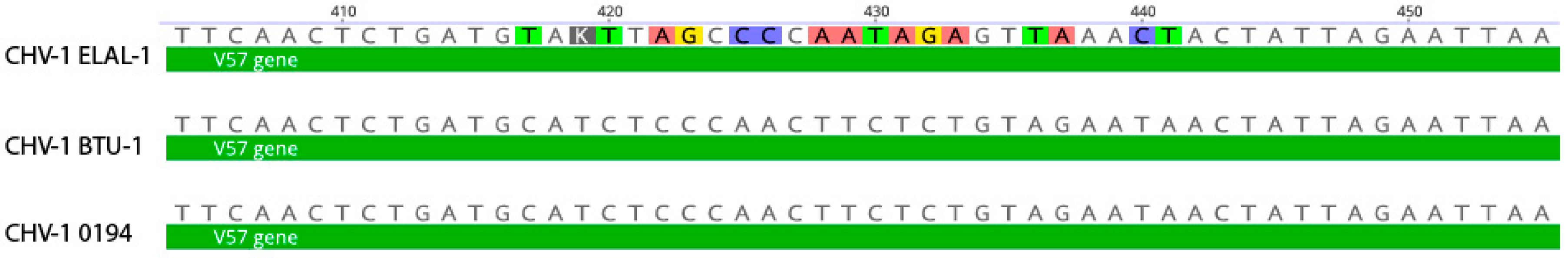
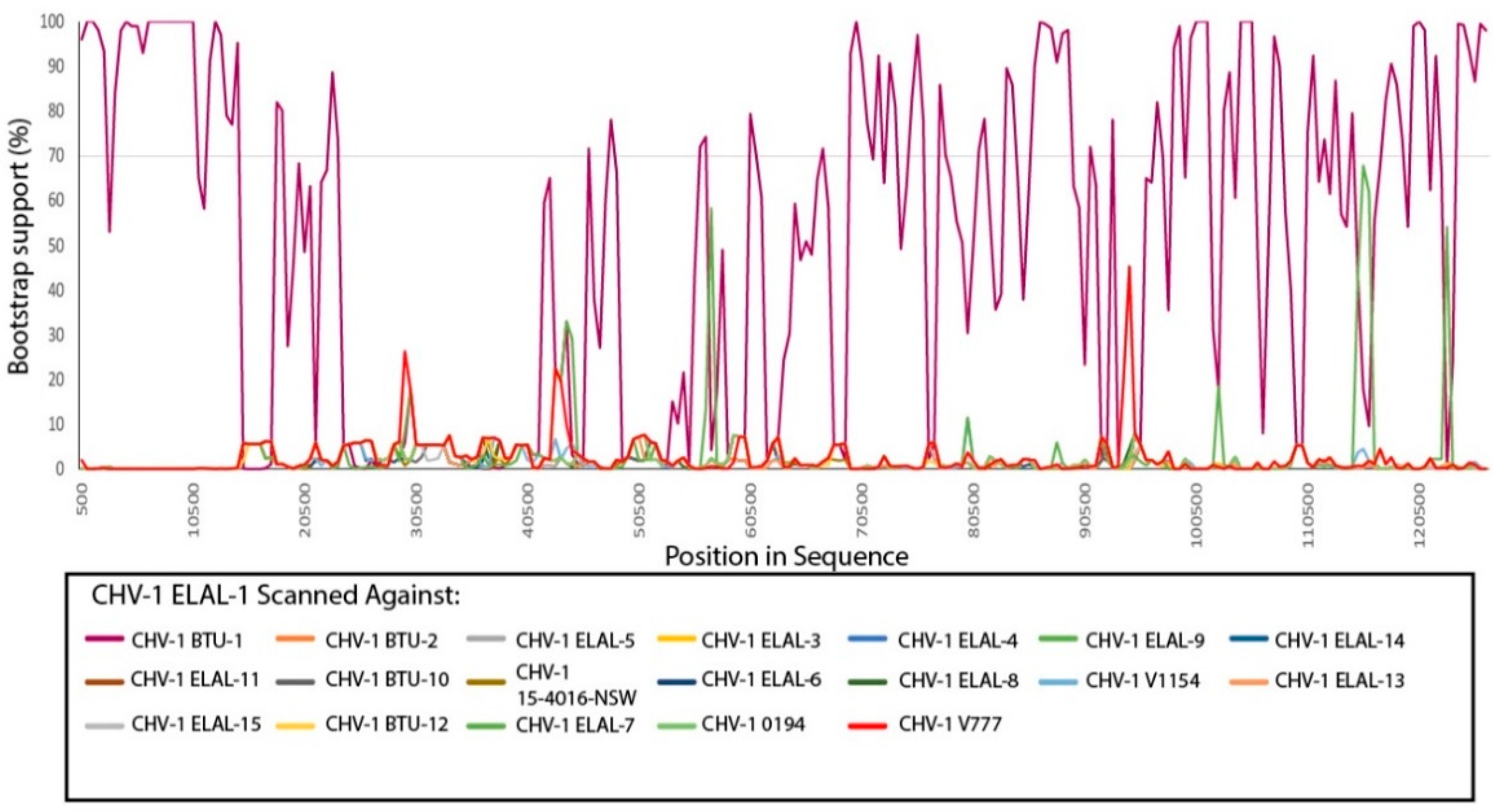
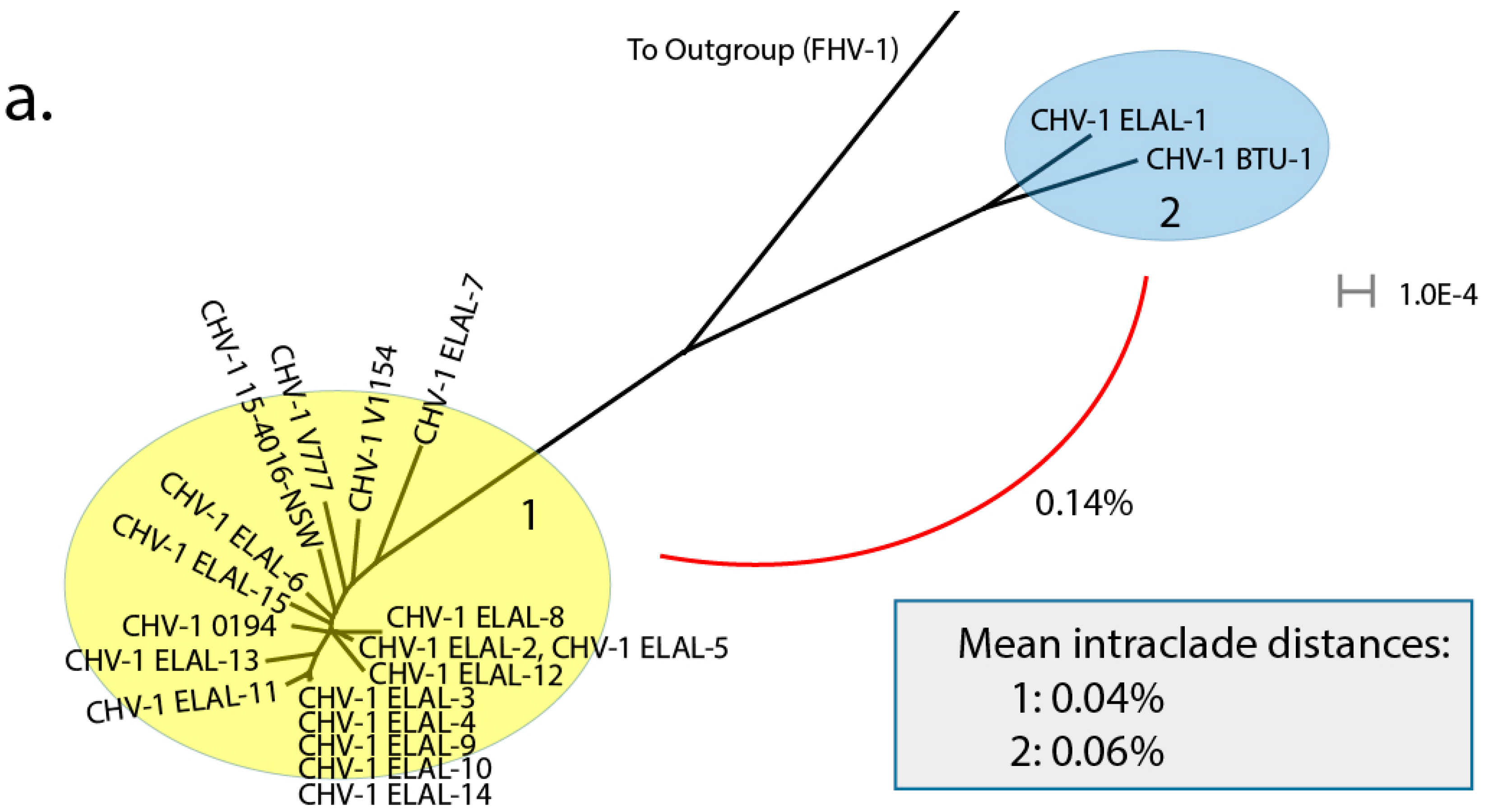
3.3. Phylogenetic and Recombination Analysis

The transversional model (TVM+F+G4) [31] was automatically chosen based on the Bayesian information criterion score (BIC) using ModelFinder [26] within IQ-Tree 2 ver 1.6.12 [27]. The resultant tree file was used to create a maximum likelihood tree (Figure 1). All bootstrap support values were 100, suggesting strong support for the recovered clade relationships. The maximum likelihood tree was visualized using Geneious Prime ver 2020.2.4.
The same tree file was also visualized using Splitstree ver 4.16.1 [28] (Figure 2) to further elucidate clade structure. Two clades were visualized. Clade 2 contained two isolates: ELAL-1 (a CHV-1 isolate collected from a host in 2019 in San Antonio, TX, USA) and BTU-1 (a CHV-1 isolate collected from a host in 2012 in Brazil). Clade 1 contained 18 isolates, most of which originated from host animals in the USA. Clade 1 also contained previously sequenced isolates from the UK [18] and Australia [19]. As expected, isolates obtained from multiple host animals in the same colony during an outbreak (ELAL-3, ELAL-4, ELAL-9, ELAL-10, ELAL-11, ELAL-14) were extremely homogenous and were grouped closely together (Figure 2b).
The overall mean genomic distance for all isolates sequenced for this study was 0.09% (calculated using MEGAX). Interclade genomic distance was 0.14%, as calculated using an alignment of consensus sequences (MAFFT ver. 7.450) from each clade. Clades were designated based on previously outlined criteria for varicelloviruses [20]. Intraclade distance was slightly lower in Clade 1 (0.04%) than in Clade 2 (0.06%).
To further investigate the origin of genomic variation between Clade 1 and Clade 2, a distance plot (Figure 3) was created using RDP ver 4.100 [24] with an alignment containing 3 CHV-1 genomes; ELAL-1 (Clade 2), BTU-1 (Clade 2) and 0194 (Clade 1). When either BTU-1 or ELAL-1 were scanned against 0194, a trough representing the increased genomic distance was visualized approximately 9 kb from the left end of the genome. This has been previously reported for BTU-1 and is due to variants in the UL50 deoxyuridine triphosphatase gene region in this isolate [20]. When BTU-1 and ELAL-1 were scanned against each other, a deep trough 93 kb from the left end of the genome was visualized, which corresponds with the CHV-1 V57 protein region.
An alignment containing the V57 gene of the same three isolates (BTU-1, ELAL-1, 0194) was created and visualized using Geneious Prime ver 2020.2.4 (Figure 4). A novel variant between positions 417 and 441 in the V57 gene of ELAL-1 was detected. Apart from this variant, the V57 sequences of the three isolates were almost identical.
Recombination detection was performed on an alignment containing all available CHV-1 genomes in RDP ver 4.100 [24]. A PHI test demonstrated very good evidence of recombination in the alignment (p < 0.00001). Evidence for recombination between ELAL-1 and BTU-1 was abundant; RDP, GENECONV, Bootscan (Recscan), MaxChi and Chimaera supported this hypothesis. The results of a manual bootscan performed in RDP4 are shown in Figure 5. Manual bootscans were also performed for all other isolates versus the remaining isolates, with complete results shown in Table S2. Using this technique, multiple isolates did not demonstrate any evidence of recombination with other isolates, including all isolates shown in Figure 2b from the same outbreak in New York State in 2008 (ELAL-3, ELAL-4, ELAL-9, ELAL-10, ELAL-11, ELAL-14) as well as ELAL-6, ELAL-12, 0194 and 15-4016-NSW. The remaining Clade 1 isolates (ELAL-2, ELAL-5, ELAL-7, ELAL-8, ELAL-13 and ELAL-15, V1154, V777) all demonstrated some evidence of recombination with other Clade 1 isolates with bootstrap support of over 70%. There was no evidence of recombination between clades.

4. Discussion

To the authors’ knowledge, the present study represents the first known description of the genomics and phylogeny of CHV-1 isolates obtained from USA-based host canids. Overall, most of the isolate genomes studied were very similar to previously sequenced isolates from the UK and Australia, with a low overall genomic distance (0.09%). Two isolates were notably different from the others and formed a clear separate clade: BTU-1 (isolated in 2012 from a host canid in Brazil) and ELAL-1 (isolated in 2019 from a host canid in TX, USA). Herein, we have described evidence for trans-boundary transmission of this virus in canid populations; ELAL-1 is very likely to have originated from a recombination event involving BTU-1. This discovery provides critical information for our collective understanding of the transmission of the virus and may play a future role in surveillance and control as the availability of whole viral genome sequencing increases. Despite robust import controls in the USA for live dogs and canine semen, there are presently no steps in place to test for CHV-1. Using a simple phylogeny assessment, we were able to create a visualization of the relationship between the CHV-1 isolates (Figure 2), which confirmed that isolates obtained from multiple host animals in the same outbreak in New York State (Figure 2b) were extremely closely related. Future sequenced isolates can be considered within this framework to determine likely phylogeny. Aside from this example of geography influencing clade organization, this effect appears to be much less pronounced for CHV-1 than for comparable viruses such as FHV-1, where geography appears to be a strong determinant of clade organization [21]. The high degree of similarity between the majority of CHV-1 genomes (Clade 1, Figure 2a) from host animals housed in a variety of geographic locations provides additional evidence for trans-boundary spread of this pathogen.
In the present study, two CHV-1 clades were detected by phylogenetic analysis. This is in agreement with previous analysis of 4 CHV-1 isolates; BTU-1 (Clade 2), 0194 (Clade 1), V777 (Clade 1) and V1154 (Clade 1) [20]. This previous work determined that the UL50 gene of BTU-1 was 12.2% distant from the remaining isolates, hypothesized to be the result of a recombination event between CHV-1 and an unknown virus [20]. In the present study, the genome of ELAL-1 was found to be very similar to BTU-1 with one exception in the V57 gene region. For comparison, the V57 gene region of ELAL-1 was approximately 1.6% distant from both BTU-1 and 0194. BLAST searches (blast.ncbi.nlm.nih.gov) (Table S3) confirmed the closest identity of ELAL-1 V57 to various other CHV-1 isolates, suggesting that positive selection is likely to be the underlying cause of this variation.
As most of the isolates were found to have homogenous genomes, it is unsurprising that variant detection (compared to 0194, the reference genome) yielded a modest number of results for each isolate. The exception was for ELAL-1, which contained 188 synonymous variants and 154 non-synonymous variants. For comparison, our set of CHV-1 isolates contained more variants than FHV-1 [21] but less than herpes simplex virus (HSV) [32,33]. As expected, most variants were found in larger regions of the CHV-1 genome (Table 3, Figure S2). It can be seen from the regression analysis in Figure S2 that a single gene (UL50, Deoxyuridine triphosphate) had a higher-than-expected total number of variants (53) relative to the gene size (918 bp). This gene had a relatively low number of variants in prior analyses of FHV-1 [21] and HSV-1 [34] and it is therefore unclear why this would be the case for CHV-1. Of possible clinical relevance is that one isolate (ELAL-1) contained variants in UL23, UL30 and UL42, all regions targeted by antiviral medications [30]. Further investigation is necessary to determine if these variants have any impact on antiviral susceptibility. The host canid from which ELAL-1 was obtained had been treated by a veterinarian with multiple ocular medications for disease related to CHV-1 infection including topical ocular idoxuridine, without significant improvement of clinical signs. Following cessation of idoxuridine and initiation of topical ocular trifluridine [35], the ocular clinical signs improved. Antiviral resistance has been previously documented for HSV [36] but not for CHV-1.
The G-C content across the CHV-1 genomes was consistent at 31.6%. As has been previously noted [18,20], this is the lowest for known varicelloviruses. For comparison, the G-C content of FHV-1 is 45.8% and 72.6% for bovine herpes virus type 1 [20]. Sequencing coverage was excellent for CHV-1, and this is likely related to the consistently low G-C content across the genome for this virus [32,37]. It has been previously suggested that ungulate varicelloviruses, which have a higher G-C content, have a greater degree of intraspecies distance than those from non-ungulates (such as CHV-1). Our results fit within this framework, with a low overall distance of 0.09% for the 20 CHV-1 genomes analyzed.
We detected evidence of recombination between multiple isolates, a process which is thought to be common in herpesviruses [20,38,39]. In line with what has been previously reported, we did not detect any evidence of recombination between clades. By monitoring future occurrences of isolates from Clade 2, this should provide a useful method to monitor the spread of CHV-1 in the USA canid population. Recombination seems to be subjectively less prevalent in this sample of CHV-1 isolates compared to viruses such as FHV-1 [21] and HSV [32]. The reason for this is unknown.
All the CHV-1 isolates sequenced for the present study were obtained from the conjunctiva of animals with ocular disease (conjunctivitis and/or keratitis). Similar to other herpesviruses, CHV-1 establishes latency and is periodically excreted at various mucosal sites such as the conjunctiva, oral cavity and genitalia [40]. It is possible that the body sampling site influenced characteristics of the viral genomes which we obtained. This is considered unlikely given that many of the genomes from ocular isolates appeared to be very similar to those obtained from other tissues such as kidney, liver and lung.

5. Conclusions

Overall, our results agreed with previous reports of the genomics and phylogeny of CHV-1 [18,19,20]. We report further evidence of a novel form of CHV-1, recently isolated from a host canid located in the USA. Together these techniques can be used to elucidate the origins and transmission of this pathogen in domestic canid populations.

Supplementary Materials

The following are available online at https://www.mdpi.com/1999-4915/12/12/1421/s1, Figure S1: Coverage (in blue) of a representative isolate (ELAL-7) along the length of the genome. A schematic of the genome is shown immediately below, with regions of low coverage (less than 100x) identified with red bars. Figure S2: Total number of variants per gene detected in CHV-1 isolates sequenced for this study plotted against the length (bp) of each gene. The solid blue line represents the linear trendline with 99% confidence intervals shown with dotted black lines. The red data point indicates the single gene where the number of detected variants was above the expected number of variants (UL50/53 variants/918 bp). Table S1: Unique non-synonymous variants organized according to gene region affected. Table S2: Results of manual bootscan in RDP4 for each CHV-1 isolate analyzed. Table S3: Results of BLAST for ELAL-1 V57 region against nt database using BLASTN (blast.ncbi.nlm.nih.gov).

Author Contributions

Conceptualization, A.C.L.; methodology, A.C.L. and L.M.C.; software, A.C.L. and L.M.C.; validation, A.C.L. and L.M.C.; formal analysis, A.C.L., L.M.C., C.-C.L.; investigation, A.C.L.; resources, A.C.L.; data curation, A.C.L. and L.M.C.; writing—original draft preparation, A.C.L., L.M.C. and M.M.; writing—review and editing, A.C.L., L.M.C., M.M., R.T.C., C.-C.L., E.C.L.; visualization, A.C.L.; supervision, A.C.L.; project administration, A.C.L.; funding acquisition, A.C.L. All authors have read and agreed to the published version of the manuscript.

Funding

This project was supported by the AKC Canine Health Foundation (02681-A). The contents of this publication are solely the responsibility of the authors and do not necessarily represent the views of the Foundation.

Acknowledgments

The authors would like to thank Vladimir Chouljenko, Aaron Kolb, Christopher Alling, Muzammel Haque and Bruna Miessler for technical assistance with various aspects of this project. The authors would also like to thank Lynsey Wagner for providing information about the host canid for one of the isolates.

Conflicts of Interest

The authors declare no conflict of interest. The funders had no role in the design of the study; in the collection, analyses, or interpretation of data; in the writing of the manuscript, or in the decision to publish the results.

References

  1. Engels, M.; Mayrbibrack, B.; Ruckstuhl, B.; Metzler, A.; Wyler, R. The Sero-Epizootiology of Canine Herpes-Virus Infections in Switzerland and Preliminary Studies with a Vaccine. Zent. Vet. Reihe B (Ger. FR) 1980, 27, 257–267. [Google Scholar]
  2. Reading, M.J.; Field, H.J. Detection of high levels of canine herpes virus-1 neutralising antibody in kennel dogs using a novel serum neutralisation test. Res. Vet. Sci. 1999, 66, 273–275. [Google Scholar] [CrossRef] [PubMed]
  3. Van Gucht, S.; Nauwynck, H.; Pensaert, M. Prevalence of canine herpesvirus in kennels and the possible association with fertility problems and neonatal death. Vlaams Diergeneeskd. Tijdschr. 2001, 70, 204–211. [Google Scholar]
  4. Carmichael, L.E.; Fabricant, J.; Squire, R.A. A Fatal Septicemic Disease of Infant Puppies Caused by Cytopathogenic Organisms with Characteristics of Mycoplasma. Proc. Soc. Exp. Biol. Med. 1964, 117, 826–833. [Google Scholar] [CrossRef]
  5. Ledbetter, E.C. Canine herpesvirus-1 ocular diseases of mature dogs. N. Z. Vet. J. 2013, 61, 193–201. [Google Scholar] [CrossRef]
  6. Karpas, A.; Garcia, F.G.; Calvo, F.; Cross, R.E. Experimental Production of Canine Tracheobronchitis (Kennel Cough) with Canine Herpesvirus Isolated from Naturally Infected Dogs. Am. J. Vet. Res. 1968, 29, 1251–1257. [Google Scholar] [PubMed]
  7. Wright, N.G.; Cornwell, H.J. Susceptibility of 6-Week Old Puppies to Canine Herpes Virus. J. Small Anim. Pract. 1970, 10, 669–674. [Google Scholar] [CrossRef]
  8. Poste, G.; King, N. Isolation of a Herpesvirus from Canine Genital Tract—Association with Infertility, Abortion and Stillbirths. Vet. Rec. 1971, 88, 229. [Google Scholar] [CrossRef]
  9. Hashimoto, A.; Hirai, K.; Fukushi, H.; Fujimoto, Y. The Vaginal Lesions of a Bitch with a History of Canine Herpesvirus-Infection. Jpn. J. Vet. Sci. 1983, 45, 123–126. [Google Scholar] [CrossRef] [Green Version]
  10. Burr, P.D.; Campbell, M.E.M.; Nicolson, L.; Onions, D.E. Detection of canine herpesvirus 1 in a wide range of tissues using the polymerase chain reaction. Vet. Microbiol. 1996, 53, 227–237. [Google Scholar] [CrossRef]
  11. Schulze, C.; Baumgartner, W. Nested polymerase chain reaction and in situ hybridization for diagnosis of canine herpesvirus infection in puppies. Vet. Pathol. 1998, 35, 209–217. [Google Scholar] [CrossRef] [PubMed] [Green Version]
  12. Lacheretz, A.; Cognard, S. Epidemiology and serological diagnosis of canine herpesvirus infection. Rev. Med. Vet. 1998, 149, 853–856. [Google Scholar]
  13. Reading, M.J.; Field, H.J. A serological study of canine herpes virus-1 infection in the English dog population. Arch. Virol. 1998, 143, 1477–1488. [Google Scholar] [CrossRef] [PubMed]
  14. Rijsewijk, F.A.M.; Luiten, E.J.; Daus, F.J.; van der Heijden, R.W.; van Oirschot, J.T. Prevalence of antibodies against canine herpesvirus 1 in dogs in The Netherlands in 1997–1998. Vet. Microbiol. 1999, 65, 1–7. [Google Scholar] [CrossRef]
  15. Ledbetter, E.C.; Nicklin, A.M.; Spertus, C.B.; Pennington, M.R.; Van de Walle, G.R.; Mohammed, H.O. Evaluation of topical ophthalmic ganciclovir gel for the treatment of dogs with experimentally induced ocular canine herpesvirus-1 infection. Am. J. Vet. Res. 2018, 79, 762–769. [Google Scholar] [CrossRef]
  16. Poulet, H.; Guigal, P.M.; Soulier, M.; Leroy, V.; Fayet, G.; Minke, J.; Merial, G.C. Protection of puppies against canine herpesvirus by vaccination of the dams. Vet. Rec. 2001, 148, 691–695. [Google Scholar] [CrossRef]
  17. Ledbetter, E.C.; Kim, K.; Dubovi, E.J.; Mohammed, H.O.; Felippe, M.J.B. Clinical and immunological assessment of therapeutic immunization with a subunit vaccine for recurrent ocular canine herpesvirus-1 infection in dogs. Vet. Microbiol. 2016, 197, 102–110. [Google Scholar] [CrossRef]
  18. Papageorgiou, K.V.; Suárez, N.M.; Wilkie, G.S.; McDonald, M.; Graham, E.M.; Davison, A.J. Genome Sequence of Canine Herpesvirus. PLoS ONE 2016, 11, e0156015. [Google Scholar] [CrossRef] [Green Version]
  19. Sarker, S.; Das, S.; Helbig, K.; Peters, A.; Raidal, S.R. Genome sequence of an Australian strain of canid alphaherpesvirus 1. Aust. Vet. J. 2018, 96, 24–27. [Google Scholar] [CrossRef]
  20. Kolb, A.W.; Lewin, A.C.; Moeller Trane, R.; McLellan, G.J.; Brandt, C.R. Phylogenetic and recombination analysis of the herpesvirus genus varicellovirus. BMC Genom. 2017, 18, 1–17. [Google Scholar] [CrossRef]
  21. Lewin, A.C.; Kolb, A.W.; McLellan, G.J.; Bentley, E.; Bernard, K.A.; Newbury, S.P.; Brandt, C.R. Genomic, Recombinational and Phylogenetic Characterization of Global Feline Herpesvirus 1 Isolates. Virology 2018, 518, 385–397. [Google Scholar] [CrossRef] [PubMed]
  22. Ledbetter, E.C.; Riis, R.C.; Kern, T.J.; Haley, N.J.; Schatzberg, S.J. Corneal ulceration associated with naturally occurring canine herpesvirus-1 infection in two adult dogs. J. Am. Vet. Med. Assoc. 2006, 229, 376–384. [Google Scholar] [CrossRef] [PubMed]
  23. Katoh, K.; Standley, D.M. MAFFT multiple sequence alignment software version 7: Improvements in performance and usability. Mol. Biol. Evol. 2013, 30, 772–780. [Google Scholar] [CrossRef] [PubMed] [Green Version]
  24. Martin, D.P.; Murrell, B.; Golden, M.; Khoosal, A.; Muhire, B. RDP4: Detection and analysis of recombination patterns in virus genomes. Virus Evol. 2015, 1. [Google Scholar] [CrossRef] [Green Version]
  25. Jin, L.; Nei, M. Limitations of the evolutionary parsimony method of phylogenetic analysis. Mol. Biol. Evol. 1990, 7, 82–102. [Google Scholar] [CrossRef] [Green Version]
  26. Kalyaanamoorthy, S.; Minh, B.Q.; Wong, T.K.F.; von Haeseler, A.; Jermiin, L.S. ModelFinder: Fast model selection for accurate phylogenetic estimates. Nat. Methods 2017, 14, 587–589. [Google Scholar] [CrossRef] [Green Version]
  27. Minh, B.Q.; Schmidt, H.A.; Chernomor, O.; Schrempf, D.; Woodhams, M.D.; von Haeseler, A.; Lanfear, R. IQ-TREE 2: New Models and Efficient Methods for Phylogenetic Inference in the Genomic Era. Mol. Biol. Evol. 2020, 37, 1530–1534. [Google Scholar] [CrossRef] [Green Version]
  28. Huson, D.H.; Bryant, D. Application of phylogenetic networks in evolutionary studies. Mol. Biol. Evol. 2006, 23, 254–267. [Google Scholar] [CrossRef]
  29. Kumar, S.; Stecher, G.; Li, M.; Knyaz, C.; Tamura, K. MEGA X: Molecular Evolutionary Genetics Analysis across Computing Platforms. Mol. Biol. Evol. 2018, 35, 1547–1549. [Google Scholar] [CrossRef]
  30. Thomasy, S.M.; Maggs, D.J. A review of antiviral drugs and other compounds with activity against feline herpesvirus type 1. Vet. Ophthalmol. 2016, 19 (Suppl. 1) (Suppl. 1), 119–130. [Google Scholar] [CrossRef] [Green Version]
  31. Posada, D. Using MODELTEST and PAUP* to select a model of nucleotide substitution. Curr. Protoc. Bioinform. 2003. [Google Scholar] [CrossRef]
  32. Kolb, A.W.; Larsen, I.V.; Cuellar, J.A.; Brandt, C.R. Genomic, phylogenetic, and recombinational characterization of herpes simplex virus 2 strains. J. Virol. 2015, 89, 6427–6434. [Google Scholar] [CrossRef] [PubMed] [Green Version]
  33. Kolb, A.W.; Adams, M.; Cabot, E.L.; Craven, M.; Brandt, C.R. Multiplex sequencing of seven ocular herpes simplex virus type-1 genomes: Phylogeny, sequence variability, and SNP distribution. Investig. Ophthalmol. Vis. Sci. 2011, 52, 9061–9073. [Google Scholar] [CrossRef] [PubMed] [Green Version]
  34. Szpara, M.L.; Parsons, L.; Enquist, L.W. Sequence variability in clinical and laboratory isolates of herpes simplex virus 1 reveals new mutations. J. Virol. 2010, 84, 5303–5313. [Google Scholar] [CrossRef] [PubMed] [Green Version]
  35. Spertus, C.B.; Mohammed, H.O.; Ledbetter, E.C. Effects of topical ocular application of 1% trifluridine ophthalmic solution in dogs with experimentally induced recurrent ocular canine herpesvirus-1 infection. Am. J. Vet. Res. 2016, 77, 1140–1147. [Google Scholar] [CrossRef] [PubMed]
  36. Piret, J.; Boivin, G. Antiviral resistance in herpes simplex virus and varicella-zoster virus infections: Diagnosis and management. Curr. Opin. Infect. Dis. 2016, 29, 654–662. [Google Scholar] [CrossRef] [PubMed]
  37. Lee, K.; Kolb, A.W.; Sverchkov, Y.; Cuellar, J.A.; Craven, M.; Brandt, C.R. Recombination Analysis of Herpes Simplex Virus 1 Reveals a Bias toward GC Content and the Inverted Repeat Regions. J. Virol. 2015, 89, 7214–7223. [Google Scholar] [CrossRef] [PubMed] [Green Version]
  38. Dohner, D.E.; Adams, S.G.; Gelb, L.D. Recombination in tissue culture between varicella-zoster virus strains. J. Med. Virol. 1988, 24, 329–341. [Google Scholar] [CrossRef] [PubMed]
  39. Henderson, L.M.; Katz, J.B.; Erickson, G.A.; Mayfield, J.E. In vivo and in vitro genetic recombination between conventional and gene-deleted vaccine strains of pseudorabies virus. Am. J. Vet. Res. 1990, 51, 1656–1662. [Google Scholar]
  40. Miyoshi, M.; Ishii, Y.; Takiguchi, M.; Takada, A.; Yasuda, J.; Hashimoto, A.; Okazaki, K.; Kida, H. Detection of canine herpesvirus DNA in the ganglionic neurons and the lymph node lymphocytes of latently infected dogs. J. Vet. Med. Sci. 1999, 61, 375–379. [Google Scholar] [CrossRef] [Green Version]
Figure 1. Maximum likelihood tree of all analyzed CHV-1 isolates with an FHV-1 C-27 outgroup (in red). Branch labels represent bootstrap support. Note the separation of two CHV-1 isolates (ELAL-1 and BTU-1) from the remaining CHV-1 isolates. CHV-1 ELAL-1 was obtained from a host animal in Texas in 2019 and BTU-1 was obtained from a host animal in Brazil in 2012.
Viruses 12 01421 g001
Figure 2. (a) Phylogenetic network of all analyzed CHV-1 isolates with a FHV-1 (C-27) outgroup. Two distinct clades are visible: 1 (yellow) and 2 (blue). The interclade distance (0.14%) is illustrated with a red line and was calculated using an alignment of consensus sequences (MAFFT ver. 7.450) from each clade. The mean intraclade distances were calculated using MEGAX. (b) Detail of subsection of clade 1 from Figure 2a. All isolates shown in this subsection were obtained from the same outbreak of CHV-1 in New York State in 2008. As expected, the genomes of these particular CHV-1 isolates are extremely homogenous.
Viruses 12 01421 g002aViruses 12 01421 g002b
Figure 3. Genomic distance plotted against sequence position for CHV-1 isolates. An alignment containing 3 isolates (ELAL-1 (Clade 2), BTU-1 (Clade 2) and 0194 (Clade 1)) was processed in RDP4 ver 4.100. Results are for a manual distance plot with a window size of 1200, step size of 500 using the Jin and Nei model. The top graph (blue line) clearly illustrates a deep trough at approximately position 9kb in the alignment when either of the clade 2 isolates are scanned against the CHV-1 reference genome (0194, Clade 1). The bottom graph (green line) shows a deep trough at approximately position 93kb in the alignment when Clade A isolates are compared. This corresponds with the V57 gene region.
Viruses 12 01421 g003
Figure 4. Variant in CHV-1 ELAL-1 isolate in the V57 gene region. A clearly identifiable region of low identity was detected centered around position 430 in the V57 gene which corresponded to a novel variant in CHV-1 ELAL-1.
Viruses 12 01421 g004
Figure 5. Manual bootscan results for recombination detection of CHV-1 ELAL-1 against all other analyzed isolates. A cutoff value of 70% bootstrap support is shown on the graph. Clear evidence of recombination was found between CHV-1 ELAL-1 and fellow Clade 2 isolate CHV-1 BTU-1, but not between CHV-1 ELAL-1 and any of the remaining isolates.
Viruses 12 01421 g005
Table 1. Details of isolates included in analysis. All isolates are canid alphaherpesvirus (CHV-1) apart from C-27, which is feline herpes virus 1 (FHV-1). M = Male, F = Female, FS = Female, spayed, MC = Male, castrated, ACS = American Cocker Spaniel, NA = not applicable/unknown.
Strain IDGenbank AccessionHost Age/Sex/BreedHost LocationDate CollectedSource Sample
ELAL-1MW3531257/FS/English BulldogSan Antonio, TX, USA6 March 2019Eye
ELAL-2MW3531268/MC/Labrador RetrieverOneonta, NY, USA6 July 2003Eye
ELAL-3MW3531270.33/F/BeagleNorth Rose, NY, USA19 November 2008Eye
ELAL-4MW3531280.25/F/BeagleNorth Rose, NY, USA19 November 2008Eye
ELAL-5MW3531297/FS/Min. SchnauzerEndwell, NY, USA26 August 2004Eye
ELAL-6MW35313010/FS/Golden RetrieverWilliston, VT, USA17 September 2014Eye
ELAL-7MW3531318/FS/Cross breedBatavia, NY, USA18 October 2007Eye
ELAL-8MW3531328/MC/ACSOvid, NY, USA25 February 2014Eye
ELAL-9MW3531330.2/M/BeagleNorth Rose, NY, USA19 November 2008Eye
ELAL-10MW3531340.2/M/BeagleNorth Rose, NY, USA19 November 2008Eye
ELAL-11MW3531350.33/F/BeagleNorth Rose, NY, USA19 November 2008Eye
ELAL-12MW35313612/MC/Cross breedWilton, CT, USA16 October 2019Eye
ELAL-13MW3531380.16/F/BeagleIthaca, NY, USA16 April 2009Eye
ELAL-14MW3531370.33/M/BeagleNorth Rose, NY, USA19 November 2008Eye
ELAL-15MW3531399/FS/Golden RetrieverBinghamton, NY, USA26 August 2008Eye
V1154KT8196310.04/NA/DalmatianUK2000Kidney
V777KT8196320.04/NA/Min. SchnauzerUK1995Lung
BTU-1KX828242NA/NA/NABrazil2012Kidney
15-4016-NSWKY0573640.08/F/Labrador RetrieverNSW, Australia2016Liver
0-194NC_030117NA/NA/NANA1985NA
C-27NC_013590 NA/NA/NANAPre 1980NA
Table 2. Sequencing details of sequenced CHV-1 isolates.
Strain IDNumber ReadsMapped ReadsMean CoverageMean Mapped Read Length (bp)
ELAL-12,865,972155,016290.2226.9
ELAL-22,963,230390,228706.8228.6
ELAL-31,292,262231,149413.2225.3
ELAL-41,848,000376,806663.9223.1
ELAL-51,430,708279,714476.8214.7
ELAL-63,053,006847,3711509.6225.4
ELAL-73,221,498219,157372.8214.5
ELAL-81,889,752298,155505.4213.2
ELAL-93,415,468416,370706213.7
ELAL-103,533,658648,2091409.8201.2
ELAL-112,356,364456,166765.2211.7
ELAL-123,126,278639,6771024.4201.1
ELAL-132,496,354326,583536.8206.8
ELAL-142,955,844856,9921404207.5
ELAL-152,985,658527,658870.8205.7
Table 3. Summary of variants detected, organized by gene involved. F = Forward, R = Reverse.
GeneGene ProductNumber Unique Synonymous VariantsNumber Unique non-Synonymous VariantsLength of Gene (bp)
CircMyristylated tegument protein CIRC42651
RL2Ubiquitin E3 ligase ICP0141005
RS1Transcriptional regulator ICP422164155(F)/4155(R)
UL1Envelope glycoprotein L00468
UL10Envelope glycoprotein M121254
UL11Myristylated tegument protein10213
UL12Deoxyribonuclease201638
UL13Tegument serine/threonine protein kinase251770
UL14Tegument protein UL1414912
UL15DNA packaging terminase subunit 1125442
UL16Tegument protein UL16421092
UL17DNA packaging tegument protein UL17042067
UL18Capsid triplex subunit 230948
UL19Major capsid protein734122
UL2Uracil-DNA glycosylase00837
UL20Envelope protein UL2020693
UL21Tegument protein UL21211572
UL22Envelope glycoprotein H112394
UL23Thymidine kinase02987
UL24Nuclear protein UL2410783
UL25DNA packaging tegument protein UL25211758
UL26Capsid maturation protease421731
UL26.5Capsid scaffold protein31846
UL27Envelope glycoprotein B112640
UL28DNA packaging terminase subunit 2322271
UL29Single-stranded DNA-binding protein543594
UL3Nuclear protein UL301597
UL30DNA polymerase catalytic subunit243540
UL31Nuclear egress lamina protein111005
UL32DNA packaging protein UL32121680
UL33DNA packaging protein UL3301381
UL34Nuclear egress membrane protein11795
UL35Small capsid protein00321
UL36Large tegument protein15429531
UL37Tegument protein UL37343039
UL38Capsid triplex subunit 1101374
UL4Nuclear protein UL431663
UL41Tegument host shutoff protein041461
UL42DNA polymerase processivity subunit121059
UL43Envelope protein UL43321203
UL44Envelope glycoprotein C111380
UL45Membrane protein UL4511528
UL46Tegument protein VP11/12011986
UL47Tegument protein VP13/14782439
UL48Transactivating tegument protein VP16311269
UL49Tegument protein VP2271795
UL49AEnvelope glycoprotein N31261
UL5Helicase-primase helicase subunit132661
UL50Deoxyuridine triphosphate2033918
UL51Tegument protein UL51146672
UL52Helicase-primase primase subunit17143063
UL53Envelope glycoprotein K841014
UL54Multifunctional expression regulator401272
UL55Nuclear protein UL5531564
UL56Membrane protein UL5605522
UL6Capsid portal protein812073
UL7Tegument protein UL722879
UL8Helicase-primase subunit632271
UL9DNA replication origin-binding helicase332535
US1Regulatory protein ICP2251942(F)/942(R)
US10Virion protein US1015588(F)/588(R)
US2Virion protein US2321176
US3Serine/threonine protein kinase US3001203
US4Envelope glycoprotein G111248
US6Envelope glycoprotein D311038
US7Envelope glycoprotein I311038
US8Envelope glycoprotein E241569
US8AMembrane protein US8A00237
US9Membrane protein US902360
V32Protein V3202417
V57Protein V5711729
V67Virion protein V6760552(F)/552(R)
Publisher’s Note: MDPI stays neutral with regard to jurisdictional claims in published maps and institutional affiliations.

