A Deep-Sequencing Workflow for the Fast and Efficient Generation of High-Quality African Swine Fever Virus Whole-Genome Sequences
Abstract
1. Introduction
2. Materials and Methods
2.1. Virus Cultivation
2.2. DNA Extraction
2.3. Illumina Sequencing
2.4. Nanopore Sequencing
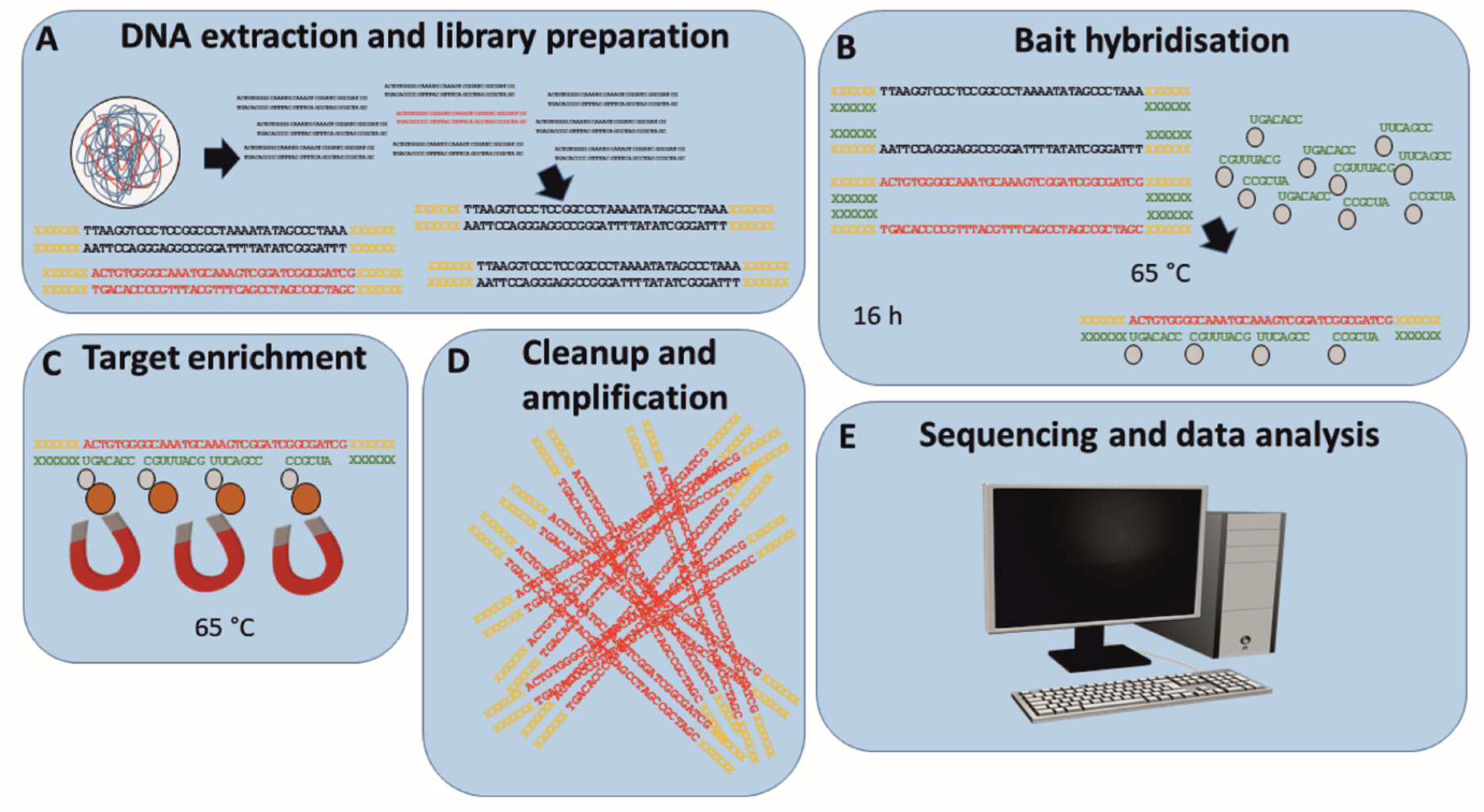
2.5. Target Enrichment
2.6. Data Analysis
2.7. PCR and Sanger Sequencing
2.8. Data Availability
3. Results
3.1. ASFV-Specific Target Enrichment Prior to Illumina Sequencing Provides High Amounts of Target Reads
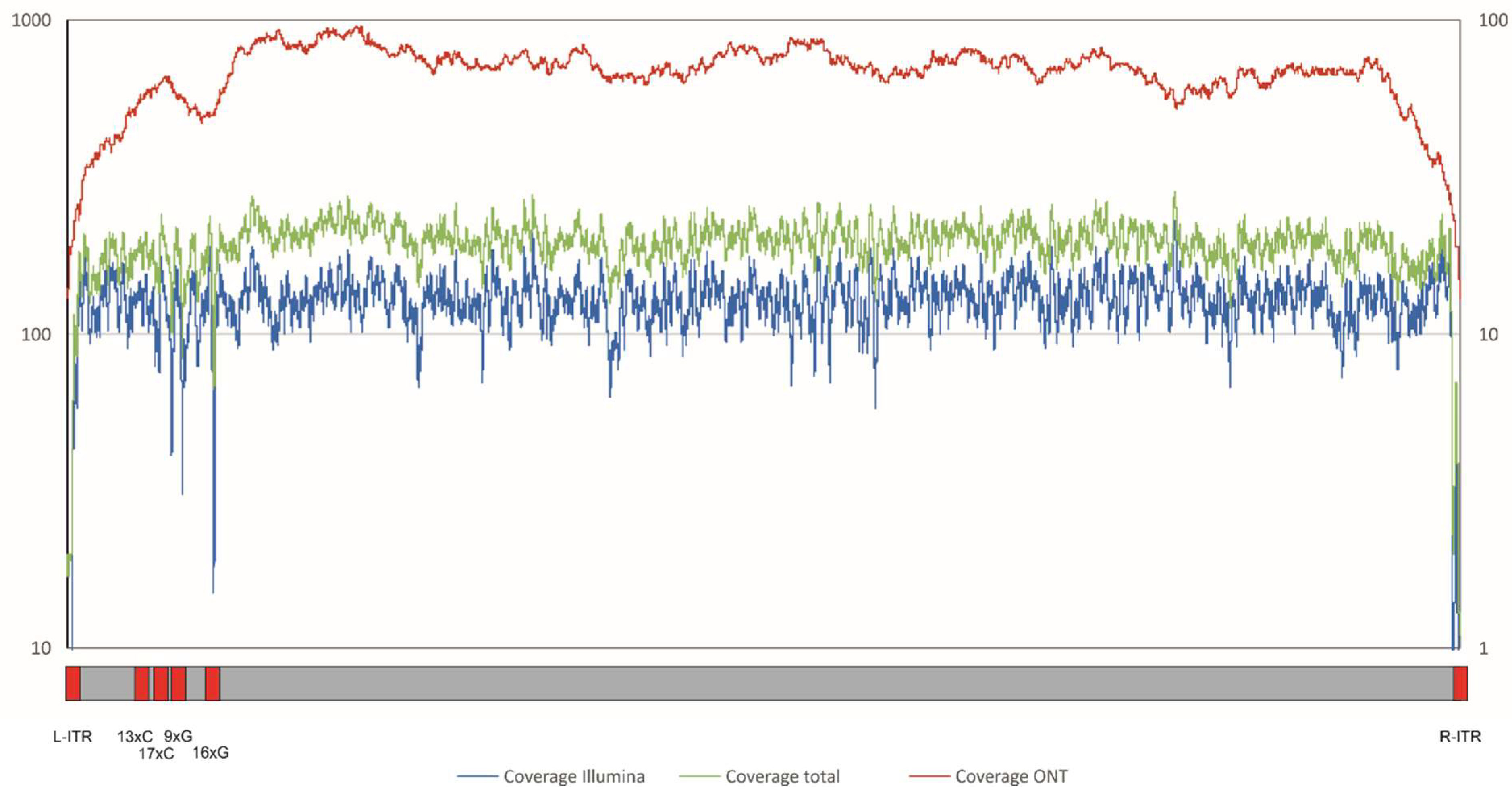
3.2. Hybrid Assembly of Nanopore and Illumina Data Provide an Improved ASFV Georgia 2007/1 Whole-Genome Sequence with Novel Information about the Genome Length
3.3. Sequence Alignment with the Improved ASFV Genome Reveals 71 Differences in Homopolymer Regions and Open Reading Frames
3.4. Application of Target Enrichment Prior to Illumina Sequencing Enabled Whole-Genome Assembly of ASFV Moldova from Organ Samples Using the New Improved Sequence as Reference
3.5. Variant Analysis Reveals Possible Single Nucleotide Polymorphisms
3.6. Alignment of ASFV Moldova 2017/1 and ASFV Georgia 2007/1 Reveals Only Single Nucleotide Differences and a Tandem Repeat Variation
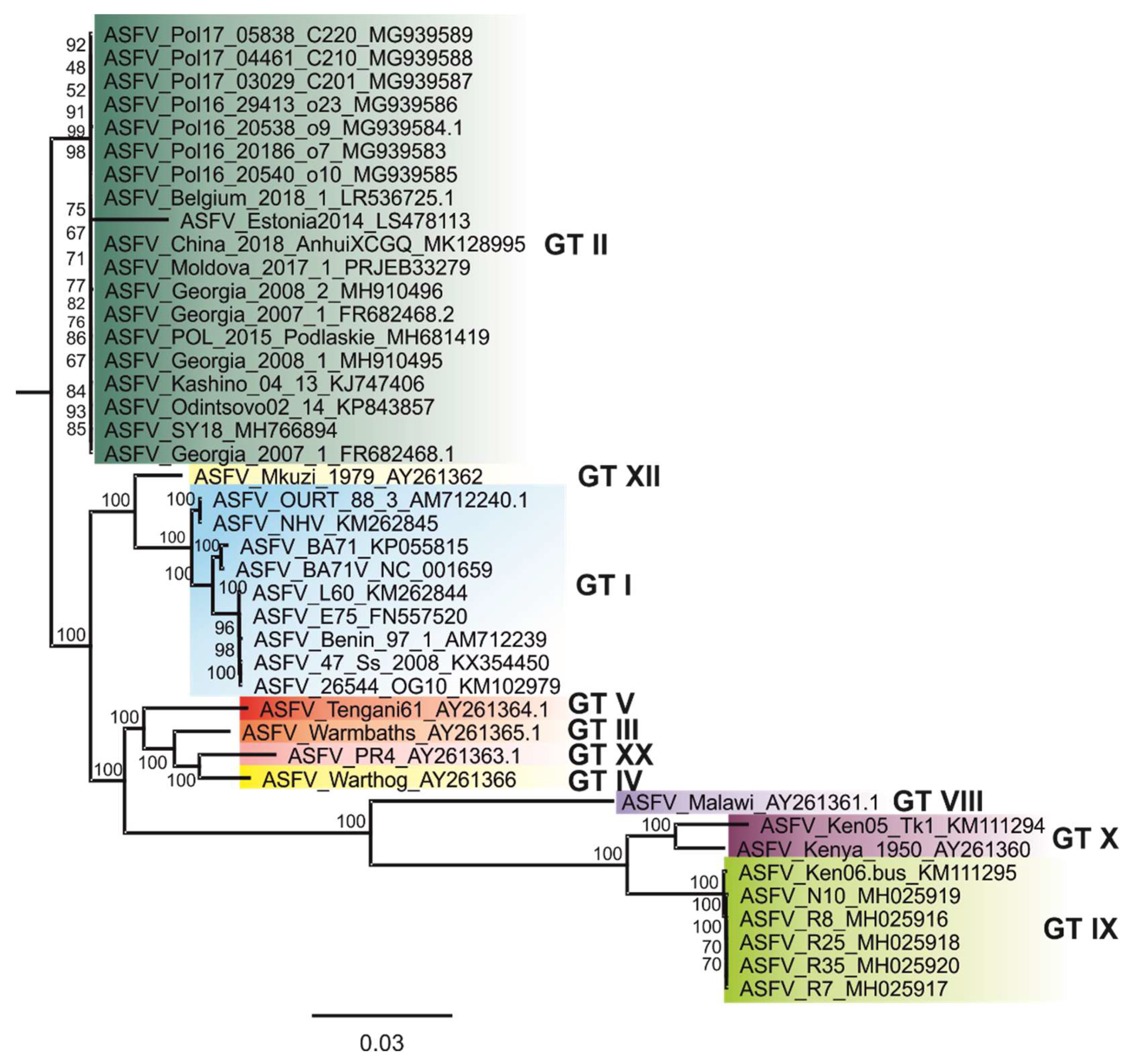
3.7. Alignment and Phylogenetic Reconstruction of All Available ASFV Whole-Genome Sequences Shows a Very High Overall Nucleotide Sequence Identity of All Eurasian Strains
4. Discussion
Supplementary Materials
Author Contributions
Funding
Acknowledgments
Conflicts of Interest
References
- Goodwin, S.; McPherson, J.D.; McCombie, W.R. Coming of age: Ten years of next-generation sequencing technologies. Nat. Rev. Genet. 2016, 17, 333–351. [Google Scholar] [CrossRef] [PubMed]
- Levy, S.E.; Myers, R.M. Advancements in Next-Generation Sequencing. Annu. Rev. Genom. Hum. Genet. 2016, 17, 95–115. [Google Scholar] [CrossRef] [PubMed]
- Bayliss, S.C.; Verner-Jeffreys, D.W.; Bartie, K.L.; Aanensen, D.M.; Sheppard, S.K.; Adams, A.; Feil, E.J. The Promise of Whole Genome Pathogen Sequencing for the Molecular Epidemiology of Emerging Aquaculture Pathogens. Front. Microbiol. 2017, 8, 121. [Google Scholar] [CrossRef] [PubMed]
- Tang, P.; Croxen, M.A.; Hasan, M.R.; Hsiao, W.W.; Hoang, L.M. Infection control in the new age of genomic epidemiology. Am. J. Infect. Control 2017, 45, 170–179. [Google Scholar] [CrossRef] [PubMed]
- Alonso, C.; Borca, M.; Dixon, L.; Revilla, Y.; Rodriguez, F.; Escribano, J.M.; Ictv Report, C. ICTV Virus Taxonomy Profile: Asfarviridae. J. Gen. Virol. 2018, 99, 613–614. [Google Scholar] [CrossRef]
- Montgomery, R.E. On A Form of Swine Fever Occurring in British East Africa (Kenya Colony). J. Comp. Pathol. 1921, 34, 159–191. [Google Scholar] [CrossRef]
- Burrage, T.G. African swine fever virus infection in Ornithodoros ticks. Virus Res. 2013, 173, 131–139. [Google Scholar] [CrossRef]
- Blome, S.; Gabriel, C.; Dietze, K.; Breithaupt, A.; Beer, M. High virulence of African swine fever virus caucasus isolate in European wild boars of all ages. Emerg. Infect. Dis. 2012, 18, 708. [Google Scholar] [CrossRef]
- Blome, S.; Gabriel, C.; Beer, M. Pathogenesis of African swine fever in domestic pigs and European wild boar. Virus Res. 2013, 173, 122–130. [Google Scholar] [CrossRef]
- Arias, M.; de la Torre, A.; Dixon, L.; Gallardo, C.; Jori, F.; Laddomada, A.; Martins, C.; Parkhouse, R.M.; Revilla, Y.; Rodriguez, F.A.J.; et al. Approaches and Perspectives for Development of African Swine Fever Virus Vaccines. Vaccines 2017, 5, 35. [Google Scholar] [CrossRef]
- Arias, M.; Sánchez-Vizcaíno, J.M. African Swine Fever Eradication: The Spanish Model. In Trends in Emerging Viral Infections of Swine, 1st ed.; Morilla, A., Yoon, K.J., Zimmerman, J.J., Eds.; Iowa State Press: Iowa City, IA, USA, 2002; pp. 133–139. [Google Scholar]
- Sanchez-Cordon, P.J.; Montoya, M.; Reis, A.L.; Dixon, L.K. African swine fever: A re-emerging viral disease threatening the global pig industry. Vet. J. 2018, 233, 41–48. [Google Scholar] [CrossRef] [PubMed]
- Sanchez-Vizcaino, J.M.; Mur, L.; Gomez-Villamandos, J.C.; Carrasco, L. An update on the epidemiology and pathology of African swine fever. J. Comp. Pathol. 2015, 152, 9–21. [Google Scholar] [CrossRef] [PubMed]
- Sanchez-Vizcaino, J.M.; Mur, L.; Martinez-Lopez, B. African swine fever: An epidemiological update. Transbound. Emerg. Dis. 2012, 59, 27–35. [Google Scholar] [CrossRef] [PubMed]
- Beltran-Alcrudo, D.; Lubroth, J.; Depner, K.; De La Rocque, S. African swine fever in the Caucasus. FAO EMPRES Watch 2008, 1, 1–8. [Google Scholar]
- Gogin, A.; Gerasimov, V.; Malogolovkin, A.; Kolbasov, D. African swine fever in the North Caucasus region and the Russian Federation in years 2007–2012. Virus Res. 2013, 173, 198–203. [Google Scholar] [CrossRef] [PubMed]
- Kolbasov, D.; Titov, I.; Tsybanov, S.; Gogin, A.; Malogolovkin, A. African Swine Fever Virus, Siberia, Russia, 2017. Emerg. Infect. Dis. 2018, 24, 796–798. [Google Scholar] [CrossRef] [PubMed]
- Chenais, E.; Stahl, K.; Guberti, V.; Depner, K. Identification of Wild Boar-Habitat Epidemiologic Cycle in African Swine Fever Epizootic. Emerg. Infect. Dis. 2018, 24, 810–812. [Google Scholar] [CrossRef]
- Garigliany, M.; Desmecht, D.; Tignon, M.; Cassart, D.; Lesenfant, C.; Paternostre, J.; Volpe, R.; Cay, A.B.; van den Berg, T.; Linden, A. Phylogeographic Analysis of African Swine Fever Virus, Western Europe, 2018. Emerg. Infect. Dis. 2019, 25, 184–186. [Google Scholar] [CrossRef]
- Zhou, X.; Li, N.; Luo, Y.; Liu, Y.; Miao, F.; Chen, T.; Zhang, S.; Cao, P.; Li, X.; Tian, K.; et al. Emergence of African Swine Fever in China, 2018. Transbound. Emerg. Dis. 2018, 65, 1482–1484. [Google Scholar] [CrossRef]
- Luka, P.D.; Achenbach, J.E.; Mwiine, F.N.; Lamien, C.E.; Shamaki, D.; Unger, H.; Erume, J. Genetic Characterization of Circulating African Swine Fever Viruses in Nigeria (2007–2015). Transbound. Emerg. Dis. 2017, 64, 1598–1609. [Google Scholar] [CrossRef]
- Luka, P.D.; Erume, J.; Mwiine, F.N.; Shamaki, D.; Yakubu, B. Comparative Sequence Analysis of Different Strains of African Swine Fever Virus Outer Proteins Encoding Genes from Nigeria, 2009–2014. J. Virol. Mol. Biol. 2016, 5, 16–26. [Google Scholar]
- Alkhamis, M.A.; Gallardo, C.; Jurado, C.; Soler, A.; Arias, M.; Sanchez-Vizcaino, J.M. Phylodynamics and evolutionary epidemiology of African swine fever p72-CVR genes in Eurasia and Africa. PLoS ONE 2018, 13, e0192565. [Google Scholar] [CrossRef] [PubMed]
- Gallardo, C.; Fernandez-Pinero, J.; Pelayo, V.; Gazaev, I.; Markowska-Daniel, I.; Pridotkas, G.; Nieto, R.; Fernandez-Pacheco, P.; Bokhan, S.; Nevolko, O.; et al. Genetic variation among African swine fever genotype II viruses, eastern and central Europe. Emerg. Infect. Dis. 2014, 20, 1544–1547. [Google Scholar] [CrossRef] [PubMed]
- Gallardo, C.; Mwaengo, D.M.; Macharia, J.M.; Arias, M.; Taracha, E.A.; Soler, A.; Okoth, E.; Martin, E.; Kasiti, J.; Bishop, R.P. Enhanced discrimination of African swine fever virus isolates through nucleotide sequencing of the p54, p72, and pB602L (CVR) genes. Virus Genes 2009, 38, 85–95. [Google Scholar] [CrossRef] [PubMed]
- Nix, R.J.; Gallardo, C.; Hutchings, G.; Blanco, E.; Dixon, L.K. Molecular epidemiology of African swine fever virus studied by analysis of four variable genome regions. Arch. Virol. 2006, 151, 2475–2494. [Google Scholar] [CrossRef]
- Onzere, C.K.; Bastos, A.D.; Okoth, E.A.; Lichoti, J.K.; Bochere, E.N.; Owido, M.G.; Ndambuki, G.; Bronsvoort, M.; Bishop, R.P. Multi-locus sequence typing of African swine fever viruses from endemic regions of Kenya and Eastern Uganda (2011–2013) reveals rapid B602L central variable region evolution. Virus Genes 2017, 54, 111–123. [Google Scholar] [CrossRef] [PubMed]
- Dixon, L.K.; Chapman, D.A.; Netherton, C.L.; Upton, C. African swine fever virus replication and genomics. Virus Res. 2013, 173, 3–14. [Google Scholar] [CrossRef]
- Michaud, V.; Randriamparany, T.; Albina, E. Comprehensive phylogenetic reconstructions of African swine fever virus: Proposal for a new classification and molecular dating of the virus. PLoS ONE 2013, 8, e69662. [Google Scholar] [CrossRef]
- Dixon, L.K.; Wilkinson, P.J. Genetic Diversity of African Swine Fever Virus Isolates from Soft Ticks (Ornithodoros moubata) Inhabiting Warthog Burrows in Zambia. J. Gen. Virol. 1988, 69, 2981–2993. [Google Scholar] [CrossRef]
- Qin, L.; Evans, D.H. Genome scale patterns of recombination between coinfecting vaccinia viruses. J. Virol. 2014, 88, 5277–5286. [Google Scholar] [CrossRef]
- Yanez, R.J.; Rodriguez, J.M.; Nogal, M.L.; Yuste, L.; Enriquez, C.; Rodriguez, J.F.; Vinuela, E. Analysis of the complete nucleotide sequence of African swine fever virus. Virology 1995, 208, 249–278. [Google Scholar] [CrossRef] [PubMed]
- Sanger, F.; Coulson, A.R. A rapid method for determining sequences in DNA by primed synthesis with DNA polymerase. J. Mol. Biol. 1975, 94, 441–448. [Google Scholar] [CrossRef]
- Haresnape, J.M.; Wilkinson, P.J. A study of African swine fever virus infected ticks (Ornithodoros moubata) collected from three villages in the ASF enzootic area of Malawi following an outbreak of the disease in domestic pigs. Epidemiol. Infect. 2009, 102, 507. [Google Scholar] [CrossRef] [PubMed]
- Chapman, D.A.; Tcherepanov, V.; Upton, C.; Dixon, L.K. Comparison of the genome sequences of non-pathogenic and pathogenic African swine fever virus isolates. J. Gen. Virol. 2008, 89, 397–408. [Google Scholar] [CrossRef] [PubMed]
- de Villiers, E.P.; Gallardo, C.; Arias, M.; da Silva, M.; Upton, C.; Martin, R.; Bishop, R.P. Phylogenomic analysis of 11 complete African swine fever virus genome sequences. Virology 2010, 400, 128–136. [Google Scholar] [CrossRef] [PubMed]
- Chapman, D.A.; Darby, A.C.; Da Silva, M.; Upton, C.; Radford, A.D.; Dixon, L.K. Genomic analysis of highly virulent Georgia 2007/1 isolate of African swine fever virus. Emerg. Infect. Dis. 2011, 17, 599–605. [Google Scholar] [CrossRef] [PubMed]
- Portugal, R.; Coelho, J.; Höper, D.; Little, N.S.; Smithson, C.; Upton, C.; Martins, C.; Leitao, A.; Keil, G.M. Related strains of African swine fever virus with different virulence: Genome comparison and analysis. J. Gen. Virol. 2015, 96, 408–419. [Google Scholar] [CrossRef] [PubMed]
- Bishop, R.P.; Fleischauer, C.; de Villiers, E.P.; Okoth, E.A.; Arias, M.; Gallardo, C.; Upton, C. Comparative analysis of the complete genome sequences of Kenyan African swine fever virus isolates within p72 genotypes IX and X. Virus Genes 2015, 50, 303–309. [Google Scholar] [CrossRef]
- Granberg, F.; Torresi, C.; Oggiano, A.; Malmberg, M.; Iscaro, C.; De Mia, G.M.; Belak, S. Complete Genome Sequence of an African Swine Fever Virus Isolate from Sardinia, Italy. Genome Announc. 2016, 4, e01220-16. [Google Scholar] [CrossRef]
- Olesen, A.S.; Lohse, L.; Dalgaard, M.D.; Wozniakowski, G.; Belsham, G.J.; Botner, A.; Rasmussen, T.B. Complete genome sequence of an African swine fever virus (ASFV POL/2015/Podlaskie) determined directly from pig erythrocyte-associated nucleic acid. J. Virol. Methods 2018, 261, 14–16. [Google Scholar] [CrossRef]
- Masembe, C.; Sreenu, V.B.; Da Silva Filipe, A.; Wilkie, G.S.; Ogweng, P.; Mayega, F.J.; Muwanika, V.B.; Biek, R.; Palmarini, M.; Davison, A.J. Genome Sequences of Five African Swine Fever Virus Genotype IX Isolates from Domestic Pigs in Uganda. Microbiol. Resour. Announc. 2018, 7, e01018-18. [Google Scholar] [CrossRef] [PubMed]
- Zani, L.; Forth, J.H.; Forth, L.; Nurmoja, I.; Leidenberger, S.; Henke, J.; Carlson, J.; Breidenstein, C.; Viltrop, A.; Höper, D.; et al. Deletion at the 5′-end of Estonian ASFV strains associated with an attenuated phenotype. Sci. Rep. 2018, 8, 6510. [Google Scholar] [CrossRef] [PubMed]
- Rodriguez, J.M.; Moreno, L.T.; Alejo, A.; Lacasta, A.; Rodriguez, F.; Salas, M.L. Genome Sequence of African Swine Fever Virus BA71, the Virulent Parental Strain of the Nonpathogenic and Tissue-Culture Adapted BA71V. PLoS ONE 2015, 10, e0142889. [Google Scholar] [CrossRef] [PubMed]
- Farlow, J.; Donduashvili, M.; Kokhreidze, M.; Kotorashvili, A.; Vepkhvadze, N.G.; Kotaria, N.; Gulbani, A. Intra-epidemic genome variation in highly pathogenic African swine fever virus (ASFV) from the country of Georgia. Virol. J. 2018, 15, 190. [Google Scholar] [CrossRef] [PubMed]
- Bacciu, D.; Deligios, M.; Sanna, G.; Madrau, M.P.; Sanna, M.L.; Dei Giudici, S.; Oggiano, A. Genomic analysis of Sardinian 26544/OG10 isolate of African swine fever virus. Virol. Rep. 2016, 6, 81–89. [Google Scholar] [CrossRef][Green Version]
- Quince, C.; Lanzen, A.; Curtis, T.P.; Davenport, R.J.; Hall, N.; Head, I.M.; Read, L.F.; Sloan, W.T. Accurate determination of microbial diversity from 454 pyrosequencing data. Nat. Methods 2009, 6, 639–641. [Google Scholar] [CrossRef] [PubMed]
- Luo, C.; Tsementzi, D.; Kyrpides, N.; Read, T.; Konstantinidis, K.T. Direct comparisons of Illumina vs. Roche 454 sequencing technologies on the same microbial community DNA sample. PLoS ONE 2012, 7, e30087. [Google Scholar]
- Borca, M.V.; O’Donnell, V.; Holinka, L.G.; Ramirez-Medina, E.; Clark, B.A.; Vuono, E.A.; Berggren, K.; Alfano, M.; Carey, L.B.; Richt, J.A.; et al. The L83L ORF of African swine fever virus strain Georgia encodes for a non-essential gene that interacts with the host protein IL-1beta. Virus Res. 2018, 249, 116–123. [Google Scholar] [CrossRef] [PubMed]
- Borca, M.V.; O’Donnell, V.; Holinka, L.G.; Sanford, B.; Azzinaro, P.A.; Risatti, G.R.; Gladue, D.P. Development of a fluorescent ASFV strain that retains the ability to cause disease in swine. Sci. Rep. 2017, 7, 46747. [Google Scholar] [CrossRef] [PubMed]
- Mazur-Panasiuk, N.; Wozniakowski, G.; Niemczuk, K. The first complete genomic sequences of African swine fever virus isolated in Poland. Sci. Rep. 2019, 9, 4556. [Google Scholar] [CrossRef]
- Bao, J.; Wang, Q.; Lin, P.; Liu, C.; Li, L.; Wu, X.; Chi, T.; Xu, T.; Ge, S.; Liu, Y.; et al. Genome comparison of African swine fever virus China/2018/AnhuiXCGQ strain and related European p72 Genotype II strains. Transbound. Emerg. Dis. 2019, 66, 1167–1176. [Google Scholar] [CrossRef] [PubMed]
- Forth, J.H.; Tignon, M.; Cay, A.B.; Forth, L.F.; Höper, D.; Blome, S.; Beer, M. Comparative Analysis of Whole-Genome Sequence of African Swine Fever Virus Belgium 2018/1. Emerg. Infect. Dis. 2019, 25, 1249. [Google Scholar] [CrossRef] [PubMed]
- Carrascosa, A.L.; Bustos, M.J.; de Leon, P. Methods for growing and titrating African swine fever virus: Field and laboratory samples. Curr. Protoc. Cell Biol. 2011, 53, 1–25. [Google Scholar]
- Wylezich, C.; Papa, A.; Beer, M.; Höper, D. A Versatile Sample Processing Workflow for Metagenomic Pathogen Detection. Sci. Rep. 2018, 8, 13108. [Google Scholar] [CrossRef] [PubMed]
- Clausen, P.; Aarestrup, F.M.; Lund, O. Rapid and precise alignment of raw reads against redundant databases with KMA. BMC Bioinform. 2018, 19, 307. [Google Scholar] [CrossRef] [PubMed]
- Li, H.; Handsaker, B.; Wysoker, A.; Fennell, T.; Ruan, J.; Homer, N.; Marth, G.; Abecasis, G.; Durbin, R. The Sequence Alignment/Map format and SAMtools. Bioinformatics 2009, 25, 2078–2079. [Google Scholar] [CrossRef] [PubMed]
- Netherton, C.; Rouiller, I.; Wileman, T. The Subcellular Distribution of Multigene Family 110 Proteins of African Swine Fever Virus Is Determined by Differences in C-Terminal KDEL Endoplasmic Reticulum Retention Motifs. J. Virol. 2004, 78, 3710–3721. [Google Scholar] [CrossRef] [PubMed]
- Goller, K.V.; Malogolovkin, A.S.; Katorkin, S.; Kolbasov, D.; Titov, I.; Höper, D.; Beer, M.; Keil, G.M.; Portugal, R.; Blome, S. Tandem Repeat Insertion in African Swine Fever Virus, Russia, 2012. Emerg. Infect. Dis. 2015, 21, 731–732. [Google Scholar] [CrossRef] [PubMed]
- Arbor_Bioscience Arbor Bioscience myBaits® Custom Target Capture Kits Homepage. Available online: https://arborbiosci.com/products/custom-target-capture/ (accessed on 8 April 2019).
- Rojo, G.; García-Beato, R.; Viñuela, E.; Salas, M.L.; Salas, J. Replication of African Swine Fever Virus DNA in Infected Cells. Virology 1999, 257, 524–536. [Google Scholar] [CrossRef] [PubMed]
- Du, S.; Traktman, P. Vaccinia virus DNA replication: Two hundred base pairs of telomeric sequence confer optimal replication efficiency on minichromosome templates. Proc. Natl. Acad. Sci. USA 1996, 93, 9693–9698. [Google Scholar] [CrossRef]
- Gonzalez, A.; Talavera, A.; Almendral, J.M.; Vinuela, E. Hairpin Loop Structure of African Swine Fever Virus-DNA. Nucleic Acids Res. 1986, 14, 6835–6844. [Google Scholar] [CrossRef] [PubMed]
- Loman, N.J.; Misra, R.V.; Dallman, T.J.; Constantinidou, C.; Gharbia, S.E.; Wain, J.; Pallen, M.J. Performance comparison of benchtop high-throughput sequencing platforms. Nat. Biotechnol. 2012, 30, 434–439. [Google Scholar] [CrossRef] [PubMed]
- Schirmer, M.; D'Amore, R.; Ijaz, U.Z.; Hall, N.; Quince, C. Illumina error profiles: Resolving fine-scale variation in metagenomic sequencing data. BMC Bioinform. 2016, 17, 125. [Google Scholar] [CrossRef] [PubMed]
- Minoche, A.E.; Dohm, J.C.; Himmelbauer, H. Evaluation of genomic high-throughput sequencing data generated on Illumina HiSeq and genome analyzer systems. Genome Biol. 2011, 12, R112. [Google Scholar] [CrossRef]
- Bonnaud, E.M.; Troupin, C.; Dacheux, L.; Holmes, E.C.; Monchatre-Leroy, E.; Tanguy, M.; Bouchier, C.; Cliquet, F.; Barrat, J.; Bourhy, H. Comparison of intra- and inter-host genetic diversity in rabies virus during experimental cross-species transmission. PLoS Pathog. 2019, 15, e1007799. [Google Scholar] [CrossRef]
- Renner, D.W.; Szpara, M.L. Impacts of Genome-Wide Analyses on Our Understanding of Human Herpesvirus Diversity and Evolution. J. Virol. 2018, 92, e00908-17. [Google Scholar] [CrossRef]
- Gong, Y.N.; Tsao, K.C.; Chen, G.W.; Wu, C.J.; Chen, Y.H.; Liu, Y.C.; Yang, S.L.; Huang, Y.C.; Shih, S.R. Population dynamics at neuraminidase position 151 of influenza A (H1N1)pdm09 virus in clinical specimens. J. Gen. Virol. 2019, 100, 752–759. [Google Scholar] [CrossRef]
- Höper, D.; Freuling, C.M.; Müller, T.; Hanke, D.; von Messling, V.; Duchow, K.; Beer, M.; Mettenleiter, T.C. High definition viral vaccine strain identity and stability testing using full-genome population data—The next generation of vaccine quality control. Vaccine 2015, 33, 5829–5837. [Google Scholar] [CrossRef] [PubMed]


| Number | Accession Number | ASFV Isolate | Country of Origin | Submission Date | Collection Date | Host | P72 Genotype | WGS Publication | Method | Coverage |
|---|---|---|---|---|---|---|---|---|---|---|
| 1 | NC_001659.2 | BA71V | Spain | 1995 | 1967 | Vero cells | I | [32] | Sanger sequencing | N/A |
| 2 | AY261360.1 | Kenya 1950 | Kenya | 2003 | 1950 | Domestic pig | X | N/A | N/A | N/A |
| 3 | AY261362.1 | Mkuzi 1979 | South Africa | 2003 | 1979 | Tick | XII | N/A | N/A | N/A |
| 4 | AY261365 | Warmbaths | South Africa | 2003 | N/A | Tick | III / I | N/A | N/A | N/A |
| 5 | AY261363.1 | Pretorisuskop/96/4 | South Africa | 2003 | 1996 | Tick | XX / I | N/A | N/A | N/A |
| 6 | AY261361.1 | Malawi Lil-20/1 | Malawi | 2003 | 1983 | Tick | VIII | [34] | N/A | N/A |
| 7 | AY261366.1 | Warthog | Namibia | 2003 | 1980 | Warthog | IV | N/A | N/A | N/A |
| 8 | AY261364.1 | Tengani 62 | Malawi | 2003 | 1962 | Domestic pig | V / I | N/A | N/A | N/A |
| 9 | AM712239.1 | Benin 97/1 | Benin | 2007 | 1997 | Domestic pig | I | [35] | Sanger sequencing | N/A |
| 10 | AM712240.1 | OURT 88/3 | Portugal | 2007 | 1988 | Domestic pig | I | [35] | Sanger sequencing | N/A |
| 11 | FN557520.1 | E75 | Spain | 2009 | 1975 | Domestic pig | I | [36] | Roche 454 GS FLX, Sanger sequencing | N/A |
| 12 | FR682468.1 | Georgia 2007/1 | Georgia | 2010 | 2007 | Domestic pig | II | [37] | Roche 454 GS FLX | N/A |
| 13 | KM102979.1 | 26544/OG10 | Italy (Sardinia) | 2014 | 2010 | Domestic pig | I | [46] | Illumina HiScanSQ, Sanger sequencing | 20 |
| 14 | KJ747406.1 | Kashino 04/13 | Russia | 2014 | 2013 | Wild boar | II | N/A | Sanger sequencing | N/A |
| 15 | KM111295.1 | Ken06.Bus | Kenya | 2014 | 2006 | Domestic pig | X | [39] | Illumina HiSeq | N/A |
| 16 | KM262844.1 | L60 | Portugal | 2014 | 1960 | Domestic pig | I | [38] | Amplicon sequencing on Roche 454 GS FLX, Sanger sequencing | N/A |
| 17 | KP055815.1 | BA71 | Spain | 2014 | 1971 | Domestic pig | I | [44] | Sanger sequencing | N/A |
| 18 | KM262845.1 | NHV | Spain | 2014 | 1968 | Domestic pig | I | [38] | Amplicon sequencing on Roche 454 GS FLX, Sanger sequencing | N/A |
| 19 | KM111294.1 | Ken05/Tk1 | Kenya | 2015 | 2005 | Tick | IX | [39] | Illumina HiSeq | N/A |
| 20 | KP843857.1 | Odintsovo_02/14 | Russia | 2015 | 2014 | Wild boar | II | N/A | Roche 454 GS FLX | N/A |
| 21 | LP643842.1 | Patent WO2015091322 | N/A | 2015 | N/A | N/A | N/A | N/A | N/A | N/A |
| 22 | KX354450.1 | 47/Ss/2008 | Italy (Sardinia) | 2016 | 2008 | Domestic pig | I | [40] | Illumina MiSeq; PacBio | N/A |
| 23 | MG939585.1 | Pol16_20540_o10 | Poland | 2018 | 2016/2017 | Sus scrofa | II | [51] | Illumina MiSeq | 20-40 |
| 24 | MG939587.1 | Pol17_03029_C201 | Poland | 2018 | 2016/2017 | Sus scrofa | II | [51] | Illumina MiSeq | 20-40 |
| 25 | MG939583.1 | Pol16_20186_o7 | Poland | 2018 | 2016/2017 | Sus scrofa | II | [51] | Illumina MiSeq | 20-40 |
| 26 | MG939588.1 | Pol17_04461_C210 | Poland | 2018 | 2016/2017 | Sus scrofa | II | [51] | Illumina MiSeq | 20-40 |
| 27 | MG939584.1 | Pol16_20538_o9 | Poland | 2018 | 2016/2017 | Sus scrofa | II | [51] | Illumina MiSeq | 20-40 |
| 28 | MG939586.1 | Pol16_29413_o23 | Poland | 2018 | 2016/2017 | Sus scrofa | II | [51] | Illumina MiSeq | 20-40 |
| 29 | MG939589.1 | Pol17_05838_C220 | Poland | 2018 | 2016/2017 | Sus scrofa | II | [51] | Illumina MiSeq | 20-40 |
| 30 | MH681419.1 | ASFV/POL/2015/Podlaskie | Poland | 2018 | 2015 | Wild boar | II | [41] | Illumina MiSeq | 103 |
| 31 | MH766894.1 | ASFV-SY18 | China | 2018 | 2018 | Domestic pig | II | N/A | N/A | N/A |
| 32 | MH025918.1 | R25 | Uganda | 2018 | 2015 | Domestic pig | IX | [42] | Illumina NextSeq 500 | 869 |
| 33 | MH025920.1 | R35 | Uganda | 2018 | 2015 | Domestic pig | IX | [42] | Illumina NextSeq 500 | 1487 |
| 34 | MH025917.1 | R7 | Uganda | 2018 | 2015 | Domestic pig | IX | [42] | Illumina NextSeq 500 | 439 |
| 35 | MH025916.1 | R8 | Uganda | 2018 | 2015 | Domestic pig | IX | [42] | Illumina NextSeq 500 | 309 |
| 36 | MH025919.1 | N10 | Uganda | 2018 | 2015 | Domestic pig | IX | [42] | Illumina NextSeq 500 | 23 |
| 37 | LS478113.1 | Estonia 2014 | Estonia | 2018 | 2014 | Domestic pig | II | [43] | Illumina MiSeq | 100 |
| 38 | MH910495.1 | Georgia 2008/1 | Georgia | 2018 | 2008 | Domestic pig | II | [45] | Illumina MiSeq | 8.5 |
| 39 | MH910496.1 | Georgia 2008/2 | Georgia | 2018 | 2008 | Domestic pig | II | [45] | Illumina MiSeq | 118 |
| 40 | MK128995.1 | China/2018/AnhuiXCGQ | China | 2019 | 2018 | Domestic pig | II | [52] | BGISEQ-500 | 271 |
| 41 | LR536725.1 | Belgium 2018/1 | Belgium | 2019 | 2018 | Wild Boar | II | [53] | Illumina MiSeq | 292 |
| ASFV | Sample Type | Library Number | Sequencing Mode | Total Reads | Total ASFV Reads | % ASFV Reads | Mean Coverage |
|---|---|---|---|---|---|---|---|
| Georgia 2007/1 | Cell culture supernatant | lib02645 | shotgun | 1,764,078 | 8309 (8150) | 0.47 (0.46) | 12.7 (12.5) |
| lib02645 | shotgun | 7,317,744 | 36,268 (33,454) | 0.5 (0.46) | 56.5 (52.1) | ||
| lib02679 | myBaits | 67,174 | 44,862 (40,234) | 66.78 (59.89) | 57.2 (51.7) | ||
| Moldova 2017/1 | Spleen | lib02487 | myBaits | 829,408 | 690,206 (207,763) | 83.89 (25.0) | 1055 (317) |
| lib02577 | shotgun | 8,232,518 | 4042 (3986) | 0.05 (0.048) | N/A |
© 2019 by the authors. Licensee MDPI, Basel, Switzerland. This article is an open access article distributed under the terms and conditions of the Creative Commons Attribution (CC BY) license (http://creativecommons.org/licenses/by/4.0/).
Share and Cite
Forth, J.H.; Forth, L.F.; King, J.; Groza, O.; Hübner, A.; Olesen, A.S.; Höper, D.; Dixon, L.K.; Netherton, C.L.; Rasmussen, T.B.; et al. A Deep-Sequencing Workflow for the Fast and Efficient Generation of High-Quality African Swine Fever Virus Whole-Genome Sequences. Viruses 2019, 11, 846. https://doi.org/10.3390/v11090846
Forth JH, Forth LF, King J, Groza O, Hübner A, Olesen AS, Höper D, Dixon LK, Netherton CL, Rasmussen TB, et al. A Deep-Sequencing Workflow for the Fast and Efficient Generation of High-Quality African Swine Fever Virus Whole-Genome Sequences. Viruses. 2019; 11(9):846. https://doi.org/10.3390/v11090846
Chicago/Turabian StyleForth, Jan H., Leonie F. Forth, Jacqueline King, Oxana Groza, Alexandra Hübner, Ann Sofie Olesen, Dirk Höper, Linda K. Dixon, Christopher L. Netherton, Thomas Bruun Rasmussen, and et al. 2019. "A Deep-Sequencing Workflow for the Fast and Efficient Generation of High-Quality African Swine Fever Virus Whole-Genome Sequences" Viruses 11, no. 9: 846. https://doi.org/10.3390/v11090846
APA StyleForth, J. H., Forth, L. F., King, J., Groza, O., Hübner, A., Olesen, A. S., Höper, D., Dixon, L. K., Netherton, C. L., Rasmussen, T. B., Blome, S., Pohlmann, A., & Beer, M. (2019). A Deep-Sequencing Workflow for the Fast and Efficient Generation of High-Quality African Swine Fever Virus Whole-Genome Sequences. Viruses, 11(9), 846. https://doi.org/10.3390/v11090846


