Unravelling the Single-Stranded DNA Virome of the New Zealand Blackfly
Abstract
1. Introduction
2. Materials and Methods
2.1. Collection of Blackflies and Isolation of Viral Nucleic Acid
2.2. High-Throughput Sequencing and Viral Genome Verification
2.3. Network Construction, Phylogenetic and Similarity Comparison Analyses
3. Results and Discussion
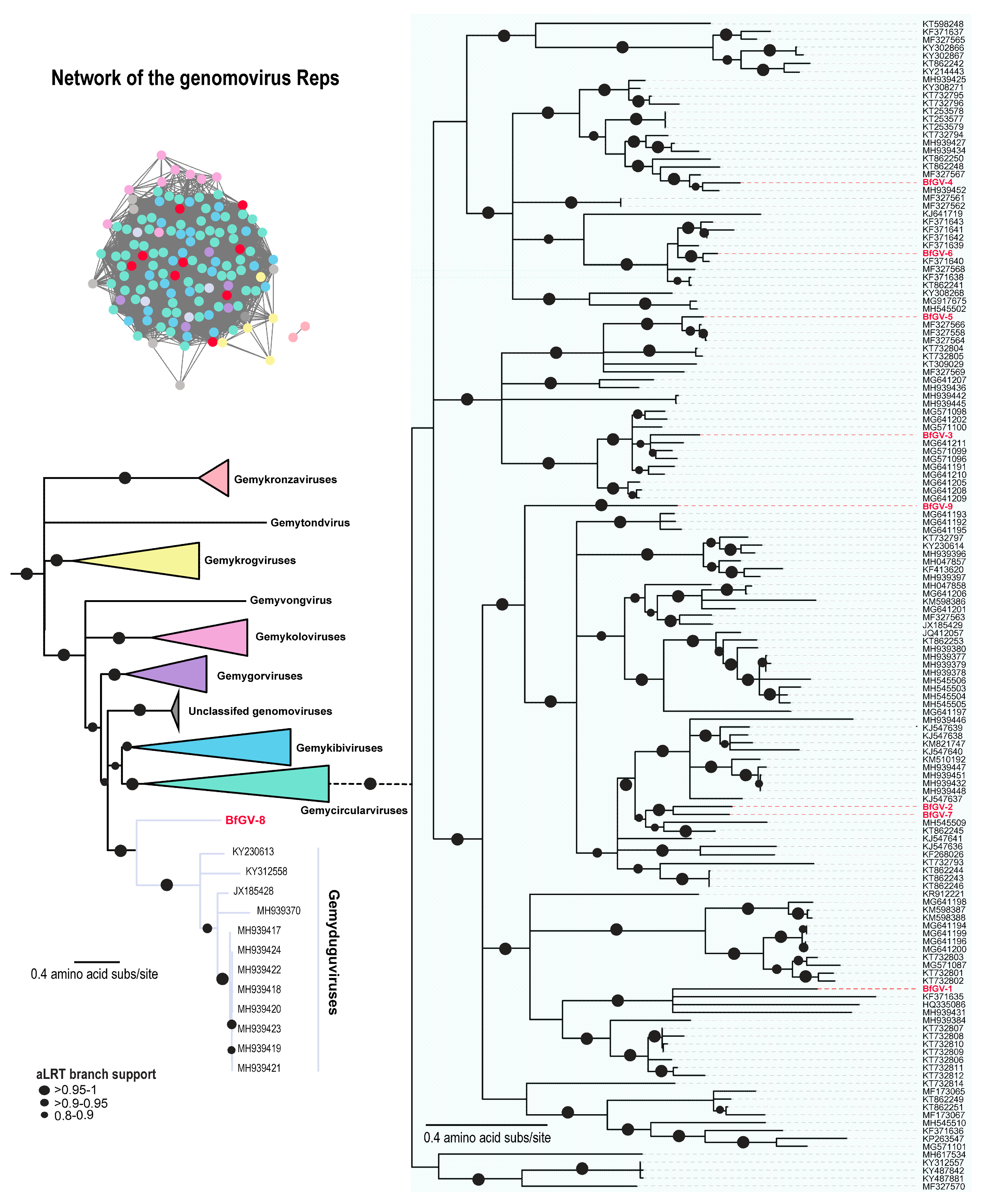
3.1. Blackfly CRESS DNA Viruses Clustering with Genomoviruses
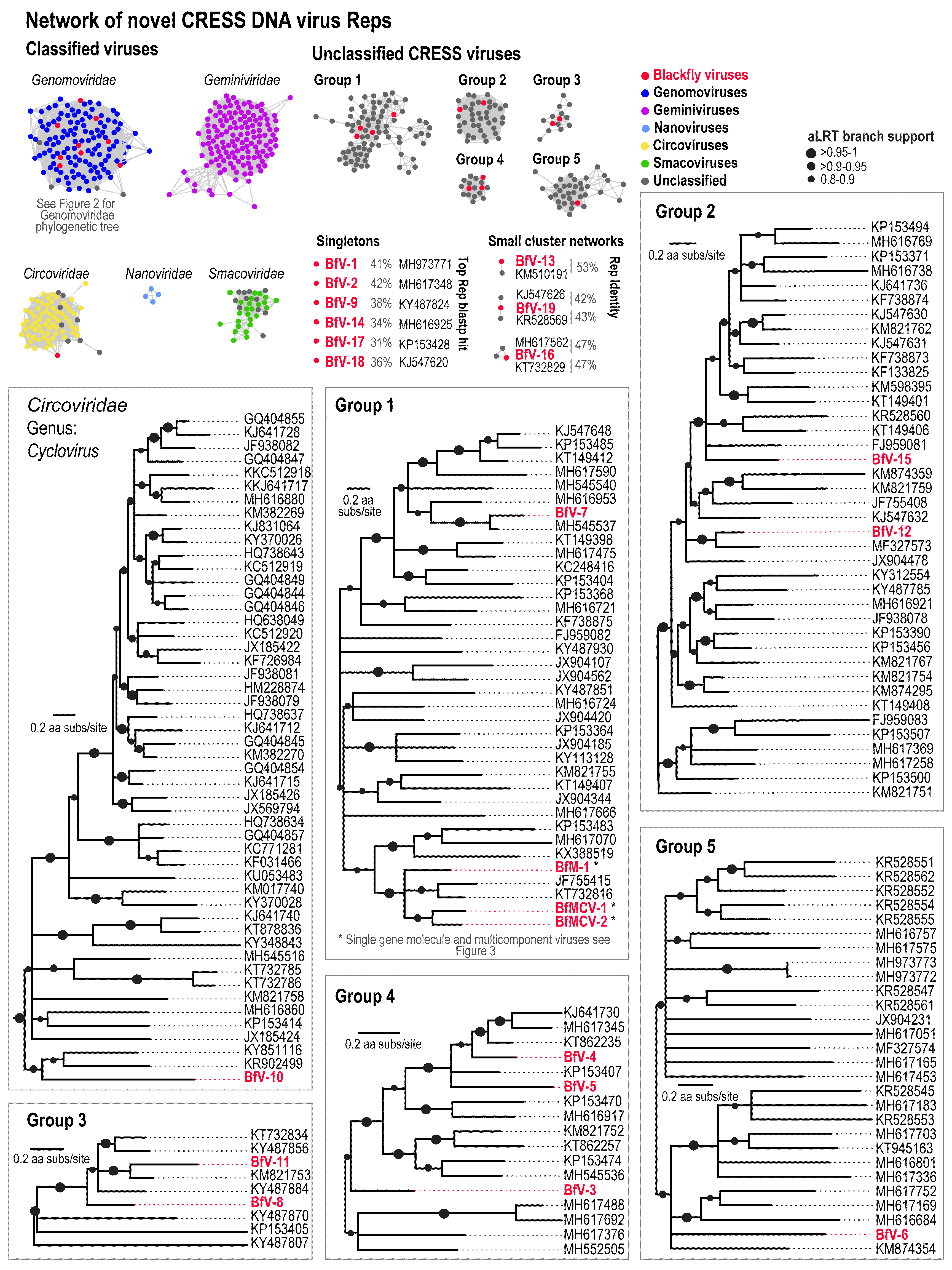
3.2. Divergent CRESS DNA Viruses
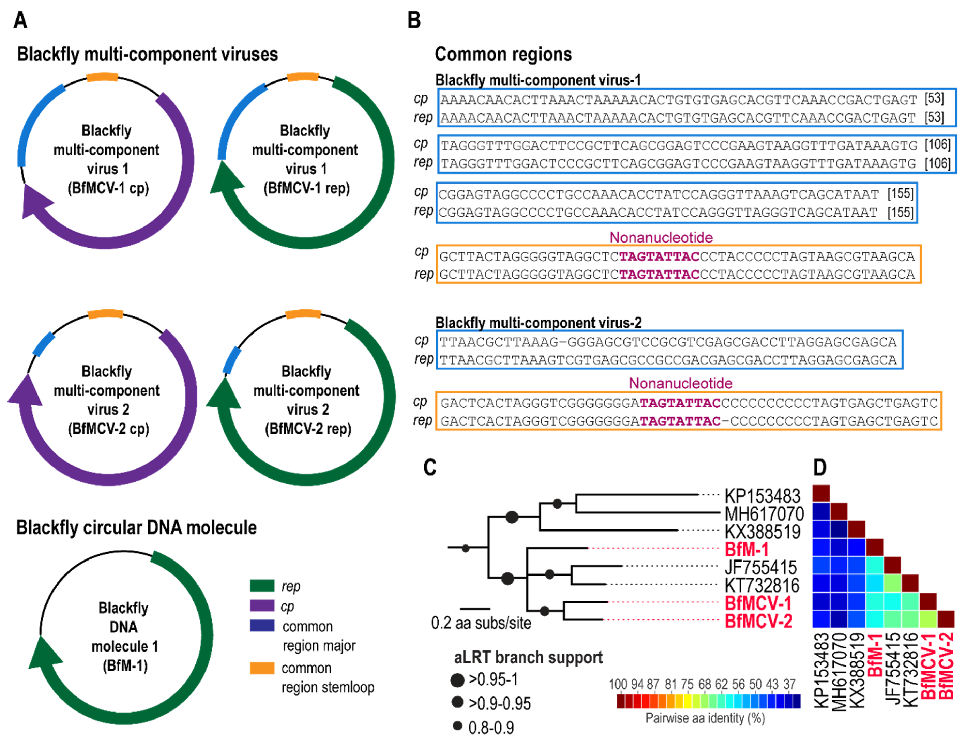
3.3. Multi-Component Viruses and Circular Rep-Encoding DNA Molecule
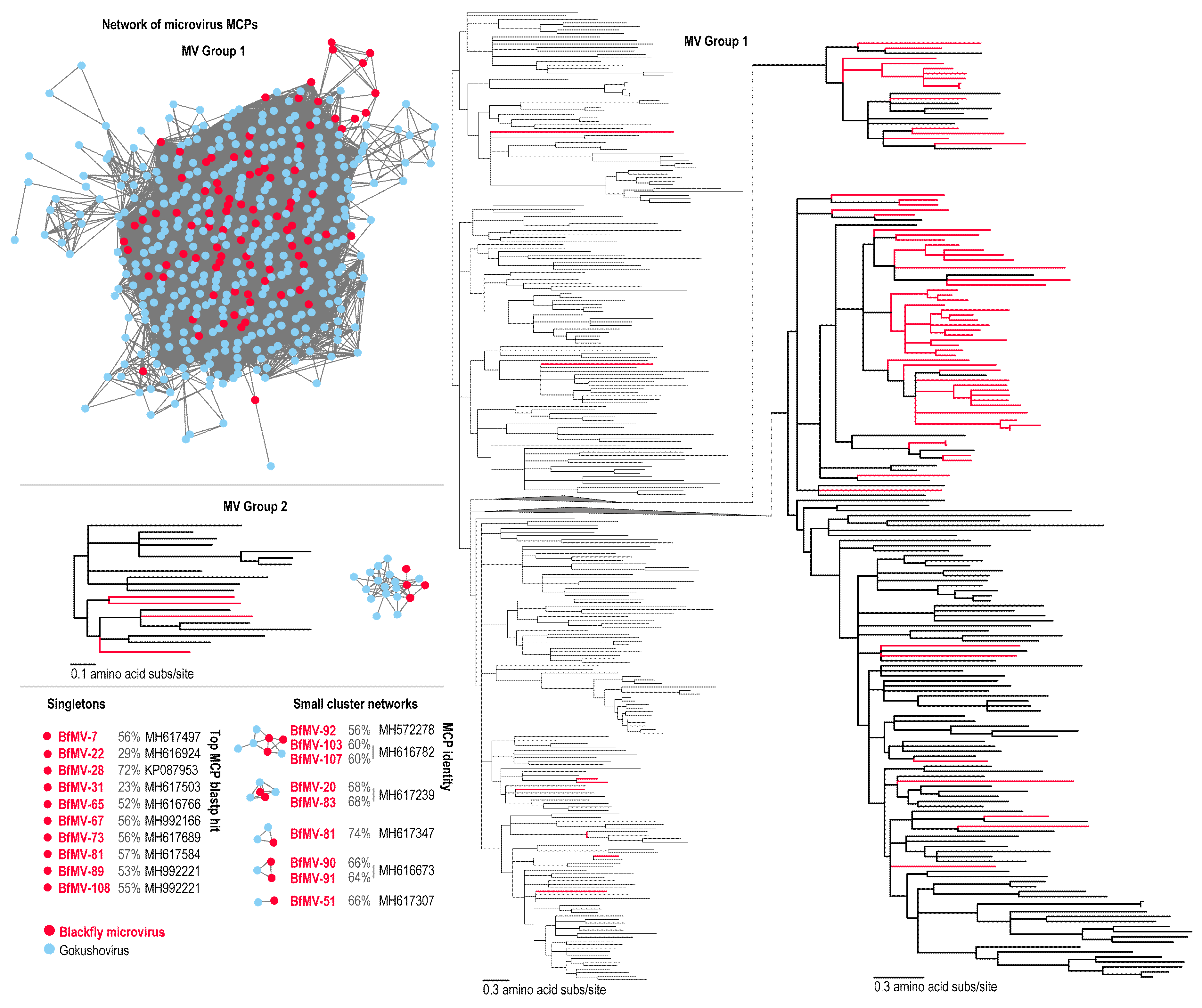
3.4. Bacteria-Infecting CRESS DNA Viruses
4. Conclusions
Supplementary Materials
Author Contributions
Funding
Conflicts of Interest
References
- Adler, P.H.; Cheke, R.A.; Post, R.J. Evolution, epidemiology, and population genetics of black flies (Diptera: Simuliidae). Infect. Genet. Evol. 2010, 10, 846–865. [Google Scholar] [CrossRef] [PubMed]
- Lustigman, S.; James, E.R.; Tawe, W.; Abraham, D. Towards a recombinant antigen vaccine against Onchocerca volvulus. Trends Parasitol. 2002, 18, 135–141. [Google Scholar] [CrossRef]
- Kalluri, S.; Gilruth, P.; Rogers, D.; Szczur, M. Surveillance of arthropod vector-borne infectious diseases using remote sensing techniques: A review. PLoS Pathog. 2007, 3, e116. [Google Scholar] [CrossRef] [PubMed]
- Hellgren, O.; Bensch, S.; Malmqvist, B. Bird hosts, blood parasites and their vectors—Associations uncovered by molecular analyses of blackfly blood meals. Mol. Ecol. 2008, 17, 1605–1613. [Google Scholar] [CrossRef] [PubMed]
- Sato, Y.; Tamada, A.; Mochizuki, Y.; Nakamura, S.; Okano, E.; Yoshida, C.; Ejiri, H.; Omori, S.; Yukawa, M.; Murata, K. Molecular detection of Leucocytozoon lovati from probable vectors, black flies (Simuliudae) collected in the alpine regions of Japan. Parasitol. Res. 2009, 104, 251–255. [Google Scholar] [CrossRef] [PubMed]
- Lichty, B.D.; Power, A.T.; Stojdl, D.F.; Bell, J.C. Vesicular stomatitis virus: Re-inventing the bullet. Trends Mol. Med. 2004, 10, 210–216. [Google Scholar] [CrossRef] [PubMed]
- Piégu, B.; Guizard, S.; Spears, T.; Cruaud, C.; Couloux, A.; Bideshi, D.K.; Federici, B.A.; Bigot, Y. Complete genome sequence of invertebrate iridovirus IIV-25 isolated from a blackfly larva. Arch. Virol. 2014, 159, 1181–1185. [Google Scholar] [CrossRef] [PubMed]
- Williams, T. Covert iridovirus infection of blackfly larvae. Proc. R. Soc. Lond. Ser. B Biol. Sci. 1993, 251, 225–230. [Google Scholar] [CrossRef]
- Crosby, T. Austrosimulium (Austrosimulium) dumbletoni n. sp. from New Zealand (Diptera: Simuliidae). N. Z. J. Zool. 1976, 3, 17–19. [Google Scholar] [CrossRef]
- Crosskey, R.W. Blackflies (Simuliidae). In Medical Insects and Arachnids; Lane, R.P., Crosskey, R.W., Eds.; Springer: Dordrecht, The Netherlands, 1993; pp. 241–287. [Google Scholar] [CrossRef]
- Rosario, K.; Mettel, K.A.; Benner, B.E.; Johnson, R.; Scott, C.; Yusseff-Vanegas, S.Z.; Baker, C.C.; Cassill, D.L.; Storer, C.; Varsani, A.; et al. Virus discovery in all three major lineages of terrestrial arthropods highlights the diversity of single-stranded DNA viruses associated with invertebrates. PeerJ 2018, 6, e5761. [Google Scholar] [CrossRef]
- Li, C.X.; Shi, M.; Tian, J.H.; Lin, X.D.; Kang, Y.J.; Chen, L.J.; Qin, X.C.; Xu, J.; Holmes, E.C.; Zhang, Y.Z. Unprecedented genomic diversity of RNA viruses in arthropods reveals the ancestry of negative-sense RNA viruses. Elife 2015, 4, e05378. [Google Scholar] [CrossRef] [PubMed]
- Ng, T.F.; Duffy, S.; Polston, J.E.; Bixby, E.; Vallad, G.E.; Breitbart, M. Exploring the diversity of plant DNA viruses and their satellites using vector-enabled metagenomics on whiteflies. PLoS ONE 2011, 6, e19050. [Google Scholar] [CrossRef] [PubMed]
- Ng, T.F.F.; Willner, D.L.; Lim, Y.W.; Schmieder, R.; Chau, B.; Nilsson, C.; Anthony, S.; Ruan, Y.; Rohwer, F.; Breitbart, M. Broad surveys of DNA viral diversity obtained through viral metagenomics of mosquitoes. PLoS ONE 2011, 6, e20579. [Google Scholar] [CrossRef] [PubMed]
- Sadeghi, M.; Altan, E.; Deng, X.; Barker, C.M.; Fang, Y.; Coffey, L.L.; Delwart, E. Virome of >12 thousand Culex mosquitoes from throughout California. Virology 2018, 523, 74–88. [Google Scholar] [CrossRef] [PubMed]
- Waits, K.; Edwards, M.J.; Cobb, I.N.; Fontenele, R.S.; Varsani, A. Identification of an anellovirus and genomoviruses in ixodid ticks. Vir. Genes 2018, 54, 155–159. [Google Scholar] [CrossRef] [PubMed]
- Kraberger, S.; Hofstetter, R.W.; Potter, K.A.; Farkas, K.; Varsani, A. Genomoviruses associated with mountain and western pine beetles. Vir. Res. 2018, 256, 17–20. [Google Scholar] [CrossRef] [PubMed]
- Zhao, L.; Rosario, K.; Breitbart, M.; Duffy, S. Eukaryotic circular rep-encoding single-stranded DNA (CRESS DNA) viruses: Ubiquitous viruses with small genomes and a diverse host range. In Advances in Virus Research; Kielian, M., Mettenleiter, T.C., Roossinck, M.J., Eds.; Elsevier: Cambridge, MA, USA, 2019; pp. 71–133. [Google Scholar] [CrossRef]
- Cherwa, J.E.; Fane, B.A. Microviridae: Microviruses and Gokushoviruses; John Wiley & Sons: Hoboken, NJ, USA, 2011. [Google Scholar] [CrossRef]
- Dayaram, A.; Potter, K.A.; Pailes, R.; Marinov, M.; Rosenstein, D.D.; Varsani, A. Identification of diverse circular single-stranded DNA viruses in adult dragonflies and damselflies (Insecta: Odonata) of Arizona and Oklahoma, USA. Infect. Genet. Evol. 2015, 30, 278–287. [Google Scholar] [CrossRef] [PubMed]
- Bankevich, A.; Nurk, S.; Antipov, D.; Gurevich, A.A.; Dvorkin, M.; Kulikov, A.S.; Lesin, V.M.; Nikolenko, S.I.; Pham, S.; Prjibelski, A.D.; et al. SPAdes: A new genome assembly algorithm and its applications to single-cell sequencing. J. Comput. Biol. 2012, 19, 455–477. [Google Scholar] [CrossRef]
- Altschul, S.F.; Gish, W.; Miller, W.; Myers, E.W.; Lipman, D.J. Basic local alignment search tool. J. Mol. Biol. 1990, 215, 403–410. [Google Scholar] [CrossRef]
- Bushnell, B. BBMap Short-Read Aligner, and Other Bioinformatics Tools; University of California: Berkeley, CA, USA, 2015. [Google Scholar]
- Ying, H.; Beifang, N.; Ying, G.; Limin, F.; Weizhong, L. CD-HIT Suite: A web server for clustering and comparing biological sequences. Bioinformatics 2010, 26, 680–682. [Google Scholar] [CrossRef]
- Gerlt, J.A.; Bouvier, J.T.; Davidson, D.B.; Imker, H.J.; Sadkhin, B.; Slater, D.R.; Whalen, K.L. Enzyme function initiative-enzyme similarity tool (EFI-EST): A web tool for generating protein sequence similarity networks. Biochim. Biophys. Acta 2015, 1854, 1019–1037. [Google Scholar] [CrossRef] [PubMed]
- Zallot, R.; Oberg, N.O.; Gerlt, J.A. Democratizedgenomic enzymology web tools for functional assignment. Curr. Opin. Chem. Biol. 2018, 47, 77–85. [Google Scholar] [CrossRef] [PubMed]
- Lopes, C.T.; Franz, M.; Kazi, F.; Donaldson, S.L.; Morris, Q.; Bader, G.D. Cytoscape web: An interactive web-based network browser. Bioinformatics 2010, 26, 2347–2348. [Google Scholar] [CrossRef] [PubMed]
- Edgar, R.C. MUSCLE: Multiple sequence alignment with high accuracy and high throughput. Nucleic Acids Res. 2004, 32, 1792–1797. [Google Scholar] [CrossRef] [PubMed]
- Guindon, S.; Dufayard, J.F.; Lefort, V.; Anisimova, M.; Hordijk, W.; Gascuel, O. New algorithms and methods to estimate maximum-likelihood phylogenies: Assessing the performance of PhyML 3.0. Syst. Biol. 2010, 59, 307–321. [Google Scholar] [CrossRef] [PubMed]
- Darriba, D.; Taboada, G.L.; Doallo, R.; Posada, D. ProtTest 3: Fast selection of best-fit models of protein evolution. Bioinformatics 2011, 27, 1164–1165. [Google Scholar] [CrossRef]
- Stover, B.C.; Muller, K.F. TreeGraph 2: Combining and visualizing evidence from different phylogenetic analyses. BMC Bioinform. 2010, 11, 7. [Google Scholar] [CrossRef]
- Muhire, B.M.; Varsani, A.; Martin, D.P. SDT: A virus classification tool based on pairwise sequence alignment and identity calculation. PLoS ONE 2014, 9, e108277. [Google Scholar] [CrossRef]
- Kraberger, S.; Polston, J.E.; Capobianco, H.M.; Alcala-Briseno, R.I.; Fontenele, R.S.; Varsani, A. Genomovirus genomes recovered from Echinothrips americanus sampled in Florida, USA. Genome Announc. 2017, 5, e00445-17. [Google Scholar] [CrossRef]
- Kraberger, S.; Cook, C.N.; Schmidlin, K.; Fontenele, R.S.; Bautista, J.; Smith, B.; Varsani, A. Diverse single-stranded DNA viruses associated with honey bees (Apis mellifera). Infect. Genet. Evol. 2019, 71, 179–188. [Google Scholar] [CrossRef]
- Yu, X.; Li, B.; Fu, Y.; Jiang, D.; Ghabrial, S.A.; Li, G.; Peng, Y.; Xie, J.; Cheng, J.; Huang, J.; et al. A geminivirus-related DNA mycovirus that confers hypovirulence to a plant pathogenic fungus. Proc. Natl. Acad. Sci. USA 2010, 107, 8387–8392. [Google Scholar] [CrossRef] [PubMed]
- Varsani, A.; Krupovic, M. Sequence-based taxonomic framework for the classification of uncultured single-stranded DNA viruses of the family Genomoviridae. Vir. Evol. 2017, 3. [Google Scholar] [CrossRef] [PubMed]
- Kraberger, S.; Waits, K.; Ivan, J.; Newkirk, E.; VandeWoude, S.; Varsani, A. Identification of circular single-stranded DNA viruses in faecal samples of Canada lynx (Lynx canadensis), moose (Alces alces) and snowshoe hare (Lepus americanus) inhabiting the Colorado San Juan Mountains. Infect. Genet. Evol. 2018, 64, 1–8. [Google Scholar] [CrossRef] [PubMed]
- Sikorski, A.; Massaro, M.; Kraberger, S.; Young, L.M.; Smalley, D.; Martin, D.P.; Varsani, A. Novel myco-like DNA viruses discovered in the faecal matter of various animals. Vir. Res. 2013, 177, 209–216. [Google Scholar] [CrossRef] [PubMed]
- Dayaram, A.; Galatowitsch, M.; Harding, J.S.; Arguello-Astorga, G.R.; Varsani, A. Novel circular DNA viruses identified in Procordulia grayi and Xanthocnemis zealandica larvae using metagenomic approaches. Infect. Genet. Evol. 2014, 22, 134–141. [Google Scholar] [CrossRef] [PubMed]
- Male, M.F.; Kraberger, S.; Stainton, D.; Kami, V.; Varsani, A. Cycloviruses, gemycircularviruses and other novel replication-associated protein encoding circular viruses in Pacific flying fox (Pteropus tonganus) faeces. Infect. Genet. Evol. 2016, 39, 279–292. [Google Scholar] [CrossRef] [PubMed]
- Kraberger, S.; Arguello-Astorga, G.R.; Greenfield, L.G.; Galilee, C.; Law, D.; Martin, D.P.; Varsani, A. Characterisation of a diverse range of circular replication-associated protein encoding DNA viruses recovered from a sewage treatment oxidation pond. Infect. Genet. Evol. 2015, 31, 73–86. [Google Scholar] [CrossRef] [PubMed]
- Tisza, M.J.; Pastrana, D.V.; Welch, N.L.; Stewart, B.; Peretti, A.; Starrett, G.J.; Pang, Y.-Y.S.; Varsani, A.; Krishnamurthy, S.R.; Pesavento, P.A. Discovery of several thousand highly diverse circular DNA viruses. BioRxiv 2019, 2019, 555375. [Google Scholar] [CrossRef]
- Steel, O.; Kraberger, S.; Sikorski, A.; Young, L.M.; Catchpole, R.J.; Stevens, A.J.; Ladley, J.J.; Coray, D.S.; Stainton, D.; Dayaram, A.; et al. Circular replication-associated protein encoding DNA viruses identified in the faecal matter of various animals in New Zealand. Infect. Genet. Evol. 2016, 43, 151–164. [Google Scholar] [CrossRef] [PubMed]
- Dayaram, A.; Galatowitsch, M.L.; Arguello-Astorga, G.R.; van Bysterveldt, K.; Kraberger, S.; Stainton, D.; Harding, J.S.; Roumagnac, P.; Martin, D.P.; Lefeuvre, P.; et al. Diverse circular replication-associated protein encoding viruses circulating in invertebrates within a lake ecosystem. Infect. Genet. Evol. 2016, 39, 304–316. [Google Scholar] [CrossRef] [PubMed]
- Rosario, K.; Marinov, M.; Stainton, D.; Kraberger, S.; Wiltshire, E.J.; Collings, D.A.; Walters, M.; Martin, D.P.; Breitbart, M.; Varsani, A. Dragonfly cyclovirus, a novel single-stranded DNA virus discovered in dragonflies (Odonata: Anisoptera). J. Gen. Virol. 2011, 92, 1302–1308. [Google Scholar] [CrossRef] [PubMed]
- Padilla-Rodriguez, M.; Rosario, K.; Breitbart, M. Novel cyclovirus discovered in the Florida woods cockroach Eurycotis floridana (Walker). Arch. Virol. 2013, 158, 1389–1392. [Google Scholar] [CrossRef] [PubMed]
- Wang, B.; Sun, L.D.; Liu, H.H.; Wang, Z.D.; Zhao, Y.K.; Wang, W.; Liu, Q. Molecular detection of novel circoviruses in ticks in northeastern China. Ticks Tick Borne Dis. 2018, 9, 836–839. [Google Scholar] [CrossRef] [PubMed]
- Dayaram, A.; Potter, K.A.; Moline, A.B.; Rosenstein, D.D.; Marinov, M.; Thomas, J.E.; Breitbart, M.; Rosario, K.; Arguello-Astorga, G.R.; Varsani, A. High global diversity of cycloviruses amongst dragonflies. J. Gen. Virol. 2013, 94, 1827–1840. [Google Scholar] [CrossRef] [PubMed]
- Kraberger, S.; Farkas, K.; Bernardo, P.; Booker, C.; Argüello-Astorga, G.R.; Mesléard, F.; Martin, D.P.; Roumagnac, P.; Varsani, A. Identification of novel Bromus- and Trifolium-associated circular DNA viruses. Arch. Virol. 2015, 160, 1303–1311. [Google Scholar] [CrossRef] [PubMed]
- Rosario, K.; Schenck, R.O.; Harbeitner, R.C.; Lawler, S.N.; Breitbart, M. Novel circular single-stranded DNA viruses identified in marine invertebrates reveal high sequence diversity and consistent predicted intrinsic disorder patterns within putative structural proteins. Front. Microbiol. 2015, 6, 696. [Google Scholar] [CrossRef] [PubMed]
- Pearson, V.M.; Caudle, S.B.; Rokyta, D.R. Viral recombination blurs taxonomic lines: Examination of single-stranded DNA viruses in a wastewater treatment plant. PeerJ 2016, 4, e2585. [Google Scholar] [CrossRef]
- Timchenko, T.; de Kouchkovsky, F.; Katul, L.; David, C.; Vetten, H.J.; Gronenborn, B. A single rep protein initiates replication of multiple genome components of faba bean necrotic yellows virus, a single-stranded DNA virus of plants. J. Virol. 1999, 73, 10173–10182. [Google Scholar]
- Phan, T.G.; Kapusinszky, B.; Wang, C.; Rose, R.K.; Lipton, H.L.; Delwart, E.L. The fecal viral flora of wild rodents. PLoS Pathog. 2011, 7, e1002218. [Google Scholar] [CrossRef]
- Schmidlin, K.; Kraberger, S.; Fontenele, R.S.; De Martini, F.; Chouvenc, T.; Gile, G.H.; Varsani, A. Genome sequences of microviruses associated with Coptotermes formosanus. Microbiol. Resour. Announc. 2019, 8, e00185-19. [Google Scholar] [CrossRef]


| Family/Subfamily | Genus | Virus Name | Accession Number | Size (nts) | ORF Orientation |
|---|---|---|---|---|---|
| Genomoviridae | Gemycircularirus | Blackfly genomovirus-1 SF02 506 | MK433242 | 2290 | Bidirectional |
| Blackfly genomovirus-2 SF02 631 | MK433234 | 2131 | Bidirectional | ||
| Blackfly genomovirus-3 SF02 1766 | MK433235 | 2138 | Bidirectional | ||
| Blackfly genomovirus-4 SF02 836 | MK433236 | 2181 | Bidirectional | ||
| Blackfly genomovirus-5 SF02 599 | MK433237 | 2182 | Bidirectional | ||
| Blackfly genomovirus-6 SF02 459 | MK433238 | 2186 | Bidirectional | ||
| Blackfly genomovirus-7 SF02 767 | MK433239 | 2195 | Bidirectional | ||
| Blackfly genomovirus-9 SF02 507 | MK433241 | 2217 | Bidirectional | ||
| Genomoviridae | Gemyduguvirus | Blackfly genomovirus-8 SF02 579 | MK433240 | 2226 | Bidirectional |
| Unclassified CRESS DNA virus | Unassigned | Blackfly DNA virus-1 SF02 666 | MK433215 | 1805 | Unidirectional |
| Blackfly DNA virus-2 SF02 583 | MK433216 | 1936 | Bidirectional | ||
| Blackfly DNA virus-3 SF02 402 | MK433217 | 2005 | Unidirectional | ||
| Blackfly DNA virus-4 SF02 664 | MK433218 | 2047 | Unidirectional | ||
| Blackfly DNA virus-5 SF02 839 | MK433219 | 2058 | Unidirectional | ||
| Blackfly DNA virus-6 SF01 308 | MK433220 | 2172 | Bidirectional | ||
| Blackfly DNA virus-7 SF02 462 | MK433221 | 2306 | Bidirectional | ||
| Blackfly DNA virus-8 SF02 1137 | MK433222 | 2337 | Bidirectional | ||
| Blackfly DNA virus-9 SF02 881 | MK433223 | 2389 | Bidirectional | ||
| Blackfly DNA virus-10 SF02 899 | MK433224 | 2444 | Bidirectional | ||
| Blackfly DNA virus-11 SF02 963 | MK433225 | 2449 | Bidirectional | ||
| Blackfly DNA virus-12 SF02 422 | MK433226 | 2466 | Unidirectional | ||
| Blackfly DNA virus-13 SF02 413 | MK433227 | 2501 | Bidirectional | ||
| Blackfly DNA virus-14 SF02 295 | MK433228 | 2583 | Bidirectional | ||
| Blackfly DNA virus-15 SF02 403 | MK433229 | 2697 | Bidirectional | ||
| Blackfly DNA virus-16 SF02 377 | MK433230 | 2704 | Bidirectional | ||
| Blackfly DNA virus-17 SF02 1426 | MK433231 | 2835 | Unidirectional | ||
| Blackfly DNA virus-18 SF02 66 | MK433232 | 3003 | Bidirectional | ||
| Blackfly DNA virus-19 SF02 380 | MK433233 | 2002 | Bidirectional | ||
| Unclassified Circular DNA molecules | Unassigned | Blackfly DNA molecule 1 - rep | MK561604 | 1157 | Unidirectional |
| Unclassified Multi-component virus | Unassigned | Blackfly multicomponent virus 1 - rep | MK561605 | 1163 | Unidirectional |
| Blackfly multicomponent virus 1 - cp | MK561606 | 1154 | Unidirectional | ||
| Blackfly multicomponent virus 2 - rep | MK561607 | 1136 | Unidirectional | ||
| Blackfly multicomponent virus 2 - cp | MK561608 | 1133 | Unidirectional | ||
| Microviridae/Gokushovirinae | Unassigned | Blackfly microvirus-1_ SF02_SP_9 | MK249160 | 4873 | Unidirectional |
| Blackfly microvirus-2_ SF02_SP_11 | MK249161 | 4544 | Unidirectional | ||
| Blackfly microvirus-3_ SF02_SP_12 | MK249162 | 4625 | Unidirectional | ||
| Blackfly microvirus-4_ SF02_SP_13 | MK249163 | 4660 | Unidirectional | ||
| Blackfly microvirus-5_ SF02_SP_14 | MK249164 | 4594 | Unidirectional | ||
| Blackfly microvirus-6_ SF02_SP_15 | MK249165 | 4675 | Unidirectional | ||
| Blackfly microvirus-7_ SF02_SP_17 | MK249166 | 4379 | Unidirectional | ||
| Blackfly microvirus-8_ SF02_SP_18 | MK249167 | 4483 | Unidirectional | ||
| Blackfly microvirus-9_ SF02_SP_20 | MK249168 | 4324 | Unidirectional | ||
| Blackfly microvirus-10_ SF02_SP_21 | MK249169 | 4656 | Unidirectional | ||
| Blackfly microvirus-11_ SF02_SP_22 | MK249170 | 4565 | Unidirectional | ||
| Blackfly microvirus-12_ SF02_SP_24 | MK249171 | 4231 | Unidirectional | ||
| Blackfly microvirus-13_ SF02_SP_25 | MK249172 | 4582 | Unidirectional | ||
| Blackfly microvirus-14_ SF02_SP_27 | MK249173 | 4694 | Unidirectional | ||
| Blackfly microvirus-15_ SF02_SP_28 | MK249174 | 4557 | Unidirectional | ||
| Blackfly microvirus-16_ SF02_SP_30 | MK249175 | 4689 | Unidirectional | ||
| Blackfly microvirus-17_ SF02_SP_31 | MK249176 | 4474 | Unidirectional | ||
| Blackfly microvirus-18_ SF02_SP_32 | MK249177 | 4841 | Unidirectional | ||
| Blackfly microvirus-19_ SF02_SP_34 | MK249178 | 4727 | Unidirectional | ||
| Blackfly microvirus-20_ SF02_SP_36 | MK249179 | 4498 | Unidirectional | ||
| Blackfly microvirus-21_ SF02_SP_38 | MK249180 | 4461 | Unidirectional | ||
| Blackfly microvirus-22_ SF02_SP_44 | MK249181 | 6565 | Unidirectional | ||
| Blackfly microvirus-23_ SF02_SP_45 | MK249182 | 4565 | Unidirectional | ||
| Blackfly microvirus-24_ SF02_SP_46 | MK249183 | 4633 | Unidirectional | ||
| Blackfly microvirus-25_ SF02_SP_49 | MK249184 | 4521 | Unidirectional | ||
| Blackfly microvirus-26_ SF02_SP_50 | MK249185 | 4731 | Unidirectional | ||
| Blackfly microvirus-27_ SF02_SP_51 | MK249186 | 4581 | Unidirectional | ||
| Blackfly microvirus-28_ SF02_SP_52 | MK249187 | 4667 | Unidirectional | ||
| Blackfly microvirus-29_ SF02_SP_54 | MK249188 | 4637 | Unidirectional | ||
| Blackfly microvirus-30_ SF02_SP_57 | MK249189 | 4980 | Unidirectional | ||
| Blackfly microvirus-31_ SF02_SP_67 | MK249190 | 4901 | Unidirectional | ||
| Blackfly microvirus-32_ SF02_SP_69 | MK249191 | 4633 | Unidirectional | ||
| Blackfly microvirus-33_ SF02_SP_71 | MK249192 | 4901 | Unidirectional | ||
| Blackfly microvirus-34_ SF02_SP_73 | MK249193 | 4870 | Unidirectional | ||
| Blackfly microvirus-35_ SF02_SP_74 | MK249194 | 4765 | Unidirectional | ||
| Blackfly microvirus-36_ SF02_SP_75 | MK249195 | 4789 | Unidirectional | ||
| Blackfly microvirus-37_ SF02_SP_78 | MK249196 | 4774 | Unidirectional | ||
| Blackfly microvirus-38_ SF02_SP_79 | MK249197 | 4711 | Unidirectional | ||
| Blackfly microvirus-39_ SF02_SP_80 | MK249198 | 4858 | Unidirectional | ||
| Blackfly microvirus-40_ SF02_SP_82 | MK249199 | 4799 | Unidirectional | ||
| Blackfly microvirus-41_ SF02_SP_83 | MK249200 | 4825 | Unidirectional | ||
| Blackfly microvirus-42_ SF02_SP_84 | MK249201 | 4779 | Unidirectional | ||
| Blackfly microvirus-43_ SF02_SP_87 | MK249202 | 4768 | Unidirectional | ||
| Blackfly microvirus-44_ SF02_SP_88 | MK249203 | 4615 | Unidirectional | ||
| Blackfly microvirus-45_ SF02_SP_89 | MK249204 | 4630 | Unidirectional | ||
| Blackfly microvirus-46_ SF02_SP_91 | MK249205 | 4733 | Unidirectional | ||
| Blackfly microvirus-47_ SF02_SP_93 | MK249206 | 4721 | Unidirectional | ||
| Blackfly microvirus-48_ SF02_SP_97 | MK249207 | 4646 | Unidirectional | ||
| Blackfly microvirus-49_ SF02_SP_98 | MK249208 | 4700 | Unidirectional | ||
| Blackfly microvirus-50_ SF02_SP_99 | MK249209 | 4706 | Unidirectional | ||
| Blackfly microvirus-51_ SF02_SP_100 | MK249210 | 4721 | Unidirectional | ||
| Blackfly microvirus-52_ SF02_SP_104 | MK249211 | 4692 | Unidirectional | ||
| Blackfly microvirus-53_ SF02_SP_106 | MK249212 | 4680 | Unidirectional | ||
| Blackfly microvirus-54_ SF02_SP_107 | MK249213 | 4706 | Unidirectional | ||
| Blackfly microvirus-55_ SF02_SP_108 | MK249214 | 4647 | Unidirectional | ||
| Blackfly microvirus-56_ SF02_SP_110 | MK249215 | 4618 | Unidirectional | ||
| Blackfly microvirus-57_ SF02_SP_115 | MK249216 | 4608 | Unidirectional | ||
| Blackfly microvirus-58_ SF02_SP_116 | MK249217 | 4596 | Unidirectional | ||
| Blackfly microvirus-59_ SF02_SP_117 | MK249218 | 4590 | Unidirectional | ||
| Blackfly microvirus-60_ SF02_SP_118 | MK249219 | 4621 | Unidirectional | ||
| Blackfly microvirus-61_ SF02_SP_120 | MK249220 | 4617 | Unidirectional | ||
| Blackfly microvirus-62_ SF02_SP_122 | MK249221 | 4591 | Unidirectional | ||
| Blackfly microvirus-63_ SF02_SP_123 | MK249222 | 4499 | Unidirectional | ||
| Blackfly microvirus-64_ SF02_SP_124 | MK249223 | 4510 | Unidirectional | ||
| Blackfly microvirus-65_ SF02_SP_125 | MK249224 | 4507 | Unidirectional | ||
| Blackfly microvirus-66_ SF02_SP_126 | MK249225 | 4581 | Unidirectional | ||
| Blackfly microvirus-67_ SF02_SP_128 | MK249226 | 4503 | Unidirectional | ||
| Blackfly microvirus-68_ SF02_SP_129 | MK249227 | 4570 | Unidirectional | ||
| Blackfly microvirus-69_ SF02_SP_130 | MK249228 | 4498 | Unidirectional | ||
| Blackfly microvirus-70_ SF02_SP_131 | MK249229 | 4558 | Unidirectional | ||
| Blackfly microvirus-71_ SF02_SP_135 | MK249230 | 4488 | Unidirectional | ||
| Blackfly microvirus-72_ SF02_SP_137 | MK249231 | 4551 | Unidirectional | ||
| Blackfly microvirus-73_ SF02_SP_138 | MK249232 | 4335 | Unidirectional | ||
| Blackfly microvirus-74_ SF02_SP_139 | MK249233 | 4547 | Unidirectional | ||
| Blackfly microvirus-75_ SF02_SP_142 | MK249234 | 4538 | Unidirectional | ||
| Blackfly microvirus-76_ SF02_SP_143 | MK249127 | 4471 | Unidirectional | ||
| Blackfly microvirus-77_ SF02_SP_145 | MK249128 | 4529 | Unidirectional | ||
| Blackfly microvirus-78_ SF02_SP_146 | MK249129 | 4521 | Unidirectional | ||
| Blackfly microvirus-79_ SF02_SP_148 | MK249130 | 4549 | Unidirectional | ||
| Blackfly microvirus-80_ SF02_SP_149 | MK249131 | 4460 | Unidirectional | ||
| Blackfly microvirus-81_ SF02_SP_150 | MK249132 | 4460 | Unidirectional | ||
| Blackfly microvirus-82_ SF02_SP_156 | MK249133 | 4443 | Unidirectional | ||
| Blackfly microvirus-83_ SF02_SP_159 | MK249134 | 4438 | Unidirectional | ||
| Blackfly microvirus-84_ SF02_SP_160 | MK249135 | 4483 | Unidirectional | ||
| Blackfly microvirus-85_ SF02_SP_162 | MK249136 | 4350 | Unidirectional | ||
| Blackfly microvirus-86_ SF02_SP_163 | MK249137 | 4484 | Unidirectional | ||
| Blackfly microvirus-87_ SF02_SP_164 | MK249138 | 4502 | Unidirectional | ||
| Blackfly microvirus-88_ SF02_SP_165 | MK249139 | 4487 | Unidirectional | ||
| Blackfly microvirus-89_ SF02_SP_166 | MK249140 | 4410 | Unidirectional | ||
| Blackfly microvirus-90_ SF02_SP_167 | MK249141 | 4459 | Unidirectional | ||
| Blackfly microvirus-91_ SF02_SP_168 | MK249142 | 4401 | Unidirectional | ||
| Blackfly microvirus-92_ SF02_SP_169 | MK249143 | 4387 | Unidirectional | ||
| Blackfly microvirus-93_ SF02_SP_174 | MK249144 | 4445 | Unidirectional | ||
| Blackfly microvirus-94_ SF02_SP_175 | MK249145 | 4400 | Unidirectional | ||
| Blackfly microvirus-95_ SF02_SP_178 | MK249146 | 4422 | Unidirectional | ||
| Blackfly microvirus-96_ SF02_SP_181 | MK249147 | 4391 | Unidirectional | ||
| Blackfly microvirus-97_ SF02_SP_183 | MK249148 | 4368 | Unidirectional | ||
| Blackfly microvirus-98_ SF02_SP_184 | MK249149 | 4293 | Unidirectional | ||
| Blackfly microvirus-99_ SF02_SP_185 | MK249150 | 4351 | Unidirectional | ||
| Blackfly microvirus-100_ SF02_SP_188 | MK249151 | 4276 | Unidirectional | ||
| Blackfly microvirus-101_ SF02_SP_189 | MK249152 | 4318 | Unidirectional | ||
| Blackfly microvirus-102_ SF02_SP_190 | MK249153 | 4205 | Unidirectional | ||
| Blackfly microvirus-103_ SF02_SP_192 | MK249154 | 4320 | Unidirectional | ||
| Blackfly microvirus-104_ SF02_SP_195 | MK249155 | 4302 | Unidirectional | ||
| Blackfly microvirus-105_ SF02_SP_206 | MK249156 | 4238 | Unidirectional | ||
| Blackfly microvirus-106_ SF02_SP_208 | MK249157 | 4237 | Unidirectional | ||
| Blackfly microvirus-107_ SF02_SP_211 | MK249158 | 4170 | Unidirectional | ||
| Blackfly microvirus-108_ SF02_SP_213 | MK249159 | 4232 | Unidirectional |
© 2019 by the authors. Licensee MDPI, Basel, Switzerland. This article is an open access article distributed under the terms and conditions of the Creative Commons Attribution (CC BY) license (http://creativecommons.org/licenses/by/4.0/).
Share and Cite
Kraberger, S.; Schmidlin, K.; Fontenele, R.S.; Walters, M.; Varsani, A. Unravelling the Single-Stranded DNA Virome of the New Zealand Blackfly. Viruses 2019, 11, 532. https://doi.org/10.3390/v11060532
Kraberger S, Schmidlin K, Fontenele RS, Walters M, Varsani A. Unravelling the Single-Stranded DNA Virome of the New Zealand Blackfly. Viruses. 2019; 11(6):532. https://doi.org/10.3390/v11060532
Chicago/Turabian StyleKraberger, Simona, Kara Schmidlin, Rafaela S. Fontenele, Matthew Walters, and Arvind Varsani. 2019. "Unravelling the Single-Stranded DNA Virome of the New Zealand Blackfly" Viruses 11, no. 6: 532. https://doi.org/10.3390/v11060532
APA StyleKraberger, S., Schmidlin, K., Fontenele, R. S., Walters, M., & Varsani, A. (2019). Unravelling the Single-Stranded DNA Virome of the New Zealand Blackfly. Viruses, 11(6), 532. https://doi.org/10.3390/v11060532


