Article

Development of Ash Dieback in South-Eastern Germany and the Increasing Occurrence of Secondary Pathogens

Bavarian State Institute of Forestry, Hans-Carl-von-Carlowitz-Platz 1, 85354 Freising, Germany
*
Author to whom correspondence should be addressed.
Forests 2016, 7(2), 41; https://doi.org/10.3390/f7020041
Submission received: 1 October 2015 / Revised: 18 January 2016 / Accepted: 3 February 2016 / Published: 15 February 2016

Abstract

:
Since its first identification in Poland in 2006, the ascomycete Hymenoscyphus fraxineus has caused massive dieback of Fraxinus excelsior in the countries of eastern, northern and central Europe. This work shows the development, expansion, and severity of the disease in south-eastern Germany for a period of four years, starting in 2010. Differences between habitats, as well as age classes have been captured. The presence and the amount of potentially resistant trees were proven over the years, to determine how high the resistance level might be. Typical disease symptoms are the wilting of leaves, necrotic lesions in the bark and reddish discolorations of branches and stems. In addition, stem necroses also appear by infection with species of Armillaria. Therefore, special attention has been given to Armillaria species in affected ash stands but also to other secondary pathogens, like ash bark beetles. It is shown that breeding galleries of Hylesinus fraxini are only found in trees that have recently died and thus Hylesinus fraxini is still acting as a secondary opportunistic pathogen. In contrast, Armillaria spp. can be considered as serious pathogens of weakened ash trees. In different ash stands, typical symptoms of infection can be found. A relationship between stem base necrotic lesions and vitality was examined. It is shown that necrotic lesions severely contribute to accelerating the mortality of ash trees. In addition to the high infection pressure by H. fraxineus, the high inoculum of Armillaria in the soil facilitates further infections and, thus, likewise endangers the survival of potentially resistant trees. In the following years, forest conversion and seed harvest in affected ash stands will have to be urgently considered to avoid tree gaps on a large scale. Furthermore, infection assays of potentially resistant trees with ensuing breeding programmes should be initially started for the conservation of this ecologically and economically important tree species.

1. Introduction

The ash dieback pathogen Hymenoscyphus fraxineus [1], which is native to Asia [2,3,4] and previously known as Hymenoscyphus pseudoalbidus, [5] is meanwhile widespread throughout Europe [6]. Symptoms of the disease range from brown lesions on leaves and branches, wilting of leaves and crown-dieback to discolorations in the wood. The ascomycete threatens ash trees (Fraxinus excelsior) of all age classes in all their growth areas, in lowlands, floodplain or mountain forests. Above all, young trees are severely affected [7] but more and more trees in older ash stands also die within a few years of infection. In the extent of this study, severity and progression of the disease in south-eastern Germany have been analyzed for a period of four years in 22 study sites. As there is currently no method to cure the trees or slow down the disease expansion, special study attention was drawn to potentially resistant trees. According to the data from studies in different countries, where the disease was described earlier, it can be expected that the resistance in ash stands against H. fraxineus is low [8,9]. Recommendations for the management of affected ash stands provide for not removing healthy or less-affected F. excelsior trees depending on their crown defoliation as the best strategy to retain possibly resistant trees [10]. Furthermore, it was suggested that trees under infection stress by H. fraxineus are more susceptible to secondary pathogens [11]. Some bark beetles are specialized on Genus Fraxinus or Oleacea. The most abundant species are Hylesinus fraxini, but also Hylesinus oleiperda and Hylesinus crenatus [12]. During nearly all of their life cycle they live inside the tissue and feed on the phloem [13]. Before the impact of ash dieback these species were not of any economic importance in the forest industry. There is some concern that these secondary biotic pest agents may increase in abundance on weakened trees and become primary pest agents [11,14]. Therefore, the role of ash bark beetles was closely investigated in the different study sites to determine if such beetles are still secondary pests even in H. fraxineus infected ash stands.
An additional problem results from the species of Armillaria. These basidiomycetes are distributed worldwide and play variable ecological roles, including causing root disease of diverse forest trees [15]. In Europe, seven Armillaria species can be distinguished. Some of them are considered as weak parasites, whereas other species, like A. mellea, A. ostoyae and A. cepistipes, belong to a group of serious pathogens [16]. A. cepistipes and A. ostoyae can frequently be found in the same forest stand, whereby their virulence depends on the production of rhizomorphs [17]. For Lithuania, where the first reports of H. fraxineus infected trees came from, the presence of A. cepistipes in declining ash stands was already reported [18]. In Denmark and south-west Germany A. gallica was associated with ash dieback, although the fungus was not considered as the main causal agent [7,19]. Not only on ash trees, but also on oaks, some Armillaria species were found jointly responsible for a large-scale mortality in Arkansas, USA [20]. Some species are able to infect the non-wounded root bark directly [21]. Armillaria species cause central butt rots, leading to a severe loss in the production of wood, and are therefore economically important. This rot normally does not spread more than a couple of meters [22].
In ash dieback affected stands in south-eastern Germany, resistance against Armillaria species has been lowered and the danger of a fungal infection has increased drastically. The interaction between Armillaria species and H. fraxineus in declining ash stands is examined. It becomes evident that Armillaria reduces not only the timber quality but also the stability of the infected trees, which are at high risk for falling down even under windless conditions [19]. Increasing mortality by combined infections of H. fraxineus and Armillaria spp. causes large tree gaps within the stands. Also potentially resistant trees are endangered within dissolving ash stands, as there is a high inoculum of rhizomorphs of Armillaria spp. within the soil and because of changing groundwater levels caused by the high mortality rate of ash trees. Therefore, forest conversion must be carried out for future prospects, and seeds of potentially resistant trees must be urgently harvested for further breeding, for the possible conservation of this endangered tree species. The aims of this study are therefore (i) to study the development and severity of the ash dieback disease in different geographic areas in south-eastern Germany in young growth, pole and matured forests; (ii) to screen for potentially resistant trees; (iii) and to clarify the role of the secondary pathogens Hylesinus fraxini and Armillaria spp. in the decline process to derive further recommendations from these findings and to prove the urgency to act resolutely in these affected regions.

2. Experimental Section
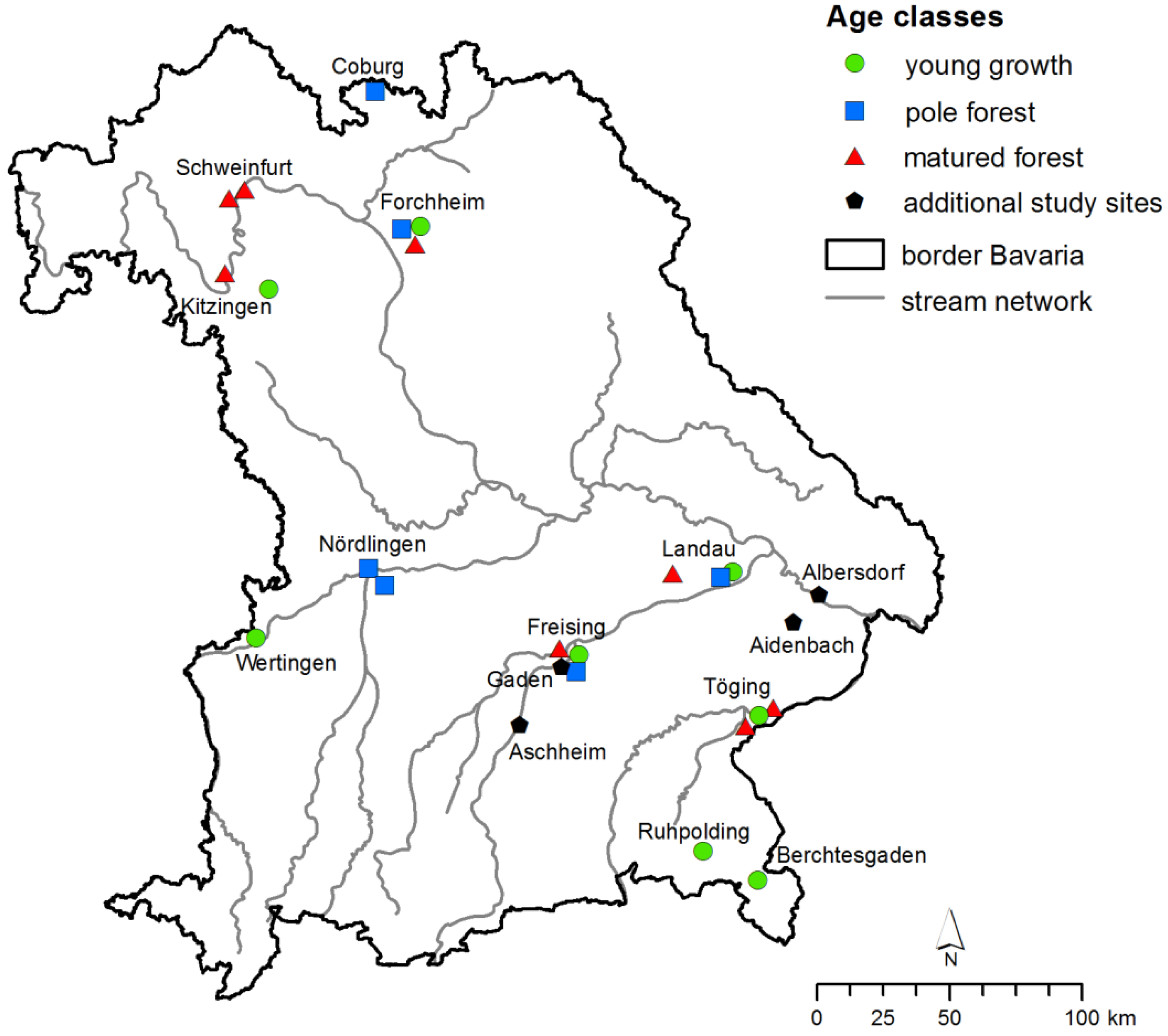
2.1. Study Sites

In 2009, 22 study sites distributed throughout south-eastern Germany were established for monitoring the ash dieback disease. The sites include eight matured stands, six pole stands and eight young growth stands (Figure 1). In 2010, the study sites were extended and additional trees were chosen for investigation. For better comparability, data are therefore shown beginning in 2010 with 230 trees in matured stands, 584 trees in pole stands and 579 trees in young growth stands.
Figure 1. The development of vitality was examined in 22 study sites from 2010 to 2014. Study sites can be distinguished into young growth, pole and matured forests. Four additional study sites were chosen in 2013 for a closer investigation of ash bark beetle- and Armillaria spp. infestation.
Forests 07 00041 g001
In 2013, four additional study sites already severely damaged by H. fraxineus, were established to investigate Armillaria spp. and ash bark beetles. They were located in Gaden (Ga), Aschheim (As), Aidenbach (Ai) and Albersdorf (Al) (Figure 1). The height above sea level slightly differed from 350 m for Aidenbach as lowest, to 493 m as the highest examined stand for Aschheim. Albersdorf at 396 m and Gaden at 425 m lie in between. The mean annual temperature is 8.3 °C. Mean annual precipitation ranges from 746 mm to 916 mm. In each stand, except for Gaden with 98 trees, 50 trees were chosen for further investigation.

2.2. Measurement of Vitality and Diameters at Breast Height

Numbering, measurement and vitality estimation with a special rating key classifying ash trees in different groups depending on their crown damage [23] were carried out for each study site. Trees belonging to the vitality classes 0–1 were less affected, vitality classes 2–3 mean moderate to serious disease symptoms, vitality class 4 trees are dying, vitality class 5 trees are already dead. The distinction between vitality class 4 and 5 was started in 2012. In 2010, in young growth stands the mean height of trees was 3 m; the mean diameter of trees at breast height (DBH) was 11.1 cm in pole stands and 38.3 cm in matured stands (Table 1).
In the additional study sites, the mean vitality was 2.15, the mean diameter of the trees in breast height was 14.5 cm (Table 2).
Table 1. Mean height and diameter of trees at breast height in the study sites, plot number indicated in brackets.
Young Growth StandsHeight (m)Pole ForestsDBH (cm)Matured ForestsDBH (cm)
Berchtesgaden (1)4.3Coburg (2)11.9Forchheim (3)26.6
Forchheim (5)2.5Forchheim (4)8.7Freising (6)37.9
Freising (8)2.1Freising (7)12.7Kitzingen (10)38.8
Kitzingen (11)2.4Landau (12)5.7Landau (14)35.8
Landau (13)2.4Nördlingen I (16)13.0Schweinfurt I (19)36.2
Ruhpolding (18)1.9Nördlingen II (17)14.7Schweinfurt II (20)33.0
Töging (23)4.2 Töging I (21)45.7
Wertingen (24)3.8 Töging II (22)52.0
Average3.011.138.3
Table 2. Mean vitality and diameter of trees at breast height in the additional study sites.
Pole ForestsMean VitalityMean DBH (cm)Minimum DBH (cm)Maximum DBH (cm)
Gaden3.1114.17.033.5
Aschheim1.8215.67.825.7
Aidenbach1,4616.68.922.7
Albersdorf2.1911.76.219.7
Average2.1514.57.525.4

2.3. Isolation and Cultivation of H. Fraxineus from Infected Branches

In young growth stands, infected branches with visible necrotic lesions from four to 10 different trees within the study site were collected and H. fraxineus was isolated as described previously [24]. From a total of 74 trees, 40 successful isolations on malt agar supplemented with antibiotic (20 g·L−1 malt extract, 100 mg·L−1 streptomycin and 15 g·L−1 agar) were performed. In earlier studies, the presence of the fungus was also confirmed by PCR analysis for six study sites [25]. In each pole stand, except for Freising, one tree was felled and samples from three symptomatic branches were used for DNA extraction. PCR analysis was performed as described in 2.5–2.6. For the pole stand in Freising, the fungal DNA was directly isolated out of fruiting bodies on the leaf litter. For mature stands, except for Freising, H. fraxineus was isolated from epicormic branches (Table 3). For the mature stand in Freising, DNA was isolated out of dead petioles.
Table 3. Detection of H. fraxineus in the study sites, plot number indicated in brackets.
Study SiteType of TissueNo. of SamplesCultivation/DetectionPCR
Young growth stands
Berchtesgaden (1)Infected branches108-
Forchheim (5)Infected branches42+*
Freising (8)Infected branches103+*
Kitzingen (11)Infected branches103+*
Landau (13)Infected branches106+*
Ruhpolding (18)Infected branches108-
Töging (23)Infected branches104+*
Wertingen (24)Infected branches106+*
Pole forests
Coburg (2)Infected branches31+
Forchheim (4)Infected branches31+
Freising (7)Fruiting bodies66+
Landau (12)Infected branches31+
Nördlingen I (16)Infected branches32+
Nördlingen II (17)Infected branches31+
Matured forests
Forchheim (3)Infected branches31+
Freising (6)Dead petioles43+
Kitzingen (10)Infected branches30-
Landau (14)Infected branches33+
Schweinfurt I (19)Infected branches32+
Schweinfurt II (20)Infected branches33+
Töging I (21)Infected branches31+
Töging II (22)Infected branches31+
(+*: detection performed in Gherghel et al. 2014).
The sampling in the mature forest in Kitzingen has to be repeated, because we could not detect the fungus out of the collected branches by PCR analysis. For all the other study sites, positive detections of the fungus were possible.

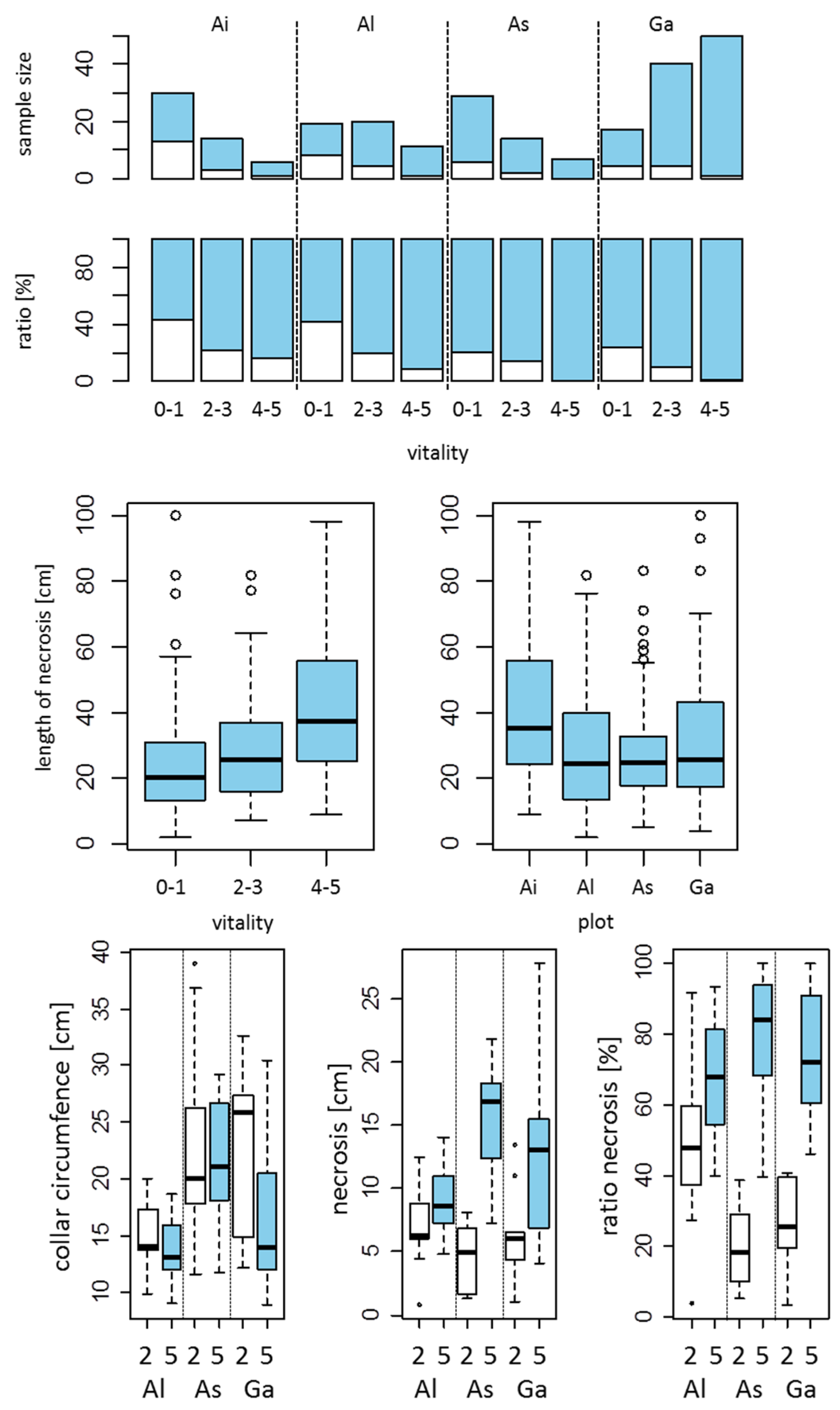
2.4. Measurements of Stem Necroses and Their Collar Circumferences

In four additional ash stands (Figure 1, black pentagons), lesions were measured by removing large portions of dying or dead bark from the root stem base following up the necroses using a bark peeler. The percentage of affected collar circumference and thus the extent of decay were determined. Only additional study sites allowed this type of sampling, because severely affected trees were destined for subsequent felling and therefore the massive wounding of the trees was justifiable.

2.5. Isolation of Armillaria spp. from Collar Lesions of Ash Trees

At the Gaden site, fungi were isolated from collar lesions of three trees in each of the four vitality classes, for a total of 12 trees. Before sampling the bark was removed with a sterilized knife. Wood pieces were pulverized with metal beads in a swing mill (Retsch MM200) at 25.0 Hz for 70 min. The DNA extraction kit Hi Yield Genomic DNA Mini Kit (Plant) from SLG (Süd-Laborbedarf, Germany) was used and DNA extraction was performed according to manufacturer´s protocol.

2.6. PCR Method

Following DNA extraction, the amplification was performed by polymerase chain reaction. An unspecific Armillaria region was amplified by using the primers already described [26]. For the detection of H. fraxineus, already published primers were used [27]. The PCR was performed in 25 µL volumes consisting of 12.5 µL Taq master mix, 5 µL of DNA, 1 µL of each 10µM primer and 5.5 µL milliQ water. Amplification was performed in a BioRad thermal cycler according to the following programme: initial denaturation followed by 34 cycles of denaturation at 94 °C for 30 s, annealing at 58 °C for 30 s and extension at 72 °C for 1 min, with a final extension at 72 °C for 7 min. The amplified products were separated on a 1% Agarose gel, stained with 5 µL Midori Green and visualized under UV light.

2.7. Assessment of Bark Beetle Infestations

Four additional ash stands (Figure 1, black pentagons) were examined for bark beetle attack. Trees of all vitality classes were screened for typical holes of maturation feeding and breeding galleries. By using a bark peeler, parts of the bark were removed und galleries became visible. In three ash stands (Ai, As, Ga) three trees belonging to the vitality class 2 were cut, respectively, and the number of maturation holes were counted on each 1 m segment. In Gaden, six trees belonging to the vitality class 5 were cut in two following years and the number and length of the breeding galleries were determined.

2.8. Calculations and Statistical Analysis

For the characterization of the pathology situation in a stand we calculated the percentage of the vitality classes. The classes were poled to groups: healthy trees belonging to the vitality classes 0 + 1, intermedium with the vitality classes 2 + 3 and dying or dead with the vitality classes 4 + 5. For these poled groups, the 95% confidence interval was calculated. For a comparison of the progress of the disease, the mean vitality per year and stand were calculated. For testing the proportion for several groups a chi-squared statistic without continuity correction was performed, using the R-function prop.test. For an analysis of the length of necrosis in relation to forest stand and poled vitality, a two-way analysis of variance was performed. The ratio length of necrosis/collar circumference was arcsinus transformed before variance analysis was conducted. Analyses and Graphics were performed using R [28]. The study site map is created with ArcGIS viewer.

3. Results

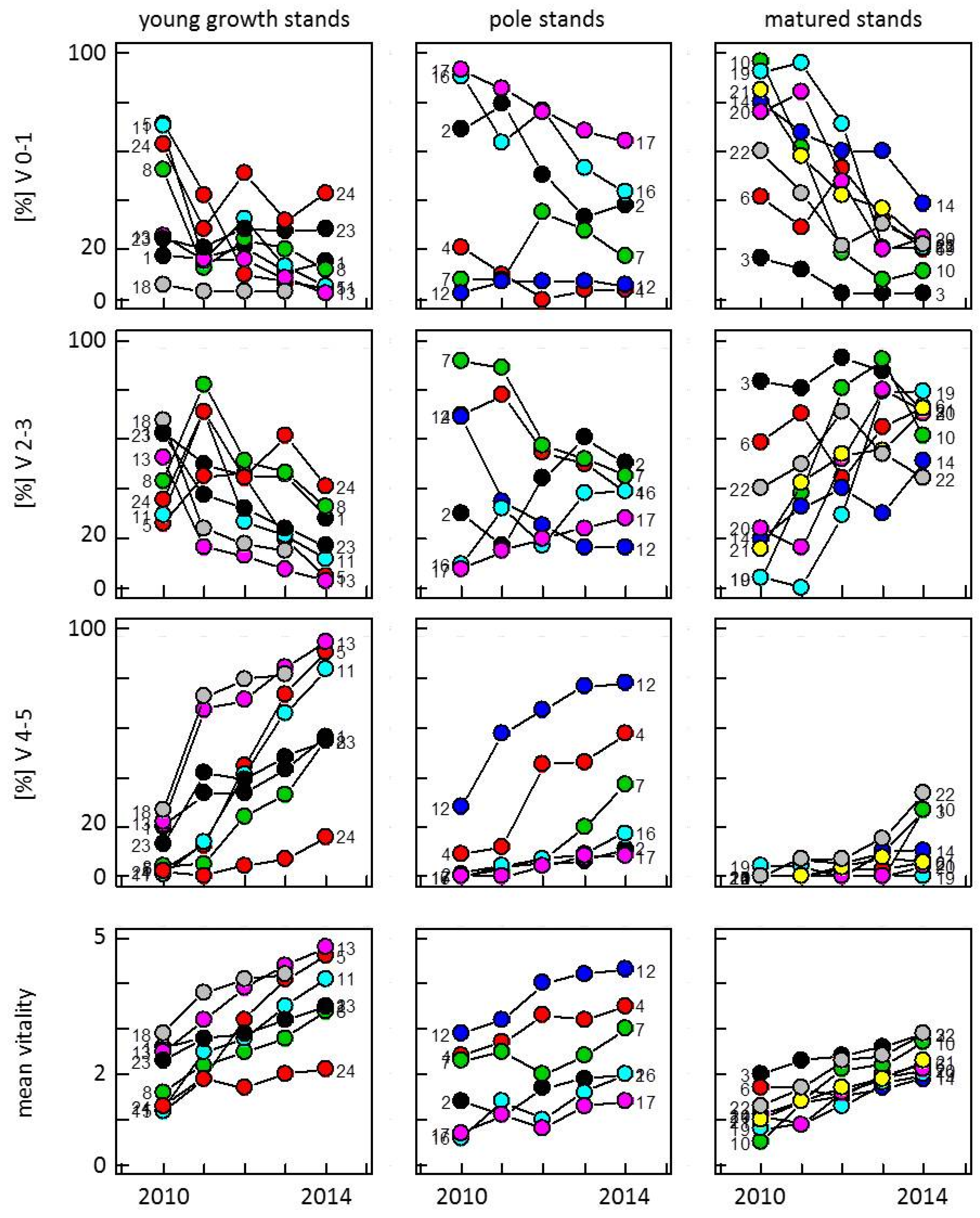
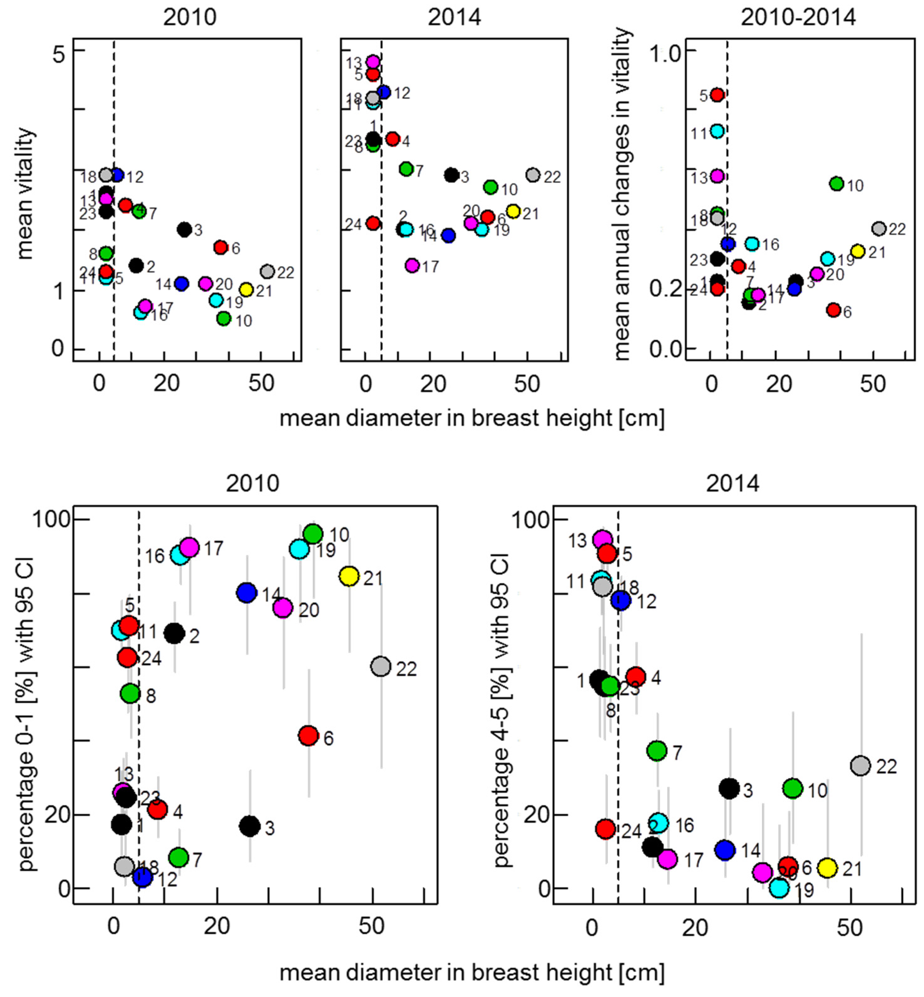
3.1. Development of Vitality in Ash Stands from 2010 to 2014

The development of the vitality of ash trees was analyzed in 22 ash stands, differing in age and habitat (Figure 1) to figure out to what extent the ash dieback disease is already established and if there is a different behavior in the progression of the disease in different regions of south-eastern Germany. Therefore, a special rating key using the vitality classes 0–5 described in Section 2.2 was applied. Typical symptoms of ash dieback, like crown defoliation or discolorations of branches could be observed in all examined study sites. Fruiting bodies were found to a large extent on ash petioles in the leaf litter in all vegetation periods. In all young growth stands, the presence of H. fraxineus as a causal agent of the disease was confirmed by its isolation and its subsequent cultivation out of infected branches (Table 3). Furthermore, PCR analysis from six isolates out of six study sites were performed and provided additional information on the genetic heterogeneity of the species [25]. For pole and matured forests, branches from one tree within each study site were harvested and with one exception the presence of the fungus was confirmed by PCR analysis (Table 3).
The results for four vegetation periods, started in 2010, are shown (Figure 2). At that time, no disease-free areas were found anymore. Therefore, we cannot give positive proof of how long the fungus had already been present in the different stands. As the degree of infestation differed significantly between the study sites, a comparison between them is rather difficult (comparison of the proportion of trees with vitality 0 and 1 per stand in 2010, x-squared = 555.93, Df = 21, p-value < 0.001). Nevertheless, we assume a nearly simultaneous infestation of the fungus in study sites located next to one another, as it is the case for Freising, Landau and Forchheim, for instance, each with one young growth, pole and matured forest.
Generally, mortality rates are high in young growth and pole stands, whereby there is a high divergence also within the age classes. With the exception of Wertingen (No. 24) that is located in the west and only shows 16% of trees that can be categorized in the vitality groups 4 and 5 in 2014, meaning that they are dying or already dead, more than 50% in Töging and Freising (No. 23, No. 8) and up to 95% of the trees in the remaining five young growth stands belong to this category.
In pole stands in 2014, the study sites in Landau, Forchheim and Freising (No. 12, 4 and 7) are massively affected by the disease with mortality rates ranging from 37% to 78%, whereby No. 12 and 4 are the youngest stands (DBH of 5.7 and 8.7), compared to the other study sites (Table 1).
In matured forests in 2014 three stands in Forchheim, Kitzingen and Töging (No. 3, 10, 22) have about 30% of trees belonging to the vitality classes 4–5 and further disease development has to be monitored in the future. Trees of the residual five stands show a drastic increase from the vitality classes 0–1 to 2–3 (Figure 2). Although the mean vitality in young growth stands in comparison to pole or matured forests was worse in 2010 (shown in Figure 3 in correlation to the diameter of the trees), differences between young growth stands in comparison to pole and matured forests regarding mean annual changes in vitality become obvious. The average decrease of vitality is much higher in most of the young growth stands compared to the older stands (Figure 3, upper row). Matured stands show a slightly higher alteration than pole stands. In 2010, with the exception of stands No. 3 and No. 6 the amount of trees in pole and matured stands show a high 0–1 ratio in comparison to the young growth stands with high variations. A high overlap of confidence intervals can be observed. In 2014, the overlap is not that high anymore and clear differences between young growth stands with older stands could be measured.
To our knowledge, for the first time these data demonstrate the longer period of time to die from the infection for older stands in comparison to young growth stands. In summary, regardless of the age and the habitat a decreasing vitality accompanied by an increasing mortality (Figure 2 and Figure 3) can be found in all ash stands. Surprisingly, there was no apparent correlation between the extent of mortality and the geographical position of the stands, although first disease symptoms could be observed in the south.
Figure 2. The amount of trees belonging to the vitality classes 0–1 (less affected), 2–3 (moderate symptoms) and 4–5 (dying or dead) is illustrated for young growth, pole- and matured stands from 2010 to 2014. The development of the mean vitality for each study site is shown at the bottom. Numbers indicate study sites (Table 1).
Forests 07 00041 g002
Figure 3. The mean vitality per stand in relation to the mean diameter at breast height (DBH) is illustrated for the years 2010 and 2014. As all the young growth stands were under a DBH of 5 cm, they were set to a DBH of 2.5 cm for illustration. The mean annual changes in vitality over the years from 2010 to 2014 in relation to the DBH are shown on the right site. At the bottom row, the percentage with 95% confidence interval of trees of the vitality class 0 and 1 for 2010 and of the vitality class 4 and 5 for 2014 in relation to the DBH is shown. Numbers indicate study sites (Table 1).
Forests 07 00041 g003

3.2. Screening for Potentially Resistant Trees

Increasing disease progression could be shown in all study sites and up to now no efficient treatments are known to cure or slow down this process. Therefore, special attention was drawn to those trees that, although under high infection pressure, still belong to the vitality classes 0–1 over the monitored period of at least four years. Based on the data shown in Figure 2, trees fulfilling these criteria were regarded as potentially resistant trees. The number of trees were determined in 2014 and are presented in Table 4. As we can assume from Figure 2, each study site has been infested for different amounts of time and the investigated period of time of four years is rather short. Therefore, we cannot compare resistance levels in different stands and we expect a high number of falsely identified resistant trees. The most likely candidate trees for resistance are expected to be trees from stands that have had the highest amount of disease pressure for the longest period of time. In young growth stands where the disease progression is fastest as it was expected, less tolerant trees could be found. With the exception of Wertingen, the average percentage of candidate tolerant trees lies close together at between 0% and 5%. In the pole stands No. 4, No. 7 and No. 12, for instance, only 1%–2% of candidate tolerant trees were left, suggesting a higher disease pressure for a longer period of time by comparison with the other pole stands that still show a range from 1% to 64%. Similar to the young growth stands, the stands in Nördlingen (No. 16, 17) located in the west have shown the most candidate tolerant trees up to now. In matured stands, where the disease progression proceeds more slowly, with the exception of Forchheim (No. 3) with no unaffected trees, candidate tolerant trees still exist between 11% and 36%. The still high number of trees belonging to the vitality classes 0–1 in pole and matured stands reflects that the disease in these areas has not progressed enough to be able to reliably identify candidate tolerant trees. It can be expected that their number decreases within the next years, as the infection pressure from H. fraxineus is still high and the disease progression has not come to a standstill yet.
Table 4. Age-dependent number of potentially resistant/tolerant trees that belong to the vitality classes 0–1 from 2010 to 2014. The percentage is calculated with the amount of trees found in 2014.
Young Growth StandsTolerant Treesn%
Berchtesgaden (1)2425
Forchheim (5)2773
Freising (8)1701
Kitzingen (11)2972
Landau (13)1781
Ruhpolding (18)0310
Töging (23)1572
Wertingen (24)84817
Sum175004
Pole standsTolerant treesn%
Coburg (2)289928
Forchheim (4)11081
Freising (7)1961
Landau (12)21242
Nördlingen (16)359836
Nördlingen (17)162564
Sum8355022
Matured standsTolerant treesn%
Forchheim (3)0410
Freising (6)43412
Kitzingen (10)32612
Landau (14)143936
Schweinfurt (19)52421
Schweinfurt (20)52421
Töging (21)41724
Töging (22)1911
Sum3621417
In addition to continued monitoring, to rigorously identify resistant trees, surviving trees should be propagated and replicated inoculation experiments performed or clone trials planted.

3.3. Extent of Armillaria Infection in Reference to the Lengths and Circumferences of Collar Lesions

In recent years, declining ash stands are not only affected by H. fraxineus itself, but also to an increasing amount by secondary pathogens. In the south and middle of the investigated area, infestations by Armillaria spp., as well as ash bark beetles became visible, whereas momentarily in the north of the country infections with these opportunistic pathogens are rare (own observations). Therefore, four additional study sites with a total of 248 trees were chosen in these areas where massive Armillaria infections could be observed (Figure 1) to clarify the role of these pathogens in the disease process. As the following experiments include extensive wounding of the trees, we could conduct measurements only on three stands that were destined for subsequent felling and did not include study sites that were used for long-term monitoring. In all four study sites, the stem discolorations at the stem base became apparent and were investigated regarding its length and Armillaria infections. Based on the occurrence of mycelial fans underneath the bark which are characteristic for Armillaria spp., and because of the occasional presence of rhizomorphs, it can be concluded that those trees were colonized by wood-inhabiting Armillaria spp. (Figure S1), although the possible co-existence of H. fraxineus within the lesions was not proven for each tree and should be included in further similar experiments.
Figure 4 shows the increase of Armillaria infestation with decreasing vitality (top row). A significant increase in the length of stem necroses could be observed in worse vitality groups (Figure 4, middle row: Df = 1, F = 28.9, p < 0.001), whereby the length of measured necroses was comparable between the different study sites (Df = 3, F = 2.53, p = 0.059). There was no significant interaction between plot and vitality (Df = 3, F = 1.03, p = 0.38).
Figure 4. Sample size and ratio of trees with Armillaria infestation (top row, blue) and without an infestation (white) are shown for four stands in 2013 (Ai–Aidenbach, Al–Albersdorf, As–Aschheim, Ga–Gaden, see Figure 1). The boxplots (middle row) show the length of stem necroses in relation to the vitality classes (left) and stands (right). Differences between vitality classes are statistically significant (two way ANOVA, vitality class: Df = 1, F = 28.9, p < 0.001, see text). The boxplots (bottom row) show the collar circumference of trees (left), the affected collar circumference by necroses (middle) and the ratio necroses/collar circumference of trees of the vitality class 2 (white) and 5 (blue) with 10 trees per vitality class. Differences between the vitality classes 2 and 5 in all three plots are statistically significant.
Forests 07 00041 g004
The mean collar circumference between the plots differed significantly (Df = 2, F = 11.3, p < 0.001) as well as in relation to vitality (Figure 4, bottom row, left: Df = 1, F = 4.87, p = 0.031). The collar circumference of trees in the vitality class 5 was smaller. There was no significant interaction between plot and vitality (Df = 2, F = 1.26, p = 0.29). Interestingly, a closer investigation of three ash stands shows some distinctions in the amount of collar rot between ash trees belonging to the vitality classes 2 and 5. Brownish to reddish discolorations were summed up at the stem base and show that the amount of discoloration, independent of collar circumference, increases significantly with decreasing vitality (Figure 4, bottom row, middle: vitality: Df = 1, F = 37.75, p < 0.001; interaction between plot and vitality: Df = 2, F = 6.93, p = 0.002). There are no significant differences in the amount of discoloration between the plots (Df = 2, F = 1.42, p = 0.25). Taking the size of collar circumference into account, using the percentage of necrotic tissue, the effect is amplified (Figure 4, bottom row, right: vitality: Df = 1, F = 62.3, p < 0.001; plot: Df = 2, F = 1.38, p = 0.26). Also, here we can see an interaction between plot and discoloration (Df = 2, F = 6.05, p = 0.004). That shows that not only the occurrence but above all the extension of the necrotic infection by Armillaria and probably H. fraxineus is relevant for the vitality and influences the progression of the disease.

3.4. Armillaria Conditioned Mortality

The extent of Armillaria infection and the impact of stem necroses on the vitality were shown on four different study sites in 2013. As three study sites underwent massive felling in the winter in 2013, Gaden was chosen for further investigations regarding the influence of these infections over a longer period of time. Figure 5 illustrates a decreasing vitality in Gaden (Ga) over a period of three years. By comparison with 2012 with 13% died back trees, a rapid increase in mortality can be observed, that is much higher than in the neighboring pole stand in Freising (No. 7, Fs), where momentarily with 17% only a small percentage of trees show necrotic symptoms at the stem base, although we cannot exclude earlier fungal infections in Gaden that would explain the difference. Necrotic lesions could be measured within all vitality classes (Figure 4). Samples from the trees belonging to the vitality classes 1–4 were taken from necrotic lesions and analyzed for H. fraxineus and Armillaria spp. Two trees belonging to the vitality class 2 were negative for both pathogens and have to be proven again on different sites of the necrosis to identify the inducer. Except from these two trees, in all other trees, both pathogens were detected positively (data not shown). Therefore, with these results the primary inducer of stem necroses cannot be determined and has to be proven in the future.
Figure 5. The development of ash vitality in Gaden (Ga) and Freising (Fs) from 2012 to 2014 is shown for trees belonging to the vitality classes 0–1, 2–3 and 4–5.
Forests 07 00041 g005
Nevertheless, earlier observations showed with less exceptions that trees normally experience a yearly decrease in vitality moving from one vitality class to the other, as can be found in the analysis of the data from the 22 study sites, exemplarily shown for Freising. For instance, also in Gaden, trees from the vitality class 4 changed into vitality class 5 most frequently (Table 5). Interestingly, the current data also show that the combination of Armillaria and H. fraxineus causes not only a yearly vitality decrease from one vitality class to the next class, but also further vitality changes (Table 5). Some trees belonging to the vitality classes 1, 2 or 3 dropped down to the vitality class 5 within one year. These data therefore indicate that the Armillaria infections accompanied by stem necroses contribute to accelerating ash mortality in weakened ash stands.
Table 5. Sum of dead ash trees in Gaden and Freising with rapid vitality changes in the years 2013 and 2014.
Vitality Dev.2012–20132013–2014
GadenFreisingGadenFreising
0–50000
1–54020
2–50140
3–54301
4–583133
5–51332910
Sum29104814

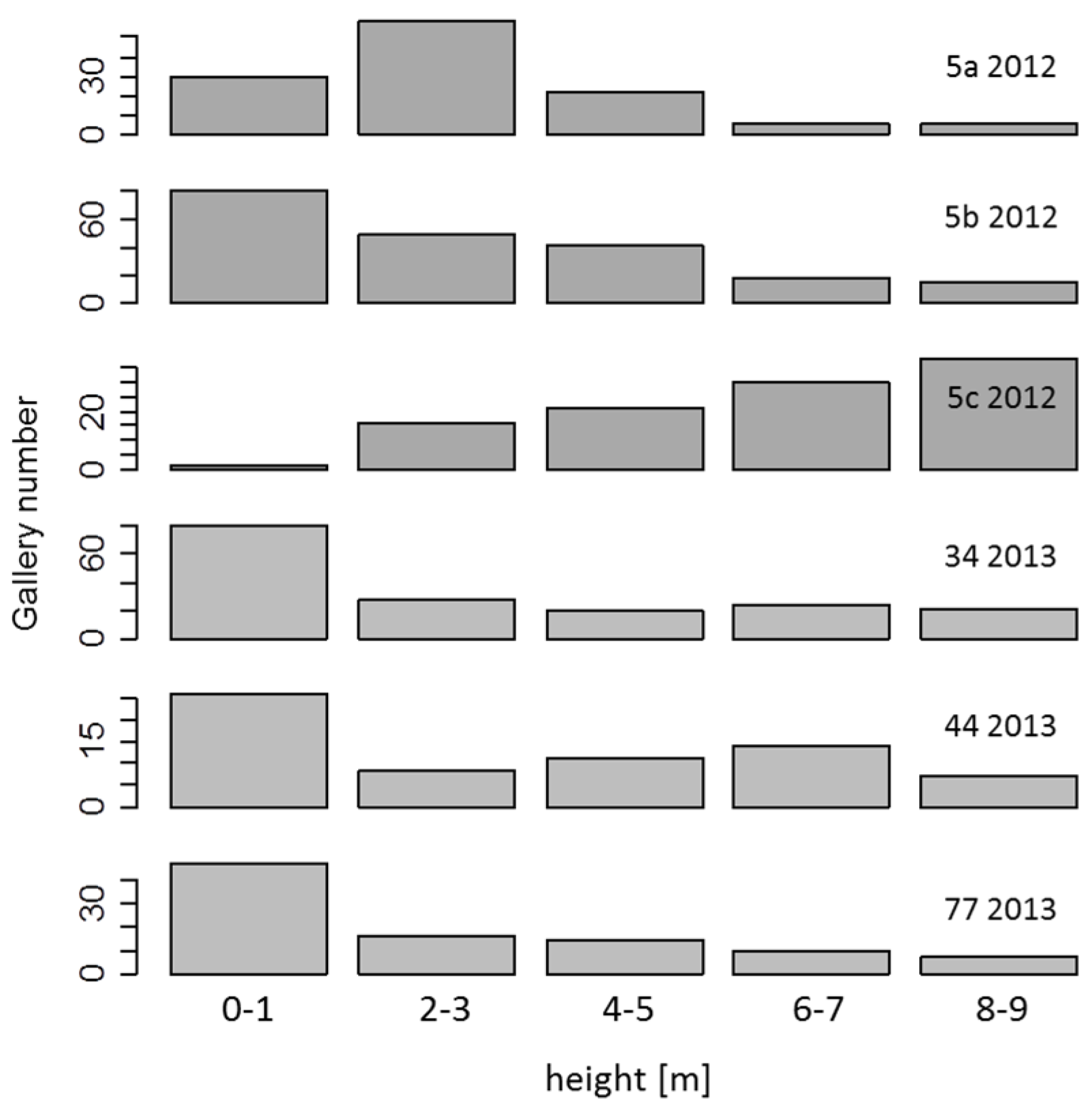
3.5. Breeding Galleries of Ash Bark Beetles Only in Recently Dead Trees

Before the occurrence of ash dieback, ash bark beetles were never considered as an economic forest health problem. Nevertheless, the high mortality within ash habitats increases the availability of breeding material for the ash bark beetles and so could have an influence on its population size and on the disease progression in H. fraxineus weakened ash stands, too. To prove the possibility of becoming primarily pests, the same four ash stands, investigated for Armillaria infection, were examined for bark beetle attack. Although the study site in Gaden was the most severely affected ash stand, ash bark beetles could be detected in the other study sites. Mostly Hylesinus fraxini was found, but also Hylesinus oleiperda. Maturation feeding could be observed over all vitality classes. In three ash stands (Aidenbach, Aschheim, Gaden), three trees belonging to the vitality class 2 were examined regarding the number of maturation holes. With decreasing height, the extent of maturation feeding has the tendency to increase (Figure S3a–c).
In all four examined ash stands, new breeding galleries could be detected only on trees belonging to the vitality class 5 (Table 6), and only on those trees which were still greenish under the bark (Figure S2). Trees that died earlier and were already brown and desiccated did not show any fresh breeding material. The number of galleries was counted for meter segments. Conspicuously, the number of galleries at the lower heights was greater than the number observed at the higher heights (Figure 6).
Figure 6. In relation to the height, the gallery numbers of six trees recently belonging to the vitality class 5 were determined in Gaden in two subsequent years. In 2012, the trees 5a–5c, and in 2013, the tress 34, 44 and 70 were measured.
Forests 07 00041 g006
Table 6. In four ash stands (Gaden (Ga), Aschheim (As), Aidenbach (Ai) and Albersdorf (Al)) the number of dead trees were counted and examined for gallery formation.
Feature/Sample AreaGaAsAiAl
Total amount of vitality 5-trees2913415
Greenish under the bark710214
Brownish under the bark22321
Recently generated breeding galleries710213
Vitality 5-trees with greenish bark and breeding galleries [%]10010010093
In addition to the number of total galleries on each tree, the length of each gallery was also determined. The medial gallery length was around 6 cm (Figure S4). These data indicate that Hylesinus fraxini still acts as a secondary pathogen unable to form breeding galleries on ash trees belonging to the vitality classes 0–4 but able to do so on trees that have recently died. They are therefore momentarily irrelevant for the progression of the disease in affected ash stands.

4. Discussion

Since its first identification in Germany in 2007 [29], the fungus H. fraxineus has spread nationwide. In south-eastern Germany, it was described for Bavaria in 2009 [30]. Since then, vitality in all ash stands has been observed to be continuously decreasing (Figure 2), and is still expected in the coming years, as the infection pressure has not lowered. At the beginning of the monitoring of the ash stands, no disease free areas were found, suggesting that since its arrival a fast spread of the disease has taken place accompanied by an intense development of the symptoms in the affected stands. Also, the degree of fungal infestation differed massively between the different study sites and it is impossible to determine retrospectively at which time the fungus arrived in each stand. We also could not find an apparent correlation between the extent of the disease and the geographical position of the stands, although first symptoms could be observed in the south. Nevertheless, we assume that in study sites located close together, a nearly simultaneous infestation of the fungus has occurred. By the analysis of the data regarding the mean annual changes in vitality, we could find notable differences between study sites that had stands of different age classes. By comparison with pole or matured stands, symptoms occur faster in young growth stands and the mortality rate is higher there (Figure 3). This can be explained by their smaller crowns and thinner stem diameters, where the fungus more quickly colonizes the whole tissue. The high mortality rates of up to 95% (Figure 2) reflect similar findings already known from Lithuania, where starting in 2005 progeny trials only showed 10% survival after eight years of constant infections with H. fraxineus [31]. Also in pole forests, a massive increase of mortality was monitored in three out of six study sites. Matured ash stands show a rapid decrease in healthy or less affected trees. The majority of the trees are moderate affected, belonging to the vitality classes 2–3 (Figure 2). The mortality within these stands is comparably low. Surprisingly, the mean annual changes of vitality were slightly higher in matured stands than in pole stands (Figure 3, top row). Therefore, we assume that the shift from the vitality classes 0–1 to the vitality classes 2–3 in matured stands proceeds faster than the shift from the vitality classes 2–3 to the vitality classes 4–5 in pole stands. In summary, these data clearly show for the first time the longer process of dying in older stands in comparison to young growth stands. It is important to follow up the disease, but, up to now, there has been a lack of clear trends in the damage of the trees that makes it difficult to predict the future development of the disease. At present, no successful treatments for the affected ash stands exist to cure or slow down the disease. For the conservation of the species, it is therefore important to screen for potentially resistant trees, which still could be found in all areas, except for two study sites (Table 4). Apparently healthy individuals stood alongside severely affected trees. As each study site has been infested by the fungus for different amounts of time, a comparison between the resistance levels is impossible. In time delayed infected ash stands, we expect also a high number of falsely identified resistant trees. The most reliable data can be obtined from the stands with the fastest disease progression. That means in young growth stands (Figure 3) and from the stands that were already massively impaired by the disease in 2010; for example, the pole stands No. 4, No. 7 and No. 12 (Figure 2). In young growth stands with only 4% and in the three mentioned pole stands with only 1%–2%, few potentially resistant candidate trees were left (Table 4). This relatively low number is comparable to the findings in other countries, where the disease has arrived earlier [8,32]. It thus seems that the total ash population has only a small fraction of genotypes that will enable it to withstand the disease [8,31,32,33]. Therefore, the advice of not removing healthy looking trees is necessary not only to be able to provide further recommendations for disease management to practitioners but also to find and protect potentially resistant ash trees for further natural or artificial selection following breeding in this area which is planned for the future.
In different countries, clones were already tested for susceptibility and it could be shown that there are no totally resistant clones, but that there are some individuals that remain in relatively good health. In southern Sweden, some clones exhibited reduced susceptibility and retained this resistance after six years under heavy infection pressure [33], thus giving hope to the likelihood of being able to conserve Fraxinus excelsior in Europe. Not only the screening for candidates is important but also the characterization of the underlying resistance mechanisms, to evaluate which genetic material can be utilized for further breeding [9] and to prove the robustness of the observed partial resistance when new virulent strains evolve. Also in south-eastern Germany [25] and other countries, a high level of fungal genetic variation was already found [34] and the virulence of the different strains has to be proven in the future. The survival of potentially resistant trees is endangered not only by the infections with new evolving virulent strains of H. fraxineus itself, but also by secondary pathogens, like the ash bark beetles or other species of Armillaria that can be increasingly found in weakened ash stands as it was proven in this study. There was some concern that the abundance of ash bark beetles increases drastically and that they could become primary pest agents [11,14]. Therefore, the role of ash bark beetles was examined in four study sites to prove if they act as primary or secondary pathogens in weakened ash stands. It could be shown that ash bark beetles are not playing a major role in the decline process at this juncture. Breeding galleries from H. fraxini can only be found in recently died back trees (Table 6). These findings are in line with observations made in Denmark, where ash trees that had died during spring were heavily colonized by bark beetles in the same growth season [7]. The amount of breeding galleries (Figure 6) as well as ash roses that appear as a consequence of maturation feeding (Figure S3) was mostly higher on the lower parts of the stem than near the top. Nevertheless, these insects should be carefully observed in the future and, if required, sanitation measures could be considered. Our findings will facilitate the detection of these beetles, because it was shown that they are not only found on the crown but also at lower heights.
A more serious problem occurs through Armillaria infections. These infections depend on the conditions of the ecosystem [35] and can cause major disease symptoms on woody plants and thus affect the ecosystem itself in its structure and function. The fungus is often associated with forest decline events and is generally considered as a contributing factor ultimately responsible for tree death following some other predisposing stresses [20]. Symptoms of the disease can be found on collar rots and were firstly described in Lithuania and Denmark [18,21,36], where ash dieback was found earlier. Recently, also reports from south-western Germany describe the disease symptoms [19]. Although it was shown that Armillaria acts as a secondary pathogen in affected ash stands [7,18], it is still difficult to distinguish if Armillaria is the primary or secondary agent responsible for the lesions at the stem base. In France, where still disease-free areas exist, Armillaria alone was never observed, which suggests a primary role for H. fraxineus in initial lesion formation [37]. However, ash trees were also found to belong to the vitality classes 0–1, which means less affected trees show stem necroses (Figure 4). These findings are in accordance with similar and earlier findings in Lithuania, where also colonization of the fungus on 80% of sound-looking trees was reported in [18]. This speaks against H. fraxineus as a primary inducer. Also in study sites in south-west Germany, a spatial dependence of collar rots was shown. This points to Armillaria as a primary inducer because of the infection path via roots and because it is not spatially independent through airborne ascospores [19]. In south-eastern Germany, no H. fraxineus free plots are available and can be examined regarding Armillaria infection alone. Investigated necrotic lesions of trees in Gaden were positive for both pathogens. Therefore, a higher amount of infected trees have to be tested in the future. Nevertheless, it can be suggested that high amounts of Armillaria inoculum in the soil, abundantly present through dying trees, facilitate the infection even of healthier trees. We could show that trees with the stem necroses also possess a significantly higher ash dieback intensity, which is in accordance with former results from different countries [7,19,36]. When starting the monitoring in 2009, as the infections became visible, the vast majority of affected ash trees showed a gradual decline from one vitality class to the next class (exemplarily shown for Freising). Three years later, in 2012, necrotic lesions and signs of Armillaria infection at the stem base became apparent in many study sites. Since then, vitality drops from several trees to more vitality classes have been observed (Table 5). Therefore, we conclude that Armillaria spp. contributes severely to the acceleration of ash mortality.
The high mortality in ash stands affected by H. fraxineus and Armillaria leads to economic losses and also compromises the survival of potentially resistant trees, as trees of all vitality classes are affected. For this reason, seeds of potentially resistant trees in severely affected ash stands should be harvested for further breeding and, taking our results into consideration, the planning of reforestation in the ash stands by using the resting shield of ash trees should be assigned more importance. The use of specifically selected alternative tree species must be considered to avoid tree gaps on a large scale. Also for forest practitioners, there must be an increased awareness of the significant risk when trees are falling spontaneously, because of their impaired stability.

Supplementary Files

Supplementary File 1

Acknowledgments

This work was funded by the Bavarian State Forest Administration (Project No. Z70). We also thank the student assistants Thomas Dichtl, Bernhard Loock and Anna Meindl who contributed to obtain the bark beetle field data and Britta Hausknecht for GIS data. Furthermore, we thank the involved forestry officers of the correspondent forest departments, and anonymous reviewers for valuable comments on the manuscript.

Author Contributions

Heike D. Lenz and Hannes Lemme conceived and designed the experiments and guided the field and laboratory research. Ludwig Straßer and Heike D. Lenz performed field sampling, Heike D. Lenz and Bernadett Bartha analyzed the data and wrote the paper.

Conflicts of Interest

The authors declare no conflict of interest.

References

  1. Baral, H.O.; Queloz, V.; Hosoya, T. Hymenoscyphus fraxineus, the correct scientific name for the fungus causing ash dieback in Europe. IMA Fungus 2014, 5, 79–80. [Google Scholar] [CrossRef] [PubMed]
  2. Zhao, Y.J.; Hosoya, T.; Baral, H.O.; Hosaka, K.; Kakishima, M. Hymenoscyphus pseudoalbidus, the correct name for Lambertella albida reported from Japan. Mycotaxon 2013, 122, 25–41. [Google Scholar] [CrossRef]
  3. Zheng, H.D.; Zhuang, W.Y. Hymenoscyphus albidoides sp. nov. and H. pseudoalbidus from China. Mycol. Prog. 2014, 13, 625–638. [Google Scholar] [CrossRef]
  4. Han, J.G.; Shrestha, B.; Hosoya, T.; Lee, K.H.; Sung, G.H.; Shin, H.D. First report of the ash dieback pathogen Hymenoscyphus fraxineus in Korea. Mycobiology 2014, 42, 391–396. [Google Scholar] [CrossRef] [PubMed]
  5. Queloz, V.; Grünig, C.R.; Berndt, R.; Kowalski, T.; Sieber, T.N.; Holdenrieder, O. Cryptic speciation in Hymenoscyphus albidus. For. Pathol. 2011, 41, 133–142. [Google Scholar] [CrossRef]
  6. Timmermann, V.; Borja, I.; Hietala, A.M.; Kirisits, T.; Solheim, H. Ash dieback: Pathogen spread and diurnal patterns of ascospore dispersal, with special emphasis on Norway. EPPO Bull. 2011, 41, 14–20. [Google Scholar] [CrossRef]
  7. Skovsgaard, J.P.; Thomsen, I.M.; Skovgaard, I.M.; Martinussen, T. Associations among symptoms of dieback in even-aged stands of ash (Fraxinus excelsior L.). For. Pathol. 2010, 40, 7–18. [Google Scholar] [CrossRef]
  8. Kjær, E.D.; McKinney, L.V.; Nielsen, L.R.; Hansen, L.N.; Hansen, J.K. Adaptive potential of ash (Fraxinus excelsior) populations against the novel emerging pathogen Hymenoscyphus pseudoalbidus. Evol. Appl. 2012, 5, 219–228. [Google Scholar] [CrossRef] [PubMed]
  9. McKinney, L.V.; Thomsen, I.M.; Kjaer, E.D.; Nielsen, L.R. Genetic resistance to Hymenoscyphus pseudoalbidus limits fungal growth and symptom occurrence in Fraxinus excelsior. For. Pathol. 2012, 42, 69–74. [Google Scholar] [CrossRef]
  10. Metzler, B.; Baumann, M.; Baier, U.; Heydeck, P.; Bressem, U.; Lenz, H. Bundesweite Zusammenstellung: Handlungsempfehlungen beim Eschentriebsterben. AFZ-Der Wald 2013, 68, 17–20. [Google Scholar]
  11. Kunca, A.; Leontovyc, R.; Zúbrik, M.; Gubka, A. Bark beetle outbreak on weakened ash trees and applied control measures. EPPO Bull. 2011, 41, 11–13. [Google Scholar] [CrossRef]
  12. Schedl, K.E. Scolytidae. In Die Käfer Mitteleuropas: Band 10; Freude, H., Harde, K.W., Lohse, G.A., Eds.; Goecke & Evers Verlag: Krefeld, Germany, 1981; pp. 34–99. [Google Scholar]
  13. Sauvard, D. General biology of bark beetles. In Bark and Wood Boring Insects in Living Trees in Europe, a Synthesis; Lieutier, F., Day, K.R., Battisti, A., Grégoire, J.C., Evans, H.F., Eds.; Springer Netherlands: Dordrecht, The Netherlands, 2004; pp. 63–88. [Google Scholar]
  14. Pfister, A. Aktuelle Schäden durch Eschenbastkäfer in der Steiermark. Forstsch. Aktuell 2012, 54, 22–25. [Google Scholar]
  15. Keča, N.; Klopfenstein, N.B.; Kim, M.S.; Solheim, H.; Woodward, S. Initial characterization of an unidentified Armillaria isolate from Serbia using LSU-IGS1 and TEF-1-α genes. For. Pathol. 2015, 45, 120–126. [Google Scholar] [CrossRef]
  16. Longa, C.M.O.; La Porta, N. Rapid identification of Armillaria species by PCR-DGGE. J. Microbiol. Methods 2014, 107, 63–65. [Google Scholar] [CrossRef] [PubMed]
  17. Prospero, S.; Holdenrieder, O.; Rigling, D. Comparison of the virulence of Armillaria cepistipes and Armillaria ostoyae on four Norway spruce provenances. For. Pathol. 2004, 34, 1–14. [Google Scholar] [CrossRef]
  18. Lygis, V.; Vasiliauskas, R.; Larsson, K.H.; Stenlid, J. Wood-inhabiting fungi in stems of Fraxinus excelsior in declining ash stands of northern Lithuania, with particular reference to Armillaria cepistipes. Scand. J. For. Res. 2005, 20, 337–346. [Google Scholar] [CrossRef]
  19. Enderle, R.; Peters, F.; Nakou, A.; Metzler, B. Temporal development of ash dieback symptoms and spatial distribution of collar rots in a provenance trial of Fraxinus excelsior. Eur. J. For. Res. 2013, 132, 865–876. [Google Scholar] [CrossRef]
  20. Kelley, M.B.; Fierke, M.K.; Stephen, F.M. Identification and distribution of Armillaria species associated with an oak decline event in the Arkansas Ozarks. For. Pathol. 2009, 39, 397–404. [Google Scholar] [CrossRef]
  21. Solla, A.; Tomlinson, F.; Woodward, S. Penetration of Picea sitchensis root bark by Armillaria mellea, Armillaria ostoyae and Heterobasidion annosum. For. Pathol. 2002, 32, 55–70. [Google Scholar] [CrossRef]
  22. Roll-Hansen, F. The Armillaria species in Europe: A literature review. Eur. J. For. Pathol. 1985, 15, 22–31. [Google Scholar] [CrossRef]
  23. Lenz, H.D.; Straßer, L.; Baumann, M.; Baier, U. Boniturschlüssel zur Einstufung der Vitalität von Alteschen. AFZ-DerWald 2012, 3, 18–19. [Google Scholar]
  24. Kowalski, T. Chalara fraxinea sp. nov. associated with dieback of ash (Fraxinus excelsior) in Poland. For. Pathol. 2006, 36, 264–270. [Google Scholar] [CrossRef]
  25. Gherghel, F.; Fussi, B.; Donges, K.; Haustein, M.; Jakob, K.M.; Müller, K.; Piškur, B.; Hauptman, T.; Lenz, H.D.; Konnert, M.; et al. The development of a species-specific test to detect Hymenoschyphus pseudoalbidus in ash tissues. For. Pathol. 2014, 44, 137–144. [Google Scholar] [CrossRef]
  26. Maphosa, L.; Wingfield, B.D.; Coetzee, M.P.A.; Mwenje, E.; Wingfield, M.J. Phylogenetic relationships among Armillaria species inferred from partial elongation factor 1-alpha DNA sequence data. Australas. Plant Pathol. 2006, 35, 513–520. [Google Scholar] [CrossRef]
  27. Johansson, S.B.K.; Vasaitis, R.; Ihrmark, K.; Barklund, P.; Stenlid, J. Detection of Chalara fraxinea from tissue of Fraxinus excelsior using species-specific ITS primers. For. Pathol. 2010, 40, 111–115. [Google Scholar] [CrossRef]
  28. R Core Team. R: A Language and Environment for Statistical Computing; R Foundation for Statistical Computing: Vienna, Austria, 2013. [Google Scholar]
  29. Schumacher, J.; Wulf, A.; Leonhard, S. Erster Nachweis von Chalara fraxinea T. Kowalski in Deutschland- ein Verursacher neuartiger Schäden an Eschen. Deutsch. Pflanzenschutzd. 2007, 59, 121–123. [Google Scholar]
  30. Leonhard, S.; Straßer, L.; Nannig, A.; Blaschke, M.; Schumacher, J.; Immler, T. Neues Krankheitsphänomen an der Esche: Das von Chalara fraxinea verursachte Eschentriebsterben ist auch in Bayern nachgewiesen. LWF Aktuell 2009, 71, 60–63. [Google Scholar]
  31. Pliura, A.; Lygis, V.; Suchockas, V.; Bartkevicius, E. Performance of twenty four European Fraxinus excelsior populations in three Lithuanian progeny trials with a special emphasis on resistance to Chalara fraxinea. Balt. For. 2011, 17, 17–34. [Google Scholar]
  32. McKinney, L.V.; Nielsen, L.R.; Hansen, J.K.; Kjaer, E.D. Presence of natural genetic resistance in Fraxinus excelsior (Oleraceae) to Chalara fraxinea (Ascomycota): An emerging infectious disease. Heredity 2011, 106, 788–797. [Google Scholar] [CrossRef] [PubMed]
  33. Stener, L.G. Clonal differences in susceptibility to the dieback of Fraxinus excelsior in southern Sweden. Scand. J. For. Res. 2013, 28, 205–216. [Google Scholar] [CrossRef]
  34. Rytkönen, A.; Lilja, A.; Drenkhan, R.; Gaitnieks, T.; Hantula, J. First record of Chalara fraxinea in Finland and genetic variation among isolates sampled from Aland, mainland Finland, Estonia and Latvia. For. Pathol. 2011, 41, 169–174. [Google Scholar] [CrossRef]
  35. Entry, J.A.; Martin, N.E.; Cromack, K.; Stafford, S.G. Light and nutrient limitation in Pinus monticola: Seedling susceptibility to Armillaria infection. For. Ecol. Manag. 1986, 17, 189–198. [Google Scholar] [CrossRef]
  36. Bakys, R.; Vasiliauskas, A.; Ihrmark, K.; Stenlid, J.; Menkis, A.; Vasaitis, R. Root rot, associated fungi and their impact on health condition of declining Fraxinus excelsior stands in Lithuania. Scand. J. For. Res. 2011, 26, 128–135. [Google Scholar] [CrossRef]
  37. Husson, C.; Caël, O.; Grandjean, J.P.; Nageleisen, L.M.; Marçais, B. Occurrence of Hymenoscyphus pseudoalbidus on infected ash logs. Plant Pathol. 2012, 61, 889–895. [Google Scholar] [CrossRef]

