Article

Physiological and Biochemical Responses of Idesia polycarpa to Botryosphaeria dothidea Infection at Different Stages of Stem Canker Disease

1
College of Forestry, Henan Agricultural University, Zhengzhou 450046, China
2
National Forestry and Grassland Administration Key Laboratory for Central Plains Forest Resources Cultivation, Zhengzhou 450046, China
3
Henan Province Engineering Technology Research Center for Idesia, Zhengzhou 450046, China
4
College of Urban Construction, Zhejiang Shuren University, Shaoxing 312028, China
5
Henan Vocational College of Agriculture, Zhengzhou 450046, China
6
Zhengzhou New Development Greening Management and Operation Co., Ltd., Zhengzhou 450046, China
*
Authors to whom correspondence should be addressed.
These authors contributed equally to this work.
Forests 2025, 16(9), 1411; https://doi.org/10.3390/f16091411
Submission received: 11 August 2025 / Revised: 29 August 2025 / Accepted: 1 September 2025 / Published: 3 September 2025

Abstract

Botryosphaeria dothidea (Moug. ex Fr.) Ces. & De Not. is a major pathogenic fungus causing stem canker in Idesia polycarpa, posing a significant threat to the growth and survival of its plantations. To elucidate the physiological and biochemical responses of the host under pathogenic stress, this study used two-year-old potted seedlings of I. polycarpa (provenance: ‘Emeishan No. 1’) and conducted artificial inoculation. Dynamic changes in physiological and biochemical indices at different disease stages were systematically monitored. The results revealed a distinct stage-specific response pattern: in the early infection stage, the activities of antioxidant enzymes (POD and SOD) increased rapidly, accompanied by significant elevations in osmotic regulators such as proline and soluble protein. In the mid-stage, malondialdehyde (MDA) content increased, while the levels of photosynthetic pigments—especially chlorophyll b and carotenoids—declined, indicating intensified oxidative damage. In the late stage, antioxidant capacity and osmotic adjustment weakened, and the photosynthetic system was continuously impaired. Correlation analysis further demonstrated significant synergistic relationships among antioxidant defense, membrane stability, osmotic regulation, and photosynthetic function. These findings enhance our understanding of the disease resistance mechanisms in I. polycarpa and provide a theoretical and practical reference for resistance evaluation and precise management of canker disease in woody species.

1. Introduction

Canker disease is a typical stem and branch disease that primarily affects the trunks and lateral branches of trees. Mild infections lead to lesion formation on the bark, impairing normal growth, while severe infections may result in dieback of upper branches or even entire plant death [1]. Historically, the causative pathogens have been known to attack broad-leaved species such as Populus przewalskii Maxim., Robinia pseudoacacia L., and Vernicia fordii (Hemsl.) Airy Shaw. In recent years, however, infections have also been reported on certain coniferous species, indicating a broadening host range [2]. This disease progresses rapidly and has strong spreading ability, posing a significant threat to forest production, especially during the early stages of afforestation, where it can lead to large-scale seedling mortality and even total reforestation failure during spring planting seasons [3]. Studies have shown that canker disease significantly affects host plants at multiple levels, including external morphological symptoms and internal physiological, biochemical, and nutritional indices, across different infection stages. During the initial phase, pathogen invasion weakens host vitality, causing round or elliptical brown lesions that gradually expand. As the disease progresses, infected tissues undergo dehydration and necrosis, often accompanied by the formation of pathogen fruiting bodies in later stages. Anatomical observations reveal that infected bark and xylem tissues exhibit pronounced browning and structural disintegration [4].
Idesia polycarpa Maxim. is a deciduous tree belonging to the family Flacourtiaceae. It is widely distributed and highly adaptable, with strong resistance to cold, drought, and poor soils, making it a valuable species for ecological restoration and afforestation programs [5,6]. Its fruits are rich in unsaturated fatty acids such as linoleic acid and alpha-linolenic acid, as well as bioactive compounds including vitamin E, squalene, and polyphenols, conferring high nutritional and medicinal value [7]. The wood of I. polycarpa is lightweight, decay-resistant, and finely textured, making it useful for furniture and construction. In addition, the fruit and seed oil have recognized therapeutic benefits, particularly for cardiovascular conditions such as hypertension and coronary heart disease [8]. In recent years, I. polycarpa has been incorporated into the national food management system in China, and its industry is rapidly expanding with promising market potential [9].
Currently, I. polycarpa is under extensive artificial cultivation in China, yet stem canker disease continues to intensify annually, severely limiting plantation development. Previous research has identified Botryosphaeria dothidea (Moug. ex Fr.) Ces. & De Not. as the primary pathogen responsible for stem canker in I. polycarpa [10]. Previous studies on woody plants have reported physiological and biochemical responses to B. dothidea infection, but most of these investigations were limited to single time points or generalized descriptions of host responses, lacking stage-specific resolution of the full process from defense activation to decline. To elucidate the plant’s response mechanisms under this pathogen-induced stress—a typical form of environmental stress—this study used two-year-old potted I. polycarpa seedlings and conducted artificial inoculation with B. dothidea. We systematically investigated changes in chlorophyll content, osmotic regulatory substances, and antioxidant enzyme activities at different stages of infection. This study aims to clarify the physiological regulation and response patterns of I. polycarpa under canker stress and provide a theoretical foundation and data support for targeted disease management and resistance breeding.

2. Materials and Methods

2.1. Experimental Materials

The experimental materials were cultivated at the Forestry Experimental Station of Henan Agricultural University, located in Zhengzhou, Henan Province, China (113°38′ E, 34°47′ N). This region has a distinct temperate climate with four distinct seasons, characterized by a maximum temperature of 43 °C, a minimum temperature of −17.9 °C, and an annual average temperature of 14.2 °C. The average annual precipitation is approximately 650.1 mm, with a frost-free period of 215 days and an annual sunshine duration of about 2400 h. The area belongs to a typical semi-arid to semi-humid continental monsoon climate zone. These values are based on long-term meteorological data from 1981 to 2010 provided by the China Meteorological Data Service Center (CMDC) [11]. In the later stage of the experiment, the materials were transferred to a controlled growth chamber in the Tree Physiology and Ecology Laboratory of Henan Agricultural University (113°82′ E, 34°79′ N). The geographical location of the planting and experimental sites is shown in Figure 1.

2.1.1. Experimental Plants

The experimental plants were two-year-old seedlings of I. polycarpa ‘Emeishan No. 1’, grown from seeds. In March 2021, seeds were mixed with moist sand and sown in nursery pots filled with a 1:1 (v/v) mixture of vermiculite and peat soil. The substrate had the following characteristics: available nitrogen 1.32 g·kg−1, available phosphorus 0.022 g·kg−1, available potassium 1.29 g·kg−1, pH 6.45, and maximum field water-holding capacity of 65%. In May 2022, the seedlings were transplanted into plastic pots (outer diameter: 29.6 cm, inner diameter: 25.4 cm, height: 19.7 cm, bottom diameter: 17.8 cm). In April 2023, uniform and healthy seedlings with no visible pests or diseases (average height: 55.8 cm; average stem base diameter: 1.2 cm) were selected and transferred to a closed growth chamber. Full-spectrum LED plant growth lights (100 W@220 V) were used for supplemental illumination under controlled conditions of a 16 h light/8 h dark photoperiod, light intensity of approximately 200 μmol·m−2·s−1 (PAR), relative humidity of 70–75%, and a constant temperature of 25 ± 3 °C. After one month of acclimation, the seedlings were used for subsequent experiments.

2.1.2. Pathogen Strain

The pathogen strain used in this study was B. dothidea, provided by the Laboratory of Tree Physiology and Ecology, Henan Agricultural University [12]. The strain was transferred from slant cultures to PDA plates and incubated in the dark at 28 °C for activation. Mycelial plugs (5.00 mm in diameter) were excised from the actively growing edges of the colonies using a sterile punch and used for inoculation.

2.2. Experimental Methods

2.2.1. Pathogen Inoculation

Uniformly growing I. polycarpa seedlings were selected for inoculation. A wound with a diameter of approximately 5.00 mm was made on one side of the stem, about 20 cm above the base, using a sterile scalpel. A mycelial plug of B. dothidea (activated on PDA) was placed onto the wound, then covered with sterile cotton and sealed with Parafilm. For the control group, sterile Potato dextrose agar (PDA) plugs of the same size were applied instead. All plants were maintained in a closed growth chamber under full-spectrum plant growth lights. Regular watering and daily misting with sterile water at the inoculation site were performed to maintain high humidity and promote fungal infection.

2.2.2. Sample Collection and Pretreatment

On the 7th, 14th, 21st, 28th, 35th, 42nd, 49th, and 56th days after inoculation with B. dothidea, 3 stem segment samples were collected from each of the experimental and control groups (a total of 48 plants). A 20 cm stem section, including the inoculation site, was excised, immediately wrapped in aluminum foil, labeled, and flash-frozen in liquid nitrogen for 1 min before being stored at −80 °C for subsequent physiological and biochemical analysis. Three biological replicates were maintained for each time point.

2.2.3. Indicator Measurement Methods

Unless otherwise specified, all physiological and biochemical measurements were performed with three biological replicates (n = 3). For each biological replicate, three technical replicates were conducted, and the mean value was used for statistical analysis.
Pathogenicity Assessment
To accurately assess the pathogenicity of B. dothidea and monitor disease progression in I. polycarpa, experimental plants were divided into two groups: one group (20 plants) was used for non-destructive, continuous observation of disease symptoms and grading, while the other was used for destructive sampling at each time point for physiological and biochemical measurements (three replicates per treatment, 48 plants in total).
Disease grading was performed every 7 days after inoculation based on a modified five-grade scale adapted from Chen et al. [13] (Table 1). The classification criteria were as follows:
  • Grade I: Callus formation without visible lesions;
  • Grade II: Lesions with browning of the stem 1–3 cm around the inoculation site;
  • Grade III: Browning exceeding 3 cm from the inoculation site;
  • Grade IV: Browning extending along the main stem;
  • Grade V: Browning of both the main stem and upper branches, with symptoms such as leaf wilting and bark exfoliation.
Disease incidence (%) was calculated as:
Disease   incidence = Number   of   diseased   plants Number   of   inoculated   plants × 100 %
Disease severity index (%) was calculated using the formula:
Disease   severity   index = Number   of   plants   at   each   grade × Corresponding   grade   value Total   number   of   investigated   plants × Maximum   grade   value × 100 %
Note: The plants used for destructive sampling (see Section 2.2.2) were solely used for physiological and biochemical measurements and were not included in the disease severity evaluation.
Determination of Chlorophyll and Carotenoid Contents
The contents of chlorophyll and carotenoids in stem tissues were determined using an ethanol–acetone extraction method. After removing surface impurities, the periderm and xylem were carefully scraped with a sterile scalpel, and the sample was cut into small fragments. A 0.1 g portion was weighed and immersed in 10 mL of 95% ethanol–acetone mixture (v/v = 1:1) in a 10 mL centrifuge tube. The extraction was conducted at room temperature in the dark for 36 h, with gentle manual shaking every 8 h. To ensure accuracy, a blank control (distilled water) and a known concentration chlorophyll standard (Sigma) were used for colorimetric calibration. Absorbance was measured using a UV-Vis spectrophotometer, with appropriate wavelengths set at 663, 646, and 470 nm. The error margin was calculated based on the standard deviation (SD) from technical replicates.
The pigment concentrations were calculated using the following equations:
Chlorophyll a (mg·L−1) = 12.7 × A663 − 2.69 × A646
Chlorophyll b (mg·L−1) = 22.9 × A646 − 4.68 × A663
Total chlorophyll (mg·L−1) = Chl a + Chl b
Carotenoids (mg·L−1) = (1000 × A470 − 2.05 × Chl a − 114.8 × Chl b)/245
Note: where A663, A646, and A470 are the absorbance values of the extract at wavelengths of 663, 646, and 470 nm, respectively. Chl a and Chl b represent the concentrations of chlorophyll a and chlorophyll b calculated from Equations (3) and (4).
Determination of Lipid Peroxidation Products and Osmoregulatory Substances
Malondialdehyde (MDA) content was determined using the thiobarbituric acid (TBA) colorimetric method.
Soluble sugar (SS) content was measured by the anthrone colorimetric method.
Soluble protein (SP) content was determined using the Coomassie Brilliant Blue G-250 dye-binding method.
Proline (Pro) content was quantified using the ninhydrin colorimetric method.
Determination of Antioxidant Enzyme Activities
Superoxide dismutase (SOD) activity was assessed by the nitro blue tetrazolium (NBT) photochemical reduction method.
Peroxidase (POD) activity was measured using the guaiacol oxidation method.
The detection methods and calculation formulas for all physiological indicators in this study were based on the Plant Physiological Index Detection Techniques by Xuekui Wang [14].

2.2.4. Data Analysis

Data were organized and preprocessed using Microsoft Excel 2019. One-way analysis of variance (ANOVA) was performed using SPSS 26.0 to evaluate the differences in physiological and biochemical indicators between treatments and controls at different infection stages. Graphs were generated using Origin 2021, and the geographical mapping of experimental sites was conducted using ArcGIS (version 10.8, Esri, Redlands, CA, USA).
Prior to correlation analysis, all physiological and biochemical data were tested for normality using the Shapiro–Wilk test and for homogeneity of variance using Levene’s test. Variables that met the assumptions of normality were analyzed using Pearson’s correlation, whereas non-normally distributed variables were log-transformed. When assumptions were not satisfied after transformation, Spearman’s rank correlation was used instead. Pearson correlation coefficients were calculated across samples pooled over all time points. Correlations are associative and do not establish causality.

3. Results

3.1. Disease Symptoms

On the 7th day after inoculation, no visible disease symptoms were observed on any seedlings. On the 14th day, some plants began to develop lesions, with bark browning observed within a 1–3 cm radius around the inoculation site. A total of 16 and 4 seedlings were classified as Grade I and Grade II, respectively, with no symptoms of Grade III or higher. On the 21st day, browning expanded beyond 3 cm around the inoculation site in some seedlings. Eleven, six, and three seedlings were assessed as Grade I, II, and III, respectively, with no occurrences of Grade IV or V. On the 28th day, lesions had expanded, and extensive browning was observed around the inoculation site. Six, six, and eight seedlings were classified as Grade I, II, and III, respectively, indicating disease progression but still no Grade IV or V symptoms. On the 35th day, the lesions darkened and deepened, with widespread browning and liquid exudation observed on some stems. The number of seedlings in Grades I, II, III, and IV was 3, 6, 10, and 1, respectively. On the 42nd day, most seedlings exhibited extensive browning of the main stem accompanied by liquid exudation, and the periderm peeled off in some cases. The number of seedlings in Grades I–V was 2, 5, 11, 1, and 1, respectively. On the 49th day, the disease continued to progress, with most stems showing extensive browning and exudation; partial periderm detachment was observed, and a few branches also showed signs of periderm peeling. The number of affected seedlings in Grades I–V was 1, 1, 11, 6, and 1, respectively. On the 56th day, all seedlings exhibited significantly expanded lesions with exudation. Partial detachment of the periderm occurred along the main stem, browning of upper branches was noted, and some seedlings displayed leaf wilting symptoms. The numbers of seedlings at Grades I, III, IV, and V were 1, 5, 11, and 3, respectively. The specific incidence rate and disease index are shown in Table 2.

3.2. Effects of B. dothidea Inoculation on Photosynthetic Pigments

To investigate the effects of B. dothidea infection on the photosynthetic pigments of I. polycarpa, we monitored the dynamic changes in the contents of chlorophyll a, chlorophyll b, total chlorophyll, and carotenoids in both the inoculated and control groups across different infection stages.
Chlorophyll a: As shown in Figure 2a, the content of chlorophyll a in the inoculated group significantly declined from the 7th day post-inoculation and was markedly lower than that in the control group on the 21st day (p < 0.001). Although a slight recovery was observed afterward, the levels remained significantly lower than the control on the 35th day and the 49th day (p < 0.05 to p < 0.001), indicating substantial inhibition of chlorophyll a during the mid- to late stages of infection.
Chlorophyll b: Figure 2b illustrates a sharp decrease in chlorophyll b content in the inoculated group on the 28th day, with extremely significant differences from the control (p < 0.001). This timing coincides with the visually observed aggravation of disease symptoms, suggesting that chlorophyll b is highly sensitive to pathogen infection.
Total chlorophyll: As depicted in Figure 2c, total chlorophyll content exhibited a general declining trend, with a slight rebound at later stages. From the 28th to 49th days, the content in the inoculated group was significantly lower than that of the control group (p < 0.05 to p < 0.001), indicating that the photosynthetic pigment system of I. polycarpa was severely impaired during the middle and late stages of the disease.
Carotenoids: As shown in Figure 2d, carotenoid content in the inoculated group was consistently and significantly lower than that of the control group throughout the inoculation period (p < 0.01 to p < 0.001), with particularly significant differences observed on the 14th–the 21st days and the 42nd–the 56th days. This indicates that pathogen stress may cause persistent suppression of carotenoid biosynthesis or stability.
In summary, I. polycarpa exhibited a general decline in photosynthetic pigment content under pathogen stress, with chlorophyll b and carotenoids showing greater sensitivity to infection. These results suggest that B. dothidea may interfere with photosynthesis by inhibiting pigment synthesis, promoting pigment degradation, or damaging thylakoid membrane structures.

3.3. Effects of B. dothidea Inoculation on Lipid Peroxidation and Osmoregulatory Substances

To investigate the effects of B. dothidea infection on lipid peroxidation and osmoregulatory compounds in I. polycarpa, we analyzed the dynamic changes in soluble protein (SP), soluble sugar (SS), proline (PRO), and malondialdehyde (MDA) contents at different infection stages in both inoculated and control groups.
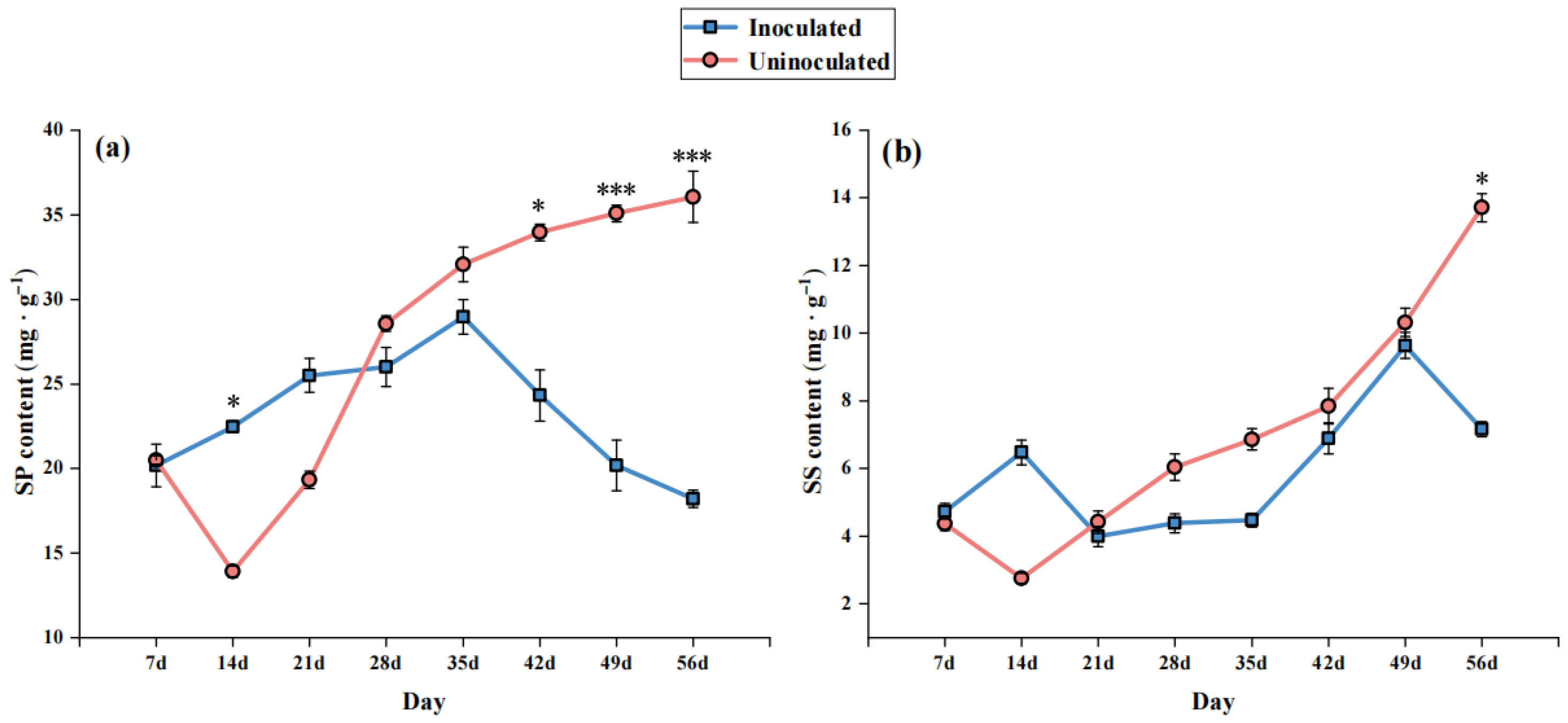
As shown in Figure 3a, SP content remained nearly unchanged on the 7th day post-inoculation. On the 14th day, the control group exhibited significantly lower SP content than the inoculated group (p < 0.05). After the 21st day, SP content in the control group continued to increase. The SP content in the inoculated group peaked on the 35 day and then declined steadily, showing the greatest difference from the control on the 49th day and the 56th day (p < 0.001). These results suggest that SP synthesis in the inoculated group was inhibited as disease progressed, weakening osmoregulatory capacity.
As shown in Figure 3b, SS content remained relatively stable from the 7th day to the 35th day in both groups. From the 35th day to the 49th day, SS levels continued to rise in both groups. On the 56th day, the control group showed significantly higher SS content than the inoculated group (p < 0.05), indicating that the capacity for SS accumulation in infected plants declined with increasing disease severity, possibly due to metabolic imbalance or disruption of synthesis pathways.
According to Figure 4, the proline (Pro) content in the inoculated group gradually increased starting from the 7th day post-inoculation and peaked on the 21st day, showing a significant increase compared with the control group (p < 0.05), indicating that proline synthesis was induced at the early stage of infection. Subsequently, Pro content declined and reached a level similar to the control on the 35th day. It then increased again and exhibited a highly significant difference on the 49th day (p < 0.01). on the 56th day, the Pro content slightly decreased but remained significantly higher than the control (p < 0.05).
As shown in Figure 5, malondialdehyde (MDA), a key marker of lipid peroxidation, reflects the degree of cell membrane damage. From the 7th day to the 42nd day post-inoculation, MDA levels in both groups showed a continuous upward trend, with the inoculated group maintaining significantly higher MDA content than the control (p < 0.05), reaching a peak on the 42nd day. Thereafter, the MDA level gradually decreased and approached that of the control by the 56th day, indicating that infection by the pathogen induced severe oxidative damage to the cellular membrane system.

3.4. Effect of B. dothidea Inoculation on Antioxidant Enzyme Activity

To investigate the effect of B. dothidea infection on the antioxidant enzyme system of I. polycarpa, the activities of peroxidase (POD) and superoxide dismutase (SOD) were measured at different infection stages. As shown in Figure 6, POD activity increased after the 7th day of inoculation, reaching a highly significant difference compared to the control on the 14th day (p < 0.001), and peaked on the 35th day, followed by a decreasing trend. SOD activity also showed an increasing trend from the 7th day to the 21st day, reaching a very significant difference on the 14th day (p < 0.01), with minor fluctuations thereafter. Both enzyme activities declined on the 49th day and the 56th day, indicating a weakened antioxidant enzyme response during the later stages of infection.

3.5. Correlation Analysis of Physiological and Biochemical Indicators

To further explore the synergistic or antagonistic relationships among physiological and biochemical parameters in I. polycarpa under B. dothidea stress, a Pearson correlation analysis was conducted using samples collected across different disease stages (Figure 7). Pearson’s correlation analysis was applied because most variables met normality assumptions after transformation, and it allowed us to quantify linear relationships among physiological indicators. For variables that did not meet these assumptions, Spearman’s correlation was also tested, yielding consistent patterns. Therefore, Pearson results were reported for clarity. The results revealed multiple significant correlations among the parameters, indicating their intrinsic interdependence during disease progression.
POD activity was significantly negatively correlated with proline (Pro) content and total chlorophyll (Chl) content (p < 0.05 and p < 0.001, respectively) and exhibited a highly significant negative correlation with SOD activity (r > 0.8, p < 0.001), suggesting a potential antagonistic regulation between these two antioxidant enzymes. Photosynthetic pigment indices showed pronounced negative correlations with both antioxidant and membrane lipid peroxidation-related indicators. Notably, total chlorophyll was strongly negatively correlated with POD activity and MDA content (p < 0.001), while carotenoids (Car) were significantly negatively correlated with MDA (p < 0.05). These results suggest that under enhanced oxidative stress and osmotic adjustment, the photosynthetic system is inhibited, pigment biosynthesis and stability are disrupted, and this is a key physiological manifestation of declining plant vitality.
In summary, the correlation analysis supports a stage-specific coordinated regulation among antioxidant defense, osmotic adjustment, membrane stability, and photosynthesis inhibition. This provides critical insight into the stress response network of I. polycarpa under canker disease pressure.

4. Discussion

4.1. Stage-Specific Physiological Response Mechanisms

I. polycarpa exhibited distinct stage-specific physiological responses under B. dothidea infection stress, which can be divided into three phases: early defense activation (the 7th day–the 14th day), intermediate coordinated regulation (the 21st day–the 35th day), and late functional decline (the 42th day–the 56th day). The timing of individual disease grades was confirmed based on the first appearance of visible symptoms at each grade, as summarized in Table 2.
In the early stage, I. polycarpa rapidly activated its antioxidant defense system, showing significantly increased POD and SOD activities along with elevated levels of osmotic adjustment substances such as proline and soluble proteins. This pattern is consistent with an acute defense response that may be linked to early ROS bursts following pathogen recognition [15,16]; however, our correlative data do not demonstrate this mechanism. During the intermediate stage, several physiological parameters (e.g., Pro, SP, MDA) peaked, indicating sustained defense and repair functions. Osmotic substances played key roles in maintaining cell water potential and membrane integrity. However, the sharp decline in photosynthetic pigments suggested that chloroplast functions were impaired, with metabolic resources likely being redirected from growth to defense [17].
In the late stage, antioxidant enzyme activities declined while MDA levels continued to rise, indicating intensified membrane lipid peroxidation and a weakening resistance system [18]. Simultaneously, chlorophyll and carotenoid contents dropped significantly, reflecting severe photosynthetic damage associated with rapid disease progression [19].
This triphasic response model—early activation, mid-stage coordination, and late imbalance—provides theoretical support for future resistance breeding and targeted disease control strategies in I. polycarpa.

4.2. Changes in Antioxidant Activity: Establishment and Decline of Early Defense

Upon pathogen infection, excessive reactive oxygen species (ROS) can damage cell membranes, proteins, and DNA. Thus, activating antioxidant enzymes to scavenge ROS is a vital defense mechanism [20]. Among them, POD and SOD serve as a frontline defense maintaining ROS homeostasis [21].
In this study, POD activity increased sharply by the 7th day post-inoculation and peaked at the 14th day (p < 0.001), while SOD activity also peaked on the 14th day (p < 0.01), indicating rapid activation of antioxidant defenses. However, both enzymes declined after the 42th day and the 21st day post-inoculation, respectively, with a pronounced decrease on the 49th–56th days, suggesting that the antioxidant system of I. polycarpa may exhibit diminished responsiveness during the late stage of infection under sustained pathogen pressure. This observation is consistent with previous reports; for example, Citrus reticulata infected by Colletotrichum gloeosporioides also displayed a “rise-then-fall” pattern in antioxidant activity [22].
These trends suggest that early increases in POD and SOD may help constrain oxidative stress, whereas their subsequent decline could be associated with elevated ROS and cellular damage, molecular confirmation is required.

4.3. MDA Accumulation: Marker of Oxidative Damage and Membrane Disruption

Malondialdehyde (MDA) is a key lipid peroxidation product that indicates the degree of oxidative damage to cellular membranes [23]. In this study, MDA levels steadily increased after B. dothidea inoculation, peaking at the 42nd day. Although levels later declined, they remained significantly higher than controls, suggesting prolonged oxidative stress. MDA levels were negatively correlated with antioxidant enzyme activity, implying that reduced antioxidant capacity aggravated membrane damage. This supports previous findings in apple and other woody plants, which suggest that MDA levels are a reliable physiological indicator of disease progression [24].

4.4. Osmotic Adjustment Substances: Early Adaptive Response and Late Metabolic Disruption

Plants typically accumulate osmolytes like proline, soluble proteins, and sugars to alleviate osmotic stress and scavenge ROS during environmental stress [25]. Here, these substances showed an early-to-mid increase followed by a decline at later stages. Proline peaked at the 21st day, but synthesis was inhibited by the 28th–35th days, indicating a breakdown in osmotic regulation. SP and SS also showed late-stage reductions, with SP displaying extremely significant differences compared to the control group by the 49th–56th days.
These findings suggest a transition from temporary adaptation to metabolic dysfunction as stress intensifies, reflecting disrupted synthesis pathways and weakened resistance systems [26].

4.5. Photosynthetic Pigments: Structural Damage and Physiological Decline

Photosynthetic pigments, including chlorophyll a, b, and carotenoids, declined under pathogen stress, especially in the mid-to-late disease stages. Chlorophyll b and carotenoids were particularly sensitive, aligning with observations of leaf yellowing and wilting in infected tissues [27]. Chlorophyll degradation is often induced by ROS accumulation, thylakoid membrane damage, and enzymatic degradation, while carotenoids contribute to ROS detoxification and membrane stability [28,29]. Their decline further weakens photosynthetic and antioxidant systems.
Thus, pigment loss reflects both structural and functional deterioration of the photosynthetic apparatus and may serve as candidate physiological indicators of disease progression pending field validation and cross-tissue verification [30]. Coupled with hyperspectral imaging or remote sensing, this offers prospects for early, non-destructive disease detection.

4.6. Coordinated Patterns Among Physiological Indicators: Consistency with a Systemic Stress Network

POD and SOD activities showed a highly significant negative correlation (p < 0.001), suggesting possible functional complementarity or temporal regulation between these two antioxidant enzymes [31]. As a marker of membrane lipid peroxidation, MDA was significantly positively correlated with osmotic adjustment substances such as Pro and SP, indicating that I. polycarpa generally initiates osmotic adjustment to buffer stress following membrane damage [32]. Meanwhile, chlorophyll was significantly negatively correlated with POD and MDA, and carotenoids were also negatively correlated with MDA (p < 0.05), implying that the photosynthetic system is readily perturbed by stress and that such inhibition is a key manifestation of physiological decline.
The correlations we observed are consistent with a coordinated response network linking antioxidant activity, osmotic adjustment, membrane stability, and photosynthetic inhibition, and they align with key features reported for Pyrus spp. and Malus domestica; studies in Populus spp. further underscore Botryosphaeria-induced constraints on photosynthesis. Cross-host comparisons strengthen the generality of our interpretation while highlighting the species-specific nuances captured in I. polycarpa.

4.7. Novelty and Limitations

Building on related studies in fruit trees, this work focuses on I. polycarpa, an ecologically and industrially important woody oil species, and—within a unified framework—jointly tracks disease severity, pigments, osmolytes, lipid peroxidation, and antioxidant enzyme activities. It reveals a stage-resolved response pattern characterized by early activation of antioxidant enzymes and mid- to late-stage declines in osmotic adjustment accompanied by damage to the photosynthetic apparatus. These species-specific data enrich the pathophysiological understanding of stem canker in I. polycarpa and offer incremental novelty and practical value. Nevertheless, certain limitations remain. All experiments were conducted under controlled pot conditions, which may not fully capture the complexity of field ecological environments. Therefore, future studies should incorporate long-term, multi-environment field trials to validate and extend our findings. Since the experiments were conducted in a closed environment free from other microbial infections, pathogen re-isolation from infected tissues was not performed. Pathogen identity was confirmed based solely on morphological characteristics, meaning that Koch’s postulates were not fully satisfied. Although the consistent appearance of typical symptoms strongly supports the pathogenicity of B. dothidea, future studies should include re-isolation and molecular validation to rigorously confirm the infection.
In addition, our correlation analysis is descriptive and does not establish causality; therefore, the proposed mechanisms remain hypotheses in the absence of molecular evidence. Future studies should integrate gene expression and signaling analyses (e.g., hormone and ROS pathways) together with multi-omics approaches to elucidate the resistance regulatory network. Meanwhile, our findings provide physiological indicators to inform the screening and evaluation of resistant genotypes and the design of biocontrol strategies; we recommend validation with larger sample sizes, multiple pathogen contexts, and under natural field conditions to enhance practical applicability and robustness.

5. Conclusions

This study systematically explored the dynamic changes in physiological and biochemical parameters of I. polycarpa under infection stress by B. dothidea, revealing a stage-specific response mechanism during disease progression. The results showed that the canker disease in I. polycarpa exhibited a clear phased pattern of development. From the 14th day after inoculation, both disease incidence and severity index increased gradually, peaking between the 49th day and the 56th day, accompanied by rapid lesion expansion, severe stem browning, and aggravated leaf wilting.
In the early stage of infection, the activities of peroxidase (POD) and superoxide dismutase (SOD) increased significantly, indicating a rapid activation of the antioxidant defense system to scavenge excess reactive oxygen species (ROS). However, in the mid-to-late stages, enzyme activities declined significantly, weakening the antioxidant capacity and exposing the cell membrane system to sustained oxidative stress. Correspondingly, malondialdehyde (MDA), a marker of lipid peroxidation, continued to accumulate and peaked at the 42nd day, indicating intensified membrane damage.
Osmoregulatory substances such as proline (Pro), soluble protein (SP), and soluble sugars (SS) increased during the early to mid-infection stages, playing key roles in osmotic pressure regulation, membrane stabilization, and protein protection. However, they declined markedly in the later stages, suggesting metabolic disturbance and reduced regulatory capacity. Meanwhile, photosynthetic pigment contents declined throughout the infection period, with chlorophyll b and carotenoids showing greater sensitivity to pathogen stress, indicating structural and functional damage to the photosynthetic system and overall physiological decline.
Correlation analysis further revealed significant synergistic relationships among these physiological parameters, outlining a coordinated response pattern of “antioxidant defense–osmotic adjustment–membrane stability–photosynthetic inhibition”.
In summary, I. polycarpa exhibited a typical three-stage defense response to canker stress: early-stage defense activation, mid-stage coordinated regulation, and late-stage system decline. This study deepens our understanding of the physiological defense mechanisms of I. polycarpa against fungal infection and provides critical theoretical guidance and key physiological indicators for future screening of resistant genotypes and the development of precise disease control strategies.

Author Contributions

Conceptualization, Z.L. (Zhi Li) and T.Z.; methodology, Q.Y., Y.Z., Y.Y., C.M., W.Z., C.C. and Z.H.; software, Q.Y., Y.Z., Y.Y., C.M., W.Z., C.C. and Z.H.; validation, Q.Y., Y.Z., Y.Y., C.M., W.Z., C.C. and Z.H.; formal analysis, Q.Y., Y.Z., Y.Y., C.M., W.Z., C.C., S.Y., L.D., Z.H., H.Z., T.Z. and Z.L. (Zhi Li); investigation, Q.Y., Y.Z., Y.Y., C.M., W.Z., C.C. and Z.H.; resources, Q.Y., Y.Z., Y.Y., C.M., W.Z., C.C., Z.L. (Zhen Liu), Y.W., X.G., Q.C., L.D., J.W., Y.R., F.L., S.Y., H.Z., T.Z. and Z.L. (Zhi Li); data curation, Q.Y., Y.Z., Y.Y., C.M., W.Z., C.C. and Z.H.; writing—original draft preparation, Q.Y., Y.Z., T.Z. and Z.L. (Zhi Li); writing—review and editing, Z.L. (Zhen Liu), Y.W., X.G., Q.C., L.D., J.W., Y.R., F.L., S.Y., H.Z., T.Z. and Z.L. (Zhi Li); visualization, Q.Y. and Y.Z.; supervision, Z.L. (Zhi Li) and T.Z.; project administration, Z.L. (Zhi Li) and T.Z.; funding acquisition, Z.L. (Zhi Li) and T.Z. All authors have read and agreed to the published version of the manuscript.

Funding

This research was funded by the Postgraduate Education Reform and Quality Improvement Project of Henan Province (YJS2024AL062/YJS2025AL63), the Henan Provincial Social Sciences Federation Research Project in China (SKL-2024-2179), the Henan Agricultural University’s “Millions of Sciences and Education Service Action” Fund Project (2024SFBQW30), and the Henan Province Postdoctoral Research Project of China (202002053). Zhejiang Shuren University Basic Scientific Research Special Funds (2025XZ034). Key Specialty Project of Ordinary Colleges and Universities in Shaoxing City, Zhejiang Province, “Landscape Architecture” (SXSZY202412).

Data Availability Statement

The datasets generated during and/or analyzed during the current study are available from the corresponding author upon reasonable request.

Acknowledgments

We thank all our laboratory members for their help with this experiment. Our gratitude extends to our lab team members, who assisted with this endeavor.

Conflicts of Interest

Author Shunyang Yao was employed by the company Zhengzhou New Development Greening Management and Operation Co., Ltd. The remaining authors declare that the research was conducted in the absence of any commercial or financial relationships that could be construed as a potential conflict of interest.

References

  1. Guo, L.; Chang, J.; He, W.; Yang, Y.; Zhang, L.; Yang, X.; Xie, S. Preliminary study on the occurrence pattern of poplar canker disease in the field. Pract. For. Technol. 2012, 10, 38–40. [Google Scholar]
  2. Kovač, M.; Diminić, D.; Orlović, S.; Zlatković, M. Botryosphaeria dothidea and Neofusicoccum yunnanense Causing Canker and Die-Back of Sequoiadendron giganteum in Croatia. Forests 2021, 12, 695. [Google Scholar] [CrossRef]
  3. Kumar, A.M.; Sharma, R.M.; Dubey, A.K.; Awasthi, O.P.; Singh, D.; Dahuja, A.; Mitra, S.V.A.C.R.; Kumar, A. Effect of weather parameters and citrus genotypes on the occurrence of citrus canker incited by Xanthomonas citri pv. citri. Indian Phytopathol. 2023, 76, 605–613. [Google Scholar] [CrossRef]
  4. Lin, F.; Chen, Z.; Deng, J.; Wang, K. Estimation of nitrogen content in rice leaves using Fourier transform mid-infrared spectroscopy. Plant Nutr. Fertil. Sci. 2009, 15, 750–755. [Google Scholar]
  5. Luo, M.; Yao, Y.; Gong, J.; Feng, A.; Zhang, W.; Yang, W.; Wang, W.; Zhao, J.; Wang, L.; Zhang, L. Investigation of bitter compounds in Idesia polycarpa and identification of associated biosynthetic genes. Ind. Crops Prod. 2025, 232, 121285. [Google Scholar] [CrossRef]
  6. Luo, X.; Liu, Y.; Lei, Y.; He, Z.; Gong, X.; Ye, M.; Xiao, Q. Genetic Diversity Analysis and Polyploid Induction Identification of Idesia polycarpa. Plants 2024, 13, 3394. [Google Scholar] [CrossRef]
  7. Tian, X.; Fang, X.; Du, M. Study on nutritional quality and antioxidant capacity of oils from different parts of Idesia polycarpa fruit. J. Chin. Cereals Oils Assoc. 2020, 35, 91–95. [Google Scholar]
  8. Yang, Y.; Miao, C.; Yuan, Q.; Zhong, W.; Hu, Z.; Chen, C.; Liu, Z.; Wang, Y.; Geng, X.; Cai, Q.; et al. Changes in the quality of Idesia polycarpa Maxim fruits from different ecotypes during the growth process. Plants 2025, 14, 2324. [Google Scholar] [CrossRef]
  9. Chen, Z.; Ai, Q.J. A new “aerial oil depot” of China is emerging—Summary of the symposium on the oil quality, value, and market prospects of oil grape (Idesia polycarpa). China For. Ind. 2019, Z2, 63–67. [Google Scholar]
  10. China Meteorological Data Service Center (CMDC). Daily Climate Data for Zhengzhou (1981–2010). Available online: http://data.cma.cn (accessed on 15 August 2025).
  11. Fang, L.; Zheng, T.; Feng, J.; Zhi, W.; Wang, Y.; Li, Z.; Cai, Q.; Geng, X.; Liu, Z. Botryosphaeria dothidea causes stem canker of Idesia polycarpa in China. Plant Pathol. 2024, 73, 1372–1381. [Google Scholar] [CrossRef]
  12. Feng, J.; Yuan, Q.; Chen, X.; Fang, L.; Zhang, T.; Liu, Z.; Wang, Y.; Geng, X.; Cai, Q.; Li, Z. Identification and chemical control of stem canker pathogen of Idesia polycarpa. Plants 2025, 14, 1393. [Google Scholar] [CrossRef]
  13. Chen, Y.; Hu, Q.; Li, Y.; Lian, H.; Li, Y. The Relativity Investigation Among Frost Injury and Dry not of Pomegranate Pomegranate in Zhengzhou; Research Progress on Pomegranate in China(I); Preparatory Group of Pomegranate Branch of Chinese Horticultural Society, Henan Agricultural University: Zhengzhou, China, 2010. [Google Scholar]
  14. Wang, X. Principles and Techniques of Plant Physiology and Biochemistry Experiments; Higher Education Press: Beijing, China, 2006. [Google Scholar]
  15. Shi, J.; Tong, R.; Yao, J.; Wang, S.; Wang, S.; Li, J.; Song, C.; Zhang, K.; Jiao, J.; Wang, M.; et al. Controlled atmosphere storage with high CO2 concentration extends storage life of fresh pomegranate fruit by regulating antioxidant capacity and respiration metabolism. BMC Plant Biol. 2025, 25, 684. [Google Scholar] [CrossRef]
  16. Mhamdi, A.; Van Breusegem, F. Reactive oxygen species in plant development. Development 2012, 139, 3901–3904. [Google Scholar] [CrossRef] [PubMed]
  17. Li, X.; Liu, J.; Hu, D.; Liu, G.; Chen, L.; Li, B.; Du, W.; Song, B. Characterization of Maize Germplasm Resistance to Common Smut and Analysis of Physiological Differences. Sci. Agric. Sin. 2025, 58, 2504–2521. [Google Scholar]
  18. Wang, Y.; Zhang, Y.; Fan, J.; Li, H.; Chen, Q.; Yin, H.; Qi, K.; Xie, Z.; Zhu, N.; Sun, X.; et al. Physiological and autophagy evaluation of different pear varieties (Pyrus spp.) in response to Botryosphaeria dothidea infection. Tree Physiol. 2024, 44, tpad139. [Google Scholar] [CrossRef] [PubMed]
  19. Liu, H.; Wu, Y.; Chen, G. Changes in antioxidative enzymes and membrane lipid peroxidation in grape leaves infected with Sclerotinia sclerotiorum. Hortic. Plant J. 2021, 7, 51–60. [Google Scholar]
  20. Kumar, S.; Shukla, V.; Tripathi, Y.N.; Aamir, M.; Divyanshu, K.; Yadav, M.; Upadhyay, R.S. Biochemical changes, antioxidative profile, and efficacy of the bio-stimulant in plant defense response against Sclerotinia sclerotiorum in common bean (Phasaeolus vulgaris L.). Heliyon 2024, 10, e23030. [Google Scholar] [CrossRef]
  21. Gill, S.S.; Tuteja, N. Reactive oxygen species and antioxidant machinery in abiotic stress tolerance in crop plants. Plant Physiol. Biochem. 2010, 48, 909–930. [Google Scholar] [CrossRef]
  22. Zhou, Y.; Li, S.; Zeng, K. Exogenous nitric oxide-induced postharvest disease resistance in citrus fruit to Colletotrichum gloeosporioide. J. Sci. Food Agric. 2016, 96, 505–512. [Google Scholar] [CrossRef]
  23. Qi, A.; Liu, C.; Lü, D.; Chen, H.; Li, Y.; Wang, A.; Shi, R. Physiological responses and cold resistance evaluation of Sapindus mukorossi seedlings from four provenances under low temperature stress. J. Cent. South Univ. For. Technol. 2024, 44, 46–54. [Google Scholar]
  24. Li, S. Physiological Characteristics of Apple Flesh Affected by Moldy Core Disease and Its Nondestructive Detection Using Near-Infrared Spectroscopy. Ph.D. Thesis, Northwest A&F University, Shaanxi, China, 2012. [Google Scholar]
  25. Szabados, L.; Savouré, A. Proline: A multifunctional amino acid. Trends Plant Sci. 2010, 15, 89–97. [Google Scholar] [CrossRef]
  26. Kissoudis, C.; Sunarti, S.; Van De Wiel, C.; Bai, Y. Responses to combined abiotic and biotic stress in tomato are governed by stress intensity and resistance mechanism. J. Exp. Bot. 2016, 67, 5119–5132. [Google Scholar] [CrossRef]
  27. Xu, Z.; Zhu, X.; Wang, S.; Liu, Z.; Feng, J.; Li, Z. Research progress on major diseases and pests of Idesia polycarpa and their control technologies. Henan For. Sci. Technol. 2021, 41, 21–25+38. [Google Scholar]
  28. Xu, Q.; Zhu, K.Q. Research progress on the degradation mechanism of chlorophyll in plants. J. Plant Physiol. 2020, 56, 401–410. [Google Scholar]
  29. Ma, X.; Zheng, J.; Zhang, X.; Hu, Q.; Qian, R. Salicylic acid alleviates the adverse effects of salt stress on Dianthus superbus by activating photosynthesis, antioxidant system and osmotic adjustment. Sci. Rep. 2016, 6, 32392. [Google Scholar]
  30. Gao, F.; Wang, Z.; Zhu, J.; Li, W.; Wang, X.; Yang, X.; Hao, Y.; An, S.; Yin, X.; Liu, X. The characterization and antifungal activities of two new Trichoderma antagonistic fungi against four apple disease pathogens. Biol. Control 2025, 200, 105689. [Google Scholar] [CrossRef]
  31. Altaf, M.A.; Shahid, R.; Ren, M.X.; Naz, S.; Altaf, M.M.; Khan, L.U.; Tiwari, R.K.; Lal, M.K.; Shahid, M.A.; Kumar, R.; et al. Melatonin improves drought stress tolerance of tomato by modulating plant growth, root architecture, photosynthesis, and antioxidant defense system. Antioxidants 2022, 11, 309. [Google Scholar] [CrossRef]
  32. Xia, T.; Yang, Y.; Zheng, H.; Han, X.; Jin, H.; Xiong, Z.; Qian, W.; Xia, L.; Ji, X.; Li, G.; et al. Efficient expression and function of a receptor-like kinase in wheat powdery mildew defense require an intron-located MYB binding site. Plant Biotechnol. J. 2021, 19, 897–909. [Google Scholar]
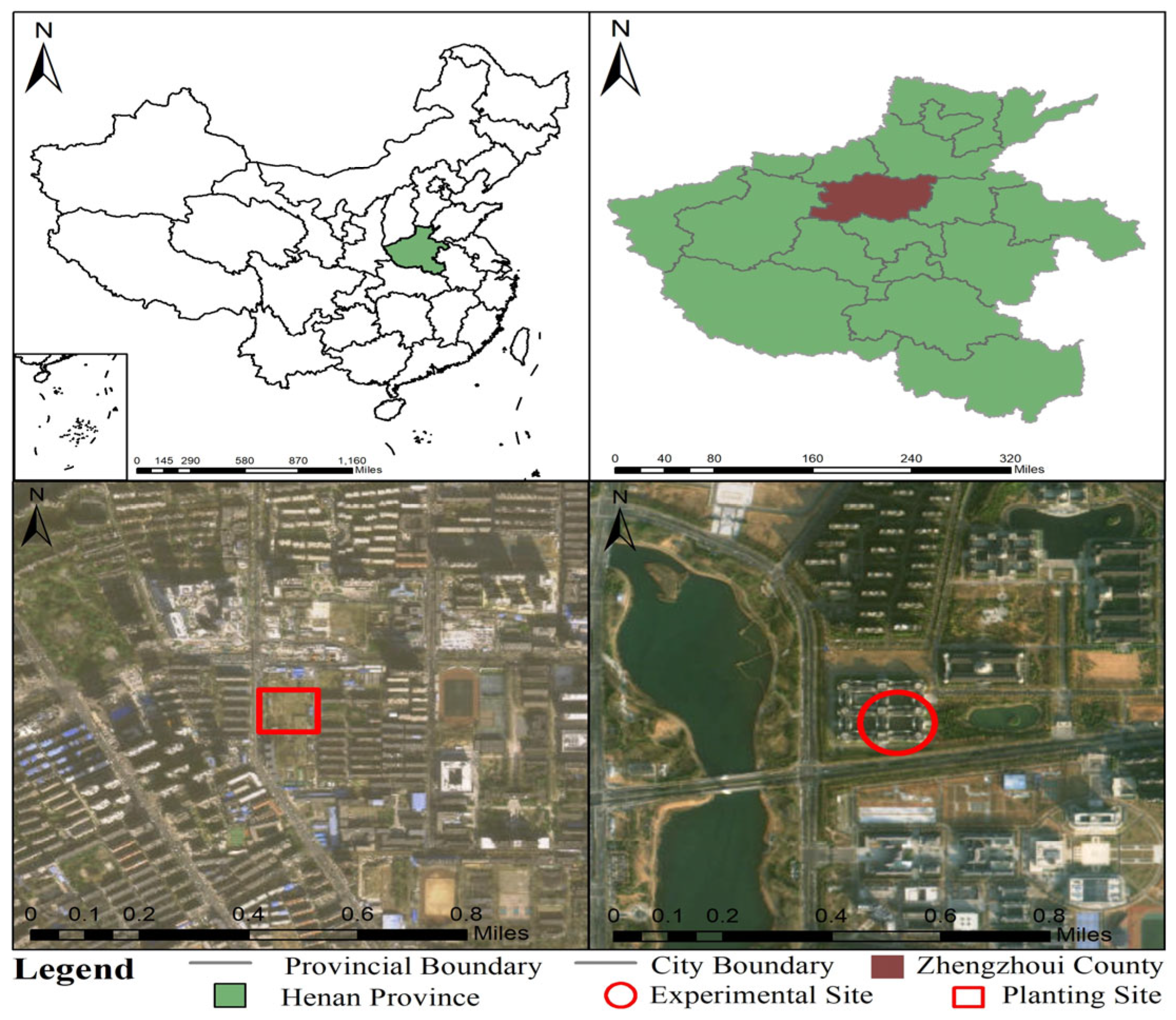
Figure 1. Geographical location of the planting site and experimental site.
Forests 16 01411 g001
Figure 2. Effects of B. dothidea inoculation on photosynthetic pigment contents in I. polycarpa seedlings. Note: * indicates a significant difference between treatments at the same time point (p < 0.05); ** indicates a highly significant difference (p < 0.01); *** indicates an extremely significant difference (p < 0.001). Values are mean ± SD (n = 3 biological replicates; 3 technical replicates each).
Forests 16 01411 g002
Figure 3. Effects of B. dothidea Inoculation on Soluble Protein (a) and Soluble Sugar (b) Contents. Note: * indicates a significant difference between treatments at the same time point (p < 0.05); *** indicates an extremely significant difference (p < 0.001). Values are mean ± SD (n = 3 biological replicates; 3 technical replicates each).
Forests 16 01411 g003
Figure 4. Effect of B. dothidea inoculation on proline (Pro) content. Note: * indicates a significant difference between treatments at the same time point (p < 0.05); ** indicates a highly significant difference (p < 0.01); Values are mean ± SD (n = 3 biological replicates; 3 technical replicates each).
Forests 16 01411 g004
Figure 5. Effect of B. dothidea inoculation on malondialdehyde (MDA) content. Note: * indicates a significant difference between treatments at the same time point (p < 0.05). Values are mean ± SD (n = 3 biological replicates; 3 technical replicates each).
Forests 16 01411 g005
Figure 6. Effect of B. dothidea inoculation on antioxidant enzyme activities. Note: (a) shows peroxidase (POD), (b) shows superoxide dismutase (SOD); * indicates a significant difference between treatments at the same time point (p < 0.05), ** indicates a very significant difference (p < 0.01), and *** indicates a highly significant difference (p < 0.001). Values are mean ± SD (n = 3 biological replicates; 3 technical replicates each).
Forests 16 01411 g006
Figure 7. Heatmap of correlations among physiological and biochemical parameters. Note: * indicates significant correlation (p < 0.05), *** indicates extremely significant correlation (p < 0.001). Correlations depict associations only and should not be interpreted as evidence of causality.
Forests 16 01411 g007
Table 1. Disease severity scale for stem canker in Idesia polycarpa seedlings [13].
GradeAssigned ValueDescription
I0No lesion.
II1very small lesions
III2Small scattered lesions
IV3Numerous lesions
V4Extensive lesions
Table 2. Disease incidence and disease index of I. polycarpa seedlings inoculated with B. dothidea.
Days After InoculationDisease Incidence (%)Disease Index
7 d00
14 d20.005.00
21 d45.0015.00
28 d70.0027.50
35 d85.0036.25
42 d90.0042.50
49 d95.0056.25
56 d95.0068.75
Note: The timing of grades I–IV corresponds to the first observation of characteristic symptoms at each assessment date. For clarity, only disease incidence and severity index are presented in Table 2, while the ratios of plants at different grades (I–IV) were used for calculating the severity index and showed consistent dynamics; therefore, they are not listed separately.
Disclaimer/Publisher’s Note: The statements, opinions and data contained in all publications are solely those of the individual author(s) and contributor(s) and not of MDPI and/or the editor(s). MDPI and/or the editor(s) disclaim responsibility for any injury to people or property resulting from any ideas, methods, instructions or products referred to in the content.